Article

Effect of Biosynthesized Nanoselenium on Controlling Tomato Root-Knot Nematode Meloidogyne incognita

1
National Key Laboratory of Agricultural Microbiology, College of Life Science and Technology, Huazhong Agricultural University, Wuhan 430070, China
2
Biochemistry Unit, Genetic Resources Department, Ecology and Dry Lands Agriculture Division, Desert Research Center, Cairo 11753, Egypt
3
Soil Fertility and Microbiology Department, Water Resources and Desert Soils Division, Desert Research Center, Cairo 11753, Egypt
4
Plant Protection Department, Ecology and Dry Lands Agriculture Division, Desert Research Center, Cairo 11753, Egypt
5
Chemistry Department, Faculty of Science, Minia University, El-Minia 61519, Egypt
6
Biology Department, College of Science, Princess Nourah bint Abdulrahman University, Riyadh 11671, Saudi Arabia
7
Agronomy Department, Faculty of Agriculture, Fayoum University, Fayoum 63514, Egypt
*
Author to whom correspondence should be addressed.
Agronomy 2023, 13(7), 1668; https://doi.org/10.3390/agronomy13071668
Submission received: 27 April 2023 / Revised: 1 June 2023 / Accepted: 13 June 2023 / Published: 21 June 2023
(This article belongs to the Special Issue Insecticide Resistance and Novel Insecticides)

Abstract

:
Tomato is a globally important fruit crop, which is easily susceptible to infection by plant-parasitic nematodes during growth. In this study, selenium nanoparticles were biosynthesized through the selenite reduction pathway in a wild-type Pseudomonas stutzeri BB19 and were characterized by uniform nanostructured needle-like forms with an average size of 95.2 nm. The nematicidal effect of biosynthesized selenium nanoparticles (BioSeNPs) at different concentrations (2, 6, and 10 ppm) during the pre- or post-infection of tomato root-knot nematode (RKN), Meloidogyne incognita, was assessed under greenhouse conditions. The BioSeNPs were applied as a foliar spray or a soil drench, compared to ethoprophos nematicide (100 mL/pot). The results showed that the ethoprophos nematicide significantly reduced the number of galls, egg mass, and eggs/egg mass of M. incognita by 94.2, 92.8, and 49.8%, respectively. BioSeNPs, as foliar sprays, significantly reduced the number of galls in post-treated infected tomato plants at 10 ppm and in pre-treated infected tomato plants at 2 ppm by 91.9 and 91.4%, respectively. Concerning the egg mass, BioSeNPs significantly reduced it in pre-treated infected tomato plants at 2 ppm as a foliar spray by 90.9%. Moreover, BioSeNPs significantly reduced the eggs/egg mass in pre-treated infected tomato plants at 2 ppm as a soil drench by 43.3%. On the other hand, the BioSeNPs considerably improved tomato growth, chlorophyll a and b, carotenoid content, and enzymes (i.e., catalase and peroxidase) activity compared to untreated infected tomato plants (negative control). Hence, the BioSeNPs show a significant application potential as a cost-effective and environmentally friendly biocontrol agent for RKN management in tomato plants.

1. Introduction

Tomato (Solanum lycopersicum L.) is a globally planted and economically important vegetable fruit that serves to meet the fundamental nutritional essentials of humans [1]. Root-knot nematodes (RKNs) are notorious pests associated with tomato crop, causing damage, ranging from 32–40% and an annual yield loss of approximately 22–30% [2]. One of the most prevalent RKNs, Meloidogyne incognita, causes biochemical and physical damage to tomato plants, such as changes in their energy and water metabolism, production of reactive oxygen species (ROS) [3], severe gall formation, yellowed and stunted leaves, and eventually the loss of whole plant [4]. Therefore, it is necessary to develop strategies for RKN control.
Currently, various methods are applied to reduce the damage of tomato crop caused by RKNs, such as biological control [5], chemical nematicides [6], and the development of resistant crops [7]. Among these, nematicides are commonly used to protect the tomato crop from RKNs because they are more effective than resistant varieties and control the cultivation environment [8]. Some nematicides, however, have also been identified as potentially serious dangers to the environment and human health, as well as higher application costs on farms [9,10]. Furthermore, several previous studies have confirmed that the development of nematode susceptibility to pesticides is additionally related to the occurrence of resistance after exposure to nematicides [11]. Traditional strategies are still meeting major obstructions. Therefore, there is a critical need for novel techniques to achieve the most significant effect with the least economic and environmental impact.
Recently, nanotechnology had excellent potential applications in plant pest and disease control [12]. Many nanomaterials can be used against plant parasitic nematodes and are directly applied either in the soil (soil drench) or on the leaves (foliar spray) [13]. Moreover, nanomaterials can be used as inducers, which can effectively activate the natural immune potential of plants through triggering with a complex integrated system of defense mechanisms [14]. More recently, the biosynthesis of nanomaterials via biological techniques using bacteria has represented a promising alternative to harmful traditional preparation methods [15].
Selenium nanoparticles (SeNPs) possess physicochemical characteristics, such as melting point, electrical/thermal conductivity, and light absorption, which enable them to exhibit excellent performance in numerous fields of science [16,17]. Many microorganisms can be used for biological synthesis to obtain SeNPs, such as Bacillus megaterium, Bacillus subtilis, Bacillus cereus, Aspergillus terreus, Enterococcus faecalis, and countless others [18,19,20]. Microorganisms can produce complex and specific nanoparticles (NPs) using their enzymes that determine unique morphological features of the SeNPs, which are often impossible to achieve through chemical synthesis [21].
In the current study, the biosynthesized SeNPs (BioSeNPs) were carried out using a wild-type Pseudomonas stutzeri strain BB19 through the selenite reduction pathway of the cell. The current study investigated the impact of BioSeNPs on the root-knot nematode M. incognita under greenhouse conditions. The study also investigated the effectiveness of BioSeNPs in controlling root galling and egg hatching in M. incognita, which infected tomato plants (Figure 1).

2. Materials and Methods

2.1. Chemicals, Bacterial and Nematode Strains, and Culture Conditions

Chemicals were supplied by Sigma-Aldrich or Merck and were of analytical grade. The chemical nematicide ethoprophos Smart-N (UPI CropScience Co., Ltd., Tianjin, China) was purchased from the local supplier Starchem Industrial Chemicals, 6th of October City, Egypt. A wild-type P. stutzeri strain BB19 previously isolated and identified from bean plants in the Baloza area, North Sinai Governorate, Egypt [22], and it was utilized to synthesize BioSeNPs in the present study. P. stutzeri cells were routinely grown in Luria-Bertani (LB) broth medium [23] at 28 °C. The culture of M. incognita was isolated from the okra plant grown in a greenhouse at El-Behera Governorate, Egypt. Perineal patterns of adult females and the morphology of second-stage juveniles (J2) were used to identify root-knot nematodes [24]. RKN egg masses were incubated for 48 h at room temperature (25 ± 2 °C) in a sodium hypochlorite solution of double-distilled H2O (ddH2O). Newly born second-stage juveniles (J2) were collected daily [25].

2.2. Biosynthesis and Purification of Selenium Nanoparticles Using P. stutzeri BB19

Biosynthesis and purification were performed according to a previously reported protocol with few modifications [26]. Briefly, 1% (v/v) of P. stutzeri inoculations were cultivated for 12 h in 250 mL Erlenmeyer flasks, usually containing 100 mL LB broth, then 500 µL of sodium selenite (1 M) was added, and incubation proceeded for 72 h at 28 °C with shaking at 150 rpm. The cultures with red color, indicating the production of BioSeNPs, were harvested by centrifugation at 8000 rpm for 10 min. After rinsing twice with ddH2O and scraping the supernatant, the pellets were resuspended in ddH2O and centrifuged twice for 30 min with 80% (w/v) sucrose to extract biomass. The separated pellets were washed with ddH2O and again resuspended to remove the impurities. The pellets were later dried and stored at −20 °C until they were used.

2.3. Characterization of Biosynthesized Selenium Nanoparticles

The rapid biosynthesis of SeNPs was characterized by a UV-visible spectrophotometer (Spectro UV-Vis double beam DUV 3500, Shimadzu Corp., Kyoto, Japan) at wavelengths between 250 to 700 nm. The SEM (Hitachi S-4500, Tokyo, Japan) and EDX were employed to investigate the size, morphology, and elemental composition of the BioSeNPs. The phase formation and crystalline nature of BioSeNPs were assayed by XRD (Model D/Max-2500, Kyoto, Japan) using Cu-Kα radiation (λ = 1.5406 Å) at a tube voltage of 40 kV and a tube current of 30 mA. The measurement was recorded over the range of 10–70 at the 2θ scale. The functional groups on the surface of fabricated materials were identified by FTIR (Model Perkin Elmer, Waltham, MA, USA), and the FTIR spectrum was recorded at wavelengths ranging from 400 to 4000 cm−1 at a resolution of 4 cm–1 in potassium bromide disk. The particle size distribution was measured by a particle analyzer (Zetasizer Nano ZS, Malvern Co., Malvern, UK) in the range of 232 nm under the following conditions: laser wavelength of 632 nm, water refractive index of 1.33, and at room temperature.

2.4. Effect of BioSeNPs on Tomato Plants Infected with M. incognita, Root Galling, and Egg Hatching of M. incognita (Greenhouse Experiments)

The experiment was conducted in the greenhouse of the Faculty of Agriculture, Cairo University, Giza, Egypt. Tomato 30-days-old uniform seedlings (cv. Super Strain B) were transplanted (seedling/pot) in a plastic pot (15 cm diameter) filled with 1 kg mixture of sand and clay (3:1, v/v). The plants were infected with 5000 nematode eggs in each pot by pouring the nematode suspension into holes with a diameter of 2–4 cm in the soil surface around the base of the plants. The commercial ethoprophos nematicide was the positive control with the recommended rate (3 L per hectare ≅ 100 mL/pot). The tomato seedlings inoculated with M. incognita without the nematicide or BioSeNPs treatments served as the negative control. Three concentrations of 2, 6, and 10 ppm of BioSeNPs were applied to tomato plants as a foliar spray, and soil drench treatment was performed individually two times; the first time was 7 days before inoculation with M. incognita (pre-infection treatment experiment), while the second time was 7 days after inoculation (post-infection treatment experiment). All treatments were replicated three times and arranged in a completely randomized design. During the experiment, the greenhouse temperature ranged between 25–30 °C, and irrigation and fertilization were applied as needed.
After 60 days from transplantation, tomato plants were uprooted, and the roots were washed and dried for 15 min in an aqueous solution of Phloxine-Bstain (0.15 g/L ddH2O) before being gently rinsed in tap water. The roots were carefully washed in tap water, and the red-stained egg masses were counted on infected roots [27]. Then, the number of galls/root system of a plant was counted visually, and egg mass and eggs per egg mass were also counted [28]. At harvest, roots were examined to count galls and egg masses on a 0–5 scale as follows [24]. Gall index: 0 = 0 gall/egg mass/root system; 1 = 1–2 galls/egg mass/root system; 2 = 3–10 gall/egg mass/root system; 3 = 11–30 gall/egg mass/root system; 4 = 31–100 gall/egg mass/root system; and 5 ≥ 100 gall/egg mass/root system.

2.5. Estimation of Growth Parameters and Photosynthetic Pigments

Growth parameters of tomato plants, including plant length, shoot, and root lengths, as well as shoot and root fresh weights, were recorded at the end of the experiment, 60 days after transplantation. Additionally, the photosynthetic pigments, including chlorophyll a, chlorophyll b, and carotenoids (mg/g fresh weight), were determined according to Mackinney [29] using 0.5 g of the fresh plant, homogenized and grounded with 5 mL of 85% of acetone, followed by filtration with wetted filter paper. This technique was repeated several times with acetone, and the filtrate volume was completed to 50 mL by 85% of acetone. Finally, the absorbance was immediately measured using the UV-Vis spectrophotometer at 662, 644, and 440 nm for calculating chlorophyll a, chlorophyll b, and carotenoids, respectively.

2.6. Assay of Antioxidant Enzymes Activity

One gram of tomato leaves was taken and homogenized to investigate the enzymatic activity, in 2 mL of 0.1 M sodium phosphate buffer (SPB), at a pH of 6.5 at 4 °C. The filtrate was centrifuged at 20,000 rpm at 4 °C for 15 min. The supernatant served as the enzyme extract for catalase (CAT) and peroxidase (POD) assays.
Catalase activity was measured by quantifying the decomposition at an absorbance of 240 nm every 10 s for 1 min. The reaction mixture comprised 50 mM of potassium phosphate buffer (pH = 7) and 10 mM H2O2, to which 30 µL of protein extract was added. CAT activity was calculated as a micromole of decomposed H2O2 per min per mg of protein [30].
Peroxidase activity was assayed colorimetrically by measuring the oxidation of pyrogallol in the presence of H2O2. The reaction mixture consisted of 1.5 mL of SPB, 20 μL of enzyme extract, 1 mL of 0.5 M pyrogallol, and 480 μL of 1% H2O2 solution (v/v). The increase in the optical density at 460 nm against blank was continuously recorded every minute. POD enzyme activity was expressed as a change in absorbance per 3 min/g fresh tissue [31].

2.7. Statistical Analysis

The obtained data from the greenhouse tests were statistically analyzed by analysis of variance using Infostat v2009 software (Córdoba, Argentina), and the means were compared by Duncan’s Multiple Range Test at a 0.05 significance level [32].

3. Results and Discussion

3.1. Biosynthesis and Characterization of Selenium Nanoparticles Using P. stutzeri BB19

The SeNPs were successfully and rapidly synthesized from P. stutzeri BB19 LB aqueous extract. The color changed from light yellow to red after 72 h of incubation (Figure 2A), which was the first evidence of selenite reduction, was followed by the synthesis of SeNPs. The surface morphology of the BioSeNPs was characterized by SEM, in which the BioSeNPs had uniform nanostructures with needle-like shapes and an average particle size of 95.2 nm (Figure 2B). These results showed a close similarity with previous results of a particle size ranging from 50 to 150 nm [33]. The preliminarily substantiated characterization of BioSeNPs was performed by UV-Visible spectrum scanning, which showed peak absorption values between 200–800 nm (UV-Visible range) and exhibited a high-intensity absorption peak at 380 nm (Figure 2C). The formation of such a peak occurs due to the Surface Plasmon Resonance (SPR) of SeNPs, and the result of a wide SPR peak strongly reveals the polydispersity of the SeNPs [34]. Similar observations were recorded in some recent studies [35,36,37,38]. In addition, the results of particle size analyzer showed that SeNPs had an average size of 50–500 nm with a high polydispersity index (Figure 2D).
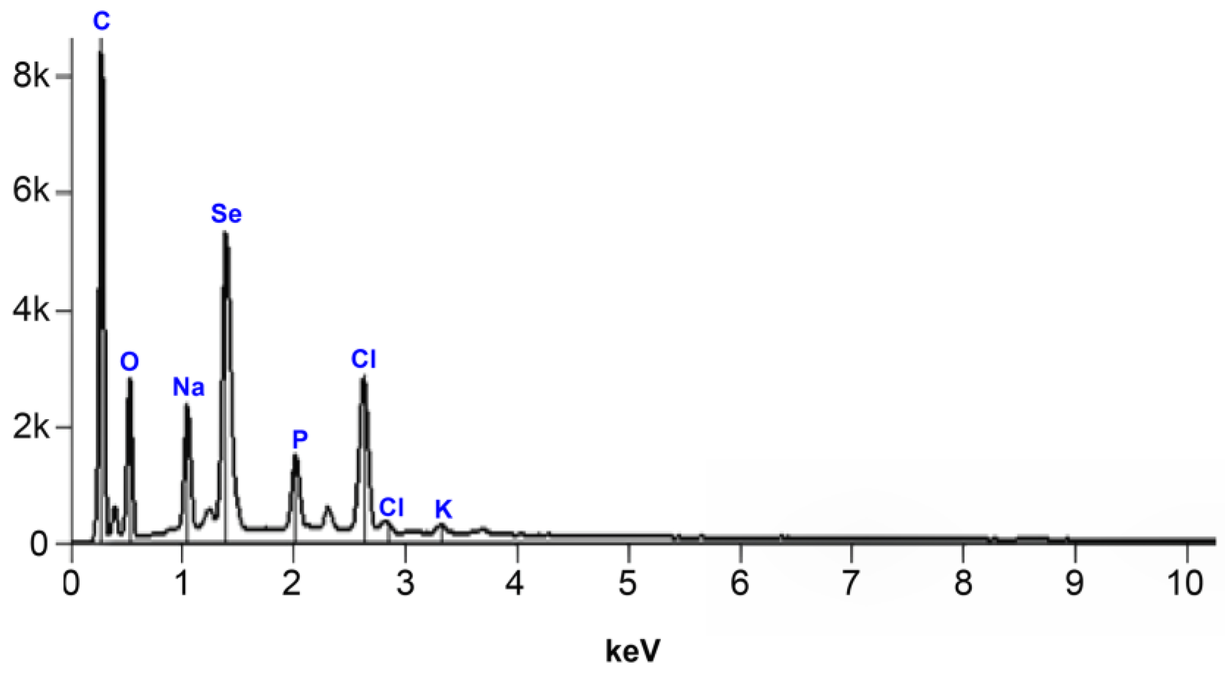
The EDX elemental analysis of BioSeNPs in Figure 3 confirms the sample composition through a high-intensity peak. The EDX intensity peaks indicated the presence of selenium, carbon, oxygen, and sodium with 14.2, 49.4, 23.3, and 4.75% by weight, respectively. These results confirm the successful preparation of SeNPs.
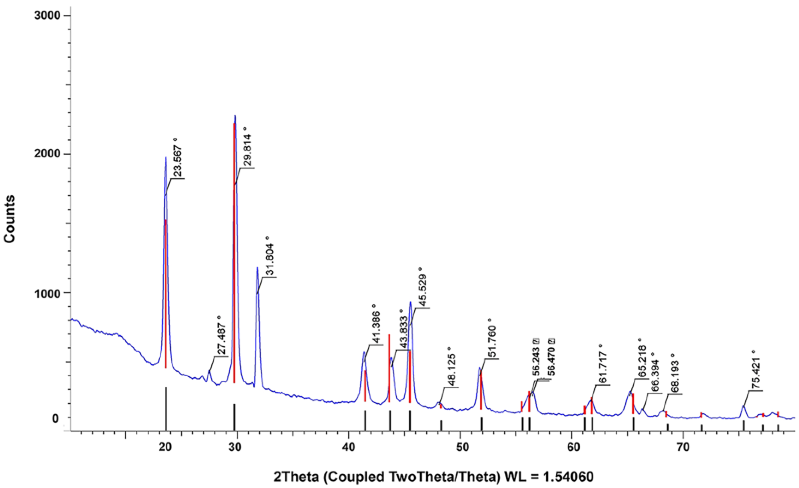
The XRD patterns of BioSeNPs are shown in Figure 4, where the diffraction intensity was observed from 2θ angles varying from 10 to 100°. The distinct sharp peaks at 23.5, 29.8, 31.8, 41.3, 43.8, 45.5, 51.7, 61.7, 65.2, and 75.4° indicated the crystalline nature of BioSeNPs, which is in agreement with previous research [39,40,41].
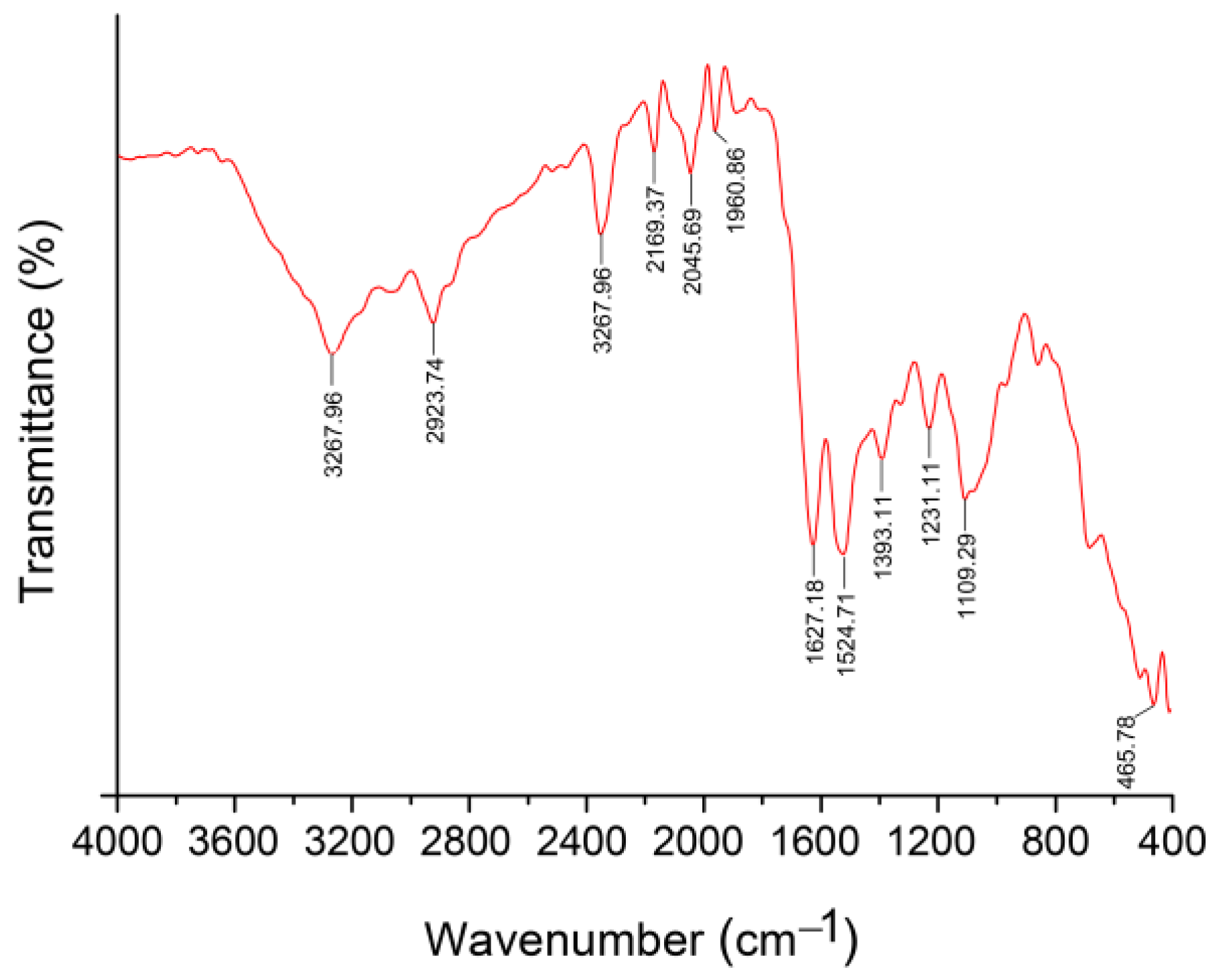
FTIR is used to determine the functional groups of organic and inorganic materials. As shown in Figure 5, the FTIR spectra show several absorption peaks at 3267, 2923, and 2850 cm−1. These peaks indicate stretching vibrations of the O–H or N–H group, asymmetric C-H stretching vibration, and symmetric C-H stretching vibration, respectively. In addition, other peaks were observed at 1627 and 1524 cm–1, which represent the stretching vibration absorption peak of C=O and bending vibration of N–H. This is most likely due to the presence of an amide group coated on the surface of the BioSeNPs. Moreover, another absorption band was observed at 1109 cm–1, representing the stretching vibration absorption peak of the C–O group. These functional groups are found in phenols, lipids, proteins, and carbohydrates, which are responsible for BioSeNPs stabilization [42,43].

3.2. Effect of BioSeNPs Treatments on Tomato Plants Infected with M. incognita under Greenhouse Conditions

3.2.1. Effect of BioSeNPs on Growth Parameters of Tomato Plants

Data in Table 1 show that the growth parameters of infected tomato seedlings significantly increased under BioSeNPs treatments. The results show that the growth parameters increased substantially after the treatment with BioSeNPs compared with infected untreated tomato plants (negative control). The BioSeNPs concentration, treatment time, and application method significantly affected plant growth. There was no significant difference between the two treatment times in plant length, root, shoot lengths, and root fresh weight, except shoot fresh weight, which has significantly increased with the post-infection treatment. Soil drenching significantly increased root length, while foliar spraying significantly increased shoot length and shoot fresh weight. However, there was no significant difference in plant length and root fresh weight between the two application methods. Post-infection treatment with BioSeNPs at 10 ppm as a soil drench resulted in the highest plant and shoot length. Pre-infection treatment with BioSeNPs at 2 ppm as a soil drench resulted in the highest root length. Post-infection treatment at all concentrations of BioSeNPs as a foliar spray resulted in the best shoot fresh weight. Consistent with previous findings, the obtained results indicated a positive role of SeNPs on growth parameters [44,45]. These might be due to the biological activity of SeNPs, where the uptake of SeNPs by the roots is slow. Then, they are oxidized rapidly inside the plant to selenite and become organic forms, improving root growth. Moreover, the interaction of SeNPs with proteins inside the plant cells and other biomolecules containing functionally active groups leads to changes in the responses of plants and improves their growth [46,47].

3.2.2. Effect of BioSeNPs on Photosynthetic Pigments of Leaves of Tomato Plants

The results of photosynthetic pigments are presented in Table 2 and show that chlorophyll a, chlorophyll b, and carotenoids increased significantly after the treatment with BioSeNPs compared with negative control. Regarding the treatment time, post-infection treatment significantly increased the chlorophyll content more than the pre-infection treatment. As for the two application methods, foliar spray recorded the highest increase in chlorophyll a and carotenoids, while soil drench recorded the highest increase in chlorophyll b. In the interaction, compared to the negative control, chlorophyll a and b showed the highest results with BioSeNPs at 10 ppm as a soil drench in post-infected treated tomato plants.
In contrast, carotenoids showed the highest results in pre-infected treated tomato plants with BioSeNPs at 2 ppm as a foliar spray compared to the negative control. The obtained results align with those reported in earlier studies, wherein SeNPs enhanced the growth and photosynthetic pigments in several plants [48,49]. The chlorophyll pigment acts as the light-capturing center and plays an essential role in photosynthesis, and any alterations in chlorophyll are harmful to plant health [50]. The mechanism behind the improved chlorophyll content has been attributed to the role of SeNPs that protected photosynthetic pigments, and this type of effect depends on the concentration of NPs used, where NPs can induce positive responses to specific doses. In contrast, they can induce the opposite effect or do not have effect on others. This behavior, called hormesis, has been reported when NPs have applied as bio-stimulants in crops [51]. The best effect of a low Se dose is reflected in the reconstruction of the lipid and fatty acids of the plastids in the cell membrane, which increase the photosynthetic rate of the plants [52].

3.2.3. Effect of BioSeNPs on Enzyme Activity of Leaves of Tomato Plants

The results presented in Table 3 show that post-infection treatment significantly enhanced CAT and POD activities more than the pre-infection treatment. Concerning the two application methods, foliar spray achieved the best CAT and POD activities, more than the soil drench. Regarding the interaction, assays of CAT and POD showed their superior activities in post-infected treated tomato plants with BioSeNPs at 10 ppm as a foliar spray by 12.1 and 178.4%, respectively, over negative control. Similarly, the literature data showed the role of SeNPs in enhancing antioxidant enzymes [48,53,54]. Activating the antioxidant enzymes has been regarded as an essential factor in enhancing plant resistance against pathogenic infection [55]. The significant antioxidant activity of SeNPs might be due to the highest level of Se, which plays a vital role in the enhancement of enzymes, such as CAT and POD, that protect plants from the damaging effects of ROS [56].

3.2.4. Effect of BioSeNPs on Number of Galls and Egg Hatching of M. incognita-infected Tomato Plants

The results in Table 4 demonstrate that treating inoculated tomato plants with chemical nematicide ethoprophos followed by BioSeNPs significantly reduced the numbers of galls, egg mass, and eggs/egg mass compared to the negative control. Concerning the treatment time, post-infection treatment significantly reduced the number of galls more than the pre-infection treatment; however, the pre-infection treatment significantly reduced the egg mass more than the post-infection treatment, whereas eggs/egg mass was not significantly different. Comparing the two application methods, the foliar spray caused a higher reduction in the number of galls and egg mass than the soil drench, while eggs/egg mass were not significantly different. In the interaction, the highest reduction in the number of root galls appeared in the positive control, which was an amount of 94.2%. In addition, the post-infected treated tomato plants with BioSeNPs at 10 ppm as foliar spray or soil drenching changed by 91.9 and 91.1%, respectively, compared to the negative control. The results of the interaction also showed that the highest reduction in egg mass was performed in infected tomato plants treated with ethoprophos nematicide (positive control) by 92.8% and pre-infected treated tomato plants with BioSeNPs at 2 ppm as a foliar spray by 90.9%, compared with the negative control. The number of eggs/egg mass showed the highest reduction in infected tomato plants treated with ethoprophos nematicide (positive control) by 49.8%, followed by pre-infected treated tomato plants with BioSeNPs at 2 ppm as a soil drench by 43.3% over the negative control. These results support the conclusion drawn from the previous research that the SeNPs showed significant suppressive activity against root-knot nematodes [44,57]. Hence, the treatment of tomato plants with BioSeNPs reduced the infestation of plants by the reduction in the number of galls, egg mass and, eggs per egg mass of M. incognita in the roots. These findings suggest the vital role of Se in plants’ life as a protector against biotic stresses, including pathogens infection by the interaction in plant cells with proteins and other biomolecules containing active groups, which enhances the signal transmission between living cells and modification of receptor proteins, which improved some plant innate mechanisms in relation to M. incognita [44,47].

4. Conclusions

In this study, the SeNPs were biosynthesized using P. stutzeri BB19 and used as a promising effective resistance inductor against tomato root-knot nematode M. incognita for the first time. The treatment of tomato plants infected with M. incognita under greenhouse conditions with BioSeNPs at 2, 6, and 10 ppm as a foliar spray or a soil drench exhibited inhibitory effects on M. incognita. Accordingly, the treatment with BioSeNPs induced tomato systemic resistance to M. incognita by the reduction in root galling, egg mass, and eggs per egg mass. Furthermore, the BioSeNPs treatments stimulated tomato growth parameters, chlorophyll a, chlorophyll b, carotenoid content, and CAT and POD enzymes activity compared with the untreated infected tomato plants. The mechanism of the activity of BioSeNPs is determined by their interaction in tomato plant cells with proteins and other biomolecules containing functionally active groups. Such interaction promotes signal transmission between living cells and modification of receptor proteins, which may lead to changes in the responses of plants to invasion. The known properties of Se as a substance with a wide range of antistress activities contributed to the development of studies of its NPs in relation to using them as biogenic elicitors of plant resistance to parasitic nematodes. Consequently, the BioSeNPs in this study show application potential as cost-effective and eco-friendly resistance inductors for the biocontrol of the tomato RKN M. incognita.

Author Contributions

Conceptualization, A.S.M.D., L.L., A.S.M.E.-N., K.M.A. and R.Y.; methodology, A.S.M.D., A.S.M.E.-N., R.Y. and A.S.; validation, A.S.M.D., N.A.-H.; formal analysis, A.S.M.D., A.S. and N.A.-H.; investigation, A.S.M.D., A.B., A.S.M.E.-N., M.H.H., T.M.B. and A.S.; collecting resources, A.S.M.D., L.L., R.Y. and A.S.M.E.-N.; data curation, A.S.M.D., K.M.A. and A.S.; writing—original draft preparation, A.S.M.D., K.M.A. and A.S.; writing—review and editing: A.S.M.D., L.L., A.B., M.H.H., A.S. and T.M.B.; supervision, A.S.M.D. and L.L.; project administration, A.S.M.D. and L.L.; funding acquisition, L.L, K.M.A., N.A.-H. and A.S. All authors have read and agreed to the published version of the manuscript.

Funding

This work was supported by the National Natural Science Foundation of China (grant number 32170124) and Princess Nourah bint Abdulrahman University Researchers Supporting Project (number PNURSP2023R402), Princess Nourah bint Abdulrahman University, Riyadh, Saudi Arabia.

Data Availability Statement

Data available in a publicly accessible repository.

Acknowledgments

The authors wish to extend their sincere appreciation to Princess Nourah bint Abdulrahman University Researchers Supporting Project (number PNURSP2023R402), Princess Nourah bint Abdulrahman University, Riyadh, Saudi Arabia, and the National Key Laboratory of Agricultural Microbiology, College of Life Science and Technology, Huazhong Agricultural University, Wuhan, China, as well as Desert Research Center, Cairo, Egypt, for providing all the necessary experimental facilities.

Conflicts of Interest

The authors declare no conflict of interest.

References

  1. Dorais, M.; Ehret, D.L.; Papadopoulos, A.P. Tomato (Solanum lycopersicum) health components: From the seed to the consumer. Phytochem. Rev. 2008, 7, 231–250. [Google Scholar] [CrossRef]
  2. Blok, V.C.; Jones, J.T.; Phillips, M.S.; Trudgill, D.L. Parasitism genes and host range disparities in biotrophic nematodes: The conundrum of polyphagy versus specialisation. Bioessays 2008, 30, 249–259. [Google Scholar] [CrossRef] [PubMed]
  3. Eves-van den Akker, S. Plant-nematode interactions. Curr. Opin. Plant Biol. 2021, 62, 102035. [Google Scholar] [CrossRef] [PubMed]
  4. Garita, S.A.; Bernardo, V.F.; Gonzalez, M.; Ripodas, J.I.; Arango, M.C.; Ruscitti, M. The false root-knot nematode: Modification of the root anatomy and alteration of the physiological performance in tomato plants. Rhizosphere 2021, 20, 100424. [Google Scholar] [CrossRef]
  5. Park, E.-J.; Jang, H.-J.; Park, J.-Y.; Yang, Y.K.; Kim, M.J.; Park, C.S.; Lee, S.; Yun, B.-S.; Lee, S.-J.; Lee, S.W.; et al. Efficacy evaluation of Streptomyces nigrescens KA-1 against the root-knot nematode Meloidogyne incognita. Biol. Control 2023, 179, 105150. [Google Scholar] [CrossRef]
  6. Qiao, K.; Liu, Q.; Zhang, S. Evaluation of fluazaindolizine, a new nematicide for management of Meloidogyne incognita in squash in calcareous soils. Crop Prot. 2021, 143, 105469. [Google Scholar] [CrossRef]
  7. Talavera, M.; Verdejo-Lucas, S.; Ornat, C.; Torres, J.; Vela, M.D.; Macias, F.J.; Cortada, L.; Arias, D.J.; Valero, J.; Sorribas, F.J. Crop rotations with Mi gene resistant and susceptible tomato cultivars for management of root-knot nematodes in plastic houses. Crop Prot. 2009, 28, 662–667. [Google Scholar] [CrossRef]
  8. D’Errico, G.; Giacometti, R.; Roversi, P.F.; d’Errico, F.P.; Woo, S.L. Mode of action and efficacy of iprodione against the root-knot nematode Meloidogyne incognita. Ann. Appl. Biol. 2017, 171, 506–510. [Google Scholar] [CrossRef]
  9. An, G.; Hong, T.; Park, H.; Lim, W.; Song, G. Oxamyl exerts developmental toxic effects in zebrafish by disrupting the mitochondrial electron transport chain and modulating PI3K/Akt and p38 Mapk signaling. Sci. Total Environ. 2023, 859, 160458. [Google Scholar] [CrossRef]
  10. Hanafi, A.; Dasenaki, M.; Bletsou, A.; Thomaidis, N.S. Dissipation rate study and pre-harvest intervals calculation of imidacloprid and oxamyl in exported Egyptian green beans and chili peppers after pestigation treatment. Food Chem. 2018, 240, 1047–1054. [Google Scholar] [CrossRef]
  11. Regmi, H.; Desaeger, J. Integrated management of root-knot nematode (Meloidogyne spp.) in Florida tomatoes combining host resistance and nematicides. Crop Prot. 2020, 134, 105170. [Google Scholar] [CrossRef]
  12. Wang, W.; Ling, Y.; Deng, L.; Yao, S.; Zeng, K. Effect of L-cysteine treatment to induce postharvest disease resistance of Monilinia fructicola in plum fruits and the possible mechanisms involved. Pestic. Biochem. Physiol. 2023, 191, 105367. [Google Scholar] [CrossRef]
  13. Tryfon, P.; Antonoglou, O.; Vourlias, G.; Mourdikoudis, S.; Menkissoglu-Spiroudi, U.; Dendrinou-Samara, C. Tailoring Ca-based nanoparticles by polyol process for use as nematicidals and pH adjusters in agriculture. ACS Appl. Nano Mater. 2019, 2, 3870–3881. [Google Scholar] [CrossRef]
  14. Salwan, R.; Sharma, M.; Sharma, A.; Sharma, V. Insights into plant beneficial microorganism-triggered induced systemic resistance. Plant Stress 2023, 7, 100140. [Google Scholar] [CrossRef]
  15. ElMekawy, A.; Hegab, H.M.; Alsafar, H.; Yousef, A.F.; Banat, F.; Hasan, S.W. Bacterial nanotechnology: The intersection impact of bacteriology and nanotechnology on the wastewater treatment sector. J. Environ. Chem. Eng. 2023, 11, 109212. [Google Scholar] [CrossRef]
  16. Huang, T.; Holden, J.A.; Heath, D.E.; O’Brien-Simpson, N.M.; O’Connor, A.J. Engineering highly effective antimicrobial selenium nanoparticles through control of particle size. Nanoscale 2019, 11, 14937–14951. [Google Scholar] [CrossRef]
  17. Luo, H.; Wang, F.; Bai, Y.; Chen, T.; Zheng, W. Selenium nanoparticles inhibit the growth of HeLa and MDA-MB-231 cells through induction of S phase arrest. Colloids Surf. B Biointerfaces 2012, 94, 304–308. [Google Scholar] [CrossRef]
  18. Hosnedlova, B.; Kepinska, M.; Skalickova, S.; Fernandez, C.; Ruttkay-Nedecky, B.; Peng, Q.; Baron, M.; Melcova, M.; Opatrilova, R.; Zidkova, J.; et al. Nano-selenium and its nanomedicine applications: A critical review. Int. J. Nanomed. 2018, 13, 2107–2128. [Google Scholar] [CrossRef] [PubMed] [Green Version]
  19. Shoeibi, S.; Mozdziak, P.; Golkar-Narenji, A. Biogenesis of selenium nanoparticles using green chemistry. Top. Curr. Chem. 2017, 375, 88. [Google Scholar] [CrossRef] [PubMed]
  20. Zare, B.; Babaie, S.; Setayesh, N.; Shahverdi, A.; Shahverdi, A. Isolation and characterization of a fungus for extracellular synthesis of small selenium nanoparticles extracellular synthesis of selenium nanoparticles using fungi. Nanomed. J. 2013, 1, 13–19. [Google Scholar]
  21. Oremland, R.S.; Herbel, M.J.; Blum, J.S.; Langley, S.; Beveridge, T.J.; Ajayan, P.M.; Sutto, T.; Ellis, A.V.; Curran, S. Structural and spectral features of selenium nanospheres produced by Se-respiring bacteria. Appl. Environ. Microbiol. 2004, 70, 52–60. [Google Scholar] [CrossRef] [PubMed] [Green Version]
  22. AbdelRazek, G.M.; Yaseen, R. Effect of some rhizosphere bacteria on root-knot nematodes. Egypt. J. Biol. Pest Control 2020, 30, 140. [Google Scholar] [CrossRef]
  23. Sambrook, J.; Russell, D.W. Molecular Cloning: A Laboratory Manual, 3rd ed.; Cold Spring Harbor Laboratory Press: Cold Spring Harbor, NY, USA, 2001. [Google Scholar]
  24. Taylor, A.L.; Sasser, J.N. Biology, Identification and Control Ofroot-Knot Nematodes (Meloidogyne Species); North Carolina State University: Raleigh, NC, USA, 1978; p. 111, vii. [Google Scholar]
  25. Hussey, R.S.; Barker, K.R. A comparison of methods of collecting inocula of Meloidogyne species, including a new technique. Plant Dis. Rep. 1973, 42, 865–872. [Google Scholar]
  26. Borah, S.N.; Goswami, L.; Sen, S.; Sachan, D.; Sarma, H.; Montes, M.; Peralta-Videa, J.R.; Pakshirajan, K.; Narayan, M. Selenite bioreduction and biosynthesis of selenium nanoparticles by Bacillus paramycoides SP3 isolated from coal mine overburden leachate. Environ. Pollut. 2021, 285, 117519. [Google Scholar] [CrossRef] [PubMed]
  27. Holbrook, C.C.; Knauft, D.A.; Dickson, D.W. A technique for screening peanut for resistance to Meloidogyne arenaria. Plant Dis. 1983, 67, 957–958. [Google Scholar] [CrossRef] [Green Version]
  28. Henderson, C.F.; Tilton, E.W. Tests with acaricides against the brown wheat mite12. J. Econ. Entomol. 1955, 48, 157–161. [Google Scholar] [CrossRef]
  29. Mackinney, G. Absorption of light by chlorophyll solutions. J. Biol. Chem. 1941, 140, 315–322. [Google Scholar] [CrossRef]
  30. Aebi, H. Catalase in vitro. Methods Enzymol. 1984, 105, 121–126. [Google Scholar]
  31. Hegedüs, A.; Erdei, S.; Horváth, G. Comparative studies of H2O2 detoxifying enzymes in green and greening barley seedlings under cadmium stress. Plant Sci. 2001, 160, 1085–1093. [Google Scholar] [CrossRef]
  32. Di Rienzo, J.A.; Casanoves, F.; Balzarini, M.G.; González, L.; Tablada, M.; Robledo, Y.C. InfoStat Versión Estudiantil; Grupo Infostat, FCA, Universidad Nacional de Córdoba: Cordoba, Argentina, 2015. [Google Scholar]
  33. Ramamurthy, C.; Sampath, K.S.; Arunkumar, P.; Kumar, M.S.; Sujatha, V.; Premkumar, K.; Thirunavukkarasu, C. Green synthesis and characterization of selenium nanoparticles and its augmented cytotoxicity with doxorubicin on cancer cells. Bioprocess Biosyst. Eng. 2013, 36, 1131–1139. [Google Scholar] [CrossRef]
  34. Mulvaney, P. Surface plasmon spectroscopy of nanosized metal particles. Langmuir 1996, 12, 788–800. [Google Scholar] [CrossRef]
  35. Avendaño, R.; Chaves, N.; Fuentes, P.; Sánchez, E.; Jiménez, J.I.; Chavarría, M. Production of selenium nanoparticles in Pseudomonas putida KT2440. Sci. Rep. 2016, 6, 37155. [Google Scholar] [CrossRef] [Green Version]
  36. Kora, A.J.; Rastogi, L. Biomimetic synthesis of selenium nanoparticles by Pseudomonas aeruginosa ATCC 27853: An approach for conversion of selenite. J. Environ. Manag. 2016, 181, 231–236. [Google Scholar] [CrossRef]
  37. Rajkumar, K.; Mvs, S.; Koganti, S.; Burgula, S. Selenium nanoparticles synthesized using Pseudomonas stutzeri (MH191156) show antiproliferative and anti-angiogenic activity against cervical cancer cells. Int. J. Nanomed. 2020, 15, 4523–4540. [Google Scholar] [CrossRef]
  38. Zhang, W.; Chen, Z.; Liu, H.; Zhang, L.; Gao, P.; Li, D. Biosynthesis and structural characteristics of selenium nanoparticles by Pseudomonas alcaliphila. Colloids Surf. B Biointerfaces 2011, 88, 196–201. [Google Scholar] [CrossRef]
  39. Alizadeh, S.R.; Abbastabar, M.; Nosratabadi, M.; Ebrahimzadeh, M.A. High antimicrobial, cytotoxicity, and catalytic activities of biosynthesized selenium nanoparticles using Crocus caspius extract. Arab. J. Chem. 2023, 16, 104705. [Google Scholar] [CrossRef]
  40. Alam, H.; Khatoon, N.; Raza, M.; Ghosh, P.C.; Sardar, M. Synthesis and characterization of nano selenium using plant biomolecules and their potential applications. BioNanoScience 2019, 9, 96–104. [Google Scholar] [CrossRef]
  41. Visha, P.; Nanjappan, K.; Selvaraj, P.; Jayachandran, S.; Elango, A.; Kumaresan, G. Biosynthesis and structural characteristics of selenium nanoparticles using Lactobacillus acidophilus bacteria by wet sterilization process. Int. J. Adv. Vet. Sci. Technol. 2015, 4, 178–183. [Google Scholar]
  42. Tugarova, A.V.; Mamchenkova, P.V.; Dyatlova, Y.A.; Kamnev, A.A. FTIR and Raman spectroscopic studies of selenium nanoparticles synthesised by the bacterium Azospirillum thiophilum. Spectrochim. Acta A Mol. Biomol. Spectrosc. 2018, 192, 458–463. [Google Scholar] [CrossRef]
  43. Kannan, S.; Mohanraj, K.; Prabhu, K.; Barathan, S.; Sivakumar, G. Synthesis of selenium nanorods with assistance of biomolecule. Bull. Mater. Sci. 2014, 37, 1631–1635. [Google Scholar] [CrossRef]
  44. Udalova, Z.V.; Folmanis, G.E.; Khasanov, F.K.; Zinovieva, S.V. Selenium nanoparticles-an inducer of tomato resistance to the root-knot nematode Meloidogyne incognita (Kofoid et White, 1919) Chitwood 1949. Dokl. Biochem. Biophys. 2018, 482, 264–267. [Google Scholar] [CrossRef]
  45. Domokos-Szabolcsy, E.; Marton, L.; Sztrik, A.; Babka, B.; Prokisch, J.; Fari, M. Accumulation of red elemental selenium nanoparticles and their biological effects in Nicotiniatabacum. Plant Growth Regul. 2012, 68, 525–531. [Google Scholar] [CrossRef]
  46. Hu, T.; Li, H.; Li, J.; Zhao, G.; Wu, W.; Liu, L.; Wang, Q.; Guo, Y. Absorption and bio-transformation of selenium nanoparticles by wheat seedlings (Triticum aestivum L.). Front. Plant Sci. 2018, 9, 597. [Google Scholar] [CrossRef] [Green Version]
  47. Husen, A.; Siddiqi, K.S. Plants and microbes assisted selenium nanoparticles: Characterization and application. J. Nanobiotechnol. 2014, 12, 28. [Google Scholar] [CrossRef] [Green Version]
  48. Hussein, H.A.A.; Darwesh, O.M.; Mekki, B.B. Environmentally friendly nano-selenium to improve antioxidant system and growth of groundnut cultivars under sandy soil conditions. Biocatal. Agric. Biotechnol. 2019, 18, 101080. [Google Scholar] [CrossRef]
  49. Ragavan, P.; Ananth, A.; Rajan, M.R. Impact of selenium nanoparticles on growth, biochemical characteristics and yield of cluster bean Cyamopsis tetragonoloba. Int. J. Environ. Agric. Biotechnol. 2017, 2, 2917–2926. [Google Scholar] [CrossRef]
  50. Reddy Pullagurala, V.L.; Adisa, I.O.; Rawat, S.; Kalagara, S.; Hernandez-Viezcas, J.A.; Peralta-Videa, J.R.; Gardea-Torresdey, J.L. ZnO nanoparticles increase photosynthetic pigments and decrease lipid peroxidation in soil grown cilantro (Coriandrum sativum). Plant Physiol. Biochem. 2018, 132, 120–127. [Google Scholar] [CrossRef]
  51. Juárez-Maldonado, A.; Ortega-Ortíz, H.; Morales-Díaz, A.B.; González-Morales, S.; Morelos-Moreno, Á.; Cabrera-De la Fuente, M.; Sandoval-Rangel, A.; Cadenas-Pliego, G.; Benavides-Mendoza, A. Nanoparticles and nanomaterials as plant biostimulants. Int. J. Mol. Sci. 2019, 20, 162. [Google Scholar] [CrossRef] [PubMed] [Green Version]
  52. Ren, Y.; Zhao, T.; Mao, G.; Zhang, M.; Li, F.; Zou, Y.; Yang, L.; Wu, X. Antitumor activity of hyaluronic acid-selenium nanoparticles in Heps tumor mice models. Int. J. Biol. Macromol. 2013, 57, 57–62. [Google Scholar] [CrossRef]
  53. Zahedi, S.M.; Abdelrahman, M.; Hosseini, M.S.; Hoveizeh, N.F.; Tran, L.P. Alleviation of the effect of salinity on growth and yield of strawberry by foliar spray of selenium-nanoparticles. Environ. Pollut. 2019, 253, 246–258. [Google Scholar] [CrossRef]
  54. Huang, C.; Qin, N.; Sun, L.; Yu, M.; Hu, W.; Qi, Z. Selenium improves physiological parameters and alleviates oxidative stress in srawberry seedlings under low-temperature stress. Int. J. Mol. Sci. 2018, 19, 1913. [Google Scholar] [CrossRef] [PubMed] [Green Version]
  55. Debona, D.; Rodrigues, F.; Rios, J.A.; Nascimento, K.J. Biochemical changes in the leaves of wheat plants infected by Pyricularia oryzae. Phytopathology 2012, 102, 1121–1129. [Google Scholar] [CrossRef] [Green Version]
  56. Bai, K.; Hong, B.; He, J.; Huang, W. Antioxidant capacity and hepatoprotective role of chitosan-stabilized selenium nanoparticles in concanavalin A-induced liver injury in mice. Nutrients 2020, 12, 857. [Google Scholar] [CrossRef] [PubMed] [Green Version]
  57. Baycheva, O.; Samaliev, H.; Udalova, Z.; Trayanov, K.; Zinovieva, S.; Folman, G. Selenium and it’s effect on plant-parasite system Meloidogyne Arenaria-Tiny Tim tomatoes. Bulgar. J. Agric. Sci. 2018, 24, 252–258. [Google Scholar]
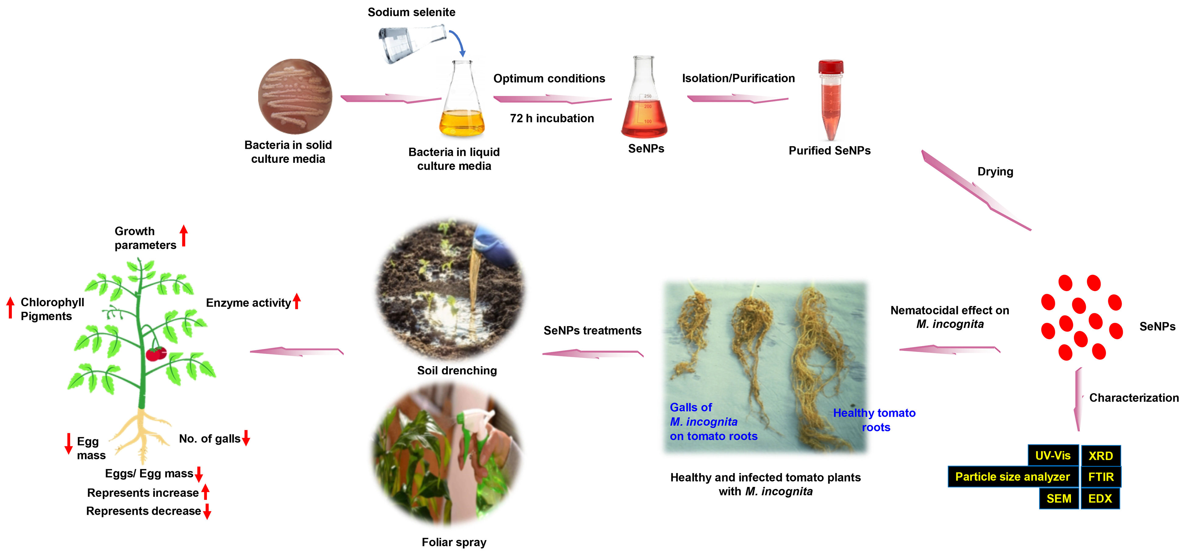
Figure 1. Schematic illustration of the procedures used in the biosynthesis of SeNPs and the control of M. incognita, which infected tomato plants. The upward red arrows indicate the measured values are increased, while the downward red arrows indicate the measured values are decreased.
Agronomy 13 01668 g001
Figure 2. Visualized P. stutzeri BB19 culture with BioSeNPs (A), SEM image (B), UV-Vis spectrum (C), and size and distribution of BioSeNPs (D). In (A), (a) the control aqueous P. stutzeri BB19 extract after 72 h of incubation; (b) the P. stutzeri BB19 extract with 500 µL sodium selenite (Na2O3Se) (1 mol L−1) after 72 h of incubation.
Agronomy 13 01668 g002
Figure 3. Corresponding results of the line scan SEM-EDS analysis of BioSeNPs.
Agronomy 13 01668 g003
Figure 4. The XRD pattern of BioSeNPs.
Agronomy 13 01668 g004
Figure 5. The FTIR spectrum of BioSeNPs.
Agronomy 13 01668 g005
Table 1. The growth parameters of tomato seedlings infected with M. incognita pre- and post-treated with different concentrations of BioSeNPs 1.
FactorPlant Length (cm)Root Length
(cm)
Shoot Length
(cm)
Root Fresh Weight (g)Shoot Fresh Weight (g)
Treatment time (TT)NSNSNSNS**
 Pre-infection (Pre)70.8 a ± 1.625.8 a ± 1.244.9 a ± 1.05.92 a ± 0.411.7 b ± 0.4
 Post-infection (Post)71.7 a ± 2.524.7 a ± 0.847.0 a ± 1.86.31 a ± 0.413.7 a ± 0.7
Application method (AM)NS***NS**
 Foliar spray (FS)72.0 a ± 1.724.0 b ± 0.948.0 a ± 1.36.05 a ± 0.413.5 a ± 0.7
 Soil drench (SD)70.4 a ± 2.426.5 a ± 1.143.9 b ± 1.56.18 a ± 0.411.9 b ± 0.4
BioSeNPs (ppm)**********
 NC61.0 d ± 2.321.7 b ± 0.939.3 d ± 1.44.80 c ± 0.111.0 c ± 0.2
 BioSe269.3 c ± 3.524.3 ab ± 2.244.9 c ± 2.34.73 c ± 0.612.7 ab ± 1.1
 BioSe675.3 b ± 2.426.8 a ± 2.048.5 b ± 1.75.64 c ± 0.413.7 ab ± 1.1
 BioSe1080.6 a ± 3.727.6 a ± 1.453.0 a ± 3.06.68 b ± 0.513.9 a ± 1.3
 PC (Ethoprophos Smart-N)70.0 c ± 1.326.0 a ± 0.944.0 c ± 0.48.73 a ± 0.512.2 bc ± 0.6
TT ×AM × BioSeNPs*********
 NC61.0 fg ± 5.321.7 d–g ± 2.239.3 fg ± 3.24.80 c–e ± 0.211.0 c–e ± 0.6
 Pre × FS × BioSe268.0 d–f ± 1.218.7 fg ± 1.849.3 c–e ± 2.92.30 f ± 0.88.67 e ± 1.5
 Pre × FS× BioSe678.3 b–d ± 6.227.7 b–e ± 6.950.7 b–d ± 3.55.33 b–e ± 0.113.8 bc ± 0.9
 Pre × FS × BioSe1071.3 c–f ± 3.220.7 e–g ± 1.850.7 b–d ± 3.55.13 b–e ± 0.612.1 c–e ± 0.6
 Pre × SD × BioSe281.7 bc ± 1.235.7 a ± 0.346.0 d–f ± 1.26.40 b–d ± 0.613.5 bc ± 1.0
 Pre × SD × BioSe678.0 b–d ± 3.531.3 a–c ± 3.246.7 d–f ± 0.36.03 b–e ± 1.013.0 cd ± 1.7
 Pre × SD × BioSe1068.3 d–f ± 0.729.0 a–d ± 0.039.3 fg ± 0.76.96 a–c ± 0.89.47 de ± 0.9
 Post × FS × BioSe275.7 b–e ± 3.525.0 b–g ± 0.050.7 b–d ± 3.56.07 b–e ± 1.117.7 a ± 1.2
 Post × FS× BioSe679.7 b–d ± 1.524.7 c–g ± 2.655.0 bc ± 1.27.30 ab ± 0.418.3 a ± 1.9
 Post × FS × BioSe1085.3 b ± 4.428.0 b–e ± 0.657.3 b ± 3.87.27 ab ± 1.417.6 a ± 3.4
 Post × SD × BioSe251.7 g ± 2.018.0 g ± 0.033.7 g ± 2.04.13 d–e ± 0.610.9 c–e ± 0.3
 Post × SD × BioSe665.0 ef ± 2.123.3 d–g ± 1.241.7 ef ± 0.93.90 ef ± 0.39.43 de ± 0.5
 Post × SD × BioSe1097.3 a ± 2.932.7 ab ± 1.564.7 a ± 1.57.35 ab ± 0.816.3 ab ± 0.3
 PC (Ethoprophos Smart-N)70.0 d–f ± 3.126.0 b–f ± 2.144.0 d–f ± 1.08.73 a ± 1.212.2 c–e ± 1.3
1 Each value represents mean ± standard error. Means in each column for each factor followed by the same letter do not differ significantly according to Duncan’s multiple range test at p ≤ 0.05. (* = p ≤ 0.05, ** = p ≤ 0.01 and NS = non-significant). NC (Negative control) denotes tomato plants inoculated with M. incognita without any treatment. PC (Positive control) denotes tomato plants treated with the chemical nematicide ethoprophos Smart-N. BioSe2, BioSe6, and BioSe10 denote 2, 6, and 10 ppm of BioSeNPs, respectively.
Table 2. The chlorophyll content of tomato seedlings infected with M. incognita pre- and post-treated with different concentrations of BioSeNPs 1.
FactorsChlorophyll a (mg/g FW)Chlorophyll b (mg/g FW)Carotenoids (mg/g FW)
Treatment time (TT)******
 Pre-infection (Pre)2.90 b ± 0.292.04 b ± 0.251.21 b ± 0.22
 Post-infection (Post)3.89 a ± 0.312.50 a ± 0.391.37 a ± 0.17
Application method (AM)*****
 Foliar spray (FS)3.47 a ± 0.292.07b ± 0.241.48 a ± 0.22
 Soil drench (SD)3.32 b ± 0.342.47 a ± 0.401.10 b ± 0.16
BioSeNPs (ppm)******
 NC1.98 e ± 0.021.58 b ± 0.020.51 e ± 0.03
 BioSe23.66 b ± 0.132.57 a ± 0.402.11 a ± 0.43
 BioSe62.51 d ± 0.161.78 b ± 0.170.61 d ± 0.14
 BioSe103.45 c ± 0.782.87 a ± 1.011.87 b ± 0.34
 PC (Ethoprophos Smart-N)5.40 a ± 0.042.54 a ± 0.381.35 c ± 0.02
TT × AM × BioSeNPs******
 NC1.98 g ± 0.041.58 c–e ± 0.040.51 ij ± 0.06
 Pre × FS × BioSe24.20 c ± 0.064.77 b ± 0.024.55 a ± 0.04
 Pre × FS× BioSe62.90 e ± 0.052.54 c ± 0.020.75 h ± 0.03
 Pre × FS × BioSe100.75 j ± 0.030.04 f ± 0.010.65 hi ± 0.03
 Pre × SD × BioSe23.05 e ± 0.012.25 cd ± 0.031.08 f ± 0.11
 Pre × SD × BioSe62.30 f ± 0.152.05 cd ± 0.080.42 j ± 0.01
 Pre × SD × BioSe101.05 i ± 0.010.50 ef ± 0.010.93 g ± 0.01
 Post × FS × BioSe23.76 d ± 0.021.37 c–e ± 0.021.36 de ± 0.08
 Post × FS× BioSe63.09 e ± 0.051.17 de ± 0.021.26 e ± 0.02
 Post × FS × BioSe105.26 b ± 0.032.54 c ± 0.032.54 c ± 0.03
 Post × SD × BioSe23.60 d ± 0.081.88 cd ± 0.061.43 d ± 0.02
 Post × SD × BioSe61.75 h ± 0.011.35 c–e ± 0.030.03 k ± 0.01
 Post × SD × BioSe106.73 a ± 0.028.42 a ± 0.023.36 b ± 0.03
 PC (Ethoprophos Smart-N)5.40 b ± 0.092.54 c ± 0.91.35 de ± 0.04
1 Each value represents mean ± standard error. Means in each column for each factor followed by the same letter do not differ significantly according to Duncan’s multiple range test at p ≤ 0.05. (* = p ≤ 0.05, ** = p ≤ 0.01). NC (Negative control) denotes tomato plants inoculated with M. incognita without any treatment. PC (Positive control) denotes tomato plants treated with the chemical nematicide ethoprophos Smart-N. BioSe2, BioSe6, and BioSe10 denote 2, 6, and 10 ppm of BioSeNPs, respectively. FW denotes fresh weight.
Table 3. The activity of some antioxidant enzymes of tomato seedlings infected with M. incognita pre- and post-treated with different concentrations of BioSeNPs 1.
FactorCAT Activity (ΔAbs 240 mg−1 Protein min−1)POD Activity (ΔAbs 460 mg−1 Protein min−1)
Treatment time (TT)****
 Pre-infection (Pre)81.4 b ± 2.721.6 b ± 1.1
 Post-infection (Post)84.1 a ± 3.323.8 a ± 2.1
Application method (AM)****
 Foliar spray (FS)86.5 a ± 2.225.7 a ± 2.1
 Soil drench (SD)79.0 b ± 3.519.6 b ± 0.8
BioSeNPs (ppm)****
 NC89.0 c ± 0.119.9 d ± 0.2
 BioSe254.9 e ± 3.221.6 c ± 2.8
 BioSe682.3 d ± 3.117.6 e ± 1.2
 BioSe1091.7 b ± 2.129.9 a ± 4.5
 PC (Ethoprophos Smart-N)95.8 a ± 0.124.4 b ± 0.1
TT ×AM × BioSeNPs****
 NC89.0 e ± 0.319.9 e ± 0.4
 Pre × FS × BioSe268.7 k ± 0.436.7 b ± 0.4
 Pre × FS× BioSe684.7 g ± 0.413.8 h ± 0.1
 Pre × FS × BioSe1082.7 h ± 0.420.8 de ± 0.2
 Pre × SD × BioSe244.5 m ± 0.315.2 g ± 0.1
 Pre × SD × BioSe675.9 i ± 0.221.9 d ± 0.2
 Pre × SD × BioSe1087.8 f ± 0.118.6 f ± 0.3
 Post × FS × BioSe261.8 l ± 0.120.8 de ± 0.4
 Post × FS× BioSe697.8 b ±0.320.9 de ± 0.5
 Post × FS × BioSe1099.8 a ± 0.0355.4 a ± 0.2
 Post × SD × BioSe244.6 m ± 0.213.7 h ± 0.4
 Post × SD × BioSe670.8 j ± 0.413.8 h ± 0.5
 Post × SD × BioSe1096.6 c ± 0.424.7 c ± 0.5
 PC (Ethoprophos Smart-N)95.8 d ± 0.124.4 c ± 0.3
1 Each value represents mean ± standard error. Means in each column for each factor followed by the same letter do not differ significantly according to Duncan’s multiple range test at p ≤ 0.05 (** = p ≤ 0.01). NC (Negative control) denotes tomato plants inoculated with M. incognita without any treatment. PC (Positive control) denotes tomato plants treated with the chemical nematicide ethoprophos Smart-N. BioSe2, BioSe6, and BioSe10 denote 2, 6, and 10 ppm of BioSeNPs, respectively.
Table 4. The number of M. incognita galls, egg mass, and eggs/egg mass per each g root of tomato seedlings pre- and post-treated with different concentrations of BioSeNPs 1.
FactorNo. of Galls (g root−1)Reduction (%)Egg Mass (g root−1)Reduction (%)Eggs/Egg Mass
(g root−1)
Reduction (%)
Treatment time (TT)***NS
 Pre-infection (Pre)846.1 a ± 165-232.9 b ± 41-345.2 a ± 16-
 Post-infection (Post)799.0 b ± 163-263.5 a ± 39-341.4 a ± 14-
Application method (AM)****NS
 Foliar spray (FS)716.1 b ± 166-225.5 b ± 41-349.9 a ± 15-
 Soil drench (SD)929.0 a ± 160-270.9 a ± 38-336.6 a ± 14-
BioSeNPs (ppm)******
 NC2484.3 a ± 50-651.0 a ± 10-456.6 a ± 18-
 BioSe2491.6 bc ± 9480.2160.7 c ± 2875.3364.2 a ± 216.37
 BioSe6454.3 c ± 5981.7189.4 b ± 1270.9357.1 a ± 88.20
 BioSe10538.4 b ± 13478.3192.9 b ± 1470.3377.2 a± 233.03
 PC (Ethoprophos Smart-N)144.0 d ± 894.247.0 d ± 192.7229.0 b ± 741.1
TT ×AM × BioSeNPs******
 NC2484.3 a ± 117-651.0 a ± 24-456.6 a ± 41-
 Pre × FS × BioSe2212.3 g ± 891.459.0 g ± 390.9378.3 c ± 617.1
 Pre × FS× BioSe6213.0 g ± 991.4129.7 f ± 380.6383.3 c ± 316.0
 Pre × FS × BioSe10443.7 e ± 482.1196.7 de ±469.8446.6 a ± 92.19
 Pre × SD × BioSe2290.0 fg ± 1388.3132.0 f ± 379.7259.0 ef ± 443.3
 Pre × SD × BioSe6755.0 d ± 1669.6181.0 e ± 672.2330.0 cd ± 1227.7
 Pre × SD × BioSe101290.0 b ± 348.0235.0 c ± 563.9366.7 c ± 2119.7
 Post × FS × BioSe2451.7 e ± 481.8141.7 f ± 778.2363.3 cd ± 320.4
 Post × FS× BioSe6383.3 ef ± 1284.5211.7 c–e ± 667.5341.7 cd ± 1325.2
 Post × FS × BioSe10200.0 g ± 1091.9120.0 f ± 681.6289.3 de ± 936.6
 Post × SD × BioSe21012.3 c ± 759.2310.0 b ± 652.4386.3 c ± 415.4
 Post × SD × BioSe6466.0 e ± 581.2235.3 c ± 463.9373.3 c ± 418.2
 Post × SD × BioSe10220.0 g ± 591.1220.0 cd ± 866.2345.3 cd ± 424.4
 PC (Ethoprophos Smart-N)144.0 g ± 1994.247.0 g ± 392.8229.0 f ± 1649.8
1 Each value represents mean ± standard error. Means in each column for each factor followed by the same letter do not differ significantly according to Duncan’s multiple range test at p ≤ 0.05. (* = p ≤ 0.05, ** = p ≤ 0.01 and NS = non-significant). NC (Negative control) denotes tomato plants inoculated with M. incognita without any treatment. PC (Positive control) denotes tomato plants treated with the chemical nematicide ethoprophos Smart-N. BioSe2, BioSe6, and BioSe10 denote 2, 6, and 10 ppm of BioSeNPs, respectively.
Disclaimer/Publisher’s Note: The statements, opinions and data contained in all publications are solely those of the individual author(s) and contributor(s) and not of MDPI and/or the editor(s). MDPI and/or the editor(s) disclaim responsibility for any injury to people or property resulting from any ideas, methods, instructions or products referred to in the content.