Article

Foliar Application of Chitosan and Phosphorus Alleviate the Potato virus Y-Induced Resistance by Modulation of the Reactive Oxygen Species, Antioxidant Defense System Activity and Gene Expression in Potato

by
Alshymaa Z. Al-Mokadem
1,2,
Abd El-Aleem M. Alnaggar
3,
Ahmed G. Mancy
4,
Ahmed R. Sofy
5,
Mahmoud R. Sofy
5,*,
Abdel Kareem S. H. Mohamed
6,
Mostafa M. A. Abou Ghazala
3,
Khaled M. El-Zabalawy
7,
Noura F. G. Salem
8,
Mohamed E. Elnosary
5 and
Mona S. Agha
9
1
Botany Department, Women’s College, Ain Shams University, Cairo 11566, Egypt
2
Chemistry Department, College of Science and Arts, Jouf University, Al-Gurayyat 77447, Saudi Arabia
3
Agriculture Botany Department, Faculty of Agriculture, Al-Azhar University, Nasr City, Cairo 11884, Egypt
4
Soil and Water Department, Faculty of Agriculture, Al-Azhar University, Nasr City, Cairo 11884, Egypt
5
Botany and Microbiology Department, Faculty of Science, Al-Azhar University, Nasr City, Cairo 11884, Egypt
6
Department of Botany and Microbiology, Faculty of Science, Al-Azhar University, Assiut Branch, Assiut 71524, Egypt
7
Environment and Bio-Agriculture Department, Faculty of Agriculture, Al-Azhar University, Nasr City, Cairo 11884, Egypt
8
Botany and Microbiology Department, Faculty of Science (Girls), Al-Azhar University, Nasr City, Cairo 11884, Egypt
9
Botany Department, Faculty of Science, Mansoura University, Mansoura 35516, Egypt
*
Author to whom correspondence should be addressed.
Agronomy 2022, 12(12), 3064; https://doi.org/10.3390/agronomy12123064
Submission received: 30 October 2022 / Revised: 24 November 2022 / Accepted: 25 November 2022 / Published: 3 December 2022

Abstract

:
Viruses pose a serious threat to the sustainable production of economically important crops around the world. In the past 20 years, potato virus Y (PVY) emerged as a relatively new and very serious problem in potatoes, even though it is the oldest known plant virus. Multiple strains of the virus cause various symptoms on the leaves and tubers of potatoes, resulting in yield reduction and poor-quality tubers. Consequently, it would be very interesting to learn what causes systemic PVY resistance in plants. Natural compounds such as chitosan (CHT) and phosphorus have been developed as alternatives to chemical pesticides to manage crop diseases in recent years. In the current study, potato leaves were foliar-sprayed with chitosan and phosphorus to assess their ability to induce PVY resistance. Compared to untreated plants, the findings demonstrated a significant decrease in disease severity and PVY accumulation in plants for which CHT and P were applied. Every treatment includes significantly increased growth parameters, chlorophyll content, photosynthetic characteristics, osmoprotectants (glycine betaine, proline, and soluble sugar), non-enzymatic antioxidants (glutathione, phenols, and ascorbic acid), enzymatic antioxidants (peroxidase, superoxide dismutase, lipoxygenase, glutathione reductase, catalase, β-1,3 glucanase, and ascorbate peroxidase), phytohormones (gibberellic acid, indole acetic acid, jasmonic acid, and salicylic acid), and mineral content (phosphorus, nitrogen, and potassium), compared to infected plants. However, compared to PVY infection values, CHT and P treatments showed a significant decrease in malondialdehyde, DPPH, H2O2, O2, OH, and abscisic acid levels. In addition, increased expression levels of some regulatory defense genes, including superoxide dismutase (SOD), ascorbic acid peroxidase (APX), relative pathogenesis-related 1 basic (PR-1b), and relative phenylalanine ammonia-lyase (PAL), were found in all treated plants, compared to PVY-infected plants. Conclusion: Phosphorus is the most effective treatment for alleviating virus infections.

1. Introduction

After maize, rice, and wheat, the potato (Solanum tuberosum L.), which is grown on 18.1 million hectares of land, is the fourth most significant crop in the world [1]. High quantities of carbohydrates, vitamins, protein, and minerals may be found in potatoes, a significant dietary source. In poor nations, it is also a source of revenue and a job opportunity [2]. It is regarded as a suitable weaning meal because of its ideal protein-to-calorie ratio, and these qualities make it an effective crop in battling global poverty and malnutrition [3]. Over a billion people use potatoes virtually every day, and they are extensively grown in more than 164 nations. In Egypt and across the globe, potatoes are grown on around 20% of the total land used for vegetable production. Egypt is one of the continent’s top producers and exporters of potatoes, and it comes in at number 14 in terms of global output of storage potatoes [4]. About 5 million metric tons of potatoes produced in Egypt are intended for human consumption, making it one of the world’s biggest exporters of potatoes. More than 759,200 MT of potatoes were exported from Egypt in 2018 as a whole. Plant viral infections endanger agriculture’s long-term survival and cost farmers a significant amount of money each year [5]. Therefore, it is crucial to concentrate on finding safe plant-disease-management options that do not affect the environment or public health and increase plant crop volume and quality in a sustainable agricultural system. For instance, plant diseases may first be regulated by genetics or prevented by plant nutrients, which are some of the most organic, environmentally benign ways of disease resistance [6]. In a small number of situations, chemical pesticides are used to treat plant viral illnesses by reducing pesticide applications and/or doses and applying nutrients, especially those related to resistance or plant disease management, where these nutrients are not thought of as a replacement for pesticides. However, since they play a crucial role in pest-management programs, pesticide residues in food crops can be reduced [7]. Thicker cell walls develop as mechanical barriers, which might impact resistance mechanisms. The production of natural defensive chemicals that fend off infections, such as antioxidants, phytoalexins, and flavonoids; certain inorganic and organic fertilizers may be useful for controlling plant illnesses by acting as resistance-stimulating agents in several plant species [8].
Numerous bacterial, viral, fungal, and insect illnesses impact potatoes. Among these illnesses, viruses may cause everything from minor symptoms to significant crop losses [9]. Regarding economic and ecological effects, the top 10 plant viruses include Potato virus Y (PVY). Potato cultivars will have different symptoms depending on the type and severity of the virus, but PVY (a member of the Potyviridae family, genus Potyvirus) has a positive single-stranded RNA genome of 9.7 kb, which infects plants mainly from the Solanaceae family and transmits it in nature through more than 40 types of aphids in a non-persistent manner. It causes a mosaic leaf pattern, vein clearing, and leaf necrosis [10].
Chitosan (CHT) possesses a broad range of distinctive bioactivities, including the ability to induce plant resistance to viral infections, suppress viral infections in plant cells, and stop the growth of phage infections in microbial cultures that are already infected [11]. In addition, CHT triggers host defense responses in monocotyledons and dicotyledons plants. The lignification of the cell wall [12], differences in ion concentrations, acidification of the phosphorylation of proteins, depolarization of membranes and cytoplasm [13], activation of the enzymes as glucanase and chitinase, production of reactive oxygen species (ROS) [14], and the biosynthesis of phytohormones such as salicylic acid and jasmonic acid [15] are a few explanations of these reactions.
There are several different ways that the presence of phosphorus nutrients in fertilization programs affects plant diseases. Furthermore, a number of vital practical aspects, with phosphorus being one of the most important nutrients, are connected to crop quality and disease resistance [16]. Phosphorus is crucial for stress tolerance, cation–anion equilibrium, stomata movement, photosynthesis, protein synthesis, energy transfer, osmoregulation, and phloem transport. In addition, phosphorus participates in several essential regulatory functions in the plant. Nearly all activities necessary for plant development and reproduction depend on protein synthesis, ionic balance control, plant stomata regulation, turgor maintenance, stress tolerance, water usage, translocation of photosynthetic products, and many other functions [17]. Additionally, it has been shown that a high phosphorus supply might lessen the severity of viral infections such as the Tobacco mosaic virus and leaf blight, which is brought on by Helminthosporium [18]. In addition, promoting the production of stronger outer barriers by epidermal cells prevents disease assaults [19]. This results in modifications to plants’ distribution and the main primary metabolite profiles as well as changes to hormonal pathways [20].
This study aimed to (1) investigate the role of chitosan (CHT) and phosphorus (P) in potato plant PVY resistance and (2) examine the influence of CHT and P on oxidative stress, osmolytes, and the antioxidant system in potato plants under PVY conditions.

2. Materials and Methods

2.1. Source of Potato Cultivar

Tubers of potato (Solanum tuberosum L.) cultivar Spunta were obtained from Kafr El-Zayat, ELGharbia governorate, and detected before cultivation and inoculation for the presence of the viruses Potato virus Y (PVY), Potato leafroll virus (PLRV), and Potato virus X (PVX) serologically, directly from sprouting tubers [21] by using the double antibody sandwich ELISA (DAS-ELISA) technique using specific polyclonal antibodies [22].

2.2. Source of the Virus Isolate

The potato cultivar Spunta with Potato virus Y (PVY) Egyptian identified was acquired by the Agriculture Botany Department, Faculty of Agriculture, Al-Azhar University, Cairo, Egypt (30°03′19.0″ N 31°19′10.0″ E). The viral isolation was verified by mechanically inoculating infectious sap on Chenopodium amaranthcolor after grinding infected leaf samples in 0.2% mercaptoethanol containing a phosphate buffer (100 mM) at pH 7.2. Because it was identified from similar local lesions that Chenopodium amaranthcolor underwent and because it afterward spread to a healthy Datura metel that belonged to the family, Solanaceae served as a propagative host. Inoculated plants were housed in a greenhouse free of insects and had daily symptom checks. As a control, the same number of seedlings at the same age are just infected with buffer.

2.3. Plant Materials

A 20 mM sample of phosphorus was prepared to form phosphorus citrate (C6H5O11P) by dissolving 2.40 g of NaH2PO4 and 1 M citric acids to adjust the pH value to 6.5, and then upon reaching 1000 mL of distilled water, the solution was finished. We bought chitosan (CHT; 50–190 kDa, Sigma-Aldrich). Chitosan (CHT 10 g) was dissolved overnight with vigorous stirring in 400 mL of filtered water with 90 mL acetic acid (1 M). After adjusting the pH to 6.5, the solution was fully dissolved in 1000 mL of distilled water (1% concentration). Before spraying, each solution was diluted with two drops of Tween 80 to improve dispersion on the leaves of potato plants.
A pot experiment was carried out in the greenhouse of the Agriculture Botany Department, Faculty of Agriculture, Al-Azhar University, Cairo, Egypt.
Twelve kilograms of sandy loam soil in plastic pots approximately 35 cm in height and 30 cm in diameter were mixed well with 5% sterilized potting compost for each pot. The potato cultivar Spunta’s tubers were grown from eye cores in plastic pots (one tuber per pot) under typical and favorable circumstances for potato plants. Before cultivation, soil samples from the pot experiment were collected, air-dried, squished, and sieved through a 2.0 mm sieve for analysis. According to Estefan et al. [23], some chemical and physical analyses of the soil samples and utilized compost are shown in Table 1. Following 23 days of growth, plants of comparable size were selected and separated into six treatments.
In the first group, the plants received an untreated (absolute control) watering (AC). In the second group, 1% chitosan was applied topically to the plants (CHT). In the third group, 20 mM phosphorus was applied topically to the plants (P). The plants in the fourth group received an infection with Potato virus Y as a challenge control (ChC). The plants in the fifth group received a foliar application of 1% chitosan (CHT) before being infected with PVY three days later. The plants in the sixth group received foliar applications of 20 mM phosphorus (P) before being infected with PVY three days later. After one-week, untreated control plants (AC), phosphorus, and CHT solution applications were once again made to the leaves.
A 100 mM phosphate-buffered solution (pH 7.5) was added to freshly infected D. metel plant leaves that had developed severe symptoms to extract the infectious sap, which was then used to create the PVY inoculation. After crushing plant tissue through two layers of muslin, the virus-containing supernatant was centrifuged for 10 min at 5000× g. As a viral inoculum, the virus-containing supernatant was used. A cotton swab dipped in the viral inoculant was used to manually infect healthy plant leaves after they had been lightly dusted with (600 mesh) carborundum [24]. Samples from the treatment facility and the control were gathered for analysis. The percentage of infected plants and the severity of the symptoms were also examined 24 days after the inoculation as follows: no symptoms are represented by 0, vein clearing by 1, mild mosaic by 3, and severe mosaic by 4.
According to Yang et al. [25], the calculation was used to determine disease severity (DS) values.
DS % = Σ D i s e a s e   g r a d e × N u m b e r   o f   p l a n t s   i n   e a c h   g r a d e   T o t a l   n u m b e r   o f   p l a n t s × H i g h e s t   d i s e a s e   g r a d e × 100

2.4. Source of the Plant Nutrition

Soil application of Hoagland nutrient solution (0.5 ionic strength) was used as a source of essential nutrients during the potato pot experiment. Four salts were used to prepare the Hoagland macronutrient solution: calcium nitrate, potassium monophosphate, potassium nitrate, magnesium sulphate and micronutrients were included, according to Hoagland and Arnon [26]. The chemical composition of the Hoagland nutrient solution used is shown in Table 2.

2.5. Growth Indices

The five plants were removed randomly from the pots, and the plant height and shoot weight were measured using a meter scale. Next, a graph sheet with leaf squares was calculated to indicate leaf area.

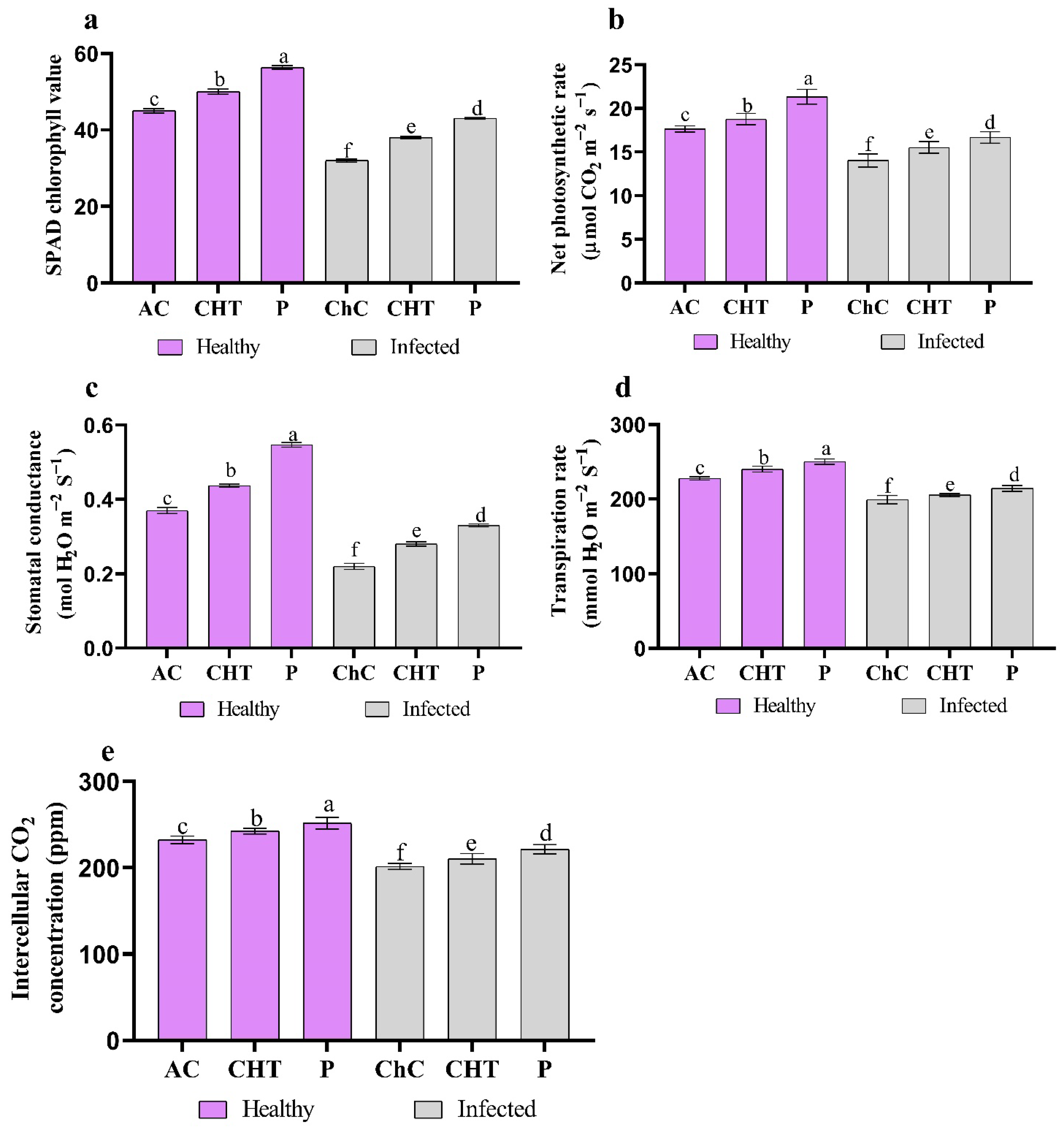
2.6. Photosynthetic Characteristics and Chlorophyll Content

The chlorophyll SPAD values in potato leaves were measured using a SPAD502 chlorophyll meter (Konica Minolta, Inc., Tokyo, Japan). The portable photosynthetic method (LICOR 6400, Lincoln, NE, USA) measures the intercellular CO2 concentration (Ci), stomatal conductance (gs), transpiration rate (E), and net photosynthetic rate (PN) in the fully extended topmost plant leaf in each treatment [27].

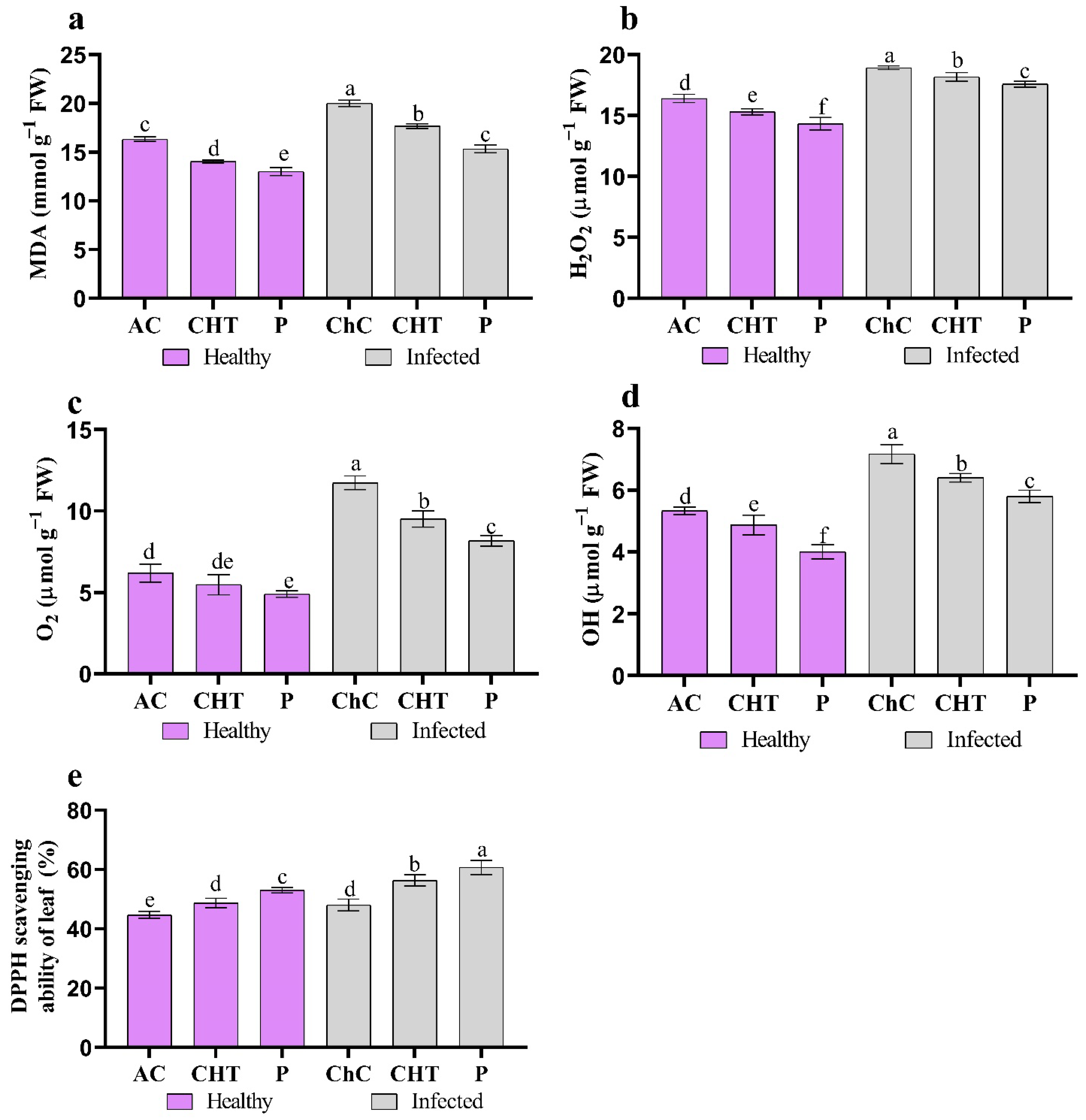
2.7. Oxidative Stress Markers

2.7.1. Proline

Bates et al.’s [28] technique was used to measure the proline content of dried potato leaves. First, the substance was extracted in equimolar amounts using glacial acetic acid, sulfosalicylic acid, and ninhydrin solution. Next, 5 mL of toluene was added after the sample had been heated to 100 °C and had cooled. Finally, the toluene layer’s absorbance was determined using a spectrophotometer at 528 nm.

2.7.2. Glycine Betaine Analysis

The fresh potato leaves (0.5 g) were crushed in 5 mL toluene–water (0.05%) solution in a 20 mL test tube. For 24 h, each tube was manually shaken at 25 °C. Following filtering, 0.5 mL of the extract was combined with 1 mL of the 2-N HCl solution and agitated for 90 min in an ice-cold water bath. Next, 10 mL of 1, 2 dichloroethane (cooled at 10 °C) was added to potassium tri-iodide (0.1 mL) solution, which included 10 g of potassium iodide and 7.5 g of iodine in 100 mL of 1-N HCl. According to Grieve and Grattan [29] approach. The optical density of the organic layer was measured at 365 nm after the top aqueous layer was removed.

2.7.3. Total Soluble Sugar Contents

Using anthrone and 80% sulphuric acid (H2SO4), Yemm and Willis [30] used techniques to calculate the total sugar contents. Test tubes containing the combination were heated for 10 min in a water bath before being chilled with cold water. A 620 nm wavelength was used to measure the optical density.

2.7.4. Phenols

According to Ziouti et al. [31], the Folin–Ciocalteu reagent (FCR) and Na2CO3 solution were used to test for the presence of phenols. First, 100 mg of dried potato leaves were mixed with 50% methanol (6.5 mL). Then, following a vortex, the samples remained in a dark environment for 95 min before being centrifuged for 5 min at 15,000× g. Next, one mL of the supernatant was mixed with 0.8 milliliters of 25% FCR, 5 milliliters of phosphoric acid (85%), and ten milliliters of distilled HCl. The tubes were then incubated for 10 min at 42 °C. Then, a spectrophotometer was used to determine the absorbance at 765 nm.

2.7.5. Glutathione (GSH)

Fresh leaf tissue (100 mg) was ground in phosphate buffer (pH 8.0) and then centrifuged at 3000× g for 15 min to determine the amount of reduced glutathione (GSH). After mixing 500 mL of 5,5-dithiobis-2-nitrobenzoic acid with 500 mL of supernatant for 10 min, the mixture was determined at 412 nm [32].

2.7.6. Ascorbic Acid (AsA)

Jagota and Dani [33] method was used to measure ascorbic acid (AsA). In the beginning, 0.2 g of leaf samples were crushed with liquid N2 and then suspended in 5% TCA (2 mL). The homogenate was then centrifuged at 10,000× g for 15 min at 5 °C. After vigorously shaking the 10% TCA and AsA extraction solution mixture, it was added to an ice bath for five minutes. Then, using distilled water to dilute the extract from 0.5 mL to 2.0 mL, 0.2 mL of diluted Folin–Ciocalteu reagent was added to the initial mixture. After 10 min at 760 nm, the absorbance of the resulting blue hue was measured.

2.8. ROS Indicators

2.8.1. Lipid Peroxidation

The peroxidation of lipids was used to determine how much lipid had been oxidized, and Heath and Packer’s [34] procedures examined the malondialdehyde (MDA) level (lipid peroxidation). Shortly after being macerated in trichloroacetic acid (TCA), fresh leaves were centrifuged at 12,000× g. The supernatant was then exposed to thiobarbituric acid for 30 min in the water bath. After the samples were cooled, optical absorbance at 532 nm was measured.

2.8.2. Hydrogen Peroxide

The 5% TCA was used to extract leaf samples, which were then centrifuged at 11,500× g for 15 min. After adding phosphate buffer (10 mM: pH 7.0) and 1 M KI to the supernatant, the absorbance at 390 nm was measured [35].

2.8.3. Superoxide Anion

Jabs et al. [36] measured leaves’ superoxide anion (O2) content by extracting them from a phosphate buffer. The extract was first given a brief incubation in hydroxylamine hydrochloride. Then, Sulphanilamide and -naphthyl were added after 20 min of incubation, and the optical density was measured spectrophotometrically at 530 nm.

2.8.4. Hydroxyl Radical

Following Babbs et al. [37], the hydroxyl radicals (OH) concentration was calculated. Deoxyribose, 20 mM KH2PO4 buffer (pH 7.4), 104 mM EDTA, 100 mM FeCl3, 100 mM ascorbate, and 1 mM H2O2, were all included in the reaction mixture’s last 1 mL. Optical density at 532 nm was measured after a 1 h incubation period at 37 °C.

2.8.5. DPPH Free Radical Scavenging Activity

Using the technique outlined by Shimada et al. [38], the free-radical-scavenging capacity of leaf extract was assessed. Each extract was mixed with 2 mL of a newly made methanolic solution, including 80 ppm of 1,1-diphenyl-2-picrylhydrazyl (DPPH) radicals. The concentration of each extract ranged from 0.2 to 10 mg/mL in methanol. After 30 min of darkness and vigorous shaking, the liquid was tested for absorbance at 517 nm. The equation computes the percentage of DPPH activity:
DPPH scavenging ability = [1 − (Ai − Aj)/Ac] × 100.
The absorbance of DPPH with extract is known as Ai; the absorbance of the extract with methanol is known as Aj; Ac is the sum of DPPH and methanol absorbance

2.9. Extraction and Examination of Potato Leaves Were Utilized to Estimate Enzymes

In this instance, 10 mL of 0.1 M phosphate buffer (pH 6.8) and 2 g of fresh potato leaves were combined before being centrifuged in an ice-cooled at 15,000× g for 20 min at 2 °C. The enzyme source was collected from the supernatant, which included the enzymes. Using a technique outlined by Marklund and Marklund [39], superoxide dismutase (SOD) activity was determined by assessing the suppression of pyrogallol auto-oxidation. A 5.5 mL sample of 0.1 M phosphate buffer (pH 6.8), 0.1 mL of the enzyme, and 0.8 mL of pyrogallol (3 mM) were added to the solution (10 mL) (dissolved in 10 mm HCl). Using a UV spectrophotometer, the reduction rate of pyrogallol was determined at 325 nm (Model 6305, Jenway). A solution comprising the enzyme extract (0.2 mL), 5.8 mL phosphate buffer (0.1 M; pH 6.8), and 2 mL of H2O2 (20 mM) was used to evaluate peroxidase (POX). A UV spectrophotometer (Model 6305, Jenway) measured the absorbance caused by the addition of 2 mL of pyrogallol (20 mM) at 470 nm [40]. Catalase (CAT) activity was examined by Chen et al.’s [41] technique. The reaction mixture with a final volume of 10 mL and 40 L of enzyme extract received 9.96 mL of 0.1 M phosphate buffer (pH 6.8) in addition to the initial volume of 10 mL using a UV spectrophotometer set to 250 nm. Using the Nakano and Asada [42] technique, ascorbate peroxidase activity (APX) was measured. The reaction mixture included enzyme extract, 5 mM ascorbate, 0.1 M potassium phosphate buffer (pH 6.8), and 0.5 mM H2O2. A determination of absorbance was made at 265 nm. The activity of glutathione reductase (GR) was assessed following Jiang and Zhang [43]. At 540 nm, the optical density determination was made. To measure lipoxygenase activity (LOX), Todd et al.’s [44] technique was used. The standard test combination contains 200 L of 20 to 40 L of linoleic acid in 40 mL of 0.1 M sodium phosphate buffer (pH 6.8). Then, 1 mL of the standard assay combination was added to 0.2 mL of LOX extract in a cuvette. The quantity of enzyme that resulted in a 234 nm increase in absorbance was determined to be one unit of LOX activity.
The enzyme β-1,3 glucanases were measured using Abeles and Forrence’s technique [45]. As a substrate, laminarin was used, while dinitro salicylic acid was used as a reagent. An optical density measurement was performed at 500 nm.

2.10. Estimation of Hormonal Contents

Endogenous gibberellic acid (GA3), indole-3-acetic acid (IAA), abscisic acid (ABA), salicylic acid (SA), and jasmonic acid (JA) were isolated from terminal buds of the potato plant and purified using the Knegt and Bruinsma [46] technique. First, a 5 g sample of wide, fresh-leaf potato was blended with cold methanol (80%) overnight at 4 °C. The extract was filtered and then evaporated to an aqueous phase using a rotary. The leftover material was dissolved in 0.1 M phosphate buffer (pH 8.0) and kept at 18 °C for 24 h. Next, the extract underwent 17,000× g centrifugation. Two grams of sample was added, washed with 0.1 M phosphate buffer (pH 8.0), and completed with 30 mL for phenol binding. After that, the organic phase was eliminated twice using diethyl ether (1:2 volume). After applying 5 N HCl to lower the aqueous phase’s pH to 2.5, the aqueous phase was twice partitioned with diethyl ether and then discarded. Using high-performance liquid chromatography (HPLC) with MeOH–acetic acid (2%) as the mobile phase and a flow rate of 1.0/min. According to Lee et al. [47], the endogenous plant hormones (GA3, IAA, JA, ABA, and SA) were separated using C18 sep-pack cartridge reversed phase.

2.11. Determination of Mineral Contents

The dried plant potato leaves were pulverized in a stainless steel mill. A 3:1 H2SO4:HClO4 solution was used to digest 0.5 g of dried plant samples, and the acid digestion solution was then diluted with redistilled water to fill a 100 mL measuring flask. The total nitrogen in acid digestion was measured using the Kjeldahl technique following the description of Bremner’s [48] procedure. Page et al. [49] used the colorimetric technique to measure the phosphorus concentration (%). According to Houba et al. [50], potassium concentration (%) was measured photometrically using a Flame photometer.

2.12. qRT-PCR Analysis of the Related Genes Regulated in Potato Plants under Different Conditions

The potato samples were gathered as previously reported in order to assess the qRT-PCR analysis of the relevant genes regulated in potato plants under various situations. First, the TRIzol reagent extracted total RNA from potato plants (Invitrogen, Carlsbad, CA, USA) [51]. Then, cDNA was generated based on instructions provided by the manufacturer (Takara, Japan). qRT-PCR was used to analyze the transcriptional levels of the genes encoding important enzymes such as Pathogenesis Related 1 Basic (PR-1b) and ROS antioxidant enzymes (SOD, APX, and LOX). The internal control was the Actin gene. Table 3 lists the qRT-PCR primer sequences. Four synchronous experiments for every gene were run under the same conditions.

2.13. Statistical Analysis

The study used a completely randomized design (CRD) with six treatments and five replications for morphology, three repetitions were used for biochemical analysis, and statistical analysis was performed using SPSS (version 28.00; IBMCorp, Armonk, NY USA) [52]. Two-way ANOVA used Fisher’s test at a 95% confidence level. The quantitative nature of the analytical data led to the parametric distribution of Levene’s test. The study of Pearson correlation and discrimination analysis is shown in the heat map. The charts were created using GraphPad Prism 8.

3. Results

3.1. Disease Severity and Reduction of Virus Infectivity

Healthy potato leaves are presented in (Figure 1a); severe mosaic was one of the PVY-infected leaves’ symptoms (Figure 1b). Exogenous CHT and P administration alone decreased the occurrence of negative virus-related symptoms, especially when P was applied to the leaves (Figure 1d). The amount of PVY in systemically infected potato leaves was calculated via ELISA (Table 4). It was found to be significantly reduced when the plants were treated with CHT and P compared to the control. Leaves sprayed with P, which causes vein clearing (Figure 1d), and leaves treated with CHT, which causes mild mosaics and vein clearing (Figure 1c), showed mild PVY symptoms. The use of P followed by CHT had the most noticeable impact. In challenge control plants (ChC), PVY infection rates were greatest (92%), while disease severity levels were highest (75.22%) (Table 4). However, when CHT and P were used instead of ChC, the infection was reduced by 60.86% and 78.26%, and the disease severity was 21.67% and 11.33%, respectively.

3.2. Physiological and Biochemical Studies

3.2.1. Changes in Plant Growth

In comparison to control plants, the PVY infection significantly reduced the plant height (31.30%), fresh weight of the shoot (15.21%), dry weight of the shoot (30.74%), and leaf area (38.61%) of the potato plants (Figure 2a–d). Furthermore, compared to the challenge control plants, potatoes that were both challenged with PVY and to which CHT and P were applied showed significant increases in all morphological characteristics. CHT and P produced the highest values across all morphological variables in both healthy and infected plants. Additionally, as compared to the challenge control plants, plants treated with P demonstrated the highest gains in plant height (30%), fresh weight of shoot (8.53%), dried weight of shoot (35.32%), and leaf area (38.35%) (Figure 2a–d).

3.2.2. Chlorophyll Content and Photosynthetic Characteristics

As compared to AC values, leaves with higher PVY symptoms in the ChC sample, SPAD chlorophyll values and photosynthetic characteristics (stomatal conductance (gs), net photosynthetic rate (PN), transpiration rate (Tr), and intercellular CO2 concentration (Ci)) decreased by 28.89%, 20.41%, 40.54%, 12.59%, and 13.34%, respectively. By applying CHT and P to the plant’s leaves, the values of SPAD chlorophyll and its photosynthetic contents rose. The adverse effects of PVY were reduced by two treatments, which resulted in increases in SPAD chlorophyll values of 18.75% and 34.38%, PN values of 10.69% and 18.77%, gs values of 27.27% and 50.00%, Ci values of 4.47% and 9.93%, and Tr values of 3.18% and 7.54%, respectively, as compared to ChC values (Figure 3). Additionally, compared to AC values, virus-free plants’ CHT and P treatments showed an enhancement in SPAD chlorophyll values and photosynthetic properties (Figure 3).

3.2.3. Oxidative Stress Markers

Proline, glycine betaine, sugar, phenol, glutathione (GSH), and ascorbic acid (AsA) content increased significantly in PVY-infected potato leaves (ChC) as compared to control values by 50%, 99.05%, 10.61%, 112.50%, 36.79%, and 34.62%, respectively. Compared to ChC values, PVY-infected potato leaves pretreated with CHT and P showed significant increases in proline content by 7.41% and 25.93%, glycine betaine by 7.18% and 18.18%, total soluble sugar by 13.678% and 29.66%, phenol by 15.69% and 34.31%, GSH by 15.86% and 29.66%, and ASA by 14.29% and 36.19% (Figure 4).

3.2.4. ROS Indicators

Comparing challenge control plants to absolute control plants, MDA, H2O2, O2, OH, and antioxidant capacity (DPPH) rose by 22.45%, 15.45%, 90.26.5%, 34.38%, and 7.46%, respectively (Figure 5). However, as compared to the ChC plants, plants with CHT and P applied showed substantially reduced MDA (11.67% and 23.34%), H2O2 (4.05% and 7.22%), O2 (19.03% and 30.39%), and OH (10.70% and 19.07%) content and antioxidant capacity (17.36% and 36.40%) (Figure 5).

3.2.5. Antioxidant Enzymes

The effects of viral infection and CHT and P therapy on the activity of antioxidant enzymes (SOD, CAT, POX, LOX, APX, β-1,3 glucanases, and GR) were presented in Figure 6. Compared to AC plants, the activity of antioxidant enzymes was much higher in the leaves of PVY-challenged plants. Additionally, SOD, CAT, POX, APX, GR, LOX, and β-1,3 glucanases rose considerably in response to the application with CHT and P compared to the ChC plant (Figure 6).

3.2.6. Phytohormones Content

Potato inoculated with PVY revealed a significant increase in the level of ABA by 93.55% and a decrease in the levels of IAA, GA3, SA, and JA by about 33.82%, 23.6%, 65.13%, and 35.42%, respectively, compared to AC plants (Figure 7). In addition, application with CHT and P caused a significant increase in GA3, JA, IAA, and SA and significantly reduced ABA levels in the PVY-infected leaves of potatoes compared to ChC plants. The most effective concentration of P caused a significant increase in IAA (43.33%), GA3 (25.74%), JA (14.29%), and SA (86.05%), and a significant reduction in ABA (31.7%) level compared to ChC plants.

3.2.7. Minerals Content

These findings allow for some important inferences to be made about the development of N, P, and K contents in potato leave treated with CHT and P in the absence or presence of PVY infection, as shown in Figure 8. According to our findings, allowing potato plants to develop under PVY-induced stress led to a considerable decrease in the amounts of N, P, and K. while under PVY infection, applying CHT and P demonstrated its significant potential to reduce PVY-stress by raising the P, N, and K levels.

3.2.8. Gene Expression

After four days of PVY inoculation, potato plants treated with CHT and P showed higher levels of pathogenesis-related gene expression than housekeeping gene (Actin), including ascorbic acid peroxidase (APX), superoxide dismutase (SOD), relative pathogenesis-related 1 basic (PR-1b), and relative phenylalanine ammonia-lyase (PAL) (Figure 9). The plants that received P showed the most pronounced increases in SOD, APX, PR-1b, and PAL expression.

3.3. Correlation

The results of the correlation analysis under PVY revealed significant correlations between the following variables: virus concentration, infection rate (%), disease severity (%), plant height, fresh and dry weight of shoots, leaf area/plant, SPAD, Pn, Gs, Tr, Ci, MDA, proline, GB, sugar, phenol, glutathione, ascorbic acid, H2O2, O2, OH, antioxidant capacity, SOD, POX, CAT, and LOX (Figure 10). In addition, proline, GB, sugar, phenol, glutathione, ascorbic acid H2O2, O2, OH, DPPH, SOD, POX, CAT, LOX, GR, APX, β 1,3-glucanase, SOD gene, APX, PR-1b, and PAL all showed positive and substantial associations with illness severity, viral concentration, and other factors. However, there were significant negative correlations between disease severity, viral concentration, and plant height, fresh weight of shoots, dry weight of shoots, leaf area/plant, SPAD, Pn, Gs, Tr, Ci, IAA, GA, JA, SA, N, P, K, and leaf area/plant (Figure 10).

4. Discussion

The current research demonstrates that CHT and P can protect potato plants against PVY by decreasing the disease’s symptoms and viral titre severity. Exogenous CHT and P administration alone decreased the emergence of negative virus-related symptoms, particularly when P was administered to the leaves. The ability of CHT and P to dissolve or penetrate the viral coat and denature the proteins and nucleic acids might thus be linked to the suppression of infectivity [53]. In addition, the antiviral effects of CHT and P either directly limit viral reproduction or subtly increase the host plant’s systemic virus resistance [54].
Compared to healthy plants, the potato’s plant growth was significantly reduced by PVY infection. In addition, potatoes exposed to PVY and given CHT and P treatments experienced significant improvements in all morphological parameters compared to challenge control plants, both directly and indirectly. Due to their high anti-pathogenic activity, phytohormone biosynthesis stimulation, soil nutrient absorption, solubilization stimulation, hardness of roots, root growth stimulation, and enhancement in carbohydrate metabolism. Callose production was shown to be induced in plant cells by CHT, and it was discovered that the amount of callose in extracts from treatment leaves correlated with resistance to PVX infection [53]. Plant defense responses limit viral infection by forming callose deposits in phloem sieve pores, which act as mechanical barriers that prevent virus particles from moving [55]. Additionally, it has been proposed that the so-called callose collar, a deposit of extracellular callose that surrounds plasmodesmata, restricts the movement of viruses from cell to cell [56]. Agnieszka et al. [57] found that phosphorus mineral nutrition improved the growth parameters, seed quality, and seed yield of two local onion genotypes plants infected with Onion yellow dwarf potyvirus (OYDV). This finding raises concerns that the beneficial effects of phosphorus on plant growth may be related to its relationship with the efficiency of the leaf CO2 assimilator [58]. Additionally, they showed that the genotype “var. Saggai” leaves significantly increased in size, measuring 31.0% in length and 17.2% in width, while the genotype “Shendi” floral stalks increased dramatically in size, measuring 28.6% in height and 33.3% in diameter in response to phosphorus treatment [59].
The role of phosphorus and chitosan during the photosynthesis process through CO2 assimilation, which increases plant growth and biomass production, may also contribute to the importance of phosphorus for the growth and morphological characteristics of potato plants under normal or even biotic stresses. A prominent feature of phosphorus deficiency was associated with a marked decrease in CO2 assimilation and reduced biomass production [60]. Through (1) the light reaction, phosphorus plays a crucial part in the photosynthesis process (absorbed light drives the electron transport chain in the thylakoid membrane to generate NADPH and ATP), and through (2) dark processes (the Calvin–Benson cycle), CO2 is transformed into carbohydrates in the chloroplast stroma using ATP and NADPH. As phosphorus shortage affects plants, the low levels of phosphorus cause a decrease in ATP generation since phosphorus, carbon dioxide, and water are the three main substrates for photosynthesis [61,62].
Zheng et al. [63] observed that treating chitosan at 0.5 g/L resulted in only negative reactions against Phytophthora infestans in the potato plant, increasing the potatoes’ length and branches. The application of P to plants can provide protection against a variety of infections [64]. P input is likely to have contributed to this since plants can devote more resources to growing more robust plants when P levels are sufficient. This process can be effective in preventing phytopathogen invasion by producing ROS and phytohormones, such as ethylene, IAA, and JA [65], boosting nutritional resources and altering primary metabolism [19]. Furthermore, the foliar application of phosphite to mango plants decreased both internal necrosis and mango wilt, according to Araujo et al. [66]. Cotton leaf curl virus can be reduced by applying phosphorous to foliage [67]. CHT may improve plant disease resistance by reducing competition between the pathogen and the host for nutrients, as with chitosan [68]. When a plant reaches this nutritional state, it can spend more time and energy constructing stronger cell walls to ward off pathogens and insects and obtain more nutrients to repair the damage [69]. During airborne pathogen infections (especially viruses and bacteria), phosphorous nutrients allow the stomata to close quickly, preventing viral invasion. As part of a plant’s innate immunity, stomata can help limit pathogen invasion [70].
Plant response to increasing atmospheric CO2 concentrations may be significantly influenced by nutrients such as phosphorus. On the other hand, phosphorus deficiency drastically inhibited photosynthetic characteristics and decreased cotton growth for both carbon dioxide concentrations (ambient 400 and 800 μmol mol−1). Cotton plant biomass production rose at elevated CO2 concentrations due to phosphorus supply (0.2, 0.05, and 0.01 mM) with taller plants, a rise in leaf number, and a larger leaf area [71].
In plants, photosynthesis is one of the most important physiological processes, so virus–plant interactions alter pigment production in chloroplasts or suppress photosystem II activity [72]. The findings of our study of chlorophyll content and photosynthetic characteristics decreased in leaves affected by PVY infection compared to unaffected plants agreed with those of Sofy et al. [73]. They observed that when tomato plants were infected with ToMV, their chlorophyll values and photosynthetic characteristics were reduced compared to control plants. Furthermore, Cacique et al. [74] observed that plants infected with Broad bean mottle virus and treated with potassium phosphate enhanced photosynthetic properties and that gs was enhanced in broad beans with photosynthetic characteristics and SPAD chlorophyll values in the infected plants. Phosphorus plays several roles in photosynthesis, including enzyme activation, ATP generation, photophosphorylation, and stomatal opening/closure regulation, according to Bindraban et al. [75]. There is a strong correlation between nutrition and various yield components, with phosphorus contributing to the transit of photoassimilates from the source to the sink [76]. A decrease in the penetration of insect–disease infestation was observed in sweet persimmon plants treated with phosphorus because phosphorus absorption into guard cells is critical for stomatal opening [77]. Direct action of phosphorus at the site of infection may result in higher photosynthetic efficiency of infected plants fed with higher phosphorus rates [78].
Healthy control plants (absolute control) had higher levels of glutathione, ascorbic acid, and phenol than inoculated plants (viral control as challenge control). Further, potato plants treated with CHT and P had higher levels of all the above content than healthy and virus-infected plants. Similar research revealed that mild-mottle-virus-infected pepper plants maintained their total GSH content at levels comparable to controls throughout the infection [79]. It has been described that glutathione can be modulated to transmit information via various signaling mechanisms [80]. In addition to protecting proteins from oxidative damage, glutathione regulates defense-related genes [81]. Increasing cellular GSH can significantly reduce viral disease symptoms and virus multiplication in some cases [79]. Plants infected by viruses and treated with CHT and P may have a greater concentration of phenolic compounds, which may contribute to strengthening the host cell walls by synthesizing suberin and lignin, which are known to act as physical barriers against pathogen spread [82].
Furthermore, phenolics, such as chitosan, are antifungal at high concentrations, protecting the host plants [83]. In stressful conditions, proline, a non-enzymatic antioxidant, may aid in buffering redox potential, stabilize subcellular components, including proteins, and scavenge free radicals and cell membranes [84]. Proline is the only suitable solute that shields plants from single oxygen and radical damage brought on by excess ROS [85]. When CHT and P are supplied sufficiently, diseased plant tissues often have a high phenol content, which aids in the resistance to disease [86]. At infection sites, mangos have been found to be resistant to Ceratocystis fimbriata infection due to phenolic buildup [66]. In addition, the potassium phosphate treatment of mango plants inhibited mango wilt and induced systemic resistance. There may be several reasons for this, including the accumulation of phenolic compounds, the formation of an antifungal barrier, and the rapid deposition of tyloses [66,87].
Oxidative damage to cell membranes is one of the harmful effects of viral infection. Next, we measured the production of reactive oxygen species, malonaldehyde (MDA), and antioxidant capacity in potato leaves infected with PVY to understand the role of CHT and P in oxidative damage. H2O2, O2, OH, MDA, and antioxidant capacity increased in ChC plants compared to AC plants. MDA is a reliable marker of free radical formation in tissues since it is a by-product of polyunsaturated fatty acid oxidation. Moreover, it can measure lipid peroxidation in plant cells as an indicator of oxidative stress and membrane damage [88].
In PVY-infected plants with severe mosaic, green vein banding, and yellow symptoms, there is an increase in MDA, H2O2, OH, and O2. Lipid peroxy-radicals and hydrogen peroxide are thought to oxidize numerous pigment molecules, which results in the bleaching of these colors. Radwan et al. [89] documented the symptoms of Cucurbita pepo and Vicia faba leaves infected with the Zucchini yellow mosaic virus and Bean yellow mosaic virus, respectively. This co-oxidation may be to blame for the mosaic and yellowing of PVY -invasive tissues.
While MDA and H2O2, OH, and O2 concentration was significantly reduced after treatment with CHT and P. Our findings showed that changes in the antioxidant activity of potato leaves treated with CHT and P under PVY stress were inversely correlated with changes in MDA concentrations. These findings corroborated those of Yang et al. [25], who found that chitosan may function as an exogenous antioxidant to increase oxidative stress resistance and lower the MDA level in garlic plants under drought stress [90].
To promote systemic resistance, CHT and P activate defense-related enzymes, namely POX, CAT, SOD, LOX, and β-1,3 glucanases. It is important for plant resistance to viral diseases that these enzymes are associated with disease pathogenesis. [91]. Viral infections produce reactive oxygen species (ROS) that damage proteins, lipids, and DNA within plant cells [92]. As a result, to reduce ROS accumulation and oxidative damage during infection, plants produce both non-enzymatic and enzymatic compounds [93]. SOD, the most important defense-system enzyme, dismutates superoxide into O2●─ and H2O2 [94]. Due to the toxic nature of ROSs and their instability, CAT converts them into less toxic and more stable components, such as water and O2●─ [95]. It is hypothesized that glucanases alter the plasmodesmata’s size exclusion limit, hence reducing viral spread [96]. Cell-to-cell virus transfer is inhibited by callose (Y-1,3-glucanase) in the neck region of the plasmodesmata, and deficiency in this enzyme blocks virus movement [97]. It has been found that glucanase is critical for the dispersal of Potato virus Y but not for its multiplication [98]. The current study found that applying CHT and P to potato plants enhanced antioxidant activity, reducing virus infection’s negative effects. Throughout the plant, phosphorus neutralizes numerous organic anions and other chemicals, maintaining pH levels between 7 and 8, which are ideal for enzyme activity. [99]. CHT and P are also necessary for enzyme activation and protein synthesis [100,101]. As shown in this study, higher concentrations of phenolic compounds are associated with increased availability of P and CHT [102]. PAL, a crucial enzyme in the phenylpropanoid pathway that allows complex phenolic compounds to be synthesized, is activated by phosphorus and chitosan [103,104]. According to Araujo et al. [66], potassium phosphites activate the phenylpropanoid pathway, decreasing internal stem necrosis and mango wilt. Additionally, phosphorus plays an important role in the process of lignin synthesis, as well as the enzyme superoxide dismutase (SOD), which protects tissues from oxidative stress in the event of pathogen and pest infections [105].
Additionally, CHT and P increased JA accumulation in infected potato plants, increasing LOX enzyme activity. The LOX system creates numerous chemicals with signaling properties, including JA, to provide the plant defense mechanism against biotic stress [106]. ABA concentration accumulates more in potato plants with PVY infection compared to unaffected plants. Similar research has revealed that Nicotiana benthamiana infected with PVY increases ABA content [107]. In plants with virus infection, ABA enters the antiviral-silencing route to prevent virus accumulation and the micro RNA (miRNA) pathway to influence the development and stability of miRNAs [108].
Additionally, ABA causes the formation of calluses in plasmodesmata, a mechanism that prevents viral cells from entering cells [109]. As a result, viruses may migrate through the vascular phloem and intercellular plasmodesmata from one cell to another. By blocking the callose-degrading enzyme β-1,3-glucanase, ABA prevents the migration of viruses [110].
Several research studies explore CHT and P as a factor that may activate the plant’s defensive system by supplying it via roots or leaf tissue [111]. The presence of CHT and P in PVY -infected potato leaves increase minerals content and reduces the likelihood of virus establishment [112]. P is an important nutrient because it is mobile [113] and can be blocked in the phloem by infections such as PVY [114]. Due to their ability to catalyze the last phase of lignin synthesis and to oxidize phenolic compounds to quinones [115], defense enzymes, including SOD, APX, PR-1b, and PAL, are usually regarded as being crucial in encouraging host resistance. Quinones are highly hazardous [116]. Additionally, increased expression of genes encoding phenylpropanoid enzymes, such as PAL, which aid in the formation of phytoalexins and other phenolic compounds, increased peroxidase activity, and increased expression of hydrolytic enzymes β-1,3-glucanase, which can destroy fungal and bacterial cell walls, may all contribute to the increased resistance of plants treated with CHT and/or P [117]. SOD and APX scavenge ROS into less dangerous and more stable substances such as oxygen and water [118]. SOD and APX activity increased in plants treated for viral infection and interacted with the virus, particularly specific virus components [119]. This encourages the retention of H2O2 in the cell, which may ultimately lead to the buildup of SA, as seen by the marked rise in SA levels [120].

5. Conclusions

This study’s objective was to examine the impact of viral infection on potato plants’ growth variables and metabolism and the results of employing CHT and P to prevent Potato virus Y infection in these plants. CHT and P increased plant resistance to PVY and reduced the severity of the illness. Less severe symptoms, stronger systemic resistance, and changes in osmolyte production, antioxidant machinery, photosynthesis, and the expression of genes associated with stress, such as SOD, APX, PR-1b, and PAL, were indicators of this. Furthermore, this was accomplished by boosting the concentration of JA and SA hormones, which are generated by the route of certain enzymes such as LOX. Therefore, phosphorus is the most effective treatment for alleviating virus infections. In addition, a crucial regulator of the phenylpropanoid pathway, PAL, helps produce phenolic chemicals that aid in plant defense. Phosphorus is thus recommended for potato plants by foliar application.

Author Contributions

M.R.S., A.R.S., A.E.-A.M.A. and A.G.M.; formal analysis M.R.S., A.R.S., A.E.-A.M.A., A.G.M., M.S.A., A.Z.A.-M. and M.E.E.; investigation, M.R.S., A.R.S., A.E.-A.M.A., A.G.M., M.S.A., A.Z.A.-M. and M.E.E.; methodology, M.R.S., A.R.S., A.E.-A.M.A., A.G.M., M.S.A., A.Z.A.-M., M.M.A.A.G., N.F.G.S., M.E.E., A.K.S.H.M. and K.M.E.-Z.; project administration, M.R.S., A.R.S. A.E.-A.M.A. and A.G.M.; software, M.R.S., A.R.S., M.S.A. and M.E.E.; resources, M.R.S., A.R.S., A.E.-A.M.A., A.G.M., M.S.A., A.Z.A.-M., M.M.A.A.G., M.E.E., A.K.S.H.M. and K.M.E.-Z.; supervision, M.R.S., A.R.S., A.E.-A.M.A. and A.G.M.; validation, M.R.S., A.R.S. A.E.-A.M.A., A.G.M., M.S.A., N.F.G.S., A.Z.A.-M. and M.E.E.; visualization, M.R.S., A.R.S. A.E.-A.M.A., A.G.M., M.S.A., N.F.G.S., A.Z.A.-M. and M.E.E.; writing—original draft, M.R.S., A.R.S., A.E.-A.M.A., A.G.M., M.S.A., A.Z.A.-M., M.M.A.A.G., M.E.E., N.F.G.S., A.K.S.H.M. and K.M.E.-Z.; writing—review and editing, M.R.S., A.R.S., A.E.-A.M.A., A.G.M., M.S.A., A.Z.A.-M., M.M.A.A.G., M.E.E., N.F.G.S., A.K.S.H.M. and K.M.E.-Z. All authors have read and agreed to the published version of the manuscript.

Funding

This research received no external funding.

Data Availability Statement

Not applicable.

Conflicts of Interest

The authors declare no conflict of interest.

References

  1. Deloko, D.C.T.; Achiangia, N.P.; Chofong, N.G.; Mbulli, A.I.; Anoumaa, M.; Sama, L.F.; Fonkou, T. Prevalence of potato viruses on potato (Solanum tuberosum L.) grown in the Western Highlands of Cameroon. J. Agric. Food Res. 2021, 5, 100192. [Google Scholar] [CrossRef]
  2. Serrem, K.; Dunay, A.; Serrem, C.; Atubukha, B.; Oláh, J.; Illés, C.B. Paucity of nutrition guidelines and nutrient quality of meals served to kenyan boarding high school students. Sustainability 2020, 12, 3463. [Google Scholar] [CrossRef] [Green Version]
  3. Zaheer, K.; Akhtar, M.H. Potato production, usage, and nutrition—A review. Crit. Rev. Food Sci. Nutr. 2016, 56, 711–721. [Google Scholar] [CrossRef]
  4. Bond, J.K. Potato utilization and markets. In The Potato: Botany, Production and Uses; CABI: Wallingford, UK, 2014; pp. 29–44. [Google Scholar]
  5. Sofy, M.R.; Mancy, A.G.; Alnaggar, A.E.A.M.; Refaey, E.E.; Mohamed, H.I.; Elnosary, M.E.; Sofy, A.R. A polishing the harmful effects of Broad Bean Mottle Virus infecting broad bean plants by enhancing the immunity using different potassium concentrations. Not. Bot. Horti Agrobot. Cluj-Napoca 2022, 50, 12654. [Google Scholar] [CrossRef]
  6. Rubio, L.; Galipienso, L.; Ferriol, I. Detection of plant viruses and disease management: Relevance of genetic diversity and evolution. Front. Plant Sci. 2020, 11, 1092. [Google Scholar] [CrossRef] [PubMed]
  7. Sofy, A.R.; Mahfouze, S.A.; El-Enany, M.A. Isozyme markers for response of wild potato species to potato spindle tuber viroid Egyptian isolate. World Appl. Sci. J 2013, 27, 1010–1022. [Google Scholar]
  8. Sofy, A.; Mousa, A.; Soliman, A.; El-Dougdoug, K.A. The limiting of climatic factors and predicting of suitable habitat for citrus gummy bark disease occurrence using GIS. Int. J. Virol. 2012, 8, 165–177. [Google Scholar] [CrossRef] [Green Version]
  9. Jones, R.A.; Barbetti, M.J. Influence of climate change on plant disease infections and epidemics caused by viruses and bacteria. CABI Rev. 2012, 7, 1–33. [Google Scholar] [CrossRef]
  10. Abd El-Aziz, M.H. The importance of Potato virus Y Potyvirus. J. Plant Sci. Phytopathol. 2020, 4, 9–15. [Google Scholar]
  11. Pieklarz, K.; Modrzejewska, Z. Sars-Cov-2—The Latest Global Threat and the Prospect of COVID-19 Therapy with the Use of Chitosan. Prog. Chem. Appl. Chitin Its Deriv. 2021, 26, 41–60. [Google Scholar] [CrossRef]
  12. Barber, M.; Bertram, R.; Ride, J. Chitin oligosaccharides elicit lignification in wounded wheat leaves. Physiol. Mol. Plant Pathol. 1989, 34, 3–12. [Google Scholar] [CrossRef]
  13. Felix, G.; Regenass, M.; Boller, T. Specific perception of subnanomolar concentrations of chitin fragments by tomato cells: Induction of extracellular alkalinization, changes in protein phosphorylation, and establishment of a refractory state. Plant J. 1993, 4, 307–316. [Google Scholar] [CrossRef]
  14. Kaku, H.; Shibuya, N.; Xu, P.; Aryan, A.P.; Fincher, G.B. N-acetylchitooligosaccharides elicit expression of a single (1→ 3)-β-glucanase gene in suspension-cultured cells from barley (Hordeum vulgare). Physiol. Plant. 1997, 100, 111–118. [Google Scholar] [CrossRef]
  15. Nojiri, H.; Sugimori, M.; Yamane, H.; Nishimura, Y.; Yamada, A.; Shibuya, N.; Kodama, O.; Murofushi, N.; Omori, T. Involvement of jasmonic acid in elicitor-induced phytoalexin production in suspension-cultured rice cells. Plant Physiol. 1996, 110, 387–392. [Google Scholar] [CrossRef] [PubMed] [Green Version]
  16. Trejo-Téllez, L.I.; Gómez-Merino, F.C. Nutrient management in strawberry: Effects on yield, quality and plant health. In Strawberries: Cultivation, Antioxidant Properties and Health Benefits; Malone, N., Ed.; Nova Science Publishers: New York, NY, USA, 2014; pp. 239–267. [Google Scholar]
  17. Hawkesford, M.; Horst, W.; Kichey, T.; Lambers, H.; Schjoerring, J.; Møller, I.S.; White, P. Functions of macronutrients. In Marschner’s Mineral Nutrition of Higher Plants; Elsevier: Amsterdam, The Netherlands, 2012; pp. 135–189. [Google Scholar]
  18. Gupta, N.; Debnath, S.; Sharma, S.; Sharma, P.; Purohit, J. Role of nutrients in controlling the plant diseases in sustainable agriculture. In Agriculturally Important Microbes for Sustainable Agriculture; Springer: Berlin/Heidelberg, Germany, 2017; pp. 217–262. [Google Scholar]
  19. Zörb, C.; Senbayram, M.; Peiter, E. Potassium in agriculture–status and perspectives. J. Plant Physiol. 2014, 171, 656–669. [Google Scholar] [CrossRef]
  20. Mohamed, H.I.; Abd-Elsalam, K.A.; Tmam, A.M.; Sofy, M.R. Silver-based nanomaterials for plant diseases management: Today and future perspectives. In Silver Nanomaterials for Agri-Food Applications; Elsevier: Amsterdam, The Netherlands, 2021; pp. 495–526. [Google Scholar]
  21. Gugerli, P.; Gehriger, W. Enzyme-linked immunosorbent assay (ELISA) for the detection of potato leafroll virus and potato virus Y in potato tubers after artificial break of dormancy. Potato Res. 1980, 23, 353–359. [Google Scholar] [CrossRef]
  22. Clark, M.F.; Adams, A. Characteristics of the microplate method of enzyme-linked immunosorbent assay for the detection of plant viruses. J. Gen. Virol. 1977, 34, 475–483. [Google Scholar] [CrossRef]
  23. Estefan, G.; Sommer, R.; Ryan, J. Methods of soil, plant, and water analysis. Man. West Asia North Afr. Reg. 2013, 3, 65–119. [Google Scholar]
  24. Sofy, A.R.; Attia, M.S.; Sharaf, A.; El-Dougdoug, K.A. Potential impacts of seed bacterization or salix extract in faba bean for enhancing protection against bean yellow mosaic disease. Nat. Sci. 2014, 12, 67–82. [Google Scholar]
  25. Yang, X.; Kang, L.; Tien, P. Resistance of tomato infected with cucumber mosaic virus satellite RNA to potato spindle tuber viroid. Ann. Appl. Biol. 1996, 129, 543–551. [Google Scholar] [CrossRef]
  26. Hoagland, D.R.; Arnon, D.I. The water-culture method for growing plants without soil. Circular. Calif. Agric. Exp. Stn. 1950, 347, 32. [Google Scholar]
  27. Faizan, M.; Faraz, A.; Yusuf, M.; Khan, S.; Hayat, S. Zinc oxide nanoparticle-mediated changes in photosynthetic efficiency and antioxidant system of tomato plants. Photosynthetica 2018, 56, 678–686. [Google Scholar] [CrossRef]
  28. Bates, L.S.; Waldren, R.P.; Teare, I. Rapid determination of free proline for water-stress studies. Plant Soil 1973, 39, 205–207. [Google Scholar] [CrossRef]
  29. Grieve, C.; Grattan, S. Rapid assay for determination of water soluble quaternary ammonium compounds. Plant Soil 1983, 70, 303–307. [Google Scholar] [CrossRef]
  30. Yemm, E.; Willis, A. The estimation of carbohydrates in plant extracts by anthrone. Biochem. J. 1954, 57, 508. [Google Scholar] [CrossRef]
  31. Ziouti, A.; El Modafar, C.; Fleuriet, A.; El Boustani, E. Les polyphenols, marqueurs potentiels de la resistance du palmier dattier (Phoenix dactylifera L.) au Fausarium oxysporum f. sp. albedinis. Bull. De Liaison-Groupe Polyphen. 1992, 16, 346. [Google Scholar]
  32. Owens, C.; Belcher, R. A colorimetric micro-method for the determination of glutathione. Biochem. J. 1965, 94, 705. [Google Scholar] [CrossRef] [PubMed]
  33. Jagota, S.; Dani, H. A new colorimetric technique for the estimation of vitamin C using Folin phenol reagent. Anal. Biochem. 1982, 127, 178–182. [Google Scholar] [CrossRef] [PubMed]
  34. Heath, R.L.; Packer, L. Photoperoxidation in isolated chloroplasts: I. Kinetics and stoichiometry of fatty acid peroxidation. Arch. Biochem. Biophys. 1968, 125, 189–198. [Google Scholar] [CrossRef]
  35. Velikova, V.; Yordanov, I.; Edreva, A. Oxidative stress and some antioxidant systems in acid rain-treated bean plants: Protective role of exogenous polyamines. Plant Sci. 2000, 151, 59–66. [Google Scholar] [CrossRef]
  36. Jabs, T.; Dietrich, R.A.; Dangl, J.L. Initiation of runaway cell death in an Arabidopsis mutant by extracellular superoxide. Science 1996, 273, 1853–1856. [Google Scholar] [CrossRef]
  37. Babbs, C.F.; Pham, J.A.; Coolbaugh, R.C. Lethal hydroxyl radical production in paraquat-treated plants. Plant Physiol. 1989, 90, 1267–1270. [Google Scholar] [CrossRef] [Green Version]
  38. Shimada, K.; Fujikawa, K.; Yahara, K.; Nakamura, T. Antioxidative properties of xanthan on the autoxidation of soybean oil in cyclodextrin emulsion. J. Agric. Food Chem. 1992, 40, 945–948. [Google Scholar] [CrossRef]
  39. Marklund, S.; Marklund, G. Involvement of the superoxide anion radical in the autoxidation of pyrogallol and a convenient assay for superoxide dismutase. Eur. J. Biochem. 1974, 47, 469–474. [Google Scholar] [CrossRef] [PubMed]
  40. Castillo, F.J.; Penel, C.; Greppin, H. Peroxidase release induced by ozone in Sedum album leaves: Involvement of Ca2+. Plant Physiol. 1984, 74, 846–851. [Google Scholar] [CrossRef] [PubMed] [Green Version]
  41. Chen, Y.; Cao, X.; Lu, Y.; Wang, X. Effects of rare earth metal ions and their EDTA complexes on antioxidant enzymes of fish liver. Bull. Environ. Contam. Toxicol. 2000, 65, 357–365. [Google Scholar] [CrossRef] [PubMed]
  42. Nakano, Y.; Asada, K. Hydrogen peroxide is scavenged by ascorbate-specific peroxidase in spinach chloroplasts. Plant Cell Physiol. 1981, 22, 867–880. [Google Scholar]
  43. Jiang, M.; Zhang, J. Water stress-induced abscisic acid accumulation triggers the increased generation of reactive oxygen species and up-regulates the activities of antioxidant enzymes in maize leaves. J. Exp. Bot. 2002, 53, 2401–2410. [Google Scholar] [CrossRef] [PubMed] [Green Version]
  44. Todd, J.F.; Paliyath, G.; Thompson, J.E. Characteristics of a membrane-associated lipoxygenase in tomato fruit. Plant Physiol. 1990, 94, 1225–1232. [Google Scholar] [CrossRef] [Green Version]
  45. Abeles, F.B.; Forrence, L.E. Temporal and hormonal control of β-1, 3-glucanase in Phaseolus vulgaris L. Plant Physiol. 1970, 45, 395–400. [Google Scholar] [CrossRef] [PubMed] [Green Version]
  46. Knegt, E.; Bruinsma, J. A rapid, sensitive and accurate determination of indolyl-3-acetic acid. Phytochemistry 1973, 12, 753–756. [Google Scholar] [CrossRef]
  47. Lee, B.; Martin, P.; Bangerth, F. The effect of sucrose on the levels of abscisic acid, indoleacetic acid and zeatin/zeatin riboside in wheat ears growing in liquid culture. Physiol. Plant. 1989, 77, 73–80. [Google Scholar] [CrossRef]
  48. Bremner, J. Determination of nitrogen in soil by the Kjeldahl method. J. Agric. Sci. 1960, 55, 11–33. [Google Scholar] [CrossRef]
  49. Page, A.; Miller, R.; Keeny, D. Methods of Soil Analysis. Part II. Chemical and Microbiological Methods; American Society of Agronomy: Madison, WI, USA, 1982. [Google Scholar]
  50. Houba, V.; Uittenbogaard, J.; Pellen, P. Wageningen evaluating programmes for analytical laboratories (WEPAL), organization and purpose. Commun. Soil Sci. Plant Anal. 1996, 27, 421–431. [Google Scholar] [CrossRef]
  51. Wang, X.; Tang, C.; Zhang, G.; Li, Y.; Wang, C.; Liu, B.; Qu, Z.; Zhao, J.; Han, Q.; Huang, L. cDNA-AFLP analysis reveals differential gene expression in compatible interaction of wheat challenged with Puccinia striiformis f. sp. tritici. BMC Genom. 2009, 10, 289. [Google Scholar] [CrossRef] [Green Version]
  52. Gomez, K.A.; Gomez, A.A. Statistical Procedures for Agricultural Research; John Wiley & Sons: Hoboken, NJ, USA, 1984. [Google Scholar]
  53. Chirkov, S. The antiviral activity of chitosan. Appl. Biochem. Microbiol. 2002, 38, 1–8. [Google Scholar] [CrossRef]
  54. Kochkina, Z.; Chirkov, S. Influence of the chitosan oligomer on the phage particles and reproduction of phage 1-97A in the culture of Bacillus thuringiensis. Microbiology 2001, 70, 706–710. [Google Scholar] [CrossRef]
  55. Beffa, R.S.; Hofer, R.-M.; Thomas, M.; Meins, F., Jr. Decreased Susceptibility to Viral Disease of [beta]-1, 3-Glucanase-Deficient Plants Generated by Antisense Transformation. Plant Cell 1996, 8, 1001–1011. [Google Scholar] [CrossRef]
  56. Stavolone, L.; Lionetti, V. Extracellular matrix in plants and animals: Hooks and locks for viruses. Front. Microbiol. 2017, 8, 1760. [Google Scholar] [CrossRef] [PubMed] [Green Version]
  57. Agnieszka, S.; Robert, P.; Del, V.L.; Silvano, S.; Gianluca, C. Interactions among genotype, environment and agronomic practices on production and quality of storage onion (Allium cepa L.)–A review. Hortic. Sci. 2017, 44, 21–42. [Google Scholar]
  58. Watanabe, M.; Walther, D.; Ueda, Y.; Kondo, K.; Ishikawa, S.; Tohge, T.; Burgos, A.; Brotman, Y.; Fernie, A.R.; Hoefgen, R. Metabolomic markers and physiological adaptations for high phosphate utilization efficiency in rice. Plant Cell Environ. 2020, 43, 2066–2079. [Google Scholar] [CrossRef]
  59. Gabbani, M.; Abubaker, M.Y.; Irabi, A.I.; Elhassan, S. Amelioration of biotic stress induced by onion yellow dwarf potyvirus on onion seed crop using nutrition. ARJA 2018, 9, 1–14. [Google Scholar]
  60. Talbi Zribi, O.; Slama, I.; Trabelsi, N.; Hamdi, A.; Smaoui, A.; Abdelly, C. Combined effects of salinity and phosphorus availability on growth, gas exchange, and nutrient status of Catapodium rigidum. Arid Land Res. Manag. 2018, 32, 277–290. [Google Scholar] [CrossRef]
  61. Badawy, I.H.; Hmed, A.A.; Sofy, M.R.; Al-Mokadem, A.Z. Alleviation of Cadmium and Nickel Toxicity and Phyto-Stimulation of Tomato Plant L. by Endophytic Micrococcus luteus and Enterobacter cloacae. Plants 2022, 11, 2018. [Google Scholar] [CrossRef] [PubMed]
  62. De Bang, T.C.; Husted, S.; Laursen, K.H.; Persson, D.P.; Schjoerring, J.K. The molecular–physiological functions of mineral macronutrients and their consequences for deficiency symptoms in plants. New Phytol. 2021, 229, 2446–2469. [Google Scholar] [CrossRef]
  63. Zheng, K.; Lu, J.; Li, J.; Yu, Y.; Zhang, J.; He, Z.; Ismail, O.M.; Wu, J.; Xie, X.; Li, X. Efficiency of chitosan application against Phytophthora infestans and the activation of defence mechanisms in potato. Int. J. Biol. Macromol. 2021, 182, 1670–1680. [Google Scholar] [CrossRef] [PubMed]
  64. Campos-Soriano, L.; Bundó, M.; Bach-Pages, M.; Chiang, S.F.; Chiou, T.J.; San Segundo, B. Phosphate excess increases susceptibility to pathogen infection in rice. Mol. Plant Pathol. 2020, 21, 555–570. [Google Scholar] [CrossRef] [Green Version]
  65. Fouda, H.M.; Sofy, M.R. Effect of biological synthesis of nanoparticles from Penicillium chrysogenum as well as traditional salt and chemical nanoparticles of zinc on canola plant oil productivity and metabolic activity. Egypt. J. Chem. 2022, 65, 1–2. [Google Scholar] [CrossRef]
  66. Araujo, L.; Bispo, W.M.S.; Rios, V.S.; Fernandes, S.A.; Rodrigues, F.A. Induction of the Phenylpropanoid Pathway by Acibenzolar-S-Methyl and Potassium Phosphite Increases Mango Resistance to Ceratocystis fimbriata Infection. Plant Dis. 2015, 99, 447–459. [Google Scholar] [CrossRef]
  67. Zafar, Z.U.; Athar, H.-U.-R. Influence of different phosphorus regimes on disease resistance in two cotton (Gossypium hirsutum L.) cultivars differing in resistance to cotton leaf curl virus (clcuv). Pak. J. Bot. 2013, 45, 617–627. [Google Scholar]
  68. Chakraborty, M.; Hasanuzzaman, M.; Rahman, M.; Khan, M.A.R.; Bhowmik, P.; Mahmud, N.U.; Tanveer, M.; Islam, T. Mechanism of plant growth promotion and disease suppression by chitosan biopolymer. Agriculture 2020, 10, 624. [Google Scholar] [CrossRef]
  69. El-Sheshtawy, H.S.; Sofy, M.R.; Ghareeb, D.A.; Yacout, G.A.; Eldemellawy, M.A.; Ibrahim, B.M. Eco-friendly polyurethane acrylate (PUA)/natural filler-based composite as an antifouling product for marine coating. App. Microbiol. Biotechnol. 2021, 105, 7023–7034. [Google Scholar] [CrossRef]
  70. Kong, X.; Zhang, C.; Zheng, H.; Sun, M.; Zhang, F.; Zhang, M.; Cui, F.; Lv, D.; Liu, L.; Guo, S. Antagonistic interaction between auxin and SA signaling pathways regulates bacterial infection through lateral root in Arabidopsis. Cell Rep. 2020, 32, 108060. [Google Scholar] [CrossRef]
  71. Singh, S.K.; Badgujar, G.; Reddy, V.R.; Fleisher, D.H.; Bunce, J.A. Carbon dioxide diffusion across stomata and mesophyll and photo-biochemical processes as affected by growth CO2 and phosphorus nutrition in cotton. J. Plant Physiol. 2013, 170, 801–813. [Google Scholar] [CrossRef]
  72. Rahoutei, J.; García-Luque, I.; Barón, M. Inhibition of photosynthesis by viral infection: Effect on PSII structure and function. Physiol. Plant. 2000, 110, 286–292. [Google Scholar] [CrossRef]
  73. Sofy, A.R.; Sofy, M.R.; Hmed, A.A.; Dawoud, R.A.; Alnaggar, A.E.-A.M.; Soliman, A.M.; El-Dougdoug, N.K. Ameliorating the adverse effects of Tomato mosaic tobamovirus infecting tomato plants in Egypt by boosting immunity in tomato plants using zinc oxide nanoparticles. Molecules 2021, 26, 1337. [Google Scholar] [CrossRef] [PubMed]
  74. Cacique, I.S.; Bispo, W.M.S.; Araujo, L.; Aucique-Pérez, C.E.; Rios, J.A.; Silva, L.C.; Rodrigues, F.Á. Potassium-modulated physiological performance of mango plants infected by Ceratocystis fimbriata. Bragantia 2017, 76, 521–535. [Google Scholar] [CrossRef] [Green Version]
  75. Bindraban, P.S.; Dimkpa, C.O.; Pandey, R. Exploring phosphorus fertilizers and fertilization strategies for improved human and environmental health. Biol. Fertil. Soils 2020, 56, 299–317. [Google Scholar]
  76. Havlin, J.L.; Schlegel, A.J. Review of phosphite as a plant nutrient and fungicide. Soil Syst. 2021, 5, 52. [Google Scholar] [CrossRef]
  77. Hossain, M.B.; Ryu, K.S. Effect of foliar applied phosphatic fertilizer on absorption pathways, yield and quality of sweet persimmon. Sci. Hortic. 2009, 122, 626–632. [Google Scholar] [CrossRef]
  78. Farahat, G.; Salama, N.H.; Elgamal, I.S.; Ghazy, N.A.; Elsharkawy, M.M. Impact of humic acid, vigamax and potassium phosphate on control of cercospora leaf spot disease. Arch. Phytopathol. Plant Prot. 2022, 55, 1661–1685. [Google Scholar] [CrossRef]
  79. Hakmaoui, A.; Pérez-Bueno, M.; García-Fontana, B.; Camejo, D.; Jiménez, A.; Sevilla, F.; Barón, M. Analysis of the antioxidant response of Nicotiana benthamiana to infection with two strains of Pepper mild mottle virus. J. Exp. Bot. 2012, 63, 5487–5496. [Google Scholar] [CrossRef] [PubMed] [Green Version]
  80. Dawood, M.F.A.; Abu-Elsaoud, A.M.; Sofy, M.R.; Mohamed, H.I.; Soliman, M.H. Appraisal of kinetin spraying strategy to alleviate the harmful effects of UVC stress on tomato plants. Env. Sci. Poll. Res. 2022, 29, 52378–52398. [Google Scholar] [CrossRef]
  81. El-Sheshtawy, H.S.; Mahdy, H.M.; Sofy, A.R.; Sofy, M.R. Production of biosurfactant by Bacillus megaterium and its correlation with lipid peroxidation of Lactuca sativa. Egypt. J. Petrol. 2022, 31, 1–6. [Google Scholar] [CrossRef]
  82. Martín, J.A.; Sobrino-Plata, J.; Rodríguez-Calcerrada, J.; Collada, C.; Gil, L. Breeding and scientific advances in the fight against Dutch elm disease: Will they allow the use of elms in forest restoration? New For. 2019, 50, 183–215. [Google Scholar] [CrossRef] [Green Version]
  83. Liu, J.; Zhang, X.; Kennedy, J.F.; Jiang, M.; Cai, Q.; Wu, X. Chitosan induces resistance to tuber rot in stored potato caused by Alternaria tenuissima. Int. J. Biol. Macromol. 2019, 140, 851–857. [Google Scholar] [CrossRef] [PubMed]
  84. Maksoud, M.A.; Bekhit, M.; El-Sherif, D.M.; Sofy, A.R.; Sofy, M.R. Gamma radiation-induced synthesis of a novel chitosan/silver/Mn-Mg ferrite nanocomposite and its impact on cadmium accumulation and translocation in brassica plant growth. Int. J. Biol. Macromol. 2022, 194, 306–316. [Google Scholar] [CrossRef] [PubMed]
  85. Dawood, M.F.; Sofy, M.R.; Mohamed, H.I.; Sofy, A.R.; Abdel-kader, H.A. Hydrogen Sulfide Modulates Salinity Stress in Common Bean Plants by Maintaining Osmolytes and Regulating Nitric Oxide Levels and Antioxidant Enzyme Expression. J. Soil Sci. Plant Nutr. 2022, 22, 3708–3726. [Google Scholar] [CrossRef]
  86. Sofy, A.R.; Abd El Haliem, N.F.; Refaey, E.E.; Hmed, A.A. Polyvalent phage CoNShP-3 as a natural antimicrobial agent showing lytic and antibiofilm activities against antibiotic-resistant coagulase-negative staphylococci strains. Foods 2020, 9, 673. [Google Scholar] [CrossRef]
  87. Zayed, M.S.; Taha, E.-K.A.; Hassan, M.M.; Elnabawy, E.-S.M. Enhance Systemic Resistance Significantly Reduces the Silverleaf Whitefly Population and Increases the Yield of Sweet Pepper, Capsicum annuum L. var. annuum. Sustainability 2022, 14, 6583. [Google Scholar] [CrossRef]
  88. Mas-Bargues, C.; Escrivá, C.; Dromant, M.; Borrás, C.; Viña, J. Lipid peroxidation as measured by chromatographic determination of malondialdehyde. Human plasma reference values in health and disease. Arch. Biochem. Biophys. 2021, 709, 108941. [Google Scholar] [CrossRef]
  89. Radwan, D.E.M.; Fayez, K.A.; Younis Mahmoud, S.; Lu, G. Modifications of antioxidant activity and protein composition of bean leaf due to Bean yellow mosaic virus infection and salicylic acid treatments. Acta Physiol. Plant. 2010, 32, 891–904. [Google Scholar] [CrossRef]
  90. Abdelaal, K.; Attia, K.A.; Niedbała, G.; Wojciechowski, T.; Hafez, Y.; Alamery, S.; Alateeq, T.K.; Arafa, S.A. Mitigation of Drought Damages by Exogenous Chitosan and Yeast Extract with Modulating the Photosynthetic Pigments, Antioxidant Defense System and Improving the Productivity of Garlic Plants. Horticulturae 2021, 7, 510. [Google Scholar] [CrossRef]
  91. Li, T.; Huang, Y.; Xu, Z.-S.; Wang, F.; Xiong, A.-S. Salicylic acid-induced differential resistance to the Tomato yellow leaf curl virus among resistant and susceptible tomato cultivars. BMC Plant Biol. 2019, 19, 173. [Google Scholar] [CrossRef] [PubMed]
  92. Vitti, A.; La Monaca, E.; Sofo, A.; Scopa, A.; Cuypers, A.; Nuzzaci, M. Beneficial effects of Trichoderma harzianum T-22 in tomato seedlings infected by Cucumber mosaic virus (CMV). BioControl 2015, 60, 135–147. [Google Scholar] [CrossRef]
  93. Mohammadi, M.A.; Han, X.; Zhang, Z.; Xi, Y.; Boorboori, M.; Wang-Pruski, G. Phosphite application alleviates Pythophthora infestans by modulation of photosynthetic and physio-biochemical metabolites in potato leaves. Pathogens 2020, 9, 170. [Google Scholar] [CrossRef] [Green Version]
  94. Abu-Shahba, M.S.; Mansour, M.M.; Mohamed, H.I.; Sofy, M.R. Effect of biosorptive removal of cadmium ions from hydroponic solution containing indigenous garlic peel and mercerized garlic peel on lettuce productivity. Sci. Hortic. 2022, 293, 110727. [Google Scholar] [CrossRef]
  95. Abbas, G.; Murtaza, B.; Bibi, I.; Shahid, M.; Niazi, N.K.; Khan, M.I.; Amjad, M.; Hussain, M. Arsenic uptake, toxicity, detoxification, and speciation in plants: Physiological, biochemical, and molecular aspects. Int. J. Environ. Res. Public Health 2018, 15, 59. [Google Scholar] [CrossRef] [Green Version]
  96. Ganusova, E.E.; Rice, J.H.; Carlew, T.S.; Patel, A.; Perrodin-Njoku, E.; Hewezi, T.; Burch-Smith, T.M. Altered expression of a chloroplast protein affects the outcome of virus and nematode infection. Mol. Plant-Microbe Interact. 2017, 30, 478–488. [Google Scholar] [CrossRef] [Green Version]
  97. Iglesias, V.A.; Meins, F. Movement of plant viruses is delayed in a β-1, 3-glucanase-deficient mutant showing a reduced plasmodesmatal size exclusion limit and enhanced callose deposition. Plant J. 2000, 21, 157–166. [Google Scholar] [CrossRef]
  98. Dobnik, D.; Baebler, Š.; Kogovšek, P.; Pompe-Novak, M.; Štebih, D.; Panter, G.; Janež, N.; Morisset, D.; Žel, J.; Gruden, K. β-1, 3-glucanase class III promotes spread of PVYNTN and improves in planta protein production. Plant Biotechnol. Rep. 2013, 7, 547–555. [Google Scholar] [CrossRef] [PubMed]
  99. Wu, B.; Wan, J.; Zhang, Y.; Pan, B.; Lo, I.M. Selective phosphate removal from water and wastewater using sorption: Process fundamentals and removal mechanisms. Environ. Sci. Technol. 2019, 54, 50–66. [Google Scholar] [CrossRef] [PubMed]
  100. Ahmed, K.B.M.; Khan, M.M.A.; Siddiqui, H.; Jahan, A. Chitosan and its oligosaccharides, a promising option for sustainable crop production-a review. Carbohydr. Polym. 2020, 227, 115331. [Google Scholar] [CrossRef] [PubMed]
  101. Bairwa, D.D.; Modhvadia, J.; Prajapat, B.S. Effect of Phosphorus and Sulphur Fertilization on Yield and Quality of Wheat (Triticum aestivum L.). Int. J. Curr. Microbiol. Appl. Sci. 2020, 9, 633–638. [Google Scholar] [CrossRef]
  102. Wright, D.M.; Jordan, G.J.; Lee, W.G.; Duncan, R.P.; Forsyth, D.M.; Coomes, D.A. Do leaves of plants on phosphorus-impoverished soils contain high concentrations of phenolic defence compounds? Funct. Ecol. 2010, 24, 52–61. [Google Scholar] [CrossRef]
  103. Chandra, S.; Chakraborty, N.; Dasgupta, A.; Sarkar, J.; Panda, K.; Acharya, K. Chitosan nanoparticles: A positive modulator of innate immune responses in plants. Sci. Rep. 2015, 5, 15195. [Google Scholar] [CrossRef] [Green Version]
  104. Nell, M.; Voetsch, M.; Vierheilig, H.; Steinkellner, S.; Zitterl-Eglseer, K.; Franz, C.; Novak, J. Effect of phosphorus uptake on growth and secondary metabolites of garden sage (Salvia officinalis L.). J. Sci. Food Agric. 2009, 89, 1090–1096. [Google Scholar] [CrossRef]
  105. Agha, M.S.; Abbas, M.A.; Sofy, M.R.; Haroun, S.A.; Mowafy, A.M. Dual inoculation of Bradyrhizobium and Enterobacter alleviates the adverse effect of salinity on Glycine max seedling. Not. Bot. Horti Agrobot. Cluj-Napoca 2021, 49, 12461. [Google Scholar] [CrossRef]
  106. ALHaithloul, H.A.S.; Khan, M.I.; Musa, A.; Ghoneim, M.M.; ALrashidi, A.A.; Khan, I.; Azab, E.; Gobouri, A.A.; Sofy, M.R.; El-Sherbiny, M. Phytotoxic effects of Acacia saligna dry leachates on germination, seedling growth, photosynthetic performance, and gene expression of economically important crops. PeerJ 2022, 10, e13623. [Google Scholar] [CrossRef]
  107. Wu, H.; Liu, L.; Chen, Y.; Liu, T.; Jiang, Q.; Wei, Z.; Li, C.; Wang, Z. Tomato SlCER1-1 catalyzes the synthesis of wax alkanes which increases the drought tolerance and fruit storability. Hortic. Res. 2022, 9, uhac004. [Google Scholar] [CrossRef]
  108. Alazem, M.; Lin, N.-S. Antiviral roles of abscisic acid in plants. Front. Plant Sci. 2017, 8, 1760. [Google Scholar] [CrossRef] [PubMed]
  109. Dawood, M.F.; Sofy, M.R.; Mohamed, H.I.; Sofy, A.R.; Abdel-Kader, H.A. N-or/and P-deprived Coccomyxa chodatii SAG 216–2 extracts instigated mercury tolerance of germinated wheat seedlings. Plant and Soil 2022, 1–29. [Google Scholar] [CrossRef]
  110. Oide, S.; Bejai, S.; Staal, J.; Guan, N.; Kaliff, M.; Dixelius, C. A novel role of PR 2 in abscisic acid (ABA) mediated, pathogen-induced callose deposition in Arabidopsis thaliana. New Phytol. 2013, 200, 1187–1199. [Google Scholar] [CrossRef] [PubMed]
  111. Iriti, M.; Picchi, V.; Rossoni, M.; Gomarasca, S.; Ludwig, N.; Gargano, M.; Faoro, F. Chitosan antitranspirant activity is due to abscisic acid-dependent stomatal closure. Environ. Exp. Bot. 2009, 66, 493–500. [Google Scholar] [CrossRef]
  112. Cataldo, E.; Fucile, M.; Mattii, G.B. Biostimulants in viticulture: A sustainable approach against biotic and abiotic stresses. Plants 2022, 11, 162. [Google Scholar] [CrossRef] [PubMed]
  113. Jones, J.; Benton, J.; Wolf, B.; Mills, H.A. Plant Analysis Handbook. A Practical Sampling, Preparation, Analysis, and Interpretation Guide; Micro-Macro Publishing, Inc.: Athens, GA, USA, 1991. [Google Scholar]
  114. Mascia, T.; Cillo, F.; Fanelli, V.; Finetti-Sialer, M.M.; De Stradis, A.; Palukaitis, P.; Gallitelli, D. Characterization of the interactions between Cucumber mosaic virus and Potato virus Y in mixed infections in tomato. Mol. Plant-Microbe Interact. 2010, 23, 1514–1524. [Google Scholar] [CrossRef] [Green Version]
  115. Campos-Vargas, R.; Saltveit, M.E. Involvement of putative chemical wound signals in the induction of phenolic metabolism in wounded lettuce. Physiol. Plant. 2002, 114, 73–84. [Google Scholar] [CrossRef] [PubMed] [Green Version]
  116. Wang, F.; Feng, G.; Chen, K. Defense responses of harvested tomato fruit to burdock fructooligosaccharide, a novel potential elicitor. Postharvest Biol. Technol. 2009, 52, 110–116. [Google Scholar] [CrossRef]
  117. Mandal, S.; Mitra, A. Reinforcement of cell wall in roots of Lycopersicon esculentum through induction of phenolic compounds and lignin by elicitors. Physiol. Mol. Plant Pathol. 2007, 71, 201–209. [Google Scholar] [CrossRef]
  118. Azarabadi, S.; Abdollahi, H.; Torabi, M.; Salehi, Z.; Nasiri, J. ROS generation, oxidative burst and dynamic expression profiles of ROS-scavenging enzymes of superoxide dismutase (SOD), catalase (CAT) and ascorbate peroxidase (APX) in response to Erwinia amylovora in pear (Pyrus communis L). Eur. J. Plant Pathol. 2017, 147, 279–294. [Google Scholar] [CrossRef]
  119. Radwan, D.E.M.; Fayez, K.A.; Mahmoud, S.Y.; Hamad, A.; Lu, G. Salicylic acid alleviates growth inhibition and oxidative stress caused by zucchini yellow mosaic virus infection in Cucurbita pepo leaves. Physiol. Mol. Plant Pathol. 2006, 69, 172–181. [Google Scholar] [CrossRef]
  120. Du, L.; Chen, E.; Wu, T.; Ruan, Y.; Wu, S. Resveratrol attenuates hydrogen peroxide-induced aging through upregulation of autophagy in human umbilical vein endothelial cells. Drug Des. Dev. Ther. 2019, 13, 747. [Google Scholar] [CrossRef] [PubMed]
Figure 1. (a) Potato healthy leaves, (b) PVY-infected leaves, (c) PVY-infected leaves treated with 1% chitosan, (d) PVY-infected leaves treated with 20 mM P.
Agronomy 12 03064 g001
Figure 2. Effect of CHT and P foliar spraying on plant height (a), fresh shoot weight (b), dried shoot weight (c), and potato leaf area (d) under absolute control (AC), as well as PVY infection (challenge control, ChC). The means (SE) for each treatment were calculated from five replications. Using Fisher’s LSD test, values with different letters (a–f) are considered to be statistically different at p < 0.05.
Agronomy 12 03064 g002
Figure 3. Effect of CHT and P foliar spraying on SPAD chlorophyll value (a), net photosynthetic rate (PN) (b), stomatal conductance (gs) (c), transpiration rate (Tr) (d), and intercellular CO2 concentration (Ci) (e) under absolute control (AC) and PVY infection (challenge control, ChC). For the means (SE) for each treatment, using Fisher’s LSD test, values with different letters (a–f) are considered to be statistically different at p < 0.05.
Agronomy 12 03064 g003
Figure 4. Effect of CHT and P foliar spraying on proline (a), glycine betaine (b), sugar (c), phenol (d), glutathione (GSH) (e), and ascorbic acid (AsA) (f) under absolute control (AC), as well as PVY infection (challenge control, ChC). The means (SE) for each treatment were calculated from 3 replications. Using Fisher’s LSD test, values with different letters (a–f) are considered to be statistically different at p < 0.05.
Agronomy 12 03064 g004
Figure 5. Effect of CHT and P foliar spraying on MDA (a), H2O2 (b), O2 (c), OH (d), and antioxidant capacity (DPPH) (e) under absolute control (AC), as well as PVY infection (challenge control, ChC). The means (SE) for each treatment were calculated from 3 replications. Using Fisher’s LSD test, values with different letters (a–f) are considered to be statistically different at p < 0.05.
Agronomy 12 03064 g005
Figure 6. Effect of CHT and P foliar spraying on SOD (a), POX (b), CAT (c), GR (d), APX (e), LOX (f), and β-1,3 glucanases (g) under absolute control (AC), as well as PVY infection (challenge control, ChC). The means (SE) for each treatment were calculated from 3 replications. Using Fisher’s LSD test, values with different letters (a–d) are considered to be statistically different at p < 0.05.
Agronomy 12 03064 g006
Figure 7. Effect of CHT and P foliar spraying on auxin (IAA) (a), gibberellin (GA3) (b), abscisic acid (ABA) (c), salicylic acid (SA) (d), and jasmonic acid (JA) (e) under absolute control (AC), as well as PVY infection (challenge control, ChC). The means (SE) for each treatment were calculated from 3 replications. According to Fisher’s LSD test, values with different letters (a–f) are considered to be statistically different at p < 0.05.
Agronomy 12 03064 g007
Figure 8. Effect of CHT and P foliar spraying on nitrogen (N) (a), phosphorus (P) (b), and potassium (K) (c) under absolute control (AC), as well as PVY infection (challenge control, ChC). The means (SE) for each treatment were calculated from 3 replications. Using Fisher’s LSD test, values with different letters (a–f) are considered to be statistically different at p < 0.05.
Agronomy 12 03064 g008
Figure 9. Effect of CHT and P foliar spraying on superoxide dismutase (SOD) (a), ascorbic acid peroxidase (APX) (b), relative pathogenesis-related 1 basic (PR-1b) (c), and relative phenylalanine ammonia-lyase (PAL) gene expression (d) under PVY infection (challenge control, ChC). The means (SE) for each treatment were calculated from 3 replications. Using Fisher’s LSD test, values with different letters (a–c) are considered to be statistically different at p < 0.05.
Agronomy 12 03064 g009
Figure 10. Based on the mean values of the different variables acquired in this research, the heat map depicts the correlation between quantitative statistical data. 1: virus concentration, 2: percentage of infection (%), 3: disease severity (%), 4: plant height, 5: fresh weight of shoot, 6: dry weight of shoot, 7: leaf area/plant, 8: SPAD, 9: Pn, 10: Gs, 11:Tr, 12: Ci, 13: MDA, 14: proline, 15: GB, 16: sugar, 17: phenol, 18: glutathione, 19: ascorbic acid, 20: H2O2, 21: O2, 22: OH, 23: antioxidant capacity, 24: SOD, 25: POX, 26: CAT, 27: LOX, 28: GR, 29: APX, 30: β-1,3-glucanase, 31: IAA, 32: GA3, 33: ABA, 34: JA, 35: SA, 36: N, 37: P, 38: K, 39: SOD gene, 40: APX gene, 41: PR-1b gene, 42: PAL gene.
Agronomy 12 03064 g010
Table 1. Chemical and physical analyses of the soil samples.
Soil Analysis
Practical Size Distribution
Coarse sand (%)Fine sand (%)Silt (%)Clay (%)Texture class
45.2227.9415.6011.24Sandy loam
Moisture content (%) at:pH
1:2.5
ECe
(dS m−1)
CEC
(cmolc kg−1)
OC
(%)
OM
(%)
CaCO3 (%)
FCPWPAW
13.014.308.707.711.743.210.190.332.01
Soluble ions mmolc   l 1
CationsAnions
Ca++Mg++Na+K+CO3=HCO3ClSO4=
2.102.5512.550.300.002.4711.53.53
Available macronutrients ( µ g   g 1 )
NPK
357.331
Compost analysis
pH (1:10)EC dS   m 1 OC %OM %C/N ratioMacronutrients %
6.902.9522.1038. 0113.31NPK
1.660.671.00
PWP = Permanent wilting point, FC = Field capacity, AW= Available water, pH = 1:10 w/v for compost water suspension, pH = 1:2.5 w/v for soil water suspension, Ece = electrical conductivity for soil paste extract, CEC = Cation exchange capacity, EC = electrical conductivity for compost water extract (1:10), OC = Organic carbon and OM = Organic matter.
Table 2. The chemical composition of the Hoagland nutrient solution used.
Macronutrients (mgL−1)Micronutrients (mgL−1)
NPKCaMgSFeMnZnCuBMo
10515.511710024641.50.500.500.050.300.05
Table 3. Forward and reverse primers sequence for SOD, APX, PR-1b, and LOX genes.
GeneForward Primer (′5–′3)Reverse Primer (′5–′3)
SODCCGACAAGCAGATTCCTCTCCAGGAGCAATTAACCCTGGA
APXGCCTTCTTCGCTGACTATGCTCCAGCGAGCTTTTCAGAAT
Pathogenesis related 1 basic (PR-1b)TGGTGATTTCACGGGGAGGGCATCCGCACACTTGTCCGCTTGCA
Lipoxygenase (LOX)GAGTTCTCCTCATGGTGTTCGTTTAAGTAGTCTGACACCCAACTT
ActinGACAACGGAACTGCACGATCTACGCTGAGCTTCATCACCA
Table 4. Influence of PVY infection and CHT, P treatments on the percentage of infection (%), disease severity (%), and virus concentration of potato leaves.
TreatmentPercentage of Infection
%
Disease Severity %Virus Concentration (Index)
Challenge control (ChC)9275.221.68
CHT + V3621.670.96
P + V2011.330.54
V: PVY-infected.
Publisher’s Note: MDPI stays neutral with regard to jurisdictional claims in published maps and institutional affiliations.

