Article

Evaluation of Seed Dormancy, One of the Key Domestication Traits in Chickpea

1
Department of Plant Biology, Mendel University in Brno, Zemědělská 1, 613 00 Brno, Czech Republic
2
Department of Botany, Palacký University Olomouc, Šlechtitelů 27, 783 71 Olomouc, Czech Republic
*
Author to whom correspondence should be addressed.
Agronomy 2021, 11(11), 2292; https://doi.org/10.3390/agronomy11112292
Submission received: 26 October 2021 / Revised: 8 November 2021 / Accepted: 10 November 2021 / Published: 12 November 2021

Abstract

:
Legume seed dormancy has been altered during the domestication process, resulting in non-dormant seeds with a testa that is readily permeable for water. Ultimately, this provides fast and uniform germination, in contrast to dormant seeds of the wild progenitor. To date, germination and seed dormancy were studied mostly in relation to two types of cultivated chickpea: kabuli and desi. We studied seed dormancy, from physiological and anatomical perspectives, in chickpea crops and compared cultivated chickpeas to the wild chickpea progenitor and set of recombinant inbred lines (RIL). There was significant difference in the macrosclereid length of parental genotypes. Cultivated chickpea (C. arietinum, ICC4958) had mean of 125 µm, while wild C. reticulatum (PI48977) had a mean of 165 µm. Histochemical staining of the seed coat also showed differences, mainly in terms of Sudan Red detection of lipidic substances. Imbibition and germination were tested and several germination coefficients were calculated. Cultivated chickpea seeds imbibed readily within 24 h, while the germination percentage of wild chickpea at various times was 36% (24 h), 46% (48 h), 60% (72 h) and reached 100% only after 20 days. RIL lines showed a broader distribution. This knowledge will ultimately lead to the identification of the underlying molecular mechanism of seed dormancy in chickpea, as well as allowing comparison to phylogenetically related legumes, such as pea, lentil and faba bean, and could be utilized in chickpea breeding programs.

1. Introduction

Legumes (Fabaceae) are a diverse family with worldwide distribution and a broad range of plant forms. They are the second most important crop plant family after Poaceae and grain legumes (pulses) account for 27% of global crop production [1], providing nutrition for millions of people. Legumes are considered to be a multipurpose crop, and can be used directly as food or in processed forms as a feed for livestock or as a rotation crop that improves soil health through nitrogen fixation [2].
Chickpea (Cicer arietinum L.) is an annual, self-pollinated legume crop [3] grown mainly in arid and semi-arid regions all over the world. It is the second most important legume crop and is a source of high quality nutrients. Chickpea seeds are rich in protein (20–30%), with a considerable amount of essential amino acids (e.g., lysine, tyrosine, glutamic acid and histidine), carbohydrates (up to 40%), minerals (e.g., phosphorus, calcium, magnesium, iron and zinc), vitamins (e.g., β-carotene) and fiber [4]. Chickpea, therefore, forms an essential part of a diet in developing countries. Moreover, farmers face climate change, a growing human population and demand for more high-quality crop food. Finding the solution for these issues is very challenging, especially in the context of increasingly limited land and water resources [2,5,6].
Chickpea was domesticated in the Fertile Crescent, which is also its place of origin. The ancestor of the cultivated C. arietinum is a wild annual plant, Cicer reticulatum Ladiz. [7], which the only relative in the primary gene pool [8,9,10] that can be interbred with cultivated chickpea [9,10]. Cultivated chickpea differs from its wild relatives in its erect plant growth habit, reduced pod dehiscence, seed coat texture, larger plant size, reduced anthocyanin pigmentation and reduced seed dormancy. These traits are considered as part of a “domestication syndrome” [11,12,13]. Nevertheless, the key to chickpea domestication was the transformation from a winter habit with an autumn sowing to a spring habit, which evaded or reduced the threat of lethal infestation of the endemic ascochyta pathogen complex [10,14]. The main differentiation between wild and spring-sown cultivated chickpea for summer cropping is the loss of responsiveness to vernalization [10].
Cultivated chickpea can be further divided into two distinct types—desi and kabuli. The desi type has small and angular typically colored seeds, with a thick seed coat. The kabuli type has larger and smoother seeds, with a thin seed coat lacking in pigmentation [2]. The seed coat thickness of the kabuli and desi types exhibits monogenic inheritance, with the thin seed coat of kabuli being recessive [15]. Kabuli and desi types are clearly separated by molecular diversity analyses [10,16] and the large-seeded kabuli appears to have evolved from the smaller-seeded desi type [10,17]. The third type is sometimes referred to as the intermediate type of chickpea, with a pea shape, medium-to-small seed size and cream color [18,19].
Seed dormancy is one of the key domestication trait of legumes [20,21] which synchronizes seed germination with optimal environmental conditions and also distributes the germination in time. Germination determines the beginning of plant growth, and the transition from seed to seedling is a crucial event in the plant life cycle [22]. Wild plant seeds are usually dormant and germinate only after they are exposed to certain environmental conditions, while crop seeds germinate immediately when water is available [20]. In comparison with other legumes, a loss of seed dormancy and reduction of pod shattering is not considered as the main key domestication trait in chickpea [10,12], but rather the change from winter to summer cropping contributed most to the bottlenecks in genetic diversity of cultivated chickpea [10,14]. However, loss of dormancy has arguably played an important role in domestication process.
There are several dormancy classes. Morphological dormancy occurs when an embryo is not completely developed and requires more time to grow and germinate. Hormone mediated dormancy is the predominant form, and is referred to as physiological dormancy. It comprises of abscisic acid (ABA) and gibberellins (GAs) metabolism. Moreover, there exist morphophysiological and combinational dormancies [20,23]. In most legume species, another class of dormancy exists: physical dormancy (often called hardseededness) which is characterized by a water-impermeable seed coat. This class exists in 17 plant families, including legumes [23], and is caused by layer of water-impermeable palisade cells. Testa thickness is substantial, but is not the sole basis of impermeability [24,25]; it is also affected by the composition of the seed coat [25,26,27]. Abundant accumulation of phenolic and polyphenolic compounds in palisade cells was confirmed in pea and Medicago truncatula Gaertn. Upon oxidation, these compounds seem to change the seed permeability [27,28,29]. Hard and impermeable seeds become permeable to water (but also gases) after mechanical abrasion, the microbial effect of the digestive tract of animals, or by cracks in the seed coat. Physical dormancy can be also broken by thermal or humidity changes in the environment. Temperature oscillation is considered to be the best option in terms of a physical dormancy-breaking mechanism, and this has been confirmed for several legumes [20,30,31]. Two to three loci have been identified as involved in seed dormancy in pea through seed coat structure and seed coat thickness [27,32]. Two genes were identified in soybean as directly involved in seed coat water impermeability [33]. However, the genetic basis and genes related to seed dormancy in chickpea have not yet been identified.
The legume seed is typically composed of an embryo, in some cases of nutritive tissue (endosperm) and a protective seed coat (testa) (Figure 1). The seed coat of legumes is developed from inner and outer integuments. The inner integument vanishes during development, while the outer integument is responsible for a typical legume seed coat structure. The layer of tightly packed palisade elongated cells (sclerenchyma cells or sclereid) is formed by the epidermis of the outer integument. Bottle-like shaped sclereid cells have thickened cell walls and are big in size, and are therefore called macrosclereids. Within macrosclereid cells exists a region that separates the terminal caps of cells from their basal parts. This region forms a line that runs across the macrosclereid layer and is referred as to as a light line. The sub-epidermal layer is differentiated into a single layer of osteosclereid cells (hourglass cells). The osteosclereid cells layer has significant intercellular spaces and thick cell walls. The macrosclereid and osteosclereid layers both contribute to the mechanical strength of the seed coat. The underlying (hypodermal) layer is formed by irregularly shaped parenchyma cells with intercellular spaces. The parenchyma cells layer serves as a nutritional tissue [20,25,34].
The aim of this study was to analyze the germination and dormancy status of cultivated (C. arietinum) and wild chickpea (C. reticulatum) and recombinant inbred lines derived from the cross of these two. Due to the assumption of a division of the RIL population into non-dormant and dormant categories, we were able to analyze the probable physical dormancy-related traits of chickpea seed coats in terms of physiology, anatomy and histochemistry. To date, a comparative study of the chickpea crop and its wild progenitor, with additional analysis of RIL population in the matter of germination, has not been carried out. Similarly, analysis of macrosclereid cell length has not been conducted with contrasting chickpea parental genotypes and the associated RIL population. We performed anatomical and physiological analysis of the chickpea seed coat as part of a comprehensive chickpea seed dormancy study that will comprise anatomical, physiological, genetic and transcriptomic approaches.

2. Materials and Methods

2.1. Germination Testing

To estimate the dormancy status of seeds, seed imbibition and germination testing was performed according to Hradilová et al. [27]. Seeds of two parental genotypes—wild dormant Cicer reticulatum (PI48977) and cultivated non-dormant Cicer arietinum (ICC4958)—and 129 recombinant inbred lines (RILs) of the F8 generation were received from Dr. C. Coyne, USDA Pullman, USA. The seeds originated from field grown (Central Ferry Farm, Pullman, WA, USA) plants. Twenty five intact dry seeds of each line were placed into 90 mm Petri dishes with filter paper (Whatman Grade 1, Sigma-Aldrich Ltd., St. Louis, MO, USA ) and moistened with 3 mL of tap water and subjected to analysis of seed imbibition and germination. A fungicide (Maxim XL035 FS, Syngenta, Basil, Switzerland) was added to prevent fungal growth. The petri dishes with seeds were incubated at 25 °C in a temperature-controlled chamber in the dark (Incubator I, Memmert, Büchenbach, Germany) and the seeds were monitored at 24 h intervals for 30 days. Water was added continuously when needed. Germination testing was performed twice (2020, 2021) and the arithmetical mean and standard deviation were counted.

2.2. Germination Data Analysis

The number of imbibed seeds (i.e., swollen seeds) was scored daily for the period of 30 days for each RIL/parental genotype. The number of germinated seeds (i.e., radicle breaking through seed coat/radicle protrusion) was also scored. The primary interest of this study is physical dormancy (PY) realized by the barrier of the seed coat for water entry, and therefore the number of imbibed seeds was used as a measure of germinated seeds in all calculations. The data contain both imbibed and fully germinated seeds.
Several mathematical measurements were used to describe the germination process. The final germination percentage (FGP), mean germination time (MGT), coefficient of velocity of germination (CVG), first day of germination (FDG), last day of germination (LDG) and time spread of germination (TSG) were calculated over a period of 30 days, according do Kader [35]. In addition, the Timson germination index (TGI) was calculated according to Ranal and Santana [36].

2.3. Anatomical and Histochemical Analysis

Samples of testa were dissected from dry seeds of both parental genotypes and selected RILs of dormant and non-dormant groups. Testa segments were saturated with a 2% sucrose solution under vacuum for 1 h, according to Janská et al. [25]. Subsequently, these segments were placed into cryo-gel (Cryo-gel Leica, Leica Biosystems, Nussloch, Germany) on the cryostat alum chuck, frozen down to −20 °C and cut with a cryotome (Leica CM1950, Leica Microsystems Europe, Breckland, UK) into 20 µm transversal sections. These were stained according the protocol of Soukup [37] with Ruthenium Red (0.05%, w/v, Sigma-Aldrich Ltd., St. Louis, MO, USA), Toluidine Blue O (0.01%, w/v, Sigma-Aldrich Ltd., St. Louis, MO, USA) and Sudan Red 7B (0.1%, w/v, Sigma-Aldrich Ltd., St. Louis, MO, USA) and observed with an Olympus IC70 microscope (Olympus Corporation., Tokyo, Japan) in a bright field. The figures were documented with a Canon EOS camera (Canon Inc., Tokyo, Japan).
The ImageJ program [38] was used to analyze five sections of seed coats of parental genotypes and selected dormant and non-dormant RILs. Each section was measured at three points in triplicate in order to quantitative evaluation of macrosclereid cells length as a measure of seed coat thickness. Statistical analyses of obtained data were performed with program Statistica 12 (StatSoft CZ, Inc., Tulsa, OK, USA, 2016).

3. Results

3.1. Germination and Dormancy Testing

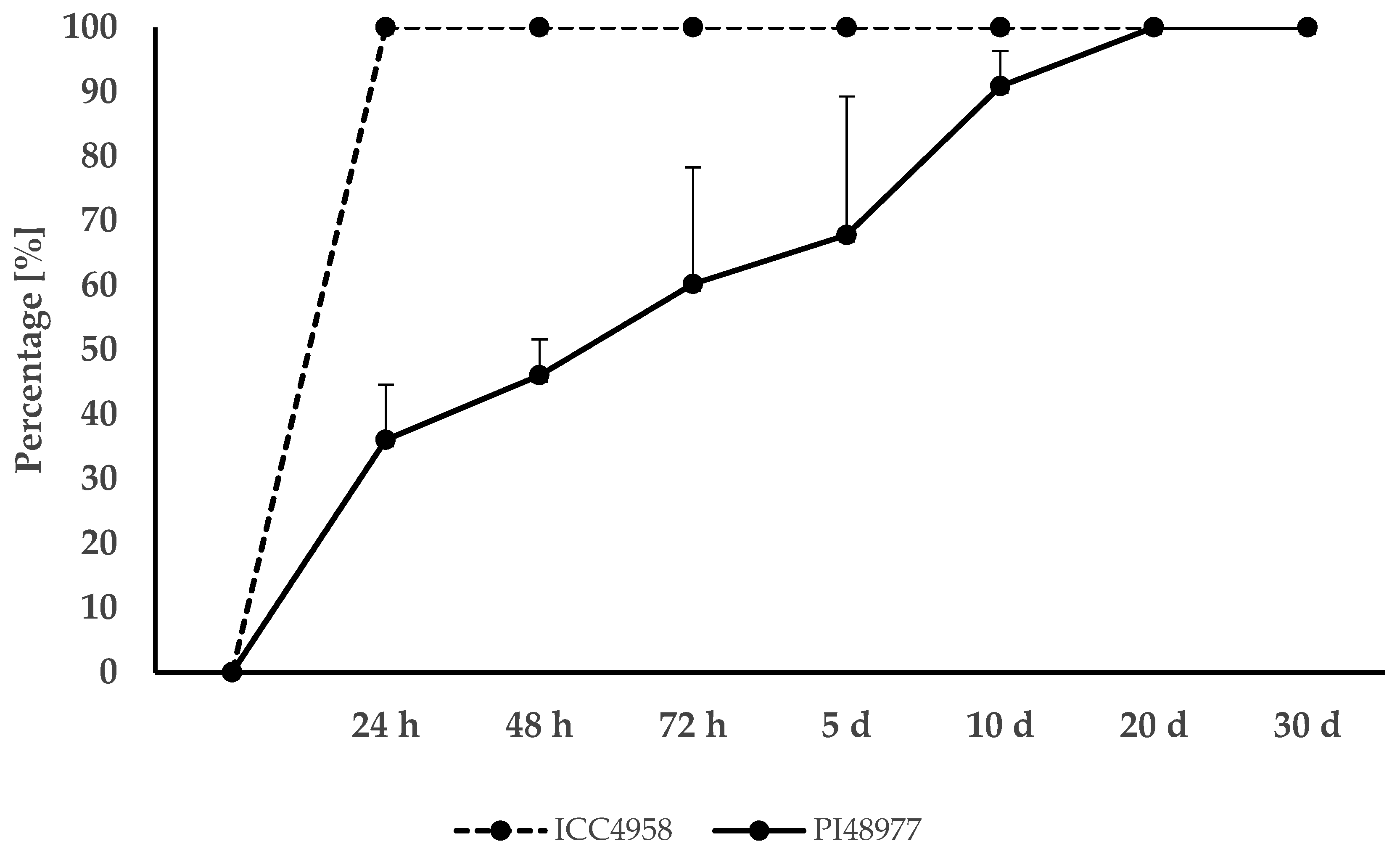
The number of imbibed seeds was used as a measure of germination in all evaluated calculations (i.e., coefficients), since the aim of this study was to evaluate physical dormancy caused by a water-impermeable seed coat. The seeds of cultivated ICC4958 imbibed easily and germinated within 24 h, while the seeds of wild PI48977 displayed certain level of dormancy that were mediated by the seed coat permeability. The germination percentage (FGP) of wild PI48977 was recorded at several time intervals of 24 h (36.1%), 48 h (46.1%), 72 h (60.2%), 5 days (67.8%), 10 days (90.9%), 20 days (100%) and 30 days (100%). Figure 2 shows the FGP comparison for parental genotypes ICC4958 and PI48977 at seven time intervals. Cultivated ICC4958 reached 100% FGP immediately after 24 h, while wild dormant PI48977 reached FGP 100% between the 10th and 20th day.
The mean germination coefficient (MGT) represented the speed of germination, and thus the lowest MGT value indicated that seed germination was fast. MGT after 30 days was for cultivated ICC4958 as one day and for wild PI48977 as five days. The first day of germination (FDG) was the same for both parental genotypes, at day 1, which indicated a fast initiation of germination. However, the last day of germination (LDG) differed, for cultivated ICC4958 where all seeds imbibed on the first day, the value was day 1, while for wild PI48977, the value was day 14. The time spread of germination (TSG) represented as the time in days between the first and last germination event. TSG for cultivated ICC4958 was 0 and for wild PI48977 it was 13 days. The coefficient of velocity indicated the rapidity of germination and differed significantly between parental genotypes. CVG after 30 days for cultivated ICC4958 was 100 and for wild PI48977 was 20. Cultivated ICC4958 had the highest possible value, and this state occurred when all seeds germinated on the first day. The Timson germination index (TGI) differed in parental genotypes after 30 days. For cultivated ICC4958 the value was 30 and for wild PI48977 the value was 26.
RILs population displayed even more variability in each of these measurements. FGP after 24 h ranged from 2–100% (Figure S1), and after 30 days ranged from 24–100% (Figure S2); MGT ranged from 1.0–23.98 days (Figure S3); CVG ranged from 4.17–100 (Figure S4); and TGI ranged from 17.57–30 (Figure S5).
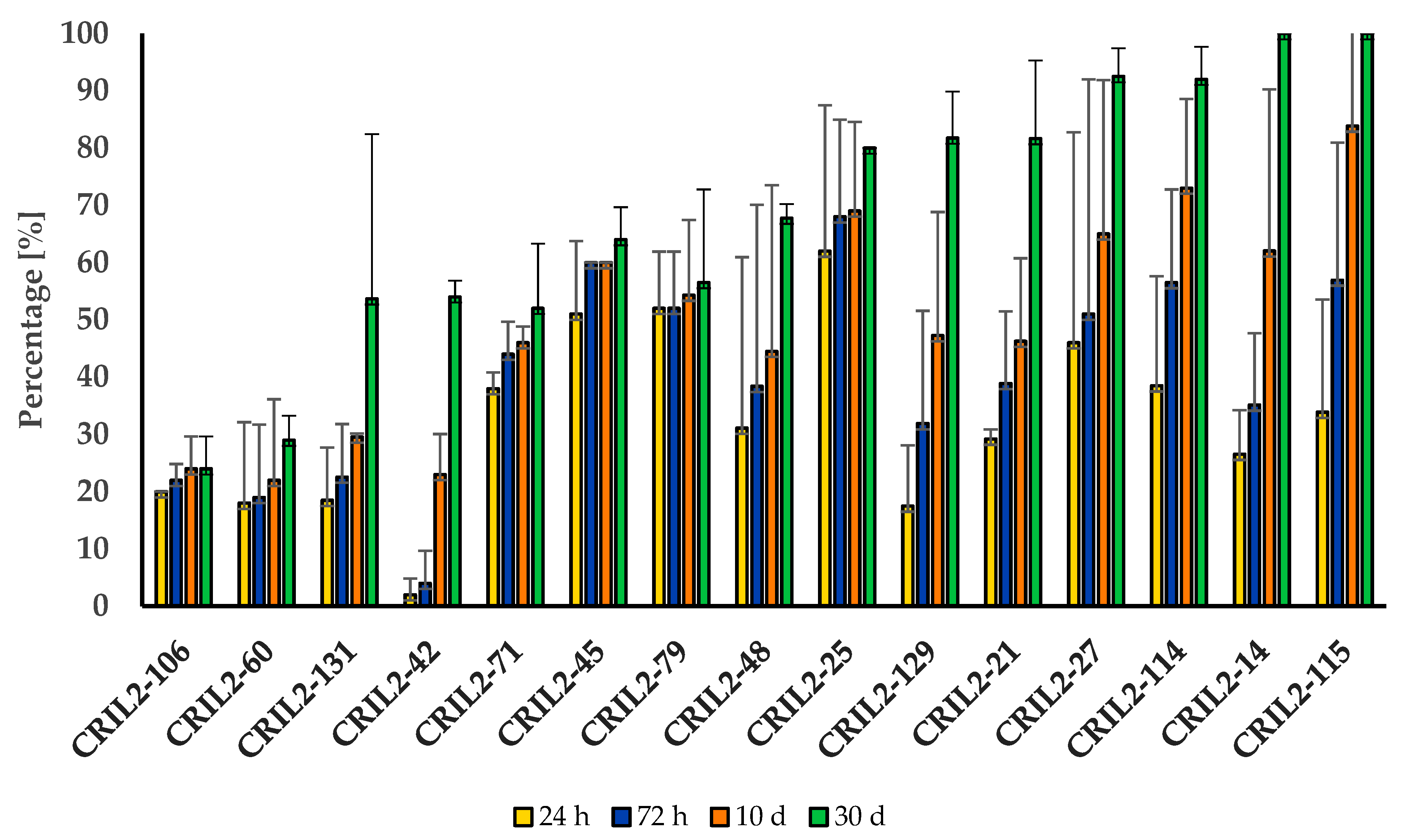
Representative non-dormant RILs were chosen based on FGP data of 24 h, when 10 RIL lines had 100% germination and, in an additional eight RILs, germination ranged from 88–99%. Dormant RILs were chosen primarily based on FGP data of 24 h with the lowest FGP values, but also with consideration of the data obtained after 30 days and the overall development of FGP data during testing. Five lines had very strong dormancy, with FGP range from 0–20% at 24 h and 10 lines were chosen as dormant, with the FGP range from 26–80%. Figure 3 shows the germination of 15 selected dormant RILs at four time intervals. Evaluation after 30 days showed that only two lines (CRIL2-14, CRIL2-115) reached 100% FGP. Table S1 summarizes data for parental genotypes and selected RILs in the categories of dormant (15 lines) and non-dormant (18 lines). The average FGP at 30 days within the dormant group of 15 RILs was 70.5% and for the non-dormant group of 18 RILs was 99.3%. Within selected lines, the MGT ranged from 1, which was the lowest possible value and meant that the population germinated on the first day, to 23.98 (CRIL2-42), with very slow germination. The dormant group of 15 RILs had an average MGT of 7.26 days while non-dormant group of 18 RILs had an average MGT of 1.2 days. The CVG value determined the rapidity of germination. Within selected RILs, the CVG ranged from 4.17 to 100. The average CVG after 30 days was for the non-dormant group of 18 RILs at 87.0 and for the dormant group of 15 RILs was 22.17. Ten lines of the non-dormant group had the highest possible CVG value of 100, which indicated that germination occurred on the first day. Within selected lines, the TGI ranged from 17.57 to 30. The average TGI after 30 days was, for the non-dormant group of 18 RILs, 29.8 and for dormant group of 15 RILs was 25.9.

3.2. Anatomical and Histochemical Analysis

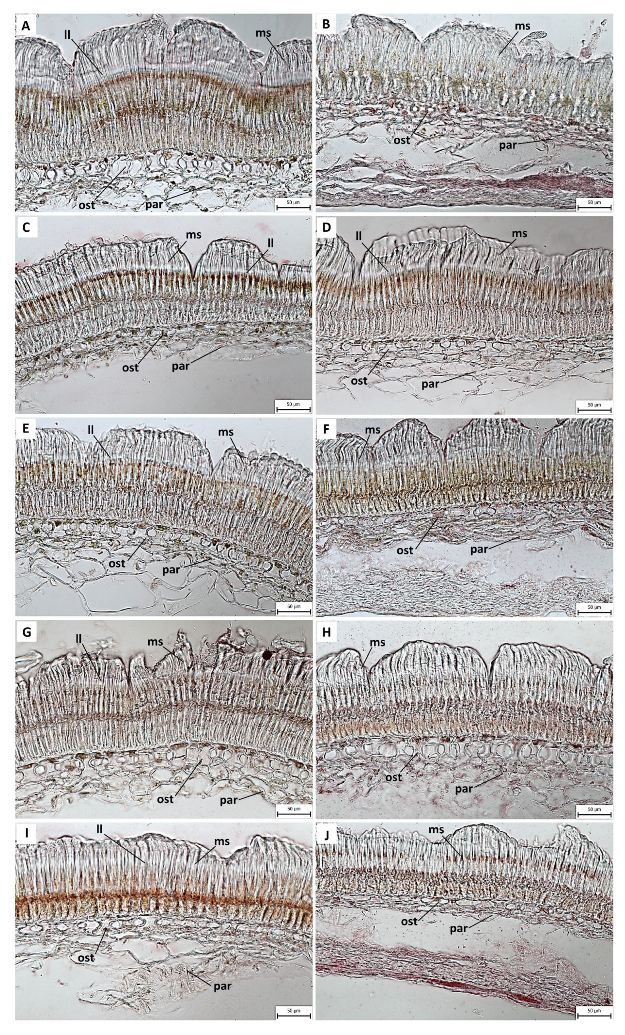
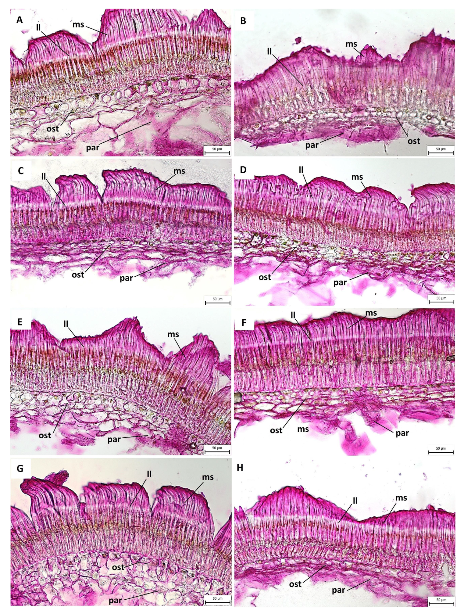
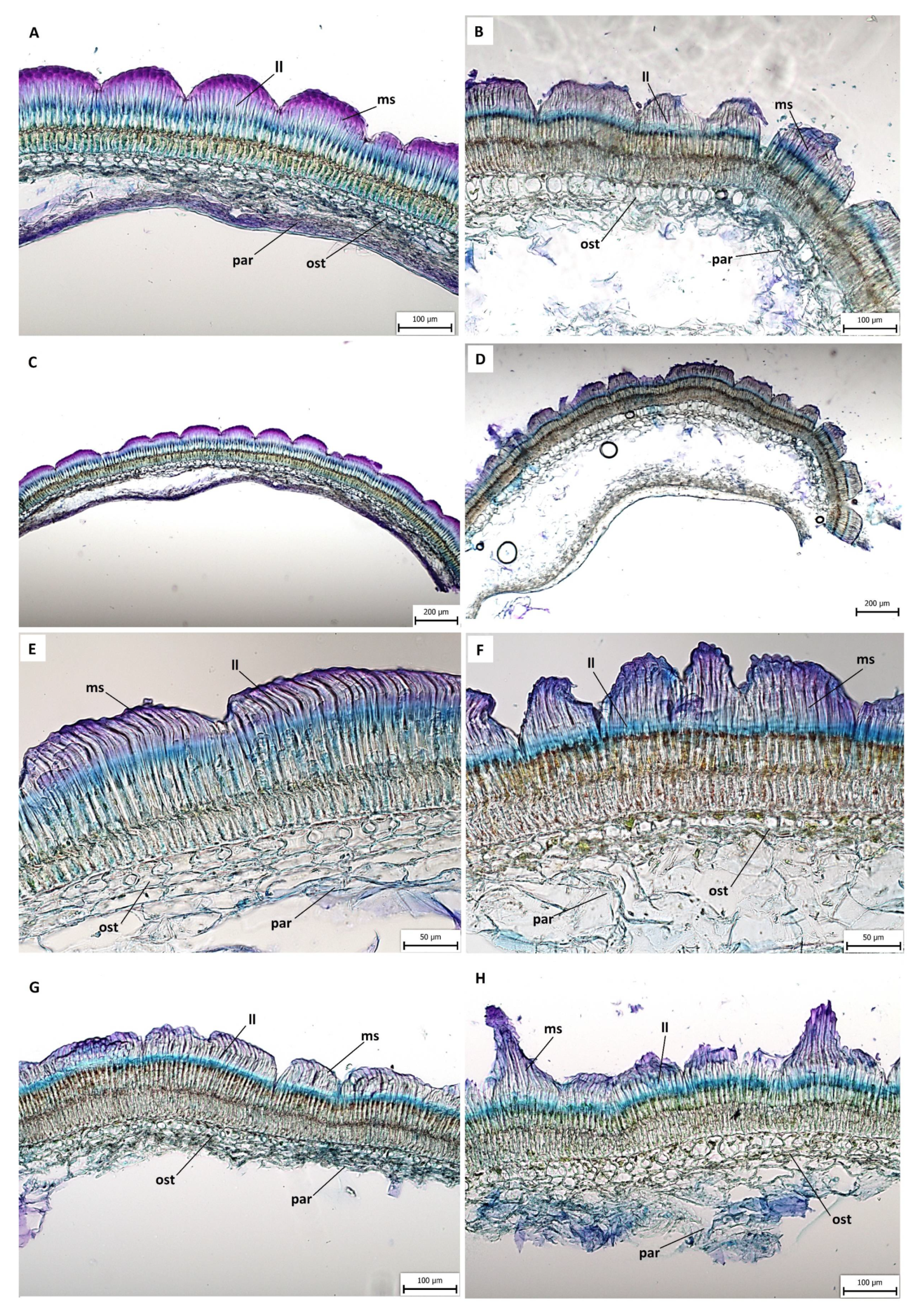
The continuity of the seed coat surface was interrupted by minor (ICC4958 and non-dormant lines; Figure 4B, Figure 5F and Figure 6E) or major (PI48977 and dormant lines; Figure 4A, Figure 5C and Figure 6F) fissures. The surface was covered with a thin cuticle. The cytological arrangement of the analyzed seed coats varied, especially in macrosclereids, and seed coat thickness was primarily managed by their length.
The lipidic character of the cuticle covering the surface can be stained by Sudan Red. Lipidic substances (Figure 4) were detected at the cuticle, but there was also a signal below the light line of the macrosclereid as a continuous strip in wild PI48977 (Figure 4A) and several lines, mostly in the dormant group (Figure 4C,E) but also in the non-dormant group (Figure 4D). In the middle part of the macrosclereid, closer to the basal part of cells, lipidic content was visible with strong staining (Figure 4G,I).
Pectin substances were revealed with Ruthenium Red (Figure 5). The seed coats had a strong staining signal in the upper parts of the macrosclereid cells (Figure 5A–H). Significant staining was observed below (Figure 5A,C,E,G) and above (Figure 5F,H) the light line. Similarly to the Sudan Red staining, Ruthenium Red showed a visible stained line in the middle of the macrosclereid cells (Figure 5C,D). A light line was clearly visible in all of the analyzed RIL lines and in both parents.
Metachromatic staining with Toluidine Blue (Figure 6) showed the difference between the cultivated chickpea and the wild parent. The seed coat surface of the cultivated ICC4958 (Figure 6A,C) had a more compact character, while the wild PI48977 (Figure 6B,D) seed coat surface had frequent protrusion and extensions of macrosclereids. Similarity between the cultivated parent and non-dormant RIL lines and the wild parent and dormant RIL lines was observed. The seed coats of the non-dormant lines (Figure 6E,G) were more compact, while the seed coats of dormant lines (Figure 6F,H) were gritty, with many macrosclereid extensions and fissures. The Toluidine Blue staining also clearly visualized the osteosclereids and the underlying parenchyma cell layer (Figure 6E,F). A light line was also present, but not clearly visible. A strong staining signal in macrosclereid tips bellow the cuticle (Figure 6A,F) was determined as the polyanionic cell wall components. This signal was present in both cultivated and wild parents and non-dormant and dormant lines (Figure 6A–H).

3.3. Length of Macroslereid

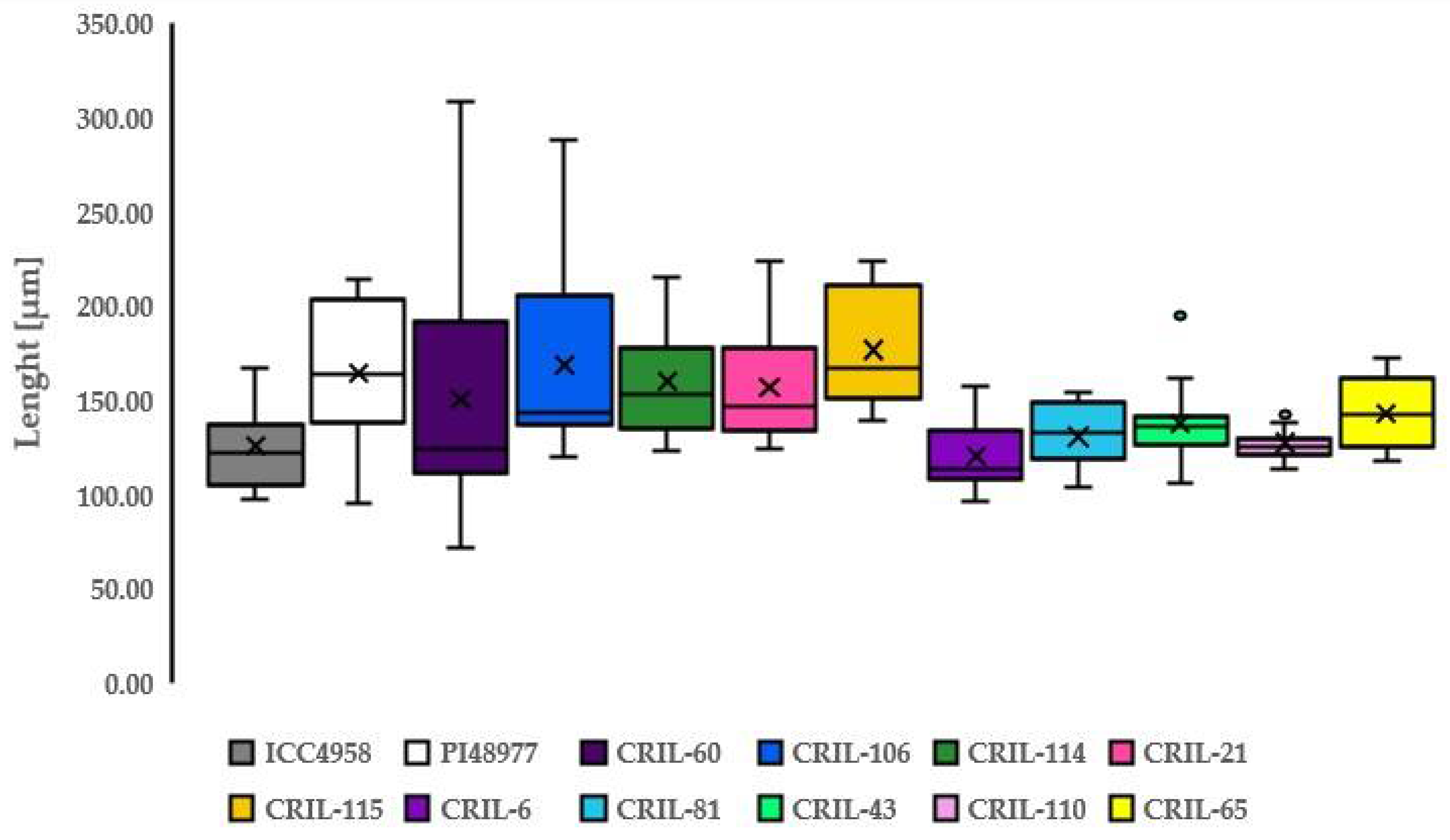
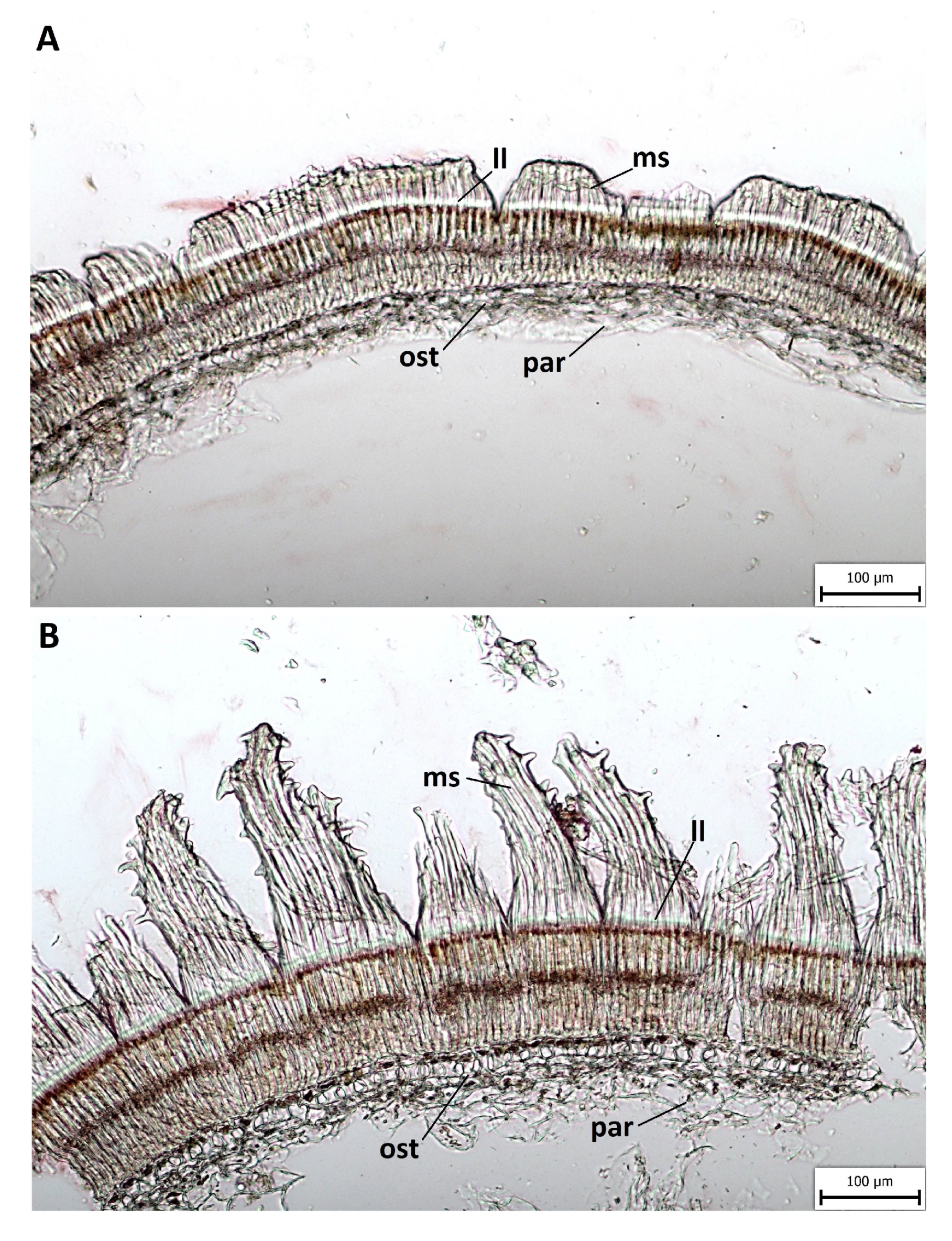
The length of the macrosclereid in cultivated ICC4958 ranged from 97.81 µm to 166.05 µm (Figure 7, Table S2), with the arithmetical mean of all sections being 125.49 µm and the standard deviation value of 21.94 (Table 1). While for wild PI48977 the upper value of the range was much higher, with the length ranging from 95.48 µm to 213.75 µm (Figure 7, Table S2), and the arithmetical mean of all sections with a value of 164.73 µm and standard deviation value of 35.30 (Table 1). There was a significant difference (p = 0.001) between the parental genotypes. The macrosclereid length of five representative dormant lines measured on five independent sections in triplicate ranged from 72.27 µm to 308.28 µm (Figure 7, Table S2). The means of the length for the dormant group (Table 2) were CRIL2-60 (150.97 µm), CRIL2-106 (168.45 µm), CRIL2-114 (159.94 µm), CRIL2-21 (157.33 µm), and CRIL2-115 (176.85 µm). The macrosclereid length of five representative non-dormant lines measured on five independent sections in triplicate ranged from 96.16 µm to 172.47 µm (Figure 7, Table S2). The means of length for the non-dormant group (Table 3) were CRIL2-6 (120.56 µm), CRIL2-81 (130.83 µm), CRIL2-43 (137.71 µm), CRIL2-110 (127.45 µm), and CRIL2-65 (138.28 µm). There was a significant difference (p = 0.0001) between the lengths of the non-dormant and dormant lines. The wild dormant parent PI48977 and dormant group of RILs had values usually above 150 µm, while the cultivated non-dormant parent and non-dormant group of RILs had values usually under 140 µm. Representation of all measured macrosclereid lengths for each parent or RIL lines with means and SD values is shown in the box-plot (Figure 7). Figure 8 shows the differences in the macrosclereid cells within the single seed of CRIL2-60 line.

4. Discussion

Most of the information about physical dormancy is based on anatomical and morphological structure studies, phenolic content determination and cuticle composition [20]. The use of domesticated crop and wild progenitors offers the advantage of comparative analysis, which can lead to identification of the underlying mechanism. It has been shown that reduction of seed coat thickness during domestication led to the reduction of seed coat impermeability [20], which we demonstrated with cultivated non-dormant and wild dormant chickpea parental genotypes. In addition to thickness, the structure of the palisade and cuticular layer [27,39] and presence or absence of cracks [27,40,41] was proposed to be associated with seed hardness [27,39], which we also noticed in the wild parent and dormant lines. Seeds of the wild chickpea parent had a thicker macrosclereid palisade layer, which supports the finding in dormant pea seed genotypes, which probably contributes to the water impermeability of seed coats [42]. However, the structural differences of seed coats are not the only and major factor [24,25,27]. Rather, the chemical composition of the testa seems to be the most important factor. It was shown that content of polysaccharides, hydroxylated fatty acids or phenol compounds is related to testa properties [26,27,43,44,45,46]. We have found substantial structural differences in seed coat properties between the wild and domesticated chickpea. Seed coats differed remarkably in terms of the presence of protrusions and discontinuity in wild parent and dormant lines. On the contrary, the surface of the seed coat of different cultivars of soybean was found to have only small discontinuities [47], which is in accordance with our cultivated chickpea parental genotype.
The use of recombinant inbred lines derived from the cross of two contrasting parental lines provides the advantage of decoupling the effect of the indirect relationship, such as pigmentation, seed content, etc., which are the result of a thousand years of selection. The testing of such RIL lines allows the dissection of traits directly related to the phenotype, such as the seed dormancy level. The evaluation of germination and seed dormancy caused by a water impermeable seed coat based on imbibed seeds data divided RILs into non-dormant and dormant groups. This strategy was previously used for pea [48] and led to the identification of differentially expressed genes [27]. In our study, we observed germination differences in 2020 and 2021 testing mostly in wild chickpea and in several lines, while the cultivated parent imbibed immediately within 24 h in both cases. In general, it cannot be stated that germination in 2020 was strictly faster or slower than in 2021; some RIL lines imbibed almost in same period of time, but some RIL lines showed substantial differences between two years of testing. The final germination percentage after 30 days in the chickpea RIL population ranged from 24–100%, which means that at least a few seeds of dormant lines imbibed. In contrast, pea accessions tested over a period of 28 days had a final germination percentage from 0–100% [48]; this means that some accessions were completely dormant and did not imbibe at all. Beside the species differences, this might also be attributed to the environment of cultivation, since pea were cultivated in glasshouses while chickpea from field grown plants. The effect of the maternal environment on seed dormancy has been shown [49] at least in Arabidopsis and other plants with physiological dormancy types, but it is likely expected also in physical dormancy, in which it was found that the release of physical dormancy of Medicago truncatula accessions from arid regions occurred to a greater extent under higher (35/15 °C) rather than lower (25/15 °C) temperature alternations [31]. In addition, the position of seed development on the parental plant predominantly influences the physiological dormancy in Brassicaceae [50] and can be expected to affect dormancy in many other plant families, including legumes.
The effect of seed coat pigmentation on seed dormancy has been extensively documented by transparent testa mutants in Arabidopsis [49] and also pigmented legume seeds generally imbibe slower than non-pigmented ones and germination is also delayed [48,51,52,53]. This was observed also in our study, where chickpea RIL lines with bigger, non-pigmented and smoother seeds imbibed earlier than smaller, pigmented and notched seeds. This was also confirmed with the seed coat browning of common bean [54,55] and chickpea [56]. Moreover, there is a difference between kabuli and desi chickpea types. Non-pigmented kabuli seeds imbibed quickly, within 4–8 h, whereas pigmented desi genotypes reached maximum water imbibing capacity at up to 16 h [57]. In our study, cultivated chickpea seeds were representative of the desi type and were usually imbibed within a few hours, maximally up to 24 h. We did not conduct our study with the cultivated kabuli type, but we assume that it would imbibe even earlier. Moreover, the thicker macrosclereid cells of desi type are responsible for the seed coat color and its polyphenolic content [34,58]. There also exists a connection with the seed coat thickness, with the thicker seed coat desi type (comprising 14% of total seed weight) and thinner seed coat kabuli type (comprising 5% of total seed weight) [34,59]. Since we studied the cultivated parent of the desi type, with a generally thicker seed coat and wild chickpea with a former seed coat not affected by domestication, it can be assumed to have RIL population with thicker seed coats. Interestingly, the thick seed coat is believed to be responsible for resistance to bruchid beetle infestation [19].
Macrosclereid lengths as a measure of seed coat thickness have been already performed in several legume species, including cultivated chickpea. While in the study of Dattatreya et al. [60] macrosclereid cells were found to be elongated, bended at one end and narrowed at both ends with lengths ranging from 37.6–155.6 µm with a mean of 94.9 µm, in our study we observed longer lengths of macrosclereid cells, with a mean of 125.49 µm. This difference is likely due to variation present in cultivated chickpea, since we used in our study the RIL lines population derived from cultivated chickpea with a slightly thicker seed coat and wild chickpea parents. Similarly, two desi genotypes’ macrosclereid cells had 141 µm and 144 µm, respectively, where substantial similarity can be noticed with our cultivated desi chickpea with the macrosclereid cell length mean of 125.49 µm, while kabuli type had a macrosclereid cells length of 110 µm [34,59]. The thin kabuli seed coat is apparently due to a thinner layer of palisade and parenchyma cells with fewer pectic polysaccharides and less proteins [34].
Crosses of desi and kabuli types were used to estimate the genetic basis of the testa thickness [15]. Although the data for length are not provided, they considered a seed coat as thick with a length over 70 µm and thin when under 70 µm. In our study, we used a significantly higher value (150 µm) as the boundary of seed coat thickness because we used population derived from the crossing of cultivated and wild chickpea, unlike in the latter study. Measurements for wild chickpea (C. reticulatum) were not conducted until this study, and therefore no comparison is feasible. Our results regarding seed coat thickness are in agreement with the seed coat differences between dormant and non-dormant pea seeds, where dormant pea accessions had a thicker seed coat (mean 139 µm) than non-dormant pea accessions (mean 97 µm) [27,48], comparable to chickpea. Moreover, non-dormant pea accessions had a variable macrosclereid cell length and values often reached the high values typical for dormant accessions (above 80 µm) [48] which was in accordance with non-dormant chickpea, where several macrosclereid measurements exceeded the boundary of 150 µm. However, macrosclereid cells of dormant pea accessions did not fall below 80 µm, which was dissimilar to chickpea where several length measurements fall below 150 µm.
Lipidic compounds stained with Sudan Red mostly occur in the cuticle on the surface of the seed coat of chickpea, which is similar to pea [25,61], with a strong signal observed in middle and basal parts of the macrosclereid cells. In addition, nearly mature pea seeds had signal above the light line [61] that can be observed in some chickpea lines. The presence of non-cuticular waxy extracellular compounds was observed in the tips of the macrosclereids above the light line in another pea study [25] and was confirmed by autofluorescence. In our study of chickpea seeds we observed the light line, the presence of which was confirmed also in cultivated and wild pea genotypes. However, the light line of wild pea genotypes was more pronounced [61], which cannot be unambiguously stated for wild chickpea. A light line that lays within the macrosclereids was interpreted variously, but its main purpose is not yet clarified [61,62,63,64,65,66]. It is believed that the light line is the result of an altered orientation of cellulose microfibrils or structural discontinuity between the lignified and non-lignified parts of the cell wall [61,67,68].
Polysaccharides present in chickpea cotyledon cell walls consist of cellulose, xyloglucan, arabinan and of pectin [69], that can be stained with Ruthenium Red. A strong staining signal was observed in the upper parts of macrosclereids, bellow the light line for several dormant lines and above the light line for several non-dormant lines. In terms of the light line, it was distinct in the majority of lines. Pectin is one of the chemical compounds that may significantly influence the physicochemical properties of different cell types during its development and the further processing of seeds [68]. This class of polysaccharides was confirmed also in the cell walls of pea seed coats where its concentration varied by location and developmental stage [34,70] and was differently localized between wild and cultivated pea seed coats [25,61]. Nevertheless, we did not conduct analysis of developmental stages, which would probably show us certain changes in pectin seed coat distribution.
The hypodermal layer, comprising of a single layer of osteosclereids, is uniformly thickened in chickpea and was obvious in all chickpea lines. As mentioned earlier in the pea study [20], osteosclereids in chickpea also contribute to the mechanical strength of the seed coat. Interestingly, the seed coat of the interspecific breeding line derived from another wild chickpea (Cicer echinospermum P.H.Davis) closely related to C. reticulatum, differed from the other desi genotypes, except for its heterogeneity in terms of osteosclereid cell shape. This heterogeneity is considered as a signature trait of C. echinospermum that has been inherited by the interspecific line. Any possible role of this trait in seed dormancy or generally in seed protection is indistinct [34].
In recent years, two types of cultivated chickpea, kabuli and desi, stand in the forefront of the interest of researchers. Therefore, this study focused also on a wild chickpea progenitor and subsequent evaluation of the RIL population was needed. Taken together, this study showed the anatomical and histochemical differences related to the level of seed dormancy in chickpea. This knowledge, in combination with genetic mapping and transcriptomic analysis of the seed coat, will ultimately lead to the identification of the underlying molecular mechanism of seed dormancy in chickpea, as well as allow comparison to phylogenetically related legumes, such as pea, lentil and faba bean, domesticated in the same region of the Fertile Crescent. In addition, information about the chemical composition of chickpea seed coats in relation to seed dormancy needs to be further determined and, along with comprehensive examination of mentioned genetic, transcriptomic and chemical analysis could be utilized in further chickpea breeding programs, particularly in relation to regulation of seed germination which is essential for successful crop establishment and final yield.

Supplementary Materials

The following are available online at https://www.mdpi.com/article/10.3390/agronomy11112292/s1, Figure S1: Final germination percentage after 24 h for chickpea RIL lines population. Representatives of dormant and non-dormant lines are highlighted, Figure S2: Final germination percentage after 30 days for chickpea RIL lines population. Representatives of dormant and non-dormant lines are highlighted, Figure S3: Mean germination time for chickpea RIL lines population. Representatives of dormant and non-dormant lines are highlighted, Figure S4: Coefficient of velocity of germination for chickpea RIL lines population. Representatives of dormant and non-dormant lines are highlighted, Figure S5: Timson germination index for chickpea RIL lines population. Representatives of dormant and non-dormant lines are highlighted, Table S1: Germination data for chickpea parental genotypes (cultivated ICC4958, wild PI48977) and representative dormant and non-dormant chickpea RIL lines, Table S2: Macrosclereid cells length of chickpea parental genotypes (cultivated ICC4958, wild PI48977), dormant lines (CRIL2-60, CRIL2-106, CRIL2-114, CRIL2-21, CRIL2-115) and non-dormant lines (CRIL2-6, CRIL2-81, CRIL2-43, CRIL2-110, CRIL2-65).

Author Contributions

Conceptualization, P.S.; methodology, V.S., L.Z. and P.S.; formal analysis, V.S., P.H., M.G. and P.S.; investigation, V.S. and P.H.; resources, P.S.; data curation, V.S. and P.S.; writing—original draft preparation, V.S., P.H. and P.S.; writing—review and editing, V.S., P.H. and P.S.; project administration, P.H. and P.S.; funding acquisition, P.H. and P.S. All authors have read and agreed to the published version of the manuscript.

Funding

This research was funded by Internal Grant Agency of Mendel University in Brno, grant number AF-IGA2020-IP011.

Acknowledgments

We thank Clarice Coyne from USDA-ARS, WSU and Eric Bishop von Wettberg from UVM for providing plant material.

Conflicts of Interest

The authors declare no conflict of interest.

References

  1. Vance, C.P.; Graham, P.H.; Allan, D.L. Biological Nitrogen Fixation: Phosphorus—A Critical Future Need? In Nitrogen Fixation: From Molecules to Crop Productivity, Current Plant Science and Biotechnology in Agriculture; Pedrosa, F.O., Hungria, M., Yates, G., Newton, W.E., Eds.; Kluwer Academic Publishers: Dordrecht, The Netherlands, 2002; pp. 509–514. [Google Scholar] [CrossRef]
  2. Merga, B.; Haji, J. Economic importance of chickpea: Production, value, and world trade. Cogent Food Agric. 2019, 5, 1615718. [Google Scholar] [CrossRef]
  3. Wang, R.; Gangola, M.P.; Irvine, C.; Gaur, P.M.; Båga, M.; Chibbar, R.N. Co-localization of genomic regions associated with seed morphology and composition in a desi chickpea (Cicer arietinum L.) population varying in seed protein concentration. Theor. Appl. Genet. 2019, 132, 1263–1281. [Google Scholar] [CrossRef]
  4. Kudapa, H.; Garg, V.; Chitikineni, A.; Varshney, R.K. The RNA-Seq-based high resolution gene expression atlas of chickpea (Cicer arietinum L.) reveals dynamic spatio-temporal changes associated with growth and development: RNA-Seq based chickpea gene expression atlas. Plant Cell Environ. 2018, 41, 2209–2225. [Google Scholar] [CrossRef] [Green Version]
  5. Afshin, A.; Micha, R.; Khatibzadeh, S.; Mozaffarian, D. Consumption of nuts and legumes and risk of incident ischemic heart disease, stroke, and diabetes: A systematic review and meta-analysis. Am. J. Clin. Nutr. 2014, 100, 278–288. [Google Scholar] [CrossRef]
  6. Shin, M.-G.; Bulyntsev, S.V.; Chang, P.L.; Korbu, L.B.; Carrasquila-Garcia, N.; Vishnyakova, M.A.; Samsonova, M.G.; Cook, D.R.; Nuzhdin, S.V. Multi-trait analysis of domestication genes in Cicer arietinumCicer reticulatum hybrids with a multidimensional approach: Modelling wide crosses for crop improvement. Plant Sci. 2019, 285, 122–131. [Google Scholar] [CrossRef]
  7. Coyne, C.J.; Kumar, S.; Wettberg, E.J.B.; Marques, E.; Berger, J.D.; Redden, R.J.; Ellis, T.H.N.; Brus, J.; Zablatzká, L.; Smýkal, P. Potential and limits of exploitation of crop wild relatives for pea, lentil, and chickpea improvement. Legume Sci. 2020, 2, e36. [Google Scholar] [CrossRef] [Green Version]
  8. Ladizinsky, G.; Adler, A. The origin of chickpea Cicer arietinum L. Euphytica 1976, 25, 211–217. [Google Scholar] [CrossRef]
  9. Ladizinsky, G. Plant Evolution under Domestication; Kluwer Academic Publishers: Dordrecht, The Netherlands, 1998. [Google Scholar]
  10. Redden, R.J.; Berger, J.D. History and origin of chickpea. In Chickpea Breeding and Management; Yadav, S.S., Redden, R.J., Chen, W., Sharma, B., Eds.; CABI: Wallingford, UK, 2007; pp. 1–13. [Google Scholar] [CrossRef]
  11. Hammer, K. Das Domestikationssyndrom. Die Kult. 1984, 32, 11–34. [Google Scholar] [CrossRef]
  12. Ladizinsky, G. Pulse domestication before cultivation. Econ. Bot. 1987, 41, 60–65. [Google Scholar] [CrossRef]
  13. Varma Penmetsa, R.; Carrasquilla-Garcia, N.; Bergmann, E.M.; Vance, L.; Castro, B.; Kassa, M.T.; Sarma, B.K.; Datta, S.; Farmer, A.D.; Baek, J.; et al. Multiple post-domestication origins of kabuli chickpea through allelic variation in a diversification-associated transcription factor. New Phytol. 2016, 211, 1440–1451. [Google Scholar] [CrossRef] [Green Version]
  14. Abbo, S.; Berger, J.; Turner, N.C. Viewpoint: Evolution of cultivated chickpea: Four bottlenecks limit diversity and constrain adaptation. Funct. Plant Biol. 2003, 30, 1081. [Google Scholar] [CrossRef] [Green Version]
  15. Gil, J.; Cubero, J.I. Inheritance of Seed Coat Thickness in Chickpea (Cicer arietinum L.) and its Evolutionary Implications. Plant Breed. 1993, 111, 257–260. [Google Scholar] [CrossRef]
  16. Iruela, M.; Rubio, J.; Cubero, J.I.; Gil, J.; Millán, T. Phylogenetic analysis in the genus Cicer and cultivated chickpea using RAPD and ISSR markers. Theor. Appl. Genet. 2002, 104, 643–651. [Google Scholar] [CrossRef] [PubMed]
  17. Moreno, M.-T.; Cubero, J.I. Variation in Cicer arietinum L. Euphytica 1978, 27, 465–485. [Google Scholar] [CrossRef]
  18. Jukanti, A.K.; Gaur, P.M.; Gowda, C.L.L.; Chibbar, R.N. Nutritional quality and health benefits of chickpea (Cicer arietinum L.): A review. Br. J. Nutr. 2012, 108, S11–S26. [Google Scholar] [CrossRef] [PubMed] [Green Version]
  19. Sastry, D.; Upadhyaya, H.D.; Srinivas, T.R. Variation for seed physical and hydration properties of chickpea (Cicer arietinum L.) mini core collection and their relevance to conservation and utilization. Plant Genet. Resour. 2019, 17, 311–324. [Google Scholar] [CrossRef]
  20. Smýkal, P.; Vernoud, V.; Blair, M.W.; Soukup, A.; Thompson, R.D. The role of the testa during development and in establishment of dormancy of the legume seed. Front. Plant Sci. 2014, 5, 351. [Google Scholar] [CrossRef] [Green Version]
  21. Smýkal, P.; Nelson, M.; Berger, J.; Von Wettberg, E. The Impact of Genetic Changes during Crop Domestication. Agronomy 2018, 8, 119. [Google Scholar] [CrossRef] [Green Version]
  22. Donohue, K.; Rubio de Casas, R.; Burghardt, L.; Kovach, K.; Willis, C.G. Germination, Postgermination Adaptation, and Species Ecological Ranges. Annu. Rev. Ecol. Evol. Syst. 2010, 41, 293–319. [Google Scholar] [CrossRef]
  23. Baskin, J.M.; Baskin, C.C.; Li, X. Taxonomy, anatomy and evolution of physical dormancy in seeds. Plant Species Biol. 2000, 15, 139–152. [Google Scholar] [CrossRef]
  24. Souza, F.H.; Marcos-Filho, J. The seed coat as a modulator of seed-environment relationships in Fabaceae. Rev. Bras. Bot. 2001, 24, 365–375. [Google Scholar] [CrossRef] [Green Version]
  25. Janská, A.; Pecková, E.; Sczepaniak, B.; Smýkal, P.; Soukup, A. The role of the testa during the establishment of physical dormancy in the pea seed. Ann. Bot. 2019, 123, 815–829. [Google Scholar] [CrossRef]
  26. Cechová, M.; Válková, M.; Hradilová, I.; Janská, A.; Soukup, A.; Smýkal, P.; Bednář, P. Towards Better Understanding of Pea Seed Dormancy Using Laser Desorption/Ionization Mass Spectrometry. Int. J. Mol. Sci. 2017, 18, 2196. [Google Scholar] [CrossRef] [Green Version]
  27. Hradilová, I.; Trněný, O.; Válková, M.; Cechová, M.; Janská, A.; Prokešová, L.; Aamir, K.; Krezdorn, N.; Rotter, B.; Winter, P.; et al. A Combined Comparative Transcriptomic, Metabolomic, and Anatomical Analyses of Two Key Domestication Traits: Pod Dehiscence and Seed Dormancy in Pea (Pisum sp.). Front. Plant Sci. 2017, 8, 542. [Google Scholar] [CrossRef] [Green Version]
  28. Marbach, I.; Mayer, A.M. Permeability of Seed Coats to Water as Related to Drying Conditions and Metabolism of Phenolics. Plant Physiol. 1974, 54, 817–820. [Google Scholar] [CrossRef]
  29. Liu, C.; Jun, J.H.; Dixon, R.A. MYB5 and MYB14 Play Pivotal Roles in Seed Coat Polymer Biosynthesis in Medicago truncatula. Plant Physiol. 2014, 165, 1424–1439. [Google Scholar] [CrossRef] [Green Version]
  30. Renzi, J.P.; Duchoslav, M.; Brus, J.; Hradilová, I.; Pechanec, V.; Václavek, T.; Machalová, J.; Hron, K.; Verdier, J.; Smýkal, P. Plasticity of Medicago truncatula seed dormancy relates to large-scale environment variation (preprint). Plant Biol. 2019. [CrossRef]
  31. Renzi, J.P.; Duchoslav, M.; Brus, J.; Hradilová, I.; Pechanec, V.; Václavek, T.; Machalová, J.; Hron, K.; Verdier, J.; Smýkal, P. Physical Dormancy Release in Medicago truncatula Seeds Is Related to Environmental Variations. Plants 2020, 9, 503. [Google Scholar] [CrossRef] [PubMed] [Green Version]
  32. Weeden, N.F. Genetic Changes Accompanying the Domestication of Pisum sativum: Is there a Common Genetic Basis to the “Domestication Syndrome” for Legumes? Ann. Bot. 2007, 100, 1017–1025. [Google Scholar] [CrossRef] [PubMed] [Green Version]
  33. Jang, S.-J.; Sato, M.; Sato, K.; Jitsuyama, Y.; Fujino, K.; Mori, H.; Takahashi, R.; Benitez, E.R.; Liu, B.; Yamada, T.; et al. A Single-Nucleotide Polymorphism in an Endo-1,4-β-Glucanase Gene Controls Seed Coat Permeability in Soybean. PLoS ONE 2015, 10, e0128527. [Google Scholar] [CrossRef]
  34. Wood, J.A.; Knights, E.J.; Choct, M. Morphology of Chickpea Seeds (Cicer arietinum L.): Comparison of desi and kabuli Types. Int. J. Plant. Sci. 2011, 172, 632–643. [Google Scholar] [CrossRef]
  35. Kader, M.A. A Comparison of Seed Germination Calculation Formulae and the Associated Interpretation of Resulting Data. J. Proc. R. Soc. N. S. Wales 2005, 138, 65–75. [Google Scholar]
  36. Ranal, M.A.; Santana, D.G. How and why to measure the germination process? Rev. Bras. Bot. 2006, 29, 1–11. [Google Scholar] [CrossRef] [Green Version]
  37. Soukup, A.; Tylová, E. Essential Methods of Plant Sample Preparation for Light Microscopy. In Plant Cell Morphogenesis, Methods in Molecular Biology; Žárský, V., Cvrčková, F., Eds.; Humana Press: Totowa, NJ, USA, 2014; pp. 1–23. [Google Scholar] [CrossRef]
  38. Rasband, W.S.; ImageJ, U.S. National Institutes of Health, Bethesda, MD, USA. 1997–2018. Available online: https://imagej.nih.gov/ij/ (accessed on 26 October 2021).
  39. Vu, D.T.; Velusamy, V.; Park, E. Structure and chemical composition of wild soybean seed coat related to its permeability. Pak. J. Bot. 2014, 46, 1847–1857. [Google Scholar]
  40. Meyer, C.J.; Steudle, E.; Peterson, C.A. Patterns and kinetics of water uptake by soybean seeds. J. Exp. Bot. 2006, 58, 717–732. [Google Scholar] [CrossRef] [Green Version]
  41. Koizumi, M.; Kikuchi, K.; Isobe, S.; Ishida, N.; Naito, S.; Kano, H. Role of Seed Coat in Imbibing Soybean Seeds Observed by Micro-magnetic Resonance Imaging. Ann. Bot. 2008, 102, 343–352. [Google Scholar] [CrossRef] [PubMed]
  42. Miao, Z.H.; Fortune, J.A.; Gallagher, J. Anatomical structure and nutritive value of lupin seed coats. Aust. J. Agric. Res. 2001, 52, 985. [Google Scholar] [CrossRef]
  43. Mullin, W.J.; Xu, W. A study of the intervarietal differences of cotyledon and seed coat carbohydrates in soybeans. Food Res. Int. 2000, 33, 883–891. [Google Scholar] [CrossRef]
  44. Mullin, W.J.; Xu, W. Study of Soybean Seed Coat Components and Their Relationship to Water Absorption. J. Agric. Food Chem. 2001, 49, 5331–5335. [Google Scholar] [CrossRef]
  45. Shao, S.; Meyer, C.J.; Ma, F.; Peterson, C.A.; Bernards, M.A. The outermost cuticle of soybean seeds: Chemical composition and function during imbibition. J. Exp. Bot. 2007, 58, 1071–1082. [Google Scholar] [CrossRef] [Green Version]
  46. Zhou, S.; Sekizaki, H.; Yang, Z.; Sawa, S.; Pan, J. Phenolics in the Seed Coat of Wild Soybean (Glycine soja) and Their Significance for Seed Hardness and Seed Germination. J. Agric. Food Chem. 2010, 58, 10972–10978. [Google Scholar] [CrossRef] [PubMed]
  47. Ma, F. Cracks in the Palisade Cuticle of Soybean Seed Coats Correlate with their Permeability to Water. Ann. Bot. 2004, 94, 213–228. [Google Scholar] [CrossRef] [PubMed] [Green Version]
  48. Hradilová, I.; Duchoslav, M.; Brus, J.; Pechanec, V.; Hýbl, M.; Kopecký, P.; Smržová, L.; Štefelová, N.; Vaclávek, T.; Bariotakis, M.; et al. Variation in wild pea (Pisum sativum subsp. elatius) seed dormancy and its relationship to the environment and seed coat traits. PeerJ 2019, 7, e6263. [Google Scholar] [CrossRef] [Green Version]
  49. Kedaffrec, E.; Nordborg, M. The maternal environment interacts with genetic variation in regulating seed dormancy in Swedish Arabidopsis Thaliana. PLoS ONE 2017, 12, e0190242. [Google Scholar] [CrossRef] [Green Version]
  50. Lu, J.J.; Tan, D.Y.; Baskin, C.C.; Baskin, J.M. Effect of Seed Position on Parental Plant on Proportion of Seeds Produced with Nondeep and Intermediate Physiological Dormancy. Front. Plant Sci. 2017, 8, 147. [Google Scholar] [CrossRef] [PubMed] [Green Version]
  51. Wyatt, J.E. Seed coat and water absorption properties of seed of near-isogenic snap bean lines differing in seed coat color. J. Am. Soc. Hortic. Sci. 1977, 102, 478–480. [Google Scholar]
  52. Werker, E.; Marbach, I.; Mayer, A.M. Relation Between the Anatomy of the Testa, Water Permeability and the Presence of Phenolics in the Genus Pisum. Ann. Bot. 1979, 43, 765–771. [Google Scholar] [CrossRef]
  53. Kantar, F.; Pilbeam, C.J.; Hebblethwaite, P.D. Effect of tannin content of faba bean (Vicia faba) seed on seed vigour, germination and field emergence. Ann. App. Biol. 1996, 128, 85–93. [Google Scholar] [CrossRef]
  54. Caldas, G.V.; Blair, M.W. Inheritance of seed condensed tannins and their relationship with seed-coat color and pattern genes in common bean (Phaseolus vulgaris L.). Theor. Appl. Genet. 2009, 119, 131–142. [Google Scholar] [CrossRef]
  55. Díaz, A.M.; Caldas, G.V.; Blair, M.W. Concentrations of condensed tannins and anthocyanins in common bean seed coats. Food Res. Int. 2010, 43, 595–601. [Google Scholar] [CrossRef]
  56. Legesse, N.; Powel, A.A. Relationship between the development of seed coat pigmentation, seed coat adherence to the cotyledons and the rate of imbibition during the maturation of grain legumes. Seed Sci. Technol. 1996, 24, 23–32. [Google Scholar]
  57. Lamichaney, A.; Kudekallu, S. Differences in seed vigour traits between desi (pigmented) and kabuli (non-pigmented) ecotypes of chickpea (Cicer arietinum) and its association with field emergence. JEB 2017, 38, 735–742. [Google Scholar] [CrossRef]
  58. Singh, U.; Manohar, S.; Singh, A.K. The anatomical structure of desi and kabuli seed coats. Int. Chickpea Newsl. 1984, 10, 26–27. [Google Scholar]
  59. Sastry, D.; Upadhyaya, H.D.; Gowda, C.L.L. Determination of physical properties of chickpea seeds and their relevance in germplasm collections. Indian J. Plant. Genet. Resour. 2014, 27, 1–9. [Google Scholar]
  60. Dattatreya, A.M.; Nanjegowda, D.K.; Viswanath, P. Microscopic detection of adulteration of Bengal gram (Cicer arietinum) flour with other legume flour based on the seed testa macrosclereids. J. Food Sci. Technol. 2011, 48, 114–119. [Google Scholar] [CrossRef] [PubMed] [Green Version]
  61. Zablatzká, L.; Balarynová, J.; Klčová, B.; Kopecký, P.; Smýkal, P. Anatomy and Histochemistry of Seed Coat Development of Wild (Pisum sativum subsp. elatius (M. Bieb.) Asch. et Graebn. and Domesticated Pea (Pisum sativum subsp. sativum L.). Int. J. Mol. Sci. 2021, 22, 4602. [Google Scholar] [CrossRef]
  62. Reeve, R.M. Ontogeny of the sclereids in the integument of Pisum sativum L. Am. J. Bot. 1946, 33, 806–816. [Google Scholar] [CrossRef]
  63. Spurný, M. Changes in the permeability of the seed coat in connection with the development of suberin adcrustations of the macrosclereids from the seed coat of the pea (Pisum sativum L.). Flora 1964, 154, 547–567. [Google Scholar] [CrossRef]
  64. Cavazza, L. Recherches sur l’impermeabilite des grains dures chez les legumineuses. Ber. Schweiz Bot. Ges. 1950, 60, 596–610. [Google Scholar]
  65. Hamly, D.H. Softening of the Seeds of Melilotus alba. Bot. Gaz. 1932, 93, 345–375. [Google Scholar] [CrossRef]
  66. Bevilacqua, L.R.; Roti-Michelozzi, G.; Modenesi, P. The watertight dormancy of Melilotus alba seeds: Further observations on the palisade cell wall. Can. J. Bot. 1989, 67, 3453–3456. [Google Scholar] [CrossRef]
  67. Martens, H.; Jakobsen, H.B.; Lyshede, O.B. Development of the strophiole in seeds of white clover (Trifolium repens L.). Seed Sci. Res. 1995, 5, 171–176. [Google Scholar] [CrossRef]
  68. Manning, J.C.; van Staden, J. The development and ultrastructure of the testa and tracheid bar in Erythrina lysistemon Hutch. (Leguminosae: Papilionoideae). Protoplasma 1985, 129, 157–167. [Google Scholar] [CrossRef]
  69. Wood, J.A.; Tan, H.-T.; Collins, H.M.; Yap, K.; Khor, S.F.; Lim, W.L.; Xing, X.; Bulone, V.; Burton, R.A.; Fincher, G.B.; et al. Genetic and environmental factors contribute to variation in cell wall composition in mature desi chickpea (Cicer arietinum L.) cotyledons: Variation in chickpea cotyledon composition. Plant Cell Environ. 2019, 41, 2195–2208. [Google Scholar] [CrossRef] [Green Version]
  70. McCartney, L.; Knox, J.P. Regulation of pectic polysaccharide domains in relation to cell development and cell properities in the pea testa. J. Exp. Bot. 2002, 53, 707–713. [Google Scholar] [CrossRef] [Green Version]
Figure 1. Anatomical structure of chickpea seed coat. Transversal section of C. reticulatum seed coat stained with Sudan Red (left). Scale bar = 50 µm. Schematic drawing (right, modified from Smýkal et al. [20]). Abbreviations: ms—macrosclereid cells, ost—osteosclereids, par—parenchyma, ll—light line.
Agronomy 11 02292 g001
Figure 2. Comparison of final germination percentage of chickpea parental genotypes—cultivated C. arietinum (ICC4958) and wild C. reticulatum (PI48977) at seven time intervals.
Agronomy 11 02292 g002
Figure 3. Comparison of final germination percentage for selected dormant chickpea RILs at four time intervals. Error lines represents standard deviation of data.
Agronomy 11 02292 g003
Figure 4. Histochemical Sudan Red staining of chickpea parental genotypes and RIL lines (A)—PI48977, (B)—ICC4958, (C)—CRIL2-60 (dormant group), (D)—CRIL2-80 (non-dormant group), (E)—CRIL2-106 (dormant group), (F)—CRIL2-51 (non-dormant group), (G)—CRIL2-114 (dormant group), (H)—CRIL2-50 (non-dormant group), (I)—CRIL2-6 (dormant group), (J)—CRIL2-89 (non-dormant group). Scale bars = 50 µm. Abbreviations: ms—macrosclereid cells, ost—osteosclereids, par—parenchyma, ll—light line.
Agronomy 11 02292 g004
Figure 5. Histochemical Ruthenium Red staining of chickpea parental genotypes and RIL lines (A)—PI48977, (B)—ICC4958, (C)—CRIL2-60 (dormant group), (D)—CRIL2-110 (non-dormant group), (E)—CRIL2-129 (dormant group), (F)—CRIL2-65 (non-dormant group), (G)—CRIL2-115 (dormant group), (H)—CRIL2-89 (non-dormant group). Scale bars = 50 µm. Abbreviations: ms—macrosclereid cells, ost—osteosclereids, par—parenchyma, ll—light line.
Agronomy 11 02292 g005
Figure 6. Toluidine Blue staining of chickpea parental genotypes and RIL lines (A)—ICC4958, (B)—PI48977, (C)—ICC4958, (D)—PI48977, (E)—CRIL2-47 (non-dormant group), (F)—CRIL2-60 (dormant group), (G)—CRIL2-80 (non-dormant group), (H)—CRIL2-129 (dormant group). Scale bars = 50 µm (A,B,E,F), 200 µm (C,D), 100 µm (G,H). Abbreviations: ms—macrosclereid cells, ost—osteosclereids, par—parenchyma, ll—light line.
Agronomy 11 02292 g006
Figure 7. Box-plot of macrosclereid cells length of chickpea parental genotypes (cultivated ICC4958, wild PI48977), dormant lines (CRIL2-60, CRIL2-106, CRIL2-114, CRIL2-21, CRIL2-115) and non-dormant lines (CRIL2-6, CRIL2-81, CRIL2-43, CRIL2-110, CRIL2-65).
Agronomy 11 02292 g007
Figure 8. Seed coat of chickpea RIL line CRIL2-60—comparison of dormant group. Seed coat differences within one seed. (A)—slightly elongated macrosclereids, (B)—extensively elongated macrosclereids. Scale bars = 100 µm. Abbreviations: ms—macrosclereid cells, ost—osteosclereids, par—parenchyma, ll—light line.
Agronomy 11 02292 g008
Table 1. Macrosclereid cells length of chickpea parental genotypes—cultivated ICC4958 and wild PI48977.
ICC4958Length [µm]PI48977Length [µm]
MeanSDMeanSD
1143.5722.411177.4541.25
2114.7814.812142.8855.14
3113.1219.003155.3915.04
4134.5931.414169.4936.84
5121.4016.105178.4731.48
Mean125.49Mean164.73
SD21.94SD35.30
Table 2. Macrosclereid cells length of 5 selected chickpea dormant RILs (CRIL2-60, CRIL2-106, CRIL2-114, CRIL2-21, CRIL2-115).
CRIL2-60CRIL2-106CRIL2-114CRIL2-21CRIL2-115
Length [µm]MeanSDMeanSDMeanSDMeanSDMeanSD
199.4523.56162.8237.65149.4713.06143.188.71158.737.89
2230.5082.61184.6190.85158.4434.61172.6244.74210.9017.58
3124.7017.28160.8441.91155.6815.23191.5728.57141.563.42
4129.3326.52151.5419.06174.7542.70148.856.63167.3416.26
5183.9467.08182.4475.41161.3944.80130.453.54205.7119.45
Mean150.97168.45159.94157.33176.85
SD65.5551.7229.1730.5930.50
Table 3. Macrosclereid cells length of five selected chickpea non-dormant RILs (CRIL2-6, CRIL2-81, CRIL2-43, CRIL2-110, CRIL2-65).
CRIL2-6CRIL2-81CRIL2-43CRIL2-110CRIL2-65
Length [µm]MeanSDMeanSDMeanSDMeanSDMeanSD
1120.2215.45120.4914.33142.6211.63128.939.82137.267.23
2117.6015.59131.8925.30127.088.67139.309.00131.4714.36
3111.7117.59142.027.76165.3527.95122.705.16158.5714.21
4132.9424.57139.9018.41129.228.83123.881.88165.657.63
5120.347.77119.8513.37124.2816.58122.457.70122.887.00
Mean120.56130.83137.71127.45143.17
SD16.1117.2320.969.0314.00
Publisher’s Note: MDPI stays neutral with regard to jurisdictional claims in published maps and institutional affiliations.