Article

Bacteria Associated with Winter Wheat Degrade Fusarium Mycotoxins and Triazole Fungicide Residues

by
Urszula Wachowska
1,*,
Katarzyna Kucharska
1,
Wioletta Pluskota
2,
Sylwester Czaplicki
3 and
Kinga Stuper-Szablewska
4
1
Department of Entomology, Phytopathology and Molecular Diagnostics, University of Warmia and Mazury in Olsztyn, Prowocheńskiego 17, 10-720 Olsztyn, Poland
2
Department of Plant Physiology, Genetics and Biotechnology, University of Warmia and Mazury in Olsztyn, Oczapowskiego 1A, 10-719 Olsztyn, Poland
3
Department of Food Plant Chemistry and Processing, Faculty of Food Sciences, University of Warmia and Mazury in Olsztyn, 1 Cieszynski Sq., 10-726 Olsztyn, Poland
4
General Chemistry Unit, University of Life Sciences in Poznań, Wojska Polskiego 42, 60-624 Poznań, Poland
*
Author to whom correspondence should be addressed.
Agronomy 2020, 10(11), 1673; https://doi.org/10.3390/agronomy10111673
Submission received: 11 September 2020 / Revised: 20 October 2020 / Accepted: 26 October 2020 / Published: 29 October 2020
(This article belongs to the Special Issue Strategies for the Control of Fusarium Head Blight in Cereals)

Abstract

:
Fusarium head blight (FHB) is the most dangerous spike disease of wheat, and triazole fungicides are generally recommended for FHB control. Bacteria isolates obtained from wheat grain were identified as members of the genus Sphingomonas based on 16S rDNA gene sequence analysis. The degradation of propiconazole and trichothecenes was analyzed by high-performance liquid chromatography. Two field experiments were conducted to determine the effectiveness of the biological treatment. All of the tested Sphingomonas isolates produced surfactin. Moreover, all strains were effective in degrading propiconazole and exhibited inhibitory effects on pathogens that cause FHB in wheat. Sphingomonas isolate S11 was selected for the field experiment because it inhibited the development of F. culmorum colonies in vitro by 48.80%, and degraded propiconazole in 15.13% after 48 h. The application of Sphingomonas S11 suspension during the growing season of winter wheat decreased the deoxynivalenol (DON) content of grain inoculated with F. culmorum more than 22-fold. Sphingomonas sp. strain S11 applied after fungicides also decreased the contamination of grain with fungi of the genus Fusarium and their mycotoxins. The analyzed bacteria can be potentially used to protect wheat against FHB pathogens, increase yields and improve grain quality by eliminating dangerous mycotoxins and propiconazole residues.

1. Introduction

Fungi of the genus Fusarium infect bread wheat throughout the growing season and cause Fusarium head blight (FHB). Fusarium head blight is the most dangerous spike disease in all wheat-growing regions which decreases yields and compromises the quality of grain due to contamination with mycotoxins, including trichothecenes [1,2,3,4,5]. Fusarium culmorum is regarded as the most virulent Fusarium species in many regions of Europe, Asia, and Africa [3,6,7,8]. This pathogen poses a serious threat to wheat yields, but wheat cultivars can differ in susceptibility to inoculation with F. culmorum. [9]. Fusarium culmorum is tolerant to varied weather conditions, and it is particularly virulent at higher temperatures [10]. Type B trichothecenes, deoxynivalenol (DON), 3-acetyl-deoxynivalenol (3ADON), 15-acetyl-deoxynivalenol (15ADON), and nivalenol (NIV), produced mainly by F. graminearum and F. culmorum, are among the most frequently identified mycotoxins in grain cereals. A characteristic feature of trichothecenes is that they exert toxic effects on humans and animals [11]. Deoxynivalenol (also known as vomitoxin) causes vomiting, digestive problems, oxidative damage, and reproductive disorders, but it is not a human carcinogen [12,13]. Its toxicity can be mitigated by bacteria. For example, Sphingomonas strain KSM1 isolated from lake water was found to catabolize DON to 16-hydroxy-DON (16-HDON) which is 10 times less phytotoxic to wheat than DON [14]. The conversion of DON to 16-HDON by strain KSM1 is mediated by the cytochrome P450 system [15].
The integrated crop management system introduced in Europe (Directive 2009/128/EC of the European Parliament and of the Council) limits fungicide use and forces producers to apply biological methods to control the spread of Fusarium fungi [16]. These methods can be applied in combination with fungicidal preparations, which are not always effective in combating FHB due to the high dynamics of FHB epidemics and the low sensitivity of various Fusarium species to triazole fungicides that are generally recommended for FHB control [17]. Propiconazole (1-[(2-(2,4-dichlorophenyl)–4–propyl–1,3-dioxolan-2–yl)]-methyl)]-1H-1,2,4 triazole) is a triazole fungicide that blocks the biosynthesis of ergosterol. This fungicide is widely applied in agriculture to protect cereal crops against FHB pathogens. It is characterized by low toxicity to mammals and a long half-life of 214–315 days in soil [18,19,20]. Propiconazole demonstrates strong affinity for soil (soil adsorption coefficient, Koc, of 1800), and its concentrations in surface runoffs may be low in the year of the treatment. Skolness et al. [21] demonstrated that plasma concentrations of 17β-estradiol and vitellogenin in female fathead minnows (Pimephales promelas) were reduced under exposure to propiconazole, and egg production decreased in groups treated with 500 and 1000 µg/l of this fungicide.
Various methods are used to reduce agricultural contamination, including saturation, recycling, pyrolysis, and combustion [22]. However, physicochemical remedy strategies are expensive and not always effective [23]. For this reason, the use of microorganisms for degrading pollutants or protecting crops against pathogens is a promising environmental remediation method [2,3,20,24,25,26]. Bacteria of the genus Sphingomonas belong to the family Sphingomonadaceae, order Sphingomonadales and class Alfaproteobacteria [27]. The generic name for these gram-negative, yellow pigment-producing bacteria was proposed by Yabuuchi et al. [28]. These bacteria were divided into four genera, and more than 130 species have been identified in the genus Sphingomonas [27,29]. Many strains of Sphingomonas bacteria are capable of degrading xenobiotic and persistent pollutants [30,31,32], including polycyclic aromatic hydrocarbons [33], polychlorinated biphenyls [34], phenanthrene [35], herbicides [36], and fungicides [37]. Sphingomonas bacteria are rarely used to protect plants against pathogens, but they are applied to promote plant growth [15,30,38,39,40,41,42,43,44]. Those microorganisms naturally colonize wheat leaves and spikes, and they can potentially inhibit pathogenic infections caused by Fusarium spp. [15].
The objective of this study was to select isolates of the genus Sphingomonas that were capable of long-term survival on wheat leaves, were able to produce surfactin, inhibit the development of F. graminearum and F. culmorum colonies in vitro, and biodegrade propiconazole. The ability of Sphingomonas isolate S11 to minimize the adverse effects of the spray inoculation of winter wheat with F. culmorum, including a decrease in yields, grain colonization by fungi of the genus Fusarium, and an increase in trichothecene concentrations in grain, was evaluated under field conditions.

2. Materials and Methods

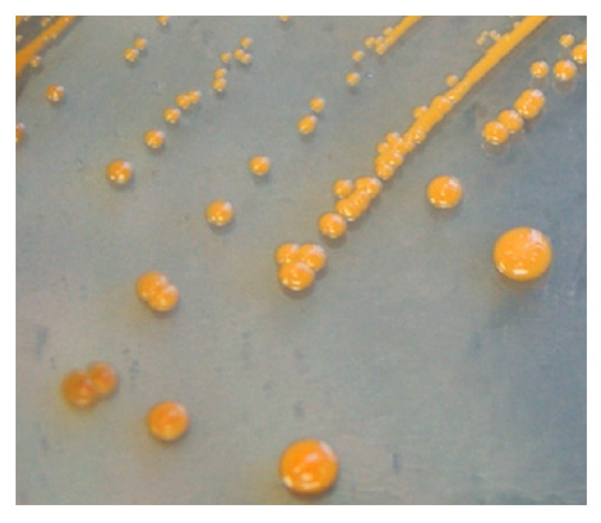
2.1. Origin and Identification of Bacterial Isolates of the Genus Sphingomonas

Six bacterial isolates of the genus Sphingomonas were obtained from the grain of winter wheat grown in the field in Poland. Bacteria were washed off from the surface of kernels [42,45,46] onto King’s B medium [47]. Gram-negative bacteria were identified according to the method proposed by Buck [48]. The morphology of bacterial colonies and cells was described based on their appearance on the PDA growth medium with pH 7.2 (Figure 1) or under a light microscope (Nikon E200, Tokyo, Japan). Oxidase activity was determined with 1% tetramethyl-p-phenylenediamine dihydrochloride as the reagent, according to the instructions attached to the test kit (Merck, Warsaw, Poland). Carbon source utilization and the enzymatic activity of the evaluated isolates were determined with the API 20NE microtest (bioMérieux, Warsaw, Poland) according to the manufacturer’s instructions. The 16S ribosomal RNA gene sequence of Sphingomonas S11 isolate were deposited in GenBank NCBI under accession number JX444564 (NCBI, National Center for Biotechnology Information, 2013) [49]. A phylogenetic tree was assembled using the neighbor-joining method in MEGA 6.0 (Tokyo, Japan) with 2000 bootstrap replications [50].

2.2. Molecular Screening for Natural Product Biosynthetic Gene Clusters

The presence of known natural product biosynthetic gene clusters in the isolate with bioactive potential was determined by PCR-based screening with the use of specific primers [51]. The following primer pairs were used: surfactin synthetase As1-F CGCGGMTACCGVATYGAGC and Ts2-R ATBCCTTTBTWDGAATGTCCGCC [52]; iturin A synthetase ituC-f AAAGGAT CCAAGCGTGCCTTTTACGGGAAA and ituC-r AAAAAGCTTAATGACGCCAGCTTTCTCTT [51]; fengycin synthetase Af2-f GAATAYMTCGGMCGTMTKGA and Tf1-r GCTTTWA DKGAATSBCCGCC; mycosubtilin synthetase Am1-f CAKCARGTSAAAATYCGMGG and Tm1-r CCDASATCAAARAADTTATC [51]. DNA was isolated from bacteria with the Bead-Beat Micro AX Gravity kit (A&A Biotechnology, Gdynia, Poland) according to the manufacturer’s procedure. The quantity and quality of the isolated DNA were determined by measuring absorbance at a wavelength of 260 nm and 280 nm (NanoMaester Gen, Warsaw, Poland). The volume of the reaction mixture was 50 μL, and the PCR reaction was conducted according to a previously described methodology [51] with certain modifications in the annealing temperature and time of the applied primers.
The amplicons obtained with As1-F and Ts2-R primers in gel electrophoresis were cut from the gel and purified with the Agarose-Out DNA Purification Kit (EURX, Gdańsk, Poland). The PCR products were cloned into pGEM-T Vector (Promega GmbH, Germany) and introduced into Escherichia coli XL1Blue cells by heat shock, according to the manufacturer’s protocols. The transformants were screened on LB agar medium supplemented with ampicillin (100 µg mL−1), IPTG (40 µL of 100 mM isopropyl β-D1-thiogalactopyranoside per plate) and X-Gal (40 µL of 20 mg/mL 5-bromo-4-chloro-indoxyl per plate). Plasmids were isolated from the transformed cells by alkaline lysis. The presence of an insert was confirmed by restriction analysis with SacI and SacII enzymes. The cloned products were sequenced using a universal primer that was complementary to the nucleotide sequences of T7 promoter (Genomed, Warszawa, Poland). The determined sequences were compared with the GenBank database using the basic local alignment search tool (BLAST) provided online by the National Center for Biotechnology Information (Maryland, MD, USA) [49].

2.3. Extraction and Purification of Surfactin

Sphingomonas sp. strains were cultured in 10 cm3 of a liquid medium (beef extract–1 g, soy peptone–5 g, NaCl–5 g, glucose–1 g, yeast extract–7 g in 1 dm3 of water) for 96 h at a temperature of 27 °C. Bacteria were removed by centrifugation (10,000× g, 30 min, 4 °C), and the resulting supernatant was sterilized by passing through a 0.22 μm syringe filter (Millipore, Darmstadt, Germany). Surfactin was determined by UPLC according to the procedure described by Mubarak et al. [53] with modifications. One millimeter of the culture liquid was passed through a 0.22 μm silicone filter and placed in a vial. The analysis was performed using the Acquity H-class UPLC system equipped with the Waters Acquity PDA detector (Waters, California, CA, USA). Chromatographic separation was performed on an Acquity UPLC® BEH C18 column (100 × 2.1 mm, particle size 1.7 μm) (Waters, Dublin, Ireland). The mobile phase consisted of acetonitrile (ACN) and 3.8 mM trifluoroacetic acid (TFA) solution (80:20 v/v) at a flow rate of 0.5 mL/min to obtain chromatograms under optimal conditions. The concentration of surfactin was determined using an internal standard at a wavelength of λ = 210 nm. The compound was identified by comparing the retention time (4 min) of the analyzed peak with the retention time of the standard, by adding a specific amount of the standard to the analyzed samples and repeating the analysis. The detection level was 1 μg/g.

2.4. Isolate Screening for Antifungal Properties

The isolates were screened for antifungal activity in a dual-culture test [51]. Potato dextrose agar (PDA) discs containing 7-day old mycelia of F. culmorum and F. graminearum were placed in the center of Petri plates with a diameter of 9 cm, filled with PDA. The pathogens obtained from wheat grain are characterized by high virulence and the potential to produce DON [54]. Suspensions of bacterial isolates with a concentration of 107 cells 1 cm−3 were placed in 5 mm openings in the PDA medium at a distance of 2 cm from the fungal discs. The plates were then incubated at 27 °C for 4 days in the dark and were observed for the inhibition of fungal mycelia. The surface area and the ellipticity ratio of pathogen colonies were calculated by multiplying the small diameter by the large diameter. Pathogen colonies cultured without bacteria were the control.

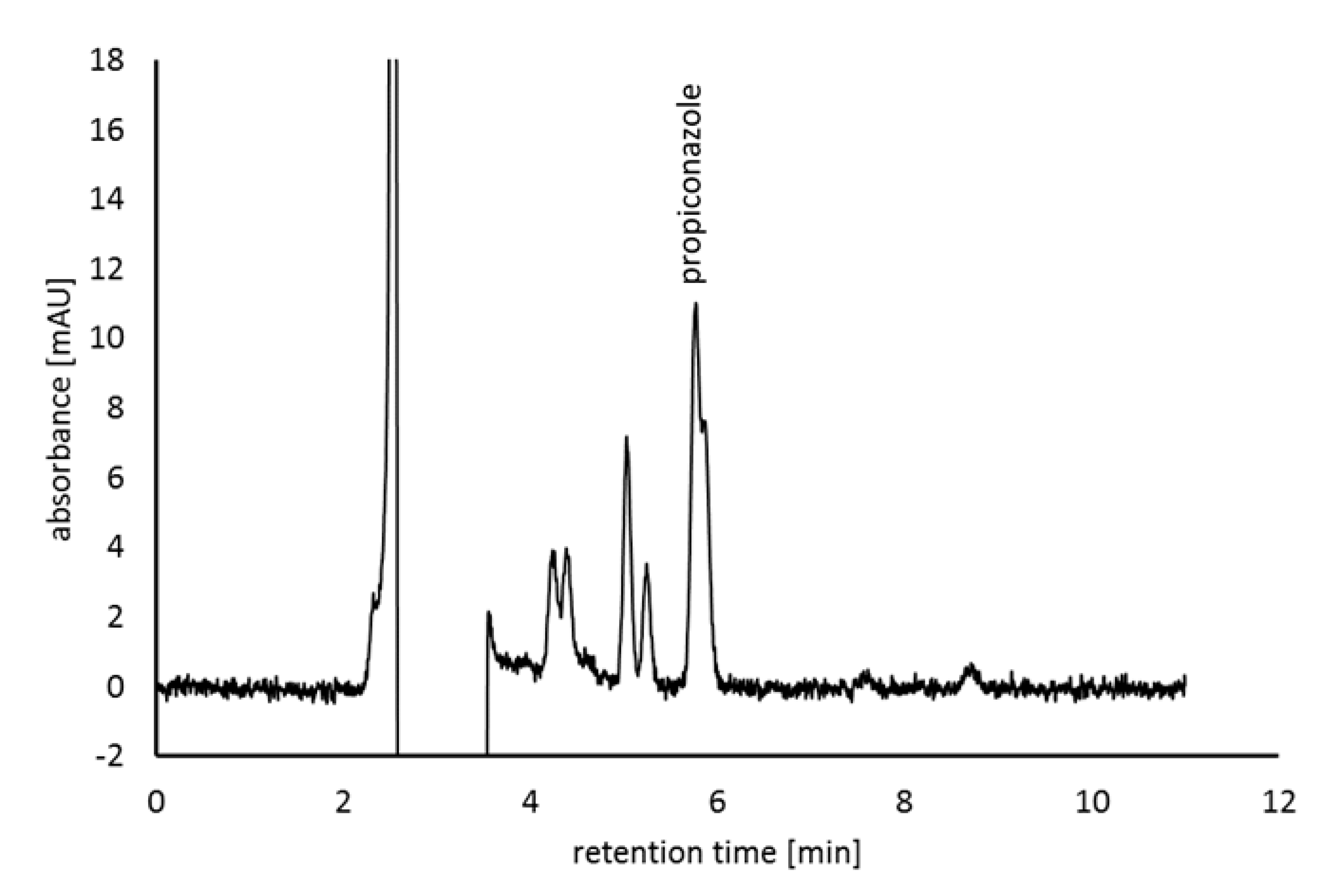
2.5. Selection of Sphingomonas sp. Isolates Capable of Biodegrading Propiconazole

Isolates demonstrating tolerance or low sensitivity to the highest propiconazole concentrations in the disc diffusion test [46] were selected for analyses of fungicide biodegradation. Isolates were cultured in 15 cm3 Falcon tubes (Bionovo, Legnica, Poland) in 10 cm3 of a liquid medium (beef extract–1 g, soy peptone–5 g, NaCl–5 g, glucose–1 g, yeast extract–7 g in 1 dm3 of water) containing propiconazole (Sigma-Aldrich, Poznań, Polska) with an initial concentration of 86.7 mg·dm−3. Bacteria were incubated for 48 h at a temperature of 27 °C. After incubation, the microorganisms were centrifuged (10,000× g, 30 min, 4 °C), and the resulting supernatant was sterilized by passing through a 0.22 μg syringe filter (Millipore, Darmstadt, Germany). Propiconazole levels were analyzed by high-performance liquid chromatography in the Agilent 1200 HPLC system (Agilent Technologies, Palo Alto, CA, USA) with a photodiode array detector. Analyses were conducted in isocratic mode with a mixture of acetonitrile and water (80:20, v/v), acidified with 0.15% formic acid as the eluent. Chromatographic separation was performed on an Agilent Eclipse XDB-C18 column (150 × 4.6 mm, 5 µm) at a temperature of 30 °C. The flow rate of the mobile phase was 0.5 cm3 min−1. Chromatographic data were registered at 220 nm wavelength based on the UV-Vis spectrum for the reference standard. Data were acquired and analyzed based on a calibration curve for the propiconazole reference standard in the HP ChemStation program (Agilent Technologies, Palo Alto, CA, USA).

2.6. Survival Rates of Sphingomonas Bacteria on the Leaves of Wheat Seedlings

Fifteen seedlings of winter wheat cvs. Bogatka and Tonacja were grown in pots (diameter 20 cm) in a controlled environment chamber with a temperature of 21 °C and constant air humidity of 70–80%. After six weeks, a cell suspension of Sphingomonas (108 cm−3) was sprayed on the leaves in the amount of 60 cm3 per pot. The experiment was conducted in four replicates. The survival rates of bacterial cells on leaf surface were determined after 1, 2, 3, 4, and 5 days of incubation. The microorganisms were washed off from the surface of leaves according to a previously described method [46].

2.7. Preparation of the Bacterial Suspension for the Field Treatment

Sphingomonas strain S11 was cultured for 7 days on Petri plates containing PDA with pH 7.2, in darkness, at a temperature of 27 °C. After incubation, bacterial colonies were washed off into 5 cm3 of sterile water (per plate) with an inoculation loop. The suspension had a cell concentration of 108 CFU per cm−3. One dm3 of the bacterial suspension was placed in a 12 dm3 backpack sprayer (Marolex Titan 12, Leśne, Poland), diluted with water to 9 dm3, and sprayed onto plots with an area of 20 m2. Plots with an area of 1 m2 were sprayed manually with a 1.5 dm3 sprayer (Marolex Master, Leśne, Poland) containing 250 cm3 of the bacterial suspension and 750 cm3 of water. Biological treatments were performed on windless days in the afternoon.

2.8. Spike Inoculation with Fusarium Culmorum

The field experiments were conducted in two variants. In the first variant, flowering wheat spikes (BBCH 65) were inoculated with an aqueous spore suspension of F. culmorum Fc32, which is potentially capable of producing DON [54]. The suspension had a concentration of 104 fungal cells cm−3. The spores were rinsed off from fungal colonies growing on PDA (Merck, Warsaw, Poland) for 14 days at 28 °C with 5 cm3 of sterile water. Twenty-four hours after the biological treatment (Bac and Integ), wheat spikes were inoculated at full flowering (BBCH 65) with a backpack sprayer (Marolex Titan 12, Leśne, Poland) containing 1 dm3 of the fungal suspension and 9 dm3 of water. Unprotected plants were the control in the first variant. In second variant, wheat spikes were sprayed with sterile water.

2.9. Antifungal Properties of Sphingomonas Strain S11 during the Growing Season of Wheat

Two field experiments were conducted in Bałdy (north-eastern Poland, N53°36′, E20°36′). Experiment 1 had a randomized block design and involved an integrated pest management (Integ) strategy. Winter wheat (Triticum aestivum L., bread wheat cv. Bogatka) was sown in plots with an area of 20 m2. Commercial fungicides containing fenpropimorph or propiconazole were applied twice at BBCH 31 and BBCH 55, respectively (Table 1). Experiment 2 had a completely randomized design, and a bacterial suspension (Bac) was applied to winter wheat cv. Tonacja sown in plots with an area of 1 m2. In both experiments, plants were fertilized with nitrogen (N) at 100 kg·ha−1, potassium (K) at 60 kg·ha−1, and phosphorus (P) at 26 kg·ha−1. The biological treatment involving a bacterial suspension of Sphingomonas S 11 was applied at full flowering (BBCH 65) [55] in Experiment 1, and it was applied three times in Experiment 2: when the first node was at least 1 cm above the tillering node (BBCH 31), in the middle of heading (BBCH 55), and at full flowering (BBCH 65). Both experiments were conducted in four replicates. Unprotected plants were the control.

2.10. Evaluation of Spike Health

The severity of FHB symptoms was determined in the hard dough stage (BBCH 87) on 100 wheat plants selected randomly from each treatment, according to the scale proposed by the European and Mediterranean Plant Protection Organization [56]. The results were expressed as the average percentage of spike surface area affected by the disease. Grain was harvested with a plot harvester in the fully ripe stage (BBCH 92). Grain yield was expressed in grams per 1 m2 of plot area.

2.11. Grain Colonization by Fungi of the Genus Fusarium

The counts of epiphytic fungi of the genus Fusarium colonizing wheat kernels were determined immediately after harvest. Grain samples of 10 g each were placed in 250 cm3 flasks filled with 90 cm3 of sterile water, and the flasks and were shaken on a shaker table (358 S, Elpin Plus, Lubawa, Poland) at 180 rpm for 30 min. Twice-diluted suspensions of fungal cells in the amount of 0.1 cm3 each were transferred to Petri plates with a diameter of 9 cm. Martin’s medium [57] was poured into the plates. The colonies of Fusarium fungi were counted after 7 days of growth. The number of colony forming units (CFU) of Fusarium fungi (N) rinsed off from 1 g of grain was calculated with the following formula: N = n/10−r × v, where v is the number of colonies on the plate, 10−r is the dilution coefficient, and v is the volume of the plated suspension.
Non-disinfected (to obtain epiphytic colonies) and surface-disinfected (to obtain endophytic colonies) kernels were placed in Petri plates filled with PDA (Merck, Warsaw, Poland). Kernels were disinfected in 1% sodium hypochlorite (NaOCl) for 5 min and rinsed with sterile water three times. A total of 100 kernels from each treatment were plated. The colony counts of Fusarium spp. growing on wheat kernels were expressed as a percentage of total kernels within epiphytes and endophytes. Fusarium spp. colonies were identified based on spore morphology [58] after 7 days of incubation at a temperature of 24 °C (Incubator En 120, Leszno, Poland).

2.12. Chemical Analysis of Ergosterol and Trichothecene

Samples containing 100 mg (ergosterol) or 10g (trichothecene) of ground grains were analyzed according to a previously described method [59]. The limit of detection for trichothecene was 0.01 mg/kg.

2.13. Statistical Analysis

The results were processed by ANOVA in the Statistica 12 program (StatSoft, Tulsa, OK, USA) [60]. The significance of differences between means was evaluated in the SNK test (p < 0.01). The sequences of the examined isolate and reference isolates were compared in the MEGA 6 program to determine their evolutionary relationships. A phylogenetic tree was developed with the use of the neighbor-joining (NJ) method. Data were verified in the Jukes-Cantor model. The reliability of phylogenetic trees was estimated in the bootstrap test with 2000 replicates. The efficacy of the tested isolates in degrading propiconazole was calculated according to the following formula: 100% − (b/a × 100%), where a is the concentration of propiconazole in control samples, and b is the concentration of propiconazole in a culture medium with bacterial isolates.

3. Results

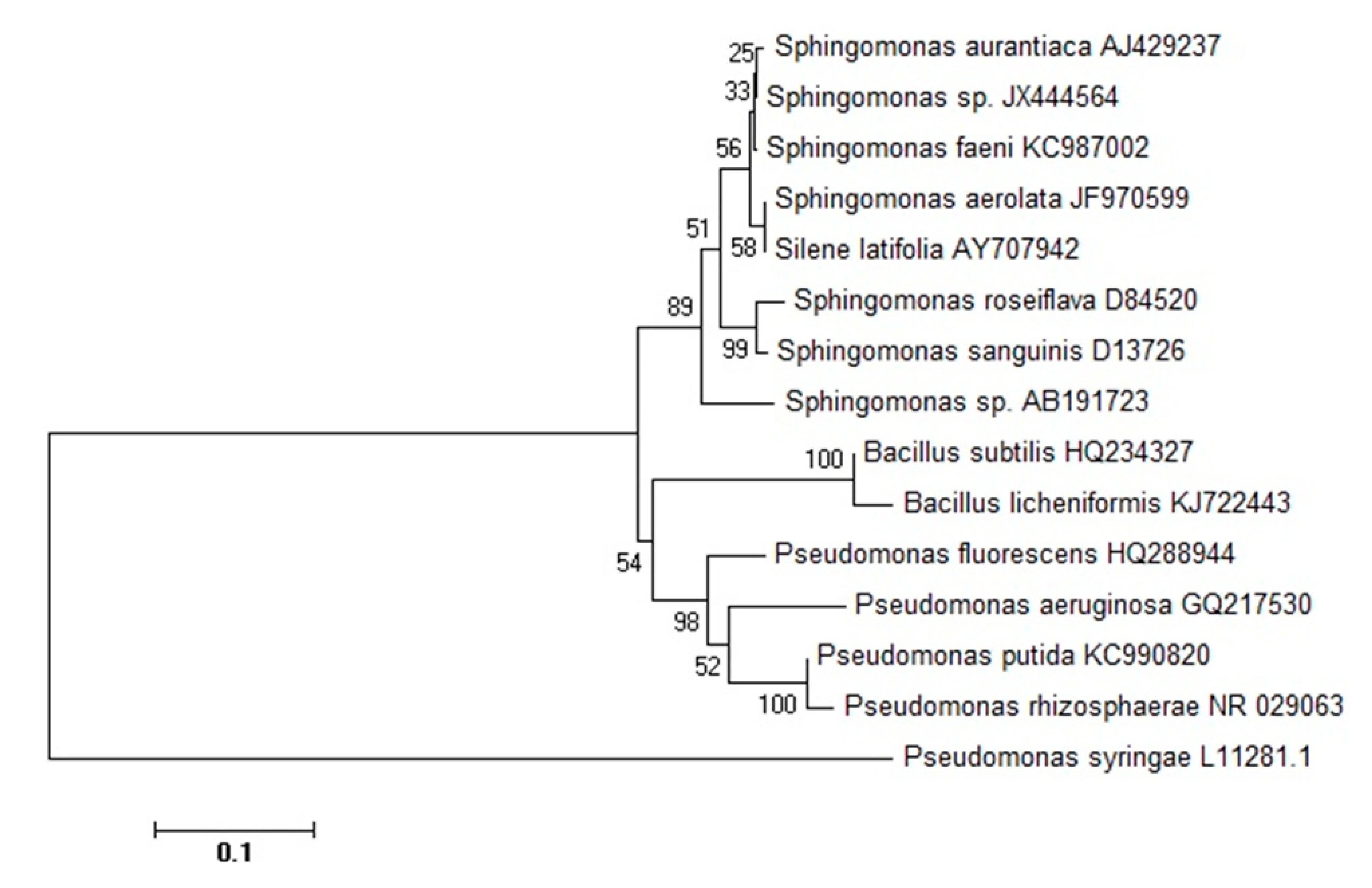
3.1. Identification and Phylogenetic Analysis of Sphingomonas sp.

The isolates were identified as Sphingomonas sp. based on their biochemical and molecular characteristics. The identified isolates were not allocated to any known species. Isolates of Sphingomonas sp. formed yellow-orange colonies with a diameter of 3–7 mm (Figure 1). All analyzed isolates of the genus Sphingomonas were gram-negative, and their cells were immobile and rod-shaped. All isolates tested positive in the catalase assay and negative in the oxidase assay. The ITS sequences in the isolates were most similar to S. aurantiaca AJ429237 and S. faeni KC987002, and less similar to other Sphingomonas species (Figure 2).

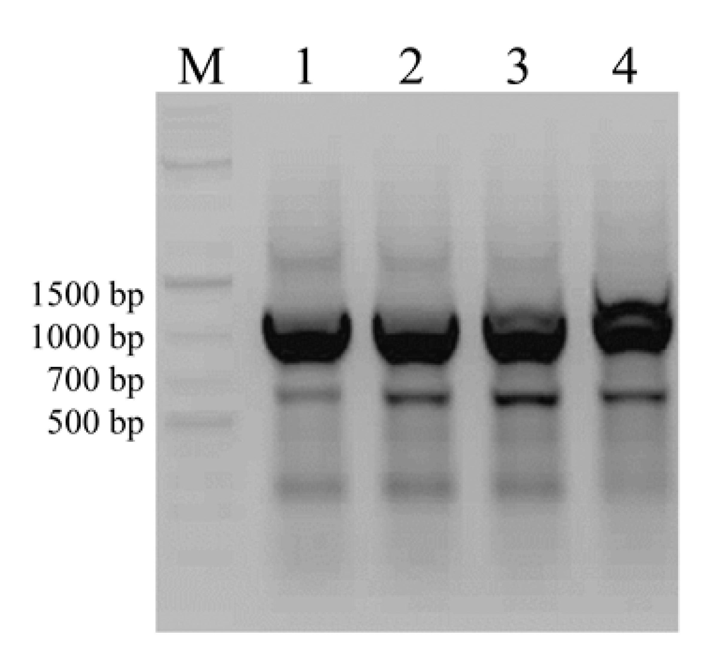
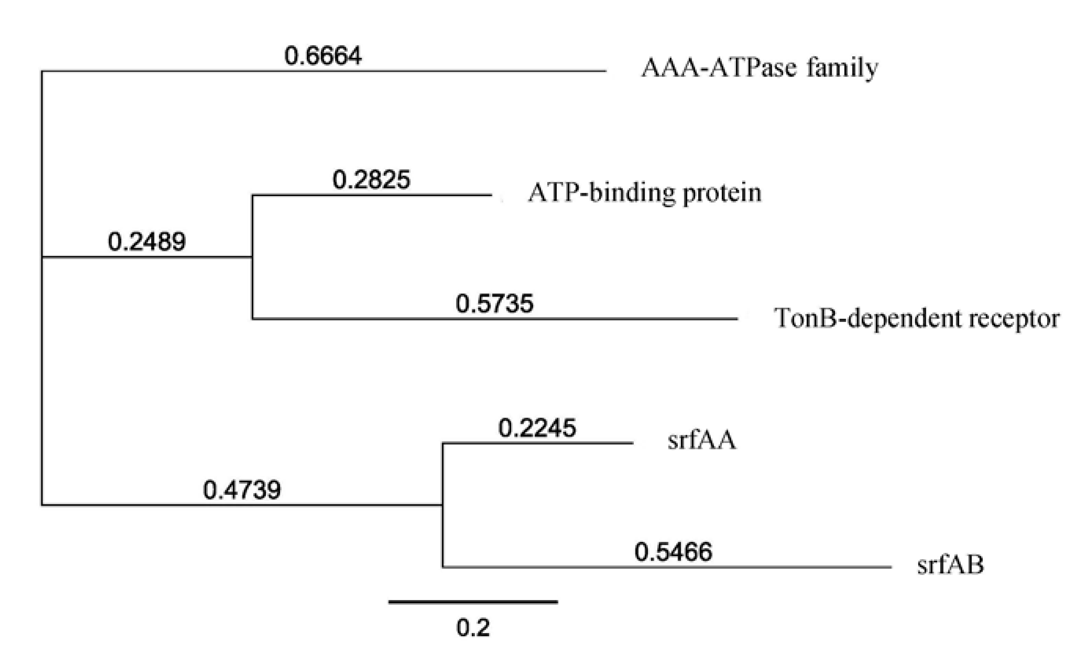
3.2. Screening for Natural Product Biosynthetic Gene Clusters by PCR

The isolates were screened by PCR with the use of four different primers specific for iturin (itu), surfactin (srf), phenazine (phz) and mycosubtilins (myc) biosynthesis genes. In the analysis involving the surfactin biosynthesis gene, the products of Sphingomonas sp. isolate S11 had an estimated size of 1000 bp and 700 bp (Figure 3). The size of the obtained amplicons exceeded the expected size of 431 and 419 bp based on the nucleotide sequences of srfAA and srfAB Bacillus subtilis (D13262.1), respectively. As1-F and Ts2-R primers for Sphingomonas sp. S11 amplified a fragment of DNA whose nucleotide sequences were similar in 74.43%, 70.54%, and 93.20% to the nucleotide sequences of unexpected genes encoding the AAA-ATPase family (WP_1301160516.1), ATP-binding cassette domain-containing protein (WP_093297233.1) and ToB-dependent receptor (WP_132909806.1) from Sphingomonas sp., respectively. The predicted amino acid sequences of the cloned genes were not similar to the predicted amino acid sequences of srfAA and srfAB proteins (Figure 4). Amplification products were not obtained with the use of primers for itu, phz, and myc marker genes.

3.3. Surfactin Production by Sphingomonas Isolates

The ability of Sphingomonas strains B4, B6, M2, M3, K2, and S11 to produce surfactin was tested in a liquid medium (Table 2). All analyzed isolates produced surfactin in a concentration range of 0.02 g (isolate S11) to 3.89 g (isolate B4) per 1 L. The rate of surfactin synthesis differed significantly across most of the tested isolates.

3.4. Propiconazole-Degrading Ability of Sphingomonas Isolates

In the disc diffusion test, the significantly largest inhibition zones around discs saturated with propiconazole were produced by Sphingomonas sp. strain S11 relative to the remaining isolates (Table 2). The ability to biodegrade propiconazole in a liquid medium was analyzed in six isolates of the genus Sphingomonas (B4, B6, M3, M2, S11, and K2). Sphingomonas sp. isolate B6 did not degrade propiconazole. Sphingomonas sp. S11 was characterized by the highest degrading ability after 48 h in a liquid medium, and it decreased propiconazole concentrations by 15.13% relative to control. Propiconazole biodegradation led to the formation of four other compounds in the mixture (retention time 2.5; 4.2; 5.1; 5.3; Figure 5). An analysis of the spectral characteristics of additional peaks in the chromatograph revealed their structural similarity to the analyzed fungicide.

3.5. Isolate Activity in Dual Cultures

The growth of F. culmorum and F. graminearum colonies on PDA in the presence of six bacterial isolates was analyzed. Sphingomonas sp. strains S11 and K2 significantly inhibited the development of F. culmorum colonies, and Sphingomonas sp. isolates B6, M3 and K2 exerted a significant influence on the shape of F. graminearum colonies (Table 3).

3.6. Survival of Bacteria on Wheat Leaves

The majority of the tested isolates adapted well to the new environment already 24 h after the application to wheat leaves. The concentration of bacterial cells on leaves remained very high, above Log(CFU + 1) = 5, for five days (Figure 6). Sphingomonas sp. isolate S11 was characterized by the highest concentration on the leaves of wheat cvs. Bogatka (Log (CFU + 1) = 7.5) and Tonacja (Log (CFU + 1) = 7.4) 24 h after application.

3.7. The Effectiveness of Sphingomonas S11 in Inhibiting FHB

The yield of winter wheat cv. Bogatka in the control treatment was determined at 479.67 g m−2, and spike inoculation with F. culmorum decreased yields by 19.88% (Table 4). The application of fungicides and the Sphingomonas sp. S11 suspension (Integ) increased the yield of non-inoculated plants by 16.81% compared to the non-inoculated control. Wheat cv. Tonacja did not overwinter well, and its yield was very low. Spike inoculation with F. culmorum additionally decreased the yield of wheat cv. Tonacja by 14.77%. Both protective treatments (Integ and Bac) reduced the severity of FHB, but the noted differences were not significant. In plots inoculated with F. culmorum, the integrated treatment (Integ) also considerably decreased the counts of epiphytic Fusarium growing on non-disinfected grain in PDA cultures. Due to the high counts of saprotrophic fungi, the ergosterol content of grain was nearly uncorrelated with the counts of Fusarium fungi.

3.8. Trichothecene Content of Grain

Type B trichothecenes were detected in all grain samples of wheat cvs. Bogatka and Tonacja (Table 5). After spike inoculation with F. culmorum, the DON content of control grain in both cultivars exceeded the safe limit (1250 μg/kg of grain). The integrated treatment decreased DON content by 69.18% in the inoculated grain of wheat cv. Bogatka and by 63.21% in non-inoculated grain. Three applications of the Sphingomonas S11 suspension (Bac) in plots with inoculated wheat cv. Tonacja decreased DON concentrations in grain 22.18-fold. In the above plots, the Bac treatment decreased the content of NIV in wheat cv. Tonacja by at least 75%. Type A trichothecenes were detected in six out of the eight analyzed grain samples. Protective treatments did not reduce the content of type A trichothecenes in grain.

4. Discussion

Agricultural ecosystems are potential sources of bacteria that can be effectively used to protect wheat against pathogens of the genus Fusarium. In this study, Sphingomonas strains isolated from wheat spikes demonstrated in vitro activity against F. culmorum and F. graminearum and antifungal activity under field conditions. Sphingomonas strain S11 effectively reduced the DON and NIV content of grain in the field experiment. Isolates of the genus Sphingomonas had been previously used as biocontrol agents to combat fusarium head blight (FHB) and powdery mildew in cereals and grasses [42] and to eliminate the symptoms of infection caused by Pseudomonas syringe on Arabidopsis thaliana leaves [38]. Sphingomonas trueperi strain S12 also effectively inhibited in vitro growth of two pathogens, Achlya klebsiana and Pythium spinosum, which cause rice seedling diseases [43]. The inhibitory effect of Sphingomonas capsulate on Rhizoctonia solani was demonstrated in vitro by Cottyn et al. [44].
In the present plot experiment, Sphingomonas strain S11 was highly effective in reducing the DON content of wheat grain. In Europe, the safe limit for grain is set at 1250 μg/kg DON. In this experiment, this threshold was exceeded only in grain from unprotected treatments where spikes were inoculated with F. culmorum. However, it should be noted that mycotoxins exert both acute and chronic toxic effects on humans and animals [13]. Three applications of the Sphingomonas S11 suspension in the field decreased DON concentration in grain more than 22-fold compared to control, suggesting the potential of bacteria Sphingomonas S11 to reduce the risk of chronic toxicity of mycotoxin in humans and animals. In a field study conducted by Pan et al. [2], Bacillus megaterium BM1 decreased the content of DON by 89.3%. In an in vitro experiment carried out by He et al. [14], Sphingomonas isolate S3-4 degraded approximately 12% of DON after 48 h and 100% of DON after 72 h to two compounds: 3-oxo-DON and 3-epi-DON. The cited authors also demonstrated that the analyzed bacterial isolate produced AKR18A1 protein. The AKR18A1 protein first catalyzes the reversible oxidation/reduction of DON to 3-oxo-DON, and in the second step, 3-oxo-DON is converted to 3-epi-DON by an unknown enzyme [15].
In the current field experiment, Sphingomonas strain S11 reduced the symptoms of fusarium head blight (FHB) and resulting in higher grain yield when applied at full flowering. Sphingomonas strain 11 was more effective than Bacillus megaterium BM1 (54.13%) applied by Pan et al. [2] in a field study, as well as the combined treatment with Lactobacillus plantarum SLG17 and Bacillus amyloliquefaciens FLN13 (49.46%), which was applied twice in the heading stage and at the beginning of flowering. Zhao et al. [61] demonstrated that B. subtilis isolate SG6 was up to 77.5% more effective than carbendazim in eliminating the symptoms of FHB compared with untreated controls.
The mechanisms underlying bacteria’s inhibitory effects on Fusarium pathogens have been extensively researched. Zhu et al. [62] demonstrated that Sphingomonas isolate CJ-5 was capable of producing chitinase and chitosanase. The molecular mass of chitinase and chitosanase was estimated at 230 kDa and 45 kDa, respectively [62]. Cheng et al. [63] found that Paenibacillus polymyxa KM2501-1 produces 11 volatile organic compounds (VOCs), of which 8 had contact nematicidal activity, 6 had fumigant activity, and 5 acted as stable chemotactic agents for Meloidogyne incognita. However, in an analysis of Bacillus megaterium KU143, Pseudomonas protegens AS15 and Sphingomonas aquatilis KU408 by Mannaa et al. [64] in a dual culture, VOCs were produced only by the first two species. These strains considerably inhibited the growth of Aspergillus flavus mycelia. The cited authors attributed the absence of antifungal activity in Sphingomonas aquatilis KU408 to its inability to produce VOCs.
In the current study, Sphingomonas S11 showed considerable antifungal potential as a biocontrol agent; therefore, it was also used after fungicide application. When applied after the fungicide, Sphingomonas sp. strain S11 exerted protective effects on wheat exposed to FHB. In our opinion, the strain is well adapted to the conditions on the surface of plants, and it directly inhibited the development of pathogens. Our previous study [42] demonstrated that the strain is able to form aggregates and survive on leaves for a long time. Sphingomonas S11 inhibited the growth of Fusarium pathogens and probably stimulated defense mechanisms in wheat. This study demonstrated that Sphingomonas S11 produces fungicidal compounds that activate plant defense responses. In the present study, all Sphingomonas sp. isolates produced surfactin within a concentration range of 0.02–3.89 g/L, which corresponds to the concentration of surfactin produced by Bacillus subtilis [65]. The structure of surfactin consists of a peptide loop with seven amino acids (L-asparagine, L-leucine, glutamic acid, L-leucine, L-valine, and two D-leucines) and a hydrophobic fatty acid chain with thirteen to fifteen carbons that allows surfactin to penetrate cellular membranes [66]. Farzaneh et al. [67] demonstrated that cell membranes in Aspergillus flavus spores were damaged by the antifungal activity of B. subtilis UTBSP1 which produces surfactin. The above inhibited the synthesis of aflatoxin in pistachio nuts. Sarwar et al. [68] reported that purified surfactin exerted potent antifungal effects against, among others, F. oxysporum, F. moniliforme, and F. solani. Fungal growth was inhibited already at a concentration of 200 ppm, and the development of F. moniliforme colonies was inhibited in 84% under exposure to 2000 ppm of surfactin. Similar results were reported by Tendulkat et al. [69], Desmyttere et al. [70], and Krishnan et al. [71] who observed that surfactin inhibited pathogen growth and modified the shape of colonies. Recent research indicates that the surfactin peptide from Bacillus velezensis directly affects fungal cell wall components and exhibits antifungal activity [72]. The fungitoxic activity of Sphingomonas sp. strain S11 could be implicated in the reduction of DON and NIV biosynthesis. Surfactin pretreatment of stored maize kernels also prevents plant and animal diseases by protecting maize kernels from the mycotoxigenic effects of phytopathogenic fungi such as F. moniliforme and Aspergillus sp. [71]. There is evidence to suggest that surfactin triggers defense responses in grapevine, tobacco, tomato, and wheat plants [73,74,75,76]. Purified surfactin induced strong natural defenses in winter wheat by stimulating both salicylic acid- and jasmonic acid-dependent signaling pathways [73].
The presence of secondary metabolite biosynthetic genes in the bacterial genome was also analyzed with the use of gene-specific primers. This is the first study to demonstrate that surfactin can by synthesized by Sphingomonas. Despite the fact that all isolates produced surfactin, the identification of srf gene markers was ambiguous. To date, surfactin has been detected in various Bacillus strains. This lipopeptide is a secondary metabolite that is synthesized by non-ribosomal peptide synthetases (NRPS) encoded by the srfA operon (srfAA, srfAB, srfAC, srfAD and sfp) in Bacillus subtilis sp. [77,78]. To date, sfr genes have been cloned only from Bacillus. Unlike other surfactant synthesis genes, the nucleotide sequences of sfr genes may not be evolutionarily conserved, which may explain why the use of degenerate primers for the amplification of sfr genes in Bacillus strains [52] did not support the detection of srf genes in the tested Sphingomonas isolate S11.
The presented study also demonstrated that Sphingomonas strain S11 can be applied during flowering as the second treatment after fungicide application. This mode of treatment was previously recommended by Palazzini et al. [1], in whose study isolates of Bacillus relezensis RC218, Brevibacillus sp. RC26, and Streptomyces sp. RC87B (inhibiting FHB and decreasing the DON content of grain) were resistant to triazole fungicides. In our study, Sphingomonas strain S11 applied after fungicide exerted protective effects on wheat exposed to FHB. The above strain also partly degrades a residual propiconazole, a triazole fungicide that is widely used to protect cereals against fungal diseases. The tested strain’s efficacy in reducing propiconazole levels was determined at 15.13%, which is lower than that reported for Pseudomonas putida isolates (maximum efficacy of 72.8%) by Sarkar et al. [37]. Kim et al. [18] observed that incomplete degradation of propiconazole resulted in the production of four structures. The cited authors identified these compounds as 2,4-dichlorophenyl-1,2,4-triazole-1-yl-methyl ketone, 1-(2,4-dichlorophenyl)-2- (1,2,4-triazole-1-yl) ethanol, as well as two structures described as 1-[2-(2,4-dichlorophenyl)-4-(hydroxypropyl)-1,3-dioxolan-2-ylmethyl]-1H-1,2,4-triazole. Bacteria’s ability to degrade fungicides can be influenced by environmental factors, including oxygen availability and temperature [37]. In a study by Sarkar et al. [37], the degradation rate of propiconazole was determined by glucose levels which played a key role in initial stages of bacterial growth. The above can be attributed to co-metabolism, where the availability of easily metabolized organic matter, such as glucose, increases the biodegradation rate of compounds that are generally not utilized by microorganisms as sources of carbon and energy [37]. Propiconazole is degraded very slowly in soil. Kim et al. [18] estimated the half-life of propiconazole in sandy loam at 315 days. After 12 months, the compound was mineralized in only up to 8% [18]. In our study, Sphingomonas S11 was characterized by satisfactory efficacy based on the rate of propiconazole degradation. This xenobiotic was slowly degraded by microorganisms in most soil samples analyzed by Kim et al. [18] and Riise et al. [19].

5. Conclusions

Integrated disease management improves the yield and quality of wheat grain. Bacterial strains colonizing spikes and grain produce surfactin, which can inhibit FHB and decrease mycotoxin concentrations in grain. These strains also degrade propiconazole, a fungicide that is widely used in agriculture. Surfactin is an ideal candidate for developing bioformulations aiming to improve the quality and quantity of wheat yields. The results of this study can also be used to explore bacterial mechanisms of action that limit the spread of economically important diseases of cereal crops.

Author Contributions

Conceptualization, U.W.; methodology, U.W., W.P., S.C. and K.S.-S.; software, U.W. and S.C.; validation, U.W. and W.P.; formal analysis, U.W.; investigation, U.W., K.K., W.P., S.C. and K.S.-S.; resources, U.W. and W.P.; data curation, U.W. and K.K.; writing—original draft preparation, U.W., K.K. and W.P.; writing—review and editing, U.W. and W.P.; visualization, U.W. and W.P.; supervision, U.W.; project administration, U.W.; funding acquisition, U.W. All authors have read and agreed to the published version of the manuscript.

Funding

This research received no external funding.

Acknowledgments

The authors are grateful to Elżbieta Kowalska and Edyta Kwiatkowska for their assistance during laboratory analyses.

Conflicts of Interest

The authors declare no conflict of interest.

References

  1. Palazzini, J.M.; Torres, A.M.; Chulze, S.N. Tolerance of triazole–based fungicides by biocontrol agents used to control Fusarium head blight in wheat in Argentina. Lett. Appl. Microbiol. 2018, 66, 434–438. [Google Scholar] [CrossRef] [PubMed]
  2. Pan, D.; Mionetto, A.; Tiscornia, S.; Bettucci, L. Endophytic bacteria from wheat grain as biocontrol agents of Fusarium graminearum and deoxynivalenol production in wheat. Mycotoxin Res. 2015, 31, 137. [Google Scholar] [CrossRef] [PubMed]
  3. Minati, M.H.; Mohammed-Ameen, M.K. Novel report on six Fusarium species associated with head blight and crown rot of wheat in Basra province, Iraq. Bull. Natl. Res. Cent. 2019, 43, 139. [Google Scholar] [CrossRef] [Green Version]
  4. Qiu, H.; Zhao, X.; Fang, W.; Wu, H.; Abubakar, Y.S.; Lu, G.; Wang, Z.; Zheng, W. Spatiotemporal nature of Fusarium graminearum—Wheat coleoptile interactions. Phytopathol. Res. 2019, 1, 26. [Google Scholar] [CrossRef] [Green Version]
  5. Rojas, E.C.; Sapkota, R.; Jensen, B.; Jørgensen, H.J.L.; Henriksson, T.; Jørgensen, L.N.; Nicolaisen, M.; Collinge, D.B. Fusarium head blight modifies fungal endophytic communities during infection of wheat spikes. Microb. Ecol. 2019, 79, 397–408. [Google Scholar] [CrossRef] [PubMed] [Green Version]
  6. Abdallah-Nekache, N.; Laraba, I.; Ducos, C.; Barreau, C.; Bouznad, Z.; Boureghda, H. Occurrence of Fusarium head blight and Fusarium crown rot in Algerian wheat: Identification of associated species and assessment of aggressiveness. Eur. J. Plant Pathol. 2019, 154, 499–512. [Google Scholar] [CrossRef]
  7. Matny, O.; Shamsallah, S.; Fahad, M.A.; Haas, M. Genetic diversity study of Fusarium culmorum: Causal agent of wheat crown rot in Iraq. J. Plant Protec. Res. 2017, 57, 43–49. [Google Scholar] [CrossRef]
  8. Xu, X.M.; Nicholson, P.; Thomsett, M.A.; Simpson, D.; Cooke, B.M.; Doohan, F.M.; Edwards, S.G. Relationship between the fungal complex causing Fusarium head blight of wheat and environmental conditions. Phytopathology 2008, 98, 69–78. [Google Scholar] [CrossRef] [Green Version]
  9. Góral, T.; Wiśniewska, H.; Ochodzki, P.; Nielsen, L.K.; Walentyn-Góral, D.; Stępień, Ł. Relationship between fusarium head blight, kernel damage, concentration of fusarium biomass, and Fusarium toxins in grain of winter wheat inoculated with Fusarium culmorum. Toxins 2019, 11, 2. [Google Scholar] [CrossRef] [Green Version]
  10. Mielniczuk, E.; Skwaryło-Bednarz, B. Fusarium head blight, mycotoxins and strategies for their reduction. Agronomy 2020, 10, 509. [Google Scholar] [CrossRef] [Green Version]
  11. Venkataramana, M.; Selvakumar, G.; Chandranayaka, S. Fusarium Mycotoxin: Toxicity and Detection. In Microbial Toxins. Toxinology; Gopalakrishnakone, P., Stiles, B., Alape-Girón, A., Dubreuil, J., Mandal, M., Eds.; Springer: Dordrecht, The Netherlands, 2008. [Google Scholar]
  12. Berthiller, F.; Krska, R.; Domig, K.J.; Kneifel, W.; Juge, N.; Schuhmacher, R.; Adam, G. Hydrolytic fate of deoxynivalenol-3-glucoside during digestion. Toxicol. Lett. 2011, 206, 264–267. [Google Scholar] [CrossRef] [PubMed] [Green Version]
  13. Ji, F.; He, D.; Olaniran, A.O.; Mokoena, M.P.; Xu, J.; Shi, J. Occurrence, toxicity, production and detection of Fusarium mycotoxin: A review. Food Prod. Process. Nutr. 2019, 1, 6. [Google Scholar] [CrossRef]
  14. He, W.; Zhang, L.; Yi, S.; Tang, X.L.; Yuan, Q.S.; Guo, M.W.; Wu, A.B.; Qu, B.; Li, H.P.; Liao, Y.C. An aldo-keto reductase is responsible for Fusarium toxin-degrading activity in a soil Sphingomonas strain. Sci. Rep. 2017, 7, 9549. [Google Scholar] [CrossRef] [PubMed] [Green Version]
  15. Ito, M.; Sato, I.; Ishizaka, M.; Yoshida, S.; Koitabashi, M.; Yoshida, S.; Tsushima, S. Bacterial cytochrome P450 system catabolizing the Fusarium toxin deoxynivalenol. Appl. Environ. Microbiol. 2013, 79, 1619–1628. [Google Scholar] [CrossRef] [PubMed] [Green Version]
  16. Baffoni, L.; Gaggia, F.; Dalanaj, N.; Prodi, A.; Nipoti, P.; Pisi, A.; Biavati, B.; Gioia, D. Microbial inoculants for the biocontrol of Fusarium spp. in durum wheat. BMC Microbiol. 2015, 15, 242. [Google Scholar]
  17. Cromey, M.G.; Lauren, D.R.; Parkes, R.A.; Sinclair, K.I.; Shorter, S.C.; Wallaceet, A.R. Control of fusarium head blight of wheat with fungicides. Australas. Plant Pathol. 2001, 30, 301–308. [Google Scholar] [CrossRef]
  18. Kim, I.S.; Shim, J.H.; Suh, Y.T. Laboratory studies on formation of bound residues and degradation of propiconazole in soils. Pest Manag. Sci. 2003, 59, 324–330. [Google Scholar] [CrossRef]
  19. Riise, G.; Lundekvam, H.; Wu, Q.L.; Haugen, L.E.; Mulder, J. Loss of pesticides from agricultural fields in SE Norway—Runoff through surface and drainage water. Environ. Geochem. Health. 2004, 26, 269–276. [Google Scholar] [CrossRef]
  20. Satapute, P.; Sanakal, R.D.; Mulla, S.I.; Kaliwal, B. Molecular interaction of the triazole fungicide propiconazole with homology modeled superoxide dismutase and catalase. Environ. Sustain. 2019, 2, 429–439. [Google Scholar] [CrossRef]
  21. Skolness, S.Y.; Blanksma, C.A.; Cavallin, J.E.; Churchill, J.J.; Durhan, E.J.; Jensen, K.M.; Johnson, R.D.; Kahl, M.D.; Makynen, E.A.; Villeneuve, D.L.; et al. Propiconazole inhibits steroidogenesis and reproduction in the Fathead Minnow (Pimephales promelas). Toxicol. Sci. 2013, 132, 284–297. [Google Scholar] [CrossRef] [Green Version]
  22. Wang, W.D.; Niu, J.L.; Cui, Z.J. Biodegradation of pesticides: A review. J. Heilongjiang August First Land Reclam. Univ. 2005, 17, 18. [Google Scholar]
  23. Jain, R.K.; Kapur, M.; Labana, S.; Lal, B.; Sarma, P.M.; Bhattacharya, D.; Thakur, I.S. Microbial diversity: Application of microorganisms for the biodegradation of xenobiotics. Curr. Sci. 2005, 89, 101–112. [Google Scholar]
  24. Mulla, S.I.; Bharagava, R.N.; Belhaj, D.; Saratale, G.D.; Bagewadi, Z.K.; Saxena, G.; Kumar, A.; Mohan, H.; Yu, C.P.; Ninnekar, H.Z. An overview of nitro group-containing compounds and herbicides degradation in microorganisms. In Microbial Metabolism of Xenobiotic Compounds Microorganisms for Sustainability; Arora, P.K., Ed.; Springer: Singapore, 2019; pp. 310–335. [Google Scholar]
  25. Porto, A.M.; Melgar, G.Z.; Kasemodel, M.C.; Nitschke, M. Biodegradation of Pesticides, Pesticides in Modern World-Pesticides Use and Management. 2011. Available online: http://www.intechopen.com/books/pesticides-in-the-modern-world-pesticides-use-and-management/biodegradation-of-pesticides (accessed on 12 October 2015).
  26. Satapute, P.; Kaliwal, B. Burkholderia sp. Strain BBK_9: A potent agent for propiconazole degradation. In Toxicity and Biodegradation Testing, Methods in Pharmacology and Toxicology; Bidoia, E.D., Montagnolli, R.N., Eds.; Humana Press: New York, NY, USA, 2018; pp. 87–108. [Google Scholar]
  27. Srinivasan, S.; Lee, J.; Kim, M.K. Sphingomonas rosea sp. nov. and Sphingomonas swuensis sp. nov., rosy colored beta-glucosidase-producing bacteria isolated from soil. J. Microbiol. 2011, 49, 610–616. [Google Scholar] [CrossRef] [PubMed]
  28. Yabuuchi, E.; Yano, I.; Oyaizu, H.; Hashimoto, Y.; Ezaki, T.; Yamamoto, H. Proposals of Sphingomonas paucimobilis gen. nov. and comb. nov., Sphingomonas parapaucimobilis sp. nov., Sphingomonas yanoikuyae sp. nov., Sphingomonas adhaesiva sp. nov., Sphingomonas capsulata comb. nov., and two genospecies of the genus Sphingomonas. Microbiol. Immunol. 1990, 34, 99–119. [Google Scholar] [CrossRef]
  29. Available online: www.bacterio.net/sphingomonas.html (accessed on 14 August 2019).
  30. Asaf, S.; Khan, A.L.; Khan, M.A.; Al-Harrasi, A.; Lee, I.J. Complete genome sequencing and analysis of endophytic Sphingomonas sp. LK11 and its potential in plant growth. Biotech 2018, 8, 389. [Google Scholar] [CrossRef] [PubMed] [Green Version]
  31. Nagata, Y.; Futamura, A.; Miyauchi, K.; Takagi, M. Two different types of dehalogenases, Lina and Linb, involved in γ-hexachlorocyclohexane degradation in Sphingomonas paucimobilis Ut26 are localized in the periplasmic space without molecular processing. J. Bacteriol. 1999, 181, 5409–5413. [Google Scholar] [CrossRef] [PubMed] [Green Version]
  32. Thelusond, J.R.; Strathmann, T.J.; Cupples, A.M. Carbamazepine, triclocarban, triclosan biodegradation and the phylotypes and functional genes associated with xenobiotic degradation in four agricultural soils. Sci. Total Environ. 2019, 657, 1138–1149. [Google Scholar] [CrossRef] [PubMed]
  33. Leys, N.M.; Ryngaert, A.; Bastiaens, L.; Top, E.M.; Verstraete, W.; Springael, D. Culture independent detection of Sphingomonas sp. EPA 505 related strains in soils contaminated with polycyclic aromatic hydrocarbons (PAHs). Microb. Ecol. 2005, 49, 443–450. [Google Scholar] [CrossRef]
  34. Davison, A.D.; Gillings, M.R.; Jardine, D.R.; Karuso, P.; Nouwens, A.S.; French, J.J.; Veal, D.A.; Altavilla, N. Sphingomonas paucimobilis BPSI-3 mutant AN2 produces a red catabolite during biphenyl degradation. J. Ind. Microbiol. Biotechnol. 1999, 23, 314–319. [Google Scholar] [CrossRef]
  35. Pan, T.; Wang, R.; Xiao, K.; Ye, W.; Dong, W.; Xu, M. Continous degradation of phenanthrene in cloud point system by reuses of Sphingomonas polyaromaticivorans cells. AMB Express 2019, 9, 8. [Google Scholar] [CrossRef]
  36. Sorensen, S.R.; Ronen, Z.; Aamand, J. Isolation from agricultural soil and characterization of a Sphingomonas sp. able to mineralize the phenylurea herbicide isoproturon. Appl. Environ. Microbiol. 2001, 67, 5403–5409. [Google Scholar] [CrossRef] [Green Version]
  37. Sarkar, S.; Seenivasan, S.; Premkumar, R. Biodegradation of propiconazole by Pseudomonas putina isolated from tea rhizosphere. Plant Soil Environ. 2009, 55, 196–201. [Google Scholar] [CrossRef] [Green Version]
  38. Innerebner, G.; Knief, C.; Vorholt, J.A. Sphingomonas strains protect Arabidopsis thaliana against leaf-pathogenic Pseudomonas syringae in a controlled model system. Appl. Environ. Microbiol. 2011, 77, 3202–3210. [Google Scholar] [CrossRef] [PubMed] [Green Version]
  39. Mukhtar, S.; Mehnaz, S.; Malik, K.A. Microbial diversity in the rhizosphere of plants growing under extreme environments and its impact on crop improvement. Environ. Sustain. 2019, 2, 329–338. [Google Scholar] [CrossRef]
  40. Shimoi, S.; Inoue, K.; Kitagawa, H.; Yamasaki, M.; Tsushima, S.; Park, P.; Ikeda, K. Biological control for rice blast disease by employing detachment action with gelatinolytic bacteria. Biol. Control 2010, 55, 85–91. [Google Scholar] [CrossRef]
  41. Vogel, C.; Innerebner, G.; Zingg, J.; Guder, J.; Vorholt, J.A. Forward genetic in planta screen for identification of plant-protective traits of Sphingomonas sp. strain Fr1 against Pseudomonas syringae DC3000. Appl. Environ. Microbiol. 2012, 78, 5529–5535. [Google Scholar] [CrossRef] [Green Version]
  42. Wachowska, U.; Irzykowski, W.; Jędryczka, M.; Stasiulewicz-Paluch, A.D.; Głowacka, K. Biological control of winter wheat pathogens with the use of antagonistic Sphingomonas bacteria under greenhouse conditions. Biocontrol Sci. Technol. 2013, 23, 1110–1122. [Google Scholar] [CrossRef]
  43. Adhikari, T.B.; Joseph, C.M.; Yang, G.; Phillips, D.A.; Nelson, L.M. Evaluation of bacteria isolated from rice for plant growth promotion and biological control of seedling disease of rice. Can. J. Microbiol. 2001, 47, 916–924. [Google Scholar] [CrossRef]
  44. Cottyn, B.; Debode, J.; Regalado, E.; Mew, T.W.; Swings, J. Phenotypic and genetic diversity of rice seed-associated bacteria and their role in pathogenicity and biological control. J. Appl. Microbiol. 2009, 107, 885–889. [Google Scholar] [CrossRef]
  45. Laca, A.; Mousia, Z.; Diaz, M.; Webb, C.; Pandiella, S. Distribution of microbial contamination within cereal grains. J. Food Eng. 2006, 72, 332–338. [Google Scholar] [CrossRef]
  46. Wachowska, U. Activity of fungicides against epiphytic yeast-like fungi of winter wheat. Pol. J. Environ. Stud. 2009, 18, 1169–1174. [Google Scholar]
  47. King, E.; Ward, M.K.; Raney, D.E. Two simple media for the demonstration of pyocyanin and fluorescein. J. Lab. Clin. Med. 1954, 44, 301–307. [Google Scholar]
  48. Buck, J.D. Nonstaining (KOH) method for determination of gram reactions of marine bacteria. Appl. Environ. Microbiol. 1982, 44, 992–993. [Google Scholar] [CrossRef] [PubMed] [Green Version]
  49. NCBI. Available online: www.ncbi.nlm.nih.gov (accessed on 5 May 2020).
  50. Wachowska, U.; Popielarczyk, C.; Duba, A.; Goriewa, K.; Kucharska, K. Evaluation of usefulness of yeast and chemical fungicides to reduce development of fungi colonizing Scot pine seedlings (in Polish). Sylwan 2016, 7, 556–563. [Google Scholar]
  51. Jasim, B.; Sreelakshmi, K.S.; Mathew, J.; Radhakrishnan, E.K. Surfactin, iturin, and fengycin biosynthesis by endophytic Bacillus sp. from Bacopa monnieri. Microb. Ecol. 2016, 72, 106–119. [Google Scholar] [CrossRef] [PubMed]
  52. Tapi, A.; Chollet-Imbert, M.; Scherens, B.; Jacques, P. New approach for the detection of non-ribosomal peptide synthetase genes in Bacillus strains by polymerase chain reaction. Appl. Gen. Mol. Biotechnol. 2010, 85, 1521–1531. [Google Scholar] [CrossRef]
  53. Mubarak, M.Q.E.; Hassan, A.R.; Hamid, A.A.; Khalil, S.; Isa, M.H.M. A simple and effective isocratic HPLC method for fast identification and quantification of surfactin. Sains Malays. 2015, 44, 115–120. [Google Scholar] [CrossRef]
  54. Wiwart, M.; Suchowilska, E.; Kandler, W.; Sulyok, M.; Wachowska, U.; Krska, R. The response of selected Triticum spp. genotypes with different ploidy levels to head blight caused by Fusarium culmorum (W.G.Smith) Sacc. Toxins 2016, 8, 112. [Google Scholar] [CrossRef] [Green Version]
  55. Meier, U. Phenological growth stages. In Phenology: An Integrative Science; Task for vegetation science; Schwarz, M.D., Ed.; Kluwer Academic Publishers: Dordrecht, The Netherlands; Boston, MA, USA; London, UK, 2003; Volume 39. [Google Scholar]
  56. EPPO Bulletin. 1998. Available online: https://bip.minrol.gov.pl/.../fung%20-%20choroby%20liści%20zbóż (accessed on 1 May 2009).
  57. Martin, J.P. Use of acid, rose Bengal and streptomycin in the plate method for estimating soil fungi. Soil Sci. 1950, 38, 215–220. [Google Scholar] [CrossRef]
  58. Leslie, J.F.; Summerell, B.A. The Fusarium Laboratory Manual; Blackwell Professional: Ames, IA, USA, 2006. [Google Scholar]
  59. Wachowska, U.; Stuper-Szablewska, K.; Perkowski, J. Yeasts isolated from wheat grain can suppress fusarium head blight and decrease trichothecene concentrations in bread wheat and durum wheat grain. Pol. J. Environ. Stud. 2020, 29, 4345–4360. [Google Scholar] [CrossRef]
  60. Statsoft, Inc. Statistica (Data Analysis Software System), Version 12. 2015. Available online: www.statsoft.com (accessed on 30 November 2019).
  61. Zhao, Y.; Selvaraj, J.N.; Xing, F.; Zhou, L.; Wang, Y.; Song, H.; Tan, X.; Sun, L.; Sangare, L.; Folly, Y.M.E.; et al. Antagonistic action of Bacillus subtilis strain SG6 on Fusarium graminearum. PLoS ONE 2014, 9, e92486. [Google Scholar] [CrossRef] [PubMed]
  62. Zhu, X.; Zhou, Y.; Feng, J. Analysis of both chitinase and chitosanase produced by Sphingomonas sp. CJ-5. J. Zhejiang Univ. Sci. 2007, 8, 831–838. [Google Scholar] [CrossRef] [PubMed] [Green Version]
  63. Cheng, W.; Yang, J.; Nie, Q.; Huang, D.; Yu, C.; Zheng, L.; Cai, M.; Thomashow, L.S.; Weller, D.M.; Yu, Z.; et al. Volatile organic compounds from Paenibacillus polymyxa KM2501-1 control Meloidogyne incognita by multiple strategies. Sci. Rep. 2017, 7, 16213. [Google Scholar] [CrossRef]
  64. Mannaa, M.; Oh, J.Y.; Kim, K.D. Biocontrol activity of volatile-producing Bacillus megaterium and Pseudomonas protegens against Aspergillus flavus and aflatoxin production on stored rice grains. Mycobiology 2017, 45, 213–219. [Google Scholar] [CrossRef] [Green Version]
  65. Abdel-Mawgoud, A.M.; Aboulwafa, M.M.; Hassouna, N.A. Characterization of surfactin produced by Bacillus subtilis isolate BS5. Appl. Biochem. Biotechnol. 2008, 150, 289–303. [Google Scholar] [CrossRef] [PubMed]
  66. Hussein, W.; Fahim, S. Detection of synthetases genes involved in non ribosomal lipopeptides (NRLPS) biosynthesis from Bacillus species by bioinformatics and PCR degenerated primers and estimation of their production. Int. J. Pharm. Bio. Sci. 2017, 8, 116–125. [Google Scholar]
  67. Farzaneh, M.; Shi, Z.Q.; Ahmadzadeh, M.; Hu, L.B.; Ghassempour, A. Inhibition of the Aspergillus flavus growth and aflatoxin B1 contamination on pistachio nut by fengycin and surfactin-producing Bacillus subtilis UTBSP1. Plant Pathol. J. 2016, 32, 209–215. [Google Scholar] [CrossRef] [Green Version]
  68. Sarwar, A.; Hassan, M.N.; Imran, M.; Iqbal, M.; Majeed, S.; Brader, G.; Sessitsch, A.; Hafeez, F.Y. Biocontrol activity of surfactin A purified from Bacillus NH-100 and NH-217 against rice bakanae disease. Microbiol. Res. 2018, 209, 1–13. [Google Scholar] [CrossRef]
  69. Tendulkar, S.R.; Saikumari, Y.K.; Patel, V.; Raghotama, S.; Munshi, T.K.; Balaram, P.; Chattoo, B.B. Isolation, purification and characterization of an antifungal molecule produced by Bacillus licheniformis BC98, and its effect on phytopathogen Magnaporthe grisea. J. Appl. Microbiol. 2007, 103, 2331–2339. [Google Scholar] [CrossRef]
  70. Desmyttere, H.; Deweer, C.; Muchembled, J.; Sahmer, K.; Jacquin, J.; Coutte, F.; Jacques, P. Antifungal activities of Bacillus subtilis lipopeptides to two Venturia inaequalis strains possessing different tebuconazole sensitivity. Front. Microbiol. 2019, 10, 2327. [Google Scholar] [CrossRef]
  71. Krishnan, N.; Velramar, B.; Velu, R.K. Investigation of antifungal activity of surfactin against mycotoxigenic phytopathogenic fungus Fusarium moniliforme and its impact in seed germination and mycotoxicosis. Pestic. Biochem. Physiol. 2019, 155, 101–107. [Google Scholar] [CrossRef] [PubMed]
  72. Park, G.; Nam, J.; Kim, J.; Song, J.; Kim, P.I.; Min, H.J.; Lee, C.W. Structure and mechanism of surfactin peptide from Bacillus velezensis antagonistic to fungi plant pathogens. Bull. Korean Chem. Soc. 2019, 40, 704–709. [Google Scholar] [CrossRef]
  73. Le Mire, G.; Siah, A.; Brisset, M.-N.; Gaucher, M.; Deleu, M.; Jijakli, M.H. Surfactin protects wheat against Zymoseptoria tritici and activates both salicylic acid- and jasmonic acid-dependent defense responses. Agriculture 2018, 8, 11. [Google Scholar] [CrossRef] [Green Version]
  74. Farace, G.; Fernandez, O.; Jacquens, L.; Coutte, F.; Krier, F.; Jacques, P.; Clément, C.; Barka, E.A.; Jacquard, C.; Dorey, S. Cyclic lipopeptides from Bacillus subtilis activate distinct patterns of defence responses in grapevine. Mol. Plant Pathol. 2015, 16, 177–187. [Google Scholar] [CrossRef]
  75. Jourdan, E.; Henry, G.; Duby, F.; Dommes, J.; Barthelemy, J.P.; Thonart, P.; Ongena, M.A.R.C. Insights into the defense-related events occurring in plant cells following perception of surfactin-type lipopeptide from Bacillus subtilis. Mol. Plant Microbe Interact. 2009, 22, 456–468. [Google Scholar] [CrossRef] [Green Version]
  76. Cawoy, H.; Mariutto, M.; Henry, G.; Fisher, C.; Vasilyeva, N.; Thonart, P.; Dommes, J.; Ongena, M. Plant defense stimulation by natural isolates of Bacillus depends on efficient surfactin production. Mol. Plant Microbe Interact. 2014, 27, 87–100. [Google Scholar] [CrossRef] [PubMed] [Green Version]
  77. Nakano, M.M.; Magnuson, R.; Myers, A.; Curry, J.; Grossman, A.D.; Zuber, P. SrfA is an operon required for surfactin production, competence development, and efficient sporulation in Bacillus subtilis. J. Bacteriol. 1991, 173, 1770–1778. [Google Scholar] [CrossRef] [Green Version]
  78. Nakano, M.M.; Corbell, N.; Besson, J.; Zuber, P. Isolation and characterization of sfp a gene that functions in the production of the lipopeptide biosurfactant, surfactin, in Bacillus subtilis. Mol. Gen. Genet. 1992, 232, 313–321. [Google Scholar] [CrossRef] [PubMed]
Figure 1. Sphingomonas sp. JX444564 (S11) colonies.
Agronomy 10 01673 g001
Figure 2. A phylogenetic tree created with the use of the neighbor-joining (NJ) method. The NJ method reflects the phylogenetic affinity between the identified bacterial species and Sphingomonas sp. JX444564 (S11). Bacterial isolates were identified based on 16S r RNA gene markers. The statistical significance of the tree was determined in the bootstrap test with 2000 replicates and >70% bootstrap support.
Agronomy 10 01673 g002
Figure 3. PCR amplification with degenerate primers (As1-F and Ts2-R) for Sphingomonas sp. strain S11 at an annealing temperature of 51 °C (line 1), 48.4 °C (line 2), 45.7 °C (line 3) and 43.1 °C (line 4). M—GeneRuler 1kb Plus DNA Ladder (Thermo Scientific, Waltham, MA, USA).
Agronomy 10 01673 g003
Figure 4. A phylogenetic tree of predicted amino acid sequences of surfactin synthase (srfAA and srfAB) and homologs of the obtained PCR products (AAA-ATPase family, ATP-binding protein, and TonB-dependent receptor) constructed using Genious Pro sequence analysis software version 4.7.6 (Michigan, IN, USA).
Agronomy 10 01673 g004
Figure 5. A chromatogram of propiconazole concentration in the sample with Sphingomonas sp. JX444564 (S11) isolate.
Agronomy 10 01673 g005
Figure 6. Survival of bacteria on the leaves of wheat cvs. Tonacja (left) and Bogatka (right). CFU—colony-forming unit. Values that do not differ significantly between columns based on the results of the SNK test (p < 0.01) are marked with identical letters.
Agronomy 10 01673 g006
Table 1. Treatments applied to winter wheat.
TreatmentFirst Node BBCH 31Middle of Heading BBCH 55Full Flowering BBCH 65
Experiment 1, cv. Bogatka
Integrated (Integ)fenpropimorph 1 (1 dm3 per ha)propiconazole 2 (1 dm3 per ha) Sphingomonas sp. S11 JX444564
Experiment 2, cv. Tonacja
Biological (Bac)Sphingomonas sp. S11 JX444564Sphingomonas sp. S11 JX444564Sphingomonas sp. S11 JX444564
1 Corbel 750 EC (fenpropimorph–79.87%, BASF, Ludwigshafen, Germany), 2 Bumper 250 SC (propiconazole–25.1%, Makhteshim Chemical Works Ltd., Beersheba, Israel).
Table 2. Activity of bacterial isolates: biodegradation of propiconazole and surfactin production.
Sphingomonas sp. StrainsSurfactin Content (g/L)Inhibition Zones in the Disc Diffusion Test (cm2)Biodegradation of Propiconazole (%)
B43.89 a0.98 c4.52
B62.36 b0 e0
M21.86 c0.67 d7.52
M31.70 d0.45 d7.91
K20.05 e0.74 c,d7.23
S110.02 e2.87 a15.13
Values that do not differ significantly between columns based on the results of the SNK test (p < 0.01) are marked with identical letters.
Table 3. Inhibitory effect of bacterial isolates of the genus Sphingomonas on Fusarium pathogens in the dual-culture test.
PathogenSphingomonas sp.Colony Area in cm2Colony Ellipticity Ratio
Fusarium culmorumControl8.69 a,b0.89 a
B49.77 a0.79 a,b,c,d
B66.92 a,b0.67 d,e
M210.00 a0.83 a,b
M36.79 a,b0.85 a,b
K24.59 c0.83 a,b
S114.88 c0.93 a
Fusarium graminearumControl7.99 a,b0.92 a
B47.05 a,b0.81 a,b,c
B64.89 b0.67 c,d,e
M28.83 a,b0.94 a
M35.57 b0.64 e
K25.49 b0.65 e
S117.55 a,b0.87 a
Values that do not differ significantly between columns based on the results of the SNK test (p < 0.01) are marked with identical letters.
Table 4. Grain yield, severity of Fusarium head blight (FHB), ergosterol (ERG) content, and colonization of winter wheat by Fusarium spp.
InoculationTreatmentYield
(g m−2)
FHB SeverityERG Content
(mg kg−1)
Fusarium spp.
CFU × 10 2 per 10 g of Grain aPercentage b
EpiphytesEndophytes
Experiment 1, cv. Bogatka
Without inoculationControl *479.40 b0.082.063.05 a15.28 a12.50
Integ560.00 a06.393.09 a8.33 a,b15.28
Inoculation F. culmorumControl384.50 c0.1819.411.59 b15.29 a12.5
Integ337.70 d04.262.06 a,b0.10 b6.94
Experiment 2, cv. Tonacja
Without inoculationControl303.301.361.112.95 a1.39 b8.33
Bac291.060.4410.062.54 a2.78 a,b12.50
Inoculation F. culmorumControl258.511.7812.363.15 a9.72 a,b0.10
Bac268.010.119.612.68 a12.50 a4.17
a—epiphytes rinsed off from kernel surfaces (F. culmorum, F. poae, F. graminearum); b—colonies growing on kernels in PDA cultures (epiphytes: F. poae, F. culmorum, F. avenaceum, F. sporotichiodes; endophytes: F. poae, F. culmorum, F. graminearum). Values that do not differ significantly between columns based on the results of the SNK test (p < 0.01) are marked with identical letters. CFU—colony-forming unit; Integ—integrated pest management with fungicide and Sphingomonas S11; Bac—biological treatment with Sphingomonas S11. *—unprotected plants.
Table 5. Content of Fusarium metabolites in winter grain (mg kg−1 sample).
InoculationTreatmentDONFUS-X3AcDON15AcDONNIVSum BSTOT-2 HT-2Sum A
Experiment 1, cv. Bogatka
ControlControl0.1060.0010.0030.0010.0250.136<LOD0.006<LOD0.006
Integ 0.0390.0010.0010.0010.0630.1050.0060.012<LOD0.018
Inoculation F.culmorumControl6.1280.0260.3140.0420.0646.5750.0170.006<LOD0.023
Integ 1.8890.0090.0360.0110.3662.3110.0180.0390.0070.066
Experiment 2, cv. Tonacja
ControlControl1.0990.0120.0440.0130.4531.6210.0070.0090.0040.020
Bac1.0860.0170.0240.0090.1121.248<LOD<LOD<LOD<LOD
Inoculation F.culmorumControl7.8080.0270.1430.0480.1678.1930.010<LOD<LOD0.010
Bac0.3520.0050.0150.0040.0360.413<LOD<LOD<LOD<LOD
LOD—limit of detection, LOD for all mycotoxins is 0.001 (mg/kg). Integ—integrated pest management with fungicide and Sphingomonas S11; Bac—biological treatment with Sphingomonas S11; DON—deoxynivalenol; FUS-X—fusarenon-X; 3ADON—3-acetyl-deoxynivalenol; 15ADON—15-acetyl-deoxynivalenol; NIV—nivalenol; Sum B—sum of type B trichothecenes; STO—scirpentriol; T-2—T-2 toxin tetraol; HT-2—HT-2 toxin; Sum A—sum of type A trichothecenes.
Publisher’s Note: MDPI stays neutral with regard to jurisdictional claims in published maps and institutional affiliations.

