Article

The Crystal Structure of the GG-Rich DNA Quadruplex Sequence GGGGTTTTGGGG in Presence of Zn2+ and K+ Ions

by
Hristina Sbirkova-Dimitrova
1,2,*,
Hristo Gerginov
1 and
Boris L. Shivachev
1,*
1
Institute of Mineralogy and Crystallography “Acad. Ivan Kostov”, Bulgarian Academy of Sciences, Acad. G. Bonchev Str., Bl. 107, 1113 Sofia, Bulgaria
2
PERIMED-2, BG16RFPR002-1.014-0007, Central District, Vasil Aprilov Blvd. 15A, 4002 Plovdiv, Bulgaria
*
Authors to whom correspondence should be addressed.
Crystals 2026, 16(4), 223; https://doi.org/10.3390/cryst16040223
Submission received: 27 February 2026 / Revised: 24 March 2026 / Accepted: 25 March 2026 / Published: 27 March 2026

Abstract

The structural characterization of GG-rich DNA sequences in presence of metal ions provides essential insight into quadruplex stability and ion-dependent conformational specifics. We report the crystal structure of the GG-quadruplex formed by the sequence GGGGTTTTGGGG in the presence of Zn2+, K+, and Na+. It was deposited in the RCSB Protein Data Bank under the accession code 9FTA. The structure was determined by single-crystal X-ray diffraction at a resolution of 2.49 Å in the space group P212121. It reveals a parallel-stranded, two-G-tetrad stabilized by K+ ions within the central channel, while Na+ and Zn2+ occupy peripheral and groove-associated sites. Zn2+ ions are engaged in noncanonical coordination interactions with phosphate oxygens and structured water molecules, contributing to lattice stabilization and subtle adjustments in groove dimensions. The T4 loop forms a compact, ordered motif that contributes to crystal packing rather than intramolecular G4 stabilization. The presence of mixed cations produces a sole lattice architecture mediated by ions that provides structural insight into how bivalent and monovalent metals mutually modulate G-quadruplex topology. These results suggest a basis for understanding the specific ion effects on G4 structures and may direct the design of metal open DNA architectures.

1. Introduction

G-quadruplexes (G4s) are noncanonical nucleic acid secondary structures formed by the stacking of planar G-tetrads, each composed of four guanine bases interconnected through Hoogsteen hydrogen bonds [1,2]. G4 structures are structurally distinct from typical Watson–Crick duplexes [3]. They need extra stabilization to maintain their structural conformation. These structures are stabilized by base stacking, hydrogen bonding, and metal cation coordination, which neutralizes the strong electrostatic repulsion of closely spaced O6 carbonyls in stacked guanines [4]. GG-rich sequences capable of forming G4s are prevalent in telomeric regions, which are gene promoters and regulatory sites [5].
Monovalent cations such as K+ and Na+ play a central role in G4 stabilization by occupying separate positions within the quadruplex channel and coordinating directly with oxygen atoms of the GG tetrads [6,7,8]. K+ ions in particular confer higher thermal stability compared to Na+ and are commonly observed in both crystallographic and solution structures [9,10,11]. Bivalent ions, including Zn2+, can also affect quadruplex folding and stability, through interactions with phosphate oxygens or structure water molecules at the peripheral [9,12].
The sequence GGGGTTTTGGGG represents a typical GG-rich motif capable of forming a parallel folded G4 complex with a short thymine loop connecting GG regions. These kinds of quadruplexes are very well suited for single-crystal X-ray diffraction [13]. While monovalent cation effects have been extensively characterized, structural data for G4s in mixed monovalent/bivalent cation environments remain limited. Mixed cation studies are relevant because biologically significant bivalent metals, such as Zn2+ and Na+, may locally modify quadruplex structure and lattice organization by binding in noncanonical coordination outside the central channel [14,15].
In this study, we report the crystal structure of the GG-rich sequence GGGGTTTTGGGG in the presence of Zn2+, K+, and Na+ (PDB entry 9FTA). The studied DNA sequence has been previously structurally characterized, and the present work focuses on its crystallization and structural behavior in the presence of mixed metal ions (Zn2+, K+, and Na+). The structure reveals a parallel-stranded, two-tetrad G4 stabilized by K+ ions in the central channel and peripheral Zn2+ ions. Examination of the TT geometry and cation positions provides insight into how metal ions cooperatively modulate quadruplex architecture and crystal packing, increasing our understanding of how metal ions affect G4 DNA [4,14].

2. Materials and Methods

2.1. Materials

The dry oligonucleotide sequences were purchased from Eurofins MWG Genomics (Ebersberg, Germany) as HPLC-purified material (Table 1). The single-stranded DNA was dissolved in nuclease-free water and annealed by heating at 75 °C for 5 min, followed by slow cooling at room temperature. Heating was applied to disrupt potential secondary structures and ensure complete strand denaturation before refolding into the G-quadruplex conformation during slow cooling.

2.2. Sample Crystallization

Macromolecular crystallization screening was performed at 293 K using the hanging drop vapor diffusion technique [16]. The screen utilized a combination of KCl, ZnCl2, Na-cacodylate, Spermine tetrachloride, MPD, and H2O. The initial screening included various combinations of sodium cacodylate (NaCaCo) (pH 6.9), alcohol (2-propanol or methylpentanediol (MPD)), cations (K+, Zn2+), and polyamines (Spermine). Optimal crystal growth for the sequences studied was observed exclusively in the presence of spermine. To prepare the double-stranded DNA (dsDNA), the sequences were reconstituted to a concentration of 2 mM and were annealed for 1 min at 75 °C. Specifically, for the 12 bp sequence (9FTA), the optimized mother solution consisted of 30 mM NaCaCo (pH 6.9), 50 mM KCl, 10 mM ZnCl2, 10% MPD, and 2 mM Spermine. Drops were prepared by mixing 1.5 μL of DNA (2 mM) with 1.5 μL of ligand (2 mM) and were equilibrated against a reservoir containing 50% MPD. Plates were maintained in a temperature-stabilized environment (16–20 °C).

2.3. Single-Crystal X-Ray Diffraction (SCXRD), Data Collection and Processing

Initial screening of the diffraction quality was performed using a D8 Venture (Bruker, Karlsruhe, Germany) diffractometer (CuKα radiation, λ = 1.54056 Å) with a microfocus source IμS and a PHOTON II CPAD (Bruker, Karlsruhe, Germany) detector. Following this assessment, high-quality crystals were collected using nylon loops, frozen in liquid nitrogen, and stored in a cryogenic dewar. Comprehensive X-ray diffraction data were collected at Elettra Synchrotron Trieste (XRD2 beamline). The experimental setup used an MD2s Microdiffractometer (Arinax Scientific, Grenoble, France), a Pilatus 6M detector (DECTRIS Ltd., Baden, Switzerland), and a superconducting wiggler source. A wavelength of 0.99 Å was isolated using a cryogenically cooled Si double-crystal monochromator. The beam was specified with a 100 μm aperture and subsequently cleaned with a 200 μm capillary. During data collection, the sample was maintained at 100 K using an open-flow nitrogen cryostat. Data processing was carried out using XDS [17] and XSCALE [18] (version 19 January 2025).

2.4. Macromolecular Structure Solution and Refinement

Molecular replacement was performed using the previously reported G-quadruplex structure (PDB ID: 3NZ7) as the initial model for structure refinement. The phases were obtained by molecular replacement (MR) with Phaser (version phaser-2.7.0) [19,20]. Refinement of the structures involved several cycles of refinement using REFMAC5 (ver. 5.8.0253) [21] and Coot (version 0.9.8.95) [22]. The metal ions were positioned from the Fo-Fc difference map using the Coot interface. Visual analyses of the model and the electron-density maps were carried out using Coot. X3DNA (web 3DNA 2.0) [23] was used to carry out structural analysis and geometrical calculations of DNA parameters. UCSF Chimera (chimera-1.19-win64) [24] and PyMOL (ver. 3.1.0) [25] were used to prepare the figures. The coordinates and structure factors have been deposited in the Protein Data Bank (PDB) as entry 9FTA.

3. Results

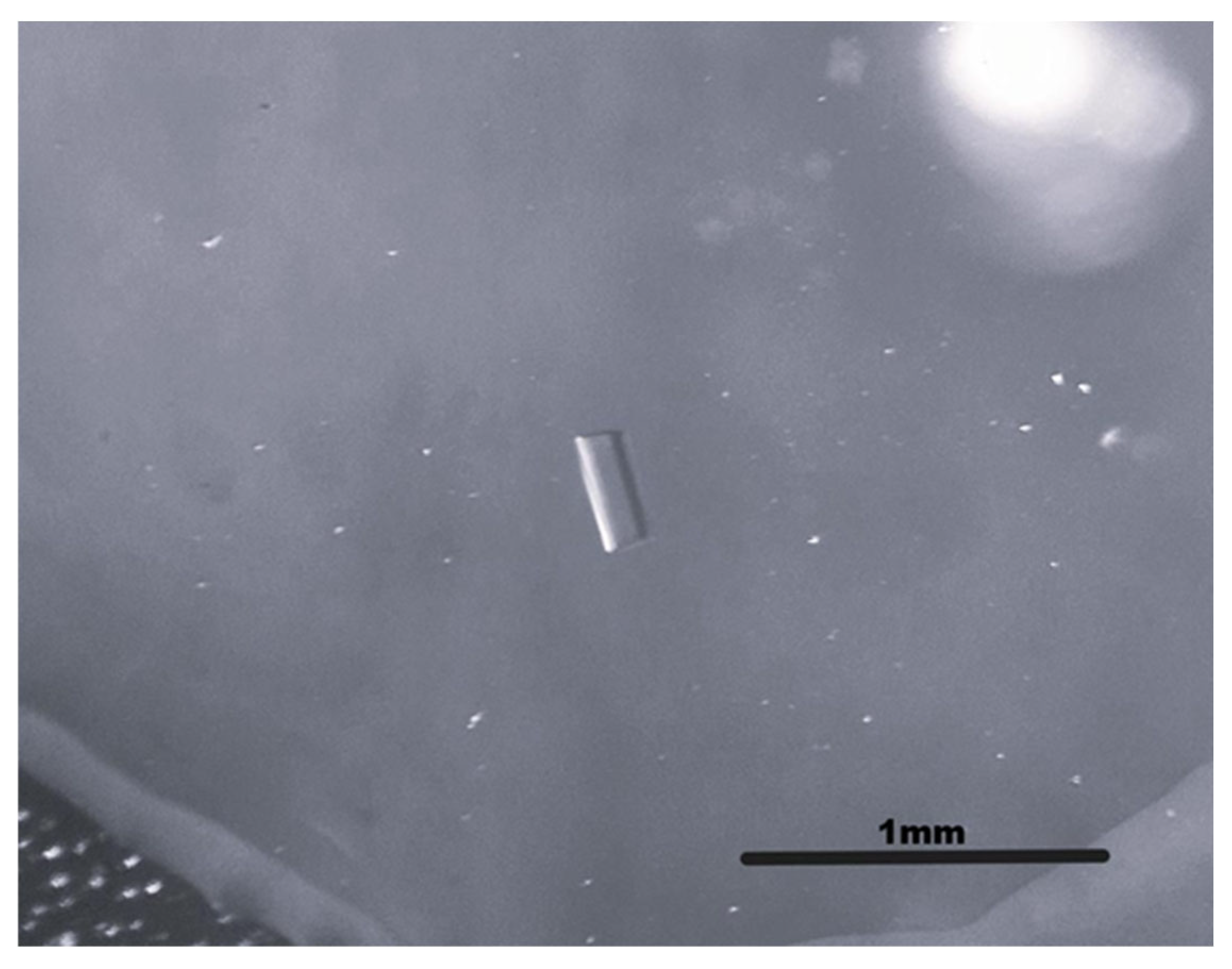
Crystals of the DNA oligomer d(GGGGTTTTGGGG) were grown in the presence of zinc and potassium ions. Crystals of good quality (0.4 × 0.2 × 0.25 mm3), suitable for single-crystal X-ray analysis, formed within 3–4 weeks (Figure 1).
Diffraction data were collected at 100 K and processed in the orthorhombic space group P212121 with unit-cell parameters a = 26.457 Å, b = 47.925 Å, c = 96.076 Å, α, β, γ = 90.00°. The structure was refined with REFMAC 5.8.0253 to 2.49 Å resolution, giving final R = 0.334 and Rfree = 0.426. The final model comprises 48 DNA residues; Zn2+, K+, and Na+ ions; and 41 water molecules. The structure has been deposited in the RCSB Protein Data Bank under accession code 9FTA. Table 2 shows the crystallographic data collection and refinement statistics parameters for 9FTA.
During cell refinement, the angles refined to values within 0.1° of 90° (α = 90.00°, β = 89.92°, γ = 90.06°). These small deviations are within experimental error and are consistent with orthorhombic symmetry, and the final model was refined with orthorhombic constraints. The relatively elevated R and Rfree values observed for the present structure primarily reflect the moderate diffraction resolution (2.49 Å), the presence of multiple independent molecules in the asymmetric unit (Z′ = 4), and partial lattice disorder typical for nucleic acid crystals [26]. These factors often lead to increased refinement statistics in DNA G-quadruplex structures, particularly when several conformationally similar molecules coexist within the asymmetric unit. Despite these values, the electron density clearly defines the stacking of the G-tetrads and the overall quadruplex topology, indicating that the global fold is reliably determined.

4. Discussion

The crystal structure of the G-rich sequence GGGGTTTTGGGG reveals a well-defined parallel-stranded DNA G-quadruplex stabilized by centrally located potassium ions [27,28]. The K+ ions occupy canonical positions between adjacent tetrads and are essential for maintaining the integrity of the quadruplex core. The overall fold and tetrad geometry are consistent with those reported for other short potassium-stabilized DNA G-quadruplexes determined by X-ray [29,30]. In addition to the monovalent cations within the quadruplex channel, peripheral Zn2+ ions are observed. These bivalent ions are coordinated by nucleobase and phosphate oxygen atoms and contribute to intermolecular contacts and crystal packing [31]. The three-dimensional separation between the centrally bound K+ ions and the externally located Zn2+ ions indicates a clear functional distinction between ions responsible for intrinsic quadruplex stabilization and those involved in lattice organization [32]. The metal ion concentrations used during crystallization are higher than those typically found in cellular environments. Therefore, the Zn2+ ions observed in the present structure are likely to reflect crystallization conditions and lattice stabilization effects rather than direct physiological binding sites. Nevertheless, the observed coordination modes illustrate possible interactions between divalent metal ions and nucleic acid phosphate groups, which may contribute to quadruplex stabilization under certain ionic environments. The structure of 9FTA provides a crystallographic demonstration of how Zn2+ can tune quadruplex folding and lattice interactions without disrupting the canonical G-tetrad core.
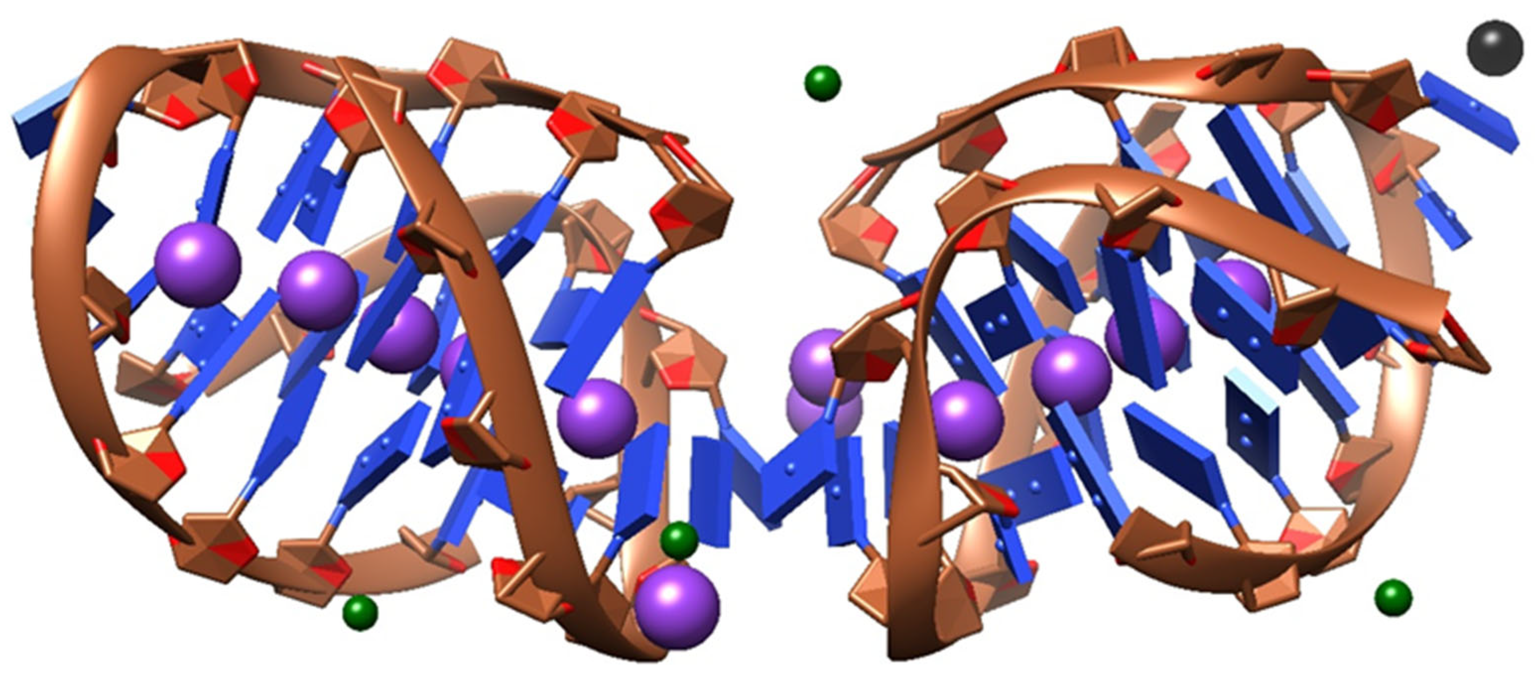
The asymmetric unit comprises four symmetry-independent DNA strands (chains A–D), each corresponding to one d(GGGGTTTTGGGG) oligonucleotide (Figure 2). These strands associate to form two independent G-quadruplex units related by non-crystallographic symmetry, consistent with the pseudo-translational symmetry identified in the diffraction data. Each quadruplex adopts a G4 architecture characteristic of parallel-stranded G-quadruplexes. Potassium ions occupy well-defined positions within the central channels of the G-quadruplexes. This coordination geometry is consistent with the canonical role of K+ in stabilizing G-quadruplex structures. The presence of multiple K+ sites within each quadruplex reflects the extended tetrad stacking and contributes significantly to the rigidity of the DNA framework. In contrast, zinc ions are located at peripheral sites outside the quadruplex channels and exhibit distinct coordination environments. Zn2+ ions mediate intermolecular contacts by coordinating to nucleobase and phosphate oxygen atoms from neighboring DNA strands, thereby linking adjacent quadruplex units within the asymmetric unit. This kind of coordination suggests that Zn2+ plays a mainly structural and packing role without altering the intrinsic topology of the G-quadruplex core. A single sodium ion is additionally observed at a solvent-exposed site, rather than a specific structural requirement. Together with several ordered water molecules, it contributes to local charge compensation. The asymmetric unit illustrates a clear functional separation between K+ and Zn2+ ions: potassium ions stabilize the internal G-quadruplex architecture, while zinc ions act as external cross-linking elements that promote crystal packing and lattice stability.
To clarify the distinct structural roles of K+ and Zn2+ ions in the present structure, it is important to note that these metals occupy clearly different positions relative to the quadruplex core [33]. Potassium ions are located inside the central channel between the stacked G-tetrads [11]. This position allows them to coordinate guanine O6 atoms and stabilize the tetrad stacking, which is a well-known feature of parallel G-quadruplex structures. In contrast, Zn2+ ions are not found within the tetrad channel. Instead, they are positioned at the outer surface of the DNA molecules. Their coordination involves phosphate oxygens, nucleobase atoms, and water molecules from neighboring strands. Through these interactions, Zn2+ links adjacent quadruplex units and stabilizes the crystal lattice [34]. Thus, the structure shows a clear functional separation: K+ ions stabilize the internal quadruplex framework, while Zn2+ ions contribute mainly to intermolecular contacts and crystal packing.
The G-quadruplex architecture observed in the present structure is consistent with previously reported parallel-stranded DNA G-quadruplexes determined by X-ray crystallography and deposited in the Protein Data Bank. The G–G interactions within the tetrads adopt typical Hoogsteen hydrogen bonding, with donor–acceptor distances in the 2.5–3.3 Å range (Table S1). Representative structures of short guanine-rich oligonucleotides crystallized in the presence of potassium ions (e.g., PDB IDs 1JPQ, 1KF1, 1XAV, 2GW0, 1D59) display closely related features, including stacked planar G-tetrads and K+ ions positioned between adjacent tetrads [35,36,37,38]. This canonical K+ coordination mode, which is considered a defining feature of stable parallel G-quadruplexes, is likewise observed in the present structure and confirms the conserved stabilizing role of potassium ions within the quadruplex core. While the internal organization of the quadruplex units closely resembles these previously reported structures, the present model differs in the amount of bivalent metal-ion involvement. In many X-rays G-quadruplex structures, lattice stabilization is dominated by direct DNA–DNA contacts, base stacking between neighboring quadruplexes, or solvent-mediated interactions [39,40]. The current structure contains well-defined Zn2+ ions at peripheral coordination sites, where they mediate intermolecular contacts between adjacent quadruplex units. These Zn2+ ions appear to play a packing-related structural role, rather than directly influencing G-quadruplex folding. Zn2+-mediated interactions are relatively uncommon among G-quadruplex structures in the PDB and have been reported only sporadically, often with limited positional definition [32,41]. Another distinguishing feature of the present structure is the presence of two crystallographically independent G-quadruplex units within the asymmetric unit, resulting in pseudo-translational symmetry. In contrast, many related G-quadruplex X-ray structures (e.g., 5HIX, 1KF1, 2GW0) contain a single quadruplex per asymmetric unit [37,42]. This difference highlights how variations in crystallization conditions, ionic composition, and sequence context can influence the organization and crystal packing without altering the fundamental topology of the G-quadruplex core. This dual metal-ion involvement broadens the range of coordination environments and packing strategies observed for DNA G-quadruplexes under different crystallographic conditions.
The structure deposited as PDB ID 3NZ7 [43], which was used as a model during the molecular replacement, represents a closely related parallel-stranded DNA. The overall fold, strand orientation, tetrad stacking, and inter-tetrad spacing in the present structure closely match those observed in 3NZ7. In both structures, K+ ions occupy equivalent positions within the central channel, underlining the structural importance of the monovalent cations. The core differences appear to be of crystal packing and metal-ion intermolecular interactions. In 3NZ7 and many other reported G-quadruplex crystal structures, lattice stabilization is achieved mainly through end-to-end stacking of quadruplex units or solvent-mediated interactions, but bivalent cations are usually absent or weakly ordered [39,40]. The 9FTA structure features well-defined Zn2+ ions at peripheral coordination sites, which mediate contacts between neighboring quadruplex units. These Zn2+ ions play a packing and structural role, without notable changes to the intrinsic G-quadruplex fold. Comparable Zn2+-mediated intermolecular interactions are rarely reported in G-quadruplexes and are less resolved when present [44].
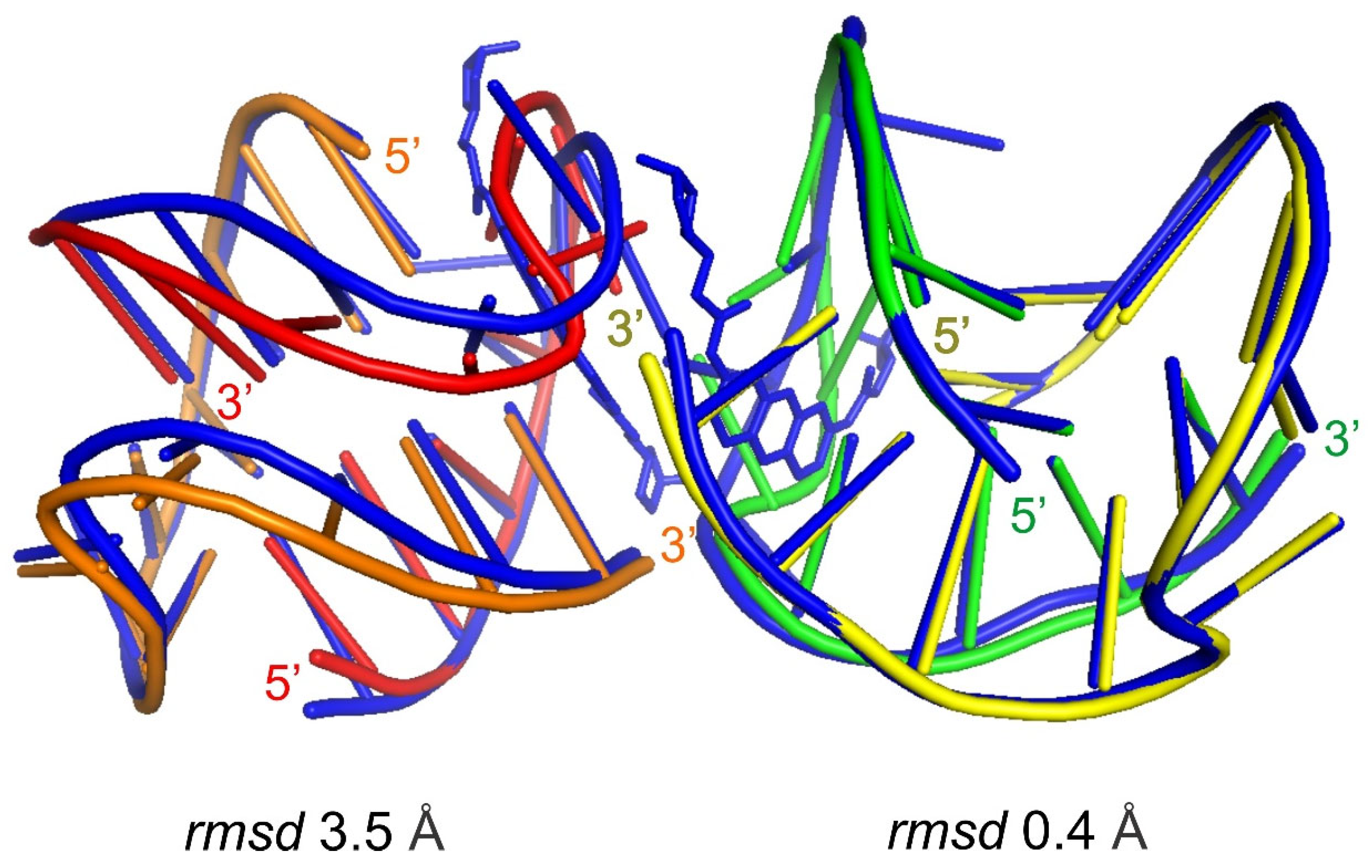
To provide a more representative structural comparison, the present structure was superposed with the previously reported model used for molecular replacement (PDB ID: 3NZ7). The analysis shows that the core quadruplex architecture is highly conserved and that the present structure closely resembles chain B of 3NZ7 (RMSD ≈ 0.4 Å), while larger deviations are observed relative to chain A (RMSD ≈ 3.6 Å), mainly due to differences in loop conformations and crystal packing. The structural relationship between the models is illustrated in Figure 3, which shows the superposition of representative structures and highlights the conserved arrangement of the stacked G-tetrads.
The base-pair morphology values [45] for shear, stretch, stagger, buckle, opening, and propeller twist obtained by w3DNA [46] are shown in Table 3 and Table S2. These parameters describe the fine structural properties of the DNA G-quadruplex and highlight the effects of ligand binding in 3NZ7 compared to the unliganded structure of 9FTA. The 9FTA structure shows notable differences in buckle, propeller twist, and opening parameters, especially in GG-rich regions, where ligand binding is supported in 3NZ7. Both structures retain the typical characteristics of G-DNA, suggesting that ligand binding induces localized conformational changes.
The base-pair morphology parameters provide a quantitative description of local conformational features within the G-quadruplex structures. They allow for a direct comparison between the 9FTA and the reference structure 3NZ7 (Table 3). Parameters such as shear, stretch, stagger, buckle, opening, and propeller twist are usually employed to characterize deviations from ideal base geometry and have been successfully applied in detailed structural analyses of nucleic acid crystal structures, including G-quadruplexes [47,48,49]. The shear and stretch values for both 9FTA and 3NZ7 remain close to zero, indicating that the guanine bases forming the G-tetrads are well aligned within the tetrad planes and experience minimal lateral displacement. The values are characteristic of regular Hoogsteen base pairing and are typical for a wide range of potassium-stabilized G-quadruplex crystal structures [32,50]. The stagger parameter shows slightly greater variability in 9FTA than in 3NZ7, reflecting small vertical displacements of guanine bases. These stagger variations could be attributed to differences in lattice environment and intermolecular contacts, and they do not destabilize the quadruplex core [27]. In the present structure, these deviations may be associated with Zn2+ mediated packing interactions absent in the reference model. The buckle values are low in both structures, consistent with the near-planarity of guanine tetrads observed in parallel-stranded G-quadruplexes [50,51]. Slightly elevated buckle values in some tetrads of 9FTA indicate localized out-of-plane distortions, which are commonly observed in nucleic acid crystal structures under packing constraints [52]. The opening parameter exhibits stable values across comparable tetrads in 9FTA. They could act as local conformational adaptive regions within the structure and also not affect the Hoogsteen hydrogen bonding [27,53]. The observed values in 9FTA are consistent with and do not suggest any weakening of the tetrad core. The propeller twist values are comparable between 9FTA and 3NZ7 and fall into the same range as for stable G-tetrads [32,51]. This conservation indicates that stacking geometry remains almost unaffected by differences in crystal packing or by the presence of peripheral Zn2+ ions.
The X3DNA analysis shows that the base-pair morphology of the present structure closely resembles that of the reference structure 3NZ7, especially for the potassium-stabilized quadruplex core. The slight deviations observed in the stagger, buckle, and opening parameters are localized and consistent with previously reported packing adaptations in crystallographic G-quadruplex structures [52,53]. These confirm that the intrinsic geometry of the G-quadruplex core is highly conserved.

5. Conclusions

The crystal structure of the G-rich DNA sequence GGGGTTTTGGGG was solved at a resolution of 2.49 Å (PDB 9FTA). The reported X-ray structure adopts a parallel-stranded G-quadruplex stabilized by stacked G-tetrads and well-defined and coordinated potassium ions. The K+ ions occupy canonical inner positions and are essential for maintaining the structural integrity of the quadruplex core. The asymmetric unit comprises two crystallographically independent G-quadruplex units formed by four symmetry-independent strands, which result in pseudo-translational symmetry. Peripheral Zn2+ ions are well resolved and mediate intermolecular contacts between neighboring quadruplex units, contributing to lattice stabilization without disturbing the intrinsic quadruplex topology. The 3D and functional separation between K+ ions, which stabilize the quadruplex core and Zn2+ ions involved in crystal packing, highlights the balancing roles of monovalent and bivalent ions in determining both local structure and crystal organization.

Supplementary Materials

The following supporting information can be downloaded at: https://www.mdpi.com/article/10.3390/cryst16040223/s1. Table S1: X3DNA results for Detailed H-bond information values in 9FTA DNA crystal structure; Table S2: X3DNA results for Base-Pair morphology: shear, stretch, stagger, buckle, opening, and propeller twist values in the 9FTA DNA crystal structure; Table S3: X3DNA results for Base-Pair morphology: shear, stretch, stagger, buckle, opening, and propeller twist values in the 3NZ7 DNA crystal structure.

Author Contributions

Conceptualization, H.S.-D. and B.L.S.; Formal analysis, H.S.-D.; Visualization, H.S.-D. and B.L.S.; Investigation (single crystal), H.S.-D. and H.G.; Investigation (PDB deposition), H.S.-D. and B.L.S.; methodology, H.S.-D., H.G. and B.L.S.; software, B.L.S.; writing—original draft, H.S.-D., H.G. and B.L.S.; Funding acquisition, H.S.-D. All authors have read and agreed to the published version of the manuscript.

Funding

This research was funded by the Bulgarian National Science Fund (BNSF), grant number KP-06-KOCT/5.

Data Availability Statement

Complete crystallographic data for the structure of 9FTA reported in this paper have been deposited in the mmCIF format on the RCSB Protein Data Bank. These data can be obtained free of charge via https://www.rcsb.org/ (accessed on 2 July 2025).

Acknowledgments

We acknowledge Elettra Sincrotrone Trieste for providing access to its synchrotron radiation facilities under proposals 20195613 and 20210594, and we thank Annie Heroux and Nicola Demitri for assistance in using beamline XRD2. The authors acknowledge the technical support from the project BG16RFPR002-1.014-0007, PERIMED-2 (2024–2029).

Conflicts of Interest

The authors declare no conflicts of interest.

References

  1. Parkinson, G.N.; Lee, M.P.; Neidle, S. Crystal structure of parallel quadruplexes from human telomeric DNA. Nature 2002, 417, 876–880. [Google Scholar] [CrossRef]
  2. Haider, S.M.; Parkinson, G.N.; Neidle, S. Structure of a G-quadruplex–ligand complex. J. Mol. Biol. 2003, 326, 117–125. [Google Scholar] [CrossRef]
  3. Bansal, M. DNA structure: Revisiting the Watson–Crick double helix. Curr. Sci. 2003, 85, 1556–1563. [Google Scholar]
  4. Phan, A.T.; Kuryavyi, V.; Patel, D.J. DNA architecture: From G to Z. Curr. Opin. Struct. Biol. 2006, 16, 288–298. [Google Scholar] [CrossRef] [PubMed]
  5. Patel, D.J.; Phan, A.T.N.; Kuryavyi, V. Human telomere, oncogenic promoter and 5′-UTR G-quadruplexes: Diverse higher order DNA and RNA targets for cancer therapeutics. Nucleic Acids Res. 2007, 35, 7429–7455. [Google Scholar] [PubMed]
  6. Collie, G.W.; Parkinson, G.N. The application of DNA and RNA G-quadruplexes to therapeutic medicines. Chem. Soc. Rev. 2011, 40, 5867–5892. [Google Scholar] [CrossRef] [PubMed]
  7. Neidle, S. Quadruplex nucleic acids as targets for anticancer therapeutics. Nat. Rev. Chem. 2017, 1, 0041. [Google Scholar] [CrossRef]
  8. Marathias, V.M.; Bolton, P.H. Determinants of DNA quadruplex structural type: Sequence and potassium binding. Biochemistry 1999, 38, 4355–4364. [Google Scholar] [CrossRef]
  9. Luo, Y.; Živković, M.L.; Wang, J.; Ryneš, J.; Foldynová-Trantírková, S.; Trantírek, L.; Verga, D.; Mergny, J.-L. A sodium/potassium switch for G4-prone G/C-rich sequences. Nucleic Acids Res. 2024, 52, 448–461. [Google Scholar] [CrossRef]
  10. Dai, J.; Carver, M.; Yang, D. Polymorphism of human telomeric quadruplex structures. Biochimie 2008, 90, 1172–1183. [Google Scholar] [CrossRef]
  11. Hud, N.V.; Schultze, P.; Sklenář, V.R.; Feigon, J. Binding sites and dynamics of ammonium ions in a telomere repeat DNA quadruplex. J. Mol. Biol. 1999, 285, 233–243. [Google Scholar] [CrossRef]
  12. Huppert, J.L.; Balasubramanian, S. Prevalence of quadruplexes in the human genome. Nucleic Acids Res. 2005, 33, 2908–2916. [Google Scholar] [CrossRef] [PubMed]
  13. Burge, S.; Parkinson, G.N.; Hazel, P.; Todd, A.K.; Neidle, S. Quadruplex DNA: Sequence, topology and structure. Nucleic Acids Res. 2006, 34, 5402–5415. [Google Scholar] [CrossRef] [PubMed]
  14. Bochman, M.L.; Paeschke, K.; Zakian, V.A. DNA secondary structures: Stability and function of G-quadruplex structures. Nat. Rev. Genet. 2012, 13, 770–780. [Google Scholar] [CrossRef] [PubMed]
  15. Deng, H.; Braunlin, W.H. Kinetics of sodium ion binding to DNA quadruplexes. J. Mol. Biol. 1996, 255, 476–483. [Google Scholar] [CrossRef]
  16. Luft, J.R.; Detitta, G.T. HANGMAN: A macromolecular hanging-drop vapor-diffusion technique. Appl. Crystallogr. 1992, 25, 324–325. [Google Scholar] [CrossRef]
  17. Kabsch, W. XDS. Biol. Crystallogr. 2010, 66, 125–132. [Google Scholar] [CrossRef]
  18. Kabsch, W. Automatic processing of rotation diffraction data from crystals of initially unknown symmetry and cell constants. Appl. Crystallogr. 1993, 26, 795–800. [Google Scholar]
  19. McCoy, A.J. Solving structures of protein complexes by molecular replacement with Phaser. Biol. Crystallogr. 2007, 63, 32–41. [Google Scholar] [CrossRef]
  20. McCoy, A.J.; Grosse-Kunstleve, R.W.; Adams, P.D.; Winn, M.D.; Storoni, L.C.; Read, R.J. Phaser crystallographic software. Appl. Crystallogr. 2007, 40, 658–674. [Google Scholar] [CrossRef]
  21. Murshudov, G.N.; Skubák, P.; Lebedev, A.A.; Pannu, N.S.; Steiner, R.A.; Nicholls, R.A.; Winn, M.D.; Long, F.; Vagin, A.A. REFMAC5 for the refinement of macromolecular crystal structures. Biol. Crystallogr. 2011, 67, 355–367. [Google Scholar] [CrossRef]
  22. Emsley, P.; Lohkamp, B.; Scott, W.G.; Cowtan, K. Features and development of Coot. Biol. Crystallogr. 2010, 66, 486–501. [Google Scholar] [CrossRef] [PubMed]
  23. Zheng, G.; Lu, X.-J.; Olson, W.K. Web 3DNA—A web server for the analysis, reconstruction, and visualization of three-dimensional nucleic-acid structures. Nucleic Acids Res. 2009, 37, W240–W246. [Google Scholar] [CrossRef] [PubMed]
  24. Pettersen, E.F.; Goddard, T.D.; Huang, C.C.; Couch, G.S.; Greenblatt, D.M.; Meng, E.C.; Ferrin, T.E. UCSF Chimera—A visualization system for exploratory research and analysis. J. Comput. Chem. 2004, 25, 1605–1612. [Google Scholar] [CrossRef] [PubMed]
  25. DeLano, W.L.; Bromberg, S. PyMOL User’s Guide; DeLano Scientific LLC.: Palo Alto, CA, USA, 2004; Volume 629. [Google Scholar]
  26. Berman, H.M.; Westbrook, J.; Feng, Z.; Gilliland, G.; Bhat, T.N.; Weissig, H.; Shindyalov, I.N.; Bourne, P.E. The protein data bank. Nucleic Acids Res. 2000, 28, 235–242. [Google Scholar] [CrossRef]
  27. Ambrus, A.; Chen, D.; Dai, J.; Bialis, T.; Jones, R.A.; Yang, D. Human telomeric sequence forms a hybrid-type intramolecular G-quadruplex structure with mixed parallel/antiparallel strands in potassium solution. Nucleic Acids Res. 2006, 34, 2723–2735. [Google Scholar] [CrossRef]
  28. Campbell, N.H.; Patel, M.; Tofa, A.B.; Ghosh, R.; Parkinson, G.N.; Neidle, S. Selectivity in ligand recognition of G-quadruplex loops. Biochemistry 2009, 48, 1675–1680. [Google Scholar] [CrossRef]
  29. Rhodes, D.; Lipps, H.J. G-quadruplexes and their regulatory roles in biology. Nucleic Acids Res. 2015, 43, 8627–8637. [Google Scholar] [CrossRef]
  30. Huppert, J.L.; Balasubramanian, S. G-quadruplexes in promoters throughout the human genome. Nucleic Acids Res. 2007, 35, 406–413. [Google Scholar] [CrossRef]
  31. Gong, J.-y.; Wen, C.-j.; Tang, M.-l.; Duan, R.-f.; Chen, J.-n.; Zhang, J.-y.; Zheng, K.-w.; He, Y.-d.; Hao, Y.-h.; Yu, Q. G-quadruplex structural variations in human genome associated with single-nucleotide variations and their impact on gene activity. Proc. Natl. Acad. Sci. USA 2021, 118, e2013230118. [Google Scholar] [CrossRef]
  32. Lim, K.W.; Amrane, S.; Bouaziz, S.; Xu, W.; Mu, Y.; Patel, D.J.; Luu, K.N.; Phan, A.T. Structure of the human telomere in K+ solution: A stable basket-type G-quadruplex with only two G-tetrad layers. J. Am. Chem. Soc. 2009, 131, 4301–4309. [Google Scholar] [CrossRef]
  33. Ida, R.; Wu, G. Direct NMR detection of alkali metal ions bound to G-quadruplex DNA. J. Am. Chem. Soc. 2008, 130, 3590–3602. [Google Scholar] [CrossRef] [PubMed]
  34. Siters, K.E.; Fountain, M.A.; Morrow, J.R. Selective binding of Zn2+ complexes to human telomeric G-quadruplex DNA. Inorg. Chem. 2014, 53, 11540–11551. [Google Scholar] [PubMed]
  35. Haider, S.; Parkinson, G.N.; Neidle, S. Crystal structure of the potassium form of an Oxytricha nova G-quadruplex. J. Mol. Biol. 2002, 320, 189–200. [Google Scholar] [CrossRef] [PubMed]
  36. Ambrus, A.; Chen, D.; Dai, J.; Jones, R.A.; Yang, D. Solution structure of the biologically relevant G-quadruplex element in the human c-MYC promoter. Implications for G-quadruplex stabilization. Biochemistry 2005, 44, 2048–2058. [Google Scholar] [CrossRef]
  37. Lee, M.P.; Parkinson, G.N.; Hazel, P.; Neidle, S. Observation of the coexistence of sodium and calcium ions in a DNA G-quadruplex ion channel. J. Am. Chem. Soc. 2007, 129, 10106–10107. [Google Scholar] [CrossRef]
  38. Kang, C.; Zhang, X.; Ratliff, R.; Moyzis, R.; Rich, A. Crystal structure of four-stranded Oxytricha telomeric DNA. Nature 1992, 356, 126–131. [Google Scholar] [CrossRef]
  39. Kuryavyi, V.; Phan, A.T.n.; Patel, D.J. Solution structures of all parallel-stranded monomeric and dimeric G-quadruplex scaffolds of the human c-kit2 promoter. Nucleic Acids Res. 2010, 38, 6757–6773. [Google Scholar]
  40. Biffi, G.; Tannahill, D.; McCafferty, J.; Balasubramanian, S. Quantitative visualization of DNA G-quadruplex structures in human cells. Nat. Chem. 2013, 5, 182–186. [Google Scholar] [CrossRef]
  41. Maizels, N.; Gray, L.T. The G4 genome. PLoS Genet. 2013, 9, e1003468. [Google Scholar]
  42. Mandal, P.K.; Baptiste, B.; Langlois d’Estaintot, B.; Kauffmann, B.; Huc, I. Multivalent Interactions between an Aromatic Helical Foldamer and a DNA G-Quadruplex in the Solid State. ChemBioChem 2016, 17, 1911–1914. [Google Scholar] [CrossRef]
  43. Campbell, N.H.; Smith, D.L.; Reszka, A.P.; Neidle, S.; O’Hagan, D. Fluorine in medicinal chemistry: β-fluorination of peripheral pyrrolidines attached to acridine ligands affects their interactions with G-quadruplex DNA. Org. Biomol. Chem. 2011, 9, 1328–1331. [Google Scholar] [CrossRef]
  44. Spiegel, J.; Adhikari, S.; Balasubramanian, S. The structure and function of DNA G-quadruplexes. Trends Chem. 2020, 2, 123–136. [Google Scholar] [CrossRef]
  45. El Hassan, M.; Calladine, C. Propeller-twisting of base-pairs and the conformational mobility of dinucleotide steps in DNA. J. Mol. Biol. 1996, 259, 95–103. [Google Scholar] [CrossRef] [PubMed]
  46. Li, S.; Olson, W.K.; Lu, X.-J. Web 3DNA 2.0 for the analysis, visualization, and modeling of 3D nucleic acid structures. Nucleic Acids Res. 2019, 47, W26–W34. [Google Scholar] [CrossRef] [PubMed]
  47. Lu, X.J.; Olson, W.K. 3DNA: A software package for the analysis, rebuilding and visualization of three-dimensional nucleic acid structures. Nucleic Acids Res. 2003, 31, 5108–5121. [Google Scholar] [CrossRef] [PubMed]
  48. Lu, X.-J.; Bussemaker, H.J.; Olson, W.K. DSSR: An integrated software tool for dissecting the spatial structure of RNA. Nucleic Acids Res. 2015, 43, e142. [Google Scholar] [CrossRef]
  49. Olson, W.K.; Bansal, M.; Burley, S.K.; Dickerson, R.E.; Gerstein, M.; Harvey, S.C.; Heinemann, U.; Lu, X.-J.; Neidle, S.; Shakked, Z. A standard reference frame for the description of nucleic acid base-pair geometry. J. Mol. Biol. 2001, 313, 229–237. [Google Scholar] [CrossRef]
  50. Webba da Silva, M. Geometric formalism for DNA quadruplex folding. Chem.–Eur. J. 2007, 13, 9738–9745. [Google Scholar] [CrossRef]
  51. Do, N.Q.; Lim, K.W.; Teo, M.H.; Heddi, B.; Phan, A.T.n. Stacking of G-quadruplexes: NMR structure of a G-rich oligonucleotide with potential anti-HIV and anticancer activity. Nucleic Acids Res. 2011, 39, 9448–9457. [Google Scholar] [CrossRef]
  52. Heddi, B.; Phan, A.T. Structure of human telomeric DNA in crowded solution. J. Am. Chem. Soc. 2011, 133, 9824–9833. [Google Scholar] [CrossRef]
  53. Venczel, E.A.; Sen, D. Parallel and antiparallel G-DNA structures from a complex telomeric sequence. Biochemistry 1993, 32, 6220–6228. [Google Scholar] [CrossRef]
Figure 1. Optical image of the observed crystals of 5′-GGGGTTTTGGGG-3′. The crystals form transparent rectangular blocks suitable for single-crystal X-ray diffraction analysis. Scale bar: 1 mm (magnification 2.5×).
Crystals 16 00223 g001
Figure 2. View of the asymmetric unit of 9FTA, visualizing the Zn2+ (in green color), K+ (in purple color), and Na+ (in black color) ions positions. Potassium ions are located in the central channel of the G-quadruplex and stabilize the stacked G-tetrads. Zinc ions are positioned at peripheral sites where they form contacts between neighboring DNA molecules. The sodium ion is located at a solvent-exposed position.
Crystals 16 00223 g002
Figure 3. Superposition of the DNA duplex from 3NZ7 onto the two crystallographically independent DNA molecules in the asymmetric unit of 9FTA. The DNA backbone is shown as cartoon, and the bases as sticks. The two duplexes of 9FTA are colored individually (chains A—red, B—orange, C—green, D—yellow), while the whole 3NZ7 structure is shown in dark blue. The overlay demonstrates a conserved quadruplex core, with the present structure closely resembling chain B of 3NZ7 (RMSD ≈ 0.4 Å), while larger deviations are observed for chain A (RMSD ≈ 3.6 Å).
Crystals 16 00223 g003
Table 1. Main characteristics of the 9FTA oligonucleotide sequence.
NameSequence (5′→3′)MW (g/mol)GC-Content (%)Extinction Coefficient (mol·cm)Tm [°C]Bases
9FTA5′-GGGGTTTTGGGG-3′3427671045004012
Table 2. Data collection and refinement statistics for 9FTA.
PDB Code9FTA
space groupP212121
cell dimensions
a, b, c, Å26.457, 47.925, 96.076
α, β, γ, °90.00, 90.00, 90.00
independent molecules4
Diffraction data
wavelength, Å0.99
resolution (refinement), Å48–2.49 (2.55–2.49)
reflections5451 (4239)
completeness, %91.1 (95.55)
I/σ (I)1.11 (2.56)
redundancy4 (3.8)
Rmerge %4.6 (5.3)
Refinement statistics
reflections used4043 (305)
R (Rfree) %33.4 (42.6)
no. of atoms1070
DNA1012
Waters41
K12
Zn4
Na1
average B factor, Å236.0
r.m.s.d.
bond lengths, Å0.005 (0.002)
bond angles, °1.175 (1.602)
Table 3. X3DNA results for Base-Pair morphology: shear, stretch, stagger, buckle, opening, and propeller twist values in 9FTA and 3NZ7 DNA crystal structures (shear, stretch, and stagger are in Å; buckle, opening, and propeller twist are in degrees).
PairShear (Å)Stretch (Å)Stagger (Å)Buckle (°)Propeller (°)Opening (°)
9FTA3NZ79FTA3NZ79FTA3NZ79FTA3NZ79FTA3NZ79FTA3NZ7
6|1G-G1.35−1.53.44−3.61−0.19−0.240.289.32−5.91−8.61−90.4190.85
7|2G-G−0.911.55−3.973.520.24−0.239.060.29−2.56−15.7794.24−89.12
8|3T-T−0.670.65−4.154.73−0.23−0.356.541.12−22.4527.61−105.9102.21
9|4G-G−1.41.56−3.613.480−0.13−1.156.881.093.1189.06−89.4
10|5G-G1.62−1.53.13−3.56−0.40.0613.73−0.49−6.798.52−86.9588.85
11|6G-G1.31.73.243.45−0.070.030.542.84−4.86−3.39−96.08−89.71
12|7G-G−0.6−1.51−3.74−3.53−0.11−0.0213.272.13−4.714.1298.9690.28
13|8G-G1.211.533.683.57−0.10.270.57−10.49−7.854.67−90.78−89.88
14|9G-G0.98−1.793.65−3.450.12−0.03−12.962.254.8111.59−96.7689.13
Disclaimer/Publisher’s Note: The statements, opinions and data contained in all publications are solely those of the individual author(s) and contributor(s) and not of MDPI and/or the editor(s). MDPI and/or the editor(s) disclaim responsibility for any injury to people or property resulting from any ideas, methods, instructions or products referred to in the content.