Article

Glass Fiber Post Pretreated with Neodymium-Doped Yttrium Orthovanadate, Toluidine Blue Activated Low-Level Laser Therapy, and Bioactive Glass: An In Vitro Analysis of SEM, Bond Strength, and Surface Roughness

by
Mohammad H. AlRefeai
1 and
Fahad Alkhudhairy
2,*
1
Department of Restorative Dentistry, Operative Division, College of Dentistry, King Saud University, Riyadh 11451, Saudi Arabia
2
Restorative Dental Sciences Department, College of Dentistry, King Saud University, Riyadh 11451, Saudi Arabia
*
Author to whom correspondence should be addressed.
Crystals 2025, 15(9), 813; https://doi.org/10.3390/cryst15090813
Submission received: 15 August 2025 / Revised: 6 September 2025 / Accepted: 9 September 2025 / Published: 17 September 2025
(This article belongs to the Section Inorganic Crystalline Materials)

Abstract

To evaluate the impact of different surface treatment regimens, Neodymium-doped yttrium orthovanadate (Nd: YVO4) laser, Toluidine blue (TB) activated Low-level laser therapy (LLLT), and Bioactive glass particles (BAGPs) on the surface roughness (Ra), surface morphology, and bond strength (BS) of Glass fiber posts (GFP) bonded to canal dentin. Forty single human rooted incisors with a closed apex were included. The endodontic treatment was performed, followed by post space preparation. Fifty-six GFP were sorted into four categories based on the conditioning method used (n = 14). Group 1: H2O2, Group 2: Nd: YVO4 laser, Group 3: TB-LLLT, and Group 4: BAGPs. Surface Ra and topographic changes were identified using a profilometer and Scanning Electron Microscopy (SEM). Post cementation was executed by utilizing self-adhesive resin cement. Analysis of BS and fracture pattern was performed using a universal testing machine and a stereomicroscope, respectively. Variance analysis with Tukey’s test was used to compare Ra and BS between the study groups at different root sections (p < 0.05). Group 2 (Nd: YVO4 laser) displayed the highest Ra scores (1051.54 ± 0.087 µm) and BS at all thirds. Whereas Group 3 TB-activated LLLT exhibited the lowest outcomes of Ra (539.39 ± 0.091) and BS at all three sections. Comparison among the investigated groups displayed that Group 1 (H2O2) and Group 2 Nd: YVO4 exhibited comparable outcomes of Ra and BS (p ˃ 0.05). Nd: YVO4 laser has the potential to roughen the surface of GFP, thereby enhancing its BS to resin cement

1. Introduction

The utilization of glass fiber posts (GFP) has seen a significant rise in contemporary practice and is advocated to improve the retention of core buildups and restorations [1]. Nevertheless, its retention within the root canal is contingent upon the bond strength (BS) at the post-cement interface, as most of the fracture occurs at this junction [2]. Therefore, it becomes imperative to precondition the post surface to get a strong bond between the GFP and the resin cement [3]. Surface treatments have been proposed to enhance the adhesion characteristics at the interface by facilitating micromechanical and chemical retention [4].
Hydrogen peroxide (H2O2) has been extensively utilized within the domain of dentistry for conditioning GFP [5,6]. It is posited that oxidizing agents such as H2O2 possess the capacity to dissolve the epoxy resin matrix of glass post, thereby resulting in the exposure of glass fibers [7]. This results in increased surface roughness (Ra), which leads to mechanical interlocking of cement to the post surface, eventually improving the BS [8]. Nevertheless, conditioning the post surface with H2O2 can undermine the post framework by reducing elastic modulus, hence compromising stress distribution, indirectly diminishing bond values [9,10]. Therefore, it becomes important to identify better alternatives for pretreating post surfaces.
The utilization of various lasers has been adopted due to their beneficial effects in multiple domains of dentistry [11,12]. In contemporary times, Neodymium-doped yttrium orthovanadate (Nd: YVO4) lasers, on account of their remarkable mechanical and physical properties, have captivated the interest of researchers [13]. The laser functions at an elevated power density and possesses a brief pulse width [14]. Preceding investigations have illustrated its efficacy in meticulously contouring the PEEK post surface, and its influence on the BS was satisfactory [15]. Nevertheless, the existing literature is deficient concerning its efficacy in conditioning the GFP and warrants further examination.
Photodynamic Therapy (PDT) is an innovative method that employs low-level laser therapy (LLLT) to trigger different dyes or photosensitizers (PS) to disinfect and condition dental materials [15,16]. Toluidine blue (TB) has gained significant attention as it is effective in combating bacterial infections and disassembling biofilms, which are commonly encountered in oral health issues. A study by Gupta and coworkers has used it as a surface conditioner for resin-based ceramics, and the results were not promising [17]. Nevertheless, its role as a GFP surface pretreatment regime on Ra and BS to resin cement needs to be addressed.
Bioactive glass particles (BAGPs) may be employed for the conditioning of ceramics, particularly in the field of prosthodontics, presenting a minimally invasive methodology for surface pretreatment [18]. These particles have the potential to enhance the BS between the indirect restoration and bonding cement [19]. This technique serves as an alternative to conventional air particle abrasion and offers a means to condition different materials while safeguarding their integrity [20]. Nonetheless, the efficacy of this methodology in enhancing the Ra and BS of GFP has yet to be thoroughly evaluated.
The present research is founded on the premise that there will be no significant variation in the Ra and surface topography of fiber posts bonded to dentin pretreated with BAGPs, TB-activated LLLT, and an Nd: YVO4 laser, in contrast to fiber posts treated with H2O2. Furthermore, it was also hypothesized that the BS of GFP to resin cement treated with modern techniques will be comparable to that of the control (H2O2). Thus, the contemporary inquiry aims to explore the impact of different surface treatments on the Ra, surface morphology, and BS of GFP bonded to resin cement.

2. Materials and Methods

Sample Preparation: Forty human single-rooted incisors with a closed apex were included. The patient, whose teeth were utilized for experimental purposes in this study, provided written consent. Using the World Health Organization sample size calculation, confidence level: 95%, absolute precision required: 1%, population mean: 12.44, population SD: 0.45, sample size: 10 cases in each group (i.e., total 40 cases) were identified [21,22]. The calculus and debris were eliminated from the tooth surface using an ultrasonic scaler (Henry Schein, Melbourne, Australia). The samples were submerged in a 0.5% thymol solution at 4 °C for about 48 h. With the help of a slow-speed saw (IsoMet 5000; Buehler, IL, USA), the crown part was sectioned at the cementoenamel junction (CEJ) level to a standardized 14 mm root length [23].
The endodontic treatment began with a 10 K patency file (Dentsply Sirona, Middle East, Riyadh, Saudi Arabia) until its tip was barely seen at the apical foramen. Working length was determined by keeping the patency file 1 mm short of the apex, making it 13 mm for each tooth. Pulpectomy was performed till 25 K file followed by root canal preparation using ProTaper Gold finishing file till F3 (Dentsply Sirona, Riyadh, Saudi Arabia). Canal disinfection was conducted with 5 mL of 2.5% NaOCl (Clorox Co, Cairo, Egypt) following a standardized protocol during the instrumentation procedure. 17% EDTA was used as a final irrigation for 1 min, followed by drying using F3 absorbent points (Dentsply Maillefer, Berlin, Germany). Canals were then filled with an F3 gutta-percha and AH plus sealer (Dentsply Maillefer) using the single cone technique. Canal orifices were then sealed with Cavite (Ariadent, Tehran, Iran). The teeth were kept in an incubator at 37 °C and 100% humidity for 7 days [24].
Post space preparation: The coronal gutta-percha measuring up to 8 mm was extracted with peeso reamers (Fibio, Anthogyr, Sallanches, France), ensuring that a minimum of 5 mm of apical gutta-percha stayed intact. The method started with Peeso reamers #1 or #2, then progressed sequentially to larger sizes #3 and #4. The verification of gutta-percha removal was performed via pre-marking the peeso reamer (rubber stop at 8 mm from the tip), periapical radiograph with the peeso reamer in place, and visual confirmation of the removed gutta-percha on the reamer flutes.
Fifty-six fiberglass DT Light Posts (VDW, Berlin, Germany) were sorted into four categories according to the conditioning method used (n = 14) [25]
Group 1: H2O2: 24% H2O2(Micko Industrial Chemicals Co. (Pvt.) Ltd., Karachi, Pakistan) was applied using a micro brush on GFP for 20 min.
Group 2: Nd: YVO4 laser: GFP were conditioned utilizing an Nd: YVO4 laser (Castech Inc., Fuzhou, China). The laser was set up to produce indentations with a vertical separation of 200 m and a depth of 150 m. The laser was placed at a right angle and kept at a distance of 197 mm for 30 s. Laser parameters include 8 ns pulse duration, a rate of 500 mm/s, a frequency of 25 kHz, a wavelength of 1064 nm, and a power density of 5.3 MW/cm2 [26,27].
Group 3: TB-LLLT: 10 μL of TBPS (Blue +T, Novateb, Iran) was combined with 0.1 mg/mL 0.9% NaCl (wt/vol) to create a solution. This mixture was applied on the post surface and then exposed to a diode laser (DX62, Konftec, New Taipei City, Taiwan) emitting at a wavelength of 635 nm, fiber tip 300 µm with an output power of 220 mW and a power density of 3.05 w/cm2 for 30 secs [28].
Group 4: BAGNP: BAGNPs (Sigma Aldrich, Berlin, Germany) with an average particle size of 27 µm were blasted using an extraoral sandblast device (Renfort basic professional, Gmbh, Germany) at an air pressure of 2 bar for 20 s, keeping the tip at 15 mm away from the surface and using a circular motion [29].
All the posts were subsequently washed and desiccated before cementation.
Scanning electron microscopy (SEM) analysis: A thin layer of gold was applied on two GPF from each cohort using a sputter coating device (Sputter Coater S 150A; Edwards, West Sussex, England, UK). The posts were anchored in stubs, and photographs of the posts’ morphological structure and energy dispersive spectroscopy (EDS) were obtained using SEM (FEI Quanta 200, The Netherlands) at 15 kV and 1000x magnification [30].
Surface Ra analysis: Profilometer (PLμ 2300, Sensofar Barcelona, Spain) was used to identify the Ra on two fiber posts from each group. The apparatus was configured to allow the stylus point to accurately scan 0.75 mm. From each sample, three measurements were collected to find the average Ra value [12].
Post bonding: Post cementation was executed by utilizing Relyx U200 self-adhesive resin cement (3M ESPE, St. Paul, MN, USA), adhering to the manufacturer’s guidelines. The cement was placed into the post space using a syringe. The GFP was placed, and any surplus cement was wiped away using an applicator tip. Photopolymerization was executed using LED light (Radii Cal, SDI) at an intensity of 800 mW/cm2, applying the light source vertically along the post for 40 s. The specimens were positioned in an oven at 37 °C to guarantee full cement hardening, then subjected to artificial ageing with a thermocycler (Automatic Thermocycling Dipping Machine) for 10,000 cycles. Every cycle involved placing samples in a water bath kept at 5 °C and another at 55 °C. Each dwell duration and transfer time between the water baths was set at 20 s.
Sample cutting and push-out testing: The roots were cut at right angles to the root axis using a low-speed saw (Isomet 1000; Buehler Ltd., Lake Bluff, Illinois, USA). Three slices were collected from each sample (coronal, middle, and apical), and every slice measured 2 mm in thickness. The thickness tolerance was measured using a digital caliper (Baker Gauges, Mumbai, India) with 0.01 mm resolution, measuring at multiple points around each slice. A push-out test was conducted on slices 2 mm each (coronal, middle, apical) at a rate of 0.5 mm/min utilizing a metallic plunger (coronal 1.4 mm, middle 1.2 mm, and apical 0.9 mm) of a universal testing machine (UTM) (Tinius Olsen, Horsham, PA, USA). The force was applied from the apical direction to the coronal until failure occurred. The bond required to fail the bond was measured in MegaPascal (MPa) [31].
Analysis of fracture patterns: Stereomicroscope at 30x magnification (ZEISS, Stemi 2000-C, Oberkochen, Germany) analyzed the failure mode. The type of fractures was categorized into adhesive, cohesive, and admixed.
Statistical evaluation: Data statistics were performed using SPSS software (version 17.0, IBM, Chicago, IL, USA). Variance analysis with Tukey’s test was used to compare Ra and BS between the study groups and different root sections (p < 0.05). Normality of data was assessed using the Kolmogorov-Smirnov test.

3. Results

SEM and EDX: Figure 1: SEM Figure 1A,B depict a traditional endodontic post design featuring a gradual taper. The surface texture is relatively consistent, characteristic of a manufactured GFP, and the post appears clean and free of defects. Figure 1C illustrates the elemental composition of GFP. Si (Silicon) shows a dominant peak, indicating it is the primary component of the glass fibers. Ca (Calcium) is likely derived from the glass composition or bioactive elements. O (Oxygen) displays high intensity, as expected in a silicate glass structure. Ta (Tantalum) is detected with multiple peaks, serving as a radio pacifier agent, and Al (Aluminum) is present, commonly acting as a glass former or modifier.
Ra Analysis: Table 1 demonstrates the GFP Ra score after using different pretreatment regimes. Group 2 Nd: YVO4 laser (1051.54 ± 0.087 µm) displayed the highest Ra scores, and Group 3 TB-LLLT (539.39 ± 0.091) exhibited the lowest outcomes of Ra. Comparison among the investigated groups displayed that Group 1 (H2O2) (1027.37 ± 0.066 µm) and Group 2 exhibited comparable outcomes of Ra (p ˃ 0.05), Group 4 (BAGPs) presented comparatively lower Ra than Groups 1 and 2, yet higher than Group 3 (p ˂ 0.05).
BS Evaluation: Table 2 demonstrates the GFP BS to root dentin after using different pretreatment regimes. The cervical section of Group 2 (Nd: YVO4 laser) (8.89 ± 0.12 MPa) displayed maximum BS, and the apical third of Group 3 TB-LLLT (4.94 ± 0.12 MPa) exhibited the lowest outcomes of bond integrity. Comparison among inspected cohorts displayed that Group 1 (H2O2) (Cervical: 8.75 ± 0.21 MPa, middle: 8.43 ± 0.15 MPa, and apical: 7.13 ± 0.09 MPa) and Group 2 (Cervical: 8.89 ± 0.12 MPa, middle: 8.57 ± 0.06 MPa, and apical: 7.01 ± 0.10 MPa) exhibited equivalent bond scores. p ˃ 0.05. Whereas Group 4 (BAGPs) (Cervical: 6.79 ± 0.35 MPa, middle: 6.39 ± 0.41 MPa, and apical: 5.94 ± 0.21 MPa) presented comparatively lower BS than Group 1 and 2 yet higher than Group 3 (Cervical: 5.87 ± 0.54 MPa, middle: 5.54 ± 0.43 MPa, and apical: 4.94 ± 0.12 MPa) (p ˂ 0.05) (Figure 2).
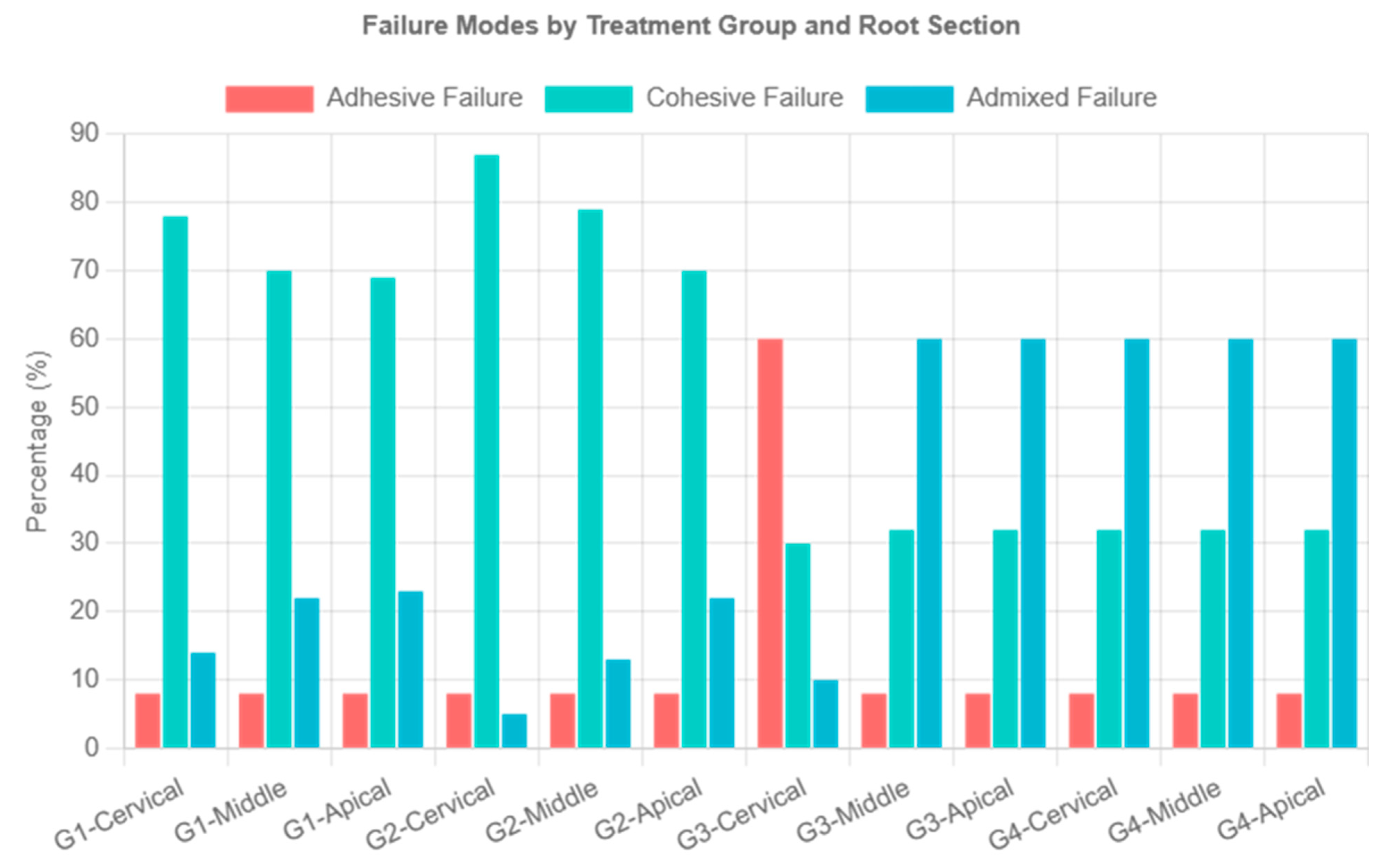
Failure Mode Assessment Figure 3 displayed the percentage of various failure modes found within each analyzed group. Samples treated with Nd: YVO4 laser and H2O2 conditioning showed cohesive failure pattern most often. In contrast, those subjected to pretreatment with BAGPs showed mixed failures. Whereas TB-LLLT predominantly resulted in adhesive failure patterns.

4. Discussion

The present research is founded on the premise that there will be no significant variation in the Ra and surface topography of GFP bonded to dentin pretreated with BAGPs, TB-activated LLLT, an Nd: YVO4 laser, in contrast to fiber posts treated with H2O2. Furthermore, it was postulated that the BS of GFP to resin cement treated with modern techniques will be comparable to the control. The GFP is composed of a blend of epoxy resin and inorganic glass fibers. The robust connection of a fiber post with the canal dentin depends on multiple factors, including Ra, cement’s wettability, and the kind of fiber utilized in the post [25].
The result of the present study showed that Nd: YVO4 laser preconditioned GFP presented comparable Ra scores and BS values to resin cement to those of the control. This aligns with the results of past in vitro studies performed by Sara Shabib et al. and Alkhudhairy et al. [26,27]. Both studies utilized an Nd: YVO4 laser for conditioning of the PEEK post and presented satisfactory outcomes. They reported that Nd: YVO4 laser exposure elevates the Ra, thereby expanding the area by forming controlled tiny grooves on the surface, resulting in the mechanical interlocking of the treated post with the resin cement [26,27]. Similarly, the presumed mechanism of action for GFP-pretreated Nd: YVO4 involves photomechanical, photochemical, and photothermal effects [28,29]. This is supported by SEM images, which show a partial degradation of fibers and matrix from the GFP after being preconditioned with Nd: YVO4. The authors of this study also suggest that thermal energy is responsible for these effects, as it leads to the breakdown of the fibers and matrix. Nonetheless, the precise mechanism by which it affects the fiber post remains unidentified.
Similarly, the use of H2O2 to condition GFP before cementation also led to acceptable outcomes regarding Ra and BS. H2O2 is an oxidizing agent that dissolves the epoxy resin of GFP [30]. Its dissolution process relies on the electrophilic attack of H2O2, leading to micromechanical interlocking between fiber posts and resin cement [31,32]. The micrograph subjected to H2O2 revealed a partial degradation of fibers and matrix. While fibers exist, the inter-fibrillar network is irregular with an increased surface area, resulting in improved BS when bonded to dentin using resin cement.
The GFP treated with BAGPs showed notably reduced Ra and BS scores when compared to H2O2 and Nd: YVO4 laser, but remained higher than those for TB-LLLT. Research analysis by Gencer et al. and Al Hamdan et al. aligns with the results of the current in vitro analysis. They indicated that BAGPs possess the ability to form precipitates and a porous layer on the hybrid ceramics, enhancing the surface area for bonding, which greatly boosts mechanical interlocking at the interface with the repair composite [33,34]. However, in the existing study, GFP conditioned with BAGP may have resulted in a weak particle-post interface, a non-uniform surface, and altered surface energy. SEM analysis revealed that the surface-treated BAGPs exhibited the presence of BAGPs on the GFP surface, with no matrix loss, and the visibility of tiny silica particles. Specimens treated with TB-LLLT displayed significantly lower Ra and BS outcomes. This follows the findings of Shono and colleagues [35]. They found that when resin-based ceramic was treated with TB, it led to reduced roughness and BS with the resin cement. The authors of this study suggested that TB-LLLT pretreatment might have created an O2 inhibition layer on the GFP surface, which hinders the polymerization of the resin cement and ultimately impacts its bond integrity. Another study indicated that TB [36], being hydrophilic like methylene blue, caused water absorption at the interface between the resin cement and post, affecting BS. SEM images further support these findings, as there is no loss of matrix and fibrillar structure, indicating its lower surface conditioning properties.
Performing a thorough analysis of the various fracture modes is crucial. This research demonstrated a distinct and specific relationship between the fracture pattern and the outcomes of BS evaluations. Samples subjected to Nd: YVO4 laser and H2O2 conditioning presented cohesive failure patterns most frequently. Conversely, GFP exposed to pretreatment with BAGPs exhibited admixed failures, while TB-LLLT showed the adhesive failure pattern the most. Clinical implications based on the outcomes of the present study included that Nd: YVO4 laser treatment of GFP offers a viable alternative to H2O2 conditioning, potentially reducing post-structural compromise. The superior and controlled performance of laser treatment may translate to improved long-term post retention and reduced endodontic failures [26,32].
This study has identified specific limitations that must be acknowledged. Conducted as a laboratory investigation, the research does not encompass all potential factors present in the oral cavity. The use of only one concentration of PS and a single set of laser parameters may have affected the outcomes. Furthermore, conducting atomic force microscopy (AFM) would have provided a better understanding of the alterations in the surface topography of GFP following different condition regimes. Beyond roughness and BS, other mechanical properties of G should be evaluated. The present study does not mention controlling for cement layer thickness, which significantly affects bond strength. Variations in cement thickness can create stress concentrations and affect polymerization, potentially confounding the results. Only thermocycling was used; mechanical loading simulation would better represent clinical conditions. Given these limitations, the results should be generalized with caution, and further laboratory and in vivo studies are essential to confirm the conclusions of this research.

5. Conclusions

Among the surface treatments evaluated, Nd: YVO4 laser demonstrated superior surface roughening and bond strength comparable to the H2O2 control, suggesting it as an effective alternative for glass fiber post conditioning. Bioactive glass particle treatment showed moderate effectiveness, providing intermediate bond strength and roughness values. Toluidine blue-activated low-level laser therapy proved least effective, showing significantly reduced surface roughness and bond strength across all root sections.

Author Contributions

Conceptualization, F.A. and M.H.A.; methodology, F.A. and M.H.A.; software, F.A. and M.H.A.; validation, F.A. and M.H.A.; formal analysis, F.A.; investigation, F.A. and M.H.A.; resources, F.A. and M.H.A.; data curation, F.A.; writing—original draft preparation, F.A. and M.H.A.; writing—review and editing, M.H.A.; visualization, F.A.; supervision, F.A.; project administration, F.A.; funding acquisition, F.A. All authors have read and agreed to the published version of the manuscript.

Funding

Ongoing Research Funding Program (ORF-2025-815) at King Saud University, Riyadh, Saudi Arabia.

Institutional Review Board Statement

The study was approved by the ethical board of King Saud University under IRB # FC-449-25.

Data Availability Statement

The data can be made available on request to the authors.

Acknowledgments

The authors gratefully acknowledge the Ongoing Research Funding Program (ORF-2025-815) at King Saud University, Riyadh, Saudi Arabia, for their financial support.

Conflicts of Interest

The authors declare no conflicts of interest.

References

  1. Wan, S.; Tan, Y.; Xie, J.; Huang, X.; Guo, L. The Effect of a Root-Dentin Pretreatment Technique Combining PIPS with MTAD Aiming to Improve the Bond Strength of Glass Fiber Post. Microsc. Res. Tech. 2020, 83, 824–833. [Google Scholar] [CrossRef] [PubMed]
  2. Maawadh, A.M.; Al Ahdal, K.; Al Deeb, L.; Alshamrani, A.S.; Almohareb, T.; Alrahlah, A. Effectiveness of Final Cavity Disinfectant Terminalia Chebula, Malachite, and Indocyanine Green, against E. Faecalis and on the Bond Interface of Fiber Post to Radicular Dentin. Photodiagn. Photodyn. Ther. 2023, 42, 103538. [Google Scholar] [CrossRef]
  3. Alshahrani, A.; Albaqami, M.; Naji, Z.; Al-Khunein, Y.; Alsubaie, K.; Alqahtani, A.; Al-Thobity, A.M. Impact of Different Surface Treatment Methods on Bond Strength between Fiber Post and Composite Core Material. Saudi Dent. J. 2021, 33, 334–341. [Google Scholar] [CrossRef]
  4. Santos, L.R.D.; Lima, D.M.; Carvalho, E.M.; Rodrigues, V.P.; Alves, C.M.C. Effect of Glass Fiber Post Surface Treatment on Bond Strength of a Self-Adhesive Resin Cement: An “in Vitro” Study. Int. J. Dent. 2021, 2021, 8856657. [Google Scholar] [CrossRef] [PubMed]
  5. Choi, Y.R.; Kim, K.N.; Kim, K.M. The Disinfection of Impression Materials by Using Microwave Irradiation and Hydrogen Peroxide. J. Prosthet. Dent. 2014, 112, 981–987. [Google Scholar] [CrossRef]
  6. Li, X.; Xu, N.; Dong, M.; Zuo, E.; Ren, X.; Hu, S. Effect of Different Surface Treatments on Bond Strength of Glass Fiber Post to Root Dentin an in Vitro Study. J. Dalian Med. Univ. 2013, 35, 424–429. [Google Scholar] [CrossRef]
  7. Mishra, L.; Khan, A.S.; Velo, M.M.d.A.C.; Panda, S.; Zavattini, A.; Rizzante, F.A.P.; Arbildo Vega, H.I.; Sauro, S.; Lukomska-Szymanska, M. Effects of Surface Treatments of Glass Fiber-Reinforced Post on Bond Strength to Root Dentine: A Systematic Review. Materials 2020, 13, 1967. [Google Scholar] [CrossRef]
  8. Punia, S.K.; Kumar, Y.; Kushwaha, S.; Choudhary, D.; Agarwal, S. Impact of Antioxidants on Bond Strength between Resin Composites and Bleached Enamel: An In Vitro Comparative Evaluation. Dent. J. Adv. Stud. 2024, 12, 106–110. [Google Scholar] [CrossRef]
  9. Qamar, Z.; Abdul, N.S.; Ali, S.; AlSheikh, R.; Alqarawi, F.K.; Niazi, F.H. Assessment of Push-out Bond Strength of Post-Surface Pretreatment before Salinization Using Hydrogen Peroxide, Aluminum Trioxide, and Natural Photosensitizers to Radicular Dentin. Photodiagnosis Photodyn. Ther. 2022, 39, 102859. [Google Scholar] [CrossRef]
  10. Aljamhan, A.; Alkhudhairy, F. Impact of Hydrofluoric Acid, Ytterbium Fiber Lasers, and Hydroxyapatite Nanoparticles on Surface Roughness and Bonding Strength of Resin Cement with Different Viscosities to Lithium Disilicate Glass Ceramic: SEM and EDX Analysis. Crystals 2025, 15, 661. [Google Scholar] [CrossRef]
  11. Alkhudhairy, F.; Vohra, F.; Naseem, M. Influence of Er,Cr:YSGG Laser Dentin Conditioning on the Bond Strength of Bioactive and Conventional Bulk-Fill Dental Restorative Material. Photobiomodul. Photomed. Laser Surg. 2020, 38, 30–35. [Google Scholar] [CrossRef]
  12. Alrabiah, M.; Alsunbul, H.; Almutairi, B.; Aljanakh, M.; Alzahrani, K.M.; Abduljabbar, T. Er, Cr: YSGG, Nd: YVO4, and Ti: Sapphire Lasers for Surface Treatment of Hybrid Ceramics on Bond Durability, Surface Texture, and Debonding to Resin Cement: A Scanning Electron Microscopy (SEM) Analysis. J. Biomater. Tissue Eng. 2023, 13, 792–798. [Google Scholar] [CrossRef]
  13. Kimura, H.; Tsuka, H.; Morita, K.; Hirata, I.; Nishio, F.; Abekura, H.; Doi, K.; Tsuga, K. Nd:YVO4 Laser Groove Treatment Can Improve the Shear Bond Strength between Dental PEEK and Adhesive Resin Cement with an Adhesive System. Dent. Mater. J. 2022, 41, 382–391. [Google Scholar] [CrossRef]
  14. Vohra, F.; Bukhari, I.A.; Sheikh, S.A.; Naseem, M.; Hussain, M. Photodynamic Activation of Irrigation (Using Different Laser Prototypes) on Push out Bond Strength of Fiber Posts. Photodiagn. Photodyn. Ther. 2020, 30, 101716. [Google Scholar] [CrossRef]
  15. Maawadh, A.M.; Almohareb, T.; Al-Hamdan, R.S.; Al Deeb, M.; Naseem, M.; Alhenaki, A.M.; Vohra, F.; Abduljabbar, T. Repair Strength and Surface Topography of Lithium Disilicate and Hybrid Resin Ceramics with LLLT and Photodynamic Therapy in Comparison to Hydrofluoric Acid. J. Appl. Biomater. Funct. Mater. 2020, 18, 2280800020966938. [Google Scholar] [CrossRef] [PubMed]
  16. Alsunbul, H.; Almutairi, B.; Aljanakh, M.; Abduljabbar, T. Hybrid Ceramic Repair Strength, Surface Roughness, and Bond Failure, Using Methylene Blue-Activated Low-Level Laser Therapy, Carbon Dioxide, and Ti: Al2O3 Laser. Photodiagn. Photodyn. Ther. 2023, 43, 103693. [Google Scholar] [CrossRef] [PubMed]
  17. Gupta, S.; Gupta, B.; Motwani, B.K.; Binalrimal, S.; Radwan, W.; Robaian, A.; Zidane, B.; Al Wadei, M.H.D.; Veeraraghavan, V.P.; Bhandi, S.; et al. The Effect of Different Surface Conditioning Techniques on the Bonding between Resin Cement & Ceramic. Coatings 2022, 12, 399. [Google Scholar] [CrossRef]
  18. Dahiya, M.S.; Tomer, V.K.; Duhan, S. Bioactive Glass/Glass Ceramics for Dental Applications. In Applications of Nanocomposite Materials in Dentistry; Woodhead Publishing: Cambridge, UK, 2019; pp. 1–25. ISBN 9780128137420. [Google Scholar]
  19. Thavornyutikarn, B.; Feltis, B.; Wright, P.F.A.; Turney, T.W. Effect of Pre-Treatment of Crystallized Bioactive Glass with Cell Culture Media on Structure, Degradability, and Biocompatibility. Mater. Sci. Eng. C 2019, 97, 188–197. [Google Scholar] [CrossRef]
  20. Edelhoff, D.; Liebermann, A.; Beuer, F.; Stimmelmayr, M.; Güth, J.-F. Minimally Invasive Treatment Options in Fixed Prosthodontics. Quintessence Int. 2016, 47, 207–216. [Google Scholar] [CrossRef]
  21. Kırmalı, Ö.; Üstün, Ö.; Kapdan, A.; Kuştarcı, A. Evaluation of Various Pretreatments to Fiber Post on the Push-out Bond Strength of Root Canal Dentin. J. Endod. 2017, 43, 1180–1185. [Google Scholar] [CrossRef] [PubMed]
  22. Al-Kheraif, A.A.; Mohamed, B.A.; Sufyan, A.O.H.; Khan, A.A.; Divakar, D.D. Photodynamic Therapy and Other Pretreatment Methods on Epoxy-Based Glass Fiber Post on the Push-out Bond Strength to Radicular Dentin. Photodiagnosis Photodyn. Ther. 2021, 36, 102526. [Google Scholar] [CrossRef]
  23. Uzun, I.; Keskin, C.; Özsu, D.; Güler, B.; Aydemir, H. Push-out Bond Strength of Oval versus Circular Fiber Posts Irradiated by Erbium-Doped Yttrium Aluminum Garnet Laser. J. Prosthet. Dent. 2016, 116, 425–430. [Google Scholar] [CrossRef]
  24. Archana, C.H.; Raju, S.M.K.; Yadhav, S.S.; Konagala, R.K.; Manthena, S.R.K.; Teja, P.R. Effect of Surface Pretreatment and Thermal Activation of Silane Coupling Agent on Bond Strength of Fiber Posts to Resin Cement. J. Contemp. Dent. Pract. 2019, 20, 1293–1296. [Google Scholar] [CrossRef] [PubMed]
  25. Alkahtany, M.F. Extrusion Bond Strength of Glass Fiber Post to Radicular Dentin after Final Irrigation Using MTAD, EDTA, Pineapple Peel Extract, and Riboflavin. Photodiagn. Photodyn. Ther. 2022, 39, 102982. [Google Scholar] [CrossRef] [PubMed]
  26. Shabib, S. Use of Nd:YVO4 Laser, Photodynamic Therapy, Sulfuric Acid and Sand Blasting on Improving Bond Integrity of PEEK to Resin Cement with Adhesive. Photodiagn. Photodyn. Ther. 2022, 39, 102865. [Google Scholar] [CrossRef]
  27. Alkhudhairy, F.; Aljamhan, A.S. Surface Conditioning of PEEK Post Using Nd: YVO4 Laser, Photodynamic Therapy, and Sulfuric Acid on the Pushout Bond Strength to Canal Dentin. Photodiagn. Photodyn. Ther. 2023, 42, 103601. [Google Scholar] [CrossRef]
  28. Mistry, S.; Roy, R.; Kundu, B.; Datta, S.; Kumar, M.; Chanda, A.; Kundu, D. Clinical Outcome of Hydroxyapatite Coated, Bioactive Glass Coated, and Machined Ti6Al4V Threaded Dental Implant in Human Jaws: A Short-Term Comparative Study. Implant Dent. 2016, 25, 252–260. [Google Scholar] [CrossRef] [PubMed]
  29. Alsunbul, H.; Alfawaz, Y.F.; Alhamdan, E.M.; Farooq, I.; Vohra, F.; Abduljabbar, T. Influence of Carbon and Graphene Oxide Nanoparticle on the Adhesive Properties of Dentin Bonding Polymer: A SEM, EDX, FTIR Study. J. Appl. Biomater. Funct. Mater. 2023, 21, 22808000231159238. [Google Scholar] [CrossRef]
  30. Mosharraf, R.; Haerian, A. Push-out Bond Strength of a Fiber Post System with Two Resin Cements. Dent. Res. J. 2011, 8, S88–S93. [Google Scholar]
  31. Saraiva, L.; Aguiar, T.; Costa, L.; Correr-Sobrinho, L.; Muniz, L.; Mathias, P. Effect of Different Adhesion Strategies on Fiber Post Cementation: Push-out Test and Scanning Electron Microscopy Analysis. Contemp. Clin. Dent. 2013, 4, 443–447. [Google Scholar] [CrossRef]
  32. Pyun, J.H.; Shin, T.B.; Lee, J.H.; Ahn, K.M.; Kim, T.H.; Cha, H.S. Effects of Hydrogen Peroxide Pretreatment and Heat Activation of Silane on the Shear Bond Strength of Fiber-Reinforced Composite Posts to Resin Cement. J. Adv. Prosthodont. 2016, 8, 94–100. [Google Scholar] [CrossRef] [PubMed]
  33. Gençer, B.K.; Şenol, A.A.; Acar, E.; Atalı, P.Y.; Tarçın, B.; Özcan, M. Effect of Surface Conditioning Protocols on the Repair Bond Strength of Resin Composite to CAD/CAM Blocks: Bioactive-Glass, Silica-Coated Alumina, or Aluminum Oxide? Eur. J. Oral Sci. 2025, 133, e70016. [Google Scholar] [CrossRef] [PubMed]
  34. Al-Hamdan, R.S.; Almutairi, B.; Abduljabbar, T. Sandblasting, Bioactive Glass Particles, and Phthalocyanine Activated via Low-Level Laser Therapy on the Surface Roughness and Shear Bond Strength of Hybrid Ceramics Bonded to Different Cement Types. Photodiagn. Photodyn. Ther. 2025, 55, 104682. [Google Scholar] [CrossRef] [PubMed]
  35. Alkhudhairy, F.; Shono, N.N. An Investigation of Y-TZP Surface Characteristics and Adhesive Properties Following Treatment with Tri-Biochemical Silica Coating, Femtosecond Laser, and Nano-Hydroxyapatite: A Scanning Electron Microscopy Evaluation. Pak. J. Med. Sci. 2025, 41, 758. [Google Scholar] [CrossRef]
  36. Mozayeni, M.A.; Vatandoost, F.; Asnaashari, M.; Shokri, M.; Azari-Marhabi, S.; Asnaashari, N. Comparing the Efficacy of Toluidine Blue, Methylene Blue and Curcumin in Photodynamic Therapy Against Enterococcus Faecalis. J. Lasers Med. Sci. 2020, 11, S49–S54. [Google Scholar] [CrossRef]
Figure 1. SEM images (A,B) show a classic endodontic post shape with a gradual taper. Relatively uniform surface texture typical of a manufactured glass fiber post. (C) exhibits the Elemental Composition of the glass fiber post. Si (Silicon): Dominant peak—primary component of glass fibers. Ca (Calcium): likely from glass composition or bioactive components. O (Oxygen): High intensity—expected in silicate glass structure. Ta (Tantalum): Multiple peaks detected—radiopacifier agent, and Al (Aluminum): Present—common glass former/modifier.
Crystals 15 00813 g001
Figure 2. (a) Post treated with H2O2 demonstrates rough, irregular surface texture with uneven degradation pattern. Resin binder appears compromised and partially removed. (b) Post-treated with Nd: YVO4 laser shows enhanced surface area for potential biological interactions. Exposed glass fibers—individual fibers becoming visible due to resin matrix dissolution. (c) Post-treated with TB-LLLT shows no loss of matrix. Minor area shows exposure of glass fiber with compromised and degraded resin binder, indicating poor surface conditioning properties. (d) Post treated with BAGP exhibits irregular particle clusters adhered to the fiber post surface. Heterogeneous coating—non-uniform distribution of bioactive glass. Coating thickness variations—some areas are more heavily coated than others. Partial loss of matrix with partial or incomplete exposure of glass fibres.
Crystals 15 00813 g002
Figure 3. Analysis of failure modes in percentage of each experimental group. Group 1 (H2O2): predominantly cohesive failures (50–60%), indicating strong bonds. Group 2 (Nd: YVO4): Highest cohesive failures (50–70%), best performance. Group 3 (TB-LLLT): High adhesive failures (50–60%), poorest bonding. Group 4 (BAGPs): High admixed failures (50–60%), intermediate performance.
Crystals 15 00813 g003
Table 1. GFP surface roughness after using different pretreatment regimes.
Experimental GroupsMean ± (SD) Ra (µm)p-Value !
Group 1H2O21027.37 ± 0.066 ap < 0.05
Group 2Nd: YVO4 laser1051.54 ± 0.087 a
Group 3LLLT-TB539.39 ± 0.091 c
Group 4BAGP793.32 ± 0.058 b
! ANOVA. Bioactive glass particles (BAGPs), Hydrogen peroxide (H2O2), Neodymium-doped yttrium orthovanadate (Nd: YVO4) laser, Low-level laser therapy (LLLT), Toluidine blue (TB). Different small superscript letters denote significant difference (p < 0.05) (Post Hoc Tukey).
Table 2. GFP BS to root dentin after using different pretreatment regimes.
Experimental GroupsMean ± SD
Cervical
Mean ± SD
Middle
Mean ± SD
Apical
p-Value !
Group 1H2O28.75 ± 0.21 a,A8.43 ± 0.15 a,A7.13 ± 0.09 a,Bp < 0.05
Group 2Nd: YVO4 laser8.89 ± 0.12 a,A8.57 ± 0.06 a,A7.01 ± 0.10 a,B
Group 3TB-LLLT5.87 ± 0.54 c,A5.54 ± 0.43 c,A4.94 ± 0.12 c,B
Group 4BAGPs6.79 ± 0.35 b,A6.39 ± 0.41 b,A5.94 ± 0.21 b,B
! ANOVA. Bioactive glass particles (BAGPs), Hydrogen peroxide (H2O2), Neodymium-doped yttrium orthovanadate (Nd: YVO4) laser, Low-level laser therapy (LLLT), Toluidine blue (TB). Statistically significant differences within the identical column are represented by distinct superscript lower-case letters (p < 0.05) (post Hoc Tukey). Data accompanied by varying upper-case letters indicates substantial disparities within each respective row (p < 0.05) (Post Hoc Tukey).
Disclaimer/Publisher’s Note: The statements, opinions and data contained in all publications are solely those of the individual author(s) and contributor(s) and not of MDPI and/or the editor(s). MDPI and/or the editor(s) disclaim responsibility for any injury to people or property resulting from any ideas, methods, instructions or products referred to in the content.