Article

Evaluation of Pre-Sterilization Cleaning Protocols on Endodontic Files Using SEM: Effects on Elemental Composition and Surface Roughness

1
Clinical Dental Science Department, College of Dentistry, Princess Nourah Bint Abdulrahman University, P.O. Box 84428, Riyadh 11671, Saudi Arabia
2
Dental Clinics Department, King Abdullah bin Abdulaziz University Hospital, Princess Nourah Bint Abdulrahman University, P.O. Box 84428, Riyadh 11671, Saudi Arabia
3
College of Dentistry, Princess Nourah Bint Abdulrahman University, P.O. Box 84428, Riyadh 11671, Saudi Arabia
4
Tissue Biobank Section, Research Department, Natural and Health Sciences Research Centre, Princess Nourah Bint Abdulrahman, P.O. Box 84428, Riyadh 11671, Saudi Arabia
*
Author to whom correspondence should be addressed.
Crystals 2025, 15(8), 684; https://doi.org/10.3390/cryst15080684
Submission received: 14 July 2025 / Revised: 19 July 2025 / Accepted: 21 July 2025 / Published: 27 July 2025

Abstract

This study evaluated the efficacy of various cleaning protocols on two nickel–titanium (NiTi) file systems—RaCe EVO(RE) and EdgeFile X7(EE)—using scanning electron microscopy (SEM) and energy-dispersive X-ray spectroscopy (EDX). Eighty-four NiTi files (42RE, 42EE) were divided into seven groups (n = 12), including a group with unused, sterilized files and a group of used files without cleaning. The remaining files were subjected to simulated clinical use, followed by different cleaning methods, such as soaking in sodium hypochlorite (NaOCl), ethanol wiping (with or without magnification), enzymatic spray, and enzymatic solution. SEM images were imported into ImageJ to quantify surface changes, while EDX assessed elemental composition. The p-value was set to ≤0.05 for significance. Apart from the unused files, calcium and phosphorus—indicators of dentin debris—were present in all groups, especially those cleaned with enzymatic spray (p ≤ 0.0001). Their percentage in RE files soaked in NaOCl or wiped with ethanol was statistically lower than the positive control (p ≤ 0.0001). Post-use, all files showed significantly higher surface asymmetry in Groups 2 and 6 (p = 0.001). Cleaning efficacy depends on the type of NiTi file. RE files responded well to both wiping and soaking, while EE required soaking for effective debris removal. Enzymatic spray was ineffective.

1. Introduction

Instrumentation of the root canal system is a critical component of root canal treatment (RCT), aiming to eliminate pathogenic bacterial biofilms and facilitate healing and treatment success. This is accomplished using both hand and rotary instruments, commonly referred to as endodontic files. These instruments, which come into direct contact with blood, pulp tissue, and saliva, are classified as critical items and therefore require effective sterilization to prevent cross-contamination [1,2,3].
Root canal instrumentation leads to the accumulation of organic and inorganic debris (biological debris) on the flutes of the files. This debris has been shown to hinder the penetration of sterilization steam, reducing disinfection efficacy [4,5]. In contemporary dental practice, the reuse of endodontic files is common; however, this trend raises concerns about whether standard sterilization methods are adequate to eliminate residual biological debris [6]. Studies indicate that sterilization alone may not suffice in removing all biological contaminants [7,8,9]. Thus, the pre-sterilization cleaning of endodontic files is essential to ensure effective sterilization and enhance patient safety [7,8,10,11,12].
Various cleaning protocols have been proposed over the years, including mechanical methods such as brushing, wiping with gauze or sponges, and chemical agents such as sodium hypochlorite (NaOCl), hydrogen peroxide, and enzymatic solutions. Often, a combination of mechanical and chemical techniques is employed to improve cleaning efficiency. Ultrasonic baths are also used to facilitate debris removal [12,13].
The effectiveness of cleaning protocols for endodontic file decontamination has been insufficiently investigated. Assessment techniques tend to rely on applying biological stains on the files and having independent observers either under magnification or with the naked eye score them. This is inherently subjective and lacks precision [10,11,14,15]. Moreover, current evidence suggests that no existing method can entirely eliminate debris from the surface of endodontic files.
Nickel–titanium (NiTi) files have become indispensable in modern endodontics due to their superior flexibility and resistance to cyclic fatigue. These properties allow NiTi instruments to adapt to complex canal anatomies, particularly in curved root canals, making them well-suited for rotary engine-driven systems and improving procedural predictability, efficiency, and consistency [16].
Given their high cost and durability, many manufacturers recommend the multiple use of NiTi files [17], further emphasizing the need for effective cleaning and sterilization protocols to prevent cross-contamination and maintain clinical safety. Recently introduced files such as Race® Evo (RE) (FKG Dentaire, La Chaux-de-Fonds, Switzerland) have be promoted for multiple use. The former is a blue heat-treated file that boasts electropolishing treatment along with alternating cutting edges, which reduces its threading into the canal and allows for better instrument control [17]. There are many other types of heat-treated NiTi files. The EdgeFile® X7 (EE) (EdgeEndo, Albuquerque, NW, USA) is made using the EdgeEndo patented FireWire heat-treatment process, which gives them extreme flexibility, along with resistance to cyclic fatigue, and it renders them one of the most popular files in the market [18].
Accordingly, the present study aims to evaluate the efficacy of various pre-sterilization cleaning protocols on these two recent and widespread NiTi endodontic file systems using scanning electron microscopy (SEM) and energy-dispersive X-ray spectroscopy (EDX). Additionally, the study investigates the impact of these protocols on the elemental composition and surface roughness of the files.

2. Materials and Methods

This in vitro study was exempted by the Institutional Review Board of Princess Nourah bint Abdulrahman University in Riyadh, Saudi Arabia (IRB no. 24-0803).

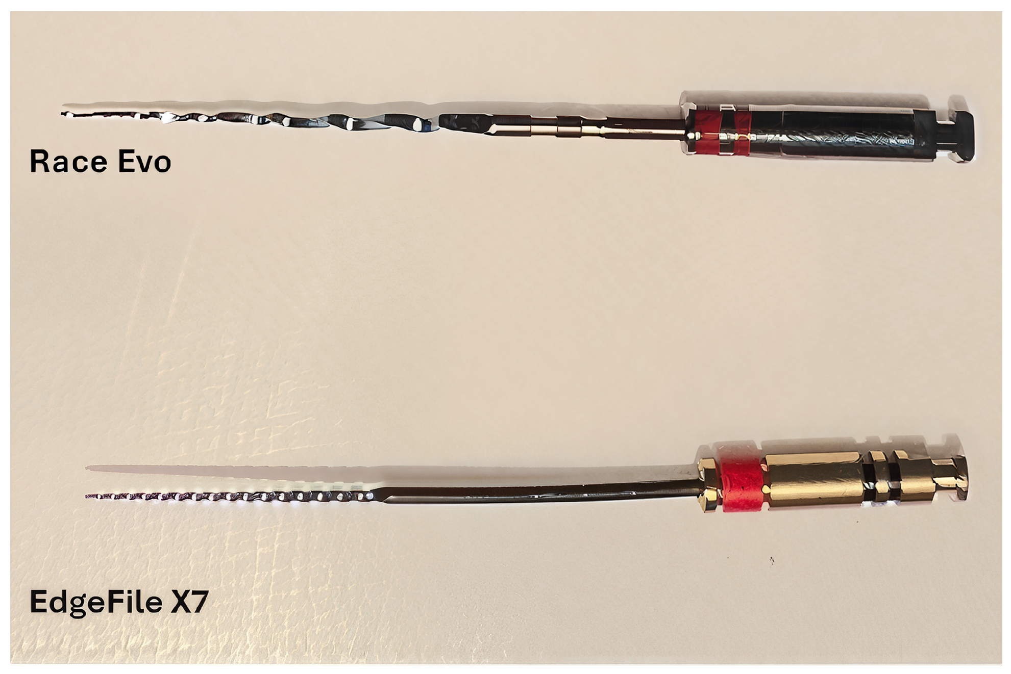
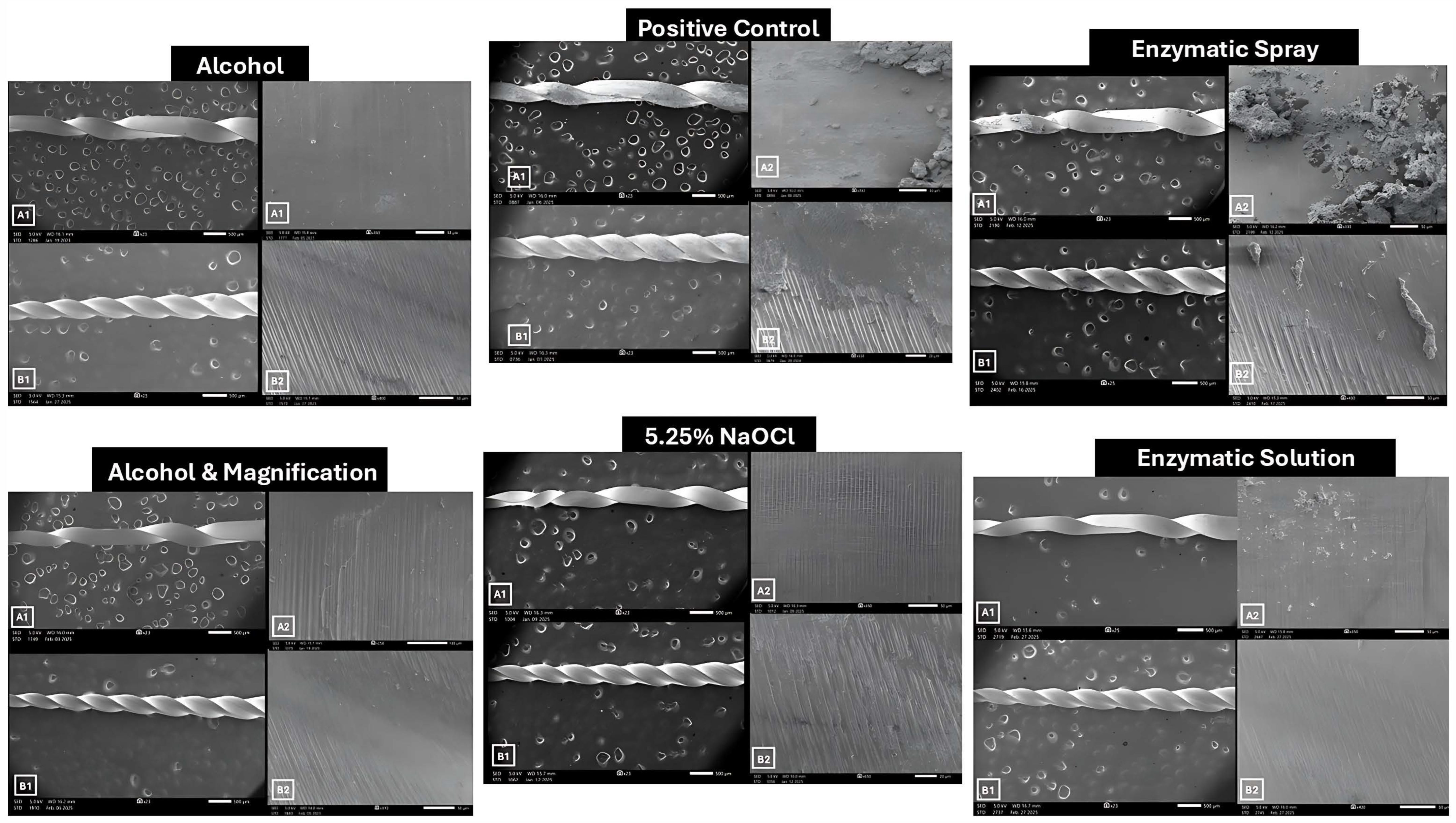
2.1. Sample Size and Preparation

A sample size calculation was performed using G*Power 3.1 software (Heinrich-Heine-Universität, Düsseldorf, Germany) to achieve a statistical power of 80% at a significance level of 0.05. The analysis indicated that 84 endodontic files were required. Two NiTi systems were selected: Race® Evo (RE) (FKG Dentaire, La Chaux-de-Fonds, Switzerland) and EdgeFile® X7 (EE) (EdgeEndo, Albuquerque, NW, USA). From each system, files of size 25 with a 0.04 taper and a length of 25 mm were used (Figure 1).
The 84 files were evenly distributed into seven experimental groups (n = 12 per group), each containing six files from each system.
  • Group 1 (Negative Control): Sterilized, unused files.
  • Group 2 (Positive Control): Used files sterilized without pre-cleaning.
  • Group 3: Files soaked in 30 mL of 5.25% sodium hypochlorite (NaOCl) for 5 min.
  • Group 4: Files wiped with 100% ethanol without magnification. Using a small, soaked sponge, a single operator wiped the files using synchronized up-and-down movements coupled with screwing movements. If debris was still observed, the process was repeated.
  • Group 5: Files wiped with 100% ethanol using 3.5× magnification dental loupes (JTL MEDIPLUS, Seoul, Republic of Korea). Using a small, soaked sponge, a single operator wiped the files using 5 up-and-down movements coupled with screwing-in movements. If debris was still observed, the process was repeated.
  • Group 6: Files coated with enzymatic spray gel (Hu-Friedy, Chicago, IL, USA) for 5 min.
  • Group 7: Files soaked in enzymatic instrument cleaner (Dürr Dental, Bietigheim-Bissingen, Germany) 2% solution. Files were placed in 30 mL of the solution for 5 min following manufacturer instructions [19].
All files, except those in Group 1, were used to instrument a single-rooted straight premolar canal following access cavity preparation using round and tapered fissure burs. The canal working length (WL) was determined to be 1 mm short of the length where the file extruded the apical foramen. To simulate the clinical conditions, the access cavities were flooded with 5.25% NaOCl. Canal preparation was performed by a single operator using a Dentsply X-Smart Endo Motor, following each manufacturer’s instructions. The settings used were as follows: RE files at 800 rpm and 1.2 N·cm torque and EE files at 350 rpm and 2 N·cm torque. All files were advanced to full working length (WL), followed by five circumferential brushing strokes within the canal.
As previous studies have shown that ultrasonic cleaning effectively removes biological debris [11,20], all cleaned files were subsequently placed in an ultrasonic bath (Famos Medizintechnik Vertriebs GmbH, Straelen, Germany) for 15 min using an enzymatic solution (Prolystica®, Steris, Mentor, OH, USA). Thereafter, all files were sterilized in a steam sterilizer (Amsco 600, Steris, Mentor, OH, USA) at 134 °C for 36 min.

2.2. SEM and EDX Analysis

The files were coated with platinum and mounted in a standardized orientation onto copper stubs using double-sided carbon tape for stability. Scanning electron microscopy (SEM) was performed using the JSM-IT500HR Field Emission SEM (JEOL Ltd., Tokyo, Japan) under high vacuum, with a 5 kV acceleration voltage and a probe current of 35. Images were captured at magnifications ranging from 60× to 700×, with resolutions between 500 μm and 100 μm. Standardized locations were imaged for all files, including the third flute from the tip (apical), the twelfth flute from the tip (middle), and the fourth flute below the shaft (coronal).
Energy-dispersive X-ray spectroscopy (EDX) was conducted to evaluate the elemental composition of the file surfaces. EDX settings included a 15 kV acceleration voltage, a probe current of 80, and magnifications ranging from 200× to 1000×, with resolutions from 100 μm to 1 μm.

2.3. Surface Roughness Analysis

Surface roughness was analyzed using ImageJ software (version 5.2, LOCI, University of Wisconsin, Madison, WI, USA) with the roughness calculation plugin. The following two parameters were used:
  • Rq (Root Mean Square Roughness): This represents the average roughness, more sensitive to surface irregularities [21,22].
  • Rsk (Surface Skewness): This reflects the asymmetry of the surface profile and indicates whether the surface has more peaks or valleys [21].
Changes in these parameters were interpreted as alterations in the vertical surface topography of the NiTi files.

2.4. Statistical Analysis

Statistical analysis was conducted using SPSS software (version 25; SPSS Inc., Chicago, IL, USA). The Shapiro–Wilk test was used to assess data normality. As the data were not normally distributed, non-parametric tests were applied, including the Mann–Whitney U test and the Kruskal–Wallis test. Statistical significance was set to p ≤ 0.05.

3. Results

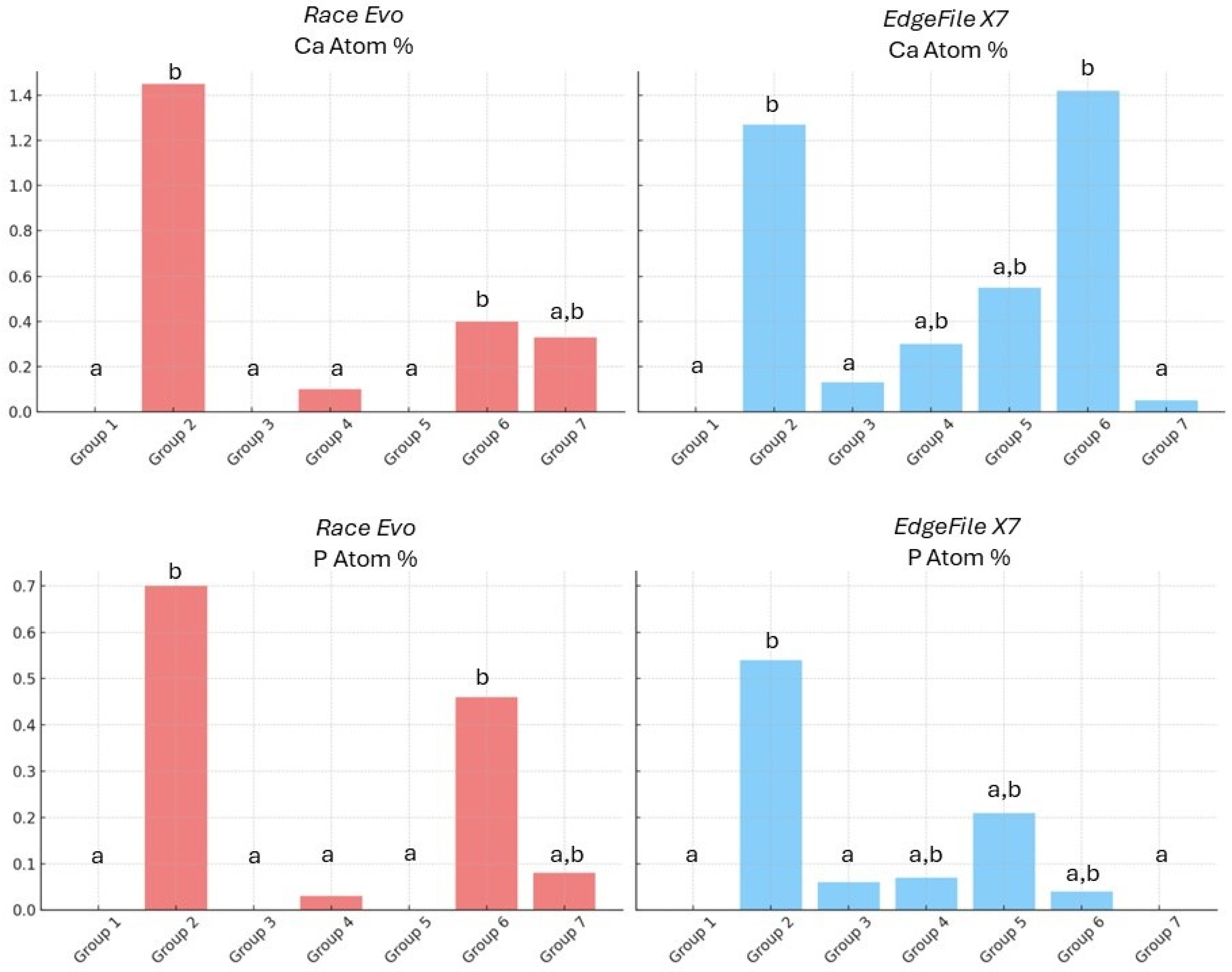
3.1. EDX Elemental Analysis

Energy-dispersive X-ray (EDX) analysis revealed that, with the exception of the unused files in Group 1 (negative control), all other groups exhibited detectable levels of calcium (Ca) and phosphorus (P). The positive control group (Group 2), consisting of used but uncleaned files, showed significantly higher levels of these elements compared to the negative control (p ≤ 0.0001).
The presence of Ca and P, indicative of hydroxyapatite—the principal inorganic component of dentin—suggests residual biological material remained on the instrument surfaces. Between the two file systems, RE files showed significantly lower calcium content than EE files (p = 0.003). RE files in Groups 3, 4, and 5 demonstrated significantly reduced levels of Ca and P compared to the positive control (p ≤ 0.0001). Similarly, EE files in Groups 3 and 7 also exhibited a significant reduction in these elements relative to the positive control (p ≤ 0.0001) (Figure 2).
Regardless of file brand, Group 6 retained significantly higher levels of Ca and P compared to the negative control (p ≤ 0.0001). Regionally, a significant difference in Ca content was noted in the middle third of the file.
In terms of core alloy composition, a statistically significant difference in nickel (Ni) and titanium (Ti) content was observed between the two NiTi file systems (p ≤ 0.0001). Race Evo files exhibited a higher mean Ni content (47.96 ± 0.32%) and Ti content (40.35 ± 0.25%) compared to EdgeFile (Ni: 43.64 ± 0.27%; Ti: 38.84 ± 0.17%). Additionally, EdgeFile showed significantly higher silicon (Si) content (0.51 ± 0.06%) compared to Race Evo (0.08 ± 0.01%) (p = 0.023).

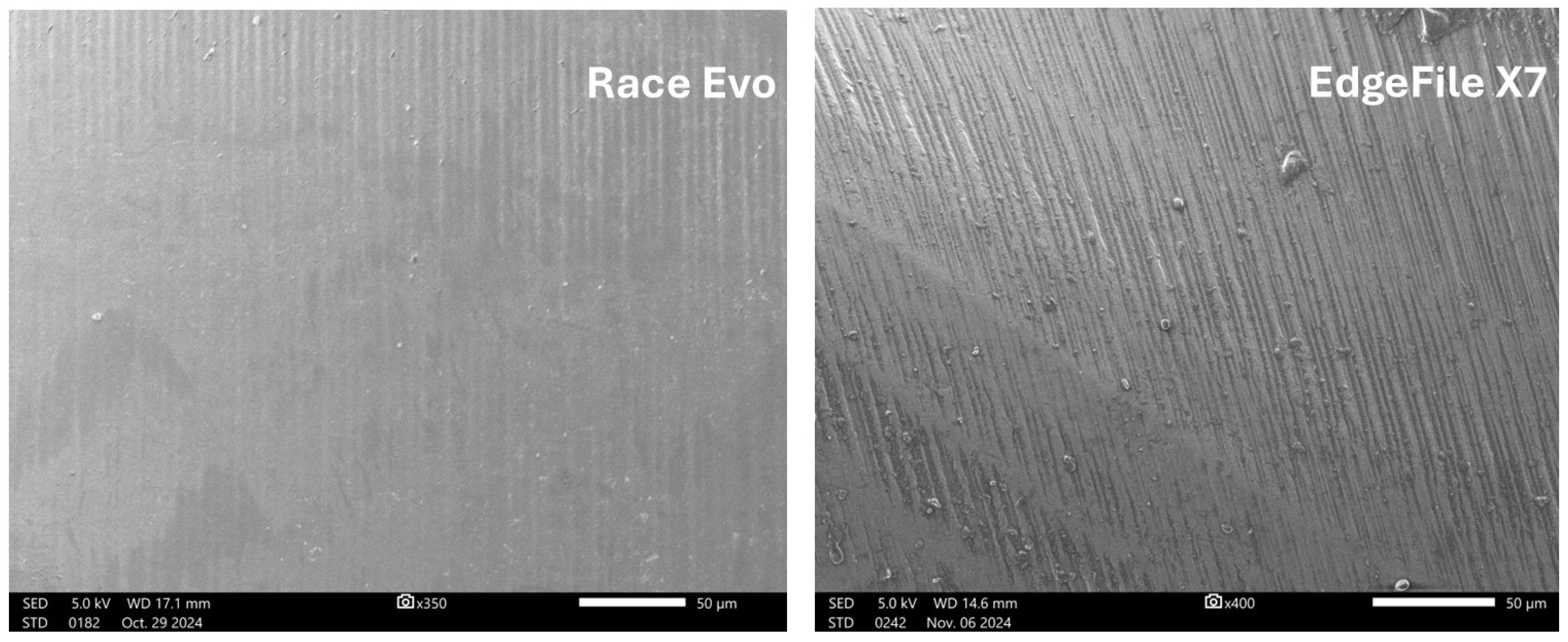
3.2. Surface Roughness

Prior to instrumentation, a significant difference in root mean square roughness (Rq) was observed between the two file brands (p = 0.001), with RE files exhibiting lower Rq values, indicating a smoother initial surface compared to EdgeFile (Figure 3). After use and cleaning, this difference in Rq was no longer significant; however, the surface asymmetry parameter (Rsk) remained statistically different across all groups (p = 0.001) (Table 1).
Following clinical simulation, RE files showed increased Rq values relative to the unused controls, with statistically significant increases in Groups 2, 4, and 6 (p = 0.01). Additionally, Rsk values were significantly elevated in Groups 2 and 6 compared to the negative control (p = 0.017), suggesting increased surface irregularity.
For EE files, Rsk values were significantly higher in all experimental groups compared to the negative control (p ≤ 0.0001), with the greatest deviation observed in Group 3 (NaOCl-soaked files). SEM images of these files revealed pronounced surface alterations, including the appearance of pits and erosion on the file surfaces (Figure 4).

4. Discussion

This study evaluated the effects of various cleaning protocols on NiTi endodontic files by analyzing their surface roughness and elemental composition post-instrumentation. Initially, Race Evo (RE) files exhibited significantly lower Rq values compared to EdgeFile (EE), indicating smoother surfaces likely due to the electropolishing process used in their manufacture [23].
Following clinical simulation and cleaning, differences in Rq between the two brands were no longer statistically significant, suggesting that both systems undergo comparable surface wear. However, Rsk values remained significantly different across all groups (p = 0.001), reflecting persistent differences in surface asymmetry and debris retention. EE files consistently demonstrated higher Rsk values, indicating greater surface irregularities. These findings were corroborated by elemental analysis, which showed significantly higher calcium (Ca) levels on EE files compared to RE. The presence of Ca and phosphorus (P), both main constituents of dentin, confirmed residual biological debris.
These results align with findings by Plotino et al. [24], who reported that increased surface roughness is associated with greater debris retention and potentially compromises sterilization, increasing the risk of mechanical failure such as file fracture. Conversely, earlier research by Eldik et al. [20] found no significant influence of file type, size, or taper on cleaning efficacy; however, that study did not evaluate elemental composition.
Although all used files—regardless of the cleaning method—retained some level of dentinal remnants, certain protocols proved more effective. RE files soaked in 5.25% NaOCl (Group 3) or wiped with alcohol prior to sterilization showed a near-complete elimination of Ca and P. NaOCl is recognized for its antimicrobial efficacy and ability to degrade organic matter [25,26]. However, it is also known to cause corrosion [27,28]. SEM images of files in Group 3 revealed visible pitting on the file surface, corresponding to significant changes in Rsk values. These findings support previous studies demonstrating NaOCl’s corrosive effects, particularly on EdgeFile instruments [29,30,31]. It is speculated that manufacturing factors such as the machining marks on the file surface can aid in the initiation of the corrosive points because it acts as crevices in the surface [31].
Despite their initially smoother surface, RE files demonstrated significant increases in Rq values after use in Groups 2, 4, and 6 (p = 0.01). The Rsk values for Groups 2 and 6 significantly increased compared to the negative control (p = 0.017), indicating the formation of a rougher, more irregular surface post-use and cleaning.
While NaOCl soaking and enzymatic cleaning reduced debris in EE files (notably in Group 7), alcohol wiping was less effective than with RE files. This difference could be attributed to variations in file design. The tighter flutes and smaller pitch of EE files may impede mechanical cleaning [32], whereas RE files—with alternating cutting blades and wider flutes—facilitate both soaking and wiping-based cleaning [33]. These findings support prior research noting that instruments with decreased pitch retain more debris [34].
Files treated with enzymatic spray demonstrated the poorest results in both brands, with high residual Ca and P and increased Rq and Rsk values. This indicates the spray may be ineffective in breaking down or flushing out dentin debris, making it an inferior cleaning option.
Despite both file systems having the same taper and tip size, they showed significantly different elemental compositions and surface responses to clinical use and sterilization. EDX analysis revealed that RE files contained higher Ni and Ti percentages than EE files, consistent with prior reports suggesting differences in alloy processing and heat treatment [35,36,37]. These metallurgical differences may influence flexibility, fatigue resistance, and clinical reusability [6].
While numerous studies have assessed cleaning protocols using staining, microscopy, and SEM [10,14,20,38], this study is the first to correlate surface roughness and elemental composition using both SEM and EDX, offering a more objective assessment of cleaning effectiveness. However, microbiological analysis was not included, which limits insight into the actual sterilization success from a clinical microbiology perspective.
This study was conducted in an in vitro setting, which may not fully replicate clinical conditions. Instruments were not exposed to vital pulp components such as blood and nerve tissue, which could affect debris composition. Moreover, while surface cleanliness was evaluated, the study did not examine the impact of cleaning protocols on mechanical properties, such as cyclic fatigue resistance—a key factor in clinical failure. Future studies should assess how these protocols affect instrument longevity and fracture resistance. Additionally, SEM and EDX analysis were restricted to standardized locations rather than the entire instrument, potentially limiting the completeness of surface assessment.

5. Conclusions

Within the limitations of this in vitro study, the efficacy of pre-sterilization cleaning methods varied depending on the NiTi file system. Race Evo files responded effectively to both wiping and soaking protocols, whereas EdgeFile X7 files—due to their tighter flute design—required soaking for optimal debris removal. Enzymatic spray was found to be the least effective cleaning method. These findings highlight the importance of tailoring cleaning protocols to the specific design and material properties of each file system. However, future studies are needed to better understand the impact of these protocols on instrument longevity and resistance to fracture.

Author Contributions

All authors have contributed significantly to this manuscript. Conceptualization, R.A.A. and R.M.B.; methodology, R.M.B.; software, S.F.; validation, R.A.A., R.M.B., and R.A. (Reem Alsaqat); formal analysis, S.F. and R.M.B.; investigation, H.A., R.A. (Renad Alsaeed), and R.A. (Raghad Alkhattabi); resources, H.A., R.A. (Renad Alsaeed), R.A. (Raghad Alkhattabi), and R.A. (Reem Alsaqat); data curation, R.M.B.; writing—original draft preparation, H.A., R.A. (Renad Alsaeed), R.A. (Raghad Alkhattabi), and R.A. (Reem Alsaqat); writing—review and editing, R.M.B. and R.A.; visualization, R.M.B.; supervision, R.M.B. and R.A.A.; project administration, R.A.A. and R.M.B.; funding acquisition, R.A.A. All authors have read and agreed to the published version of the manuscript.

Funding

This research was funded by the Princess Nourah bint Abdulrahman University Researchers Supporting Project number (PNURSP2025R162), Princess Nourah bint Abdulrahman University, Riyadh, Saudi Arabia.

Data Availability Statement

The datasets generated and analyzed in this study are available from the corresponding author upon request.

Conflicts of Interest

The authors declare no conflicts of interest.

References

  1. Rutala, W.A.; Weber, D.J. Disinfection and Sterilization: An Overview. Am. J. Infect. Control 2013, 41, S2–S5. [Google Scholar] [CrossRef]
  2. Spaulding, E. Chemical Disinfection of Medical and Surgical Materials. In Disinfection, Sterilization, and Preservation; Lea & Febiger: Philadelphia, PA, USA, 1968; pp. 517–531. ISBN 0683307401. [Google Scholar]
  3. Dioguardi, M.; Laneve, E.; Di Cosola, M.; Cazzolla, A.P.; Sovereto, D.; Aiuto, R.; Laino, L.; Leanza, T.; Alovisi, M.; Troiano, G.; et al. The Effects of Sterilization Procedures on the Cutting Efficiency of Endodontic Instruments: A Systematic Review and Network Meta-Analysis. Materials 2021, 14, 1559. [Google Scholar] [CrossRef]
  4. Sureshchandra, B.; Malik, M.; Naik, R. Endodontic Files: Are Routine Sterilization Procedures Effective? Endodontology 2011, 23, 47. [Google Scholar] [CrossRef]
  5. Reams, G.J.; Baumgartner, J.C.; Kulid, J.C. Practical Application of Infection Control in Endodontics. J. Endod. 1995, 21, 281–284. [Google Scholar] [CrossRef]
  6. Jovanovic-Medojevic, M.; Opacic-Galic, V.; Geler, K.; Pavlica, D. Comparison of the Efficiency of Cleaning and Disinfection Protocols for Hand Endodontic Instruments. Vojnosanit. Pregl. 2023, 80, 596–603. [Google Scholar] [CrossRef]
  7. Popovic, J.; Gasic, J.; Zivkovic, S.; Petrovic, A.; Radicevic, G. Evaluation of Biological Debris on Endodontic Instruments after Cleaning and Sterilization Procedures. Int. Endod. J. 2010, 43, 336–341. [Google Scholar] [CrossRef]
  8. Khullar, P.; Raisingani, D.; Gupta, S.; Bishen, K.A. Evaluation of Biological Debris on Reusable Endodontic Instruments Subjected to Different Cleaning Methods Prior to Sterilization. Int. J. Infect. Control 2013, 9, 2. [Google Scholar] [CrossRef]
  9. Morrison, A.; Conrod, S. Dental Burs and Endodontic Files: Are Routine Sterilization Procedures Effective? Tex. Dent. J. 2010, 127, 295–300. [Google Scholar]
  10. Ziauddin, S.; Bhandary, S.; Pramod, J.; Srinivasan, R.; Mahesh, M. A Comparative Evaluation of the Effectiveness of Different Cleaning Protocols on Removal of Biological Debris on Endodontic Instruments—An in Vitro Study. Endodontology 2013, 25, 19. [Google Scholar] [CrossRef]
  11. Parashos, P.; Linsuwanont, P.; Messer, H. A Cleaning Protocol for Rotary Nickel-titanium Endodontic Instruments. Aust. Dent. J. 2004, 49, 20–27. [Google Scholar] [CrossRef]
  12. Generali, L.; Generali, P.; Bertani, P.; Cavani, F.; Checchi, V.; Filippini, T.; Veneri, F. Quantitative Evaluation of Debris Removal from NiTi Rotary Endodontic Instruments After Different Cleaning Procedures. Dent. J. 2025, 13, 49. [Google Scholar] [CrossRef]
  13. Linsuwanont, P.; Parashos, P.; Messer, H.H. Cleaning of Rotary Nickel–Titanium Endodontic Instruments. Int. Endod. J. 2004, 37, 19–28. [Google Scholar] [CrossRef]
  14. Arruda-Vasconcelos, R.; Chantre, L.G.F.; Lopes, R.S.C.; Lopes, C.C.; Barbosa-Ribeiro, M.; Gomes, B.P.F.D.A. Application of Forensic Luminol for Blood Detection in Endodontic Files. Rev. Odontol. UNESP 2017, 46, 227–231. [Google Scholar] [CrossRef]
  15. Cayo-Rojas, C.F.; Brito-Ávila, E.; Aliaga-Mariñas, A.S.; Hernández-Caba, K.K.; Saenz-Cazorla, E.D.; Ladera-Castañeda, M.I.; Cervantes-Ganoza, L.A. Cleaning of Endodontic Files with and without Enzymatic Detergent by Means of the Manual Method versus the Ultrasonic Method. J. Int. Soc. Prev. Community Dent. 2021, 11, 307–315. [Google Scholar] [CrossRef]
  16. Zinelis, S.; Eliades, T.; Eliades, G. A Metallurgical Characterization of Ten Endodontic Ni-Ti Instruments: Assessing the Clinical Relevance of Shape Memory and Superelastic Properties of Ni-Ti Endodontic Instruments. Int. Endod. J. 2010, 43, 125–134. [Google Scholar] [CrossRef]
  17. RaceEvo|FKG Dentaire RaceEvo|FKG Dentaire. Available online: https://fkg.ch/download-center/ (accessed on 31 May 2025).
  18. Anonymous. Extremely Flexible Files. Br. Dent. J. 2021, 231, 311. [Google Scholar] [CrossRef]
  19. Dürr Dental. Available online: https://www.duerrdental.com/en/products/hygiene/hygiene-preparations/instruments/id-212-instrument-disinfection/ (accessed on 2 January 2025).
  20. Eldik, D.V.; Zilm, P.S.; Rogers, A.H.; Marin, P.D. A SEM Evaluation of Debris Removal from Endodontic Files after Cleaning and Steam Sterilization Procedures. Aust. Dent. J. 2004, 49, 128–135. [Google Scholar] [CrossRef]
  21. Fuss, F.K. The Effect of Surface Skewness on the Super/Postcritical Coefficient of Drag of Roughened Cylinders. Procedia Eng. 2011, 13, 284–289. [Google Scholar] [CrossRef]
  22. Finishing Surface. Comparing Surface Roughness Units and Standards. Available online: https://finishingsurface.com/comparing-surface-roughness-units-and-standards (accessed on 2 February 2025).
  23. Lopes, N.I.D.A.; Silva, L.Á.D.O.; Santos, L.D.A.; Buono, V.T.L. Surface Characterization of NiTi Superelastic and Shape Memory Alloys After Electrolytic Polishing. Mater. Res. 2017, 20, 572–579. [Google Scholar] [CrossRef]
  24. Plotino, G.; Costanzo, A.; Grande, N.M.; Petrovic, R.; Testarelli, L.; Gambarini, G. Experimental Evaluation on the Influence of Autoclave Sterilization on the Cyclic Fatigue of New Nickel-Titanium Rotary Instruments. J. Endod. 2012, 38, 222–225. [Google Scholar] [CrossRef]
  25. Koskinen, K.P.; Stenvall, H.; Uitto, V. Dissolution of Bovine Pulp Tissue by Endodontic Solutions. Eur. J. Oral Sci. 1980, 88, 406–411. [Google Scholar] [CrossRef]
  26. Hand, R.E.; Smith, M.L.; Harrison, J.W. Analysis of the Effect of Dilution on the Necrotic Tissue Dissolution Property of Sodium Hypochlorite. J. Endod. 1978, 4, 60–64. [Google Scholar] [CrossRef]
  27. Stokes, O.W.; Di Fiore, P.M.; Barss, J.T.; Koerber, A.; Gilbert, J.L.; Lautenschlager, E.P. Corrosion in Stainless-Steel and Nickel-Titanium Files. J. Endod. 1999, 25, 17–20. [Google Scholar] [CrossRef]
  28. Edie, J.W.; Andreasen, G.F.; Zaytoun, M.P. Surface Corrosion of Nitinol and Stainless Steel Under Clinical Conditions. Angle Orthod. 1981, 51, 319–324. [Google Scholar]
  29. Han-Hsing Lin, J.; Karabucak, B.; Lee, S.-M. Effect of Sodium Hypochlorite on Conventional and Heat-Treated Nickel-Titanium Endodontic Rotary Instruments—An in Vitro Study. J. Dent. Sci. 2021, 16, 738–743. [Google Scholar] [CrossRef]
  30. Uslu, G.; Özyürek, T.; Yılmaz, K. Effect of Sodium Hypochlorite and EDTA on Surface Roughness of HyFlex CM and HyFlex EDM Files. Microsc. Res. Tech. 2018, 81, 1406–1411. [Google Scholar] [CrossRef]
  31. Sood, K.; Mohan, B.; Lakshminarayanan, L. Effect of Cleaning and Sterilization Procedures on Niti Rotary Files—An Sem and Eds Study. Endodontology 2006, 18, 34. [Google Scholar] [CrossRef]
  32. EdgeEndo File X7. Available online: https://web.edgeendo.com/edgefile-x7/ (accessed on 6 January 2025).
  33. Almohareb, R.A.; Barakat, R.; Albakri, A.; Altamimi, M. Effect of Autoclaving Cycles on the Cyclic Fatigue Resistance of Race and Race Evo Nickel-Titanium Endodontic Rotary Files: An In Vitro Study. Metals 2021, 11, 1947. [Google Scholar] [CrossRef]
  34. Liang, Y.; Yue, L. Evolution and Development: Engine-Driven Endodontic Rotary Nickel-Titanium Instruments. Int. J. Oral Sci. 2022, 14, 12. [Google Scholar] [CrossRef]
  35. Faus-Llácer, V.; Hamoud-Kharrat, N.; Marhuenda Ramos, M.T.; Faus-Matoses, I.; Zubizarreta-Macho, Á.; Ruiz Sánchez, C.; Faus-Matoses, V. Influence of the Geometrical Cross-Section Design on the Dynamic Cyclic Fatigue Resistance of NiTi Endodontic Rotary Files—An In Vitro Study. J. Clin. Med. 2021, 10, 4713. [Google Scholar] [CrossRef]
  36. Ruiz-Sánchez, C.; Faus-Llácer, V.; Faus-Matoses, I.; Zubizarreta-Macho, Á.; Sauro, S.; Faus-Matoses, V. The Influence of NiTi Alloy on the Cyclic Fatigue Resistance of Endodontic Files. J. Clin. Med. 2020, 9, 3755. [Google Scholar] [CrossRef]
  37. Barakat, R.M.; Almohareb, R.A.; Algahtani, F.N.; Altamimi, A.A.; Alfuraih, J.I.; Bahlol, L.S.; Jamleh, A. Comprehensive Characterization of Blue Wire NiTi File Failure: A Comparative Analysis of Cyclic Fatigue and Torsional Resistance Properties. Coatings 2024, 14, 361. [Google Scholar] [CrossRef]
  38. Guandalini, B.; Vendramini, I.; Leonardi, D.P.; Tomazinho, F.S.F.; Tomazinho, P.H. Comparative Analysis of Four Cleaning Methods of Endodontic Files. RSBO 2015, 11, 154–158. [Google Scholar] [CrossRef]
Figure 1. Nickel–titanium (NiTi) endodontic files used in the study. Files shown are size 25, with a 0.04 taper and 25 mm length from two systems, Race® Evo and EdgeFile® X7.
Crystals 15 00684 g001
Figure 2. Bar charts illustrating the atomic percentage of calcium (Ca) and phosphorous (P) on the surfaces of Race Evo and EdgeFileX7 files across the seven experimental groups. The data represents mean values obtained via EDX analysis, reflecting the presence of residual biological material. The different letters (a, b) over the bars indicate the presence of statistical significance.
Crystals 15 00684 g002
Figure 3. Representative SEM images of unused and sterilized NiTi files, highlighting differences in surface roughness between Race Evo and EdgeFile X7. Race Evo files exhibited smoother surfaces compared to EdgeFile prior to clinical use.
Crystals 15 00684 g003
Figure 4. SEM images of both NiTi file systems following use and cleaning: (A) Race Evo and (B) EdgeFile X7. For each group, the left panel (1) shows a general overview of the file, while the right panel (2) displays a higher magnification of one of the flutes, highlighting surface alterations such as debris retention or pitting.
Crystals 15 00684 g004
Table 1. Mean and median values of surface roughness parameters (Rq and Rsk) for Race Evo and EdgeFile X7 NiTi files across the experimental groups. Within each file brand, values labeled with different letters (a, b) indicate a statistically significant difference between those values (p < 0.05).
File SystemGroupRqRsk
Mean ± SDMedianMean ± SDMedian
Race Evo122.69 ± 5.7922.41 a0.41 ± 0.220.40 a
230.51 ± 5.7629.16 b0.58 ± 0.190.53 b
327.74 ± 6.7728.52 a,b0.49 ± 0.130.44 a,b
429.26 ± 0.2229.53 b0.45 ± 0.180.44 a,b
529.11 ± 6.6229.11 a,b0.52 ± 0.150.52 a,b
635.28 ± 11.7330.86 b0.57 ± 0.210.57 b
728.23 ± 5.7629.06 a,b0.51 ± 0.170.53 a,b
EdgeFile X7131.98 ± 8.8132.53 a0.44 ± 0.170.46 a
231.87 ± 5.6032.54 a0.70 ± 0.210.72 b
328.61 ± 7.6728.67 a0.86 ± 0.150.88 b
428.98 ± 6.3128.30 a0.76 ± 0.180.80 b
528.28 ± 5.9926.78 a0.68 ± 0.180.72 b
630.28 ± 3.2829.98 a0.75 ± 0.170.76 b
729.73 ± 5.1330.34 a0.68 ± 0.160.67 b
Disclaimer/Publisher’s Note: The statements, opinions and data contained in all publications are solely those of the individual author(s) and contributor(s) and not of MDPI and/or the editor(s). MDPI and/or the editor(s) disclaim responsibility for any injury to people or property resulting from any ideas, methods, instructions or products referred to in the content.

