Article

Crystallographic Combinations: Understanding Polymorphism and Approximate Symmetry in N-(1,3-Thiazol-2-yl)benzamide

by
Johannes C. Voigt
1,2,
Michael J. Hall
2 and
Paul G. Waddell
2,*
1
Institute of Organic Chemistry, Justus Liebig University Giessen, Heinrich-Buff-Ring 17, 35392 Giessen, Germany
2
School of Natural and Environmental Sciences, Bedson Building, Newcastle University, Newcastle upon Tyne NE1 7RU, UK
*
Author to whom correspondence should be addressed.
Crystals 2025, 15(7), 657; https://doi.org/10.3390/cryst15070657
Submission received: 19 June 2025 / Revised: 16 July 2025 / Accepted: 16 July 2025 / Published: 18 July 2025
(This article belongs to the Special Issue Celebrating the 10th Anniversary of International Crystallography)

Abstract

A new polymorph of N-(1,3-thiazol-2-yl)benzamide crystallises in the monoclinic space group Pc with four crystallographically independent molecules (Z′ = 4) in the asymmetric unit. Where the previously reported polymorphs exhibit two distinct hydrogen-bonded dimer geometries exclusively, the asymmetric unit of the new polymorph comprises both. Approximate symmetry was observed to relate the molecules of these dimers. These approximate symmetry elements combine to form a structure with distorted P21/c space group symmetry, rationalising the unexpectedly high number of crystallographically independent molecules.

Graphical Abstract

1. Introduction

Polymorphism is an important phenomenon in crystallography that occurs when a substance can adopt more than one distinct crystal structure with variations in the molecular conformation and/or crystal packing [1,2,3,4]. Polymorphs can exhibit different physical properties, thus the phenomenon is not only interesting crystallographically speaking but also relevant in a pharmaceutical context, as changes in properties can also affect the application of a molecule [3,5].
Another important yet often underexplored frontier in crystallography concerns crystal structures with more than one molecule in the asymmetric unit. The number of molecules in the asymmetric unit of a homomolecular crystal is referred to as Z-prime (Z′). Higher Z′ values (Z′ > 1) and structures of this kind reflect the complexity of real-world crystallisation phenomena that deviate from idealised symmetry [6]. Variations in conformation, intermolecular interactions and molecular packing, and other concepts such as ‘frustration’ and approximate symmetry, can give rise to higher Z′ values, and the investigation of such structures often reveals the influence of these factors [6,7,8]. Higher Z′ values are somewhat rare, as structures with Z′ > 1 currently account for only ca. 10% of entries in the Cambridge Structural Database (CSD, version 5.46, update 1 February 2025), and the frequency decreases further as Z′ increases [9].
In addition, crystal structures with Z′ > 1 are notoriously difficult to predict and are often disregarded completely by crystal structure prediction (CSP) algorithms [8]. As such, the systematic study of such compounds will surely provide insights to the crystallographic community by further establishing the rationale behind their formation. Investigating a structure with higher Z′ is particularly intriguing in the context of polymorphism as it provides a unique opportunity to explore the supramolecular landscape governing polymorphic behaviour in the solid state [10].
N-(1,3-Thiazol-2-yl)benzamide (Figure 1) is a benzamide containing a hetereoaromatic thiazolyl substituent. This molecule and its derivatives have found use in a wide range of applications in different fields, e.g., as quorum sensing inhibitors, borodifluoride complexes and gelators [11,12,13,14]. In addition, scaffolds based on N-(1,3-thiazol-2-yl)benzamide are found in medicinal chemistry, in the development of ligands targeting biologically relevant metals [15], as well as in the development of inhibitors of other enzymatic targets [16,17,18,19,20,21,22,23].
Two crystal structures of N-(1,3-thiazol-2-yl)benzamide have been reported: polymorph I, grown from EtOH, and polymorph II, for which crystallisation details are not available—both of which crystallise in different centrosymmetric, monoclinic space groups with one molecule in the asymmetric unit (Z′ = 1) [24,25]. In this work, we report the discovery of a third polymorph (polymorph III) with four crystallographically independent molecules in the asymmetric unit (Z′ = 4) and compare its structure with the previously reported polymorphic forms I and II. The analysis focuses on variations in conformation, hydrogen-bonded dimer geometries and overall crystal packing, which are key to describing the differences between the three forms. The analysis demonstrates that polymorph III combines features of the previous two structures, and the observation of the approximate symmetry elements and how they combine to form the structure further highlights the differences between the three polymorphs and rationalises the relatively high Z′ value observed.

2. Experimental Section

N-(1,3-Thiazol-2-yl)benzamide was prepared via an adapted protocol [26]. Chemicals and solvents were used as purchased from Sigma-Aldrich or Alfa Aesar.
2-Aminothiazol (100 mg, 1 mmol) was added to a Schlenk flask, followed by the addition of DCM (4 mL), and the reaction mixture was stirred until all solids had dissolved. Benzoyl isothiocyanate (0.134 mL, 1 mmol) was then added dropwise and the mixture was stirred at room temperature overnight, resulting in the formation of a yellow precipitate. The reaction mixture was then dissolved in EtOAc (75 mL) and washed with brine (2 × 100 mL); the organic layer was separated and dried over MgSO4 and the solvent removed under reduced pressure to give the crude product as a yellow powder (124.4 mg).
The crude material was equally distributed across four small glass vessels, into each of which was added EtOH, EtOAc, DCM or acetone until the sample had fully dissolved. Samples were allowed to slowly evaporate over one to two weeks, until crystals had formed.
Single crystals of polymorph I suitable for SCXRD were obtained from EtOH at room temperature and confirmed by unit cell measurement. The remaining material from unsuccessful crystallisations was resubjected to slow evaporation crystallisation from EtOH. Single crystals suitable for SCXRD were obtained from the EtOH solution of the residue initially dissolved in EtOAc at room temperature, giving the new form, polymorph III.
Single-crystal diffraction data were collected on an XtaLAB Synergy HyPix-Arc 100 diffractometer using copper radiation (λCuKα  =  1.54184 Å) at 150 K, using an Oxford Cryosystems CryostreamPlus open-flow N2 cooling device.
Intensities were corrected for absorption using a multifaceted crystal model created by indexing the faces of the crystal for which data were collected [27]. Cell refinement, data collection and data reduction were undertaken via the software CrysAlisPro [28].
All structures were solved using SHELXT [29] and refined by SHELXL [30] using the Olex2 interface [31]. All non-hydrogen atoms were refined anisotropically and hydrogen atoms were positioned with idealised geometry, with the exception of those bound to heteroatoms, the positions of which were located using peaks in the Fourier difference map. The displacement parameters of the hydrogen atoms were constrained using a riding model, with U(H) set to be an appropriate multiple (1.2 or 1.5) of the Ueq value of the parent atom.

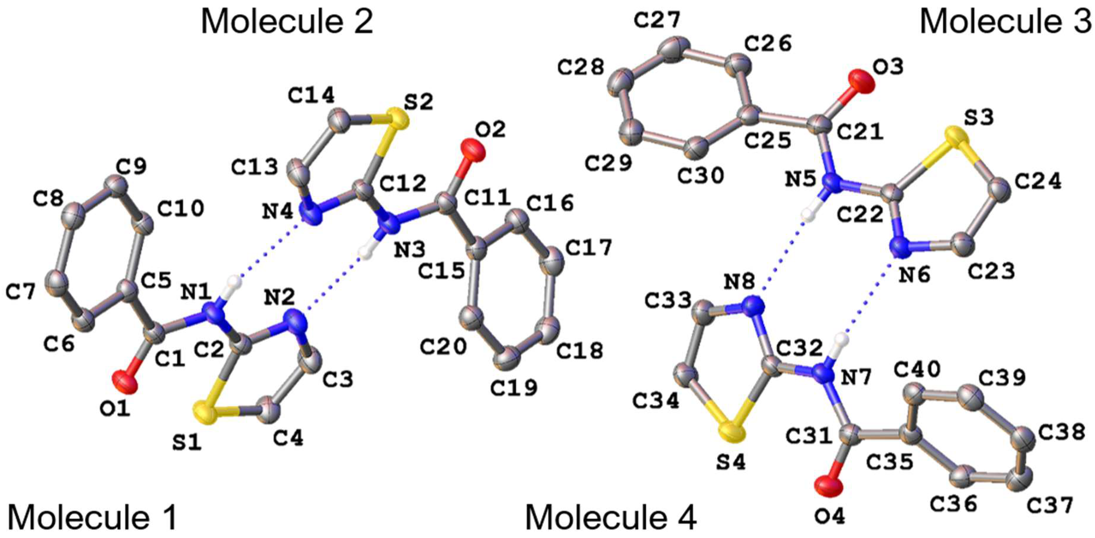
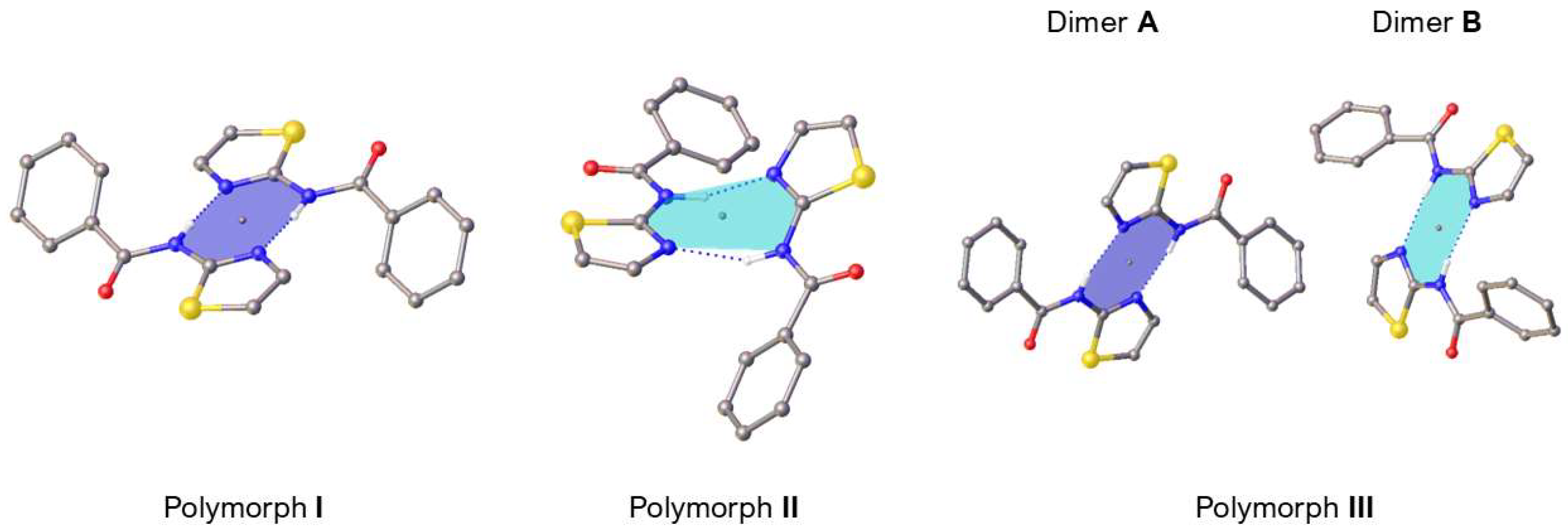
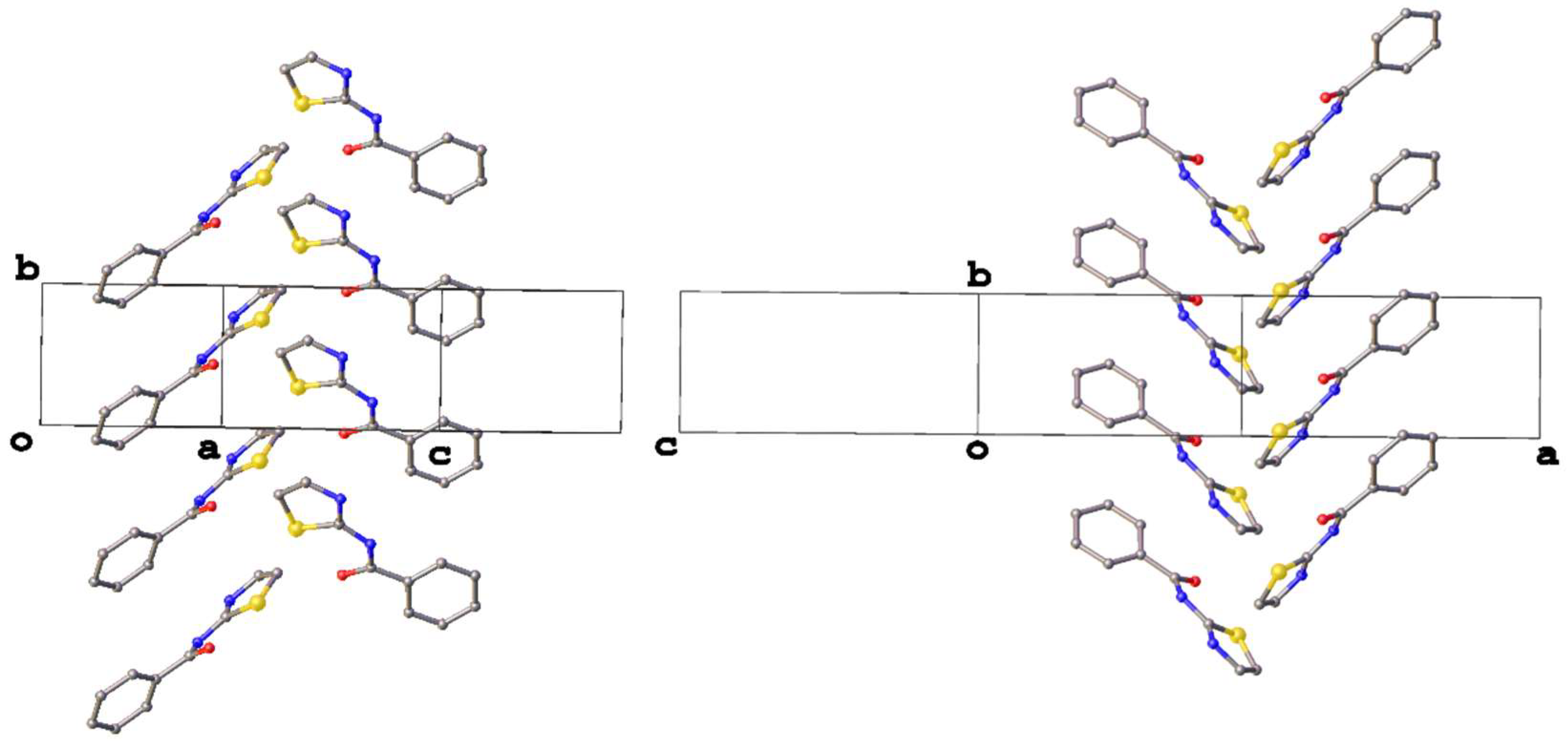
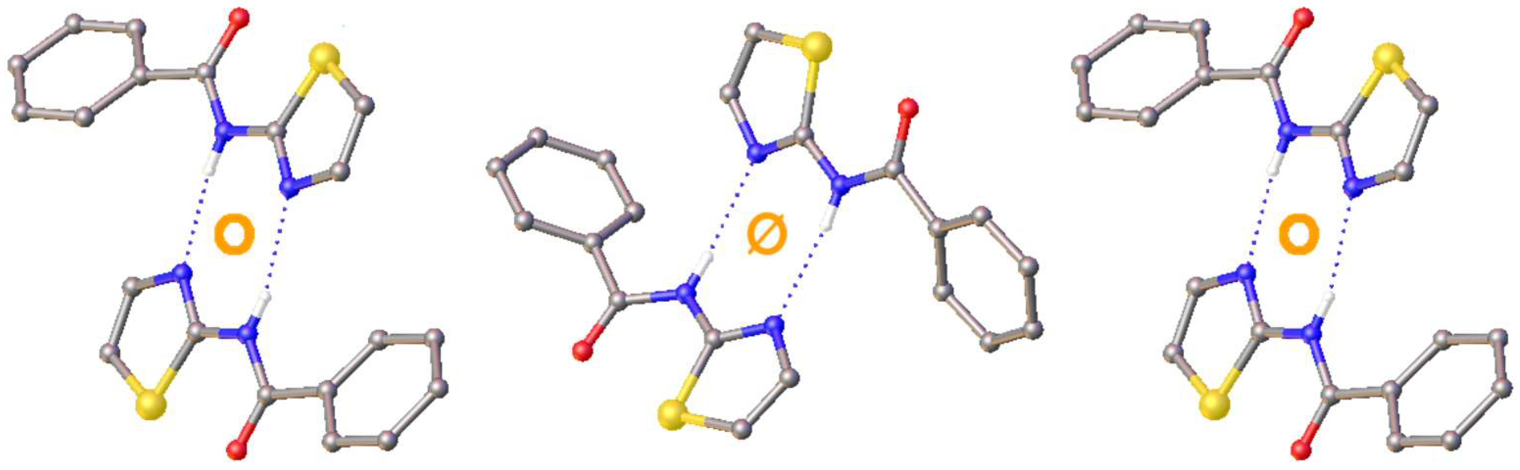
3. Results and Discussion

The structure of N-(1,3-thiazol-2-yl)benzamide reported in this work represents a third polymorph (polymorph III) of this compound (Figure 2), complementing the two crystalline forms that have previously been described (Table 1). The first polymorph (polymorph I) crystallises in the centrosymmetric, monoclinic space group P21/c with one molecule in the asymmetric unit (Z′ = 1) (CSD Refcode: NORLAI), while the second polymorph (polymorph II) was found to crystallise in the centrosymmetric, monoclinic space group C2/c, also with one molecule in the asymmetric unit (Z′ = 1) (CSD Refcode: NORLAI01) [24,25]. By way of contrast, polymorph III crystallises in a non-centrosymmetric monoclinic space group (Pc) with a high number of crystallographically independent molecules in the asymmetric unit (Z′ = 4).
Considering the asymmetric unit of polymorph III, confirmation of the fact that the four molecules are symmetry-independent can be seen in an overlay diagram using the amide moiety as the centre (Figure 3). It is clear from this figure that the conformation of each molecule is different, and this can be quantified by the torsion angles relating to the phenyl and thiazolyl groups (Table 2). Although the S-C-N-C torsions differ only slightly in magnitude, there appears to be a greater variation in the C-C-N-C angle, with molecule 2 representing the most significant outlier.
When compared to the two known polymorphs, the torsion angles describing the conformation of the phenyl unit (C-C-C-N) for I and II fall within the range of those of III but differ significantly from each other. This variation can be attributed to the lack of significant conjugation between the phenyl ring and the amide group, which allows for rotation about the C1–C5 bond.
For the torsion angle describing the conformation of the thiazolyl unit, S-C-N-C, one finds very similar magnitudes, all less than 8°, indicating a conformation close to planarity across all polymorphs. This is most likely due to restricted rotation as a result of conjugation brought about by the delocalisation of charge between N1 and N2. The planarity of the thiazolyl ring relative to the amide carbonyl may be further maintained by the presence of intramolecular S⋯O interactions [14].
The relative orientation of the two rings in each molecule, as denoted by the sign of the torsion angles, relates also to the formation of dimer units in each of the three polymorphs (Figure 4). These dimers are formed of hydrogen bonds (Table 3), where the amide proton acts as a donor to the thiazolyl nitrogen atom to give a ring motif with the graph set R 2 2 (8) [32]. In polymorph I, the two molecules that comprise the dimer are related by inversion symmetry, whereas in II they are related by a two-fold rotation about the rotation axis of the C2/c space group symmetry, with the dimer appearing twisted out of plane as a result.
Although both dimers exhibit similar hydrogen bond distances, there is a discrepancy in the angle of this interaction as a result of the interplanar angles of the thiazolyl rings of the constituent molecules. In polymorph I, the crystallographic inversion centre lies in the mean plane defined by the thiazolyl ring, resulting in centrosymmetric dimers with coplanar thiazolyl rings. In polymorph II, the crystallographic two-fold axis is not orthogonal to the thiazolyl planes, resulting in a ~37° dihedral angle between the two thiazolyl planes of the dimer. As the strength of a hydrogen bond scales with angle, this observation suggests that the hydrogen bonding in polymorph II is weaker than that of polymorph I [33].
In the structure of polymorph III, as there are four molecules in the asymmetric unit, there are two distinct dimers: dimer A, formed of molecules 1 and 2, and dimer B, formed of molecules 3 and 4. These dimers form ring motifs with the graph set R 2 2 (8) in a similar fashion to those of I and II (Table 4).
As each of the four molecules comprising the two dimers in polymorph III are crystallographically independent, they are not related by crystallographic symmetry, but closer inspection reveals that they do appear to be related by approximate symmetry. The two molecules of dimer A are related by an approximate inversion, whereas dimer B forms about an approximate two-fold rotation axis. As such, it can be said that polymorph III combines aspects of the structures of polymorphs I and II where the dimers form across a true inversion centre and rotation axis, respectively. To illustrate this, the dimers of polymorphs I and II can be overlaid with the dimer of III that displays the corresponding approximate symmetry, i.e., polymorph I with dimer A of III and polymorph II with dimer B of III (Figure 5).
In addition to demonstrating the nature of the approximate symmetry in polymorph III, this overlay and the interplanar angles within the dimers also show how much this approximate symmetry deviates from ideal symmetry. For dimer A, the interplanar angle is 4.23 (8)°, deviating slightly from the 0.0 (3)° in polymorph I, where the thiazolyl rings are arranged in a perfectly planar manner across the inversion centre. The equivalent angle in dimer B, 31.47 (9)°, is closer to that of polymorph II and is indicative of a similar relationship between the two molecules of the respective dimers.
The differences between the three polymorphs can be understood not only by the geometries of individual molecules and dimers but also by the relationship between the dimers corresponding to the overall crystal packing.
The similarities and differences between the three polymorphs are best demonstrated by viewing each along the crystallographic [010] direction (Figure 6). Polymorph III appears very similar to polymorph I in this direction, with both forming columns or stacks. Where they do differ is in the orientation of the dimers along the [100] direction. In polymorph I, the dimers are related by a pure translation in this direction, whereas in polymorph III, adjacent dimers are observed to adopt different orientations.
This similarity is brought sharply into focus through a thorough analysis of the approximate symmetry in the structure of polymorph III. As polymorph III crystallises in Pc, there is a glide plane coplanar with (001), including a translational component of half the unit cell in the [010] direction and no crystallographic inversion centres as the space group is non-centrosymmetric. However, closer inspection reveals further approximate symmetry elements.
In addition to the approximate inversion relating the molecules of dimer A and the approximate two-fold rotation relating the molecules of dimer B, there are also approximate screw axes between adjacent dimers in the [010] direction (Figure 7). As a result of the approximate inversion at the centre of dimer A, the two screw axes are also related by approximate inversion and exhibit opposite helicity. Viewing the structure down the [010] direction reveals how these approximate symmetry elements combine in such a way that the structure can be described as a distorted version of a structure with the symmetry of a different space group (Figure 8). This is consistent with many other examples of structures in Pc that mimic the symmetry of other higher-symmetry space groups [34].
Polymorph III can be considered a distorted P21/c structure as the centres of the dimers and the screw axes appear to mirror the arrangement of the inversion centres and screw axes in the space group P21/c. It would seem in this case that the approximate two-fold rotation is merely local to and affects only the dimer unit and that the approximate symmetry relating the two molecules of dimer B could be considered an approximate inversion. Although, in this case, the molecules of dimer B would only partially superimpose upon inversion, the dimers on either side in the [100] direction are clearly related by an approximate inversion through the centre of dimer B (Figure 9).
As such, the structure can be considered a distorted P21/c structure, with an approximate unit cell of the same dimensions as the true cell but with the origin at an approximate inversion centre. The asymmetric unit in this approximate cell will comprise only two molecules (Z′ = 2) as the multiplicity of the general position in P21/c is 4, compared to 2 for Pc, and both the true and approximate cells contain eight molecules in total.

4. Conclusions

The crystal structure of a third polymorph of N-(1,3-thiazol-2-yl)benzamide (polymorph III) has been reported and compared to the known forms, polymorph I and polymorph II. All three structures crystallise as hydrogen-bonded dimers in the solid state, but where in polymorph I the molecules of the dimer are related by an inversion centre, those of polymorph II are related by a two-fold rotation. An analysis of the newly discovered form reveals that this structure combines the features of polymorphs I and II, with an asymmetric unit comprising two dimers—one similar to that observed in polymorph I with approximate inversion symmetry and the other similar to that of polymorph II with an approximate rotation axis—resulting in four crystallographically independent molecules (Z′ = 4) in the asymmetric unit.
The combination of these approximate symmetry elements in the structure of polymorph III results in packing consistent with distorted P21/c space group symmetry. As this approximate unit cell would have Z′ = 2, it is clear that the presence of approximate symmetry and the two distinct dimer geometries are at the root of the high Z′ observed for polymorph III.
This analysis raises interesting questions about the formation of this ‘hybrid’ form of N-(1,3-thiazol-2-yl)benzamide comprising the dimer units previously exclusive to polymorphs I and II. Do these dimers form in solution prior to nucleation, and if so, what exact conditions favour the formation of one over the other? Alternatively, do the dimers form at the nucleation stage and whichever is observed will propagate through the growth of the crystal? In any case, further study of this relatively simple molecule should provide greater insights into its polymorphism, approximate symmetry and the combination of these two phenomena.

Author Contributions

Conceptualization, M.J.H.; Formal analysis, P.G.W.; Investigation, J.C.V.; Writing—original draft, J.C.V. and P.G.W.; Writing—review & editing, M.J.H. and P.G.W.; Supervision, P.G.W.; Project administration, M.J.H. All authors have read and agreed to the published version of the manuscript.

Funding

This research received no external funding.

Data Availability Statement

CCDC 2457230 contains the full crystallographic data for this article. These are available free of charge at www.ccdc.cam.ac.uk/data_request/cif (accessed on 15 July 2025), by emailing data_request@ccdc.cam.ac.uk or by contacting the Cambridge Crystallographic Data Centre, 12 Union Road, Cambridge, CB2 1EZ, UK; Fax: +44-1223-336033.

Acknowledgments

JCV is grateful to Deutsche Akademische Austauchdienst (DAAD) for support through the PROMOS scholarship.

Conflicts of Interest

The authors declare no competing financial interests.

References

  1. Brog, J.-P.; Chanez, C.-L.; Crochet, A.; Fromm, K.M. Polymorphism, what it is and how to identify it: A systematic review. RSC Adv. 2013, 3, 16905. [Google Scholar] [CrossRef]
  2. Cruz-Cabeza, A.J.; Bernstein, J. Conformational polymorphism. Chem. Rev. 2014, 114, 2170–2191. [Google Scholar] [CrossRef] [PubMed]
  3. Cruz-Cabeza, A.J.; Reutzel-Edens, S.M.; Bernstein, J. Facts and fictions about polymorphism. Chem. Soc. Rev. 2015, 44, 8619–8635. [Google Scholar] [CrossRef] [PubMed]
  4. Cruz-Cabeza, A.J.; Feeder, N.; Davey, R.J. Open questions in organic crystal polymorphism. Commun. Chem. 2020, 3, 142. [Google Scholar] [CrossRef] [PubMed]
  5. Nogueira, B.A.; Castiglioni, C.; Fausto, R. Color polymorphism in organic crystals. Commun. Chem. 2020, 3, 34. [Google Scholar] [CrossRef] [PubMed]
  6. Steed, K.M.; Steed, J.W. Packing problems: High Z′ crystal structures and their relationship to cocrystals, inclusion compounds, and polymorphism. Chem. Rev. 2015, 115, 2895–2933. [Google Scholar] [CrossRef] [PubMed]
  7. Brock, C.P. High-Z′ structures of organic molecules: Their diversity and organizing principles. Acta Crystallogr. B Struct. Sci. Cryst. Eng. Mater. 2016, 72, 807–821. [Google Scholar] [CrossRef] [PubMed]
  8. Waddell, P.G. A lot to unpack: A decade in high Z ′ crystal structures. CrystEngComm 2025, 27, 578–589. [Google Scholar] [CrossRef]
  9. Groom, C.R.; Bruno, I.J.; Lightfoot, M.P.; Ward, S.C. The Cambridge Structural Database. Acta Crystallogr. B Struct. Sci. Cryst. Eng. Mater. 2016, 72, 171–179. [Google Scholar] [CrossRef] [PubMed]
  10. Clegg, W. Crystallographic curiosities: Polymorphism and structures with Z′ 1. Acta Crystallogr. C Struct. Chem. 2019, 75, 833–834. [Google Scholar] [CrossRef] [PubMed]
  11. Sharma, N.; Srivastava, N.; Kaushal, A.; Das, B.; Vashistha, A.; Kumar, L.; Kumar, R.; Kumar Yadav, A. Synthesis, in Silico Study and Biological Evaluation of N-(Benzothiazol/Thiazol-2-yl)benzamide Derivatives as Quorum Sensing Inhibitors against Pseudomonas aeruginosa. Chem. Biodivers. 2023, 20, e202300647. [Google Scholar] [CrossRef] [PubMed]
  12. Potopnyk, M.A.; Lytvyn, R.; Danyliv, Y.; Ceborska, M.; Bezvikonnyi, O.; Volyniuk, D.; Gražulevičius, J.V. N,O π-Conjugated 4-Substituted 1,3-Thiazole BF2 Complexes: Synthesis and Photophysical Properties. J. Org. Chem. 2018, 83, 1095–1105. [Google Scholar] [CrossRef] [PubMed]
  13. Wang, Z.; Zhang, Y.; Li, X.; Zhou, Y.; Sheng, W. One pot synthesis and crystal of N-benzoyl(benzo)thiazol-2-amine borodifluoride complexes (NBO) with aggregation induced luminescence properties. J. Mol. Struct. 2025, 1321, 139761. [Google Scholar] [CrossRef]
  14. Yadav, P.; Ballabh, A. N-(thiazol-2-yl)benzamide derivatives as a new series of supramolecular gelators: Role of methyl functionality and S⋯O interaction. J. Solid State Chem. 2020, 281, 121027. [Google Scholar] [CrossRef]
  15. Madjroh, N.; Mellou, E.; Davies, P.A.; Söderhielm, P.C.; Jensen, A.A. Discovery and functional characterization of N-(thiazol-2-yl)-benzamide analogs as the first class of selective antagonists of the Zinc-Activated Channel (ZAC). Biochem. Pharmacol. 2021, 193, 114782. [Google Scholar] [CrossRef] [PubMed]
  16. Abbasi, M.A.; Raza, H.; Rehman, A.U.; Siddiqui, S.Z.; Nazir, M.; Mumtaz, A.; Shah, S.A.A.; Seo, S.-Y.; Hassan, M. Synthesis, Antioxidant and In-Silico Studies of Potent Urease Inhibitors: N-(4-{(4-Methoxyphenethyl)-(substituted)aminosulfonyl}phenyl)acetamides. Drug Res. 2019, 69, 111–120. [Google Scholar] [CrossRef] [PubMed]
  17. Iino, T.; Tsukahara, D.; Kamata, K.; Sasaki, K.; Ohyama, S.; Hosaka, H.; Hasegawa, T.; Chiba, M.; Nagata, Y.; Eiki, J.; et al. Discovery of potent and orally active 3-alkoxy-5-phenoxy-N-thiazolyl benzamides as novel allosteric glucokinase activators. Bioorg. Med. Chem. 2009, 17, 2733–2743. [Google Scholar] [CrossRef] [PubMed]
  18. In, E.-J.; Lee, Y.; Koppula, S.; Kim, T.-Y.; Han, J.-H.; Lee, K.-H.; Kang, T.-B. Identification and Characterization of NTB451 as a Potential Inhibitor of Necroptosis. Molecules 2018, 23, 2884. [Google Scholar] [CrossRef] [PubMed]
  19. Sams, A.G.; Mikkelsen, G.K.; Larsen, M.; Langgård, M.; Howells, M.E.; Schrøder, T.J.; Brennum, L.T.; Torup, L.; Jørgensen, E.B.; Bundgaard, C.; et al. Discovery of phosphoric acid mono-{2-(E/Z)-4-(3,3-dimethyl-butyrylamino)-3,5-difluoro-benzoylimino-thiazol-3-ylmethyl} ester (Lu AA47070): A phosphonooxymethylene prodrug of a potent and selective hA(2A) receptor antagonist. J. Med. Chem. 2011, 54, 751–764. [Google Scholar] [CrossRef] [PubMed]
  20. Satoh, A.; Nagatomi, Y.; Hirata, Y.; Ito, S.; Suzuki, G.; Kimura, T.; Maehara, S.; Hikichi, H.; Satow, A.; Hata, M.; et al. Discovery and in vitro and in vivo profiles of 4-fluoro-N-4-6-(isopropylamino)pyrimidin-4-yl-1,3-thiazol-2-yl-N-methylbenzamide as novel class of an orally active metabotropic glutamate receptor 1 (mGluR1) antagonist. Bioorg. Med. Chem. Lett. 2009, 19, 5464–5468. [Google Scholar] [CrossRef] [PubMed]
  21. Uto, Y.; Ogata, T.; Kiyotsuka, Y.; Miyazawa, Y.; Ueno, Y.; Kurata, H.; Deguchi, T.; Yamada, M.; Watanabe, N.; Takagi, T.; et al. Novel and potent inhibitors of stearoyl-CoA desaturase-1. Part II: Identification of 4-ethylamino-3-(2-hydroxyethoxy)-N-5-(3-trifluoromethylbenzyl)thiazol-2-ylbenzamide and its biological evaluation. Bioorg. Med. Chem. Lett. 2009, 19, 4159–4166. [Google Scholar] [CrossRef] [PubMed]
  22. van Muijlwijk-Koezen, J.E.; Timmerman, H.; Vollinga, R.C.; Frijtag von Drabbe Künzel, J.; de Groote, M.; Visser, S.; IJzerman, A.P. Thiazole and thiadiazole analogues as a novel class of adenosine receptor antagonists. J. Med. Chem. 2001, 44, 749–762. [Google Scholar] [CrossRef] [PubMed]
  23. Wu, G.; Qiu, X.-L.; Zhou, L.; Zhu, J.; Chamberlin, R.; Lau, J.; Chen, P.-L.; Lee, W.-H. Small molecule targeting the Hec1/Nek2 mitotic pathway suppresses tumor cell growth in culture and in animal. Cancer Res. 2008, 68, 8393–8399. [Google Scholar] [CrossRef] [PubMed]
  24. Zonouzi, A.; Mirzazadeh, R.; Rahmani, H.; Ng, S.W. N-(1,3-Thia-zol-2-yl)benzamide. Acta Crystallogr. Sect. E Struct. Rep. Online 2009, 65, o817. [Google Scholar] [CrossRef] [PubMed]
  25. Galal, S.A.; Abdelsamie, A.S.; Tokuda, H.; Suzuki, N.; Lida, A.; Elhefnawi, M.M.; Ramadan, R.A.; Atta, M.H.E.; El Diwani, H.I. Part I: Synthesis, cancer chemopreventive activity and molecular docking study of novel quinoxaline derivatives. Eur. J. Med. Chem. 2011, 46, 327–340. [Google Scholar] [CrossRef] [PubMed]
  26. Barnikow, G.; Bödeker, J. Isothiocyanate. XXV. Umsetzungen von Benzoylisothiocyanat mit Amino-N-heterocyclen. J. Prakt. Chem. 1971, 313, 1148–1154. [Google Scholar] [CrossRef]
  27. Clark, R.C.; Reid, J.S. The analytical calculation of absorption in multifaceted crystals. Acta Crystallogr. A Found. Crystallogr. 1995, 51, 887–897. [Google Scholar] [CrossRef]
  28. Rigaku. CrysAlisPro R; Tokyo, Japan. 2024. Available online: https://rigaku.com/products/crystallography/x-ray-diffraction/crysalispro (accessed on 10 June 2025).
  29. Sheldrick, G.M. SHELXT—Integrated space-group and crystal-structure determination. Acta Crystallogr. A Found. Adv. 2015, 71, 3–8. [Google Scholar] [CrossRef] [PubMed]
  30. Sheldrick, G.M. A short history of SHELX. Acta Crystallogr. A 2008, 64, 112–122. [Google Scholar] [CrossRef] [PubMed]
  31. Dolomanov, O.V.; Bourhis, L.J.; Gildea, R.J.; Howard, J.A.K.; Puschmann, H. OLEX2: A complete structure solution, refinement and analysis program. J. Appl. Crystallogr. 2009, 42, 339–341. [Google Scholar] [CrossRef]
  32. Etter, M.C. Encoding and decoding hydrogen-bond patterns of organic compounds. Acc. Chem. Res. 1990, 23, 120–126. [Google Scholar] [CrossRef]
  33. Wood, P.A.; Allen, F.H.; Pidcock, E. Hydrogen-bond directionality at the donor H atom—Analysis of interaction energies and database statistics. CrystEngComm 2009, 11, 1563. [Google Scholar] [CrossRef]
  34. Brock, C.P. Prevalence and Significance of Approximate Symmetry in Organic Pc Structures. Helv. Chim. Acta 2023, 106, e202200170. [Google Scholar] [CrossRef]
Figure 1. N-(1,3-Thiazol-2-yl)benzamide with the numbering scheme used in this article.
Crystals 15 00657 g001
Figure 2. The asymmetric unit of polymorph III with four crystallographically independent molecules. Ellipsoids are drawn at the 50% probability level. Hydrogen atoms not involved in hydrogen bonding have been omitted for clarity.
Crystals 15 00657 g002
Figure 3. Two perspectives of the overlay of molecule 1 (magenta), molecule 2 (red), molecule 3 (blue) and molecule 4 (cyan) in the asymmetric unit of molecule polymorph III.
Crystals 15 00657 g003
Figure 4. The hydrogen-bonded dimers in the structures of polymorphs I–III. Hydrogen atoms not involved in hydrogen bonding have been omitted for clarity.
Crystals 15 00657 g004
Figure 5. Overlay of dimer A of III (red) with the dimer of I (yellow) and dimer B of III (blue) with the dimer of II (green).
Crystals 15 00657 g005
Figure 6. Comparison of the [010] plane in polymorphs I to III.
Crystals 15 00657 g006
Figure 7. View of the approximate screw axes along [010] in the structure of polymorph III comprising molecules 1 and 2 (left) and molecules 3 and 4 (right).
Crystals 15 00657 g007
Figure 8. View of the structure of polymorph III along [010] with approximate screw axes (green) and inversion centres (orange). Where the orange circles have a line through them, the inversion symmetry is broken, manifesting as a two-fold rotation.
Crystals 15 00657 g008
Figure 9. A close-up view of the packing in polymorph III, showing that the two equivalents of dimer A to the left and right are related by an approximate inversion through the centre of dimer B (centre).
Crystals 15 00657 g009
Table 1. Crystal data and structural refinement details for the three polymorphs of N-(1,3-thiazol-2-yl)benzamide.
Polymorph IPolymorph IIPolymorph III
Empirical formulaC10H8N2OSC10H8N2OSC10H8N2OS
Formula weight204.24203.243204.24
Temperature/K123298150
Crystal systemMonoclinicMonoclinicMonoclinic
Space groupP21/cC2/cPc
a/Å12.0142 (2)15.9169 (5)20.3396 (3)
b/Å5.0581 (1)10.0631 (4)5.07500 (10)
c/Å15.4090 (3)12.5115 (5)20.3274 (3)
α/°909090
β/°99.093 (1)97.7186 (14)116.755 (2)
γ/°909090
Volume/Å3924.62 (3)1985.85 (13)1873.62 (6)
Z, Z′4, 18, 18, 4
ρcalcg/cm31.4671.3601.448
μ/mm−10.3130.292.786
F(000)424.0848.0 *848.0
RadiationMoKα (λ = 0.71073)MoKα (λ = 0.71073)CuKα (λ = 1.54184)
2Θ range for data collection/°3.44 to 556.02 to 60.14.866 to 154.336
Index ranges−15 ≤ h ≤ 15, −6 ≤ k ≤ 6, −18 ≤ l ≤ 20−22 ≤ h ≤ 22, −13 ≤ k ≤ 14, −17 ≤ l ≤ 17−24 ≤ h ≤ 25, −5 ≤ k ≤ 6, −25 ≤ l ≤ 25
Reflections collected6130549332148
Independent reflections2104 [Rint = 0.0164]3427 [Rint = 0.030]7102 [Rint = 0.0436]
Data/restraints/parameters2104/0/1313427/0/1277102/3/518
Goodness-of-fit on F21.0741.7231.018
Final R indices [I >= 2σ (I)]R1 = 0.0290, wR2 = 0.0842R1 = 0.0400, wR2 = 0.0690R1 = 0.0269, wR2 = 0.0652
Largest diff. peak/hole/e Å−30.37/−0.210.43/−0.510.23/−0.17
Flack parametern/an/a0.374 (12)
* Reported as 840.0 in the CIF.
Table 2. Selected torsion angles for the three polymorphs of N-(1,3-thiazol-2-yl)benzamide.
Polymorph IPolymorph IIPolymorph III
C10-C5-C1-N1/° *37.5 (2)25.01 (4)25.8 (4)
C20-C15-C11-N3/° −31.0 (4)
C30-C25-C21-N5/° 22.9 (5)
C40-C35-C31-N7/° 42.0 (4)
S1-C2-N1-C1/° **3.8 (2)−4.04 (2)−2.3 (4)
S2-C12-N3-C11/° 2.6 (4)
S3-C22-N5-C21/° −5.4 (4)
S4-C32-N7-C31/° 7.7 (4)
* Equivalent to the torsion angles C10-C5-C4-N2 and C9-C5-C6-N3 in I or II, respectively; ** equivalent to the torsion angles S1-C3-N2-C4 and S1-C7-N3-C6 in I or II, respectively.
Table 3. Comparison of the hydrogen-bond geometry in polymorphs I and II of N-(1,3-thiazol-2-yl)benzamide.
D—H⋯APolymorph IPolymorph II
d(D—H)/Å0.88 (2)0.960 (2)
d(H⋯A)/Å2.04 (2)2.030 (2)
d(DA)/Å2.922 (2)2.900 (2)
(D—H⋯A)/°173 (2)149.9 (2)
For atom labelling, see SI, D—H⋯A in I: N3—H⋯N1, in II: N3—H⋯N4.
Table 4. Comparison of the hydrogen-bond geometry in the dimers in polymorph III of N-(1,3-thiazol-2-yl)benzamide.
Dimer ADimer B
D—H⋯AN1—H1⋯N4N3—H3A⋯N2N5—H5⋯N8N7—H7A⋯N6
d(D—H)/Å0.81 (2)0.83 (4)0.82 (3)0.83 (3)
d(H⋯A)/Å2.22 (2)2.09 (4)2.16 (4)2.21 (3)
d(DA)/Å3.014 (3)2.918 (3)2.977 (3)3.014 (3)
(D—H⋯A)/°168 (3)173 (3)172 (3)164 (3)
Disclaimer/Publisher’s Note: The statements, opinions and data contained in all publications are solely those of the individual author(s) and contributor(s) and not of MDPI and/or the editor(s). MDPI and/or the editor(s) disclaim responsibility for any injury to people or property resulting from any ideas, methods, instructions or products referred to in the content.

