Article

Investigation of In Vitro Corrosion and Wear Properties of Biomedical Coatings Applied to Ti6Al4V Alloy Manufactured by Selective Laser Melting

by
Ali İhsan Bahçepinar
* and
İbrahim Aydin
Manisa Vocational School, Manisa Celal Bayar University, Manisa 45140, Turkey
*
Author to whom correspondence should be addressed.
Crystals 2025, 15(4), 316; https://doi.org/10.3390/cryst15040316
Submission received: 27 February 2025 / Revised: 10 March 2025 / Accepted: 21 March 2025 / Published: 27 March 2025

Abstract

:
This study focuses on enhancing the biomedical performance of PBF-LB Ti6Al4V, produced using Selective Laser Melting (SLM), an advanced manufacturing technology widely used for patient-specific medical devices and implants. Hydroxyapatite (HA), titanium (Ti), and bilayer Ti/HA coatings were applied, using the powder flame spray coating technique. A comprehensive analysis was conducted to examine the microstructural, chemical, and mechanical properties of the coatings. Surface analysis was performed using a scanning electron microscope (SEM), chemical composition was determined by energy-dispersive spectroscopy (EDS), crystal structure was analyzed via X-ray diffraction (XRD), and surface roughness was evaluated through topographic analyses. Additionally, in vitro wear and corrosion resistance tests, crucial for biomedical applications, were conducted. In wear tests, HA coatings exhibited the lowest wear resistance with the highest wear rate (73.83 × 10⁻³ mm3/N·m), while Ti coatings showed the highest wear resistance (6.32 × 10⁻³ mm3/N·m), and Ti/HA coatings demonstrated an intermediate performance (34.22 × 10⁻³ mm3/N·m). Corrosion tests revealed that bilayer Ti/HA coatings provided the best protection (0.00009 mm/year), followed by Ti coatings (0.0002 mm/year) and HA coatings (0.003 mm/year). The results indicate that Ti/HA coatings offer the most suitable biomedical performance.

1. Introduction

Selective Laser Melting (SLM) is an advanced manufacturing technology used for producing patient-specific medical devices and implants. This technology enables the production of complex and porous implants that cannot be manufactured using traditional methods such as forging or casting [1]. SLM has gained significant attention in recent years due to the growing demand for patient-specific implants and its ability to facilitate rapid production without delays in surgical procedures [2].
The Ti6Al4V alloy stands out as an orthopedic implant due to its low density, high strength-to-weight ratio, excellent corrosion resistance, and favorable biocompatibility [3]. However, these metallic implants can release ions into the body because of wear during implantation applications, potentially causing allergic reactions [3,4]. Additionally, Ti6Al4V metallic implants are not bioactive, which prevents them from directly bonding with living bone [5]. To address these drawbacks of metal implants, bioactive and composite coatings are utilized [6].
Hydroxyapatite (HA) is a bioactive material with the same chemical composition as bone, commonly used as a coating material on the surfaces of metallic implants [7,8]. When applied as a coating on metal implants, HA enhances surface energy and bioactivity, thereby improving the osseointegration process [8]. However, due to its poor mechanical properties, HA cannot be used directly as an implant material. When HA’s bioactivity is combined with the superior mechanical properties of metallic implants, it results in an excellent biomaterial [9]. In addition to enhancing osseointegration, HA acts as a barrier between metal and bone, preventing the release of ions into the body [10].
In some implant applications, the detachment of single-layer HA coatings from the surface can occur. To address this issue, double-layer coatings are applied to enhance the adhesion strength of the coating to the implant surface and to provide superior wear resistance. Ti/HA coatings are among the most commonly used types of such coatings [6].
The TiN phase obtained as a result of Ti coating is a bioactive ceramic material with high hardness, wear resistance, and corrosion resistance. Due to its biocompatibility, it is used as a coating material to enhance the wear and corrosion properties of orthopedic and dental implants [11,12,13,14,15,16]. This, in turn, reduces ion release caused by wear on metal implants [11,12]. Physical Vapor Deposition (PVD), Chemical Vapor Deposition (CVD), and thermal spray coating methods are employed to produce Ti and HA coatings [15]. Among thermal spray coating methods, the flame spray technique is preferred due to its low investment cost, high efficiency, ease of use, versatility, and environmentally friendly nature [17,18,19].
Some studies on bilayer biomedical coatings in the literature are as follows: Quirama et al. reported that a hydroxyapatite (HA) coating developed using a TiN/TiO2 interlayer has great potential for biomedical applications [20]. Mohseni et al. stated that the Ti/TiN interlayer increased the adhesion strength of HA coatings by 44.57% compared to HA coatings without an interlayer [21]. Qui et al. applied gradient transition HA/TiN/Ti composite coatings on the surface of Ti6Al4V substrates using reactive and magnetron sputtering techniques. They reported that HA/TiN/Ti composite coatings exhibited better corrosion resistance and improved metal ion barrier properties [22]. Lenis et al. reported that multilayer HA-Ag/TiN-Ti coatings obtained by radio frequency magnetron sputtering have potential for biomedical applications [23]. Sargın et al. reported that the HA coatings obtained using Ti and TiO2 interlayers exhibited bioactive properties [24]. In addition to these studies, research on HA coating applications for implant materials produced by Selective Laser Melting (SLM) is limited [25,26,27,28,29]. In particular, there is no study on the use of the commercially viable powder flame spraying method. The performance of biomedical coatings applied to SLM-produced materials is important from both a literature perspective and for implantation applications.

2. Materials and Methods

Within the scope of this study, the PBF-LB Ti6Al4V used as substrate material was manufactured using the Selective Laser Melting method with the EOS-M 290 (EOS GmbH, Krailling, Germany) model machine in dimensions of 10 mm (width) × 10 mm (length) × 2 mm (height) and Ø10 mm (diameter) × 5 mm (height). The chemical composition of the powders (EOS Titanium Ti64, E.O.S. GmbH, Krailling, Germany) used in the alloy is provided in Table 1. The alloys produced were subjected to heat treatment at 540 °C for 2 h in a Nabertherm Lh 120/12 (Nabertherm GmbH, Lilienthal, Germany) furnace under a flow of pure argon gas at a rate of 6 L per minute. Yang et al. reported that the heat treatment applied at 540 °C led to the formation of a stable (α + β) structure [30].
Before coating, the produced PBF-LB Ti6Al4V was sandblasted using an SK 1000 Injection Sandblasting Machine (Saykar Metallurgy, Istanbul, Turkey) with 40–70 µm glass beads. The purpose of this process was to clean the alloy powders on the surface and improve the adhesion of the coating powders. After sandblasting, the samples were subjected to ultrasonic cleaning for 5 min sequentially with ethanol, acetone, and deionized water to remove surface contaminants. HA coating processes were performed using a 5PM-II Powder Flame Spray gun (Metal Coat, Jodhpur, India) with Captal 60 HA powder (Plasma Biotal Ltd., Tideswell, UK). Ti coating processes were carried out using a 6PM-II Powder Flame Spray gun (Metal Coat, Jodhpur, India) with Metco 4010D (Wohlen, Switzerland) titanium powder. In the powder flame spray method, oxygen and propane were used to generate the flame, while air was used as the propellant gas. The coating process was carried out from a distance of 40 cm at a 90° angle to the surface. All parameters related to the coating process are provided in Table 2.
The morphological structure of the coatings was examined using a Zeiss Gemini 500 (Carl Zeiss AG, Oberkochen, Germany) scanning electron microscope (SEM). In addition, energy-dispersive spectrometer (EDS) analyses of the coating surfaces were performed. The phases contained in the coatings were determined using the X-ray diffraction (XRD) method. For the XRD analyses of the coatings, a PANalytical Empyrean model X-ray Diffraction Device (Malvern Panalytical, Almelo, Netherlands) was utilized. Phase analyses of the obtained XRD patterns were conducted using the HighScore Plus XRD software version 3.0. The surface roughness of the coatings was measured using a PCE-RT 1200 model device (PCE-RT 1200, Meschede, Germany). For each sample, measurements were taken 8 times, and the results were averaged. The electrochemical tests of the coatings were performed using a Gamry Interface 1010 E (Gamry Instruments, Warminster, PA, ABD) potentiostat/galvanostat in a three-electrode system, consisting of a coated sample surface with a 0.78 cm2 working electrode, a graphite counter electrode, and a saturated calomel electrode (SCE) as the reference electrode. A lactate-containing Ringer’s solution, equivalent in composition to fluids found in the human body, was used for all electrochemical tests. Open circuit potential measurements were conducted for 120 s, followed by electrochemical scans within a ±0.5 V range at a scan rate of 1 mV/s. Based on the results of the corrosion tests, the corrosion potential (Ecorr), corrosion current density (Icorr), and the corresponding corrosion rates were determined using the Tafel extrapolation method [32]. Wear tests for the coatings were conducted using a CSM Instruments Tribometer (CSM, Peseux, Switzerland). A 6 mm diameter Al2O3 ball was used as the counter material. The wear process was performed at room temperature in a lactate-containing Ringer’s solution at a sliding speed of 2 cm/s, a total sliding distance of 50 m, and an applied load of 2 N.

3. Results and Discussion

The cross-sectional SEM images of Ti/HA bilayer coatings obtained by the flame spray coating method are shown in Figure 1. In this coating, the thickness of the first layer (Ti) was measured as an average of 90.33 µm, while the thickness of the second layer (HA) was measured as an average of 256.1 µm, resulting in a total coating thickness of approximately 346.43 µm. These coating thicknesses are expected to prevent ion release from PBF-LB Ti6Al4V and enhance the functionality of implants. Alontsevaa et al. applied a double-layer Ti/HA coating on titanium implants using the microplasma spray method and demonstrated that the mechanical strength was enhanced while bone integration improved with 200–300 µm porous titanium and a 100 µm HA coating [33]. Guillem-Marti et al. obtained a porous Ti-HA coating with a thickness of 596 ± 74 μm using the cold gas spraying method and reported that the coating exhibited high porosity, strong bonding strength, and biocompatibility [34]. Sargin et al. applied Ti, HA, and Ti/HA coatings on PEEK surfaces using the plasma spray method and measured coating thicknesses as 41.15 ± 7.43 μm (Ti), 84.7 ± 19.17 μm (HA), and 139.88 ± 9.65 μm (Ti/HA), respectively. Their study revealed that HA coatings contributed to the formation of bone-like apatite by releasing Ca and P, while no Ti ion release was detected in Ti-coated samples [24].
The SEM images presented in Figure 2 illustrate various microstructural features identified with specific color-coded arrows: yellow arrows indicate microcracks, red arrows indicate macrocracks, orange arrows indicate pores, purple arrows indicate semi-melted particles, green arrows indicate unmelted particles, and blue arrows indicate splats. The SEM images in Figure 2a show that Ti particles are partially or completely melted, forming a homogeneous coating layer on the surface. No unmelted Ti particles were observed on the surface; this finding was confirmed by the XRD analysis results given in Figure 3. Another noteworthy feature on the coating surface is the presence of macro- and microcracks. The EDS analysis results, presented in Figure 2b, reveal that the coatings are composed of Ti, N, and O elements. This composition is further corroborated by the XRD analysis results. The SEM images presented in Figure 2c,e reveal the presence of splats, which are disk-like structures formed by the impact and subsequent cooling of molten or partially molten HA particles on the substrate surface, a fundamental feature of the thermal spray coating process. Additionally, the pores observed in the SEM images arise either from insufficient spreading of molten particles upon impact or from air bubbles trapped between particles. While these pores can be beneficial for the biological compatibility of the coating, an excessive porosity level can negatively affect mechanical strength [35]. The small number of unmelted particles observed is attributed to the particles either not receiving sufficient energy or cooling before reaching the surface [36]. In the Ti/HA bilayer coatings shown in Figure 2e, macrocrack formation, which is commonly seen in single-layer HA coatings, is not observed. This is related to the thermal expansion coefficients of the materials. The thermal expansion coefficient of the titanium substrate is lower than that of the hydroxyapatite coating. This mismatch leads to tensile stresses during cooling, which can cause cracks to form on the coating or result in delamination from the substrate. Such issues adversely affect the mechanical durability and longevity of the coating [37,38]. The thermal expansion coefficient of TiN (9.35 × 10⁻⁶ K−1), positioned between Ti-6Al-4V (8.50 × 10⁻⁶ K−1) and HA (11.5 × 10⁻⁶ K−1), balances the thermal expansion difference, reducing internal stresses within the coating. This prevents macrocrack formation and enhances wear and corrosion resistance [23,39]. Kazemi et al. [40] reported that TiN/HA coatings exhibit greater resistance to crack formation compared to single-layer HA coatings.
When examining the XRD analysis results presented in Figure 3, the Ti coatings reveal the presence of TiN (ICDD File No: 01-087-0632) and Ti3O (ICDD File No: 01-076-1644) phases. Additionally, no pure titanium peaks were detected on the coating surface. This indicates that during the flame spray coating process, the Ti powders completely reacted with the nitrogen present in the carrier gas (air) [41,42]. The Ti3O phase is a result of the oxidation of titanium and is formed due to the rapid cooling of molten Ti powders during the flame spray process [42]. In both single-layer HA and double-layer Ti/HA coatings, HA (ICDD File No: 00-009-0432) and β-TCP (ICDD File No: 00-009-169) phases are observed. In HA coatings, the 2 Theta peaks at 31.03 and 52.78 correspond to the β-TCP phase, while all other peaks represent HA. In double-layer HA coatings, the 2 Theta peak at 30.94 corresponds to the β-TCP phase, and all remaining peaks are attributed to HA.
The presence of HA and β-TCP phases offers distinct advantages in biomedical applications. The combination of these two phases enhances the biological and mechanical properties of the material. HA is well known for its bone-like structure and excellent biocompatibility, while β-TCP, with its higher solubility, can be absorbed more quickly by bone tissue and promotes bone regeneration. The coexistence of these phases optimizes both the biomechanical durability and the biological degradation rate of the material, making it suitable for applications that require a balance of strength and bioactivity [43,44,45].
When examining the surface roughness results presented in Figure 4, it is observed that the surface roughness value of the Ti coatings is 9.75 ± 1.63 µm, the surface roughness value of the HA coatings is 6.51 ± 0.92 µm, and the surface roughness value of the bilayer Ti/HA coatings is 12.48 ± 1.4 µm. The surface roughness of biomedical materials is a critical factor for osteoblast cell adhesion and bone tissue growth [24,46,47,48,49]. Therefore, it is considered that the high roughness values obtained in bilayer Ti/HA coatings could make a significant contribution to enhancing the bioactivity of implants.
Figure 5 presents the potentiodynamic polarization curves of the coatings applied to the PBF-LB Ti6Al4V. Table 3 provides detailed electrochemical corrosion parameters obtained using the Tafel extrapolation method. The Ti/HA double-layer coating demonstrates the best performance in terms of corrosion resistance, exhibiting the lowest corrosion current density (0.000153 μA) and corrosion rate (0.00009 mm/year). Additionally, the most positive Ecorr value (237.259 mV) supports the formation of a robust passive layer against corrosion [50,51,52], while the low Icorr value minimizes the corrosion rate [53,54,55]. In contrast, the HA coating shows the weakest performance, with the highest corrosion current density (0.051 μA) and corrosion rate (0.003 mm/year). Although the Ti coating offers more balanced protection, it falls short compared to the Ti/HA bilayer coating. Overall, the Ti/HA double-layer coating stands out as the most suitable option for applications requiring high durability and biocompatibility, such as biomedical uses. Similarly, Kazemi et al. [40] reported that the TiN/HA double-layer coating is more resistant to corrosion than standalone HA or TiN coatings.
Figure 6 presents the friction coefficients and wear rates of the coatings applied to the PBF-LB Ti6Al4V. Figure 7 displays the SEM images of the wear surfaces and the EDS analyses results obtained after the wear test. When examining the SEM images and EDS analysis results provided in Figure 7a, it is observed that in HA coatings, a significant portion of the coating has been removed from the surface, with only a small amount adhering due to friction effects. The EDS analyses clearly detected substrate metal elements, indicating that the coating exhibits insufficient adhesion under mechanical loads. In contrast, the Ti coating demonstrated better wear resistance, with less pronounced wear tracks. Indentations and fractures in the splats were observed, but substrate metal elements were not detected in the EDS analyses, confirming the high effectiveness of the coating in protecting the surface. For the Ti/HA double-layer coating, the intermediate Ti layer provided significant strength, remaining unaffected by wear. However, the HA coating on the surface was partially removed. These findings suggest that the mechanical strength of HA coatings can be improved by incorporating Ti, and that the multilayer structure positively influences wear resistance. These observations are further supported by wear rates calculated based on weight loss measurements.

4. Conclusions

In this study, hydroxyapatite (HA), titanium (Ti), and bilayer Ti/HA coatings applied to PBF-LB Ti6Al4V fabricated via Selective Laser Melting (SLM) were comprehensively analyzed. The microstructural, chemical, and mechanical properties of the coatings were evaluated using scanning electron microscopy (SEM), energy-dispersive spectroscopy (EDS), surface roughness measurements, phase analysis (XRD), and in vitro wear and corrosion tests.
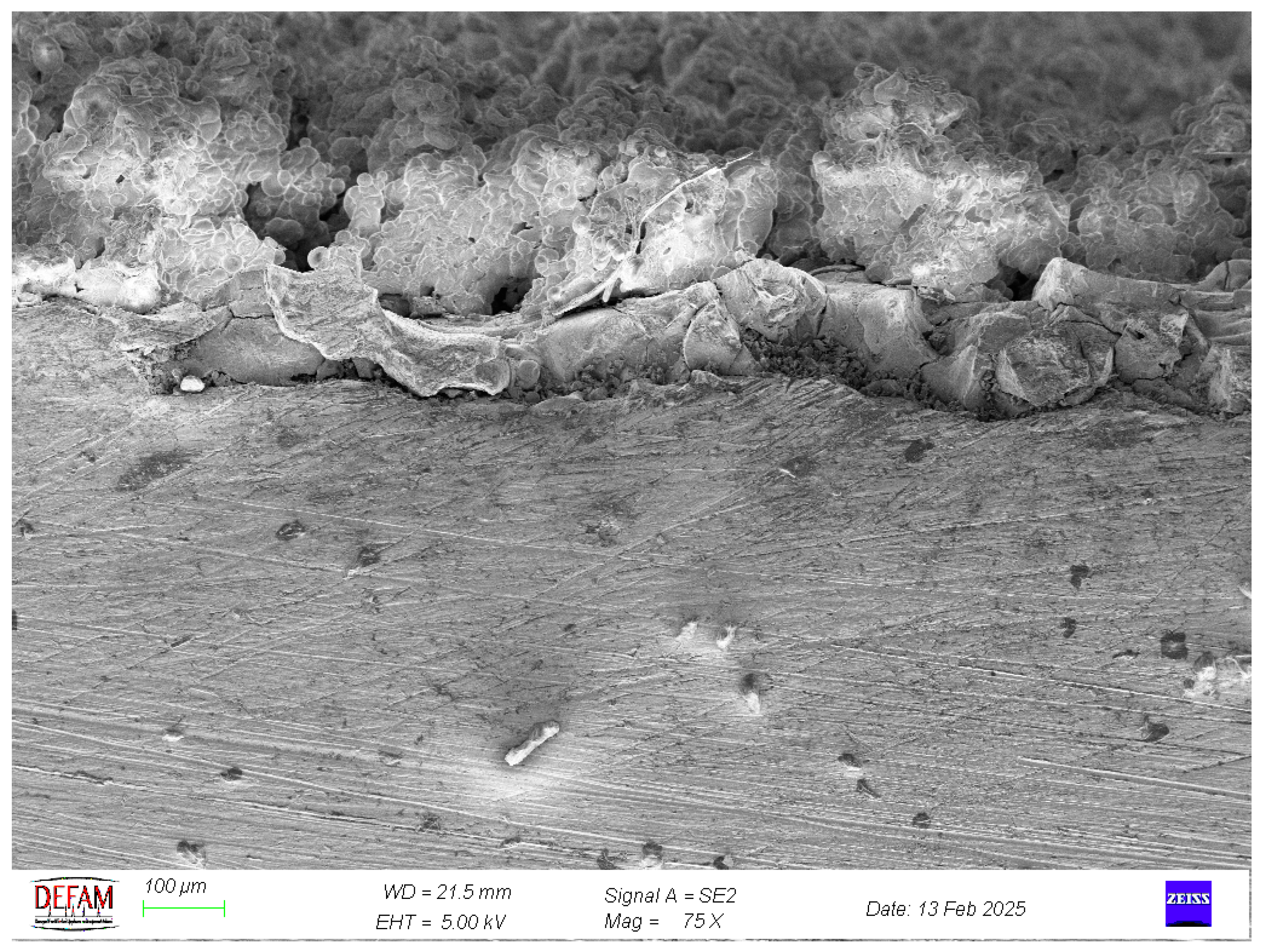
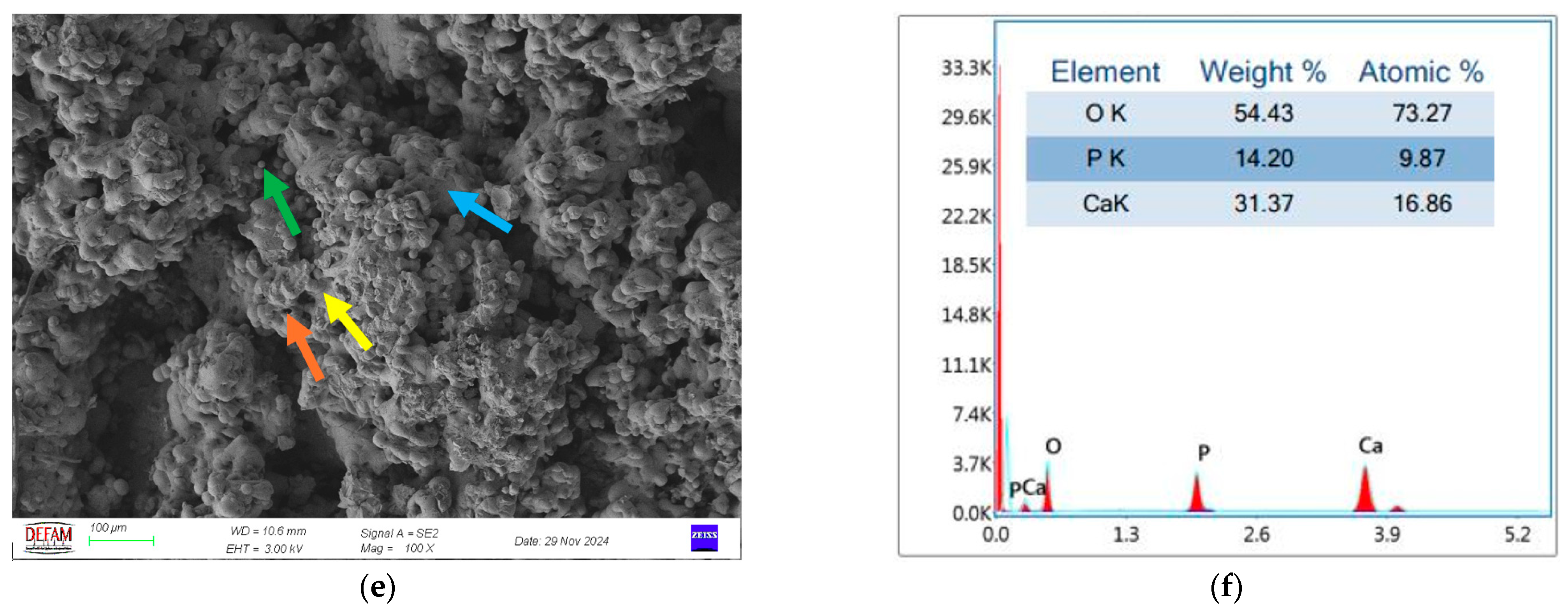
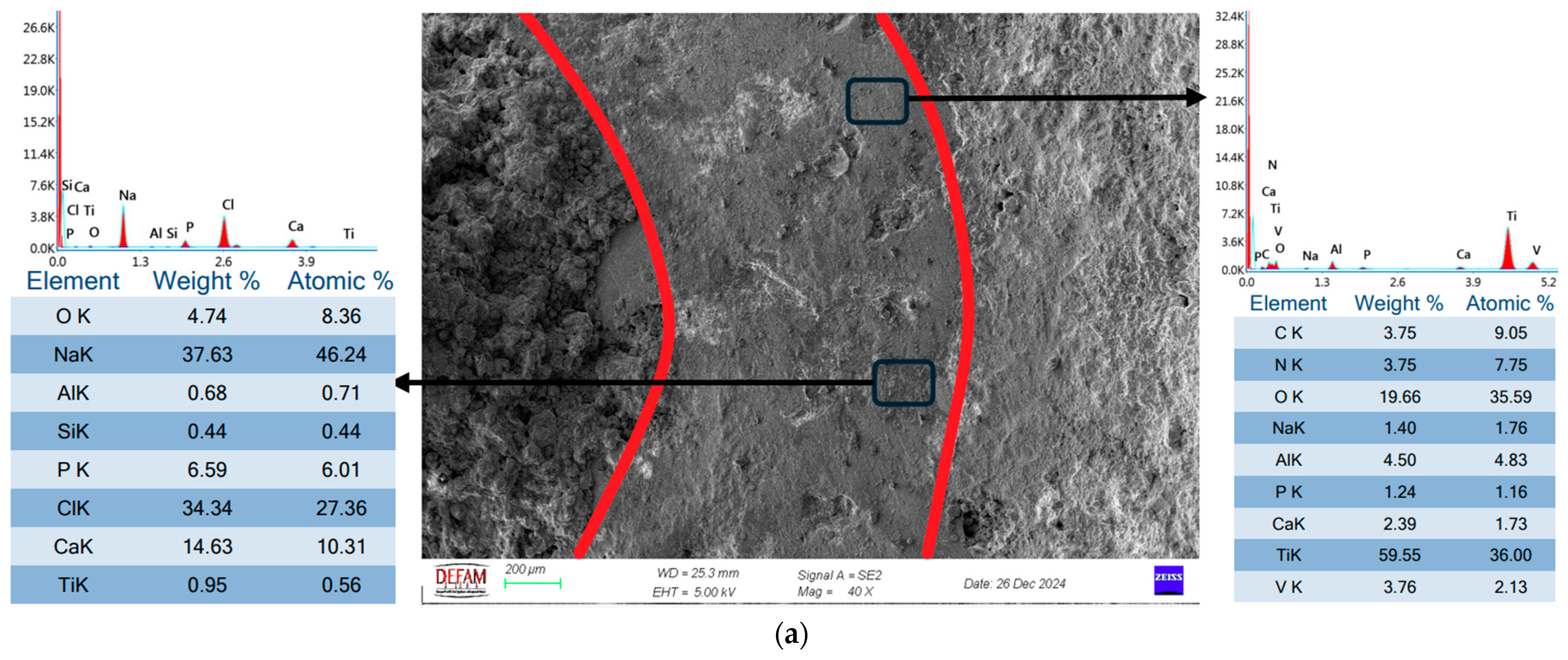
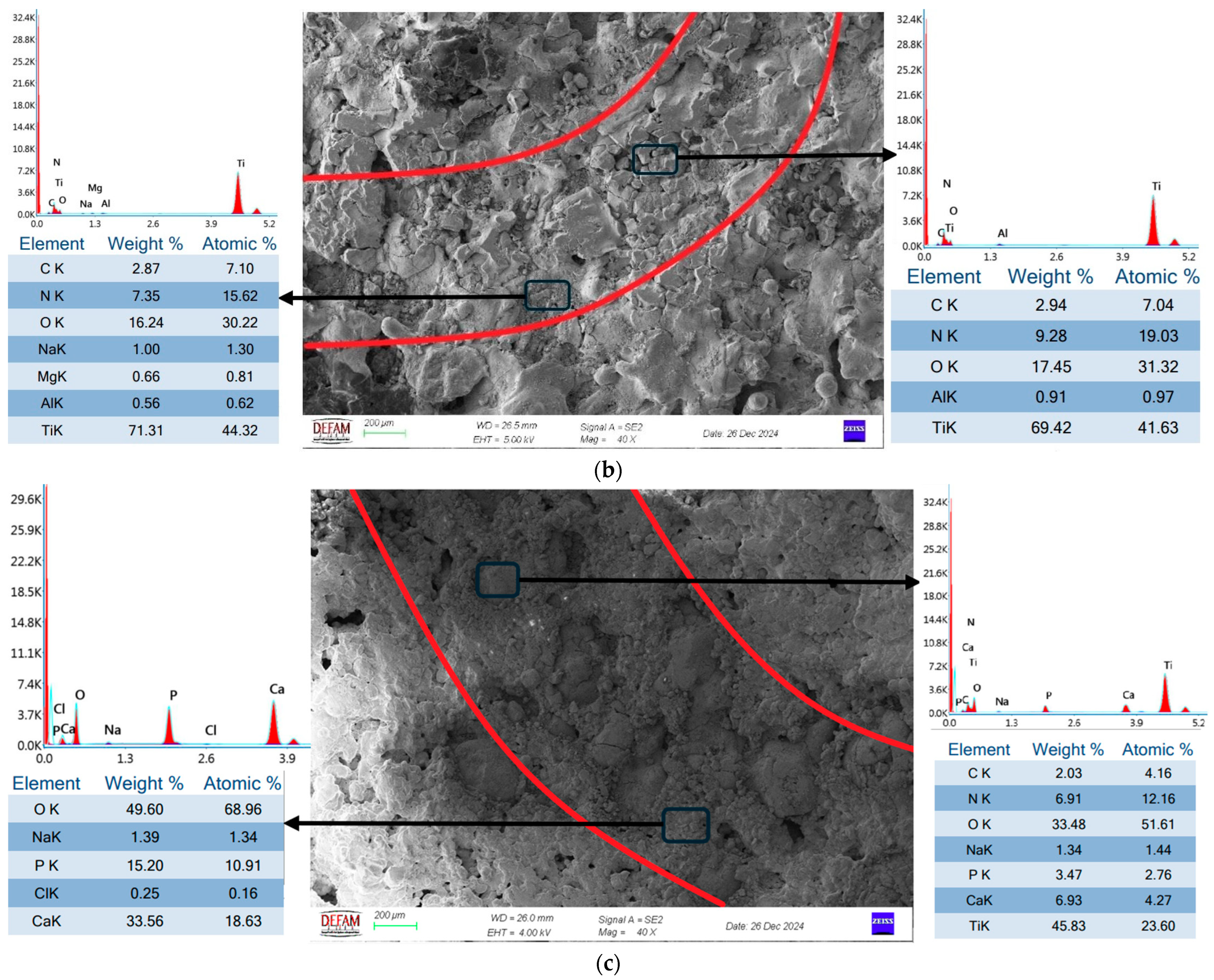
  • SEM and EDS analyses demonstrated that bilayer Ti/HA coatings minimized microcrack formation, thereby enhancing mechanical durability and biocompatibility. The Ca/P ratio of 1.67, determined through EDS analysis, further validated the superior biocompatibility of these coatings.
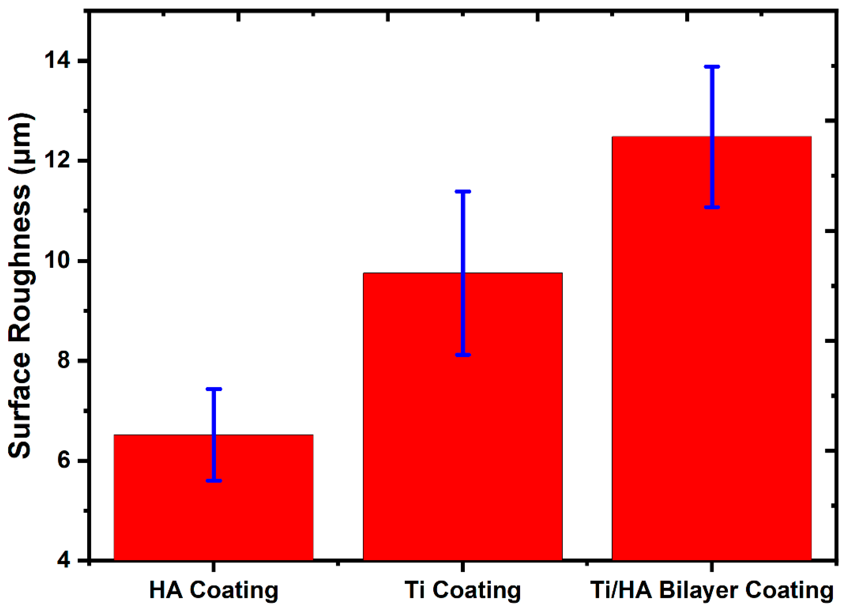
  • Surface roughness analyses revealed that Ti coatings exhibited a roughness value of 9.75 ± 1.63 μm, HA coatings 6.51 ± 0.92 μm, and bilayer Ti/HA coatings 12.48 ± 1.4 μm. The highest surface roughness value observed in bilayer coatings is a critical feature that promotes cellular adhesion, making it advantageous for biomedical applications.
  • In wear tests, Ti coatings displayed the lowest wear rate at 6.32 × 10⁻³ (mm3/N⋅m), while HA coatings exhibited the highest wear rate at 73.83 × 10⁻³ (mm3/N⋅m). Bilayer Ti/HA coatings showed an intermediate wear performance with a wear rate of 34.22 × 10⁻³ (mm3/N⋅m). These findings highlight the contribution of the Ti layer to wear resistance and the role of the bilayer structure in improving durability.
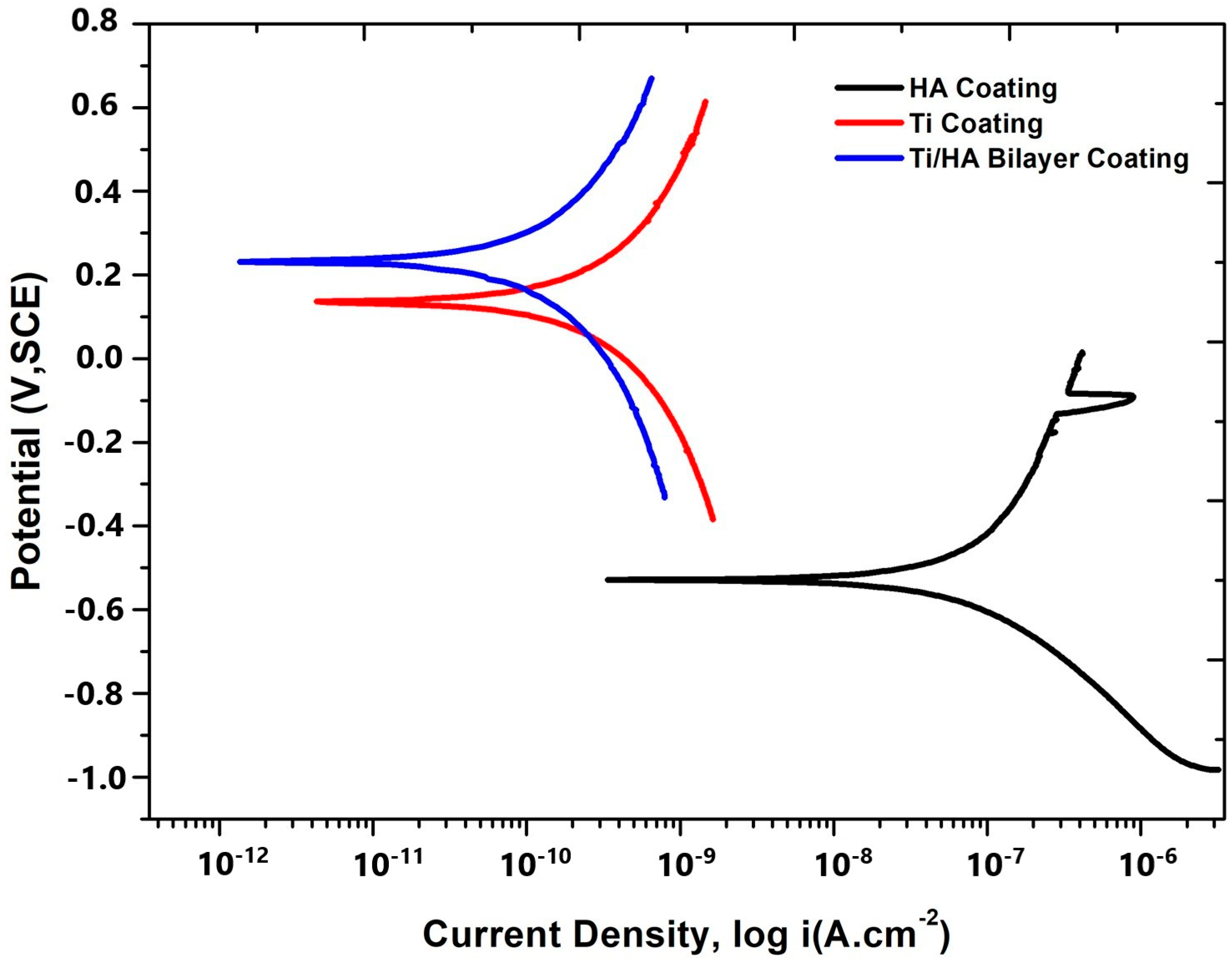
  • In corrosion tests, the bilayer Ti/HA coating demonstrated the best performance with a corrosion current of 0.000153 μA and a corrosion rate of 0.00009 mm/year, Ti coatings exhibited a corrosion current of 0.000351 μA and a corrosion rate of 0.0002 mm/year, showing lower resistance compared to bilayer coatings but higher than HA coatings. HA coatings, with a corrosion current of 0.051 μA and a corrosion rate of 0.003 mm/year, showed the lowest corrosion resistance.
  • In conclusion, bilayer Ti/HA coatings provide significant advantages in terms of biomedical performance by preventing microcrack formation and exhibiting superior wear and corrosion resistance. These findings contribute valuable insights to the limited literature on SLM-based coating applications and serve as a solid foundation for future research.

Author Contributions

Conceptualization, A.İ.B. and İ.A.; methodology, A.İ.B. and İ.A.; investigation, A.İ.B. and İ.A.; writing—original draft preparation, A.İ.B.; writing—review and editing, İ.A.; visualization, A.İ.B. and İ.A.; supervision, İ.A. All authors have read and agreed to the published version of the manuscript.

Funding

This research received no external funding.

Data Availability Statement

Data are contained within the article.

Conflicts of Interest

The authors declare no conflicts of interest.

References

  1. Zhou, L.; Yuan, T.; Li, R.; Tang, J.; Wang, M.; Mei, F. Microstructure and mechanical properties of selective laser melted biomaterial Ti-13Nb-13Zr compared to hot-forging. Mater. Sci. Eng. A 2018, 725, 329–340. [Google Scholar] [CrossRef]
  2. Munir, K.; Biesiekierski, A.; Wen, C.; Li, Y. Selective Laser Melting in Biomedical Manufacturing. In Metallic Biomaterials Processing and Medical Device Manufacturing; Elsevier: Amsterdam, The Netherlands, 2020; pp. 235–269. [Google Scholar] [CrossRef]
  3. Amirnejad, M.; Rajabi, M.; Jamaati, R. Importance of individual evaluation of crystallographic texture and microstructure effects on biocompatibility and corrosion performance of Ti6Al4V alloy. Met. Mater. Int. 2023, 29, 343–356. [Google Scholar] [CrossRef]
  4. Thomas, P.; Weik, T.; Roider, G.; Summer, B.; Thomsen, M. Influence of surface coating on metal ion release: Evaluation in patients with metal allergy. Orthopedics 2016, 39, S24–S30. [Google Scholar] [CrossRef]
  5. Levine, B.R.; Sporer, S.; Poggie, R.A.; Della Valle, C.J.; Jacobs, J.J. Experimental and clinical performance of porous tantalum in orthopedic surgery. Biomaterials 2006, 27, 4671–4681. [Google Scholar] [CrossRef]
  6. Kazemi, M.; Ahangarani, S.; Esmailian, M.; Shanaghi, A. Investigating the corrosion performance of Ti-6Al-4V biomaterial alloy with hydroxyapatite coating by artificial neural network. Mater. Sci. Eng. B 2022, 278, 115644. [Google Scholar] [CrossRef]
  7. Ritwik, A.; Saju, K.K.; Vengellur, A.; Saipriya, P.P. Development of thin-film hydroxyapatite coatings with an intermediate shellac layer produced by dip-coating process on Ti6Al4V implant materials. J. Coat. Technol. Res. 2022, 19, 597–605. [Google Scholar] [CrossRef]
  8. Singh, H.; Kumar, R.; Prakash, C.; Singh, S. HA-based coating by plasma spray techniques on titanium alloy for orthopedic applications. Mater. Today Proc. 2022, 50, 612–628. [Google Scholar] [CrossRef]
  9. Ghadami, F.; Hamedani, M.A.; Rouhi, G.; Saber-Samandari, S.; Dehghan, M.M.; Farzad-Mohajeri, S.; Mashhadi-Abbas, F. The correlation between osseointegration and bonding strength at the bone-implant interface: In-vivo & ex-vivo investigations on hydroxyapatite and hydroxyapatite/titanium coatings. J. Biomech. 2022, 144, 111310. [Google Scholar] [CrossRef]
  10. Kylmäoja, E.; Holopainen, J.; Abushahba, F.; Ritala, M.; Tuukkanen, J. Osteoblast attachment on titanium coated with hydroxyapatite by atomic layer deposition. Biomolecules 2022, 12, 654. [Google Scholar] [CrossRef]
  11. Van Hove, R.P.; Sierevelt, I.N.; van Royen, B.J.; Nolte, P.A. Titanium-nitride coating of orthopaedic implants: A review of the literature. BioMed Res. Int. 2015, 2015, 485975. [Google Scholar] [CrossRef]
  12. Piscanec, S.; Ciacchi, L.C.; Vesselli, E.; Comelli, G.; Sbaizero, O.; Meriani, S.; De Vita, A. Bioactivity of TiN-coated titanium implants. Acta Mater. 2004, 52, 1237–1245. [Google Scholar] [CrossRef]
  13. Liu, D.G.; Zheng, L.; Liang, Y.; Li, H.; Liu, J.Q.; Luo, L.M.; Wu, Y.C. Preparation, biocompatibility, and biotribological properties of TiN-incorporated graphite-like amorphous carbon bio-ceramic composite films. Ceram. Int. 2018, 44, 6810–6816. [Google Scholar] [CrossRef]
  14. Subramanian, B.; Muraleedharan, C.V.; Ananthakumar, R.; Jayachandran, M. A comparative study of titanium nitride (TiN), titanium oxy nitride (TiON) and titanium aluminum nitride (TiAlN), as surface coatings for bio implants. Surf. Coat. Technol. 2011, 205, 5014–5020. [Google Scholar] [CrossRef]
  15. Santecchia, E.; Hamouda, A.M.S.; Musharavati, F.; Zalnezhad, E.; Cabibbo, M.; Spigarelli, S. Wear resistance investigation of titanium nitride-based coatings. Ceram. Int. 2015, 41, 10349–10379. [Google Scholar] [CrossRef]
  16. Kumar, C.S.; Gupta, S.; Sharma, A.K. Characterization of TiN−HA nanocomposite developed through rapid microwave sintering route at high temperature. Int. J. Appl. Ceram. Technol. 2025, e15070. [Google Scholar] [CrossRef]
  17. Croitoru, C.; Roata, I.C.; Pascu, A.; Stanciu, E.M.; Hulka, I.; Stoian, G.; Lupu, N. Photocatalytic surfaces obtained through one-step thermal spraying of titanium. Appl. Surf. Sci. 2020, 504, 144173. [Google Scholar] [CrossRef]
  18. Amin, S.; Panchal, H. A review on thermal spray coating processes. Transfer 2016, 2, 556–563. [Google Scholar]
  19. Kahraman, N.; Gülenç, B. Abrasive wear behaviour of powder flame sprayed coatings on steel substrates. Mater. Des. 2002, 23, 721–725. [Google Scholar] [CrossRef]
  20. Quirama, A.; Echavarria, A.M.; Meza, J.M.; Osorio, J.; Bejarano, G. Improvement of the mechanical behavior of the calcium phosphate coatings deposited onto Ti6Al4V alloy using an intermediate TiN/TiO2 bilayer. Vacuum 2017, 146, 22–30. [Google Scholar] [CrossRef]
  21. Mohseni, E.; Zalnezhad, E.; Bushroa, A.R.; Hamouda, A.M.; Goh, B.T.; Yoon, G.H. Ti/TiN/HA coating on Ti–6Al–4V for biomedical applications. Ceram. Int. 2015, 41, 14447–14457. [Google Scholar] [CrossRef]
  22. Qi, J.; Chen, K.; Teng, S.; Li, X. Preparation and characterisation of gradient HA/TiN/Ti coatings for biomedical applications. Surf. Eng. 2024, 40, 826–837. [Google Scholar] [CrossRef]
  23. Lenis, J.A.; Bejarano, G.; Rico, P.; Ribelles, J.G.; Bolívar, F.J. Development of multilayer Hydroxyapatite-Ag/TiN-Ti coatings deposited by radio frequency magnetron sputtering with potential application in the biomedical field. Surf. Coat. Technol. 2019, 377, 124856. [Google Scholar]
  24. Sargin, F.; Erdogan, G.; Kanbur, K.; Turk, A. Investigation of in vitro behavior of plasma sprayed Ti, TiO2 and HA coatings on PEEK. Surf. Coat. Technol. 2021, 411, 126965. [Google Scholar] [CrossRef]
  25. Suntharavel Muthaiah, V.M.; Rajput, M.; Tripathi, A.; Suwas, S.; Chatterjee, K. Electrophoretic Deposition of Nanocrystalline Calcium Phosphate Coating for Augmenting Bioactivity of Additively Manufactured Ti-6Al-4V. ACS Mater. Au 2021, 2, 132–142. [Google Scholar] [CrossRef]
  26. Yan, C.; Hao, L.; Hussein, A.; Wei, Q.; Shi, Y. Microstructural and surface modifications and hydroxyapatite coating of Ti-6Al-4V triply periodic minimal surface lattices fabricated by selective laser melting. Mater. Sci. Eng. C 2017, 75, 1515–1524. [Google Scholar] [CrossRef]
  27. Luo, J.; Jia, X.; Gu, R.; Zhou, P.; Huang, Y.; Sun, J.; Yan, M. 316L stainless steel manufactured by selective laser melting and its biocompatibility with or without hydroxyapatite coating. Metals 2018, 8, 548. [Google Scholar] [CrossRef]
  28. Stango, S.A.X.; Karthick, D.; Swaroop, S.; Mudali, U.K.; Vijayalakshmi, U. Development of hydroxyapatite coatings on laser textured 316LSS and Ti-6Al-4V and its electrochemical behavior in SBF solution for orthopedic applications. Ceram. Int. 2018, 44, 3149–3160. [Google Scholar] [CrossRef]
  29. Fazel, M.; Salimijazi, H.R.; Shamanian, M.; Minneboo, M.; Modaresifar, K.; van Hengel, I.A.; Zadpoor, A.A. Osteogenic and antibacterial surfaces on additively manufactured porous Ti-6Al-4V implants: Combining silver nanoparticles with hydrothermally synthesized HA nanocrystals. Mater. Sci. Eng. C 2021, 120, 111745. [Google Scholar] [CrossRef]
  30. Yang, Y.; Zhao, M.; Wang, H.; Zhou, K.; He, Y.; Mao, Y.; Xie, D.; Lv, F.; Shen, L. Microstructure and fatigue performance of Ti6Al4V produced by laser powder bed fusion after post-heat treatment. Appl. Sci. 2023, 13, 1828. [Google Scholar] [CrossRef]
  31. EOS GmbH. EOS Titanium Ti64 Material Data Sheet. EOS. 2022. Available online: https://www.eos.info/03_system-related-assets/material-related-contents/metal-materials-and-examples/metal-material-datasheet/titan/ti64/eos_ti64_9011-0014_9011-0039_m290_mds_06-22_en.pdf (accessed on 13 March 2025).
  32. Kumrular, B.; Cicek, O.; Dağ, İ.E.; Avar, B.; Erener, H. Evaluation of the Corrosion Resistance of different types of Orthodontic fixed Retention Appliances: A preliminary Laboratory Study. J. Funct. Biomater. 2023, 14, 81. [Google Scholar] [CrossRef]
  33. Alontseva, D.L.; Abilev, M.B.; Zhilkashinova, A.M.; Voinarovych, S.G.; Kyslytsia, O.N.; Ghassemieh, E.; Russakova, A.; Łatka, L. Optimization of hydroxyapatite synthesis and microplasma spraying of porous coatings onto titanium implants. Adv. Mater. Sci. 2018, 18, 79–94. [Google Scholar] [CrossRef]
  34. Guillem-Marti, J.; Cinca, N.; Punset, M.; Cano, I.G.; Gil, F.J.; Guilemany, J.M.; Dosta, S. Porous titanium-hydroxyapatite composite coating obtained on titanium by cold gas spray with high bond strength for biomedical applications. Colloids Surf. B Biointerfaces 2019, 180, 245–253. [Google Scholar] [PubMed]
  35. Khor, K.A.; Li, H.; Cheang, P. Impact formation and microstructure characterization of thermal sprayed hydroxyapatite/titania composite coatings. Biomaterials 2003, 24, 2233–2243. [Google Scholar] [CrossRef]
  36. Sun, Y.; Zhang, X.; Luo, M.; Hu, W.; Zheng, L.; Huang, R.; Greven, J.; Hildebrand, F.; Yuan, F. Plasma Spray vs. Electrochemical Deposition: Toward a Better Osteogenic Effect of Hydroxyapatite Coatings on 3D-Printed Titanium Scaffolds. Front. Bioeng. Biotechnol. 2021, 9, 705774. [Google Scholar] [CrossRef]
  37. Farnoush, H.; Muhaffel, F.; Cimenoglu, H. Fabrication and characterization of nano-HA-45S5 bioglass composite coatings on calcium-phosphate containing micro-arc oxidized CP-Ti substrates. Appl. Surf. Sci. 2015, 324, 765–774. [Google Scholar] [CrossRef]
  38. Ansari, Z.; Kalantar, M.; Soriente, A.; Fasolino, I.; Kharaziha, M.; Ambrosio, L.; Raucci, M.G. In-situ synthesis and characterization of chitosan/hydroxyapatite nanocomposite coatings to improve the bioactive properties of Ti6Al4V substrates. Materials 2020, 13, 3772. [Google Scholar] [CrossRef] [PubMed]
  39. Qi, J.; Yang, Y.; Zhou, M.; Chen, Z.; Chen, K. Effect of transition layer on the performance of hydroxyapatite/titanium nitride coating developed on Ti-6Al-4V alloy by magnetron sputtering. Ceram. Int. 2019, 45, 4863–4869. [Google Scholar] [CrossRef]
  40. Kazemi, M.; Ahangarani, S.; Esmailian, M.; Shanaghi, A. Investigation on the corrosion behavior and biocompatibility of Ti-6Al-4V implant coated with HA/TiN dual layer for medical applications. Surf. Coat. Technol. 2020, 397, 126044. [Google Scholar] [CrossRef]
  41. Kobayashi, A. Formation of TiN coatings by gas tunnel type plasma reactive spraying. Surf. Coat. Technol. 2000, 132, 152–157. [Google Scholar] [CrossRef]
  42. Yanchun, D.; Dianran, Y.; Jining, H.; Jianxin, Z.; Lisong, X.; Xiangzhi, L. Studies on nanocrystalline TiN coatings prepared by reactive plasma spraying. J. Nanomater. 2008, 2008, 690951. [Google Scholar] [CrossRef]
  43. Dorozhkin, S.V. Bioceramics of calcium orthophosphates. Biomaterials 2010, 31, 1465–1485. [Google Scholar] [CrossRef] [PubMed]
  44. Gross, K.A.; Berndt, C.C. Biomedical application of apatites. Rev. Mineral. Geochem. 2002, 48, 631–672. [Google Scholar] [CrossRef]
  45. Sopyan, I.; Mel, M.; Ramesh, S.; Khalid, K.A. Porous hydroxyapatite for artificial bone applications. Sci. Technol. Adv. Mater. 2007, 8, 116. [Google Scholar] [CrossRef]
  46. Levingstone, T.J. Optimisation of Plasma Sprayed Hydroxyapatite Coatings. Ph.D. Thesis, Dublin City University, Dublin, Ireland, 2008. [Google Scholar]
  47. Albrektsson, T.; Johansson, C. Osteoinduction, osteoconduction and osseointegration. Eur. Spine J. 2001, 10, S96–S101. [Google Scholar] [CrossRef]
  48. Tirrell, M.; Kokkoli, E.; Biesalski, M. The role of surface science in bioengineered materials. Surf. Sci. 2002, 500, 61–83. [Google Scholar] [CrossRef]
  49. Roşu, R.A.; Şerban, V.A.; Bucur, A.I.; Dragoş, U. Deposition of titanium nitride and hydroxyapatite-based biocompatible composite by reactive plasma spraying. Appl. Surf. Sci. 2012, 258, 3871–3876. [Google Scholar] [CrossRef]
  50. Khethier Abbass, M.; Jasim, A.N.; Jasim, M.; Khashan, K.S.; Issa, M.J. Improving Bio Corrosion Resistance of the Single Layer of Nano Hydroxyapatite and Nano YSZ Coating on the Ti6Al4V Alloy Using Electrophoretic Deposition. Solid State Technol. 2020, 63, 18584–18597. [Google Scholar]
  51. Coşkun, M.İ.; Karahan, İ.H.; Yücel, Y.; Golden, T.D. Optimization of electrochemical step deposition for bioceramic hydroxyapatite coatings on CoCrMo implants. Surf. Coat. Technol. 2016, 301, 42–53. [Google Scholar] [CrossRef]
  52. Drevet, R.; Aaboubi, O.; Benhayoune, H. In vitro corrosion behavior of electrodeposited calcium phosphate coatings on Ti6Al4V substrates. J. Solid State Electrochem. 2012, 16, 3069–3077. [Google Scholar] [CrossRef]
  53. Butt, M.S.; Afsheen, R.; Saeed, H.; Javed, N.; Ghaffar, A. Promoting Surgical Pin Performance: Chitosan and Hydroxyapatite-Based Nano-composite Coatings for Antimicrobial and Corrosion Protection. Results Eng. 2024, 25, 103714. [Google Scholar] [CrossRef]
  54. Corona-Gomez, J.; Sandhi, K.K.; Yang, Q. Wear and corrosion behaviour of nanocrystalline TaN, ZrN, and TaZrN coatings deposited on biomedical grade CoCrMo alloy. J. Mech. Behav. Biomed. Mater. 2022, 130, 105228. [Google Scholar] [CrossRef] [PubMed]
  55. Hikov, T.; Nikolova, M.; Petrov, P. Effect of TiN/TiO2 multilayer coatings on the properties of stainless steel for biomedical applications. J. Phys. Conf. Ser. 2020, 1492, 012029. [Google Scholar] [CrossRef]
Figure 1. Cross-sectional SEM image of bilayer Ti/HA coatings applied to PBF-LB Ti6Al4V.
Crystals 15 00316 g001
Figure 2. The 100 X SEM images and EDS analysis results of biomedical coatings applied to PBF-LB Ti6Al4V (a,b) Ti coating; (c,d) HA coating; (e,f) Ti/HA bilayer coating (yellow arrows: microcracks, red arrows: macrocracks, orange arrows: pores, purple arrows: semi-melted particles, green arrows: unmelted particles, blue arrows: splats).
Crystals 15 00316 g002aCrystals 15 00316 g002b
Figure 3. XRD analysis results.
Crystals 15 00316 g003
Figure 4. Surface roughness results of coatings.
Crystals 15 00316 g004
Figure 5. Electrochemical potential curves of PBF-LB Ti6Al4V applied coatings.
Crystals 15 00316 g005
Figure 6. Friction coefficient and wear rate results of biomedical coatings applied to PBF-LB Ti6Al4V.
Crystals 15 00316 g006
Figure 7. SEM-EDS analyses of wear tracks from in vitro wear testing of biomedical coatings applied to PBF-LB Ti6Al4V: (a) Ha coating; (b) Ti coating; and (c) Ti/HA bilayer coating.
Crystals 15 00316 g007aCrystals 15 00316 g007b
Table 1. Chemical composition of PBF-LB Ti6Al4V powders [31].
Ti
(Wt.%)
Al
(Wt.%)
V
(Wt.%)
O (ppm)N
(ppm)
C
(ppm)
H
(ppm)
Fe
(ppm)
Density (g/cm3)
Balance5.5–6.753.5–4.5<2000<500<800<150<30004.41
Table 2. Coating process parameters.
Spraying ParameterValue (For HA Coating)Value (for Ti Coating)
Propane flow rate (L·min−1)1719
Oxygen flow rate (L·min−1)2023
Air flow rate (L·min−1)1010
Spray distance (cm)4040
Spray angle90°90°
Table 3. Corrosion parameters from the potentiodynamic polarization curves of PBF-LB Ti6Al4V applied coatings.
Coating TypeEcorr (mV)Beta a (mV/Decade)Beta cİcorr (μA)Corrosion Rate (mm/Year)
HA Coating−527.586390.4237.40.0510.003
Ti Coating137.658726.77050.0003510.0002
Ti/Ha Bilayer Coating237.259648.97130.0001530.00009
Disclaimer/Publisher’s Note: The statements, opinions and data contained in all publications are solely those of the individual author(s) and contributor(s) and not of MDPI and/or the editor(s). MDPI and/or the editor(s) disclaim responsibility for any injury to people or property resulting from any ideas, methods, instructions or products referred to in the content.

