Article

Electrophoretic Co-Deposition of Chitosan and Cu-Doped Bioactive Glass 45S5 Composite Coatings on AISI 316L Stainless Steel Substrate for Biomedical Applications

by
Sayed Mohammad Reza Mahmoudabadi
,
Abbas Bahrami
*,
Mohammad Saeid Abbasi
,
Mojtaba Rajabinezhad
,
Benyamin Fadaei Ardestani
and
Farnaz Heidari Laybidi
Department of Materials Engineering, Isfahan University of Technology, Isfahan 84156-83111, Iran
*
Author to whom correspondence should be addressed.
Crystals 2025, 15(6), 549; https://doi.org/10.3390/cryst15060549
Submission received: 7 May 2025 / Revised: 24 May 2025 / Accepted: 7 June 2025 / Published: 8 June 2025

Abstract

:
The growing demands for highly functional biomedical implants necessitate introducing innovative and easy-to-apply surface functionalization techniques, especially when it comes to stainless steel substrates. This study investigated the co-deposition of chitosan and Cu-doped bioactive glass on AISI 316L steel surfaces, with the latter providing a matrix in which fine bioactive glass powders are distributed. Cu-doping into the matrix of bioactive glass was conducted to assess its influence on the bioactivity, antibacterial properties, and structural integrity of the coating. The microstructure, mechanical properties, phase composition, and surface roughness of coated specimens were investigated through a scanning electron microscope (SEM), X-ray diffraction analysis (XRD), energy-dispersive X-ray spectroscopy (EDS), inductively coupled plasma (ICP), contact angles, adhesion tensile tests, and laser profilometry analyses. Results of adhesion tests indicated that Cu addition did not have a major implication for the mechanical properties of the coating layers. Results also revealed that the Cu-doped bioactive glass featured a hydrophilic and a rather uneven surface, both being upsides for biomedical properties. The cytotoxicity and antibacterial assessments showed promising cell viability and antibacterial properties of the deposited coatings.

1. Introduction

Various factors, such as infections and poor implant/tissue integration can cause the failure of orthopedic implants. The surface is an absolutely crucial interface that controls the mechanical bonding of the tissue/implant and the susceptibility of the implant to the microbial infections and biofilm formation [1,2,3]. The biocompatibility of the implant surface is also a fundamental issue for the bone tissue regeneration in both temporary and permanent orthopedic implants. Stainless steel grades, which are usually used for biomedical purposes, are known to have rather low biocompatibility and relatively weak osseointegrations [4]. In addition, stainless steel alloys contain some harmful elements and ions that can adversely implicate the health of surrounding tissues. Thus, it is crucial to apply biocompatible and non-toxic coatings on stainless steel orthopedic implants to minimize release of toxic elements and to enhance implant/tissue integrations. With that said, surface modification of stainless steels with bioactive layers appears to be extremely important. Surface infections and the lack of integration and its associated implant loosening can compromise the implant functioning and its stability inside the body, in which case a revision surgery will be required, a costly and of course a painful operation [5,6]. Since the initial introduction, Bioglass®45S5 has shown significant potential as a material candidate for biomedical implants and a promising substitution for autografts [7]. Bioactive glass grades are used in biomedical applications due to their bioactivity and biocompatibility, making them excellent choices for bioactive coatings on implants [8,9]. Setting a bioactive glass layer at the tissue/implant interface results in a smooth replacement of damaged bone tissues, resulting in successful tissue regeneration with minimum associated risks [10,11,12]. Furthermore, bioactive glass coating layers can enhance the integration of metal implants into the tissue by inducing apatite crystal formation at the implant/tissue interface. They can also minimize the corrosion rate and ion release at the implant interface [13]. Bioactive glass 45S5, known as one of the most-used grades, is composed of 45.0 SiO2, 24.5 CaO, 24.5 Na2O, and 6.0 P2O5 (in wt.%) [14]. When the bioactive glass comes into contact with body fluids, it releases Ca and P ions, creating strong chemical/biological connections with the hard tissues in the living organisms [15]. Therefore, bioactive glass coatings can improve the bioactivity/biocompatibility and henceforward the performance of commercial implants. When successfully applied, bioactive glass coatings are therefore expected to enhance the implant/tissue integration [16]. These characteristics make bioactive glass-containing coating a promising material for various applications, including biomedical applications.
Copper oxide (CuO) is a transition metal oxide that has lately gained loads of attentions due to its encouraging properties, such as its narrow bandgap, its non-toxic nature, and its relatively low costs [17]. CuO also demonstrates excellent antibacterial performance in various aqueous environments, including biological ones [18,19,20]. CuO nanoparticles can release copper ions, which can affect the bacterial metabolism and enzyme functioning of bacteria, with both cases being important when it comes to antibacterial properties. CuO nanoparticles have exhibited proven antibacterial activities against various Gram-positive and Gram-negative bacteria, such as Staphylococcus aureus, Escherichia coli, Pseudomonas aeruginosa, and Bacillus subtilis [21,22,23,24]. CuO nanoparticles can therefore be used as antibacterial entities in various biomedical applications, such as wound healing, water treatment, food packaging, and biomedical implants [25]. In the latter case, the antibacterial mechanism of CuO is believed to be due to the generation of reactive oxygen species (ROS) and physical damages to the structure of bacteria. ROS interactions with microorganisms results in lipid peroxidation at the outer cell wall of living organisms and subsequently in the damage at the cell walls and the DNA inside the cell, with both ending up in cell demise [26,27,28,29].
Natural polymers are increasingly used in biomedical applications due to their chemical resemblance to the extracellular structure of tissues, their attractive biological characteristics, and their degradation rates, regulated by cells or enzymes [30]. Chitosan is one of the most abundant natural polysaccharides in the world, with unique features and properties that makes it suitable in various fields, such as the food and medical sectors [31,32,33,34]. Chitosan is obtained from the de-acetylation of chitin, which is a biopolymer that forms the outer covering of crustacea, the protective layer of insects, and the cell walls of algae and fungi [35]. Chitosan is a cationic polysaccharide and a promising natural biopolymer for applications in tissue engineering, biocompatible coating layers, and drug delivery systems. The main characteristics of chitosan include its biodegradability, biocompatibility, and ability to immobilize proteins, nucleic acids, and virus drugs. Also, chitosan stimulates cellular attachments, and it does have some level of antibacterial responses, making it an essential component in wound dressing applications. If applied as an external coating layer, chitosan is expected to have chemical resistance and to provide corrosion protection for the substrate. Last but not least is its relatively easy deposition, its good mechanical properties, and its low cost. Due to these superior properties, there is growing attention on using chitosan-based composite coating layers in different biomedical applications [31,36].
In recent years, various coating materials such as hydroxyapatite, bioactive glass, TiO2, ZnO, and polymer-based composites have been extensively reported for improving the bioactivity and antibacterial performance of metallic implants [37]. Each material presents specific advantages: for instance, hydroxyapatite is highly osteoconductive, while TiO2 and ZnO offer notable antibacterial properties. However, challenges such as limited mechanical stability, weak adhesion, or insufficient antibacterial efficacy under physiological conditions still persist. Therefore, developing multifunctional coatings that simultaneously enhance bioactivity, corrosion resistance, and antibacterial performance remains a crucial need [37,38,39,40]. This study advances the current state-of-the-art research by proposing a novel chitosan/Cu-doped 45S5 bioactive glass composite coating on AISI 316L stainless steel using electrophoretic deposition (EPD). The innovation lies in the synergistic integration of three key components: (1) chitosan, a natural biopolymer with intrinsic biocompatibility and mild antibacterial activity; (2) 45S5 bioactive glass, known for its attribution in promoting apatite formation and bone regeneration; and (3) copper, known for its antibacterial properties. Unlike previous studies that focus on binary systems (e.g., chitosan with HAp or BG alone), the current approach incorporates copper-doped bioactive glass into a chitosan matrix, aiming to achieve a bioactive, antibacterial, and mechanically robust composite coating. Moreover, EPD offers a low-cost, scalable, and versatile technique that allows for uniform co-deposition of biopolymers and nanoparticles, enabling fine control over coating morphology and thickness. This paper provides insights into the EPD parameters and their effects on the microstructure, adhesion, and biological response of chitosan/Cu-BG coatings, thereby contributing to the development of surface modification approaches for orthopedic implants.

2. Materials and Methods

2.1. Materials

Bioactive glass 45S5 bioglass (Na2O, CaO, SiO2) powders (Araz Powder Pajouhan Co., Esfahan, Iran) with a particle size of approximately 200–300 nanometers, chitosan (Mystic Moments, Fordingbridge UK), acetic acid (Merck, 100%, Darmstadt, Germany), ethanol (Merck, 100%, Darmstadt, Germany), and distilled water (Sky, Esfahan, Iran) have been used. The substrate that was used for the deposition, was made of AISI 316L stainless steel. Then, bioactive glass powder, doped with 5 wt.% Cu (5CB), 10 wt.% Cu (10CB), and 20 wt.% Cu (20CB) powders (Araz Powder Pajouhan Co., Esfahan, Iran) were used to investigate the effects of Cu addition.

2.2. Sample Preparation Methods

To prepare the substrate, AISI 316L steel substrate was first carefully grinded from 80 to 800 grit to prepare the surface. Then, to clean the surface of samples, samples were sonicated for 20 min. This way, the substrate was prepared for the deposition. To synthesize bioactive glass powders, a solution was prepared by mixing 2.25 mL tetraethyl orthosilicate (TEOS), 0.15 mL triethyl phosphate (TEP), and 25 mL ethanol (Solution A). Separately, 12.38 mL deionized water, 8.12 mL ethanol, and 5.5 mL ammonia were combined to form a separate solution. The latter solution was rapidly added to the first solution under continuous stirring, and the mixture was stirred for 10 min to ensure homogeneity. To dope copper into the structure of bioactive glass, subsequently, 1.35 g calcium nitrate, 0.84 g sodium nitrate, and 0.8 g copper nitrate were introduced into the mixture, followed by an additional 2 h of stirring. The resulting mixture was filtered and subsequently dried in an oven for 24 h. Thereafter, it underwent heat treatment at 700 °C for a duration of 3 h in a furnace (Carbolite Rapid Heating Chamber Furnace-RWF, Neuhausen, Germany) under an air atmosphere at a heating rate of 10 °C/min to complete the synthesis process.
In order to do the deposition process, 40 mL of distilled water was mixed with 0.2 g of bioactive glass, then 1/2 mL of acetic acid was added, and the solution was stirred for 2 min on a stirrer until a homogeneous mixture was attained. Then, the mixture was put in the ultrasonic device for 20 min to disperse particles well. Subsequently, the mixture was put on the stirrer again, and while blending, 0.03 g of chitosan was added to the mixture. Then, 60 mL of ethanol was added to the mixture immediately, and the final solution was left to blend well for 10 min. After 10 min, the mixture was ready for electrophoresis. After the preparation of the solution and the substrate, initially the deposition of chitosan was carried out by the electrophoretic method. In this method, the distance between the cathode and the anode was kept at 1 cm. The voltage and duration of this process were changed as shown in Table 1. Various test parameters were chosen to find out the optimal coating parameters.
The best time and voltage for the coating deposition were determined based on their adhesion strength test using the ASTM-D3359 standard [41]. Results showed that the 10 min deposition time with the voltage of 20 volts ends up in the best-coated samples in terms of adhesion strength.

2.3. Characterizations

Surface wettability, indicative of hydrophilicity, was evaluated by measuring the contact angle between the sample surfaces and water droplets using a goniometer (Ezdiad Bardasht Fars, IFT-CA, Sari, Iran). Additionally, the cross-sectional morphology of the samples was examined using scanning electron microscopy (SEM, Philips30 Xl, Eindhoven, the Netherlands). Energy dispersive spectroscopy (EDS) and elemental mapping analyses were also conducted in this study. The tape test method, one of the most common adhesion tests according to the ASTM-D3359 standard, was also used to investigate the adhesion strength of the coating. Following the ASTM-D3359 standard, the calculation of the percentage of detached coating after the test was performed using Image J software (2015). Surface laser profilometry (kahroba, LPM-D1/Iran-Resolution: in the direction x: 80 µm, in the direction y: 100 µm, Tehran, Iran) was utilized to examine the surface roughness of the coating. An MTT (3-(4,5-dimethylthiazol-2-yl)-2,5-diphenyltetrazolium bromide) assay was used to evaluate the biocompatibility and cell viability of the synthesized samples. This test measures the change in the MTT color in the mitochondria of live cells, shifting from yellow to purple formazan crystals, providing a photometric estimate of the number of viable cells.
X-ray diffraction (XPERT-PRO Diffractometer, Philips, Eindhoven, the Netherlands) with a wavelength of 0.15406 nm was employed to analyze the crystallographic structure of both the powders and the deposited layers. A stereomicroscope was also utilized to examine the surface structure of the deposited layers.

2.4. Antibacterial Evaluations

Given that bacterial inflammation is one of the primary causes of implant failures and that it poses major health risks, it is important to investigate antibacterial properties of coated specimens. Colony counting remains one of the most commonly employed techniques in microbiology for quantifying bacterial populations, wherein the number of viable bacterial cells within a defined volume is determined to estimate cell density. By placing a specific volume of cultured cells on a Petri dish containing a growth medium, the number of cultured cells can be counted. In this investigation, the representative Gram-positive bacteria Staphylococcus aureus 6538ATCC and Gram-negative bacteria Escherichia coli 10536ATCC were prepared at a specific concentration according to the half McFarland concentration in normal saline solution. In this case, samples were incubated for 24 h at a temperature of 37 ± 2 °C. The culture medium combined with the bacterial suspension served as the positive control for both bacterial strains. Subsequently, serial dilution was carried out on each sample. Specific amounts of dilution were spread onto agar culture media and incubated for 24 h at a temperature between 37 and 39 °C. Following incubation, the total colony count was determined at each concentration, and any changes relative to the control sample were assessed [42].

2.5. In Vitro and Biocompatibility Studies

After the preliminary tests, the biocompatibility of the samples was evaluated by immersing specimens in simulated body fluid (SBF) at 37 °C for a duration of four weeks (28 days) to monitor the formation of hydroxyapatite crystals. At weekly intervals, a sample was taken from the solution and its surface was analyzed using scanning electron microscopy (SEM) to detect and characterize precipitated crystals over the surface. Moreover, inductively coupled plasma (ICP) analysis was conducted on the solution to quantify the calcium and phosphorus concentrations.

3. Results and Discussion

3.1. Topography of Coating Layers

Figure 1 demonstrates stereomicroscopic images of chitosan-Cu-doped bioactive glass coatings with different copper percentages (all coating layers were deposited with a voltage of 20 V for 10 min). Greenish spots are bumps, ascribed to agglomerated Cu-doped bioactive glass, while the blue areas are from the chitosan matrix. As can be seen, in all cases the distribution of green spots is homogeneous, inferring that the deposition takes place in a similar regime in all compositions and that the deposition results in a rather rough top layer, with hardly any abnormal particle aggregations.
In order to further evaluate the topography of coating layers, Ra, Rq, Rz, and Rt of all coating layers were compared. Ra (being a very important surface characteristic) for the Chit-5CB sample is almost 3 µm, while those for the Chit-10CB and Chit-20CB samples are almost 4.3 µm, implying that bioactive glass addition tends to make the surface slightly rougher (see Figure 2)—a finding very much in agreement with stereographic images. Additionally, the thicknesses of the coatings are 250 µm for Chit-BG, 310 µm for the Chit-5CB sample, 410 µm for the Chit-10CB sample, and 470 µm for the Chit-20CB sample. By comparing all these parameters, it can be concluded that with an increase in the percentage of copper as dopant, the thickness of the coatings increases. Additionally, based on the results of Ra shown in Figure 2, it can be observed that the surface roughness does not change significantly with the increase in the percentage of copper. It appears that when the percentage of copper in the bioactive glass increases, the ionic conductivity of the solution increases, resulting in an increased mass flow, and accordingly, an increase in coating thickness [43,44].

3.2. Wettability Evaluation of Coating Layers
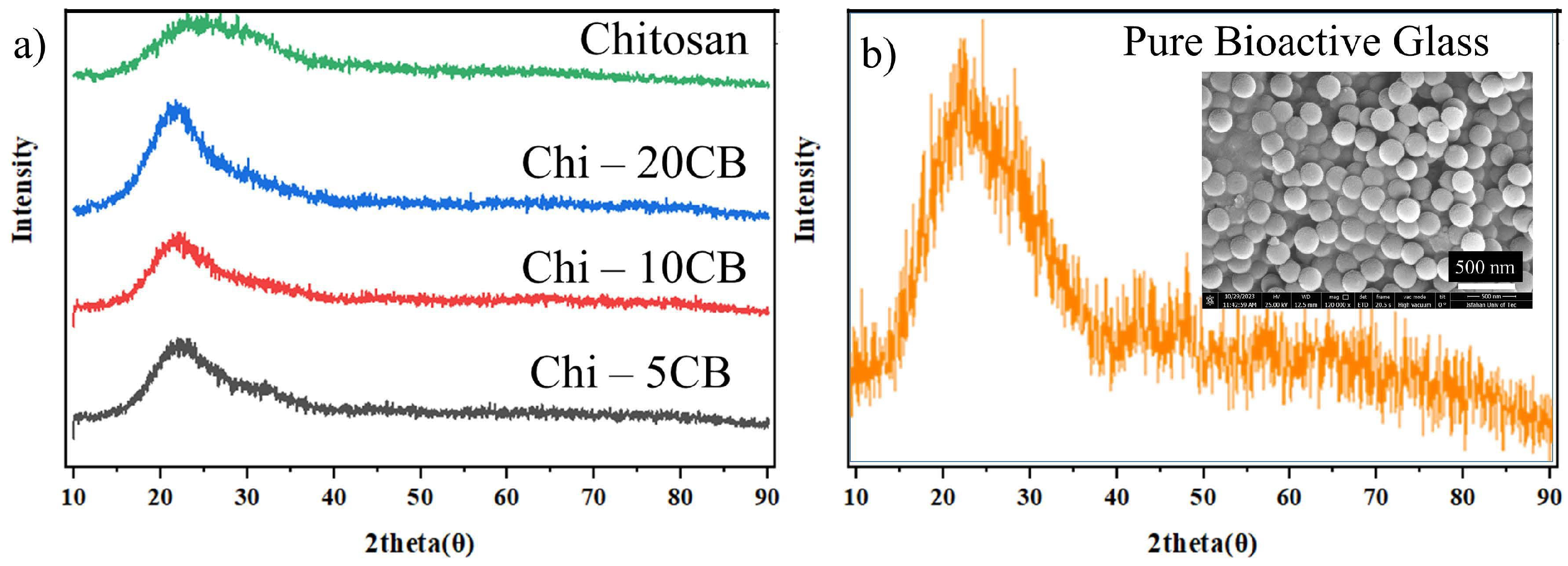
The evaluation of wettability serves as a fundamental scientific investigation into the intrinsic properties of biomaterials. According to Figure 3, the average contact angle of the Chitosan-5CB sample (bioactive glass, coating with 5% copper content) is almost the same as that of the base chitosan layer. The contact angle slightly increases when the content of copper in the bioactive glass is increased to 10%, followed by a decrease down to values less than 90° in samples containing 20% copper. Given that in all cases the contact angle is close to 90 degrees, one can conclude that deposited layers are hydrophilic and that the ceramic content of the coating has marginal implications for the wettability of deposited coatings. When it comes to the wettability of a coated layer, the relationship between the surface roughness and contact angle has a prominent role. As shown earlier, the surface topographies of coatings in all samples are pretty much similar, and that justifies the similarity of the wetting angles in deposited layers [43].

3.3. XRD Results

XRD patterns taken from composite coating samples as well as that of pure bioactive glass are shown in Figure 4. As can be seen, the pure bioactive glass has an amorphous structure. Also, as expected, the XRD pattern of chitosan does not contain any sharp peaks. Therefore, adding bioactive glass to the chitosan matrix ends up in a typical amorphous pattern. There are hardly any noticeable differences between the XRD patterns of composite samples with different contents of copper.

3.4. Adhesion Test

The adhesion strength evaluation of three coatings, Chit-5CB, Chit-10CB, and Chit-20CB, was evaluated as per the ASTM-D3359 standard. According to this standard, the adhesion strength can be evaluated based on the detached area after the pull-off test, where the highest adhesion strength is classified as 5B, while the weakest one is 0B (see Table 2). Results of adhesion strength show that the highest adhesion strength was attained in sample Chit-5CB, with its coating classification being 3B (see Figure 5). Given that the composition is almost similar in all cases, the observed increase in the strength can be ascribed to the surface roughness. It appears that the surface roughness in samples Chit-10CB and Chit-20CB is too much to create an effective mechanical inter-lock. It is likely that ceramic-based surface bumps create stress concentration and mechanically weak spots, which are prone for detachments, thus leading to a reduction in coating adhesion. The increase in the roughness above a certain level might also create some gaps at the coating/substrate interface, which also has an adverse attribution to the adhesion strength [45,46,47]. Given that the adhesion strength is an absolutely important factor and that coatings with 1B and 2B classifications have very weak adhesion strength, sample Chit-5CB was chosen as the optimum sample and biological/biomedical experiments were conducted on this composition only.

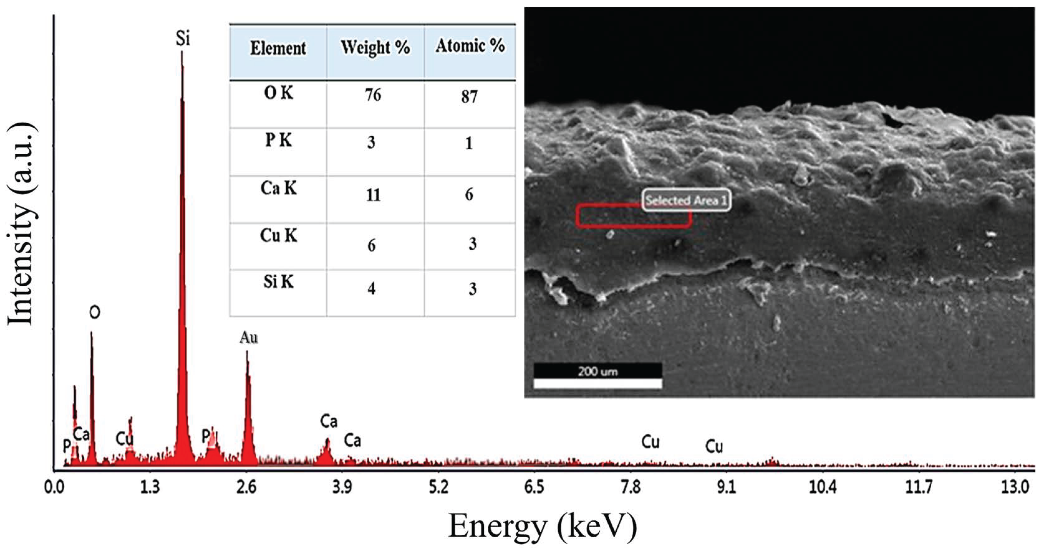
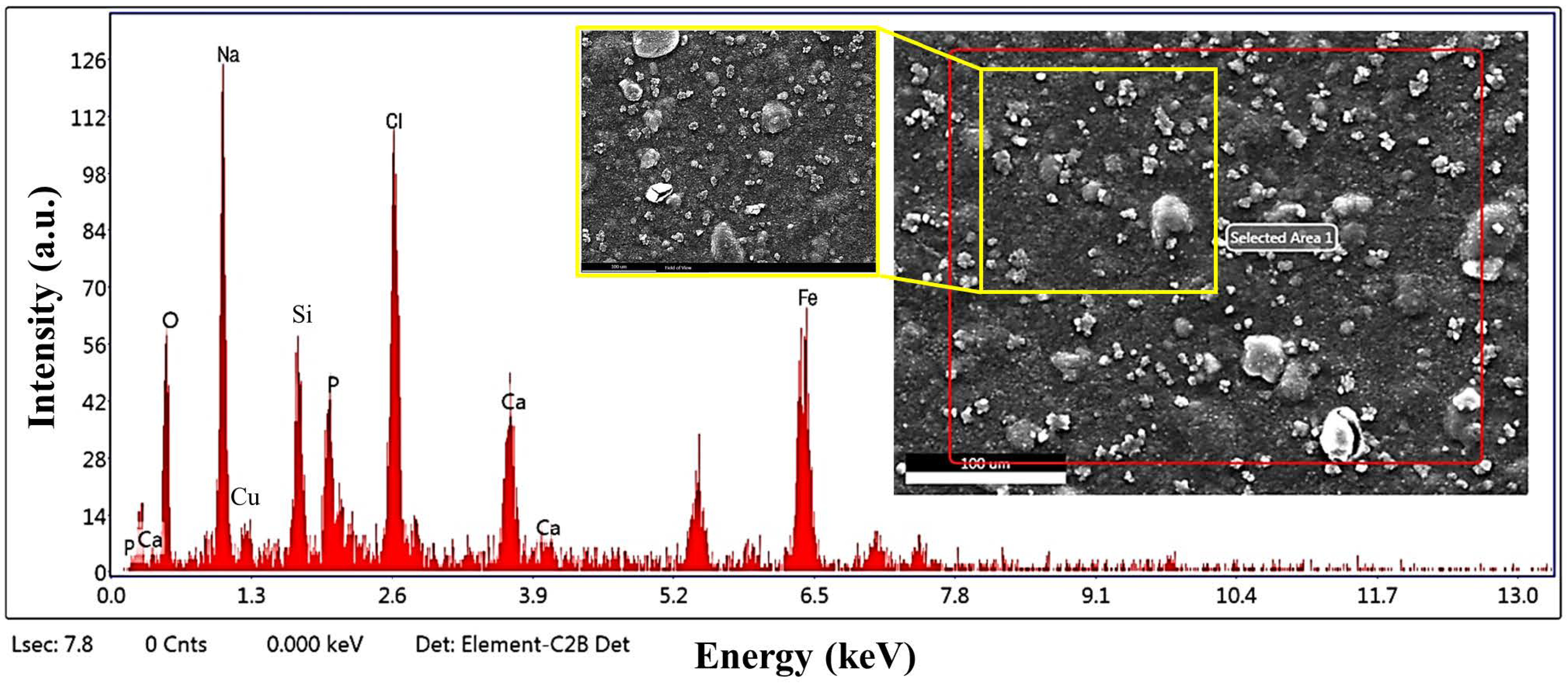
3.5. SEM/EDS Analysis of Chit-5CB Sample (The Optimum Sample)

Figure 6 depicts SEM micrograph/EDS analysis from the cross-section of the optimum sample (Chit-5CB). Surface bumps are clearly seen in this image. Bioactive glass 45S5 is rich in Si and O, and that is why the sharpest peaks are from Si and O. Calcium and phosphorus can also be clearly seen in the EDS pattern, which is also to be expected in 45S5 composition. The doped copper in the coating has also been detected by EDS analysis. It is also noticeable that the coating layer is rather dense, with hardly any indication of micro-cracks or voids or any other abnormality. Also, the thickness is rather uniform, inferring that the deposition has ended up in a uniform layer deposition.

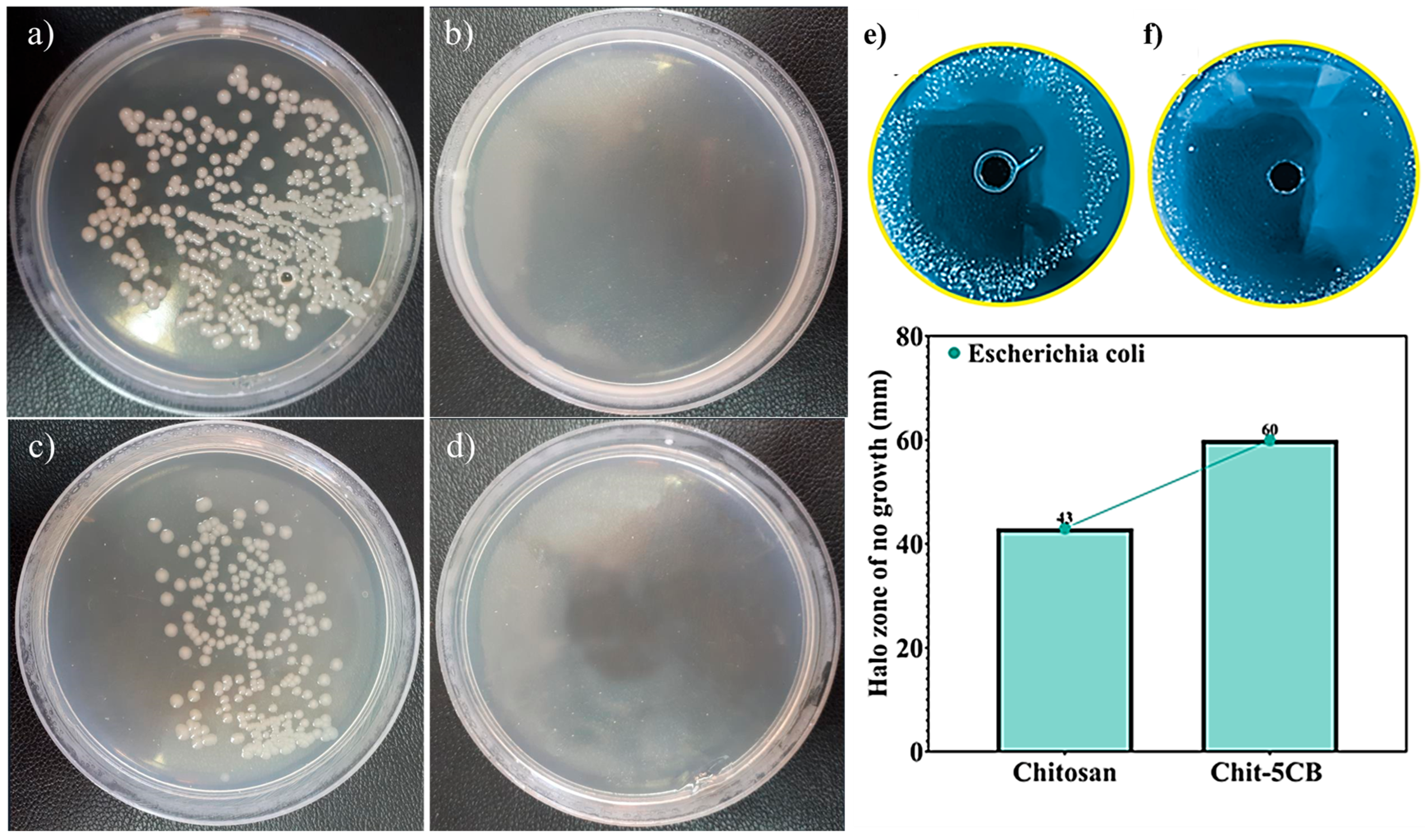
3.6. Antibacterial Assessments
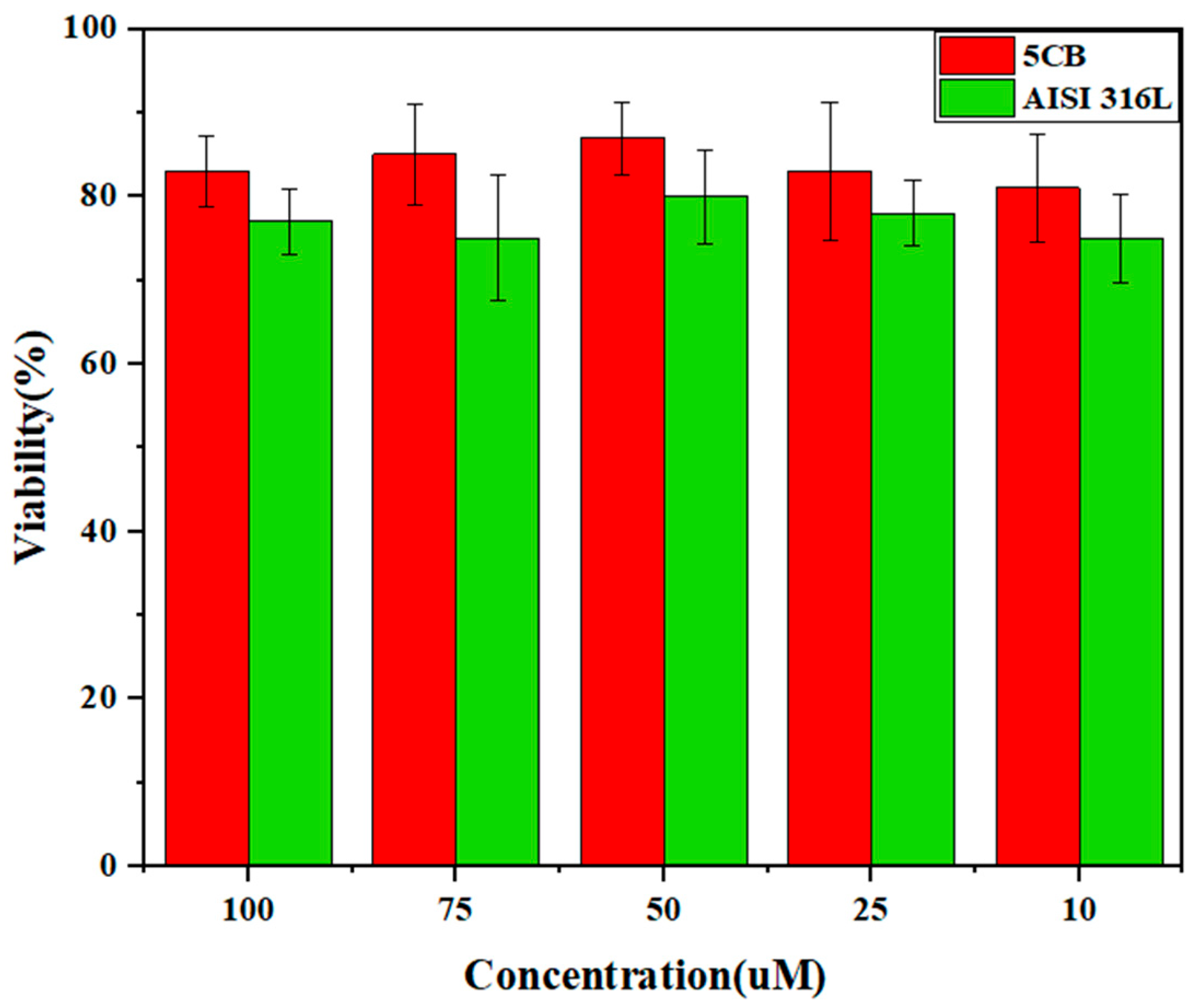
Counting bacterial cells, or bacterial colony count, is one of the most widely used antibacterial tests, in which case, the number of bacterial cells in a specific volume is counted to determine the concentration of cells. By placing a specific volume of cultured cells on a Petri dish containing a culture medium, the number of cultured cells can be counted. If the cells are properly distributed in the culture medium, it can be assumed that each cell forms a single colony. Therefore, the colonies are countable and based on the specific volume of the culture medium distributed on the plate, the concentration of cells can be calculated. Performing bacterial colony counts is useful for estimating the potency of bacterial infections, such as bacterial infections. Figure 7 and Table 3 show the impact of the Chit-5CB sample on bacterial viability assessed by comparing the number of colonies formed. The results demonstrate a significant reduction in colony counts for both Escherichia coli and Staphylococcus strains. Notably, exposure to the Chit-5CB sample led to a complete inhibition of bacterial growth, with colony numbers reduced to zero relative to the control, highlighting the potent antibacterial efficacy of the composite. This effect is likely attributable to the presence of copper within the coating, which exerts its bactericidal action through mechanisms such as the generation of reactive oxygen species (ROS), ion release, and disruption of bacterial cell membranes. Despite the structurally thicker peptidoglycan layer in the cell wall of Staphylococcus compared to E. coli, both strains exhibited comparable susceptibility to the composite material [48], implying that the proposed coating composition has similar effects in both Gram-positive and Gram-negative strains. Also, Figure 7 shows the growth inhibition analysis for two samples, chitosan and Chit-5CB. As it is clear, both samples show a significant growth inhibition diameter, such that for the sample without copper, the growth inhibition diameter is 43 mm and for the sample with copper, the growth inhibition diameter is 60 mm. On the other hand, it can be said that in general, copper ions react with phosphate groups in the cell membrane of Escherichia coli bacteria and cause cell membrane destruction and release of cell contents. On the other hand, copper produces free radical oxygen species in the presence of oxygen and causes damage to the DNA and cell membranes of bacteria [49,50].

3.7. MTT Test

An MTT (3-(4,5-dimethylthiazol-2-yl)-2,5-diphenyltetrazolium bromide) test was carried out to evaluate the cell viability in contact with the coating layer and the substrate (see Table 4 and Figure 8). The MTT test was conducted according to the standard ISO 10993-5:2009, which is based on the measurement of the shift in the MTT color of alive cells’ mitochondria from yellow to purple formazan crystals. The MTT viability test is essentially a photometric analysis of viable cell numbers. The control sample in this test was an untreated well without added specimen (specimen). To evaluate cytotoxicity, seeding of 1 × 10⁴ MG63 cell samples with DMEM culture media (containing 10% fetal bovine serum, 1% penicillin, and streptomycin) was carried out in 96-well plates in triplicate. Different concentrations (100, 75, 50, 25, and 10 μm) of the nanoparticle solution were added to the wells and incubation was carried out in a 5% CO2 atmosphere at 37 °C for 24 h. After incubation, 20 microliters of 5 mg/mL MTT dye was added to each well. Three hours later, the MTT color was extracted and DMSO solvent was applied to dissolve the purple crystals formed. An ELISA reader quantified the dye dissolved in the DMSO solvent. Wells with living cells exhibited higher optical density (OD) as compared to those with dead cells. Cytotoxicity and cell viability assessments were conducted according to the following criteria: a sample is considered non-cytotoxic if its viability is over 70%; otherwise, it is considered cytotoxic. The control sample is expected to have 100% viability, inferring that in control condition, cells have perfect cell survival. To ensure that measurements are reliable, the standard deviation for each experimental point should be under 15%. The viability percentage of cells in the analyzed specimens was compared to the control sample. The results indicate that the bioactive glass-containing coating exhibits a significantly higher cell viability percentage compared to the uncoated substrate, suggesting that the applied layer is highly biocompatible and demonstrates minimal cytotoxicity.

3.8. Bioactivity Analysis

To evaluate the bioactivity of the samples, scanning electron microscopy (SEM), energy-dispersive X-ray spectroscopy (EDS), and inductively coupled plasma (ICP) analysis were performed following a four-week immersion period in simulated body fluid (SBF). The SEM images (Figure 9) clearly reveal the formation of spherical and granular structures on the surface, which are indicative of hydroxyapatite (HA) deposition—a well-established sign of in vitro bioactivity. The presence of a relatively dense and uniform layer across the surface suggests the material’s high potential to induce nucleation and growth of apatite phases. The corresponding EDS spectrum confirms the presence of significant amounts of calcium (Ca) and phosphorus (P), the primary elements comprising hydroxyapatite. Additionally, elements such as silicon (Si), iron (Fe), copper (Cu), and sodium (Na) were also detected, possibly originating from the glass network or ionic residues from the SBF. Notably, the appearance of sodium and chlorine (Cl) peaks in the EDS spectrum indicates the potential formation of salt deposits, such as NaCl, on the sample surface. This is justifiable considering that the EDS area analysis captures signals from the entire surface composition, including both the deposited apatite layer and any surface salt accumulations. The formation of such precipitates is common in long-term SBF studies and is often due to solution evaporation and re-precipitation of ions onto the sample surface [51]. To further support the surface analysis, Table 5 is given, which depicts the variation in calcium and phosphorus ion concentrations in the simulated body fluid (SBF) over a four-week period, as determined by inductively coupled plasma (ICP) analysis. At the outset, the calcium and phosphorus concentrations in the SBF were 47.2 mg/dL and 36.1 mg/dL, respectively. After the first week of immersion, these values dropped significantly to 17.6 mg/dL for calcium and 4.5 mg/dL for phosphorus, indicating a rapid ionic exchange between the SCB sample surface and the solution. This phenomenon is a hallmark of bioactive glass behavior, in which Na⁺ and Ca2⁺ ions are exchanged with H⁺ or H3O⁺ ions in the surrounding solution. This ion exchange process facilitates the degradation of the silicate network, resulting in the release of calcium and phosphorus ions into the medium. In the second and third weeks, the concentrations of Ca and P continued to decrease, reaching 9.5 and 2.8 mg/dL, and then 8.6 and 1.6 mg/dL, respectively. This gradual decline suggests the consumption of these ions during the nucleation and growth of the hydroxyapatite layer on the surface. A slight increase in calcium (11.4 mg/dL) and phosphorus (2.14 mg/dL) concentrations was observed in the fourth week, which may be attributed to the partial dissolution of previously formed apatite or a dynamic equilibrium between precipitation and dissolution processes. In summary, the results of surface (SEM and EDS) and solution (ICP) analyses collectively demonstrate that the SCB sample exhibits significant bioactivity. It not only possesses the ability to release bioactive ions but also promotes the formation of a stable apatite layer, which is crucial for bonding with bone tissue in vivo. The detection of sodium and chlorine peaks is associated with superficial salt precipitates and does not detract from the overall interpretation of surface bioactivity, although it should be considered during EDS data evaluation.
The slight increase in the Ca and P levels noted in this latter stage may be attributed to the natural mobility of surface interactions mainly between coating and simulated body fluid (SBF). In particular, such variations often reflect minor localized dissolution events occurring on boundaries in areas of low crystalline stability or within the apatite layer. These disintegration phenomena temporarily bring the ions back into the fluid—thus, a brief increase in measured concentrations. This process is usually reorganized in a more stable crystalline form of rapidly re- or predetermined ha, which positively contributes to the remodeling of the surface and eventually increases structural integrity. Similar fluctuations have been widely documented in the study of bioactive glass and calcium phosphate-based coatings, where minor ionic concentration variations are signs of a healthy and dynamic balance rather than unstable or declining. Therefore, these transient changes do not negatively affect the long-term stability of the coating or its bioactive behavior. Instead, they display the ability of coatings to actively react to their environment, thus supporting continuous, effective transplantation–tuct interactions in extended periods [52,53,54].
Copper ions added to bioactive glass (Cu2) are expected to improve the bioactivity of the coating by affecting how it grows as a crystal. When copper is included in bioactive glass, it slightly replaces the structure of the glass, making it easier to slowly release into bio-liquids. This controlled disintegration helps release essential minerals such as calcium, phosphate, and silicate ions, which creates an ideal chemical environment on the coating surface for the early formation of the apatite crystals. In addition, copper ions efficiently increase the ability to exchange ions with a fluid, which increases the concentration of hydroxyl groups (OH⁻). During this initial phase, minerals first form a low organized calcium phosphate layer called unknowable calcium phosphate (ACP). Copper ions help to stabilize this ACP, for this it becomes easier for it to crystallize in hydroxyapatite. Essentially, copper ions reduce the energy barrier required for the growth of the crystal, making the entire process smooth and more efficient. Additionally, the copper can directly stimulate the crystal to change the local electrochemical environment on the surface of the coating or to help indirectly, which can improve both the volume and quality of the final ha layer. To clarify these details for future readers, we have improved and expanded this explanation in the revised manuscript [55,56,57]

4. Conclusions

This study aimed to deposit a composite coating consisting of chitosan and Cu-doped bioactive glass with varying copper concentrations, applied to AISI 316L stainless steel substrates for potential biomedical implant applications. The coatings were deposited using the electrophoretic deposition (EPD) technique. To determine the optimal deposition conditions, preliminary experiments were conducted using chitosan–bioglass coatings at different voltages and deposition times. Surface roughness, contact angle, and adhesion strength tests were performed, and based on coating uniformity and mechanical results, a voltage of 20 V for 10 min was selected as the optimal condition for the EPD process. These parameters were subsequently used for the deposition of chitosan/Cu-doped bioglass coatings with 5%, 10%, and 20% (designated as 5CB, 10CB, and 20CB). The coated samples were characterized through surface roughness measurements, contact angle analysis, and adhesion tests. Among the three samples, the 5CB coating demonstrated superior performance in terms of lower surface roughness, appropriate wettability, higher adhesion strength, and better coating uniformity. Therefore, the 5CB sample was selected as the optimal composition for further biological evaluation. Subsequent investigations on the 5CB coating included in vitro cytocompatibility assessment using the MTT assay, antibacterial tests, elemental analysis via EDS for hydroxyapatite formation, and ion release evaluation through ICP. The results revealed that the 5CB coating exhibited excellent antibacterial activity against both Gram-positive and Gram-negative bacteria. However, its bioactivity was limited, as evidenced by minimal formation of calcium–phosphate-rich layers during the immersion tests. In conclusion, the chitosan/Cu-doped bioglass composite coatings, deposited under optimized EPD conditions, provided a uniform, adherent, and antibacterial surface, indicating its potential for use in orthopedic implant applications, although its bioactivity requires further enhancement.

Author Contributions

Formal analysis, S.M.R.M., B.F.A., and A.B.; Investigation, S.M.R.M. and M.S.A.; Data curation, M.S.A. and M.R.; Writing—review and editing, M.R., F.H.L., and A.B.; Supervision, A.B., F.H.L., and M.S.A., All authors have read and agreed to the published version of the manuscript.

Funding

This research received no external funding.

Data Availability Statement

Data are contained within the article.

Conflicts of Interest

The authors declare no conflicts of interest.

References

  1. Deng, R.; Wei, R.; Zhang, Y.; Zhao, C.; Liang, J.; Bai, Q.; Li, H.; Ouyang, C.; He, Q.; Liu, S.; et al. Investigation of variable-temperature wear characteristics of austenitic stainless steel coatings fabricated via laser energy deposition. Wear 2025, 572–573, 205993. [Google Scholar] [CrossRef]
  2. Xu, K.; Xiao, X.; Wang, L.; Lou, M.; Wang, F.; Li, C.; Ren, H.; Wang, X.; Chang, K. Data-Driven Materials Research and Development for Functional Coatings. Adv. Sci. 2024, 11, 2405262. [Google Scholar] [CrossRef]
  3. Heidari Laybidi, F.; Bahrami, A. Antibacterial properties of ZnO-containing bioactive glass coatings for biomedical applications. Mater. Lett. 2024, 365, 136433. [Google Scholar] [CrossRef]
  4. Wang, S.; Zhao, X.; Hsu, Y.; He, Y.; Wang, F.; Yang, F.; Yan, F.; Xia, D.; Liu, Y. Surface modification of titanium implants with Mg-containing coatings to promote osseointegration. Acta Biomater. 2023, 169, 19–44. [Google Scholar] [CrossRef] [PubMed]
  5. Amirtharaj Mosas, K.K.; Chandrasekar, A.R.; Dasan, A.; Pakseresht, A.; Galusek, D. Recent Advancements in Materials and Coatings for Biomedical Implants. Gels 2022, 8, 323. [Google Scholar] [CrossRef] [PubMed]
  6. González-Reyna, M.A.; Espinosa-Medina, M.A.; Esparza, R.; Hernández-Martinez, A.R.; Maya-Cornejo, J.; Estévez, M. Anticorrosive Effect of the Size of Silica Nanoparticles on PMMA-Based Hybrid Coatings. J. Mater. Eng. Perform. 2021, 30, 1054–1065. [Google Scholar] [CrossRef]
  7. Taveri, G.; Hanzel, O.; Sedláček, J.; Toušek, J.; Neščaková, Z.; Michálek, M.; Dlouhý, I.; Hnatko, M. Consolidation of Bioglass® 45S5 suspension through cold isostatic pressing. Ceram. Int. 2021, 47, 4090–4096. [Google Scholar] [CrossRef]
  8. Rajabinezhad, M.; Bahrami, A.; Abbasi, M.S.; Karampoor, M.R. A promising multi-component, multi-functional, drug-releasing Cu-doped Fe3O4/bioactive glass/Chitosan coating, applied on stainless steel substrate for biomedical applications. Int. J. Biol. Macromol. 2025, 310, 143296. [Google Scholar] [CrossRef]
  9. Vafa, E.; Bazargan-Lari, R.; Bahrololoom, M.E. Electrophoretic deposition of polyvinyl alcohol/natural chitosan/bioactive glass composite coatings on 316L stainless steel for biomedical application. Prog. Org. Coat. 2021, 151, 106059. [Google Scholar] [CrossRef]
  10. Wang, M.; Zheng, Y.; Yin, C.; Dai, S.; Fan, X.; Jiang, Y.; Liu, X.; Fang, J.; Yi, B.; Zhou, Q.; et al. Recent Progress in antibacterial hydrogel coatings for targeting biofilm to prevent orthopedic implant-associated infections. Front. Microbiol. 2023, 14, 1343202. [Google Scholar] [CrossRef]
  11. Daly, A.C.; Freeman, F.E.; Gonzalez-Fernandez, T.; Critchley, S.E.; Nulty, J.; Kelly, D.J. 3D Bioprinting for Cartilage and Osteochondral Tissue Engineering. Adv. Heal. Mater. 2017, 6, 1700298. [Google Scholar] [CrossRef] [PubMed]
  12. Fernandes, H.R.; Gaddam, A.; Rebelo, A.; Brazete, D.; Stan, G.E.; Ferreira, J.M.F. Bioactive Glasses and Glass-Ceramics for Healthcare Applications in Bone Regeneration and Tissue Engineering. Materials 2018, 11, 2530. [Google Scholar] [CrossRef] [PubMed]
  13. Brunello, G.; Elsayed, H.; Biasetto, L. Bioactive Glass and Silicate-Based Ceramic Coatings on Metallic Implants: Open Challenge or Outdated Topic? Materials 2019, 12, 2929. [Google Scholar] [CrossRef]
  14. Agnes, C.J.; Karoichan, A.; Tabrizian, M. The Diamond Concept Enigma: Recent Trends of Its Implementation in Cross-linked Chitosan-Based Scaffolds for Bone Tissue Engineering. ACS Appl. Bio Mater. 2023, 6, 2515–2545. [Google Scholar] [CrossRef]
  15. Bhaskar, P.; Kumar, R.; Maurya, Y.; Ravinder, R.; Allu, A.R.; Das, S.; Gosvami, N.N.; Youngman, R.E.; Bødker, M.S.; Mascaraque, N.; et al. Cooling rate effects on the structure of 45S5 bioglass: Insights from experiments and simulations. J. Non-Cryst. Solids 2020, 534, 119952. [Google Scholar] [CrossRef]
  16. Pádua, A.S.; Gavinho, S.R.; Vieira, T.; Hammami, I.; Silva, J.C.; Borges, J.P.; Graça, M.P.F. In Vitro Characterization of Doped Bioglass 45S5/HAp Coatings Obtained by CoBlastTM Deposition. Coatings 2023, 13, 1775. [Google Scholar] [CrossRef]
  17. Abbasi, M.S.; Yousef Saber, F.; Bahrami, A.; Torkian, S.; Hosseini-Abari, A. Towards an anticancer bioactive glass-Fe3O4/Pectin/Pectic oligosaccharide coatings for biomedical applications. Surf. Interfaces 2024, 45, 103874. [Google Scholar] [CrossRef]
  18. Rajabinezhad, M.; Abbasi, M.S.; Laybidi, F.H.; SharifianJazi, M.; Khodaei, M.; Bahrami, A. Structural, mechanical and biomedical properties of 3D-printed Cu-doped Fe3O4/58S bioactive glass/polycaprolactone composite scaffold for bone tissue regeneration. Bioprinting 2025, 47, e00400. [Google Scholar] [CrossRef]
  19. Heidarpour, S.; Heidari Laybidi, F.; Bahrami, A.; Abbasi, M.S.; Hosseini-Abari, A.; Ashrafian, B. Antibacterial properties of anthracite/CuO nanocomposites. Mater. Lett. 2023, 352, 135139. [Google Scholar] [CrossRef]
  20. Koné, K.E.; Bouich, A.; Soucase, B.M.; Soro, D. Manufacture of different oxides with high uniformity for copper zinc tin sulfide (CZTS) based solar cells. J. Mol. Graph. Model. 2023, 121, 108448. [Google Scholar] [CrossRef]
  21. Vasiliev, G.; Kubo, A.-L.; Vija, H.; Kahru, A.; Bondar, D.; Karpichev, Y.; Bondarenko, O. Synergistic antibacterial effect of copper and silver nanoparticles and their mechanism of action. Sci. Rep. 2023, 13, 9202. [Google Scholar] [CrossRef] [PubMed]
  22. Ulloa-Ogaz, A.L.; Piñón-Castillo, H.A.; Muñoz-Castellanos, L.N.; Athie-García, M.S.; Ballinas-Casarrubias, M.D.L.; Murillo-Ramirez, J.G.; Flores-Ongay, L.Á.; Duran, R.; Orrantia-Borunda, E. Oxidative damage to Pseudomonas aeruginosa ATCC 27833 and Staphylococcus aureus ATCC 24213 induced by CuO-NPs. Environ. Sci. Pollut. Res. 2017, 24, 22048–22060. [Google Scholar] [CrossRef]
  23. Sathiyavimal, S.; Vasantharaj, S.; Kaliannan, T.; Garalleh, H.A.L.; Garaleh, M.; Brindhadevi, K.; Chi, N.T.L.; Sharma, A.; Pugazhendhi, A. Bio-functionalized copper oxide/chitosan nanocomposite using Sida cordifolia and their efficient properties of antibacterial, anticancer activity against on breast and lung cancer cell lines. Environ. Res. 2023, 218, 114986. [Google Scholar] [CrossRef]
  24. Sathiyavimal, S.; FDurán-Lara, E.; Vasantharaj, S.; Saravanan, M.; Sabour, A.; Alshiekheid, M.; Lan Chi, N.T.; Brindhadevi, K.; Pugazhendhi, A. Green synthesis of copper oxide nanoparticles using Abutilon indicum leaves extract and their evaluation of antibacterial, anticancer in human A549 lung and MDA-MB-231 breast cancer cells. Food Chem. Toxicol. 2022, 168, 113330. [Google Scholar] [CrossRef] [PubMed]
  25. Verma, N.; Kumar, N. Synthesis and Biomedical Applications of Copper Oxide Nanoparticles: An Expanding Horizon. ACS Biomater. Sci. Eng. 2019, 5, 1170–1188. [Google Scholar] [CrossRef]
  26. Baratipour, S.; Bahrami, A.; Eslami, A.; Alihosseini, F. Toward Antibacterial Anodized Surfaces in Ti6Al4V Alloy Using Silver Nanoparticles and Pectin Coating for Biomedical Applications. J. Mater. Eng. Perform. 2024. [Google Scholar] [CrossRef]
  27. Liu, H.; Yu, Z.; Liu, L.; Dong, S. Cell Wall Binding Strategies Based on Cu3SbS3 Nanoparticles for Selective Bacterial Elimination and Promotion of Infected Wound Healing. ACS Appl. Mater. Interfaces 2024, 16, 33038–33052. [Google Scholar] [CrossRef]
  28. Miguel, S.P.; Moreira, A.F.; Correia, I.J. Chitosan based-asymmetric membranes for wound healing: A review. Int. J. Biol. Macromol. 2019, 127, 460–475. [Google Scholar] [CrossRef]
  29. Rajamma, R.; Gopalakrishnan Nair, S.; Abdul Khadar, F.; Baskaran, B. Antibacterial and anticancer activity of biosynthesised CuO nanoparticles. IET Nanobiotechnol. 2020, 14, 833–838. [Google Scholar] [CrossRef]
  30. Avcu, E.; Baştan, F.E.; Abdullah, H.Z.; Rehman, M.A.U.; Avcu, Y.Y.; Boccaccini, A.R. Electrophoretic deposition of chitosan-based composite coatings for biomedical applications: A review. Prog. Mater. Sci. 2019, 103, 69–108. [Google Scholar] [CrossRef]
  31. Wang, W.; Xue, C.; Mao, X. Chitosan: Structural modification, biological activity and application. Int. J. Biol. Macromol. 2020, 164, 4532–4546. [Google Scholar] [CrossRef] [PubMed]
  32. Burgos-Díaz, C.; Opazo-Navarrete, M.; Palacios, J.L.; Barahona, T.; Mosi-Roa, Y.; Anguita-Barrales, F.; Bustamante, M. Synthesis of New Chitosan from an Endemic Chilean Crayfish Exoskeleton (Parastacus pugnax): Physicochemical and Biological Properties. Polymers 2021, 13, 2304. [Google Scholar] [CrossRef] [PubMed]
  33. Zhang, B.; Lan, W.; Xie, J. Chemical modifications in the structure of marine polysaccharide as serviceable food processing and preservation assistant: A review. Int. J. Biol. Macromol. 2022, 223, 1539–1555. [Google Scholar] [CrossRef] [PubMed]
  34. Khubiev, O.M.; Egorov, A.R.; Kirichuk, A.A.; Khrustalev, V.N.; Tskhovrebov, A.G.; Kritchenkov, A.S. Chitosan-Based Antibacterial Films for Biomedical and Food Applications. Int. J. Mol. Sci. 2023, 24, 10738. [Google Scholar] [CrossRef] [PubMed]
  35. Muxika, A.; Etxabide, A.; Uranga, J.; Guerrero, P.; de la Caba, K. Chitosan as a bioactive polymer: Processing, properties and applications. Int. J. Biol. Macromol. 2017, 105, 1358–1368. [Google Scholar] [CrossRef]
  36. Laybidi, F.H.; Bahrami, A.; Abbasi, M.S.; Mokhtari, M.A.; Dehkordi, M.J.; Karampoor, M.R. The promising application of pectin/ɛ-polylysine as coating material on anodized titanium surfaces for orthopedic implants: Preparation, characterization and biomedical properties. Colloids Surf. A Physicochem. Eng. Asp. 2025, 704, 135517. [Google Scholar] [CrossRef]
  37. Oliveira, J.A.M.; de Santana, R.A.C.; Wanderley Neto Ade, O. Electrophoretic deposition and characterization of chitosan-molybdenum composite coatings. Carbohydr. Polym. 2021, 255, 117382. [Google Scholar] [CrossRef]
  38. Maciąg, F.; Moskalewicz, T.; Kowalski, K.; Łukaszczyk, A.; Hadzhieva, Z.; Boccaccini, A.R. The Effect of Electrophoretic Deposition Parameters on the Microstructure and Adhesion of Zein Coatings to Titanium Substrates. Materials 2021, 14, 312. [Google Scholar] [CrossRef]
  39. Hu, S.; Li, W.; Finklea, H.; Liu, X. A review of electrophoretic deposition of metal oxides and its application in solid oxide fuel cells. Adv. Colloid Interface Sci. 2020, 276, 102102. [Google Scholar] [CrossRef]
  40. Manzur, J.; Akhtar, M.; Aizaz, A.; Ahmad, K.; Yasir, M.; Minhas, B.Z.; Avcu, E.; Ur Rehman, M.A. Electrophoretic Deposition, Microstructure, and Selected Properties of Poly(lactic-co-glycolic) Acid-Based Antibacterial Coatings on Mg Substrate. ACS Omega 2023, 8, 18074–18089. [Google Scholar] [CrossRef]
  41. ASTM D3359-23; Standard Test Methods for Rating Adhesion by Tape Test. ASTM: West Conshohocken, PA, USA, 2023.
  42. Zhang, E.; Zhao, X.; Hu, J.; Wang, R.; Fu, S.; Qin, G. Antibacterial metals and alloys for potential biomedical implants. Bioact. Mater. 2021, 6, 2569–2612. [Google Scholar] [CrossRef]
  43. Deen, I.; Selopal, G.S.; Wang, Z.M.; Rosei, F. Electrophoretic deposition of collagen/chitosan films with copper-doped phosphate glasses for orthopaedic implants. J. Colloid Interface Sci. 2022, 607, 869–880. [Google Scholar] [CrossRef] [PubMed]
  44. Abbasi, M.S.; Bahrami, A.; Hosseini-Abari, A.S.; Saber, F.Y. Synthesis and characterization of composite coating of iron oxide and bioactive glass, coated by electrophoretic co-deposition method for biomedical applications. J. Adv. Mater. Eng. 2023, 42, 1–13. [Google Scholar]
  45. Hong, W.; Guo, F.; Chen, J.; Wang, X.; Zhao, X.; Xiao, P. Bioactive glass–chitosan composite coatings on PEEK: Effects of surface wettability and roughness on the interfacial fracture resistance and in vitro cell response. Appl. Surf. Sci. 2018, 440, 514–523. [Google Scholar] [CrossRef]
  46. Sanguedolce, M.; Rotella, G.; Curcio, F.; Pelaccia, R.; Siciliani, V.; Cassano, R.; Orazi, L.; Filice, L. Effects of Ti6Al4V mechanical and thermal surface modification on the adhesion of a chitosan-bioactive glass coating. Int. J. Adv. Manuf. Technol. 2023, 125, 4621–4629. [Google Scholar] [CrossRef]
  47. Przykaza, K.; Jurak, M.; Kalisz, G.; Mroczka, R.; Wiącek, A.E. Characteristics of Hybrid Bioglass-Chitosan Coatings on the Plasma Activated PEEK Polymer. Molecules 2023, 28, 1729. [Google Scholar] [CrossRef] [PubMed]
  48. Muñoz-Tebar, N.; Pérez-Álvarez, J.A.; Fernández-López, J.; Viuda-Martos, M. Chitosan Edible Films and Coatings with Added Bioactive Compounds: Antibacterial and Antioxidant Properties and Their Application to Food Products: A Review. Polymers 2023, 15, 396. [Google Scholar] [CrossRef]
  49. Gritsch, L.; Lovell, C.; Goldmann, W.H.; Boccaccini, A.R. Fabrication and characterization of copper(II)-chitosan complexes as antibiotic-free antibacterial biomaterial. Carbohydr. Polym. 2018, 179, 370–378. [Google Scholar] [CrossRef] [PubMed]
  50. El-Kattan, N.; Ibrahim, M.A.; Emam, A.N.; Metwally, K.; Youssef, F.S.; Nassar, N.A.; Mansour, A.S. Evaluation of the antimicrobial activity of chitosan- and curcumin-capped copper oxide nanostructures against multi-drug-resistant microorganisms. Nanoscale Adv. 2025, 7, 2988–3007. [Google Scholar] [CrossRef]
  51. Harding, J.L.; Krebs, M.D. Bioinspired Deposition-Conversion Synthesis of Tunable Calcium Phosphate Coatings on Polymeric Hydrogels. ACS Biomater. Sci. Eng. 2017, 3, 2024–2032. [Google Scholar] [CrossRef]
  52. Jeong, J.; Kim, J.H.; Shim, J.H.; Hwang, N.S.; Heo, C.Y. Bioactive calcium phosphate materials and applications in bone regeneration. Biomater. Res. 2019, 23, 4. [Google Scholar] [CrossRef] [PubMed]
  53. Jaita, P.; Chokethawai, K.; Randorn, C.; Boonsri, K.; Pringproa, K.; Thongkorn, K.; Watcharapasorn, A.; Jarupoom, P. Enhancing bioactivity and mechanical performances of hydroxyapatite–calcium sulfate bone cements for bone repair: In vivo histological evaluation in rabbit femurs. RSC Adv. 2024, 14, 23286–23302. [Google Scholar] [CrossRef] [PubMed]
  54. Skliarenko, Y.; Kolomiiets, V.V.; Balatskyi, V.V.; Galuza, Y.; Koryak, O.S.; Macewicz, L.L.; Ruban, T.P.; Firstov, S.A.; Ulianchych, N.V.; Piven, O.O. The impact of the physicochemical properties of calcium phosphate ceramics on biocompatibility and osteogenic differentiation of mesenchymal stem cells. BMC Res. Notes 2024, 17, 295. [Google Scholar] [CrossRef]
  55. Ai, F.; Chen, L.; Yan, J.; Yang, K.; Li, S.; Duan, H.; Cao, C.; Li, W.; Zhou, K. Hydroxyapatite scaffolds containing copper for bone tissue engineering. J. Sol-Gel Sci. Technol. 2020, 95, 168–179. [Google Scholar] [CrossRef]
  56. Kubiak-Mihkelsoo, Z.; Kostrzębska, A.; Błaszczyszyn, A.; Pitułaj, A.; Dominiak, M.; Gedrange, T.; Nawrot-Hadzik, I.; Matys, J.; Hadzik, J. Ionic Doping of Hydroxyapatite for Bone Regeneration: Advances in Structure and Properties over Two Decades—A Narrative Review. Appl. Sci. 2025, 15, 1108. [Google Scholar] [CrossRef]
  57. Gritsch, L.; Maqbool, M.; Mouriño, V.; Ciraldo, F.E.; Cresswell, M.; Jackson, P.R.; Lovell, C.; Boccaccini, A.R. Chitosan/hydroxyapatite composite bone tissue engineering scaffolds with dual and decoupled therapeutic ion delivery: Copper and strontium. J. Mater. Chem. B 2019, 7, 6109–6124. [Google Scholar] [CrossRef]
Figure 1. Stereomicroscopic images (all on a scale of 100 µm) for chitosan-Cu-doped bioglass with different copper concentrations: (a) Chitosan-5CB, (b) Chitosan-10CB, (c) Chitosan-20CB, (d) Chitosan-BG (reference sample).
Crystals 15 00549 g001
Figure 2. Laser profilometry results of deposited coating layers.
Crystals 15 00549 g002
Figure 3. Variation of contact angles in deposited samples (Angles are measured from the blue lines).
Crystals 15 00549 g003
Figure 4. The X-ray diffraction (XRD) patterns of (a) composite samples and (b) pure bioactive glass.
Crystals 15 00549 g004
Figure 5. The results of adhesion test according to ASTM-D3359 standard on deposited samples.
Crystals 15 00549 g005
Figure 6. FESEM/EDS of the deposited coating layer (sample Chitosan-5CB).
Crystals 15 00549 g006
Figure 7. Bacterial colony count assay: (a) control sample tested against Staphylococcus at a 10⁻⁶ dilution; (b) Chit-5CB sample tested against Staphylococcus at a 10⁻⁶ dilution; (c) control sample tested against Escherichia coli at a 10⁻⁶ dilution; (d) Chit-5CB sample tested against Escherichia coli at a 10⁻⁶ dilution; (e) chitosan inhibitor growth analysis; (f) Chit-5CB inhibitor growth analysis.
Crystals 15 00549 g007
Figure 8. The results of the MTT assay after a duration of 24 ± 2 h on the 316L and Chit-5CB at five different concentrations (100, 75, 50, 25, and 10 micromolar) with three replicates.
Crystals 15 00549 g008
Figure 9. Scanning electron microscopy (SEM) images and elemental analysis of the 5CB sample after four weeks of immersion in simulated body fluid (SBF) (The analysis is from the highlighted box).
Crystals 15 00549 g009
Table 1. Deposition parameters (time and voltage) used in this investigation.
NumberTimeVoltage
15 min15 V
210 min15 V
315 min15 V
420 min15 V
55 min20 V
610 min20 V
715 min20 V
820 min20 V
Table 2. Classification of the adhesion base on ASTM-D3359.
Classification%Area Removed
5B0% (NONE)
4BLESS THAN 5%
3B5–15%
2B15–35%
1B35–65%
0BGREATER THAN 65%
Table 3. Variation in bacterial count in response to exposure to the 5CB sample.
Bacterial Growth Variation Relative to the Control Group, Expressed in Colony Forming (CFU/mL)
Bacterial strainSamplesNumber of colonies (R × 105)Number of colonies (R × 106)Dilution (10−8)
Staphylococcus5CB00↓(21.2–42.5) × 108
Control sample42585-
Escherichia coli5CB00↓(10.1–22.5) × 108
Control sample20345-
Table 4. MTT test results of (a) AISI 316L, (b) Chit-5CB samples in different concentrations of 100, 25, 50, 75, and 100 µM.
Sample—Concentrations100 (µM)75 (µM)50 (µM)25 (µM)10 (µM)
AISI 316L7775 (±10%)80 (±7%)78 (±5%)75
Chit-5CB8385 (±7%)87 (±5%)83 (±10%)81
Control Sample100100100100100
Table 5. Analysis of calcium and phosphorus ion concentration variations in the SBF solution following immersion of the coatings at the end of the first, second, and third weeks, as determined by inductively coupled plasma (ICP) testing.
Calcium (Ca) mg/dLPhosphorus (P) mg/dL
SBF47.236.1
The first week17.64.5
The second week9.52.8
The third week8.61.6
The fourth week11.42.14
Disclaimer/Publisher’s Note: The statements, opinions and data contained in all publications are solely those of the individual author(s) and contributor(s) and not of MDPI and/or the editor(s). MDPI and/or the editor(s) disclaim responsibility for any injury to people or property resulting from any ideas, methods, instructions or products referred to in the content.

