Abstract
As part of crystal growth experiments on transition metal oxidotellurates using chemical vapor transport reactions or hydrothermal conditions, single crystals of NiIITeVIO4 and CuIITeIV2O5 were obtained for the first time in the form of new modifications, as revealed by crystal structure determinations from X-ray data. In the course of these investigations, the crystal structure model of the only phase of NiIITeVIO4 reported so far (from now on named α-) was corrected. Both α-(space group P21/c, Z = 2) and the new β-polymorph of NiIITeVIO4 (space group I41/a, Z = 8) can be considered derivatives (hettotypes) of the rutile structure (aristotype), as shown by detailed symmetry relationships. For CuTe2O5 also, only one crystalline phase has been described so far (from now on named α-) that corresponds to the mineral rajite (space group P21/c, Z = 2). Its anion comprises two different trigonal-pyramidal TeO3 groups linked through corner-sharing into a ditellurite unit. The anion part of the new β-CuTe2O5 modification (space group P21/c, Z = 2), likewise, comprises two TeIV atoms but is more complex. Here, one TeIV atom exhibits a coordination number of 4 and is part of a TeO2/2O2/1] chain, and the other has a coordination number of 5 and is part of a TeO2/2O3/1]2 dimer. The two types of anions are linked into a tri-periodic framework where both TeIV atoms are stereochemically active. The α- and β-CuTe2O5 modifications show no closer structural relationship, which is also reflected in their clearly different Raman spectra. Data mining for knowledge discovery in a structure database reveals that polymorphism is a rather common phenomenon for the family of inorganic oxidotellurates.
1. Introduction
The large family of oxidotellurates has a very broad spectrum of compositions and crystal structures. This is partly due to the fact that tellurium can be tetravalent or hexavalent in these compounds, and mixed-valent TeIV/VI compounds also occur. The distinct ability to condense the corresponding anions (commonly referred to as “tellurites” for oxidotellurates(IV) and “tellurates” for oxidotellurates(VI)) also contributes to the structural diversity. A recent review of the crystal chemistry of this family of compounds summarized and categorized their peculiar structural features [].
First-row transition metal oxidotellurates(VI) with composition MTeO4 (M = Co, Ni, Cu, Zn) exhibit interesting structural and physical properties: CoTeO4 adopts the monoclinic dirutile structure type and is a wide-bandgap material []; NiTeO4, structurally determined from powder synchrotron data, is isotypic with CoTeO4 and shows a robust electrochemical activity against Li metal with a high reversible specific capacity []; CuTeO4 is reported to crystallize with an order/disorder structure [] and shows quasi-one-dimensional, antiferromagnetic correlations at high temperature []; ZnTeO4 is assumed to have great second-harmonic generation coefficients and a large birefringence, as predicted by first principles techniques on the basis of a theoretically deduced crystal structure [].
In order to understand and accurately calculate the physical properties mentioned above, a reliable crystal structure model is of utmost importance. Furthermore, in the context of structure–property relationships, it is desirable to have highly crystalline and single-phase material available for the measurement of other physical properties related to long-range ordering and correlated phenomena. For these reasons, single-crystal growth experiments were carried out for the two phases NiTeO4 and CuTeO4. In the case of NiTeO4, the existing structure model [] is highly questionable. For crystal growth of this compound, chemical vapor transport reactions (CVTR) [] were applied, the use of which had already been shown to be successful with the closely related compound CoTeO4 []. For CuTeO4, single-crystal growth was primarily intended to investigate the reported differences in the crystal structure models obtained from single-crystal diffraction [] and powder diffraction [] data. For crystal growth of CuTeO4, the hydrothermal synthesis method [] was chosen, but under considerably different pressure and temperature conditions than in the original report (650 °C, 2000 bar []) or under slightly different conditions than applied for preparation of polycrystalline material (210 °C, autogenous pressure, filling degree of the container ≈ 50% []).
In the course of these experiments, two crystalline modifications of NiTeO4 (one known and one new) as well as a new crystalline modification of the unexpected product CuTe2O5 were obtained. The results are reported and discussed here.
2. Materials and Methods
2.1. Crystal Growth Experiments
NiTeO4. Polycrystalline NiTeO4 was prepared by heating a stoichiometric 1:1 mixture of Ni(NO3)2·6H2O and Te(OH)6 in a porcelain crucible at 550 °C for one week under multiple intermediate grinding. Powder X-ray diffraction measurement revealed the product to be single-phase according to previously reported data []. About 0.5 g of the resulting ochre-colored powder was filled into a silica glass ampoule (volume ≈ 20 cm3) together with 50 mg PtCl2 and sealed under vacuum. The ampoule was heated in a horizontal two-zone furnace for three weeks, applying a temperature gradient of 640 °C (source region) → 580 °C (sink region). Under these conditions, PtCl2 decomposes and the released chlorine gas acts as a transport agent [,]. At the end of the experiment, three different crystal types were present in the sink zone, which were identified optically under a microscope and by subsequent single-crystal X-ray diffraction: yellow block-like NiIITeIV2O5 crystals [] as the main phase, brownish-orange plates of known dirutile-type NiTeO4 [] (subsequently referred to as α-NiTeO4) as a minor phase, and very few brownish-green plate-like crystals of a new modification of NiTeO4 (subsequently referred to as β-NiTeO4).
CuTeO4. Cu(NO3)2·3H2O (0.284 g), Te(OH)6 (0.270 g), and KOH (0.132 g) were mixed in the molar ratio 1:1:2 and placed in a Teflon container with an inner volume of ≈6 mL to which water was added (filling degree ≈ 70%). Then the mixture was mechanically stirred for 30 min, and the container was sealed and placed in a steel autoclave at 210 °C for one week under autogenous pressure. Very few bottle-green plate-like crystals of a new modification of CuTe2O5 (subsequently referred to as β-CuTe2O5) were obtained as the only solid reaction product. These crystals were used for the single-crystal X-ray diffraction and Raman spectroscopic measurements.
2.2. X-Ray Diffraction Measurements and Crystal Structure Analysis
Powder X-ray diffraction (PXRD) measurements were performed on a PANalytical X’Pert II Pro type PW 3040/60 diffractometer (Malvern Panalytical, Malvern, Worshestershire, UK) using Cu-Kα1,2-radiation and an X’Celerator detector.
Single-crystal X-ray diffraction data were recorded on a Bruker Kappa Apex-II single-crystal diffractometer (Bruker AXS, Madison, WI, USA) equipped with a CCD detector and using graphite-monochromatized Mo-Kα-radiation. Instrument software packages (Apex-4 and Saint []) were used for optimized measurement strategies (>99% completeness at θmax) and for data reduction; absorption effects were corrected with Sadabs []. The crystal structures were solved with Shelxt [], refined with Shelxl [] and graphically represented with Atoms []. For the crystal structure of α-NiTeO4, atom labels and coordinates were assigned in accordance with the previously reported isotypic crystal structure of CoTeO4 [].
Crystal structure and refinement data are compiled in Table 1. Further details of the crystal structure investigations may be obtained from the joint CCDC/FIZ Karlsruhe online deposition service: https://www.ccdc.cam.ac.uk/structures/ by quoting the deposition numbers specified at the end of Table 1.

Table 1.
Data collection and refinement details.
Bond valence sums (BVS) [] were calculated using the bond valence parameters provided by Brese and O’Keeffe [] for Ni–O and Cu–O bonds. For the Te–O bonds, the revised bond valence parameters by Mills and Christy [] were used.
2.3. Raman Spectroscopy
Raman spectra of α- and β-CuTe2O5 were recorded using an RM 1000 micro-Raman system (Renishaw plc, Wolton-under-Edge, UK) in the range from 4000 to 40 cm−1 on randomly oriented single crystals. The 488 nm excitation line of a 20 mW Ar laser was focused with a 50×/0.75 objective lens slightly below the sample surface. Raman intensities were collected with a thermo-electrically cooled CCD array detector. The resolution of the system was 5–6 cm−1, and the wavenumber accuracy was ±1 cm−1, both calibrated with the Rayleigh line and the 520.5 cm−1 line of a Si standard.
3. Results and Discussion
3.1. Crystal Growth
The desired target compounds NiTeO4 and CuTeO4 could not be obtained as single-phase materials under the selected experimental conditions.
During CVTR of NiTeO4, a partial reduction of the TeVI-containing educt occurred under the formation of the TeIV-containing phase NiTe2O5 as the main product in the sink region, whereas the expected phase NiTeO4 was obtained only as a minor product both in its already known monoclinic dirutile-type α-phase and in a previously unknown tetragonal form, the β-phase. The remaining material in the source region consisted mainly of Ni3TeO6 [] as revealed by PXRD measurements; some additional reflections were present that could not be assigned to any known phase. Possible equilibrium reactions for the gas phase transport of NiTeO4 are:
NiTeO4(s) + 2Cl2(g) ⇌ NiCl2(g) + TeOCl2(g) +1.5O2(g)
NiTeO4(s) + 2Cl2(g) ⇌ NiCl2(g) + TeCl2(g) + 2O2(g)
The simultaneous occurrence of NiTe2O5 and NiTeO4 in the sink region indicates a nonstationary transport behavior [], probably caused by an incongruent dissolution of the solid in the source region under the consideration of several (other) independent equilibria, e.g., (3) and (4). As noted in [], then “the targeted and possibly pure phase deposition of a substance notably depends on the composition of the initial solid, the transport agent, and the transport conditions, as well as the duration of the transport”.
3NiTeO4(s) ⇌ Ni3TeO6(s)+ 2TeO2(s) + O2(g)
2NiTeO4(s) ⇌ NiTe2O5(s)+ NiO(s) + O2(g)
NiTe2O5(s) + 3Cl2(g) ⇌ NiCl2(g) + 2TeOCl2(g) +1.5O2(g)
The oxygen partial pressure plays a crucial role in the CVTR of an oxide material, as can be seen in Equations (1) and (2) in the case of NiTeO4. If the oxygen partial pressure is higher than the dissociation pressure of the employed oxide material, the solid will be deposited in the source region, whereas a product with a lower oxygen content will be deposited if the partial pressure of oxygen is below the dissociation pressure of the employed oxide material []. Under the selected CVTR conditions for NiTeO4, the oxygen partial pressure appears to lie just between these limits. It should be noted that CVTR of polycrystalline CoTeO4 using TeCl4 instead of Cl2 as a transport agent under otherwise identical conditions, as described in Section 2.1, led to large, single-phase dirutile-type CoTeO4 crystals [], which are isotypic with α-NiTeO4.
Similarly, under the given hydrothermal conditions for the intended crystal growth of CuTeO4, TeVI was reduced to TeIV, resulting in the formation of the new β-CuTe2O5 modification. The elimination of oxygen from the employed telluric acid (or more realistically an oxidotellurate(VI) anion formed under the given basic conditions) is decisive here. This assumption is supported by the information provided in [] where it is stated that CuTeO4 was only obtained hydrothermally by the addition of hydrogen peroxide, which prevents the tendency to release oxygen under the reduction of TeVI to TeIV.
On the other hand, hydrothermal experiments for the targeted synthesis of β-CuTe2O5 by using stoichiometric amounts of Cu:Te in a ratio of 1:2 and K2TeIVO3 as the tellurium source under otherwise identical conditions, as described in Section 2.1, led in all cases to the formation of the known modification [] (subsequently referred to as α-CuTe2O5), which is also known as the mineral rajite []. Crystals of α-CuTe2O5 obtained this way were identified by single-crystal X-ray diffraction and were used for the subsequent Raman spectroscopic measurement.
3.2. Structure Description of α-NiTeO4
The crystal structure of the known polymorph α-NiTeO4 has originally been determined on the basis of laboratory PXRD data, with the Ni and Te atoms equally occupying the same general site (multiplicity 4, Wyckoff letter e) of space group P21/n []. In a recent powder synchrotron study, it was shown that the original model was incorrect in this regard, and next to two O positions at general sites, two distinct positions associated with an inversion center were assigned for Ni (2 b) and Te (2 c) []. However, the current redetermination of α-NiTeO4 based on single-crystal X-ray data clearly showed that this assignment is also incorrect and that the two positions must be swapped. This is confirmed by the calculation of the global instability index (GII) []. As guide values, for stable crystal structures, GII is given as <0.1 valence units, and for unstable crystal structures, as GII > 0.2 valence units []. The calculation of GII using the program bVsoft [] with the reported structure data [] reveals a very high value of 0.62 valence units, which clearly indicates a highly questionable crystal structure model. Taking into account a shift by ½ 0 0 in the unit cell relative to the positions given in [], Ni is located on position 2 a and Te on position 2 d in the corrected model presented here. The latter shows a significant improvement in terms of the accuracy of bond lengths, as a comparison in Table 2 shows. Correspondingly, much more satisfactory BVS values close to the expected values of 2, 6, and 2 for Ni, Te, and O are obtained.

Table 2.
α-NiTeO4 and β-NiTeO4. Selected bond lengths/Å, angles/°, as well as bond valence sums (BVS)/v.u.
α-NiTeO4 crystallizes in a dirutile-type structure in the monoclinic space group P21/c. In the crystal structure, the Ni1 atom (site symmetry ) is octahedrally surrounded by O atoms with three pairs of similar Ni–O bond lengths (Table 2; mean value 2.058 Å), in good agreement with the literature value for six-coordinate NiII (2.070(54) Å []). The Te1 atom (site symmetry ) is in oxidation state +VI and shows the common octahedral coordination [] with bond lengths in the characteristic range (mean 1.932 Å, literature value 1.923(39) Å []). Edge-sharing occurs between alternating NiO6 and TeO6 octahedra, resulting in the formation of strands that extend parallel to [101] (Figure 1a). Each strand is surrounded by four symmetry-equivalent neighboring strands, each of which is arranged at an angle of approximately 90° relative to the central strand along the [101] direction, and connected to the central strand via common corners (Figure 1b).
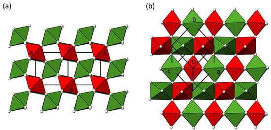
Figure 1.
(a) The crystal structure of α-NiTeO4 in a projection along [101]; (b) a section of the α-NiTeO4 structure in a projection along [1]. NiO6 octahedra are given in green and TeO6 octahedra in red color, with O atoms as colorless spheres of arbitrary radius.
3.3. Structure Description of β-NiTeO4
The new polymorph β-NiTeO4 crystallizes in the tetragonal space group I41/a. The asymmetric unit comprises one NiII atom with site symmetry (8 c), one TeVI atom with site symmetry (8 d), and two O atoms located in general sites (16 f). Like in the α-form, both Ni and Te sites in β-NiTeO4 are octahedrally surrounded by O atoms, and the individual bond lengths (Table 2) in the two modifications are nearly identical, with mean Ni–O and Te–O bond lengths of 2.057 Å and 1.929 Å, respectively, for the β-modification. Likewise, the connection of the octahedral building units is similar in the two modifications. In the crystal structure of β-NiTeO4, the strands made of edge-sharing alternating NiO6 and TeO6 octahedra extend along [001] (Figure 2a), and neighboring strands are, in turn, twisted by 90° against this strand and connected via common corners (Figure 2b).
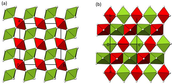
Figure 2.
(a) The crystal structure of β-NiTeO4 in a projection along [001]; (b) a section of the β-NiTeO4 structure in a projection along [110]. Color codes are as in Figure 1.
3.4. Structure Relation of α- and β-NiTeO4 to Rutile
Both modifications crystallize in hettotypes of the rutile aristotype and can be described as a twofold (α-NiTeO4) or eightfold (β-NiTeO4) cation-ordered superstructure. As in rutile itself [], the characteristic connection patterns, namely, strands of edge-linked octahedra that are fused to neighboring strands via common corners, can, therefore, be found in both polymorphs. Likewise, the typical environments of the oxygen atoms in the rutile structure are also observed in both polymorphs, i.e., each oxygen atom is bound in a (distorted) trigonal-planar manner to three positively charged atoms. The symmetry relationships [] between the rutile aristotype and the two hettotypes are graphically represented in Figure 3 in the form of a Bärnighausen tree []. Baur has described the structural relationships of rutile and its known derivatives in detail [] and has already mentioned the symmetry descent from rutile to the monoclinic dirutile type (referred to there as the CUU type), which is adapted by α-NiTeO4 and described in detail for isotypic CoTeO4 []. However, the crystal structure of the β-modification has not yet been described and, therefore, represents a new derivative of the rutile structure type. A direct symmetry derivation of β-NiTeO4 from rutile is not possible and proceeds via two hypothetical intermediate stages for which no representatives are yet known. In a first step, a translationengleiche (t) symmetry reduction of index 2 takes place, followed by a klassengleiche (k) symmetry reduction of index 2 under doubling of the unit cell. In the last step, another klassengleiche symmetry reduction k2 occurs under the ordering of the hypothetical M site into two cationic sites (Ni and Te) and a splitting of the hypothetical X site into two O sites. Due to the resulting I-centering, the cell volume is quadrupled.
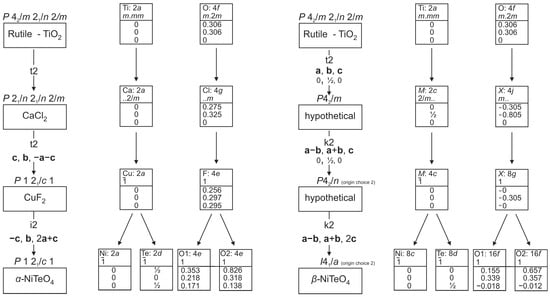
Figure 3.
Bärnighausen trees showing the symmetry relationship between the rutile aristotype and the α-NiTeO4 (left) and β-NiTeO4 (right) hettotypes.
The simultaneous occurrence of crystals of α- and β-NiTeO4 in the transport ampoule suggests that the energetic differences between the two polymorphic forms are small. However, the synthesis of polycrystalline NiTeO4 by solid-state reactions has always been reported to yield the monoclinic α-form [,], indicating that this may be the thermodynamically stable modification. Based on symmetry relationships, a direct transition from the tetragonal β-form, which has a larger crystal volume, to the monoclinic α-form, which has a smaller volume, is not possible. It is, therefore, likely that any phase transition that occurs between the two phases is of a reconstructive nature.
3.5. Structure Description of β-CuTe2O5
The asymmetric unit of β-CuTe2O5 consists of one formula unit, with all atoms on general positions (multiplicity 4, Wyckoff letter e) of space group P21/c. Selected bond lengths and angles are collated in Table 3.

Table 3.
β-CuTe2O5. Selected bond lengths/Å, angles/°, as well as bond valence sums (BVS)/v.u.
The copper(II) atom shows the characteristic [4+2] Jahn–Teller distortion [] with four short equatorial Cu–O bonds between 1.9523(18) and 1.9950(18) Å and two longer axial bonds of 2.4727(19) and 2.5635(19) Å, leading to a tetragonally distorted CuO6 octahedron. The mean Cu–O bond length (2.153 Å) corresponds almost perfectly to the literature value of 2.130(232) Å []. Individual CuO6 octahedra are linked through edge-sharing into CuO4/2O2/1] chains extending parallel to [100] whereby the long bonds are those to the edge-sharing atoms (Figure 4a).
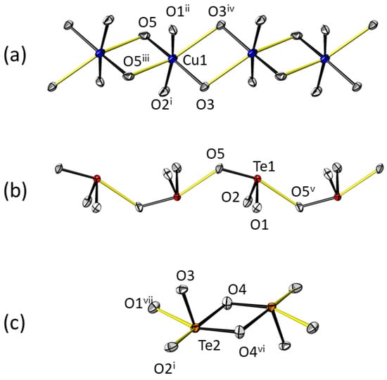
Figure 4.
The arrangement of structure elements from the main building units within the crystal structure of β-CuTe2O5. (a) The formation of a CuO4/2O2/1] chain from CuO6 octahedra; (b) the formation of a TeO2/2O2/1] chain from Te1O4 bisphenoids; (c) the formation of a TeO2/2O3/1]2 dimer from Te2O5 square pyramids. Black bonds refer to the inner coordination sphere, and yellow bonds to the outer coordination sphere. Displacement ellipsoids are given at the 74% probability level; symmetry codes refer to Table 3.
The coordination environments of the two TeIV atoms are clearly divided into two parts. If only Te–O bond lengths smaller than 2.1 Å are taken into account as being part of the inner coordination sphere, both TeIV atoms have three nearest oxygen neighbors in the form of the frequently observed TeO3 trigonal pyramid [] as a coordination polyhedron: Te1 shows an almost equidistant Te–O bond length distribution (range 1.8979(18)–1.9067(18) Å), while Te2 shows clear differences in these bond lengths, with one very short (1.8585(17) Å), one intermediate (1.9635(17) Å), and one longer bond (2.0413(17) Å), which, for the latter, can be related to the formation of centrosymmetric edge-sharing dimers, with the shortest bond being that to the terminal O atom. The outer coordination spheres of the two TeIV atoms include more remote O atoms within a threshold of 2.5 Å, and satisfactory BVS parameters for the two Te atoms can only be obtained if these longer bonds partake in the calculations (Table 3). Then Te1 is surrounded by one additional O atom at a distance of 2.4953(18) Å, with the resulting polyhedron having the shape of a distorted bisphenoid. Although the mean bond length of 2.050 Å for the Te1O4 polyhedron is greater than the literature value (1.984(123) Å) []), the deviation is within the single standard deviation.
The Te1O4 polyhedra share corner atoms (O5) to build up TeO2/2O2/1] chains propagating parallel to [010] whereby short and long bond lengths to the corner atoms alternate along the chain (Figure 4b). On the other hand, Te2 has even two additional bonding O atoms in the outer coordination sphere with distances at 2.3041(17) and 2.4026(18) Å, resulting in a distorted square pyramid as coordination polyhedron (mean Te–O bond length is 2.114 Å; literature value 2.251(551) Å []). Two Te2O5 polyhedra share an edge (O4 and its symmetry-related counterpart) forming a centrosymmetric dimer TeO2/2O3/1]2 (Figure 4c). In the crystal structure, these dimers are linked to the chains involving Te1 through common corners, making up a polymeric tri-periodic oxidotellurate(IV) framework. The lone-pair electrons Ψ of both TeIV atoms are stereochemically active and point into the free spaces of this arrangement (Figure 5). The fractional coordinates of Ψ were computed with LPLoc [] and amount to x = −0.0038, y = 0.6334, z = 0.13323 for Ψ1 at the Te1 atom (distance Te1–Ψ1 = 0.865 Å; LP radius 1.05 Å); x = 0.5033, y = 0.8586, z = 0.1934 for Ψ2 at the Te2 atom (distance Te2–Ψ2 = 0.742 Å; LP radius 0.98 Å).
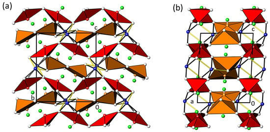
Figure 5.
(a) The crystal structure of β-CuTe2O5 in a projection along [100], and (b) in a projection along [010]. Te1O4 polyhedra are given in red, Te2O5 polyhedra in orange, Cu atoms as blue, O atoms as colorless, and the free electron pair Ψ as green spheres of arbitrary radii. Black bonds refer to short Cu–O bonds, and yellow bonds to long Cu–O bonds.
The crystal structure of β-CuTe2O5 has a completely different arrangement compared to that of α-CuTe2O5. In the latter (space group P21/c, Z = 4) [], the CuII atom likewise shows Jahn–Teller distortions but with one shorter (2.30 Å) and one longer (2.78 Å) bond to the remote O atoms (Figure 6a). The CuO6 octahedra share an edge to form CuO2/2O4/1]2 double octahedra, with the dimers arranged into rows parallel [001]. The two TeIV atoms, again under the consideration of only tightly bound O atoms, each have a trigonal-pyramidal environment. Through corner-sharing, a bent Te2O5 ditellurite group is formed with a Te–O–Te angle of 120.8°. The terminal Te–O2 bonds are somewhat shorter than those of the Te–O–Te bridge, in agreement with many examples in the literature []. One additional O atom is in the outer coordination sphere of Te2 at 2.40 Å and connects the isolated ditellurite groups into [100] chains (Figure 6b).
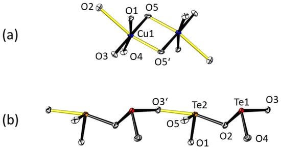
Figure 6.
The arrangement of structure elements from the main building units within the crystal structure of α-CuTe2O5 []. (a) The formation of a CuO2/2O4/1]2 dimer from CuO6 octahedra; (b) the formation of a Te2O2/2O3/1] chains from Te1O3 and Te2O[3+1] polyhedra. Displacement ellipsoids and color codes for bonds are as in Figure 3.
The Cu2O10 dimers and the ditellurite units are linked through corner- and edge-sharing into a tri-periodic framework, again with the lone-pair electrons Ψ of both Te atoms stereochemically active (coordinates calculated with LPLoc []: x = 0.0920, y = 0.1104, z = 0.1943 for Ψ1 at the Te1 atom (distance Te1–Ψ1 = 1.257 Å; LP radius 1.26 Å); x = 0.7149, y = 0.3671, z = 0.2078 for Ψ2 at the Te2 atom (distance Te2–Ψ2 = 1.16 Å; LP radius 1.18 Å). The crystal structure of α-CuTe2O5 is shown in Figure 7.
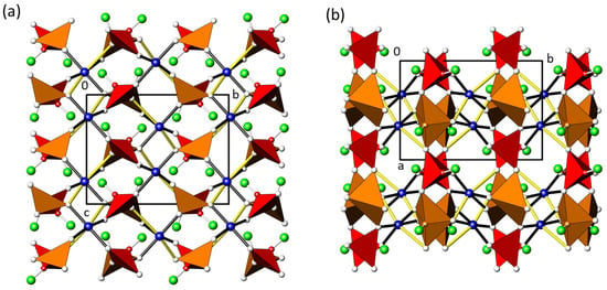
Figure 7.
(a) The crystal structure of β-CuTe2O5 in a projection along [100], and (b) in a projection along [001]. Te1O3 polyhedra are given in red, Te2O4 polyhedra in orange, Cu atoms as blue, O atoms as colorless, and the free electron pair E as green spheres of arbitrary radii. Black bonds refer to short Cu–O bonds, and yellow bonds to long Cu–O bonds.
The different structural arrangements of the building blocks in the two polymorphs make it appear very unlikely that a possible structural phase transition is enantiotropic. Based on the empirically derived Ostwald–Volmer rule, according to which the polymorph with the lower density is metastable, it might be assumed that β-CuTe2O5 (density 5.91 g/cm−3 (V/Z = 112.1 Å3) versus 5.76 g/cm−3 (V/Z = 115.04 Å3) for α-CuTe2O5) represents the thermodynamically stable form. There are, however, many known exceptions to this rule, e.g., graphite has a much lower density of 2.3 g cm–3 compared to diamond with 3.5 g cm–3, but it is the thermodynamically stable form. Moreover, the fact that CuTe2O5 has, so far, only been described in the form of the α-modification [] and as naturally occurring rajite [] speaks against the assumption that β-CuTe2O5 is the thermodynamically stable modification. It should be mentioned that in hydrothermal experiments for the production of α-CuTe2O5 (rajite) using CuSO4 and TeO2 as starting materials, “another phase or phases whose diffraction peaks do not match any records in the database” were also obtained in some of the experiments []. It does not seem unlikely that this unknown phase could be β-CuTe2O5.
3.6. Raman Spectroscopy of α- and β-CuTe2O5
The Raman spectra of α- and β-CuTe2O5 are shown in Figure 8, and the proposed assignments of Raman bands are compiled in Table 4.
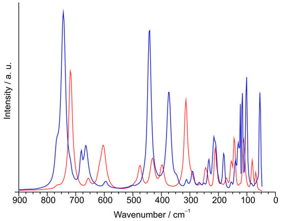
Figure 8.
Raman spectra of α-CuTe2O5 (blue) and β-CuTe2O5 (red).

Table 4.
Assignment of Raman bands.
The general spectral pattern of α-CuTe2O5 is similar to that previously reported for MgTe2O5 [], but interestingly, band positions are clearly shifted to lower wave numbers for α-CuTe2O5. This effect can probably be related to the differences in the outer oxygen coordination of the TeIV atoms in both structures; also, all Te–O bond lengths are slightly longer for the two TeIV atoms in α-CuTe2O5 (1.877, 1.883, 1.931; 1.859, 1.866, 2.019, 2.402 Å []) than for the unique TeIV atom in MgTe2O5 (1.8498 (11), 1.8588 (12), 1.9900 (9), 2.4048 (13) Å []).
In the crystal structure of α-CuTe2O5, the shape of the ditellurite anion is analogous to that of diselenite, Se2O72– (cf. for example [,]). Fifteen internal vibrations are expected for the seven-atomic Te2O52– anion as follows: two antisymmetric vibrations of the terminal TeO2 groups, two symmetrical vibrations of the terminal TeO2 groups, one antisymmetric Te–O–Te bridge vibration, one symmetric Te–O–Te bridge vibration, two deformations of the terminal TeO2 groups, one deformation of the Te–O–Te bridge, four O–TeO2 “rocking” vibrations, and two torsional vibrations. As the real symmetry of the anion in the crystal lattice is C1, the appearance of all these vibrations in the Raman effect can be expected. Notwithstanding and as usually observed in the case of crystal spectra, a reduction or an increase in the number of bands is also possible due to factor group effects and/or intensity problems.
As already discussed [], it is assumed that for the terminal TeO2-groups νs > νas in a similar way as for the analogous Se2O52− anion []. On the other hand, this assumption is clearly supported by intensity criteria since symmetric stretching modes in the Raman effect are usually very intense. For this reason, the most intense Raman band at 744 cm–1 is assigned to this mode. The weak shoulder on the higher energy side of this band (763 cm–1) is probably due to correlation field effects. The next doublet at 680 and 665 cm−1 is assigned to components of the corresponding antisymmetric stretching.
Regarding the Te–O–Te bridge motions, they are usually very weak in the Raman spectra [], a fact also observed in the case of diselenites []. We have tentatively assigned the weak band at 595 cm–1 to the antisymmetric mode, whereas the corresponding symmetric mode is only seen as a very weak feature at 507 cm–1. The relative positions of these two vibrations are also supported by a previous estimation based on a simple molecular model, showing the dependence of these two vibrations from the Te–O–Te angle []. The deformation mode of this bridge cannot be identified with certainty, but it probably lies near or below 200 cm–1 [].
In the lower energy region, the strongest Raman band (441 cm–1) is assigned to one of the expected TeO2 deformational modes, whereas the four expected “rocking” motions have been identified between 375 and 292 cm–1. The remaining band includes the expected torsional modes, the Te–O–Te bridge vibration, and some external (“lattice”) modes.
It is interesting to mention that the present Raman spectrum of synthetic α-CuTe2O5 closely resembles one of the spectra recorded for a sample of the mineral rajite. However, the corresponding analysis of these spectra is rather incomplete as the authors have not made structural considerations and based their assignment only on comparisons with the TeO32– anion, completely neglecting the presence of bands related to the Te2O52– bridge vibrations [].
The higher structural complexity of β-CuTe2O5 is clearly reflected in its Raman spectrum. For the sake of simplicity, only the Te–O bonds from the inner coordination sphere are considered relevant for the following analysis. In this case, the polymeric anion is constructed by the condensation of trigonal-pyramidal Te1O3 units with (Te2)2O4 units formed by edge-sharing of two trigonal-pyramidal TeO3 moieties.
The strongest Raman line (719 cm–1) can undoubtedly be related to the terminal Te–O bonds. Also, in this case, the weak shoulder preceding this band at higher energy may be generated by correlation field effects. The second group of bands can be assigned to Te–O–Te bridges involving the chains of condensed Te(1)O3 moieties. They appear better defined and at somewhat higher energies than in the case of α-CuTe2O5, probably as a consequence of the condensation of these groups. The following three bands are of more complex origin, involving different types of vibrations. In this region, one expects vibrations of the dimeric Te(2)2O2 units and deformational modes of the different polyhedra. The dimers with the double bridge can ideally be treated as a tetra-atomic system with D2h symmetry and, thus, possess six normal modes of vibration. Among them, three are Raman active: 2Ag + B1g. A comparison with other similar systems suggests that also here the strongest Raman line, and that at 245 cm–1, may be mixed to some extent with one of these vibrational modes []. Nevertheless, the vibrational modes related to these double bridges are mainly expected to lie around 450–400 cm–1. On the other hand, in this region and, also, at some lower energy, deformational modes of the TeOn polyhedra are expected. For comparison, δ(TeO3) modes of “free” TeO32− units are located at around 360–320 cm–1 []. The last group of bands (300–100 cm–1) includes torsional and deformational modes of different Te–O–Te bridges as well as external (“lattice”) vibrations.
3.7. Polymorphism Observed in Oxidotellurates
The observed polymorphism of NiTeO4 and CuTe2O5 is not a new phenomenon in the oxidotellurate family. A database search for polymorphic forms of purely inorganic oxidotellurates of general formula AxTeyOz (A is any other element, Te = TeIV, TeVI) in the Inorganic Crystal Structure Database (ICSD [], data release 2024.1) revealed a large number of cases, some even with up to five different polymorphs (like for CaTeO3 or SrTeO3), as the compilation of alphabetically arranged phases and their crystal data in Table 5 shows.

Table 5.
Structure data of polymorphic oxidotellurate phases with general composition AxTeyOz. Cases in which the polymorph was named with Greek letters or information on the measurement temperatures was provided are marked accordingly (HT = high temperature; LT = low temperature; HP = high pressure); cases where the existence of a polymorph was described without structure data are also included.
| Phase | Space Group | a/Å | b/Å | c/Å | α/° | β/° | γ/° | V/ų | Z | Ref. |
|---|---|---|---|---|---|---|---|---|---|---|
| Ag2Te2O6 | P21/m | 5.4562(5) | 7.4009(7) | 6.9122(7) | 90 | 101.237(2) | 90 | 273.77 | 2 | [] |
| P21/n | 5.9099(5) | 11.6831(8) | 8.0305(7) | 90 | 100.424(7) | 90 | 545.32 | 4 | [] | |
| P21/c | 6.4255(10) | 6.9852(11) | 13.204(2) | 90 | 90.090(3) | 90 | 592.64 | 4 | [] | |
| BaTeO3 | P21/m | 4.633(4) | 5.943(5) | 7.104(5) | 90 | 106.4(1) | 90 | 187.64 | 2 | [] |
| Pnma | 14.784(2) | 6.129(1) | 12.350(2) | 90 | 90 | 90 | 1119.05 | 12 | [] | |
| BaTe2O6 | Cmcm | 5.569(2) | 12.796(4) | 7.320(3) | 90 | 90 | 90 | 521.63 | 4 | [] |
| HP | P21/m | 5.40473 | 7.18472 | 6.54906 | 90 | 115.2374 | 90 | 230.03 | 2 | [] |
| CaTeO3 α | P43 | 12.1070(10) | 12.1070(10) | 11.0911(18) | 90 | 90 | 90 | 1625.73 | 20 | [] |
| β | P1 | 25.6220(4) | 10.2426(2) | 11.3327(2) | 107.218(10) | 110.245(2) | 33.0190(10) | 1520.42 | 18 | [] |
| β‘ 250 K | P | 25.6149(4) | 10.3921(2) | 11.2440(2) | 108.6080(10) | 112.394(2) | 32.3130(10) | 1479.25 | 18 | [] |
| γ | P21 | 8.4010(17) | 5.6913(11) | 22.680(5) | 90 | 110.82(3) | 90 | 1013.58 | 12 | [] |
| δ 673 K | P21ca | 13.3647(6) | 6.5330(3) | 8.1896(3) | 90 | 90 | 90 | 715.04 | 8 | [] |
| CaTe2O5 | P21/c | 9.382(2) | 5.7095(14) | 11.132(3) | 90 | 115.109(4) | 90 | 539.95 | 4 | [] |
| mica-like | [] | |||||||||
| Ca4Te5O14 | Pbca | 10.9536(16) | 16.556(2) | 15.779(2) | 90 | 90 | 90 | 2861.49 | 8 | [] |
| HP | P21/c | 11.0272(1) | 12.0588(1) | 10.1038(1) | 90 | 91.8490(10) | 90 | 1342.85 | 4 | [] |
| CdTeO3 α | P21/c | 7.790(1) | 11.253(2) | 7.418(1) | 90 | 113.5(1) | 90 | 596.34 | 8 | [] |
| β | Pnma | 7.45850(3) | 14.52185(6) | 11.04584(5) | 90 | 90 | 90 | 1196.37 | 16 | [] |
| CdTe2O5 100 K | P21/c | 9.4535(5) | 5.5806(3) | 10.8607(5) | 90 | 114.4300(10) | 90 | 521.66 | 4 | [] |
| mica-like | [] | |||||||||
| Cd3TeO6 | P21/n | 5.4986(3) | 5.6383(3) | 8.0191(5) | 90 | 90.00(5) | 90 | 248.61 | 2 | [] |
| R3 | 9.1620(2) | 9.1620(2) | 11.0736(3) | 90 | 90 | 120 | 805.00 | 6 | [] | |
| Co3TeO6 | C2/c | 14.8167(18) | 8.8509(11) | 10.3631(14) | 90 | 94.900(10) | 90 | 1354.06 | [] | |
| HP | R3 | 5.1937(6) | 5.1937(6) | 13.8237(17) | 90 | 90 | 120 | 322.93 | 3 | [] |
| Co5TeO8 | Fd3m | 8.55719(19) | 8.55719(19) | 8.55719(19) | 90 | 90 | 90 | 626.61 | 12 | [] |
| P4332 | 8.55185(4) | 8.55185(4) | 8.55185(4) | 90 | 90 | 90 | 625.42 | 4 | [] | |
| Cs2TeO4 | Pcmn | 11.6982(15) | 6.6747(9) | 8.5018(9) | 90 | 90 | 90 | 663.83 | 4 | [] |
| 773 K | P63/mmc | 6.8661(8) | 6.8661(8) | 8.897(1) | 90 | 90 | 120 | 363.24 | 2 | [] |
| CuTeO3 | Pmcn | 7.604(6) | 5.837(4) | 12.705(6) | 90 | 90 | 90 | 563.91 | 8 | [] |
| P21/n | 5.214(1) | 9.108(2) | 5.965(1) | 90 | 95.06(1) | 90 | 282.17 | 4 | [] | |
| CuTe2O5 α | P21/c | 6.871(2) | 9.322(2) | 7.602(2) | 90 | 109.08(1) | 90 | 460.17 | 4 | [] |
| β | P21/c | 6.6599(4) | 7.6233(4) | 8.8413(5) | 92.6910(10) | 448.38(4) | 4 | this work | ||
| Cu3TeO6 | Ia | 9.537(1) | 9.537(1) | 9.537(1) | 90 | 90 | 90 | 867.43 | 8 | [] |
| Pcca | 9.745(3) | 9.749(2) | 9.771(2) | 90 | 90 | 90 | 928.28 | 8 | [] | |
| HP | Ibca | 9.2444(3) | 9.3018(1) | 9.3757(4) | 90 | 90 | 90 | 806.21 | 8 | [] |
| Hg2Te2O7 α | C2/c | 12.910(4) | 7.407(2) | 13.256(4) | 90 | 112.044(5) | 90 | 1174.93 | 8 | [] |
| β | Aba2 | 7.4405(12) | 23.713(4) | 13.522(2) | 90 | 90 | 90 | 2385.78 | 16 | [] |
| Li2Te2O5 | P21/n | 10.355(3) | 4.702(1) | 10.860(3) | 90 | 110.13(1) | 90 | 496.46 | 4 | [] |
| Pnaa | 5.194(1) | 8.170(2) | 24.165(5) | 90 | 90 | 90 | 1025.44 | 8 | [] | |
| Mg3TeO6 | R3 | 8.615(3) | 8.615(3) | 10.315(3) | 90 | 90 | 120 | 663.00 | 6 | [] |
| HP | R3 | 5.1382(7) | 5.1382(7) | 13.8070(19) | 90 | 90 | 120 | 315.68 | 3 | [] |
| MnTeO3 α | Pbca | 10.0662(3) | 8.0958(2) | 14.1654(4) | 90 | 90 | 90 | 1154.39 | 16 | [] |
| β HP | Pnma | 6.1443(4) | 7.7866(7) | 5.4408(4) | 90 | 90 | 90 | 260.30 | 4 | [] |
| γ | Pbca | 7.4535(12) | 6.4164(11) | 12.843(2) | 90 | 90 | 90 | 614.21 | 8 | [] |
| MnTe2O5 α | P42/nbc | 8.82(5) | 8.82(5) | 13.04(5) | 90 | 90 | 90 | 1014.41 | 8 | [] |
| β | Pbcn | 7.3114(4) | 10.9216(6) | 6.1711(3) | 90 | 90 | 90 | 492.78 | 4 | [] |
| Mn2TeO6 HT | P42/mnm | 4.64205(11) | 4.64205(11) | 9.0750(2) | 90 | 90 | 90 | 195.54 | 2 | [] |
| LT | P21/c | 9.102948(10) | 13.046326(9) | 6.465927(6) | 90 | 90.0331(4) | 90 | 767.88 | 8 | [] |
| Mn3TeO6 | R | 8.8673(10) | 8.8673(10) | 10.6729(12) | 90 | 90 | 120 | 726.77 | 6 | [] |
| HP | P21/n | 5.29370(1) | 5.45203(1) | 7.80894(1) | 90 | 89.62514(5) | 90 | 225.37 | 2 | [] |
| MoTe2O7 α | P21/c | 4.286 | 8.618 | 15.945 | 90 | 95.68 | 90 | 586.06 | 4 | [] |
| Pna21 | 17.5632(5) | 4.9107(2) | 12.9518(4) | 90 | 90 | 90 | 1117.06 | 8 | [] | |
| Mo5TeO16 | P21/c | 10.038(2) | 14.431(2) | 8.1617(6) | 90 | 90.85(1) | 90 | 1182.16 | 4 | [] |
| Pm2a | 20.010(1) | 4.0650(2) | 7.2254(4) | 90 | 90 | 90 | 587.72 | 2 | [] | |
| Na2TeO4 α | P21/c | 10.632(5) | 5.161(2) | 13.837(11) | 90 | 103.27(4) | 90 | 738.99 | 8 | [] |
| β | Pbcn | 5.798(6) | 12.24(1) | 5.214(5) | 90 | 90 | 90 | 370.02 | 4 | [] |
| γ | Pnna | 13.5390(7) | 8.4169(4) | 6.7781(4) | 90 | 90 | 90 | 772.40 | 8 | [] |
| NiTeO4 α | P21/c | 5.5285(6) | 4.6500(5) | 5.5449(6) | 90 | 113.120(2) | 90 | 131.10(2) | 2 | this work |
| β | I41/a | 9.2627(5) | 9.2627(5) | 6.0988(4) | 90 | 90 | 90 | 523.26(7) | 8 | this work |
| Ni3TeO6 | R3 | 5.103(2) | 5.103(2) | 13.755(10) | 90 | 90 | 120 | 310.20 | 3 | [] |
| HP | Pnma | 5.9588(1) | 7.5028(1) | 5.2143(1) | 90 | 90 | 90 | 233.12 | 4 | [] |
| PbTeO3 α | C2/c | 26.73 | 4.6 | 18.06 | 90 | 106 | 90 | 2134.60 | 24 | [] |
| β | P41 | 5.304(3) | 5.304(3) | 11.900(6) | 90 | 90 | 90 | 334.78 | 4 | [] |
| γ | P | 7.0185(3) | 10.6166(4) | 11.9616(5) | 78.548(3) | 82.992(2) | 84.048(2) | 864.12 | 10 | [] |
| Sc2TeO6 | P321 | 8.7406(7) | 8.7406(7) | 4.7985(4) | 90 | 90 | 120 | 317.48 | 3 | [] |
| HP | 7.2943(3) | 5.1252(2) | 10.9502(4) | 90 | 103.8800(10) | 90 | 397.41 | 4 | [] | |
| SrTeO3 α | C2 | 28.151(6) | 5.8970(10) | 15.261(3) | 90 | 122.09(3) | 90 | 2146.35 | 24 | [] |
| β 473 K | C2/c | 28.206(6) | 5.9210(10) | 28.528(6) | 90 | 114.16(3) | 90 | 4347.06 | 48 | [] |
| γ 583 K | C2 | 28.262(6) | 5.9350(10) | 15.434(3) | 90 | 122.21(3) | 90 | 2190.40 | 24 | [] |
| δ 780 K | C2/m | 28.438(6) | 5.9500(10) | 15.550(3) | 90 | 122.45(3) | 90 | 2220.32 | 24 | [] |
| ε | P21/c | 6.7759(1) | 7.2188(1) | 8.6773(2) | 90 | 126.4980(7) | 90 | 341.20 | 4 | [] |
| Te(OH)6 | P21/n | 6.495(1) | 9.320(1) | 11.393(1) | 90 | 133.88 | 90 | 497.10 | 4 | [] |
| F4132 | 15.699(2) | 15.699(2) | 15.699(2) | 90 | 90 | 90 | 3869.15 | 32 | [] | |
| Te3O3(PO4)2 | P21/c | 12.375(2) | 7.317(1) | 9.834(1) | 90 | 98.04(1) | 90 | 881.70 | 4 | [] |
| β 203 K | P21/n | 11.115(5) | 4.7033(19) | 17.287(7) | 90 | 106.086(5) | 90 | 868.33 | 4 | [] |
| Tl2Te2O5 α | P21/n | 7.119(1) | 12.138(2) | 8.439(2) | 90 | 114.28(3) | 90 | 664.72 | 4 | [] |
| β | [] |
The causes and structural effects of polymorphism in the listed oxidotellurates are manifold—preparation conditions, temperature and/or pressure-dependent thermodynamic (meta-)stabilities, monotropic/enantiotropic transitions, reconstructive/displacive transitions, just to name but a few—and depend on an individual case. A detailed consideration is beyond the scope of this brief overview, and the interested reader is, therefore, referred to the original literature.
4. Conclusions
The original aim of this study, namely, the growth of NiTeO4 and CuTeO4 crystals as single-phase material, could not be achieved. At the selected average temperature of 620 °C under CVTR conditions, NiTeO4 is at the limit of its thermal stability, whereas CuTeO4 has no stability range under the selected hydrothermal conditions at 210 °C. This explains the observation that instead of the expected phases NiTeO4 and CuTeO4, single crystals of NiTe2O5 and of NiTeO4 (in the form of the known α-polymorph and of a previously unknown β-polymorph) and of so far unknown β-CuTe2O5, respectively, were obtained in very small amounts. Apart from crystal structure determinations from single-crystal X-ray data for α-, β-NiTeO4, and β-CuTe2O5, as well as Raman measurements for the latter, the scarcity of available material made further investigations and measurements of physical properties impossible. Hence, experimental details for the growth of α-, β-NiTeO4, and β-CuTe2O5 single crystals in larger quantities and as single-phase material must be optimized in the future. These time-consuming experiments could not be carried out as part of the present structural study. Only through the successful synthesis of the individual polymorphic forms will it be possible to obtain precise information about their thermodynamic stabilities and their thermal behaviors in relation to any occurring phase transitions.
Author Contributions
Professor Enrique J. Baran (E.J.B.) passed away in 2024 and is included as a co-author for his contributions to this article. Conceptualization, M.W.; methodology, M.W.; software, M.W.; validation, M.W. and E.J.B.; formal analysis, M.W. and E.J.B.; investigation, M.W. and E.J.B.; resources, M.W.; data curation, M.W.; writing—original draft preparation, M.W. and E.J.B.; writing—review and editing, M.W.; visualization, M.W.; project administration, M.W. All authors have read and agreed to the published version of the manuscript.
Funding
This research received no external funding.
Data Availability Statement
Part of the data presented in this study are available in The Cambridge Crystallographic Data Centre (CCDC) and can be obtained free of charge via www.ccdc.cam.ac.uk/structures.
Acknowledgments
The X-ray centre of TU Wien is acknowledged for providing free access to X-ray diffraction instruments.
Conflicts of Interest
The authors declare no conflicts of interest.
References
- Christy, A.G.; Mills, S.J.; Kampf, A.R. A review of the structural architecture of tellurium oxycompounds. Mineral. Mag. 2016, 80, 415–545. [Google Scholar] [CrossRef]
- Weil, M.; Pramanik, P.; Maltoni, P.M.; Clulow, R.; Rydh, A.; Wildner, M.; Blaha, P.; King, G.; Ivanov, S.A.; Mathieu, R.; et al. CoTeO4—A wide-bandgap material adopting the dirutile structure type. Mater. Adv. 2024, 5, 3001–3013. [Google Scholar] [CrossRef]
- Patel, A.K.; Panda, M.R.; Rani, E.; Singh, H.; Samatham, S.S.; Nagendra, A.; Jha, S.N.; Bhattacharyya, D.; Suresh, K.G.; Mitra, S. Unique Structure-Induced Magnetic and Electrochemical Activity in Nanostructured Transition Metal Tellurates Co1–XNixTeO4 (x = 0, 0.5, and 1). ACS Appl. Energy Mater. 2020, 3, 9436–9448. [Google Scholar] [CrossRef]
- Falck, L.; Lindquist, O.; Mark, W.; Philippot, E.; Moret, J. The crystal structure of CuTeO4. Acta Crystallogr. 1978, B34, 1450–1453. [Google Scholar] [CrossRef]
- Hasan, Z.; Zoghlin, E.; Winiarski, M.; Arpino, K.E.; Halloran, T.; Tran, T.T.; McQueen, T.M. Quasi-one-dimensional exchange interactions and short-range magnetic correlations in CuTeO4. Phys. Rev. B 2024, 109, 195168. [Google Scholar] [CrossRef]
- Chang, Y.H.R.; Yoon, T.L.; Lim, T.L.; Koh, P.W.; Goh, E.S. Effects of oxygen variation on the improved structural stability, electronic and optical properties of ZnTeO compounds. J. Phys. Condens. Matter 2020, 32, 225701. [Google Scholar] [CrossRef]
- Binnewies, M.; Glaum, R.; Schmidt, M.; Schmidt, P. Chemical Vapor Transport Reactions; De Gruyter: Berlin, Germany; Boston, MA, USA, 2012. [Google Scholar]
- Rabenau, A. The Role of Hydrothermal Synthesis in Preparative Chemistry. Angew. Chem. Int. Ed. Engl. 1985, 24, 1026–1040. [Google Scholar] [CrossRef]
- Schöneborn, M.; Hoffbauer, W.; Schmedt auf der Günne, J.; Glaum, R. Beiträge zur Kristallchemie und zum thermischen Verhalten von wasserfreien Phosphaten, XXXVII. Synthese, Kristallstruktur und kernresonanzspektroskopische Untersuchung von In2Ti6(PO4)6[Si2O(PO4)6]—Eine Hybride aus den NASICON und M4[Si2O(PO4)6] Strukturtypen. Z. Naturforsch. 2006, 61b, 741–748. [Google Scholar]
- Platte, C.; Trömel, M. Nickelditellurat(IV): Sauerstoffkoordinationszahl Fünf am vierwertigen Tellur. Acta Crystallogr. 1981, B37, 1276–1278. [Google Scholar] [CrossRef]
- APEX-4 and SAINT; Bruker AXS Inc.: Madison, WI, USA, 2021.
- Krause, L.; Herbst-Irmer, R.; Sheldrick, G.M.; Stalke, D. Comparison of silver and molybdenum microfocus X-ray sources for single-crystal structure determination. J. Appl. Cryst. 2015, 48, 3–10. [Google Scholar] [CrossRef] [PubMed]
- Sheldrick, G.M. SHELXT—Integrated space-group and crystal-structure determination. Acta Crystallogr. 2015, A71, 3–8. [Google Scholar] [CrossRef] [PubMed]
- Sheldrick, G.M. Crystal structure refinement with SHELXL. Acta Crystallogr. 2015, C71, 3–8. [Google Scholar]
- ATOMS for Windows; Shape Software: Kingsport, TN, USA, 2006.
- Brown, I.D. The Chemical Bond in Inorganic Chemistry: The Bond Valence Model; Oxford University Press: Oxford, UK, 2002. [Google Scholar]
- Brese, N.E.; O’Keeffe, M. Bond-valence parameters for solids. Acta Crystallogr. 1991, B47, 192–197. [Google Scholar] [CrossRef]
- Mills, S.J.; Christy, A.G. Revised values of the bond-valence parameters for TeIV–O, TeVI–O and TeIV–Cl. Acta Crystallogr. 2013, B69, 145–149. [Google Scholar] [CrossRef] [PubMed]
- Newnham, R.E.; Meagher, E.P. Crystal structure of Ni3TeO6. Mater. Res. Bull. 1967, 2, 549–554. [Google Scholar] [CrossRef]
- Binnewies, M.; Schmidt, M.; Schmidt, P. Chemical Vapor Transport Reactions—Arguments for Choosing a Suitable Transport Agent. Z. Anorg. Allg. Chem. 2017, 643, 1295–1311. [Google Scholar] [CrossRef]
- Hanke, K.; Kupčik, V.; Lindqvist, O. The crystal structure of CuTe2O5. Acta Crystallogr. 1973, B29, 963–970. [Google Scholar] [CrossRef]
- Williams, S.A. Rajite, naturally occurring cupric pyrotellurite, a new mineral. Mineral. Mag. 1979, 43, 91–92. [Google Scholar] [CrossRef][Green Version]
- Isasi, J. New MM’O4 oxides derived from the rutile type: Synthesis, structure and study of magnetic and electronic properties. J. Alloys Comps. 2001, 322, 89–96. [Google Scholar] [CrossRef]
- Salinas-Sanchez, A.; Garcia-Muñoz, J.L.; Rodriguez-Carvajal, J.; Saez-Puche, R.; Martinez, J.L. Structural characterization of R2BaCuO5 (R = Y, Lu, Yb, Tm, Er, Ho, Dy, Gd, Eu and Sm) oxides by X-ray and neutron diffraction. J. Solid State Chem. 1992, 100, 201–211. [Google Scholar]
- Brown, I.D. Recent Developments in the Methods and Applications of the Bond Valence Model. Chem. Rev. 2009, 109, 6858–6919. [Google Scholar] [CrossRef] [PubMed]
- Chen, H.; Wong, L.L.; Adams, S. SoftBV–a software tool for screening the materials genome of inorganic fast ion conductors. Acta Crystallogr. 2019, B75, 18–33. [Google Scholar] [CrossRef]
- Gagné, O.C.; Hawthorne, F.C. Bond-length distributions for ions bonded to oxygen: Results for the transition metals and quantification of the factors underlying bond-length variation in inorganic solids. IUCrJ 2020, 7, 581–629. [Google Scholar] [CrossRef] [PubMed]
- Gagné, O.C.; Hawthorne, F.C. Bond-length distributions for ions bonded to oxygen: Results for the non-metals and discussion of lone-pair stereoactivity and the polymerization of PO4. Acta Crystallogr. 2018, B74, 79–96. [Google Scholar]
- Baur, W.H. The rutile type and its derivatives. Crystallogr. Rev. 2007, 13, 65–113. [Google Scholar] [CrossRef]
- Müller, U. Symmetry Relationships Between Crystal Structures; Oxford University Press: Okford, UK, 2013. [Google Scholar]
- Bärnighausen, H. Group-Subgroup Relations between Space Groups: A useful Tool in Crystal Chemistry. MATCH Commun. Math. Chem. 1989, 9, 139–175. [Google Scholar]
- Halcrow, M.A. Jahn–Teller distortions in transition metal compounds, and their importance in functional molecular and inorganic materials. Chem Soc. Rev. 2013, 42, 1784–1795. [Google Scholar] [CrossRef] [PubMed]
- Hamani, D.; Masson, O.; Thomas, P. Localization and steric effect of the lone electron pair of the tellurium Te4+ cation and other cations of the p-block elements. A systematic study. J. Appl. Crystallogr. 2020, 53, 1243–1251. [Google Scholar] [CrossRef]
- Majzlan, J.; Notz, S.; Haase, P.; Kamitsos, E.I.; Tagiara, N.S.; Dachs, E. Thermodynamic properties of tellurite (β-TeO2), paratellurite (α-TeO2), TeO2 glass, and Te(IV) phases with stoichiometry M2Te3O8, MTe6O13, MTe2O5 (M2+ = Co, Cu, Mg, Mn, Ni, Zn). Geochemistry 2022, 82, 12591. [Google Scholar] [CrossRef]
- Baran, E.J. Das Schwingungsspektrum des Ditellurit-Ions. Z. Anorg Allg Chem. 1978, 442, 112–118. [Google Scholar] [CrossRef]
- Weil, M. Redetermination of MgTe2O5. Acta Crystallogr. 2005, E61, i237–i239. [Google Scholar] [CrossRef]
- Simon, A.; Paetzold, R. Untersuchungen an Selen–Sauerstoff-Verbindungen. III. Die Struktur des Pyroselenitions. Z. Anorg. Allg. Chem. 1960, 303, 39–45. [Google Scholar] [CrossRef]
- Frost, R.L.; Dickfos, M.J.; Keeffe, E.C. Raman spectroscopic study of the tellurite minerals: Rajite and denningite. Spectrochim. Acta 2008, 71A, 1512–1515. [Google Scholar] [CrossRef] [PubMed][Green Version]
- Jezowska-Trzebiatowska, B. Theory and importance of oxygen bridge-bonding. Pure Appl. Chem. 1971, 27, 89–111. [Google Scholar] [CrossRef]
- Siebert, H. Anwendungen der Schingungsspektroskopie in der Anorganischen Chemie; Springer: Berlin/Heidelberg, Germany, 1966. [Google Scholar]
- Zagorac, D.; Müller, H.; Ruehl, S.; Zagorac, J.; Rehme, S. Recent developments in the Inorganic Crystal Structure Database: Theoretical crystal structure data and related features. J. Appl. Crystallogr. 2019, 52, 918–925. [Google Scholar] [CrossRef] [PubMed]
- Klein, W.; Curda, J.; Peters, E.M.; Jansen, M. Neue Silber(I)-oxotellurate(IV/VI). Z. Anorg. Allg. Chem. 2005, 631, 2893–2899. [Google Scholar] [CrossRef]
- Weil, M. New silver tellurates—The crystal structures of a third modification of Ag2Te2O6 and of Ag4TeO5. Z. Anorg. Allg. Chem. 2007, 633, 1217–1222. [Google Scholar] [CrossRef]
- Koçak, M.; Platte, C.; Trömel, M. Über verschiedene Formen von BaTeO3. Z. Anorg. Allg. Chem. 1979, 453, 93–97. [Google Scholar] [CrossRef]
- Koçak, M.; Platte, C.; Trömel, M. Bariumhexaoxoditellurat(IV,VI). Sauerstoffkoordinationszahl fünf am vierwertigen Tellur. Acta Crystallogr. 1979, B35, 1439–1441. [Google Scholar] [CrossRef]
- Mishra, K.K.; Achary, S.N.; Chandra, S.; Ravindran, T.R.; Pandey, K.K.; Tyagi, A.K.; Sharma, S.M. Study of Phase Transformation in BaTe2O6 by in Situ High-Pressure X-ray Diffraction, Raman Spectroscopy, and First-Principles Calculations. Inorg. Chem. 2016, 55, 227–238. [Google Scholar] [CrossRef] [PubMed]
- Stöger, B.; Weil, M.; Zobetz, E.; Giester, G. Polymorphism of CaTeO3 and solid solutions CaxSr1−xTeO3. Acta Crystallogr. 2009, B65, 167–181. [Google Scholar] [CrossRef] [PubMed]
- Poupon, M.; Barrier, N.; Petit, S.; Clevers, S.; Dupray, V. Hydrothermal Synthesis and Dehydration of CaTeO3(H2O): An Original Route to Generate New CaTeO3 Polymorphs. Inorg. Chem. 2015, 54, 5660–5670. [Google Scholar] [CrossRef]
- Weil, M.; Stöger, B. A non-twinned polymorph of CaTe2O5 from a hydrothermally grown crystal. Acta Crystallogr. 2008, C64, i79–i81. [Google Scholar]
- Redman, M.J.; Chen, J.H.; Binnie, W.P.; Mallio, W.J. Mica-Like Tellurites of Calcium, Strontium, and Cadmium. J. Am. Ceram. Soc. 1970, 53, 645–648. [Google Scholar] [CrossRef]
- Weil, M. New phases in the systems Ca–Te–O and Cd–Te–O: The calcium tellurite(IV) Ca4Te5O14 and the cadmium compounds Cd2Te3O9 and Cd2Te2O7 with mixed-valent oxotellurium(IV/VI) anions. Solid State Sci. 2004, 6, 29–37. [Google Scholar] [CrossRef]
- Weil, M.; Heymann, G.; Huppertz, H. The high-pressure polymorph of Ca4Te5O14 and the mixed-valent compound Ca13 TeVI2/3- TeIV3.75 O15 (BO3)4(OH)3. Eur. J. Inorg. Chem. 2016, 2016, 3574–3579. [Google Scholar] [CrossRef]
- Krämer, V.; Brandt, G. Structure of cadmium tellurate(IV), CdTeO3. Acta Crystallogr. 1985, C41, 1152–1154. [Google Scholar] [CrossRef]
- Poupon, M.; Barrier, N.; Petit, S.; Boudin, S. A new β-CdTeO3 polymorph with a structure related to α-CdTeO3. Dalton Trans. 2017, 46, 1927–1935. [Google Scholar] [CrossRef]
- Eder, F.; Weil, M. The crystal structure of a new CdTe2O5 polymorph, isotypic with ε-CaTe2O5. Acta Crystallogr. 2020, E76, 831–834. [Google Scholar]
- Burckhardt, H.G.; Platte, C.; Trömel, M. Cadmiumorthotellurat(VI) Cd3TeO6: Ein pseudoorthorhombischer Kryolith im Vergleich mit Ca3TeO6. Acta Crystallogr. 1982, B38, 2450–2452. [Google Scholar] [CrossRef]
- Weil, M.; Veyer, T. A new form of Cd3TeO6 revealing dimorphism. Acta Crystallogr. 2018, E74, 1561–1564. [Google Scholar]
- Becker, R.; Johnsson, M.; Berger, H. A new synthetic cobalt tellurate: Co3TeO6. Acta Crystallogr. 2006, C62, i67–i69. [Google Scholar]
- Selb, E.; Buttlar, T.; Janka, O.; Tribus, M.; Ebbinghaus, S.G.; Heymann, G. Multianvil high-pressure/high-temperature synthesis and characterization of magnetoelectric HP-Co3TeO6. J. Mater. Chem. C 2021, 9, 5486–5496. [Google Scholar] [CrossRef]
- Podchezertsev, S.; Barrier, N.; Pautrat, A.; Suard, E.; Retuerto, M.; Alonso, J.A.; Fernández-Díaz, M.T.; Rodríguez-Carvajal, J. Influence of polymorphism on the magnetic properties of Co5TeO8 spinel. Inorg. Chem. 2021, 60, 13990–14001. [Google Scholar] [CrossRef] [PubMed]
- Epifano, E.; Volfi, A.; Abbink, M.; Nieuwland, H.; van Eijck, L.; Wallez, G.; Banerjee, D.; Martin, P.M.; Smith, A.L. Investigation of the Cs(Mo,Te)O4 Solid Solution and Implications on the Joint Oxyde-Gaine System in Fast Neutron Reactors. Inorg. Chem. 2020, 59, 10172–10184. [Google Scholar] [CrossRef] [PubMed]
- Lindqvist, O. The Crystal Structure of CuTeO3. Acta Chem. Scand. 1972, 26, 1423–1430. [Google Scholar] [CrossRef]
- Pertlik, F. Dimorphism of hydrothermal synthesized copper tellurite CuTeO3: The structure of a monoclinic representative. J. Solid State Chem. 1987, 71, 291–295. [Google Scholar] [CrossRef]
- Hostachy, A.; Coing-Boyat, J. Structure cristalline de Cu3TeO6. C. R. Seances Acad. Sci. Ser. B 1968, 267, 1435–1438. [Google Scholar]
- Missen, O.P.; Mills, S.J.; Canossa, S.; Hadermann, J.; Nénert, G.; Weil, M.; Libowitzky, E.; Housley, R.M.; Artner, W.; Kampf, A.R.; et al. Polytypism in mcalpineite. A study of natural and synthetic Cu3TeO6. Acta Crystallogr. 2022, B78, 20–32. [Google Scholar] [CrossRef]
- Qu, J.; Yan, L.; Liu, H.; Tao, Q.; Zhu, P.; Li, Z.; Wang, X. Pressure-induced structural phase transition in corundum-related class Cu3TeO6. High Press. Res. 2021, 41, 318–327. [Google Scholar] [CrossRef]
- Weil, M. Dimorphism in mercury(II) tellurite(IV) tellurate(VI): Preparation and crystal structures of α- and β-Hg2Te2O7. Z. Kristallogr. 2003, 218, 691–698. [Google Scholar] [CrossRef]
- Cachau-Herreillat, D.; Norbert, A.; Maurin, M.; Philippot, E. Etude cristallochimique comparée et conductivité ionique des deux variétés Li2Te2O5 α et β. J. Solid State Chem. 1981, 37, 352–361. [Google Scholar] [CrossRef]
- Schulz, H.; Bayer, G. Structure determination of Mg3TeO6. Acta Crystallogr. 1971, B27, 815–821. [Google Scholar] [CrossRef]
- Selb, E.; Declara, L.; Bayarjargal, L.; Podewitz, M.; Tribus, M.; Heymann, G. Crystal Structure and Properties of a UV-Transparent High-Pressure Polymorph of Mg3TeO6 with Second Harmonic Generation Response. Eur. J. Inorg. Chem. 2019, 2019, 4668–4676. [Google Scholar] [CrossRef]
- Eder, F.; Weil, M. Phase formation studies and crystal structure refinements in the MnII/TeIV/O/(H) system. Z. Anorg. Allg. Chem. 2022, 648, e202200205. [Google Scholar] [CrossRef]
- Kohn, K.; Inoue, K.; Horie, O.; Akimoto, S.-I. Crystal chemistry of MSeO3 and MTeO3 (M = Mg, Mn, Co, Ni, Cu, and Zn). J. Solid State Chem. 1976, 18, 27–37. [Google Scholar] [CrossRef]
- Walitzi, E.M. Die Kristallstruktur von Denningit, (Mn,Ca,Zn)Te2O5. Ein Beispiel für die Koordination um vierwertiges Tellur. Tschermaks Mineral. Petrogr. Mitt. 1965, 10, 241–255. [Google Scholar] [CrossRef]
- Johnston, M.G.; Harrison, W.T.A. Manganese tellurite, β-MnTe2O5. Acta Crystallogr. 2002, E58, i59–i61. [Google Scholar] [CrossRef]
- Matsubara, N.; Damay, F.; Vertruyen, B.; Barrier, N.; Lebedev, O.I.; Boullay, P.; Elkaim, E.; Manuel, P.; Khalyavin, D.D.; Martin, C. Mn2TeO6: A distorted inverse trirutile structure. Inorg. Chem. 2017, 56, 9742–9753. [Google Scholar] [CrossRef]
- Weil, M. Mn3TeO6. Acta Crystallogr. 2006, E62, i244–i245. [Google Scholar] [CrossRef]
- Arévalo-López, A.M.; Solana-Madruga, E.; Aguilar-Maldonado, C.; Ritter, C.; Olivier Mentré, O.; Attfield, J.P. Magnetic frustration in the high-pressure Mn2MnTeO6 (Mn3TeO6-II) double perovskite. Chem. Commun. 2019, 55, 14470–14473. [Google Scholar] [CrossRef] [PubMed]
- Arnaud, Y.; Averbuch-Pouchot, M.T.; Durif, A.; Guidot, J. Structure cristalline de l’oxide mixte de molybdene-tellure: MoTe2O7. Acta Crystallogr. 1976, B32, 1417–1420. [Google Scholar] [CrossRef]
- Ling, J.; Zhang, H.; Yuan, K.; Burgess, D.; Hu, J.; Hu, M. Hydrothermal syntheses and crystal structures of molybdenum tellurites. J. Solid State Chem. 2020, 287, 121317. [Google Scholar] [CrossRef]
- Arnaud, Y.; Guidot, J. Structure cristalline de l’oxyde mixte de molybdene-tellure: Mo5TeO16. Acta Crystallogr. 1977, B33, 2151–2155. [Google Scholar] [CrossRef]
- Forestier, P.; Goreaud, M. Structure cristalline de l’oxyde a valence mixte TeMo5O16 orthorombique. C. R. Acad. Sci. Ser. II 1991, 312, 1141–1145. [Google Scholar]
- Daniel, F.; Maurin, M.; Moret, J.; Philippot, E. Etude structurale d’un nouveau tellurate alcalin: Na2TeO4. Evolution de la coordination du tellure(VI) et du cation quand on passe du cation lithium au sodium. J. Solid State Chem. 1977, 22, 385–391. [Google Scholar] [CrossRef]
- Kratochvil, B.; Jensovsky, L. The crystal structure of sodium metatellurate. Acta Crystallogr. 1977, B33, 2596–2598. [Google Scholar] [CrossRef]
- Weil, M.; Stöger, B.; Larvor, C.; Raih, I.; Gierl-Mayer, C. The hydrous sodium oxotellurates(VI) Na[TeO(OH)5], Na2[TeO2(OH)4], Na4[Te2O6(OH)4](H2O)6, and a third polymorph of anhydrous Na2[TeO4]. Z. Anorg. Allg. Chem. 2017, 643, 1888–1897. [Google Scholar] [CrossRef]
- Martínez-Lope, M.J.; Retuerto, M.; Alonso, J.A.; Sánchez-Benítez, J.; Fernández-Díaz, M.T. High-pressure synthesis and neutron diffraction investigation of the crystallographic and magnetic structure of TeNiO3 perovskite. Dalton Trans. 2011, 40, 4599–4604. [Google Scholar] [CrossRef]
- Mariolacos, K. Die Kristallstruktur von PbTeO3. Anz. Oesterr. Akad. Wiss. Math.-Naturwiss. Kl. 1969, 106, 129–131. [Google Scholar]
- Sciau, P.; Lapasset, J.; Moret, J. Structure de la phase quadratique de PbTeO3. Acta Crystallogr. 1986, C42, 1688–1690. [Google Scholar] [CrossRef]
- Weil, M.; Shirkhanlou, M.; Füglein, E.; Libowitzky, E. Determination of the correct composition of anhydrous lead(II) oxotellurate(IV) as PbTeO3, crystallizing as a new polymorph. Crystals 2018, 8, 51. [Google Scholar] [CrossRef]
- Höss, P.; Schleid, T. Sc2Te5O13 und Sc2TeO6: Die ersten Oxotellurate des Scandiums. Z. Anorg. Allg. Chem. 2007, 633, 1391–1396. [Google Scholar] [CrossRef]
- Ziegler, R.; Tribus, M.; Hejny, C.; Heymann, G. Single-crystal structure of HP-Sc2TeO6 prepared by high-pressure/high-temperature synthesis. Crystals 2021, 11, 1554. [Google Scholar] [CrossRef]
- Zavodnik, V.E.; Ivanov, S.A.; Stash, A.I. The α-phase of SrTeO3 at 295 K. Acta Crystallogr. 2007, E63, i75–i76. [Google Scholar] [CrossRef]
- Zavodnik, V.E.; Ivanov, S.A.; Stash, A.I. On the thermal evolution of the crystal structure of SrTeO3: The β-form at 473 K. Acta Crystallogr. 2007, E63, i111–i112. [Google Scholar] [CrossRef]
- Zavodnik, V.E.; Ivanov, S.A.; Stash, A.I. The γ-phase of SrTeO3 at 583 K. Acta Crystallogr. 2007, E63, i151. [Google Scholar]
- Zavodnik, V.E.; Ivanov, S.A.; Stash, A.I. The δ-phase of SrTeO3 at 780 K. Acta Crystallogr. 2008, E64, i52. [Google Scholar]
- Stöger, B.; Weil, M.; Baran, E.J.; Gonzalez Baro, A.C.; Malo, S.; Rueff, J.M.; Petit, S.; Lepetit, M.B.; Raveau, B.; Barrier, N. The dehydration of SrTeO3(H2O)—A topotactic reaction for preparation of the new metastable strontium oxotellurate(IV) phase ε-SrTeO3. Dalton Trans. 2011, 40, 5538–5548. [Google Scholar] [CrossRef]
- Lindqvist, O.; Lehmann, M.S. A neutron diffraction refinement of the crystal structure of telluric acid, Te(OH)6 (mon). Acta Chem. Scand. 1973, 27, 85–95. [Google Scholar] [CrossRef]
- Mullica, D.F.; Korp, J.D.; Mulligan, W.O.; Beall, G.W.; Bernal, I. Neutron Structural Refinement of Cubic Orthotelluric Acid. Acta Crystallogr. 1980, B36, 2565–2570. [Google Scholar] [CrossRef]
- Mayer, H.; Weil, M. Synthese und Kristallstruktur von Te3O3(PO4)2, einer Verbindung mit fünffach koordiniertem Tellur(IV). Z. Anorg. Allg. Chem. 2003, 629, 1068–1072. [Google Scholar] [CrossRef]
- Li, L.; Zhuang, R.-C.; Mi, J.-X.; Huang, Y.-X. A New Modification of Tellurite Phosphate: β-Te3O3(PO4)2. Chin. J. Struct. Chem. 2018, 37, 1417–1425. [Google Scholar]
- Jeansannetas, B.; Thomas, P.; Champarnaud-Mesjard, J.C.; Frit, B. Crystal structure of α-Tl2Te2O5. Mater. Res. Bull. 1998, 33, 1709–1716. [Google Scholar] [CrossRef]
- Jeansannetas, B.; Marchet, P.; Thomas, P.; Champarnaud-Mesjard, J.C.; Frit, B. New investigations within the TeO2-rich part of the Tl2O–TeO2 system. J. Mater. Chem. 1998, 8, 1039–1042. [Google Scholar] [CrossRef]
Disclaimer/Publisher’s Note: The statements, opinions and data contained in all publications are solely those of the individual author(s) and contributor(s) and not of MDPI and/or the editor(s). MDPI and/or the editor(s) disclaim responsibility for any injury to people or property resulting from any ideas, methods, instructions or products referred to in the content. |
© 2025 by the authors. Licensee MDPI, Basel, Switzerland. This article is an open access article distributed under the terms and conditions of the Creative Commons Attribution (CC BY) license (https://creativecommons.org/licenses/by/4.0/).