Article

Ceramic Bracket Surface Treated with Hydrofluoric Acid, Er, Cr: YSGG Laser, and Phthalocyanine Activated via Low-Level Laser Therapy on Surface Roughness and Shear Bond Strength Bonded to Enamel via Unmodified and Sepiolite-Modified Orthodontic Adhesive-A SEM, EDX, and DC Evaluation

by
Salem Almoammar
1,
Muhammad Abdullah Kamran
1,*,
Abdulrahman Alshehri
2,
Wael Awadh
2,
Amirah Mesfer Alshahrani
3 and
Ibrahim Alshahrani
4
1
Department of Pedodontics and Orthodontic Sciences, College of Dentistry, King Khalid University, Abha 62521, Saudi Arabia
2
Division of Orthodontics, Department of Preventive Dental Sciences, College of Dentistry, Jazan University, Ash Shati 82943, Saudi Arabia
3
Orthodontic Department, Armed Forces Hospital Southern Region, Ministry of Defense, Abha-Aseer 62413, Saudi Arabia
4
Department of Pediatric Dentistry and Orthodontic Sciences, College of Dentistry, King Khalid University, Abha 62521, Saudi Arabia
*
Author to whom correspondence should be addressed.
Crystals 2025, 15(12), 1010; https://doi.org/10.3390/cryst15121010
Submission received: 18 October 2025 / Revised: 20 November 2025 / Accepted: 21 November 2025 / Published: 24 November 2025
(This article belongs to the Section Inorganic Crystalline Materials)

Abstract

Influence of surface pretreatment Hydrofluoric acid (HFA), Erbium yttrium scandium gallium garnet (Er, Cr: YSGG) laser (ECL), and Phthalocyanine (Pc) photosensitizer (Ps) activated by Low-level laser therapy (LLLT) via a light-emitting diode (LED) device on surface roughness (Ra) and shear bond strength (SBS) of ceramic bracket bonded to enamel via unmodified and Sepiolite-modified adhesive. Sixty non-cavitated human maxillary premolars were obtained. Ninety ceramic brackets were classified into three groups based on different pretreatment methods: Group 1: HFA; Group 2: ECL; and Group 3: Pc-LLLT. Twenty samples from each cohort were allocated into two subgroups by adhesive type: unmodified Transbond XT(A) and adhesive-modified Sep-NPs(B) (n = 10). Ra was measured using profilometry followed by surface topography via SEM, SBS via universal testing machine, and degree of conversion (DC) through FTIR spectroscopy. ANOVA and Tukey’s post hoc tests compared Ra, SBS, and DC across groups (p ˂ 0.05). Maximum Ra was observed in the ECL group (1087.43 ± 0.043 µm), while Group 3 (Pc-LLLT) showed the lowest Ra (706.53 ± 0.054 µm). Maximum SBS was recorded in Group 2B (ECL + SepNPs modified adhesive) (8.79 ± 0.48 MPa), while Group 3A (Pc-LLLT + unmodified adhesive) (5.23 ± 0.32 MPa) showed minimum bond integrity. ECL serves as an appropriate substitute for HFA in improving Ra and SBS of ceramic brackets to enamel. SepNPs improved the SBS of orthodontic adhesive to enamel with no significant difference in DC.

1. Introduction

Currently, a growing number of adults are pursuing orthodontic care mainly for aesthetic purposes [1]. They are seeking orthodontic treatment that is of high quality and is minimally invasive. This resulted in the creation of new brackets with a more attractive and acceptable look [2]. Ceramic brackets are widely utilized in modern orthodontics because of their aesthetic appeal, compatibility with oral tissues, and resistance to thermal and chemical alterations [3]. It was identified that their shear bond strength (SBS) to the enamel surface is comparable to that of metal brackets [4]. Despite their aesthetic advantages, ceramic brackets present clinical challenges, including increased brittleness during debonding and inconsistent bonding reliability to enamel [5].
In prosthodontics, it is a standard practice to pretreat ceramic materials to improve their surface roughness (Ra) and bonding strength to the substrate. Hydrofluoric acid (HFA) is considered the standard pretreatment method for preparing ceramics [6]. HFA selectively dissolves the silica (SiO2) component within the glassy matrix of feldspathic and lithium disilicate ceramics through a chemical reaction that forms silicon tetrafluoride (SiF4) gas and hexafluorosilicic acid (H2SiF6) [7]. This selective dissolution creates microporosities ranging from 5 to 30 μm in depth, significantly increasing the available surface area for micromechanical retention [8]. According to Faltermeier and colleagues, HFA effectively conditions the surface of ceramic restorations before attaching ceramic brackets [9]. However, HFA poses significant occupational and patient safety hazards. Dermal contact can cause severe chemical burns penetrating to deep tissue layers, while inhalation of HFA vapors during clinical application may result in pulmonary edema [10]. Consequently, it is crucial to find better alternatives that do not endanger human biological tissues.
In addition to acid etching, laser technology presents a viable alternative for the conditioning of ceramic surfaces [11,12]. Among the various laser types, the Erbium yttrium scandium gallium garnet (Er, Cr: YSGG) laser (ECL) has garnered significant recognition among orthodontic practitioners for its efficacy in enamel conditioning and the reversal of bleached enamel [13,14]. Likewise, ECL has been effectively employed in the repair of zirconia and lithium disilicate ceramics, yielding promising results [15,16,17]. However, the role of ECL as a ceramic bracket surface conditioner in relation to their Ra and SBS with enamel remains to be elucidated and warrants further investigation.
Photodynamic therapy (PDT), particularly in the modality of low-level laser therapy (LLLT), has been employed to modify the surfaces of various dental materials. LLLT, often referred to as cold laser therapy (CLT), represents a methodology that utilizes low-intensity red and near-infrared light to promote tissue regeneration and mitigate inflammation [18,19,20]. Several investigations indicated that CLT may enhance the adhesive strength between ceramic restorations and resin-based cement [18,21]. Phthalocyanine (Pc), a synthetic tetrapyrrolic macrocycle, functions as a Type II photosensitizer generating singlet oxygen (1O2) upon light activation at 630–680 nm wavelength [22,23]. When activated by low-level laser therapy (LLLT), Pc-generated reactive oxygen species (ROS) may modify ceramic surfaces through multiple oxidative mechanisms: (1) oxidation of residual organic contaminants from manufacturing processes (e.g., mold release agents, silane coupling agents) that persist despite routine cleaning [24], (2) formation of surface hydroxyl groups (-OH), carboxyl groups (-COOH), and carbonyl groups (C=O) through oxidation of the ceramic matrix [25], and (3) creation of surface oxides that enhance surface energy and wettability [26].While ceramic brackets undergo routine cleaning before clinical use, manufacturing residues at the nanoscale level often remain on bracket bases, potentially interfering with optimal bonding.
Beyond the conditioning regimen, the role of the adhesive in improving the SBS results of different ceramics to the dental substrate is clear. In the field of restorative dentistry, incorporating nanoparticles (NPs) into resin adhesives is a common approach to enhance SBS [27]. Sepiolite nanoparticles (SepNPs), due to their high aspect ratio and nanofibrous morphology, create a reinforced polymer network that enhances the mechanical properties of the adhesive interface through improved stress distribution and crack deflection mechanisms [28,29]. The use of SepNPs is seen as a promising method to enhance the mechanical properties of dentin bonding agents, as noted by Niazi and colleagues [30]. However, their use in orthodontic adhesives for bonding orthodontic brackets is still largely unexplored and requires further research.
The prevailing exploration was established on the proposition that there will be no significant difference in the Ra and bond integrity of ceramic brackets pretreated with ECL and Pc-LLLT compared to HFA. Moreover, it was also anticipated that the bond strength of unmodified orthodontic adhesive utilized for bonding ceramic brackets to enamel would be comparable to that of Sep-NPs modified orthodontic adhesive, with no significant difference in degree of conversion (DC). Consequently, the study seeks to evaluate the influence of surface pretreatment of ceramic brackets via ECL and Pc-LLLT on the DC, Ra, and bond integrity of ceramic brackets to enamel employing both unmodified and Sep-NPs modified orthodontic adhesives.

2. Materials and Methods

Preparation of the tooth sample: Sixty human maxillary premolars were selected for the study. Sample size calculation was performed using G*Power software version 3.1.9.7 (Heinrich-Heine-Universität Düsseldorf, Düsseldorf, Germany). A sample size of n = 10 per subgroup was calculated to detect this difference with 80% power at α = 0.05 significance level (effect size d = 1) [31,32]. Teeth exhibiting restorations, visible cavities, whitening treatments, discolorations, fractures, or hyperplasia were not part of the selection. The included teeth underwent polishing with pumice, and roots were cut at the cemento-enamel junction (CEJ) using a diamond disk (Horico, Berlin, Germany). Each tooth crown was set in epoxy resin inside a brass specimen cup, with the facial surface protruding above the cup’s edge. The final polishing of the surface was performed using diamond paste (Ultra Dent Diamond polish paste, South Jordan, UT, USA) [33].
Ceramic bracket: A total of ninety ceramic brackets, base area: 11.8 ± 0.3 mm2 (Sapphire brackets, MBT 0.022, Perfect SB Clear Bracket; Hubit Co., Ltd., Seoul, Republic of Korea) were classified into three distinct cohorts based on surface pretreatment regime (n = 30).
Group 1: HFA: A 9.6% HFA (Porch-Etch, Reliance Orthodontic Products, Itasca, IL, USA) solution was applied to the ceramic bracket base for 2 min, followed by thorough rinsing with distilled water for 30 s and air-drying for 20 s using oil-free compressed air [18].
Group 2: ECL. ECL (Waterlase; Biolase Technology, Irvine, CA, USA) was used to pretreat the ceramic bracket surface in this group. The laser parameters were set at a wavelength of 2780 nm, power output of 4.5 W, energy density of 17.7 J/cm, with a pulse duration of 140 μs. The laser tip diameter of 600 µm with a cross-sectional area of 0.00283 cm2 and irradiance of 1590 W/cm2 (4.5 W/0.00283 cm2) was maintained about 1 mm away perpendicular to the bracket base, and exposure duration was established at 20 s with a repetition frequency of 20 Hz [34,35]. Laser application was performed by a single calibrated operator (M.A.K.) with 5+ years of dental laser experience.
Group 3: PC-LLLT. Phthalocyanine (50 mg, Santa Cruz Biotechnology, Inc., California, CA, USA) was dissolved in 100 mL of sterile phosphate-buffered saline (pH 7.4) to achieve a final concentration of 0.5 mg/mL. The solution was sonicated for 15 min to ensure complete dissolution and filtered through a 0.22 μm sterile filter. The resultant mixture was carefully smeared on the bracket base for 2 min. A light-emitting diode (LED) device (EtchMaster®; Reliance Orthodontic Products, Itasca, IL, USA) with emission wavelength of 630 ± 10 nm (red light spectrum) and power density of 2 mW/cm2 was used for photodynamic activation. The LED device provides non-coherent, low-intensity light suitable for photosensitizer activation without thermal effects. The bracket base coated with Pc solution was irradiated continuously for 60 s at 1 mm. All the bracket bases were rinsed with water. All surface preparation was performed by a single operator.
Ra evaluations. Five brackets underwent Ra assessment utilizing a contact profilometer (AEP NanoMap-LS, Monrovia, CA, USA) fitted with a 5 µm diamond tip, with a cutoff value established at 0.25 mm, contact force: 0.5 mg, scan length of 2000 μm, and scan speed: 10 μm/s. The mean roughness score was determined by averaging values obtained from three distinct locations [36].
Analysis of surface topographic changes. Five random samples from each pretreatment group were mounted on aluminum stubs and sputter-coated with gold for 180 s under vacuum (Baltec SCD sputter, Scotia, NY, USA) and visualized at multiple magnifications under SEM (Vega3 Tescan, Tescan Orsay Holding, Brno, Czech Republic) at 10 kV voltage at different magnifications [37]. Additionally, SepNPs were obtained from Sigma-Aldrich (Steinheim, Germany) and examined using SEM to analyze surface topography and EDX.
Adhesive Modification with 1% SepNPs. Silanization Protocol: Sepiolite nanoparticles (1.0 g, Sigma-Aldrich, Steinheim, Germany) were ultrasonicated in 50 mL anhydrous acetone for 15 min. The suspension pH was adjusted to 4.5–5.0 using 0.1 M acetic acid in acetone to optimize silane coupling reactions. At this acidic pH, silane molecules undergo controlled hydrolytic activation without excessive self-polymerization [38].
3-Methacryloxypropyltrimethoxysilane (0.1 g, 97%, Thermo Scientific, Berlin, Germany) was added dropwise with stirring (500 rpm, 10 min); then, the mixture was refluxed at 80 °C for 3 h under nitrogen. Functionalized particles were isolated by centrifugation (8000 rpm, 10 min), washed three times with acetone, and vacuum-dried at 60 °C for 24 h [39]. Adhesive Preparation: Functionalized sepiolite (0.10 g, 1.0 wt%) was incorporated into Transbond XT adhesive (10.0 g, 3M Unitek, Monrovia, CA, USA) using high-speed mixing (2000 rpm, 5 min) followed by ultrasonication (40 kHz, 15 min) to achieve uniform distribution [40,41].
Dispersion Verification: Rheological testing (Physica MCR 301, Anton Paar, Austria) showed viscosity increased from 42.3 ± 3.1 Pa·s (unmodified) to 48.7 ± 3.8 Pa·s (modified) at 10 s−1 shear rate, indicating minimal aggregation while maintaining shear-thinning behavior [42,43]. Dynamic light scattering (Zetasizer Nano ZS, Malvern, UK) of diluted samples (1:1000 in ethanol) revealed a z-average particle size of 187 ± 23 nm with a polydispersity index of 0.24 ± 0.03, confirming a narrow size distribution without large aggregates (>1000 nm) [44].
DC analysis. DC was assessed through an IRTracer-100 FTIR spectrometer (Shimadzu Corporation, Kyoto, Japan) equipped with LabSolutions IR software (Shimadzu Corporation, Kyoto, Japan). Orthodontic adhesives, both unmodified and modified, containing SepNPs, were analyzed in both cured and uncured states. The spectrometer features a potassium bromide disc that was placed with adhesives in both cured and uncured forms. Free radical polymerization of methacrylate-based orthodontic adhesives involves the conversion of carbon-carbon double bonds (C=C, aliphatic) into single bonds (C–C) as monomers crosslink into polymer networks. In FTIR spectroscopy, this conversion is monitored by measuring the decrease in absorbance of the aliphatic C=C stretch peak at 1638 cm−1 relative to the aromatic C=C reference peak at 1607 cm−1, which remains constant during polymerization. The uncured adhesive exhibits a strong absorbance peak at 1638 cm−1 corresponding to unreacted methacrylate C=C double bonds. Following photopolymerization, this peak diminishes proportionally to the extent of double bond conversion, while the aromatic C=C peak at 1607 cm−1 from the aromatic rings in BisGMA serves as an internal reference that remains unchanged [45].
DC (%) = 100 × [1 − (C_aliphatic/C_aromatic)/(U_aliphatic/U_aromatic)].
where C = Cured sample absorbanc; U = Uncured sample absorbance; aliphatic = Peak at 1638 cm−1 (methacrylate C=C); aromatic = Peak at 1607 cm−1 (aromatic C=C, internal reference).
Bonding process. Twenty brackets from each group were allocated into two subgroups according to the adhesive type used, i.e., unmodified Transbond XT (3M Unitek, Monrovia, CA, USA) (A) and adhesive with Sep-NPs integration (B) (n = 10). A 37% phosphoric acid gel (3M Unitek, Monrovia, CA, USA) was used on the enamel for 20 s, rinsed for 10 s, and dried for 10 s. A consistent thin layer of Transbond XT Primer (3M Unitek, St. Paul, MN, USA) was carefully smeared on enamel. Subsequently, both modified and unmodified Transbond XT adhesive were applied onto the ceramic bracket base, followed by removal of excess adhesive from the bracket margins, and cured with an LED device wavelength of 450 ± 10 nm (Radii Cal, SDI, Delhi, India) for 10 s from each edge of the bracket [46].
Thermal aging. All bonded brackets experienced simulated aging through a thermocycler (Julabo, Seelbach, Germany) for 10,000 cycles. The samples were immersed in the water bath at temperatures from 5 °C to 55 °C. Every cycle included a transmission phase lasting 10 s, succeeded by an immersion phase of 30 s [47].
Bond strength assessment. Samples underwent failure testing for shear strength using a universal testing machine (Instron® 5542; Pfungstadt, Germany) at a speed of 1 mm/min. The highest critical stress required to cause bond failure was identified. The SBS was reported in Megapascals by using the following formula:
σ shear = Fmax/A bracket base surface area.
Adhesive Residue Index (ARI). A stereomicroscope (SM80, Swift microscope, Carlsbad, CA, USA) was used to identify the type of failure. The type of bond failure was evaluated with ARI, where the scoring was determined by the amount of material remaining on the enamel surface. Score 0: No adhesive found; Score 1: Residual adhesive is less than 50%; Score 2: Over 50% of the adhesive found; Score 3: The adhesive remained with a distinct impression of the bracket base [46,48].
Statistical evaluation. All statistical analyses were performed using SPSS Statistics software version 26.0 (IBM Corporation, Armonk, NY, USA). The Kolmogorov–Smirnov test assessed the normality of the data. ANOVA and Tukey’s post hoc tests compared the Ra, SBS, and DC across various investigated groups. p ˂ 0.05 (Figure 1).

3. Results

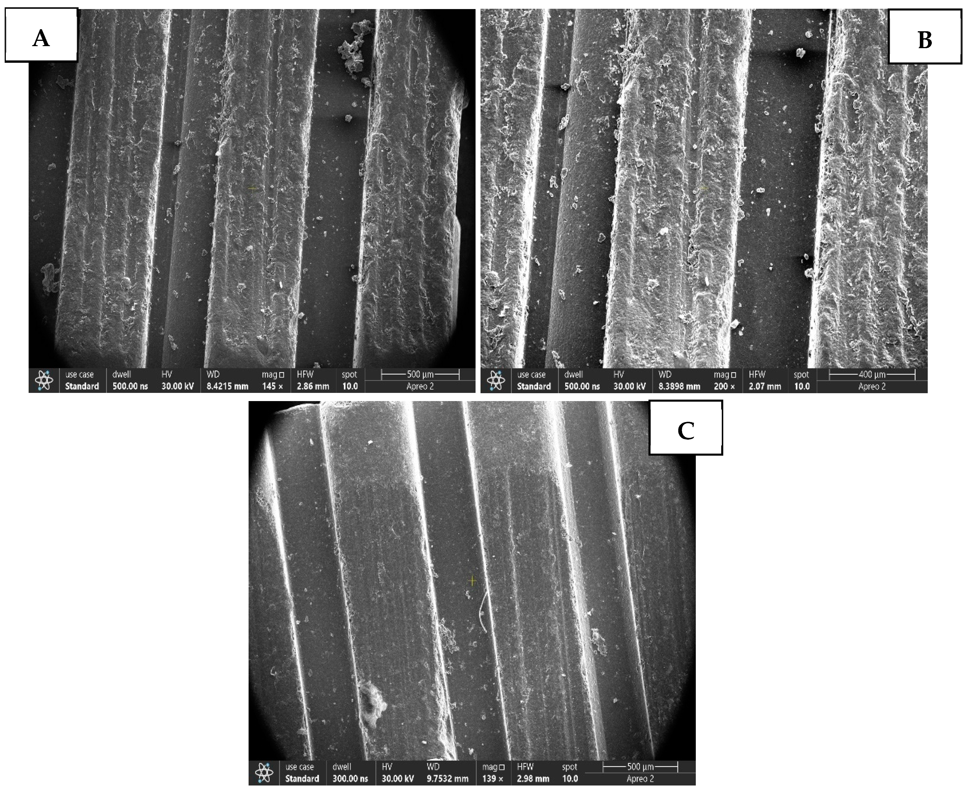
Surface Characterization of the Ceramic Base following different conditioning regimes. Figure 2: (A) The SEM image of a ceramic base treated with HFA displays a notably uneven, rough surface, indicative of chemical etching on the ceramic material. The surface is characterized by distinct parallel ridge formations, likely reflecting the mesh pattern of the bracket base. These ridges are not smooth, featuring a significant granular texture and microporosity. The presence of irregular, interconnected pores give the surface a “sponge-like” or “honeycomb” appearance. (B) The SEM image of a surface treated with an Er, Cr: YSGG laser shows vertical ridges (mesh base pattern) with much greater surface irregularity. Numerous circular-to-elliptical depressions, ranging from 50 to 150 nm in diameter, are visible on the ridge surfaces. A multitude of elevated peaks and protrusions, measuring 20–80 nm in height, contribute to a “mountainous” topography. (C) The SEM image of a ceramic bracket base treated with PC-LLLT reveals a predominantly smooth, glassy surface. It exhibits sparse, shallow surface features, lacking the extensive porosity or ablation craters observed in ceramic brackets treated with HFA and Er, Cr: YSGG.
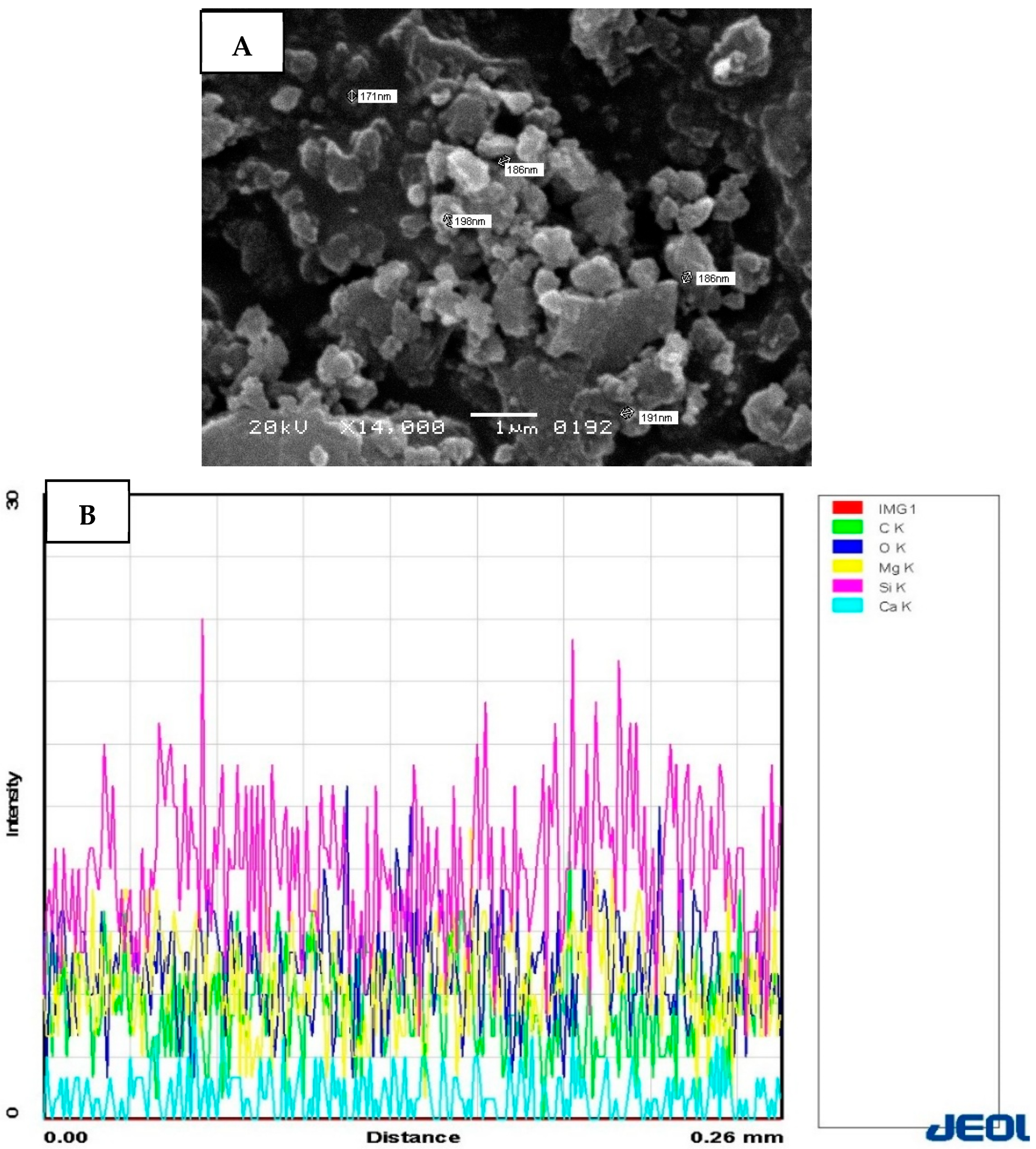
Surface Characterization of Sep NPs and EDX assessment. Figure 3: (A) SEM image of sepiolite nanoparticles demonstrating aggregated morphology. Particle size range: 107–186 nm. Magnification: ×14,000; Scale bar: 1 μm. (B) The EDX analysis reveals that Oxygen (O K, dark blue) has the highest intensity at 61%, followed by Silicon (Si K, magenta) at 20%, Magnesium (Mg K, yellow) at 13%, Carbon (C K, green) at 4%, and Calcium (Ca K, cyan) at 2%, which is considered a minor impurity.
Ra assessment. Table 1 presents the Ra after the application of various surface conditioners on the ceramic bracket. The highest mean Ra value (1087.43 ± 0.043 µm) was observed in Group 2 (ECL). In contrast, Group 3 (Pc-LLLT) exhibited the lowest Ra measurement (706.53 ± 0.054 µm). Comparative analysis indicated that Group 1 (HFA) (1031.62 ± 0.072 µm) showed similar Ra values to those of Group 2 (p ˃ 0.05).
SBS analysis. Table 2 shows the bonding efficacy of ceramic brackets to enamel after the application of surface conditioners. The maximum SBS was recorded in Group 2B (ECL + SepNPs modified adhesive) (8.79 ± 0.48 MPa). In contrast, group 3A (Pc-LLLT + Unmodified adhesive) (5.23 ± 0.32 MPa) exhibited the minimum bond integrity. A comparative analysis revealed that Group 1A (HFA + Unmodified adhesive) (7.86 ± 0.41 MPa) and Group 2A (ECL + Unmodified adhesive) (7.93 ± 0.33 MPa) did not display any statistically significant difference in bond strength results (p ˃ 0.05). Similarly, Group 1B (HFA + SepNPs modified adhesive) (8.63 ± 0.52 MPa) and Group 2B yielded analogous SBS scores (p ˃ 0.05). However, Group 3A (Pc-LLLT + Unmodified adhesive) and Group 3B (Pc-LLLT + SepNPs modified adhesive) (6.16 ± 0.48 MPa) exhibited a significant difference in their SBS (p ˂ 0.05).
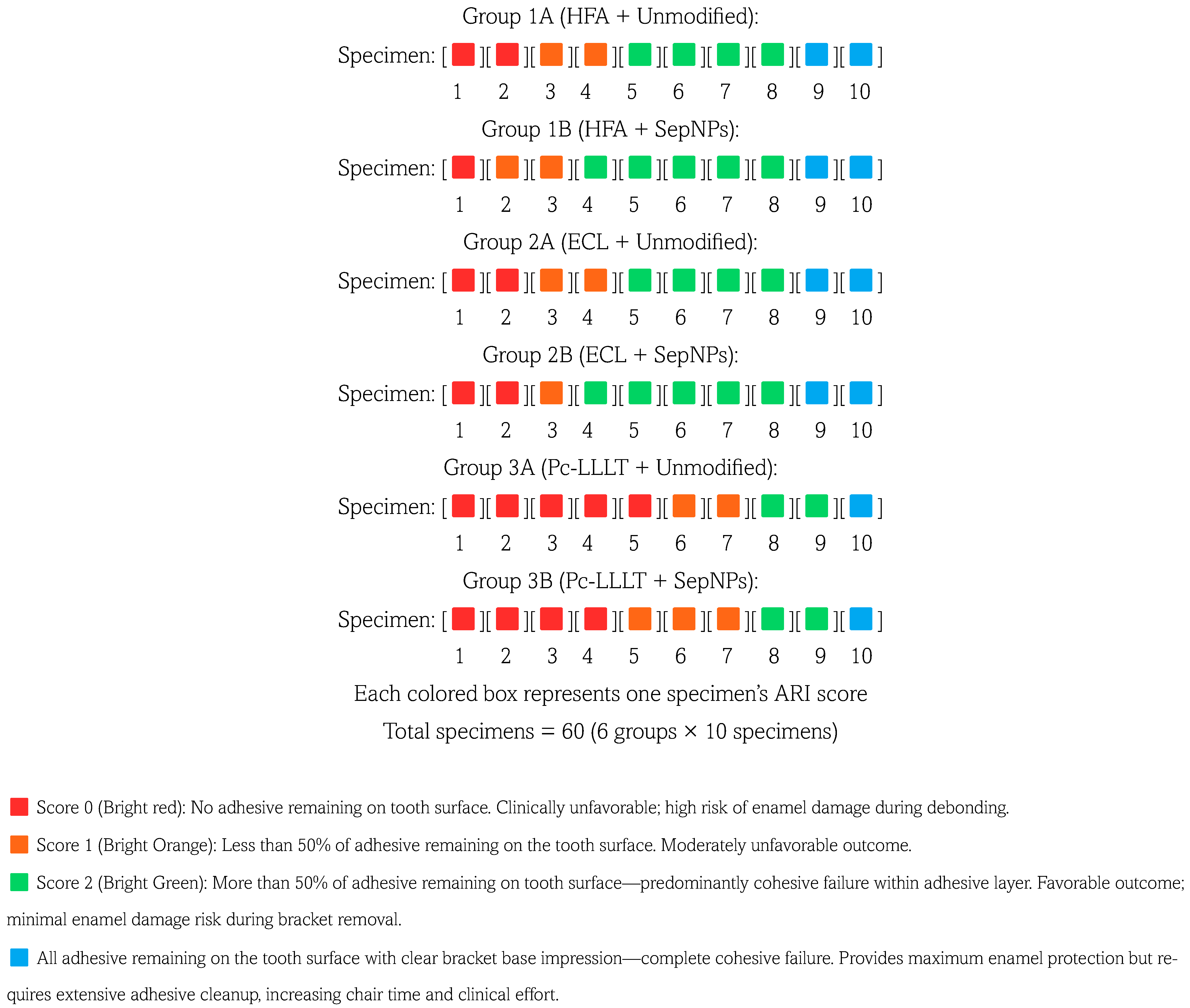
Failure mode analysis. Figure 4 illustrates the distribution of failure modes across the various groups under investigation. Groups 1A, 1B, 2A, and 2B predominantly exhibited admixed fractures. Conversely, adhesive failures were most frequently observed in Groups 3A and 3B. Failure assessment according to the ARI was performed by independent experienced clinicians, kappa score of 0.89.
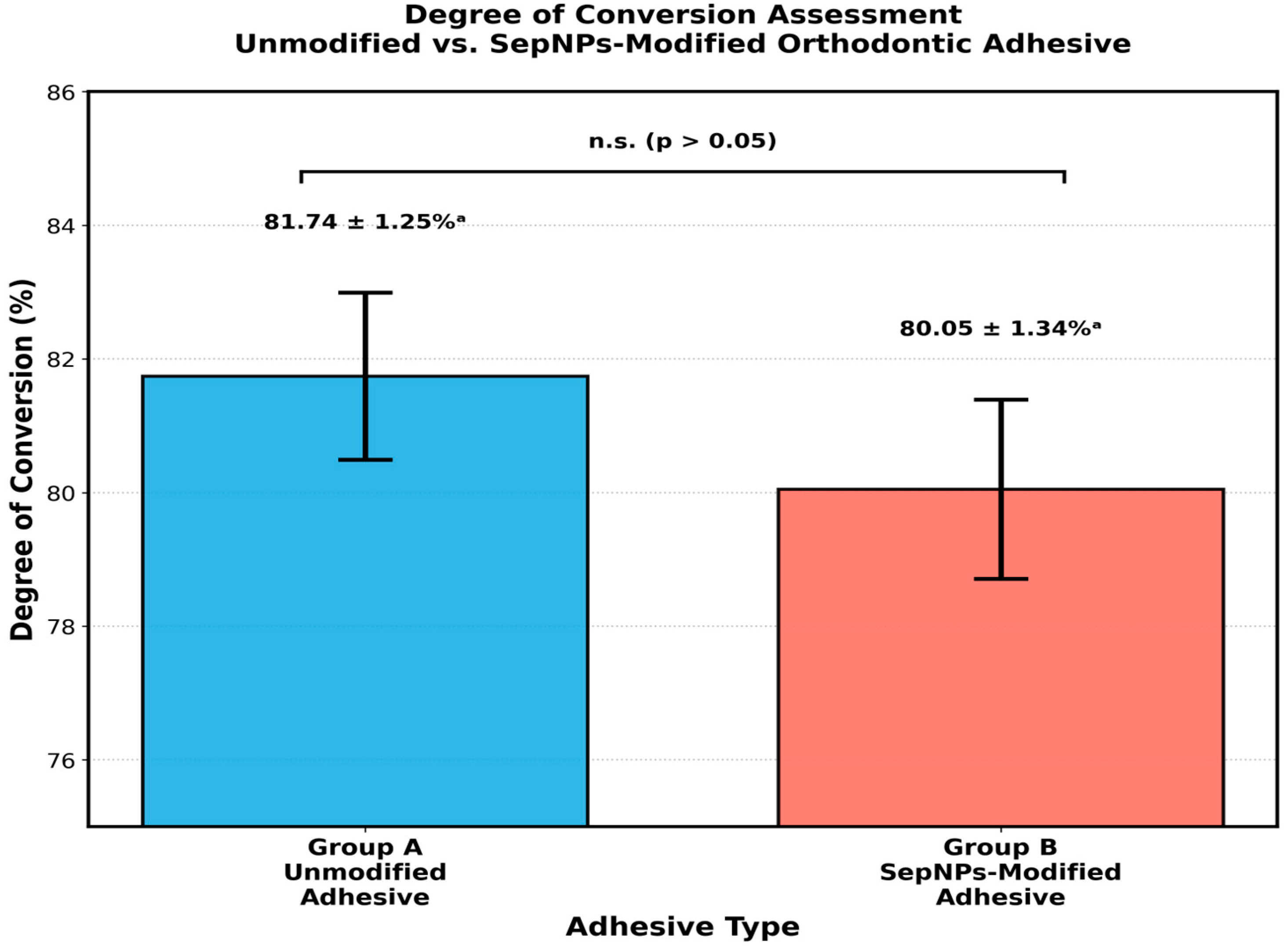
DC assessment. Figure 5 displayed the DC in percentage among the tested groups. Group A (Unmodified orthodontic adhesive) (81.74 ± 1254) and Group B (SepNPs modified orthodontic adhesive) (80.05 ± 1345) displayed comparable DC (p ˃ 0.05).

4. Discussion

The present study was based on the premise that there would be no significant difference in Ra and SBS of ceramic brackets bonded to enamel treated with ECL and Pc-LLLT compared to HFA. Additionally, it was also forecasted that the SBS of unmodified orthodontic adhesive used for bonding ceramic brackets to enamel would be similar to that of the adhesive integrated with SepNPs, with no significant difference in DC. The results of the present study showed that ceramic brackets treated with HFA and ECL exhibited similar levels of Ra and bond strength. Nevertheless, Pc-LLLT showed reduced scores for Ra and bond strength outcomes, thereby partially disproving the primary null hypothesis. Concerning the adhesive, it was found that the adhesive modified with SepNPs showed notably superior bond strength compared to the unmodified adhesive, with no significant difference in DC, partially disproving the second hypothesis.
The selection of appropriate surface conditioning protocols must balance two competing clinical objectives: achieving sufficient bond strength to prevent bracket failure during treatment, while maintaining safe debonding characteristics that minimize enamel damage risk. The clinically acceptable range for orthodontic bracket bond strength has been established as 6–8 MPa based on extensive clinical and laboratory evidence [49,50]. Bond strengths below 6 MPa result in unacceptably high bracket failure rates during orthodontic treatment, necessitating multiple rebonding procedures that increase treatment time, cost, and patient discomfort [51]. Conversely, bond strengths exceeding 13 MPa substantially increase the risk of enamel fracture, avulsion, or tear-out during bracket removal, potentially causing permanent enamel damage requiring restorative intervention [52]. Analysis of our results within this clinical framework reveals distinct performance profiles among the investigated surface conditioning methods. Groups 1A (HFA + unmodified: 7.86 ± 0.41 MPa), 1B (HFA + SepNPs: 8.63 ± 0.52 MPa), 2A (Er, Cr: YSGG + unmodified: 7.93 ± 0.33 MPa), and 2B (Er, Cr: YSGG + SepNPs: 8.79 ± 0.48 MPa) all achieved bond strength values within or slightly above the optimal 6–8 MPa range. While Groups 1B and 2B marginally exceeded 8 MPa, these values remain substantially below the 13 MPa enamel damage threshold, suggesting adequate safety margins for clinical debonding. Furthermore, the ARI score distribution in these groups (60–70% achieving Scores 2–3) confirms predominantly cohesive failure patterns that protect enamel integrity during bracket removal.
In marked contrast, Group 3A (Pc-LLLT + unmodified: 5.23 ± 0.32 MPa) fell below the 6 MPa minimum threshold, indicating clinically inadequate bonding performance. Group 3B (Pc-LLLT + SepNPs: 6.16 ± 0.48 MPa), while technically meeting the minimum 6 MPa threshold, provides no safety margin for clinical variation. The corresponding ARI score distribution in Groups 3A and 3B (50% and 40% Score 0, respectively) further confirms inadequate bonding, with failure occurring predominantly at the enamel-adhesive interface rather than through cohesive fracture. These findings emphasize that Pc-LLLT ceramic bracket conditioning, even when combined with nanoparticle-enhanced adhesives, does not provide clinically reliable bonding performance for orthodontic applications.
Ceramic brackets offer several advantages, primarily due to their discreet, tooth-colored appearance and increased comfort from their smoothly rounded edges [4]. Additionally, ceramic braces are robust and long-lasting, effectively addressing a range of orthodontic issues [6]. In the present study, concerning surface conditioning of ceramic brackets, it was noted that ECL showed comparable scores of Ra and bond strengths to HFA (control). This can be explained by the fact that ECL enhances Ra via thermo-mechanical ablation [53]. ECL transforms laser energy into heat, leading to micro-explosions and the vaporization of the ceramic substance [35]. Previous evidence reported that the bond scores of a ceramic surface are greatly affected by Ra [54]. Typically, a rise in Ra resulted in enhanced bonds since it offers a greater surface area for mechanical interlocking with the adhesive, ultimately augmenting the SBS [55]. SEM analysis further confirms this finding by indicating an increase in Ra, leading to a surface that resembled an irregular texture with numerous elevated peaks and protrusions (20–80 nm height), creating a “mountainous” topography. There was no evidence of melting, crystallization, or carbonization on any of the ceramic bracket surfaces.
Similarly, the ceramic bracket base treated with HFA exhibited commendable efficacy in enhancing both Ra and SBS to enamel. HFA is predominantly recognized for its capability to dissolve the glassy matrix through a reaction with SiO2, resulting in the formation of silicon tetrafluoride gas and hexafluoro silicic acid, which ultimately leads to the development of microporosities that augment the surface area and strengthen the bond between the bracket and the enamel [6,56]. SEM of the HFA conditioned ceramic bracket base illustrates a roughened and irregular surface topography, attributed to the selective dissolution of the glass matrix.
The present study evaluated LLLT with Pc photosensitizer as a novel surface conditioning method for ceramic bracket bases. While this approach represents a first application of photodynamic surface modification in orthodontic bonding, the results demonstrate that Pc-LLLT produced significantly lower bond strengths compared to conventional HFA and Er, Cr: YSGG laser treatments. Pc, which is part of the phenothiazine family like methylene blue, possesses hydrophobic properties. Wainwright and his team noted that methylene blue’s poor performance is due to its water absorption capability [57]. Similarly, Al-Hamdan and his colleagues found that Pc dyes are cationic, leading to electrostatic repulsion when they encounter negatively charged surfaces, thus hindering the adhesion of various materials [58]. Furthermore, light activation of Pc (630 nm, 60 s, 192 mJ/cm2) generates Type II photochemical reactions producing singlet oxygen (1O2) and other reactive oxygen species via energy transfer from triplet-state Pc to ground-state molecular oxygen. The generated 1O2 can oxidatively attack the methacrylate double bonds in the adhesive primer applied before Pc activation, reducing polymerizable sites, Polymer chains in the cured adhesive, creating carbonyls and peroxides that weaken mechanical properties [59].
The present research has shown that an orthodontic adhesive modified with 1% Sep-NPs significantly enhances bond integrity compared to its unmodified counterpart, irrespective of the conditioner used on the ceramic bracket. This finding is consistent with the in vitro study by Kim et al., which revealed that an adhesive containing silica (SiO2) nanofiller achieved the highest bond strength at a nanofiller concentration of 1wt.% However, when the concentration was increased, the nanofillers tended to cluster, which did not improve bond strength [60].
The orthodontic adhesive containing Sep-NPs at a 1% concentration showed no notable reduction in DC compared to the original adhesive formula. This aligns with the results of a laboratory study by Samad and colleagues, which indicated that NP concentrations between 0.5 and 1 weight percent (wt.%) did not affect viscosity or negatively impact the conversion rate [61]. Previous studies have highlighted that the NP proportion is crucial for the adhesive’s mechanical properties, as higher concentrations might weaken the adhesive’s mechanical integrity by lowering the DC [37,62]. A possible reason for this observed effect could be that the fillers obstruct the path of the curing light [63].
A significant limitation of this in vitro study is that laboratory tests may not effectively replicate the complex biomechanical conditions of the oral environment, including temperature fluctuations, moisture exposure, masticatory forces, and aging effects. While the present study evaluated shear bond strength as the primary mechanical parameter, additional mechanical testing, such as tensile bond strength, flexural properties of the adhesive-enamel interface, and fatigue resistance under cyclic loading, would provide a more comprehensive understanding of long-term clinical performance. Furthermore, advanced surface characterization techniques, including atomic force microscopy (AFM) for nanoscale topography assessment and X-ray photoelectron spectroscopy (XPS) for chemical composition analysis, could offer deeper insights into the bonding mechanisms and surface modifications induced by different conditioning protocols. A single concentration of PS and a singular laser parameter were used, which could affect the results. This study utilized a single brand of ceramic brackets and orthodontic adhesive, which limits the generalizability of findings to other commercially available systems. Different ceramic compositions, bracket base designs, and adhesive formulations may respond differently to the surface conditioning protocols investigated. Future investigations should incorporate longitudinal clinical trials to validate laboratory findings, utilize multiple ceramic bracket systems and adhesive formulations, and employ comprehensive mechanical testing protocols, including fatigue and creep analysis. More lab-based and in vivo research is required to confirm these outcomes.
Clinical Significance: The findings of the present study provide clinicians with a safe, effective alternative to hazardous hydrofluoric acid for ceramic bracket conditioning, while demonstrating that sepiolite-modified adhesives can enhance bracket retention without compromising polymerization.

5. Conclusions

Within the limitations of this in vitro study, Er, Cr: YSGG laser surface conditioning of ceramic bracket combined with sepiolite nanoparticle-modified adhesive, demonstrated promising shear bond strength values for orthodontic bracket bonding to the tooth surface. However, these laboratory findings cannot be directly extrapolated to clinical practice. Before clinical implementation, the modified adhesive system must undergo comprehensive preclinical evaluation in accordance with established regulatory frameworks. Under the European Union Medical Device Regulation (EU MDR 2017/745), novel dental adhesives containing nanoparticles require rigorous biocompatibility assessment, including cytotoxicity, sensitization, and genotoxicity testing, along with detailed physicochemical characterization of nanoparticle release and degradation profiles. Similarly, the United States Food and Drug Administration mandates premarket notification (510(k)) or premarket approval pathways that necessitate demonstration of substantial equivalence or safety and effectiveness data. Future investigations should include long-term bond durability studies under simulated oral conditions, evaluation of adhesive-ceramic interface aging, and comprehensive biological safety testing to establish the clinical viability of this approach.

Author Contributions

Conceptualization, S.A. and M.A.K.; methodology, S.A., M.A.K., A.A., W.A. and A.M.A.; software, I.A.; validation, S.A., M.A.K. and I.A.; formal analysis, A.A., A.M.A. and W.A.; investigation, M.A.K.; resources, A.M.A., I.A. and W.A.; data curation, A.M.A.; writing—original draft preparation, M.A.K., A.M.A. and A.A.; writing—review and editing, S.A., A.M.A. and I.A.; visualization, A.A.; supervision, M.A.K.; project administration, S.A.; funding acquisition, M.A.K. All authors have read and agreed to the published version of the manuscript.

Funding

The authors extend their appreciation to the Deanship of Research and Graduate Studies at King Khalid University for funding this work through the Small Research Project under grant number RGP1/91/46.

Institutional Review Board Statement

The study was approved by the ethical board of King Khalid University under IRB/KKUCOD/ETH/2024-25/041.

Data Availability Statement

The original contributions presented in this study are included in the article. Further inquiries can be directed to the corresponding author.

Conflicts of Interest

The authors declare no conflicts of interest.

References

  1. Gudipaneni, R.K.; Aldahmeshi, R.F.; Patil, S.R.; Alam, M.K. The Prevalence of Malocclusion and the Need for Orthodontic Treatment among Adolescents in the Northern Border Region of Saudi Arabia: An Epidemiological Study. BMC Oral Health 2018, 18, 16. [Google Scholar] [CrossRef]
  2. Omran, R.; Dowie, A. Increased Demand for Orthodontic Treatments during the COVID-19 Pandemic: A Commentary. Br. Dent. J. 2023, 234, 84–87. [Google Scholar] [CrossRef]
  3. Koaban, A.M.; Alwadai, J.M.; Alghamdi, A.M.; Alsiwat, F.J.; Dashti, A.I.; Nasser, M.M.; Alhazmi, M.A.; Aljaroudi, E.M.; Alanazi, S.B.; Almanjhi, W.A. Recent Advances in Orthodontic Brackets: From Aesthetics to Smart Technologies. Cureus 2025, 17, e85385. [Google Scholar] [CrossRef] [PubMed]
  4. Reddy, Y.G.; Sharma, R.; Singh, A.; Agarwal, V.; Agrawal, V.; Saurab, C. The Shear Bond Strengths of Metal and Ceramic Brackets: An In-Vitro Comparative Study. J. Clin. Diagn. Res. 2013, 7, 1495–1497. [Google Scholar] [CrossRef] [PubMed]
  5. Nimplod, P.; Tansalarak, R.; Sornsuwan, T. Effect of the Different Debonding Strength of Metal and Ceramic Brackets on the Degree of Enamel Microcrack Healing. Dent. Press J. Orthod. 2021, 26, e2119177. [Google Scholar] [CrossRef]
  6. Türk, T.; Saraç, D.; Saraç, Y.Ş.; Elekdaǧ-Türk, S. Effects of Surface Conditioning on Bond Strength of Metal Brackets to All-Ceramic Surfaces. Eur. J. Orthod. 2006, 28, 450–456. [Google Scholar] [CrossRef] [PubMed]
  7. Alqahtani, S.A.; Alsaeed, A.Y.; Alqahtani, S.M.; Shafqat, S.; Niaz, M.O.; Ishtiaque, Y.; Abrar, E. Lithium Disilicate Ceramics Pretreated with Ti: Sapphire Fs, Er; Cr: YSGG, and Er: YAG Lasers: A Study on Mechanical Properties, Energy Dispersive Spectroscopy, Color Alterations, and Surface Topography. Microsc. Res. Tech. 2025, 88, 3029–3036. [Google Scholar] [CrossRef]
  8. Almutairi, B.; Al-Qahtani, A.S.; Shabbir, T.; Leemani, M.J.; Unar, J.; Manzar, N.; Abduljabbar, T. A Surface Topographical Analysis of Lithium Disilicate Ceramics Pretreated with Rose Bengal, Er:YAG Laser, and Ceramic Primer on Bond Integrity, Surface Roughness, and Bond Failure to Composite Resin. Sci. Adv. Mater. 2024, 16, 800–806. [Google Scholar] [CrossRef]
  9. Faltermeier, A.; Reicheneder, C.; Götzfried, P.; Proff, P. Bonding Orthodontic Ceramic Brackets to Ceramic Restorations: Evaluation of Different Surface Conditioning Methods. Mater. Sci. Appl. 2013, 4, 10–14. [Google Scholar] [CrossRef]
  10. Kim, K.J.; Park, Y.T.; Yoo, J.H.; Park, T.H. Hydrofluoric Acid Burns. Exog. Dermatol. 2004, 3, 12–18. [Google Scholar] [CrossRef]
  11. García-Sanz, V.; Paredes-Gallardo, V.; Bellot-Arcís, C.; Mendoza-Yero, O.; Doñate-Buendía, C.; Montero, J.; Albaladejo, A. Effects of Femtosecond Laser and Other Surface Treatments on the Bond Strength of Metallic and Ceramic Orthodontic Brackets to Zirconia. PLoS ONE 2017, 12, e0186796. [Google Scholar] [CrossRef] [PubMed]
  12. Erdur, E.A.; Basciftci, F.A. Effect of Ti:Sapphire Laser on Shear Bond Strength of Orthodontic Brackets to Ceramic Surfaces. Lasers Surg. Med. 2015, 47, 512–519. [Google Scholar] [CrossRef]
  13. Alkhudhairy, F.; Naseem, M.; Bin-Shuwaish, M.; Vohra, F. Efficacy of Er Cr: YSGG Laser Therapy at Different Frequency and Power Levels on Bond Integrity of Composite to Bleached Enamel. Photodiagnosis Photodyn. Ther. 2018, 22, 34–38. [Google Scholar] [CrossRef] [PubMed]
  14. Hoteit, M.; Nammour, S.; Zeinoun, T. Evaluation of Enamel Topography after Debonding Orthodontic Ceramic Brackets by Different Er,Cr:YSGG and Er:YAG Lasers Settings. Dent. J. 2020, 8, 6. [Google Scholar] [CrossRef] [PubMed]
  15. Maawadh, A.M.; Almohareb, T.; Al-Hamdan, R.S.; Al Deeb, M.; Naseem, M.; Alhenaki, A.M.; Vohra, F.; Abduljabbar, T. Repair Strength and Surface Topography of Lithium Disilicate and Hybrid Resin Ceramics with LLLT and Photodynamic Therapy in Comparison to Hydrofluoric Acid. J. Appl. Biomater. Funct. Mater. 2020, 18, 2280800020966938. [Google Scholar] [CrossRef]
  16. Vohra, F.; Labban, N.; Al-Hussaini, A.; Al-Jarboua, M.; Zawawi, R.; Alrahlah, A.; Naseem, M. Influence of Er;Cr:YSGG Laser on Shear Bond Strength and Color Stability of Lithium Disilicate Ceramics: An In Vitro Study. Photobiomodul. Photomed. Laser Surg. 2019, 37, 483–488. [Google Scholar] [CrossRef]
  17. Alrabeah, G.; Alhamid, R.F.; Alamer, B.A.; Alrajhi, F.N.; Binrayes, A.; Habib, S.R. Impact of Various Surface Treatments on the Shear Bond Strength Between Lithium Disilicate Ceramics and Resin Cement. Front. Mater. 2024, 11, 1496749. [Google Scholar] [CrossRef]
  18. Alsunbul, H.; Almutairi, B.; Aljanakh, M.; Abduljabbar, T. Hybrid Ceramic Repair Strength, Surface Roughness, and Bond Failure, Using Methylene Blue-Activated Low-Level Laser Therapy, Carbon Dioxide, and Ti: Al2O3 Laser. Photodiagnosis Photodyn. Ther. 2023, 43, 103693. [Google Scholar] [CrossRef]
  19. Alkhudhairy, F.; AlFawaz, Y.F. Pretreatment of Hybrid Ceramics Using Ho: YAG, Low-Level Laser Therapy Activated Malachite Green, and Non-Thermal Plasma on Surface Roughness, Bond Strength, and Color Change, SEM and EDX Analysis. Ceramics 2024, 7, 944–957. [Google Scholar] [CrossRef]
  20. Alhamdan, E.M. Repair Bond Strength and Surface Roughness of Zirconia Ceramics Treated via Carbon Dioxide Laser, Malachite Green, and Sandblasting. A Lab-Based Study. J. Ceram. Sci. Technol. 2025, 16, 85–92. [Google Scholar] [CrossRef]
  21. Alanazi, A.M.; Khan, N.A.; Khan, A.A.; Ansari, Z.; Shabbir, T.; Leemani, M.J. Lithium Disilicate Ceramics Surface Pre-Treatment Using Low-Level Laser Therapy-Activated Riboflavin, and Ti: Al2O3 Laser on the Colour Change, Surface Roughness, and Shear Bond Strength to Adhesive Cement: An In Vitro Sem Valuation. Ceram.-Silik. 2025, 69, 93–100. [Google Scholar] [CrossRef]
  22. Galstyan, A. Turning Photons into Drugs: Phthalocyanine-Based Photosensitizers as Efficient Photoantimicrobials. Chem. Eur. J. 2021, 27, 1903–1920. [Google Scholar] [CrossRef]
  23. Spesia, M.B.; Durantini, E.N. Evolution of Phthalocyanine Structures as Photodynamic Agents for Bacteria Inactivation. Chem. Rec. 2022, 22, e202100292. [Google Scholar] [CrossRef]
  24. Liu, M.; Zhang, S.; Wang, Z. Advanced Oxidation Processes of Organic Contaminants. Toxics 2024, 12, 579. [Google Scholar] [CrossRef]
  25. Shirase, S.; Shinohara, K.; Tsurugi, H.; Mashima, K. Oxidation of Alcohols to Carbonyl Compounds Catalyzed by Oxo-Bridged Dinuclear Cerium Complexes with Pentadentate Schiff-Base Ligands Under a Dioxygen Atmosphere. ACS Catal. 2018, 8, 6939–6947. [Google Scholar] [CrossRef]
  26. Al-Hamdan, R.S.; Almutairi, B.; Abduljabbar, T. Sandblasting, Bioactive Glass Particles, and Phthalocyanine Activated via Low-Level Laser Therapy on the Surface Roughness and Shear Bond Strength of Hybrid Ceramics Bonded to Different Cement Types. Photodiagnosis Photodyn. Ther. 2025, 55, 104682. [Google Scholar] [CrossRef]
  27. Alzahrani, A.H.; Ebrahim, M.I.; Felemban, M.F.; Alqarni, A.A.; Algahtani, F.S.; Shawli, H.T.; Al Humayyani, N.; Meshni, A.A.; Al Moaleem, M.M. Nanoparticle Augmentation of Adhesive Systems: Impact on Tensile Strength in Fiberglass Post Placement Within Root Dentin. Med. Sci. Monit. 2024, 30, e943502-1. [Google Scholar] [CrossRef]
  28. Fallahzadeh, F.; Safarzadeh-Khosroshahi, S.; Atai, M. Dentin Bonding Agent with Improved Bond Strength to Dentin Through Incorporation of Sepiolite Nanoparticles. J. Clin. Exp. Dent. 2017, 9, e738–e742. [Google Scholar] [CrossRef] [PubMed]
  29. Iqbal Khan, Z.; Habib, U.; Binti Mohamad, Z.; Razak Bin Rahmat, A.; Amira Sahirah Binti Abdullah, N. Mechanical and Thermal Properties of Sepiolite Strengthened Thermoplastic Polymer Nanocomposites: A Comprehensive Review. Alex. Eng. J. 2022, 61, 975–990. [Google Scholar] [CrossRef]
  30. Niazi, F.H.; Alotaibi, B.; Abdulla, A.M.; AlTowayan, S.A.R.; Ahmed, S.Z.; Alshehri, D.; Samran, A.; Alsuwayyigh, N.; Luddin, N. Modified Experimental Adhesive with Sepiolite Nanoparticles on Caries Dentin Treated with Femtosecond Laser and Photodynamic Activated Erythrosine. An In Vitro Study. Photodiagnosis Photodyn. Ther. 2024, 49, 104306. [Google Scholar] [CrossRef] [PubMed]
  31. Falkensammer, F.; Freudenthaler, J.; Pseiner, B.; Bantleon, H.P. Influence of Surface Conditioning on Ceramic Microstructure and Bracket Adhesion. Eur. J. Orthod. 2012, 34, 498–504. [Google Scholar] [CrossRef]
  32. Saraç, Y.Ş.; Külünk, T.; Elekdaǧ-Türk, S.; Saraç, D.; Türk, T. Effects of Surface-Conditioning Methods on Shear Bond Strength of Brackets Bonded to Different All-Ceramic Materials. Eur. J. Orthod. 2011, 33, 667–672. [Google Scholar] [CrossRef]
  33. Abdullah Kamran, M. Remineralization of Eroded Enamel for Improved Orthodontic Bracket Bonding: An In Vitro Study. Korean J. Orthod. 2025, 55, 244–253. [Google Scholar] [CrossRef]
  34. Samih, H.; Aboulazm, K.; Abd El-Motie, M. Debonding of Ceramic Brackets with ER,CR:YSGG Laser. Egypt. Orthod. J. 2014, 46, 27–39. [Google Scholar] [CrossRef]
  35. Mirhashemi, A.H.; Hossaini, S.M.H.; Etemadi, A.; Kharazifard, M.J.; Bahador, A.; Soudi, A. Effect of Er:YAG and Er,Cr:YSGG Lasers on Ceramic Bracket Debonding from Composite Blocks. Front. Dent. 2019, 16, 88–95. [Google Scholar] [CrossRef]
  36. Shabib, S. Use of Nd:YVO4 Laser, Photodynamic Therapy, Sulfuric Acid and Sand Blasting on Improving Bond Integrity of PEEK to Resin Cement with Adhesive. Photodiagnosis Photodyn. Ther. 2022, 39, 102865. [Google Scholar] [CrossRef]
  37. Al-Hamdan, R.S.; Almutairi, B.; Kattan, H.F.; Alsuwailem, N.A.; Farooq, I.; Vohra, F.; Abduljabbar, T. Influence of Hydroxyapatite Nanospheres in Dentin Adhesive on the Dentin Bond Integrity and Degree of Conversion: A Scanning Electron Microscopy (SEM), Raman, Fourier Transform-Infrared (FTIR), and Microtensile Study. Polymers 2020, 12, 2948. [Google Scholar] [CrossRef] [PubMed]
  38. Xie, Y.; Xiao, Z.; Militz, H.; Hao, X. Silane Coupling Agents Used in Natural Fiber/Plastic Composites. In Handbook of Composites from Renewable Materials; Wiley: Hoboken, NJ, USA, 2017; Volume 1–8, pp. 407–430. ISBN 9781119441632. [Google Scholar]
  39. Sabzi, M.; Mirabedini, S.M.; Zohuriaan-Mehr, J.; Atai, M. Surface Modification of TiO2 Nano-Particles with Silane Coupling Agent and Investigation of Its Effect on the Properties of Polyurethane Composite Coating. Prog. Org. Coat. 2009, 65, 222–228. [Google Scholar] [CrossRef]
  40. Khan, A.S.; Alhamdan, Y.; Alibrahim, H.; Almulhim, K.S.; Nawaz, M.; Ahmed, S.Z.; Aljuaid, K.; Ateeq, I.S.; Akhtar, S.; Ansari, M.A.; et al. Analyses of Experimental Dental Adhesives Based on Zirconia/Silver Phosphate Nanoparticles. Polymers 2023, 15, 2614. [Google Scholar] [CrossRef]
  41. Kasraei, S.H.; Atai, M.; Khamverdi, Z.; Nejad, S.K. The effect of nanofiller addition to an experimental dentin adhesive on microtensile bond strength to human dentin. Front. Dent. 2009, 6, 36–41. [Google Scholar]
  42. Marovic, D.; Tarle, Z.; Hiller, K.A.; Müller, R.; Ristic, M.; Rosentritt, M.; Skrtic, D.; Schmalz, G. Effect of Silanized Nanosilica Addition on Remineralizing and Mechanical Properties of Experimental Composite Materials with Amorphous Calcium Phosphate. Clin. Oral Investig. 2014, 18, 783–792. [Google Scholar] [CrossRef]
  43. Chen, M.H. Critical Reviews in Oral Biology & Medicine: Update on Dental Nanocomposites. J. Dent. Res. 2010, 89, 549–560. [Google Scholar]
  44. Alqerban, A. Effectiveness of Riboflavin and Rose Bengal Photosensitizer Modified Adhesive Resin for Orthodontic Bonding. Pharmaceuticals 2021, 14, 48. [Google Scholar] [CrossRef]
  45. Messias, A.M.; Galvão, M.R.; Boaventura, J.M.C.; Jacomassi, D.P.; Bernardi, M.I.B.; Bagnato, V.S.; Rastelli, A.N.S.; Andrade, M.F. Degree of Conversion of Different Composite Resins Photo-Activated with Light-Emitting Diode and Argon Ion Laser. Laser Phys. 2015, 25, 025601. [Google Scholar] [CrossRef]
  46. Alnazeh, A.A.; Kamran, M.A.; Almoammar, S.; Al Jearah, M.M.; Qasim, M.; Alshahrani, I. Visible light-activated curcumin-doped zinc oxide nanoparticles integrated into orthodontic adhesive on Micro-tensile bond strength, degree of conversion, and antibacterial effectiveness against Staphylococcus Aureus. An investigation using scanning electron microscopy and energy-dispersive X-ray spectroscopy. J. Photochem. Photobiol. B Biol. 2024, 253, 112888. [Google Scholar] [CrossRef]
  47. Kamran, M.A.; Asiri, A.M.; Alfaifi, A.M.A.; Almukawwi, A.H.A.; Mughaddi Alwadai, J.; Alqahtani, S.J. Fluoride-Activated Via Er:YAG, Diode, and Femtosecond Lasers for Reversing Bleached Enamel for Improved Orthodontic Bonding. Photobiomodulation Photomed. Laser Surg. 2025, 43–50. [Google Scholar] [CrossRef] [PubMed]
  48. Jacobson, A.; Shojaei, A.R.; Thompson, B.D.; Kulkarni, G.V.; Titley, K.C. Adhesive Remnant Index (ARI) Revisited. An In Vitro Assessment of Clinically Debonded Orthodontic Brackets. Am. J. Orthod. Dentofac. Orthop. 2006, 130, 120. [Google Scholar] [CrossRef]
  49. Reynolds, I.R. A Review of Direct Orthodontic Bonding. Br. J. Orthod. 1975, 2, 171–178. [Google Scholar] [CrossRef]
  50. Bishara, S.E.; Oonsombat, C.; Soliman, M.M.A.; Ajlouni, R.; Laffoon, J.F. The Effect of Tooth Bleaching on the Shear Bond Strength of Orthodontic Brackets. Am. J. Orthod. Dentofac. Orthop. 2005, 128, 755–760. [Google Scholar] [CrossRef]
  51. Scribante, A.; Contreras-Bulnes, R.; Montasser, M.A.; Vallittu, P.K. Orthodontics: Bracket Materials, Adhesives Systems, and Their Bond Strength. Biomed Res. Int. 2016, 2016, 1329814. [Google Scholar] [CrossRef]
  52. Ryf, S.; Flury, S.; Palaniappan, S.; Lussi, A.; Van Meerbeek, B.; Zimmerli, B. Enamel Loss and Adhesive Remnants Following Bracket Removal and Various Clean-up Procedures In Vitro. Eur. J. Orthod. 2012, 34, 25–32. [Google Scholar] [CrossRef]
  53. Kaygısız, E.; Eğilmez, F.; Ergün, G.; Yüksel, S.; Nagas, I.Ç. The Effects of Er,Cr:YSGG Laser on Shear Bond Strength of Orthodontic Lingual Brackets to CAD/CAM Ceramic Systems. Eur. Oral Res. 2023, 57, 122–127. [Google Scholar] [CrossRef]
  54. Alkhudhairy, F.; Naseem, M.; Ahmad, Z.H.; Alnooh, A.N.; Vohra, F. Efficacy of phototherapy with different conventional surface treatments on adhesive quality of lithium disilicate ceramics. Photodiagnosis Photodyn. Ther. 2019, 25, 292–295. [Google Scholar] [CrossRef]
  55. Alkan Demetoğlu, G.; Talay Çevlïk, E. Evaluation of the Effect of Etching with Ytterbium Fiber Laser on the Bond Strength, Color Stability, and Fracture Analysis of Lithium Disilicate Ceramics to Bovine Teeth: An In Vitro Study. BMC Oral Health 2025, 25, 607. [Google Scholar] [CrossRef] [PubMed]
  56. Albaker, A.M.; Al Deeb, L.; Alhenaki, A.M.; Aldeeb, M.; Al Ahdal, K.; Abduljabbar, T.; Vohra, F. Bonding Integrity and Compressive Strength of Re-Bonded, Surface Conditioned and Er Cr YSGG Laser Treated Lithium Disilicate Ceramics. J. Appl. Biomater. Funct. Mater. 2020, 18, 2280800020910954. [Google Scholar] [CrossRef]
  57. Wainwright, M.; Phoenix, D.A.; Rice, L.; Burrow, S.M.; Waring, J. Increased Cytotoxicity and Phototoxicity in the Methylene Blue Series via Chromophore Methylation. J. Photochem. Photobiol. B Biol. 1997, 40, 233–239. [Google Scholar] [CrossRef] [PubMed]
  58. AlFawaz, Y.F.; Almutairi, B.; Kattan, H.F.; Zafar, M.S.; Farooq, I.; Naseem, M.; Vohra, F.; Abduljabbar, T. Dentin bond integrity of hydroxyapatite containing resin adhesive enhanced with graphene oxide nano-particles—An SEM, EDX, micro-Raman, and microtensile bond strength study. Polymers 2020, 12, 2978. [Google Scholar] [CrossRef] [PubMed]
  59. Baptista, M.S.; Cadet, J.; Di Mascio, P.; Ghogare, A.A.; Greer, A.; Hamblin, M.R.; Lorente, C.; Nunez, S.C.; Ribeiro, M.S.; Thomas, A.H.; et al. Type I and Type II Photosensitized Oxidation Reactions: Guidelines and Mechanistic Pathways. Photochem. Photobiol. 2017, 93, 912–919. [Google Scholar] [CrossRef]
  60. Kim, J.W.; Kim, L.U.; Kim, C.K. Size Control of Silica Nanoparticles and Their Surface Treatment for Fabrication of Dental Nanocomposites. Biomacromolecules 2007, 8, 215–222. [Google Scholar] [CrossRef]
  61. Kumar, U.; Kumar, D.; Gosai, K.N.; Dalal, D.; Pragnya, B.; Nagarajan, S. Effectiveness of nanoparticles in enhancing bond strength in adhesive dentistry. J. Pharm. Bioallied Sci. 2024, 16 (Suppl. S4), S3772–S3774. [Google Scholar] [CrossRef]
  62. Alhenaki, A.M.; Attar, E.A.; Alshahrani, A.; Farooq, I.; Vohra, F.; Abduljabbar, T. Dentin Bond Integrity of Filled and Unfilled Resin Adhesive Enhanced with Silica Nanoparticles—An Sem, Edx, Micro-Raman, Ftir and Micro-Tensile Bond Strength Study. Polymers 2021, 13, 1093. [Google Scholar] [CrossRef] [PubMed]
  63. Alqarawi, F.K.; Alkahtany, M.F.; Almadi, K.H.; Ben Gassem, A.A.; Alshahrani, F.A.; Alrefeai, M.H.; Farooq, I.; Vohra, F.; Abduljabbar, T. Influence of Different Conditioning Treatments on the Bond Integrity of Root Dentin to Rgo Infiltrated Dentin Adhesive. Sem, Edx, Ftir and Microraman Study. Polymers 2021, 13, 1555. [Google Scholar] [CrossRef] [PubMed]
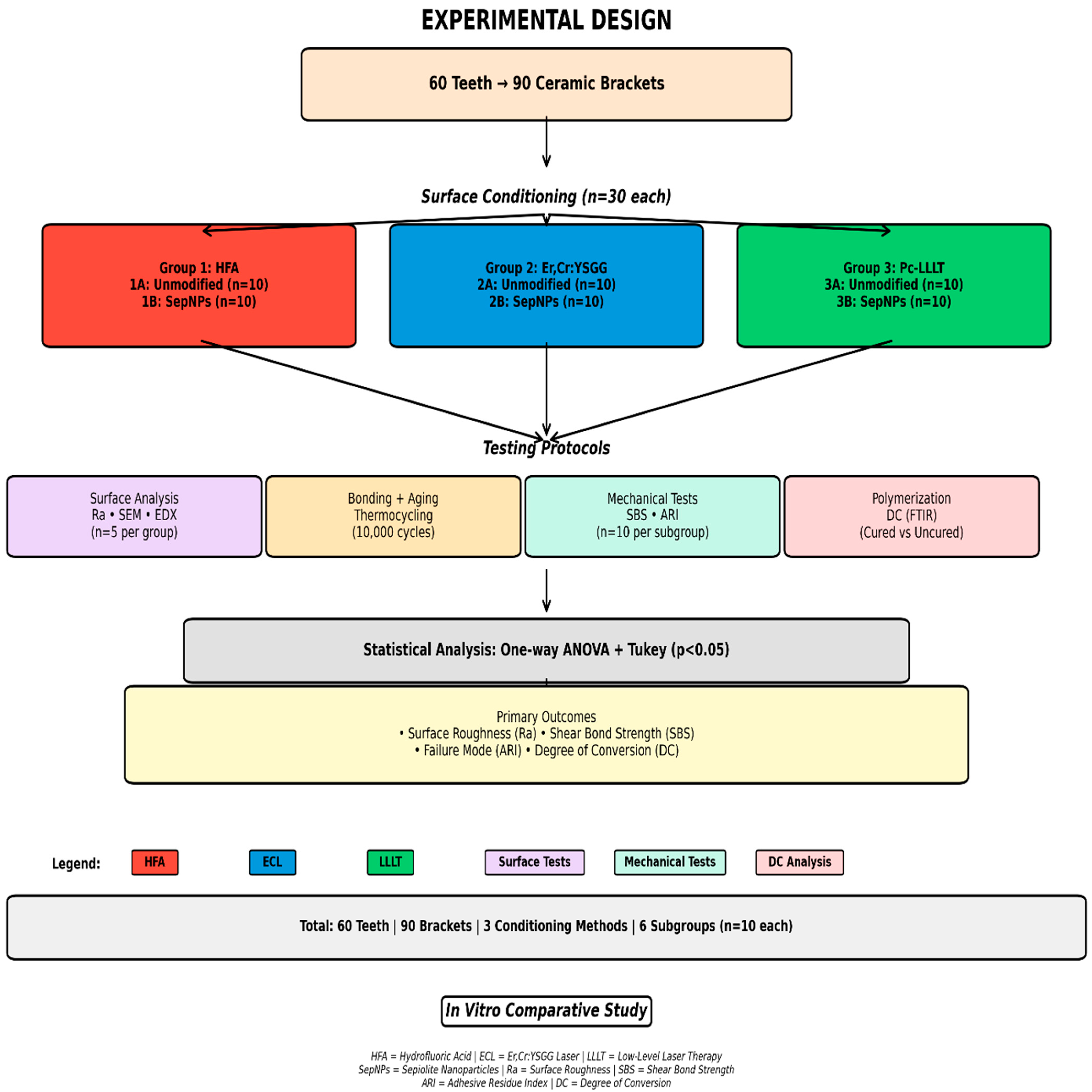
Figure 1. Methodological flow chart.
Crystals 15 01010 g001
Figure 2. SEM characterization of ceramic bracket surface morphology after conditioning. (A) HFA treatment shows irregular topography with interconnected microporosities and honeycomb texture along parallel ridge structures (scale bar: 500 μm). (B) Er, Cr: YSGG laser treatment exhibits pronounced surface roughness with ablation craters (50–150 nm diameter) and elevated peaks (20–80 nm height) (scale bar: 400 μm). (C) Pc-LLLT treatment demonstrates minimal surface modification with predominantly smooth appearance and sparse shallow features (scale bar: 500 μm).
Crystals 15 01010 g002
Figure 3. (A) Scanning electron micrograph of sepiolite nanoparticles showing aggregated morphology with individual particles ranging from 107 to 186 nm in diameter. The nanoparticles exhibit an irregular, clustered arrangement with rough surface texture. Magnification: ×14,000; Scale bar: 1 μm; Accelerating voltage: 20 kV. (B) The EDX graph shows X-Axis (Horizontal): “Distance” from 0.00 to 0.26 mm (260 μm). This is the path the electron beam traveled across your sample. Y-Axis (Vertical): “Intensity” (signal strength). Higher peaks = more of that element at that location. The elemental distribution via EDX shows Oxygen (O K, dark blue): Highest overall intensity 61%, Silicon (Si K, magenta) 20%, Magnesium (Mg K, yellow) 13%, Carbon (C K, green) 4% and Calcium (Ca K, cyan) 2% (minor impurity).
Crystals 15 01010 g003
Figure 4. Failure mode distribution among the different investigated groups.
Crystals 15 01010 g004
Figure 5. DC comparison showing no significant difference between unmodified (81.74 ± 1.25%) and SepNPs-modified (80.05 ± 1.34%) adhesives (p > 0.05). Both groups marked with a indicate the same statistical subset.
Crystals 15 01010 g005
Table 1. Surface roughness (Ra) of ceramic brackets after using different surface pretreatments.
Investigated GroupsMean ± SD Ra (µm)p-Value!
Group 1: HFA1031.62 ± 0.72 a<0.05
Group 2: ECL1087.43 ± 0.43 a
Group 3: Pc-LLLT706.53 ± 0.54 b
! ANOVA (p < 0.05). Hydrofluoric acid (HFA), Er, Cr: YSGG laser (ECL), Low-level laser therapy (LLLT), Phthalocyanine (Pc). Different superscript lower-case letters denote statistically significant differences within the same column, Post Hoc Tukey (p < 0.05).
Table 2. SBS of ceramic brackets to enamel after using different surface conditioners.
Investigated GroupsSBS Mean ± SD (MPa)p-Value!
Group 1A: HFA + Unmodified adhesive7.86 ± 0.41 a<0.05
Group 1B: HFA + SepNPs modified adhesive8.63 ± 0.52 b
Group 2A: ECL + Unmodified adhesive7.93 ± 0.33 a
Group 2B: ECL + SepNPs modified adhesive8.79 ± 0.48 b
Group 3A: Pc-LLLT + Unmodified adhesive5.23 ± 0.32 d
Group 3B: Pc-LLLT+ SepNPs modified adhesive6.16 ± 0.48 c
! ANOVA (p < 0.05). Hydrofluoric acid (HFA), Er, Cr: YSGG laser (ECL), Low-level laser therapy (LLLT), Phthalocyanine (Pc). Different superscript lower-case letters denote statistically significant differences within the same column, Post Hoc Tukey (p < 0.05).
Disclaimer/Publisher’s Note: The statements, opinions and data contained in all publications are solely those of the individual author(s) and contributor(s) and not of MDPI and/or the editor(s). MDPI and/or the editor(s) disclaim responsibility for any injury to people or property resulting from any ideas, methods, instructions or products referred to in the content.