Article

Extremely Fast and Efficient Removal of Congo Red Using Cationic-Incorporated Hydroxyapatite Nanoparticles (HAp: X (X = Fe, Ni, Zn, Co, and Ag))

by
Sandeep Eswaran Panchu
1,
Saranya Sekar
1,
Elayaraja Kolanthai
2,
Moorthy Babu Sridharan
1 and
Narayana Kalkura Subbaraya
1,*
1
Crystal Growth Centre, Anna University, Chennai 600 025, India
2
Department of Materials Science & Engineering, Advanced Materials Processing and Analysis Centre, University of Central Florida, Orlando, FL 32826, USA
*
Author to whom correspondence should be addressed.
Crystals 2023, 13(2), 209; https://doi.org/10.3390/cryst13020209
Submission received: 6 December 2022 / Revised: 7 January 2023 / Accepted: 17 January 2023 / Published: 24 January 2023
(This article belongs to the Special Issue A New Frontier in Biomineral Interactions and Biomineralization)

Abstract

:
Congo red (CR) is a stable anionic diazo dye that causes allergic reactions with carcinogenic properties. The rapid removal of CR using cation-incorporated nanohydroxyapatite (pristine HAp: X (X = Fe, Ni, Zn, Co, and Ag)) was investigated. The pristine and cation ion-doped HAp adsorbents were coprecipitated and subjected to hydrothermal and ultrasound treatments and subsequent microwave drying. The dopant ions significantly engineered the crystallite size, crystallinity, particle size (decreased 38–77%), shape (a rod to sphere modification by the incorporation of Ag+, Ni2+, and Co2+ ions), and colloidal stability (CS) of the adsorbent. These modifications aided in the rapid removal of the CR dye (98%) within one minute, and the CR adsorption rate was found to be significantly higher (93–99%) compared to previously reported rates. Furthermore, the kinetic, Langmuir, Freundlich, and DKR isotherms and thermodynamic results confirmed that the CR adsorption on the HAp was due to the strong chemical adsorption process. The order of the maximum CR adsorption capacity was Fe-HAp > HAp > Ag-HAp > Co-HAp > Zn-HAp. Whereas the CR regeneration efficiency was Fe-HAp (92%) > Ag-HAp (42%) > Ni-HAp (30%), with the other adsorbents exhibiting a poor recycling efficiency (1–16%). These results reveal Fe-HAp as a potential adsorbent for removing CR without the formation of byproducts.

1. Introduction

Over the last few decades, water pollution and drinking water shortages have become more prevalent due to the proliferation of industries and their pollutants. Among them, organic effluent has been improperly discharged into wastewater from the textile, printer, paper, soap, rubber mill, plastic, and paint industries [1,2,3]. Consequently, dye pollution is a major environmental crisis, as over 7 × 10⁵ tons of dyes are produced annually. Synthetic azo dyes are the most prevalent dye contaminants, which discharge 100 tons of waste annually into wastewater streams [4,5,6]. Accordingly, CR is the most common and highly stable industrial dye containing aromatic rings, making it an arduous challenge to degrade or remediate [1,4,5]. Moreover, even very small amounts of dyes in water cause a significant reduction in sunlight penetration, thereby disrupting the biochemical activity of aquatic organisms. In addition, dye contamination poses carcinogenic, mutagenic, and hazardous effects on living systems. Hence, their removal becomes paramount to safeguarding the environment and public health from their toxicities [7,8,9,10].
A variety of methods have been used to remove CR effluent from water, including photocatalytic (redox reaction), flocculation, coagulation, ion exchange, reverse osmosis, membrane filtration, and adsorption [2,3,11,12]. Compared to other techniques, the adsorption technique is best suited for practical applications, since it is simple, cheap, easy to reuse, and has a high adsorption capacity [8,13,14]. In addition, other methods have more significant disadvantages, including increased operating and maintenance costs, need for trained technicians, and production of second-hand toxic waste. However, choosing an absorbent that suits practical purposes is vital, since the absorption technique is highly dependent on adsorbent characteristics. Accordingly, hydroxyapatite (HAp) is a promising adsorbent because of its low cost and ability to be easily modified to meet the desired properties. In addition, it has a hydrophilic nature and porous structure, is insoluble in water, and can easily alter the surface charge and particle morphology [11,13,15].
HAp belongs to the apatite family, and the generalized formula for apatite is M10 (XO4)6Y2. M is a divalent metal ion, such as Ca2+, Fe2+, Mg2+, Zn2+, Cd2+, and Pb2+; XO43− is represented by a trivalent anion, such as PO43−, AsO43−, and VO43−; and Y is a monovalent anion, which can be occupied by OH, F, Cl, etc. Synthetic HAp has a similar chemical composition to natural dental and bone minerals, and its generalized form is Ca10(PO4)6(OH)2 [16,17,18]. Several studies have reported that HAp has excellent biocompatibility, making it a promising material for use in a wide range of biomedical applications. These include bone and tooth replacements, drug delivery systems, and tissue engineering [11,19,20]. HAp is a well-known water purification material, having excellent properties for removing industrial effluents from wastewater. Furthermore, HAp has been modified using numerous techniques, among which the incorporation of ions is a simple and efficient approach for improving HAp’s adsorption efficiency [17,19].
Therefore, we explored different cationic ions incorporated into HAp lattices to modify the adsorbent’s characteristic behavior in the present work. In addition, the pristine and modified HAp were used to examine the adsorption of CR dye from water using batch adsorption techniques. Variables such as the effect of the conduct time, concentration, pH, temperature, and regeneration of the CR adsorption were examined. The adsorption mechanism was analyzed by different isotherms: Lagergren pseudo-first and second-order kinetics; Langmuir; Freundlich; and Dubinin–Kaganer–Raduskevich (DKR) isotherm. In addition, the Gibbs free energy, entropy, and enthalpy involved in the adsorption process were determined.

2. Materials and Methods

The following chemicals were used in the present study: calcium nitrate tetrahydrate (Ca(NO3)2·4H2O ≥ 99%), di-ammonium hydrogen orthophosphate ((NH4)2HPO4 ≥ 98%), iron (II) chloride (FeCl2 ≥ 99%), nickel (II) nitrate hexahydrate (Ni(NO3)2·6H2O ≥ 98.5%), zinc nitrate hexahydrate (Zn(NO3)2·6H2O ≥ 99%), cobalt (II) nitrate hexahydrate (Co(NO3)2·6H2O ≥ 98%), silver nitrate (AgNO3 ≥ 99%), hydrochloric acid (HCL; 0.1 Normality), sodium hydroxide (NaOH ≥ 98%), and ammonia solution (NH4OH, 30%). In this experiment, all reagents used were of analytical grade and were purchased from Sigma Aldrich (St. Louis, MO, USA). The adsorbent and adsorbate were prepared with triple-distilled water.

2.1. Synthesis of HAp and Ion-Doped HAp Nanoparticles

A total of 100 mL of 1 mole of calcium was added dropwise into 100 mL of 0.6 mole of phosphate while the ammonia solution was added to maintain the precipitate pH at 12 and stirred for 2 h. The precipitate slurry was transferred into a 100 mL Teflon-lined stainless-steel autoclave and kept in an oven at 150 °C for 12 h. Subsequently, the slurry was further treated with ultrasonication for 15 min to obtain uniformly distributed nanoparticles. The final product was collected by centrifugation, and triple-distilled water was used to remove the unreacted ions. Finally, the collected product was dried in a microwave for 10 min. The ion-doped HAp: X (X = Fe, Ag, Ni, Zn, and Co) was obtained by mixing 0.1 mole of dopant ion solution with 1 mole of calcium solution, and the rest of the procedure mimicked the HAp synthesis.

2.2. Characterization

The Rigaku Ultima IV (PANalytical, AH Almelo, The Netherlands) was used to record the X-ray diffraction patterns of the synthesized adsorbents using monochromatic Cu Kα radiation (λ = 0.154 nm). The KBr pellet technique was used to analyze the HAp functional group using Fourier transform infrared spectrometers (FTIR-JASCO-6300, JASCO International Co. Ltd., Tokyo, Japan). The adsorbent particle size and morphology were analyzed using a high-resolution scanning electron microscope (FEI Quanta FEG 200, FEI Company, Hillsboro, OR, USA) and a high-resolution transmission electron microscope (Talos F200S, Thermo Fisher Scientific Inc, Waltham, MA, USA). The zeta potential of the adsorbent was examined by dynamic light scattering (Malvern Zeta-sizer Nano-ZS, Malvern instruments limited, Malvern, UK). The CR adsorption capacity and ion leaching tests were used to measure the absorption spectra by utilizing a UV-Vis spectrometer (JASCO V-730, JASCO International Co. Ltd., Tokyo, Japan). A Eu-Tech pH meter was used to determine the adsorbate pH.

2.3. Batch Adsorption Study

All adsorption experiments were carried out with a 40 mg adsorbent dosage immersed in 40 mL of CR effluent at the required concentration and stirred at 200 rpm. The 500 mg/L stock solution of the CR was prepared, and the required concentration was obtained by serial dilution. The adsorption kinetics were conducted at periodic time intervals of 0–60 min. The equilibrium adsorption isotherm was carried out for various initial concentrations (pH 7), with an equilibrium conduct time of 1 min. The CR removal efficiency was investigated at different pH (3 to 11) values using the equilibrium concentration (200 mg/L) and time (1 min). Finally, the temperature effect (303 to 333 K) was examined at 200 mg/L at pH 7 for 1 min. All batch adsorption measurements were carried out in triplicate, and the average values were taken for use in this work. The CR adsorption capacity and removal efficiency of the HAp and modified HAp were calculated using Equations (1) and (2).
A d s o r p t i o n c a p c i t y ( Q e ) = ( C o - C f ) × ( V M )
R e m o v a l % = C o - C f C o × 100 %
where Qe is the equilibrium adsorption capacity (mg/g); Co and Cf are the initial and final concentrations of CR (mg/L), respectively; and V (L) and M (g) are the volume of the CR solution and the mass of the adsorbent dosage [3,5].

3. Results and Discussion

The X-ray diffraction patterns of the HAp and cation-doped HAp reveal that the synthesized adsorbents were crystalline in nature and in good agreement with the HAp phase (JCPDS No. 09-0432), as shown in Figure 1a [21,22]. However, the incorporated Ni2+ ion in the HAp structure could replace the Ca2+ ions at CaII sites, which could form β-Ni(OH)2. The minor peak at (001) was well matched with JCPDS No. 14-0117 [23,24], corresponding to β-Ni(OH)2 (Figure 1a). Since HAp has two distinct Ca2+ sites based on Ca2+ coordination: CaI site (bonds only of the PO43− group) and CaII site (bonds both of the OH and PO43− groups). In addition, the Ni2+ ions had high electroactivity and low hydration ionic radii compared to Ca2+ and other metal ions (Fe2+, Ag+, Zn2+, and Co2+). In addition, the dopant did not show any other apparent peaks, implying the absence of any other secondary phases. The absence of peak shifts confirms that the dopant ions did not replace the Ca2+ ion in the HAp lattice. The crystalline parameters, crystallite size, crystallinity, microstrain, and dislocation density were calculated from Equations (3)–(6) [25,26,27].
C r y s t a l l i t e - S i z e ( D c ) = K B λ β cos θ
C r y s t a l l i n i t y ( X c ) = ( K c β ) 3
D i s l o c a t i o n - d e n s i t y ( δ ) = 1 ( D c ) 2
M i c r o s t r a i n ( ε ) = ( β 4 tan θ )
where β, λ, and KB are the full width half maximum (FWHM), wavelength of the X-ray (Cu Kα, λ = 0.154 nm), and broadening constant (0.9), respectively, and Kc is the constant at 0.24 [28]. The dislocation density and microstrain in the crystalline lattice were increased by the incorporation of dopant ions (Figure 1b). Accordingly, the HAp major peaks showed a broadening after the ion doping, which indicates a decrease in the crystallite size and crystallinity (Figure 1c). Hence, these two factors strongly affected the periodic arrangement, resulting in the crystallinity and crystallite size being decreased drastically compared to the pristine HAp nanoparticles.
The FTIR spectra of the synthesized adsorbent exhibited the constituent molecular vibrational peaks of the HAp (Figure 2). The broad vibrational bond at 3750–3550 cm−1 indicates the stretching vibration of the absorbed water molecules [29]. The characteristic vibration of HAp occurred at 1650 cm−1, which is attributed to the bending vibration of OH ions. The phosphate groups in the HAp exhibited three vibrational modes: symmetric stretching, asymmetric stretching, and bending, which occurred at 980, 580, and 1020–1100 cm−1 [30]. Furthermore, an additional bond at 1390 cm−1 represented the C-O stretching vibration due to the absorption of CO32− from the atmosphere, which replaced the PO43− sites [30,31].
The influence of the dopant ion on the morphology of the HAp and particle size were revealed by the FE-SEM micrographs (Figure 3). The HAp exhibited uniformly distributed nanorods with an average particle size of 65 × 25 ± 5 nm. The particle morphology and size were significantly changed after ion doping. Accordingly, the Fe-HAp and Zn-HAp exhibited a nanorod-like morphology with an average particle size of 40 × 18 ± 5 nm and 100 × 30 ± 5 nm, respectively. However, the morphology changed from rod to sphere for the Ag-HAp, Ni-HAp, and Co-HAp, and the average particle size of the Ag-HAp, Ni-HAp, and Co-HAp were 35 ± 5, 15 ± 5, and 175 ± 5 nm, respectively.

3.1. Adsorption of CR

3.1.1. Effect of Contact Time and Kinetic Isotherm

The adsorption kinetics was the most significant factor for determining the minimum operational time to reach the equilibrium adsorption state. Accordingly, the adsorption kinetics was carried out for 0 to 60 min. A total of 40 mg of the adsorbent dosage was mixed in 40 mL of 40 mg/L of CR concentration (pH 7) at ambient room temperature (303 K). Figure 4a shows the adsorption kinetics of the HAp and ion-doped HAp nanoparticles, which indicated a highly rapid CR adsorption (98%) within 1 min by the HAp, Fe-HAp, and Ag-HAp, whereas 75% and 50% CR adsorptions were observed using the Ni-HAp (1 min), Co-HAp (1 min), and Zn-HAp (10 min), respectively. To the best of our knowledge, the rapid CR adsorption within 1 min is the lowest adsorption time reported so far. Lagergren pseudo-first and second-order kinetics were fitted with the experimental kinetic data to identify the adsorption mechanism and their rate. The linear expression is given in Equations (7) and (8) [32,33].
L o g ( Q e - Q t ) = L o g ( Q e ) - ( K 1 2.303 ) t
t Q t = 1 ( K 2 × Q e 2 ) + ( 1 Q t ) t
where Qe and Qt indicate the equilibrium and periodic time interval adsorption capacity (mg/g), respectively. K1 (min−1) and K2 (g/mg min−1) are the pseudo-first and second-order rate constants, respectively. The values of Qe and K1 were calculated from the intercept and slope in the linear fit of log (Qe − Qt) vs. time (t) (Figure 4b). The values of K2 and Qe were measured from the intercept and slope in a linear plot of 1/Qt vs. time (t) (Figure 4c) [34]. The correlation coefficient (R2) and the experimental and theoretical data were more well matched with the pseudo-second order kinetics than the first-order kinetics (Table 1). Consequently, the kinetic isotherm results suggest that the CR adsorption was via chemisorption rather than physisorption. Furthermore, the rapid absorption rate (K2) was in the order of Fe-HAp > A-HAp > HAp > Ni-HAp > Co-HAp > Zn-HAp. Based on these results, the equilibrium adsorption contact time of 1 min was fixed for the subsequent analysis.

3.1.2. Effect of Initial Concentration and Adsorption Isotherm

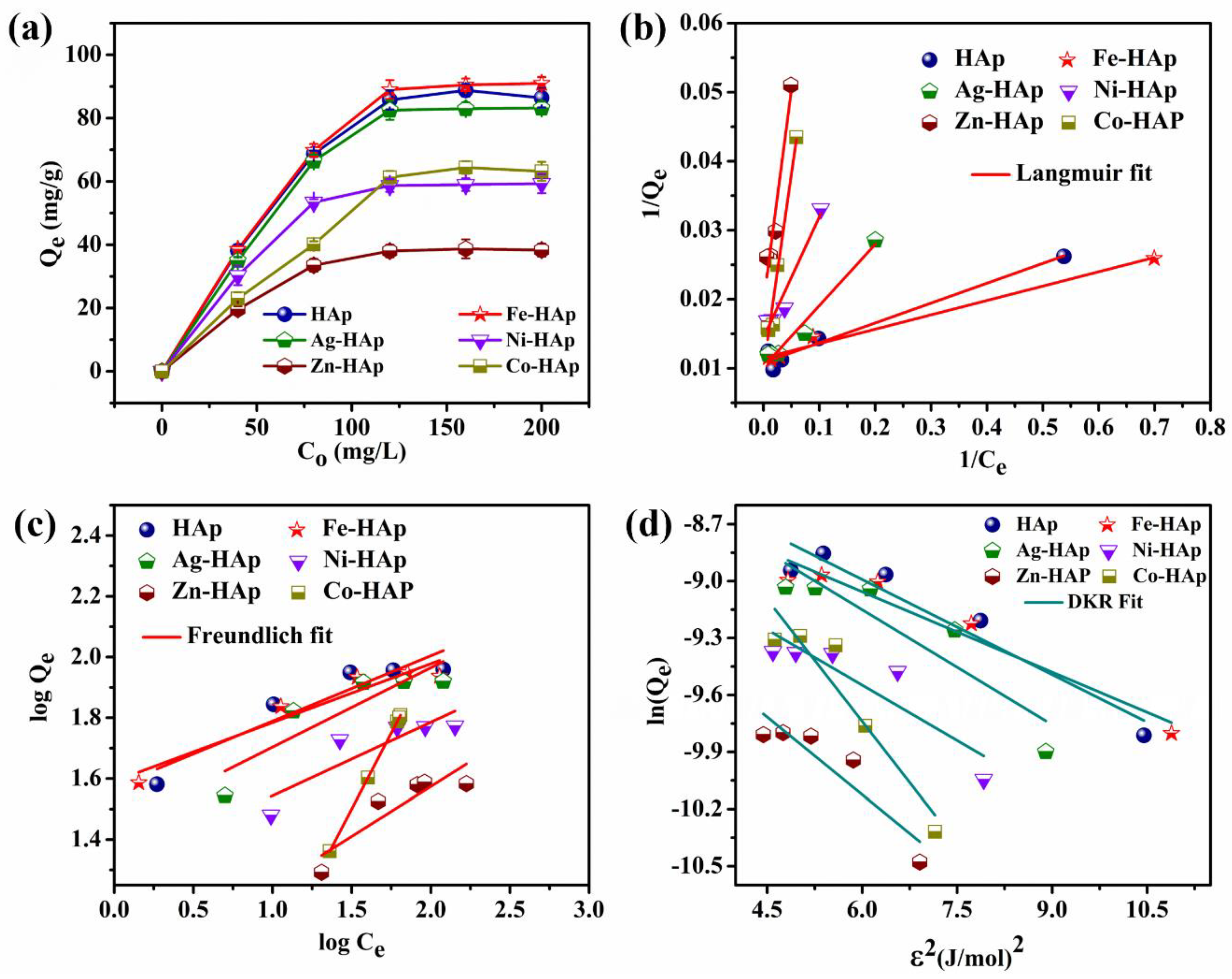
Figure 5a exhibits the maximum CR adsorption capacities of the HAp and modified HAp, with the different initial concentrations (40 to 200 mg/L). A total of 40 mg of the adsorbent was mixed at various CR concentrations at pH 7. The adsorption capacity was enhanced synergistically with an increase in the concentration of CR. Thus, the results indicate that the higher concentration provided a low resistance path and high driving force between the adsorbate and adsorbent, respectively. Accordingly, the maximum adsorption capacity increased from 38 to 86 mg/g for HAp, 39 to 91 mg/g for Fe-HAp, 35 to 83 mg/g for Ag-HAp, 30 to 59 mg/g for Ni-HAp, 20 to 38 mg/g for Zn-HAp, and 23 to 63 mg/g for Co-HAp. The maximum CR adsorption capacity of Ni-HAp, Zn-HAp, and Co-HAp was lower compared to HAp, Fe-HAp, and Ag-HAp. The higher adsorption capacity possessed by Fe-HAp qualified it as a potential adsorbent for removing CR from water compared to the other reports (Table 2). The equilibrium adsorption data were fitted to different isotherms of Langmuir, Freundlich, and DKR. These isotherm results explore the adsorption mechanism and type of the adsorption (i.e., monolayer or multilayer). The linear expression of the Langmuir, Freundlich, and DKR isotherms is given in Equations (9)–(11) [35,36,37,38]. In addition, the dimensionless Langmuir constant (RL), expressed in Equation (12), can be used to explore the adsorption favorability. The value of RL specifies the shape of the isotherm, such as if the CR adsorption is linear (RL = 1), disagreeable (RL > 1), agreeable (0 < RL > 1), or irreversible (RL = 0) [39,40].
C e Q e = 1 Q m K L + C e Q m
L o g Q e = L o g K F + L o g C e n
ln Q e = ln Q m - β ε 2
R L = 1 ( 1 + K L C o )
ε = R T ln ( 1 + 1 C e )
E = 1 - 2 β
where Ce is the equilibrium concentration (mg/L), and Qm and KF are the maximum Langmuir and Freundlich adsorption capacities (mg/g), respectively. KL (L/mg) and 1/n are the Langmuir and Freundlich constants, respectively. The 1/n exhibiting nature of the Freundlich adsorption: if the adsorption was linear ((1/n) = 1), agreeable (0.1< (1/n) > 1), disagreeable ((1/n) > 1), or irreversible ((1/n) = 0) [42]. β is the DKR adsorption mean energy (mol/J)−2; ε is the Polanyi potential (Equation (13)); R is the universal gas constant of 8.314 (J/mol K); and T is the absolute temperature (K). Furthermore, E is the Gaussian energy distribution (Equation (14)) from the DKR isotherm, which suggests the adsorption was via physisorption (E < 8 kJ/mol), chemical ion exchange (8 kJ/mol ≤ E ≤ 16 kJ/mol), or a strong chemical adsorption process (E > 16 kJ/mol).
The values of Qm and KL, KF and 1/n, and Qm and β were calculated from the intercept and slope in the liner fits of the Langmuir, Freundlich, and DKR isotherms, respectively (Figure 5b–d) [45,46,47]. The isotherms confirmed that the adsorption of CR was by monolayer and chemisorption mechanisms due to the fact of their linear regression R2 values, and the maximum adsorption capacity of the experimental and theoretical values were a better match for the Langmuir isotherms than the Freundlich and DKR isotherms (Table 3). Furthermore, the RL values obtained the adsorption favorability for the Langmuir isotherms, whereas the 1/n value of the Freundlich suggested an unfavorable adsorption of CR. In addition, the calculated Gaussian energy distribution from the DKR isotherm signified that the CR adsorption was via a chemical ion exchange process while using Ag-HAp, Zn-HAp, and Co-HAp, whereas CR adsorption occurred via strong chemisorption rather than the chemical ion exchange process while using HAp, Fe-HAp, and Ni-HAp.

3.1.3. Effect of pH

A study of the influence of pH on the effects is essential to find the maximum adsorption capacity and the adsorption behavior at different pH levels. The solution pH value strongly affects the adsorbent characteristics of the surface charge and CS. The adsorbent surface charge at different pH values was calculated from the pH drift method, as shown in Figure 6a [48,49,50,51]. The adsorbent’s zero-point charge (pHpzc) was effectively changed by incorporating different ions in the HAp lattice. Accordingly, the pHpzc of HAp, Fe-HAp, Ag-HAp, Ni-HAp, Zn-HAp, and Co-HAp were 7.5, 7.8, 8.3, 7.3, 7.1, and 6.5, respectively. Therefore, the Fe2+ and Ag+ ion incorporation prominently extended a positive surface charge, which increased the electrostatic interaction between the adsorbent and CR molecules compared to the other adsorbents. In addition, the cationic modification significantly improved the CS (excluding the Co2+ ion), and it was calculated from the zeta potential (ζ). A higher ζ represents a high CS and vice versa (Figure 6b). Accordingly, the Fe-HAp (−38 mV) obtained a higher ζ than the rest of the adsorbents (−16 mV for HAp, −21 mV for Ag-HAp, −29 mV for Ni-HAp, −25 mV for Zn-HAp, and −4 mV for Co-HAp), causing an increase in the uniform particle distribution, which led to the enhancement of the accessibility of the active sites for CR adsorption. Hence, the CR adsorption was enhanced in a wide range of pH values while using Fe-HAp compared to other adsorbents (Figure 6c). However, all of the adsorbents exhibited a decreasing CR adsorption trend at an alkaline pH, which indicates that the increase in the electrostatic repulsive force between the adsorbent and CR molecules was due to the fact of a similar charge. Whereas in acidic medium, the adsorbent and CR molecules had opposite charges, which increased the electrostatic interaction leading to an enhancement in the adsorption capacity.

3.1.4. Effect of Temperature

Figure 7a presents the CR adsorption at different isothermal temperatures (303, 313, 323, and 333 K). The thermodynamic parameter of the adsorption-free energy (∆G°) was calculated from Equations (15)–(17) [14,52].
G ° = - R T ln K d
K d = ρ Q e C e
G ° = H ° - T S °
where Kd is the dimensionless constant, ρ is the density of water (1000 g/L), ∆H° is the enthalpy, and ∆S° is the entropy. All adsorbents had a low adsorption capacity while the temperature increased from 303 to 333 K. The adsorbents acquired sufficient energy to aggregate the particles between them at higher isothermal temperatures. Consequently, the number of available active sites decreased, and the interaction between the CR and adsorbent was inhibited significantly, which drastically reduced the adsorption capacity. Furthermore, the negative value of ∆G° indicates that the adsorption of CR was spontaneous; nevertheless, the adsorption spontaneity was increased in the order of Fe-HAp > Ag-HAp > HAp > Co-HAp > Ni-HAp > Zn-HAp. However, the adsorption spontaneity increased with the increase in the temperature for Fe-HAp and Ni-HAp, whereas in the case of the other adsorbents, the adsorption spontaneity decreased significantly. The entropy (∆S°) and enthalpy (∆H°) of the given system were calculated from the van’t Hoff linear plot’s intercept and slope, respectively (Figure 7b, Table 4) [14,41]. The positive value of ∆H° suggests that the CR adsorption was endothermic, which indicates that the particle absorbed the energy from its surroundings while the temperature increased. Accordingly, the absorbed energy significantly improved the particles’ interaction with each other, resulting in an increase in the particle aggregation, which caused a decrease in the adsorption capacity. In general, physisorption occurs when ∆H°, which indicates that the energy involved in the adsorption is less than 84 kJ/mol, and chemisorption occurs when the energy involved is between 84 and 420 kJ/mol [12]. Accordingly, for the CR adsorption on the HAp and cation-modified HAp, ∆H° exhibiting chemisorption was confirmed. Furthermore, the negative ∆S° point to the insignificant changes in the internal structures of the HAp and cationic-modified HAp, which ensured that the adsorption occurred by chemisorption rather than the chemical ion-exchange process. These two results are well consistent with the adsorption isotherm analyses.

3.2. Regeneration

The recycling efficiency is the most significant factor for practical applications. Accordingly, NaOH was used to regenerate the CR-adsorbed HAp and cation-modified HAp by ultrasonication treatment for 1 h. Subsequently, the regenerated samples were centrifuged with triple-distilled water. The adsorption of CR was determined for seven successive recycles (Figure 7c), and the recycling efficiency of Fe-HAp maintained 92% of the adsorption capacity compared to the 1st cycle, whereas the recycling efficiencies of the HAp, Ag-HAp, Ni-HAp, Zn-HAp, and Co-HAp were low (1–42%). Among the ion-incorporated HAp, the Fe-HAp displayed sustained adsorbent characteristics. The CR adsorption by the HAp was due to (i) the replacement of sulfate (SO32−) ions by the phosphate/carbonate ion (PO43−/CO32−) in the HAp lattice; (ii) the surface complexation between NH3+ and OH/PO43− sites; (iii) the surface complexation between sulfate ions ( O = S - O - ) and Ca2+ sites [3,53]. Accordingly, the CR adsorption by the HAp and Co-HAp was reduced drastically after the first cycle because of the low CS of the adsorbents. The other metal ion-doped HAp (Fe-HAp Ag-HAp, Ni-HAp, and Zn-HAp) exhibited a gradual decrease in the removal efficiency in the subsequent cycles of regeneration, which indicates that the cation dopant inhibited the ion exchange activity as well as favored the surface complexation process. The high colloidal stability of the Fe-HAp boosted the desorption of CR, thereby improving the recycling efficiency compared to the other adsorbents.

3.3. Structural Analysis after CR Adsorption

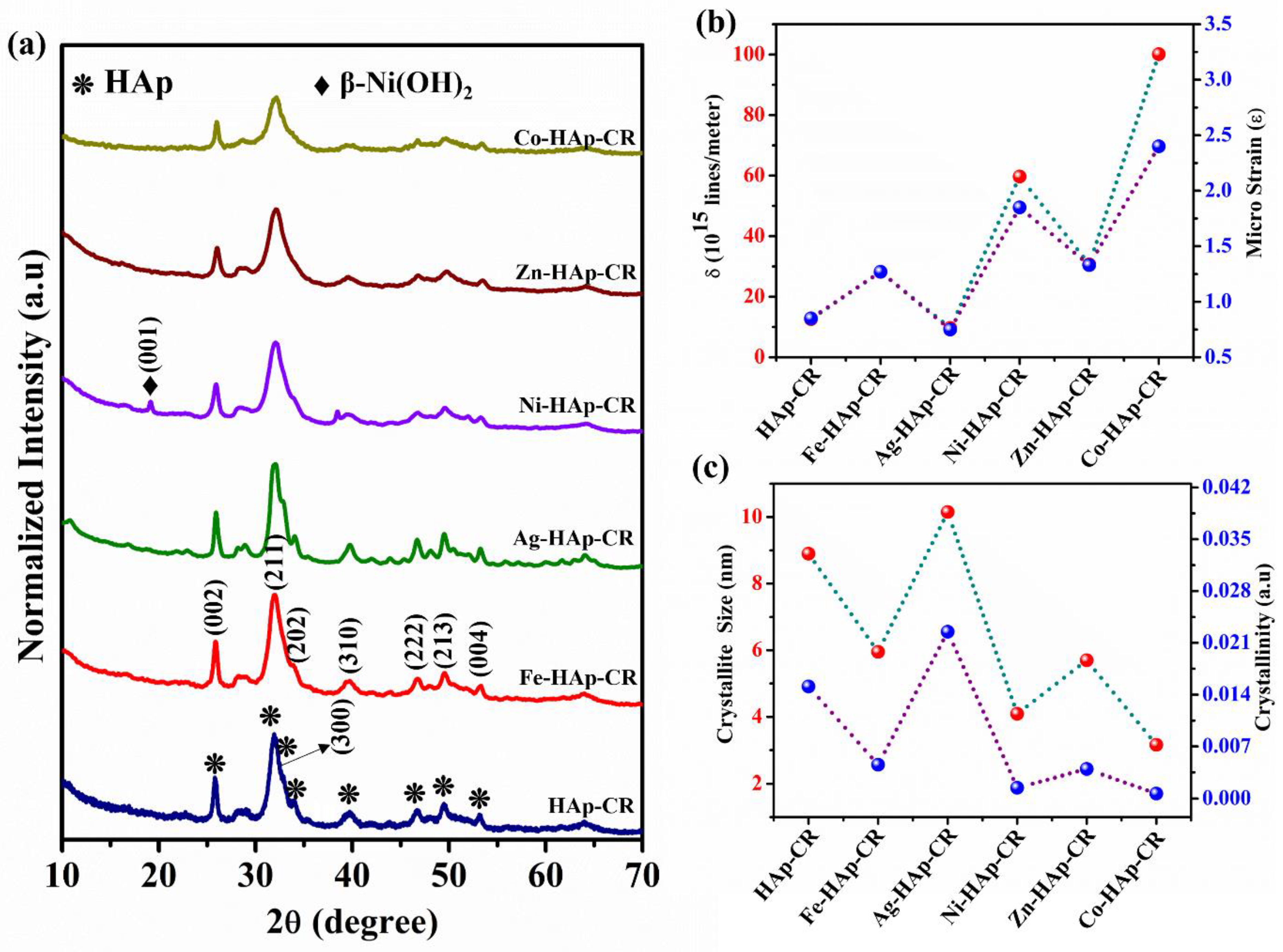
The XRD patterns of the CR-adsorbed pristine HAp and cation-incorporated HAp nanoparticles exhibited a similar HAp phase and were well matched with JCPDS No. 09-0432 (Figure 8a). In addition, the impurity phase of β-Ni(OH)2 in Ni-HAp was observed at 2θ = 19.04°. The calculated crystallite size, crystallinity, dislocation density, and microstrain after the CR adsorption of both the pristine HAp and cation-incorporated HAp nanoparticles did not show any significant variation (excluding Co-HAp). However, after CR adsorption, the Co-HAp exhibited a low crystallinity and crystallite size along with a high dislocation density and microstrain due to adsorption by the chemical ion exchange process (Figure 8b,c).
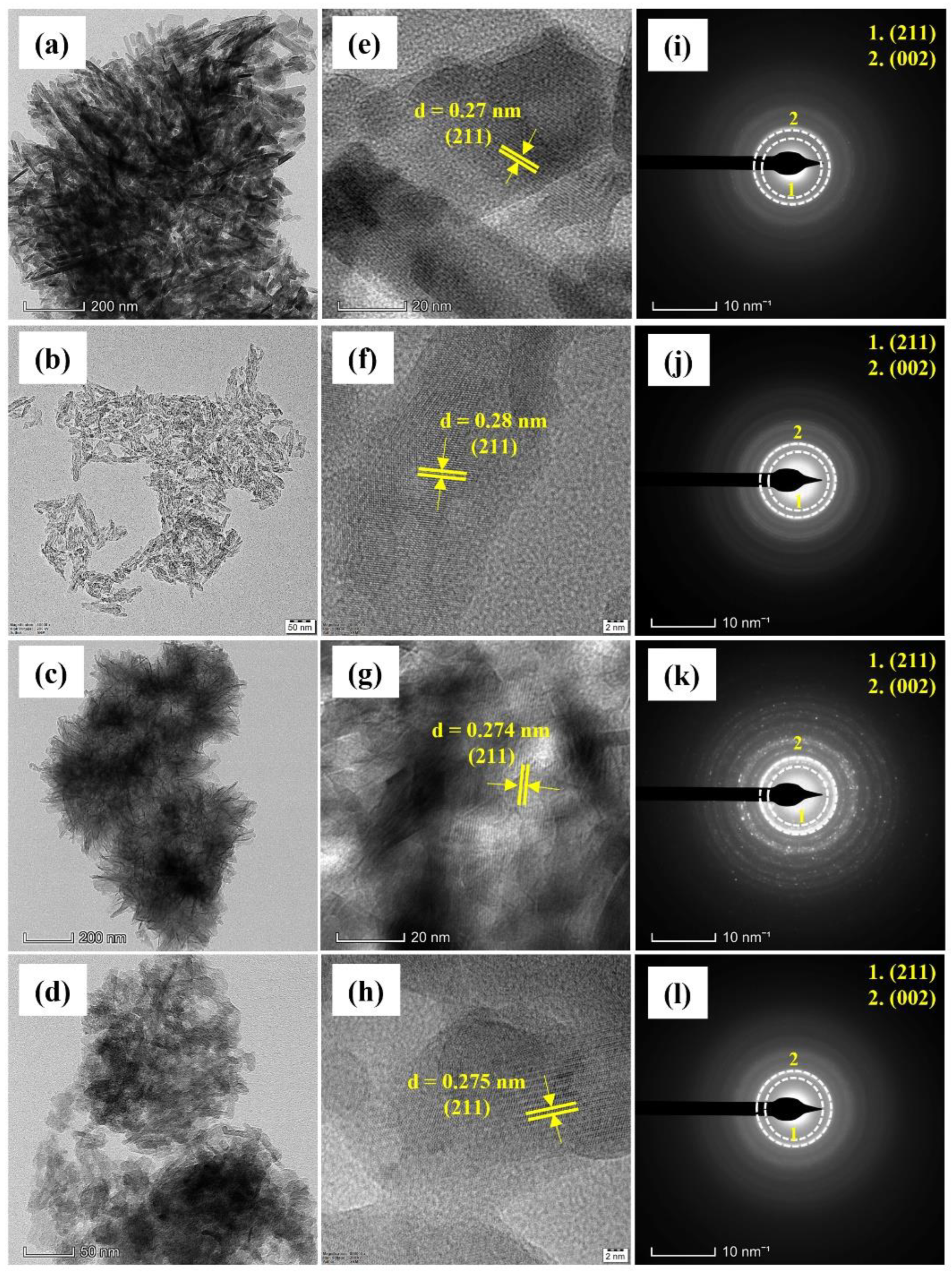
HR-TEM micrographs were used to investigate the morphological and microstructural changes before and after the CR-adsorbed HAp and Fe-HAp (Figure 9). The HR-TEM micrographs of the HAp and Fe-HAp before and after adsorption showed a uniform distribution with a rod-like morphology; the particle size of the HAp was 75 nm × 32 nm and that of Fe-HAp was 25 nm × 13 nm (Figure 9a,b). However, the CR-adsorbed HAp had flower-like agglomerates (200 nm × 20 nm) (Figure 9c), leading to a decrease in the recycling efficiency, whereas similar flower-like agglomerates were observed in the CR-adsorbed Fe-HAp (33 × 14 nm) (Figure 9d). The microstructural analysis of the HAp, Fe-HAp, HAp-CR, and Fe-HAp-CR exhibited well-resolved lattice fringes with a d-space of 0.275 nm, which are attributed to the (211) plane of the HAp phase (Figure 9e–h). Furthermore, the selective electron diffraction patterns of the HAp, Fe-HAp, HAp-CR, and Fe-HAp-CR indicate that the (211) and (002) planes’ corresponding interplanar distances were 0.27 and 0.34 nm, respectively (Figure 9i–l). These results indicate that the HAp and Fe-HAp phases were stable and did not show any significant modification in the HAp phase before and after CR adsorption, which were in good agreement with XRD analysis.

3.4. Quantification of Dopant Cations for before and after CR Adsorption

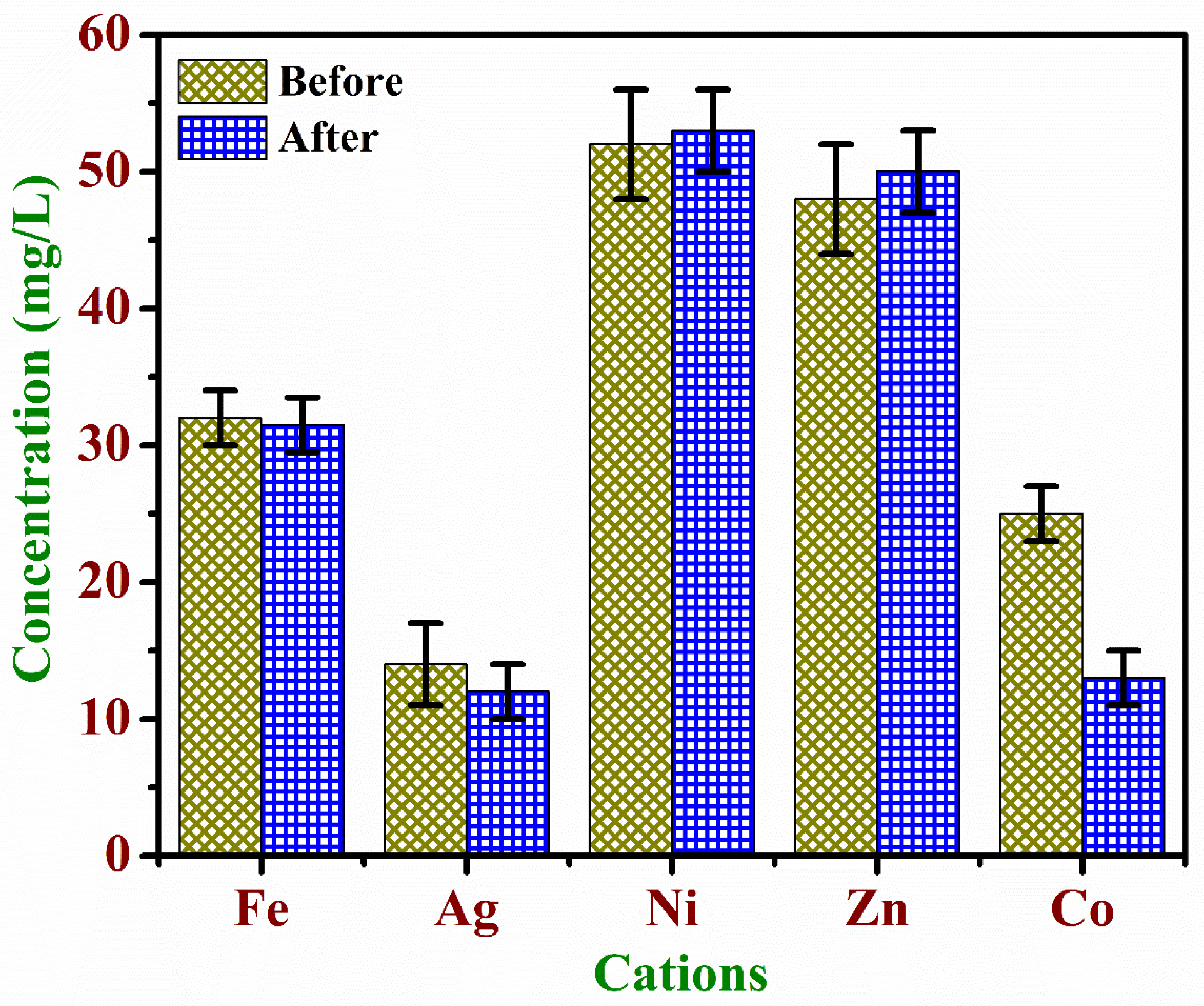
The leaching of cations before and after the adsorption of CR is shown in Figure 10. The estimated incorporation of metallic ions before adsorption in the Fe-HAp, Ag-HAp, Ni-HAp, Zn-HAp, and Co-HAp was 32, 14, 52, 48, and 25 mg/L, respectively. Accordingly, the amount of ion incorporated, in ascending order, is Ni-HAp > Zn-HAp > Fe-HAp > Co-HAp > Ag-HAp. Moreover, after the CR adsorption, apart from Ag-HAp and Co-HAp, all of the adsorbents exhibited insignificant leaching (<2%). The Ag-HAp and Co-HAp had low colloidal stability, which increased the ion exchange process during the CR adsorption process. Hence, 14% of Ag+ (2 mg/L) and 52% of Co2+ (12 mg/L) ions were leached out from the Ag-HAp and Co-HAp particles, respectively.

4. Conclusions

This work elucidated the synthesis of colloidally stable HAp nanoparticles using a combination of hydrothermal and ultrasonication treatments followed by microwave drying. The presence of dopant ions enhanced the adsorbent properties without any change of phase, as confirmed by XRD, FTIR, and SEM analyses. The incorporation of cations (Fe2+, Ag+, Ni2+, and Zn2+) in the HAp lattice enhanced the CS (31–138%), whereas the particle size was reduced compared to the pristine HAp. Moreover, the particle morphology was modified from rod to sphere by the incorporation of Ag+, Ni2+, and Co2+ ions. The equilibrium adsorption was achieved within 1 min, and the maximum CR adsorption capacities of HAp, Fe-HAp, Ag-HAp, Ni-HAp, Zn-HAp, and Co-HAp were 110, 128, 105, 71, 58, and 81 mg/g, respectively. The adsorption process was due to the strong chemical adsorption rather than an ion-exchange process, and the adsorbed CR on the HAp surface was a monolayer. The thermodynamic results suggest that the CR adsorption process was spontaneous and endothermic. Hence, the CR adsorption capacity and rate were dramatically increased compared to previous reports. Furthermore, the Fe-HAp exhibited a consistent recycling efficiency (92%) over seven cycles, when compared to other ion-doped HAp particles (1% to 42%). The overall results of this study demonstrate that the Fe-HAp has a highly stable adsorption over a wide pH range, multiple recycling efficiencies, and rapid adsorption within 1 min. The Fe-HAp is a potential, cost-effective, and alternative adsorbent for removing CR from water without the formation of secondary byproducts.

Author Contributions

Conceptualization, S.E.P.; Methodology, S.E.P.; Writing—Original Draft, S.E.P.; Validation, S.E.P. and S.S.; Visualization, S.S.; Writing—Review and Editing, N.K.S., E.K. and M.B.S.; Supervision, N.K.S.; Investigation, N.K.S.; Project Administration, N.K.S.; All authors have read and agreed to the published version of the manuscript.

Funding

Department of Science and Technology, through the scheme of the Technology Development Programme-1010 (No: DST/TM/WTI/2K16/219(G)-A).

Informed Consent Statement

Not applicable.

Data Availability Statement

Not applicable.

Acknowledgments

The authors would like to thank the government of India for the financial support from the Department of Science and Technology, through the scheme of the Technology Development Programme-1010 (No: DST/TM/WTI/2K16/219(G)-A). One of the authors (Narayana Kalkura Subbaraya) thanks the University Grants Commission, India, for the award of a Basic Scientific Research (UGC-BSR) Faculty Fellowship (No. F.4-5(11)2019 (BSR)).

Conflicts of Interest

The authors declare no conflict of interest.

References

  1. Liu, J.; Wang, N.; Zhang, H.; Baeyens, J. Adsorption of Congo Red Dye on FexCo3−xO4 Nanoparticles. J. Environ. Manag. 2019, 238, 473–483. [Google Scholar] [CrossRef] [PubMed]
  2. Sathiyavimal, S.; Vasantharaj, S.; Shanmugavel, M.; Manikandan, E.; Nguyen-Tri, P.; Brindhadevi, K.; Pugazhendhi, A. Facile Synthesis and Characterization of Hydroxyapatite from Fish Bones: Photocatalytic Degradation of Industrial Dyes (Crystal Violet and Congo Red). Prog. Org. Coatings 2020, 148, 105890. [Google Scholar] [CrossRef]
  3. Hou, H.; Zhou, R.; Wu, P.; Wu, L. Removal of Congo Red Dye from Aqueous Solution with Hydroxyapatite/Chitosan Composite. Chem. Eng. J. 2012, 211–212, 336–342. [Google Scholar] [CrossRef]
  4. Raval, N.P.; Shah, P.U.; Shah, N.K. Adsorptive Amputation of Hazardous Azo Dye Congo Red from Wastewater: A Critical Review. Environ. Sci. Pollut. Res. 2016, 23, 14810–14853. [Google Scholar] [CrossRef]
  5. Litefti, K.; Freire, M.S.; Stitou, M.; González-Álvarez, J. Adsorption of an Anionic Dye (Congo Red) from Aqueous Solutions by Pine Bark. Sci. Rep. 2019, 9, 16530. [Google Scholar] [CrossRef] [Green Version]
  6. Khan, A.; Bhoi, R.; Saharan, V.K.; George, S. Synthesis of Titanium Doped Hydroxyapatite Using Waste Marble Powder for the Degradation of Congo Red Dye in Wastewater. Mater. Today Proc. 2022, 57, 1645–1653. [Google Scholar] [CrossRef]
  7. Ausavasukhi, A.; Kampoosaen, C.; Kengnok, O. Adsorption Characteristics of Congo Red on Carbonized Leonardite. J. Clean. Prod. 2016, 134, 506–514. [Google Scholar] [CrossRef]
  8. Azeez, L.; Adebisi, S.A.; Adejumo, A.L.; Busari, H.K.; Aremu, H.K.; Olabode, O.A.; Awolola, O. Adsorptive Properties of Rod-Shaped Silver Nanoparticles-Functionalized Biogenic Hydroxyapatite for Remediating Methylene Blue and Congo Red. Inorg. Chem. Commun. 2022, 142, 109655. [Google Scholar] [CrossRef]
  9. Han, R.; Ding, D.; Xu, Y.; Zou, W.; Wang, Y.; Li, Y.; Zou, L. Use of Rice Husk for the Adsorption of Congo Red from Aqueous Solution in Column Mode. Bioresour. Technol. 2008, 99, 2938–2946. [Google Scholar] [CrossRef]
  10. Foroughi-Dahr, M.; Abolghasemi, H.; Esmaili, M.; Shojamoradi, A.; Fatoorehchi, H. Adsorption Characteristics of Congo Red from Aqueous Solution onto Tea Waste. Chem. Eng. Commun. 2015, 202, 181–193. [Google Scholar] [CrossRef]
  11. Ghanavati Nasab, S.; Semnani, A.; Teimouri, A.; Kahkesh, H.; Momeni Isfahani, T.; Habibollahi, S. Removal of Congo Red from Aqueous Solution by Hydroxyapatite Nanoparticles Loaded on Zein as an Efficient and Green Adsorbent: Response Surface Methodology and Artificial Neural Network-Genetic Algorithm. J. Polym. Environ. 2018, 26, 3677–3697. [Google Scholar] [CrossRef]
  12. Lian, L.; Guo, L.; Guo, C. Adsorption of Congo Red from Aqueous Solutions onto Ca-Bentonite. J. Hazard. Mater. 2009, 161, 126–131. [Google Scholar] [CrossRef]
  13. Bensalah, H.; Younssi, S.A.; Ouammou, M.; Gurlo, A.; Bekheet, M.F. Azo Dye Adsorption on an Industrial Waste-Transformed Hydroxyapatite Adsorbent: Kinetics, Isotherms, Mechanism and Regeneration Studies. J. Environ. Chem. Eng. 2020, 8, 103807. [Google Scholar] [CrossRef]
  14. Zhang, Z.; Moghaddam, L.; O’Hara, I.M.; Doherty, W.O.S. Congo Red Adsorption by Ball-Milled Sugarcane Bagasse. Chem. Eng. J. 2011, 178, 122–128. [Google Scholar] [CrossRef] [Green Version]
  15. Rajendran, S.; Yadav, V.K.; Gacem, A.; Algethami, J.S.; Alqahtani, M.S.; Aldakheel, F.M.; Binshaya, A.S.; Alharthi, N.S.; Khan, I.A.; Islam, S. Functionalized Microbial Consortia with Silver-Doped Hydroxyapatite (Ag@HAp) Nanostructures for Removal of RO84 from Industrial Effluent. Crystals. 2022, 12, 970. [Google Scholar] [CrossRef]
  16. Frank-Kamenetskaya, O.V.; Rozhdestvenskaya, I.V.; Rosseeva, E.V.; Zhuravlev, A.V. Refinement of apatite atomic structure of albid tissue of Late Devon conodont. Crystallogr. Rep. 2014, 59, 41–47. [Google Scholar] [CrossRef]
  17. Karalkeviciene, R.; Raudonyte-Svirbutaviciene, E.; Gaidukevic, J.; Zarkov, A.; Kareiva, A. Solvothermal Synthesis of Calcium-Deficient Hydroxyapatite via Hydrolysis of α -Tricalcium Phosphate in Different Aqueous-Organic Media. Crystals 2022, 12, 253. [Google Scholar] [CrossRef]
  18. Vidotto, M.; Grego, T.; Petrović, B.; Somers, N.; Antonić Jelić, T.; Kralj, D.; Matijaković Mlinarić, N.; Leriche, A.; Dutour Sikirić, M.; Erceg, I.; et al. A Comparative EPR Study of Non-Substituted and Mg-Substituted Hydroxyapatite Behaviour in Model Media and during Accelerated Ageing. Crystals 2022, 12, 297. [Google Scholar] [CrossRef]
  19. Marincaș, L.; Turdean, G.L.; Toșa, M.; Kovács, Z.; Kovács, B.; Barabás, R.; Farkas, N.I.; Bizo, L. Hydroxyapatite and Silicon-Modified Hydroxyapatite as Drug Carriers for 4-Aminopyridine. Crystals 2021, 11, 1124. [Google Scholar] [CrossRef]
  20. Thakur, D.; Yeh, S.C.; Cheng, R.H.; Loke, S.S.; Wei, H.H.; Cheng, P.Y.; Lai, Y.C.; Chen, H.Y.; Huang, Y.R.; Ding, S.W. Varying Synthesis Conditions and Comprehensive Characterization of Fluorine-Doped Hydroxyapatite Nanocrystals in a Simulated Body Fluid. Crystals 2022, 12, 139. [Google Scholar] [CrossRef]
  21. Ashok, M.; Meenakshi Sundaram, N.; Narayana Kalkura, S. Crystallization of Hydroxyapatite at Physiological Temperature. Mater. Lett. 2003, 57, 2066–2070. [Google Scholar] [CrossRef]
  22. Kolanthai, E.; Ganesan, K.; Epple, M.; Kalkura, S.N. Synthesis of Nanosized Hydroxyapatite/Agarose Powders for Bone Filler and Drug Delivery Application. Mater. Today Commun. 2016, 8, 31–40. [Google Scholar] [CrossRef]
  23. Qu, Y.; Zhou, W.; Miao, X.; Li, Y.; Jiang, L.; Pan, K.; Tian, G.; Ren, Z.; Wang, G.; Fu, H. A New Layered Photocathode with Porous NiO Nanosheets: An Effective Candidate for p-Type Dye-Sensitized Solar Cells. Chem.—Asian J. 2013, 8, 3085–3090. [Google Scholar] [CrossRef] [PubMed]
  24. Kettaf, S.; Guellati, O.; Harat, A.; Kennaz, H.; Momodu, D.; Dangbegnon, J.; Manyala, N.; Guerioune, M. Electrochemical Measurements of Synthesized Nanostructured β-Ni(OH)2 Using Hydrothermal Process and Activated Carbon Based Nanoelectroactive Materials. SN Appl. Sci. 2019, 1, 34. [Google Scholar] [CrossRef]
  25. Kumaresan, N.; Ramamurthi, K.; Ramesh Babu, R.; Sethuraman, K.; Moorthy Babu, S. Hydrothermally Grown ZnO Nanoparticles for Effective Photocatalytic Activity. Appl. Surf. Sci. 2017, 418, 138–146. [Google Scholar] [CrossRef]
  26. Mote, V.; Purushotham, Y.; Dole, B. Williamson-Hall Analysis in Estimation of Lattice Strain in Nanometer-Sized ZnO Particles. J. Theor. Appl. Phys. 2012, 6, 2–9. [Google Scholar] [CrossRef] [Green Version]
  27. Kamal, H.; Hezma, A.M. Spectroscopic Investigation and Magnetic Study of Iron, Manganese, Copper and Cobalt-Doped Hydroxyapatite Nanopowders. Phys. Sci. Int. J. 2015, 7, 137–151. [Google Scholar] [CrossRef] [Green Version]
  28. Li, Z.Y.; Lam, W.M.; Yang, C.; Xu, B.; Ni, G.X.; Abbah, S.A.; Cheung, K.M.C.; Luk, K.D.K.; Lu, W.W. Chemical Composition, Crystal Size and Lattice Structural Changes after Incorporation of Strontium into Biomimetic Apatite. Biomaterials 2007, 28, 1452–1460. [Google Scholar] [CrossRef]
  29. Mercado, D.F.; Magnacca, G.; Malandrino, M.; Rubert, A.; Montoneri, E.; Celi, L.; Bianco Prevot, A.; Gonzalez, M.C. Paramagnetic Iron-Doped Hydroxyapatite Nanoparticles with Improved Metal Sorption Properties. A Bioorganic Substrates-Mediated Synthesis. ACS Appl. Mater. Interfaces 2014, 6, 3937–3946. [Google Scholar] [CrossRef] [Green Version]
  30. Destainville, A.; Champion, E.; Bernache-Assollant, D.; Laborde, E. Synthesis, Characterization and Thermal Behavior of Apatitic Tricalcium Phosphate. Mater. Chem. Phys. 2003, 80, 269–277. [Google Scholar] [CrossRef]
  31. Sarath Chandra, V.; Baskar, G.; Suganthi, R.V.; Elayaraja, K.; Ahymah Joshy, M.I.; Sofi Beaula, W.; Mythili, R.; Venkatraman, G.; Narayana Kalkura, S. Blood Compatibility of Iron-Doped Nanosize Hydroxyapatite and Its Drug Release. ACS Appl. Mater. Interfaces 2012, 4, 1200–1210. [Google Scholar] [CrossRef]
  32. Liu, Z.; He, X.; Yang, X.; Ding, H.; Wang, D.; Ma, D.; Feng, Q. Synthesis of Mesoporous Carbon Nitride by Molten Salt-Assisted Silica Aerogel for Rhodamine B Adsorption and Photocatalytic Degradation. J. Mater. Sci. 2021, 56, 11248–11265. [Google Scholar] [CrossRef]
  33. Nayak, B.; Samant, A.; Patel, R.; Misra, P.K. Comprehensive Understanding of the Kinetics and Mechanism of Fluoride Removal over a Potent Nanocrystalline Hydroxyapatite Surface. ACS Omega 2017, 2, 8118–8128. [Google Scholar] [CrossRef] [Green Version]
  34. Ali, A.A.; Ahmed, I.S.; Elfiky, E.M. Auto-Combustion Synthesis and Characterization of Iron Oxide Nanoparticles (α-Fe2O3) for Removal of Lead Ions from Aqueous Solution. J. Inorg. Organomet. Polym. Mater. 2021, 31, 384–396. [Google Scholar] [CrossRef]
  35. Ghaedi, M.; Biyareh, M.N.; Kokhdan, S.N.; Shamsaldini, S.; Sahraei, R.; Daneshfar, A.; Shahriyar, S. Comparison of the Efficiency of Palladium and Silver Nanoparticles Loaded on Activated Carbon and Zinc Oxide Nanorods Loaded on Activated Carbon as New Adsorbents for Removal of Congo Red from Aqueous Solution: Kinetic and Isotherm Study. Mater. Sci. Eng. C. 2012, 32, 725–734. [Google Scholar] [CrossRef]
  36. Su, Y.; Cui, M.; Zhu, J.; Wu, Y.; Wei, Y.; Bian, S. A Facile Synthesis of Nanosheet-Structured Barium Carbonate Spheres with a Superior Adsorption Capacity for Cr(VI) Removal. J. Mater. Sci. 2018, 53, 4078–4088. [Google Scholar] [CrossRef]
  37. Rabiee Faradonbeh, M.; Dadkhah, A.A.; Rashidi, A.; Tasharofi, S.; Mansourkhani, F. Newly MOF-Graphene Hybrid Nanoadsorbent for Removal of Ni(II) from Aqueous Phase. J. Inorg. Organomet. Polym. Mater. 2018, 28, 829–836. [Google Scholar] [CrossRef]
  38. Dabrowski, A. Adsorption—From Theory to Practice. Adv. Colloid Interface Sci. 2001, 93, 135–224. [Google Scholar] [CrossRef]
  39. Espinoza-Sánchez, M.A.; Arévalo-Niño, K.; Quintero-Zapata, I.; Castro-González, I.; Almaguer-Cantú, V. Cr(VI) Adsorption from Aqueous Solution by Fungal Bioremediation Based Using Rhizopus Sp. J. Environ. Manag. 2019, 251, 109595. [Google Scholar] [CrossRef]
  40. Ma, T.; Wu, Y.; Liu, N.; Wu, Y. Iron Manganese Oxide Modified Multi-Walled Carbon Nanotube as Efficient Adsorbent for Removal of Organic Dyes: Performance, Kinetics and Mechanism Studies. J. Inorg. Organomet. Polym. Mater. 2020, 30, 4027–4042. [Google Scholar] [CrossRef]
  41. Namasivayam, C.; Kavitha, D. Removal of Congo Red from Water by Adsorption onto Activated Carbon Prepared from Coir Pith, an Agricultural Solid Waste. Dye. Pigment. 2002, 54, 47–58. [Google Scholar] [CrossRef]
  42. Parvin, S.; Biswas, B.K.; Rahman, M.A.; Rahman, M.H.; Anik, M.S.; Uddin, M.R. Study on Adsorption of Congo Red onto Chemically Modified Egg Shell Membrane. Chemosphere 2019, 236, 124326. [Google Scholar] [CrossRef]
  43. Ghaedi, M.; Tavallali, H.; Sharifi, M.; Kokhdan, S.N.; Asghari, A. Preparation of Low Cost Activated Carbon from Myrtus Communis and Pomegranate and Their Efficient Application for Removal of Congo Red from Aqueous Solution. Spectrochim. Acta Part A Mol. Biomol. Spectrosc. 2012, 86, 107–114. [Google Scholar] [CrossRef]
  44. Bhattacharyya, K.G.; Sharma, A. Azadirachta Indica Leaf Powder as an Effective Biosorbent for Dyes: A Case Study with Aqueous Congo Red Solutions. J. Environ. Manag. 2004, 71, 217–229. [Google Scholar] [CrossRef] [PubMed]
  45. Vijayaraghavan, K.; Padmesh, T.V.N.; Palanivelu, K.; Velan, M. Biosorption of Nickel(II) Ions onto Sargassum wightii: Application of Two-Parameter and Three-Parameter Isotherm Models. J. Hazard. Mater. 2006, 133, 304–308. [Google Scholar] [CrossRef] [PubMed]
  46. Foo, K.Y.; Hameed, B.H. Insights into the Modeling of Adsorption Isotherm Systems. Chem. Eng. J. 2010, 156, 2–10. [Google Scholar] [CrossRef]
  47. Hutson, N.D.; Yang, R.T. Theoretical Basis for the Dubinin-Radushkevitch (D-R) Adsorption Isotherm Equation. Adsorption 1997, 3, 189–195. [Google Scholar] [CrossRef]
  48. Sekar, S.; Panchu, S.E.; Kolanthai, E.; Rajaram, V.; Subbaraya, N.K. Enhanced Stability of Hydroxyapatite/Sodium Alginate Nanocomposite for Effective Fluoride Adsorption. Mater. Today Proc. 2022, 58, 909–917. [Google Scholar] [CrossRef]
  49. Sekar, S.; Panchu, S.E.; Kolanthai, E.; Subbaraya, N.K. Enhanced Fluoride Adsorption and Regeneration Efficiency of Cross-linker-free Mesoporous Hydroxyapatite/Chitosan Nanocomposites. Res. Chem. Intermed. 2022, 48, 4857–4882. [Google Scholar] [CrossRef]
  50. Panchu, S.E.; Sekar, S.; Rajaram, V.; Kolanthai, E.; Panchu, S.J.; Swart, H.C.; Narayana Kalkura, S. Enriching Trace Level Adsorption Affinity of As3+ Ion Using Hydrothermally Synthesized Iron-Doped Hydroxyapatite Nanorods. J. Inorg. Organomet. Polym. Mater. 2022, 32, 47–62. [Google Scholar] [CrossRef]
  51. Panchu, S.E.; Sekar, S.; Kolanthai, E.; Rajaram, V.; Subbaraya, N.K. Bioremediation: Removal of Fluoride and Methylene Blue from Water Using Eco-Friendly Bio-Adsorbents. Mater. Today Proc. 2022, 58, 871–881. [Google Scholar] [CrossRef]
  52. Vimonses, V.; Lei, S.; Jin, B.; Chow, C.W.K.; Saint, C. Kinetic Study and Equilibrium Isotherm Analysis of Congo Red Adsorption by Clay Materials. Chem. Eng. J. 2009, 148, 354–364. [Google Scholar] [CrossRef]
  53. Guan, Y.; Cao, W.; Wang, X.; Marchetti, A.; Tu, Y. Hydroxyapatite Nano-Rods for the Fast Removal of Congo Red Dye from Aqueous Solution. Mater. Res. Express. 2018, 5, 065053. [Google Scholar] [CrossRef]
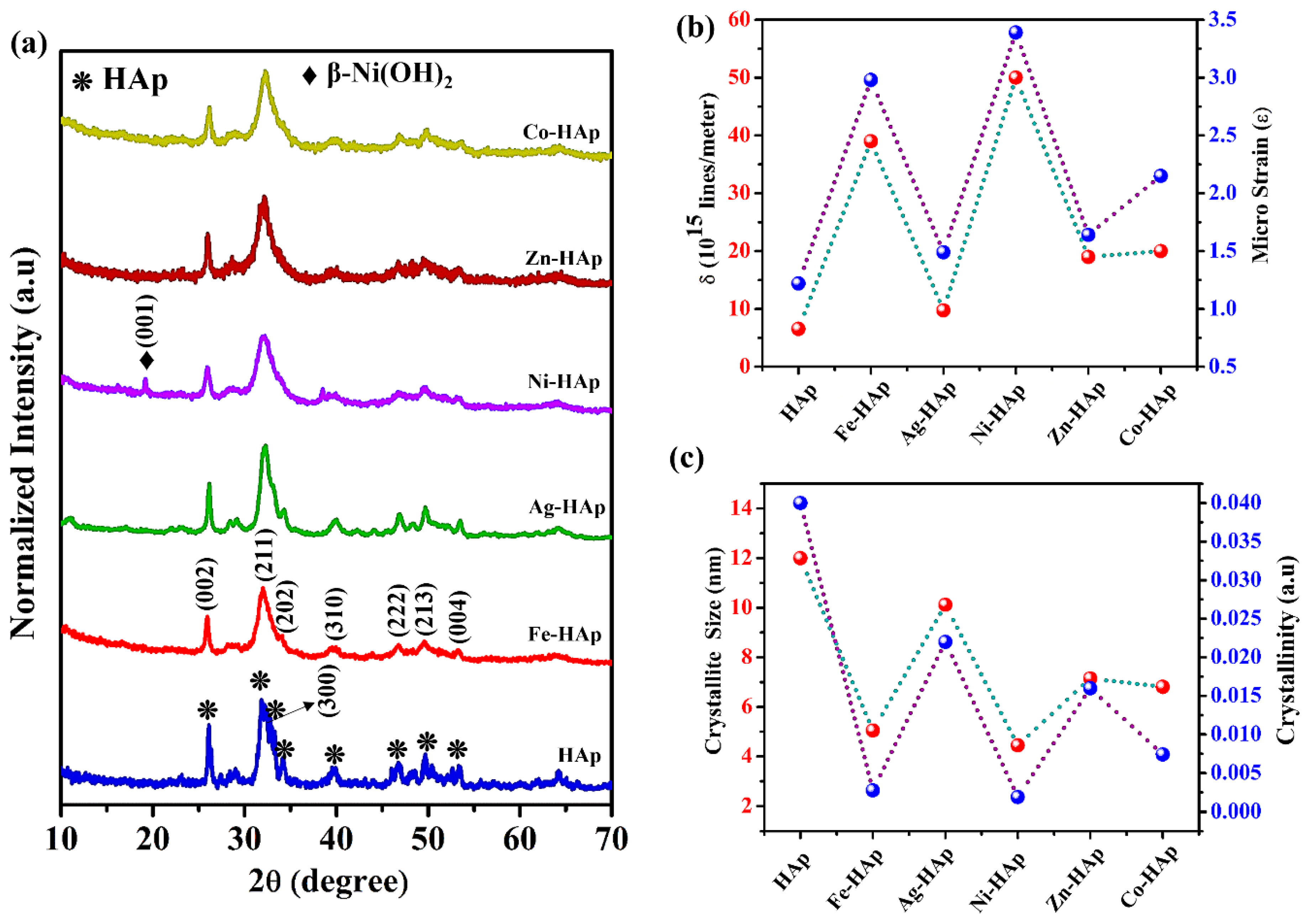
Figure 1. (a) X-ray diffraction spectrum of pristine HAp and different cation-incorporated HAp nanoparticles. The cation’s influence on the crystalline parameter: (b) dislocation density (red) and microstrain (blue); (c) crystallite size (red) and crystallinity (blue).
Crystals 13 00209 g001
Figure 2. FTIR spectra of the HAp and different cation−incorporated HAp nanoparticles.
Crystals 13 00209 g002
Figure 3. SEM micrograph: (a) HAp; (b) Fe-HAp; (c) Ag-HAp; (d) Ni-HAp; (e) Zn-HAp; (f) Co-HAp.
Crystals 13 00209 g003
Figure 4. (a) Effect of contact time (Co = 40 mg/L, pH = 7, temperature = 303 K, volume = 40 mL, dosage = 40 mg, and rpm = 200); kinetic isotherms: (b) pseudo first−order and (c) pseudo−second order.
Crystals 13 00209 g004
Figure 5. (a) Initial concentration effect of the CR adsorption (time = 1 min, pH = 7, dosage = 40 mg, volume = 40 mL, temperature = 303 K, and rpm = 200). Adsorption−isotherms: (b) Langmuir; (c) Freundlich; (d) DKR.
Crystals 13 00209 g005
Figure 6. (a) pHpzc of the adsorbent; (b) colloidal stability measured from the zeta potential; (c) pH (time = 1 min, Co = 200 mg/L, dosage = 40 mg, volume = 40 mL, rpm = 200, and temperature = 303 K).
Crystals 13 00209 g006
Figure 7. (a) Temperature (time = 1 min, Co = 200 mg/L, pH= 7, dosage = 40 mg, volume = 40 mL, and rpm = 200); (b) van’t Hoff linear plot; (c) regeneration (time = 1 min, Co = 200 mg/L, pH = 7, dosage = 40 mg, volume = 40 mL, rpm = 200, and temperature = 303 K).
Crystals 13 00209 g007
Figure 8. (a) X-ray diffraction spectrum of CR-adsorbed pristine HAp and different cation-incorporated HAp nanoparticles. The CR adsorption influence of the crystalline parameter of the HAp and cation-modified HAp nanoparticles: (b) dislocation density (red) and microstrain (blue); (c) crystallite size (red) and crystallinity (blue).
Crystals 13 00209 g008
Figure 9. High-resolution TEM micrograph: (a,e,i) HAp; (b,f,j) Fe-HAp; (c,g,k) HAp-CR; (d,h,l) Fe-HAp-CR.
Crystals 13 00209 g009
Figure 10. Leaching of cations before and after adsorption of CR.
Crystals 13 00209 g010
Table 1. Kinetic isotherms of CR adsorption using HAp and cationic-modified HAp nanoparticles.
Adsorbent Qexp (mg/g)First-Order KineticsSecond-Order Kinetics
Qm (mg/g)K1
(min−1)
R2Qm (mg/g)K2
(mg/g·min−1)
R2
HAp38.522.830.03420.965938.290.02590.9999
Fe-HAp38.861.420.02740.921338.920.02530.9999
Ag-HAp38.673.690.02380.916238.450.02570.9997
Ni-HAp30.211.470.03010.906130.210.03100.9999
Zn-HAp19.751.240.02610.923619.490.05050.9999
Co-HAp30.36.200.05100.939330.860.03260.9999
Table 2. The CR adsorption capacity of the present work was compared with other adsorbents reported in the literature.
AdsorbentspHTime (min)Qe
(mg/g)
No of Cycles/RCE%Ref.
Ag NPs-functionalized hydroxyapatite290483/80[8]
Tea Waste772023--[10]
Ca-bentonite748085--[12]
B93-HAp5.5201226/64[13]
Sugarcane bagasse7144038--[14]
Silver nanoparticles loaded on activated carbon (Ag-NPs-AC)4–71467--[35]
Activated carbon8407--[41]
Egg shell membrane4.5180117--[42]
Activated carbon-Pomegranante (AC-PG)79019--[43]
Neem leaf powder (NLP)6.730072--[44]
HAp71877/1This work
Fe-HAp917/92
Ag-HAp837/42
Ni-HAp607/30
Zn-HAp397/3
Co-HAp647/16
RCE—recycling efficiency.
Table 3. Adsorption isotherms of CR adsorption using pristine and cationic modified HAp nanoparticles.
Isotherm Adsorbents
HApFe-HApAg-HApNi-HApZn-HApCo-HAp
Qexp (mg/g)869183593863
LangmuirQm (mg/g)86.9592.5985.0359.4439.2864.23
KL (mg/L)0.02870.02080.08860.17490.61760.5625
RL0.14830.19370.05340.02770.00810.0088
R20.97690.98350.98930.98830.97990.9869
FreundlichKF (mg/g)37.1538.9027.5419.958.121.00
1/n4.655.183.834.143.059.45
R20.844470.889270.722960.709210.734811
DKRQm (mg/g)79.8984.5376.1258.9137.7359.99
β (mol/J)2−1.67 × 10−9−1.41 × 10⁻⁹−2.02 × 10⁻⁹−1.94 × 10⁻⁹−2.71 × 10⁻⁹−4.27 × 10⁻⁹
E (kJ/mol)17.618.815.816.213.610.9
R20.90390.92630.76770.74670.79580.8610
Table 4. Thermodynamic parameters of CR adsorption by HAp and cationic-modified HAp nanoparticles.
AdsorbentsG° (kJ/mol)H° (kJ/mol)S° (kJ/mol)
303 K313 K323 K333 K
HAp−16.44−16.36−16.21−16.08266−2.218
Fe-HAp−16.94−17.18−17.29−17.38189−0.526
Ag-HAp−16.53−16.53−16.33−16.27202−0.122
Ni-HAp−15.18−15.24−15.41−15.58186−0.111
Zn-HAP−13.94−13.89−13.59−12.84277−3.608
Co-HAp−15.52−15.31−14.98−14.81260−2.463
Disclaimer/Publisher’s Note: The statements, opinions and data contained in all publications are solely those of the individual author(s) and contributor(s) and not of MDPI and/or the editor(s). MDPI and/or the editor(s) disclaim responsibility for any injury to people or property resulting from any ideas, methods, instructions or products referred to in the content.