Coordination Compounds Featuring Non-Toxic Chiral 1,4-Dicarboxylic Acids and Copper(II)


Abstract
1. Introduction
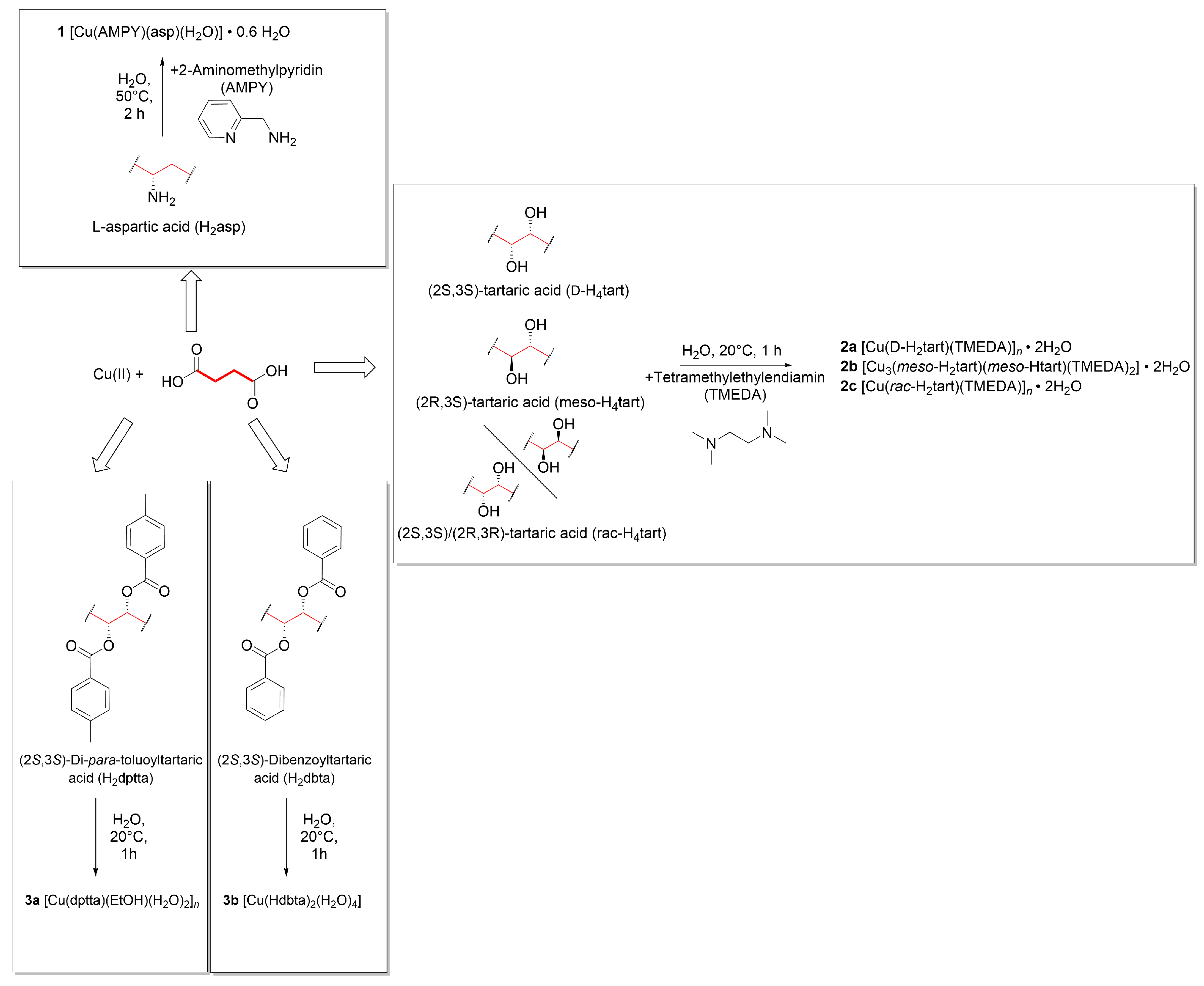
2. Results and Discussion
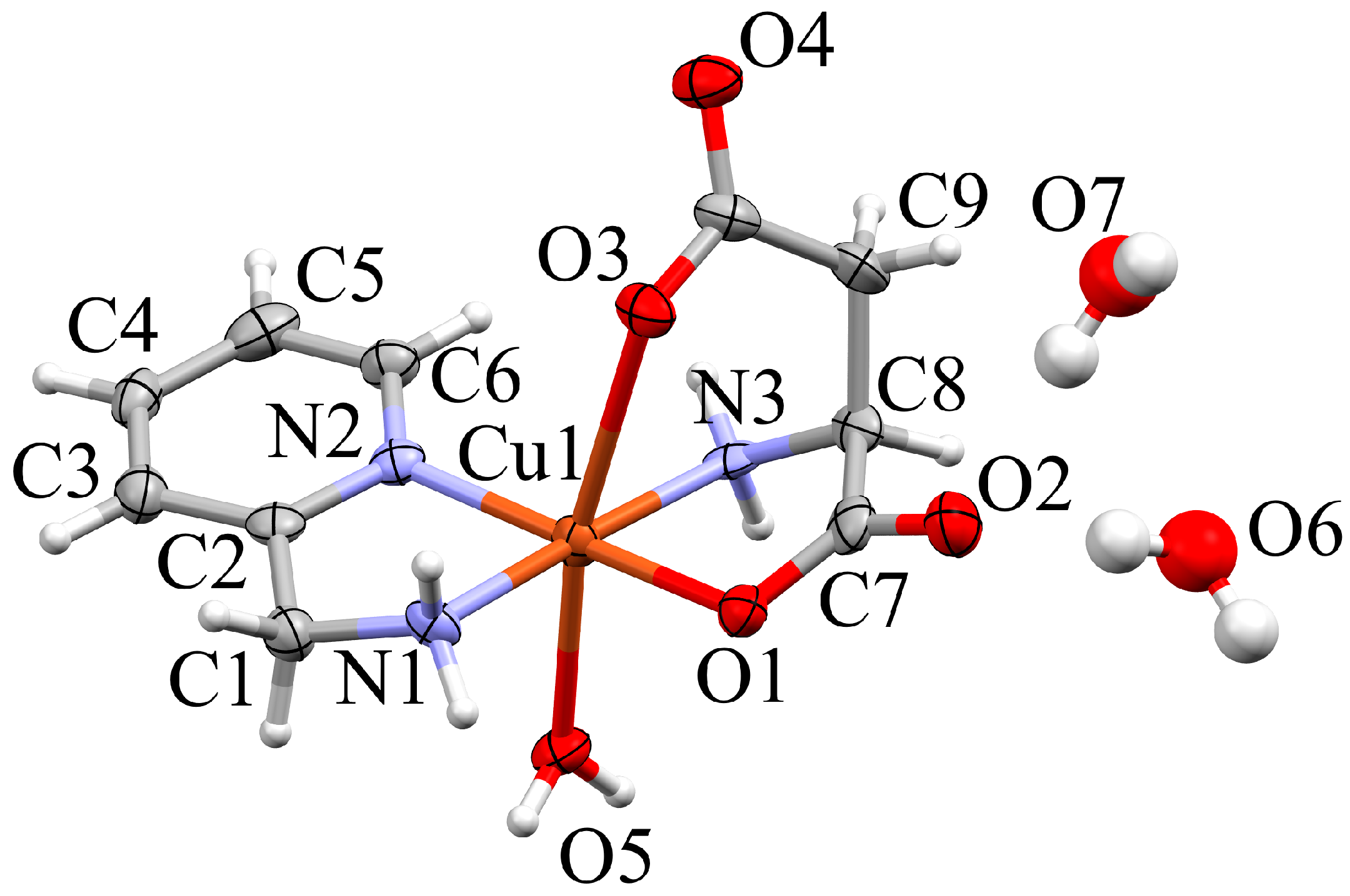
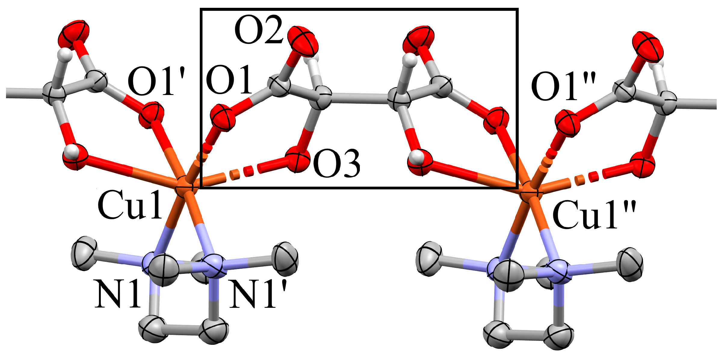
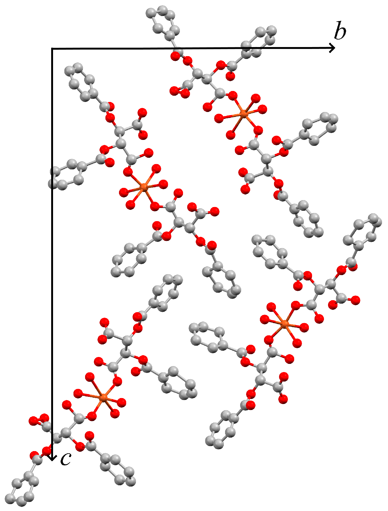
2.1. Compound 1
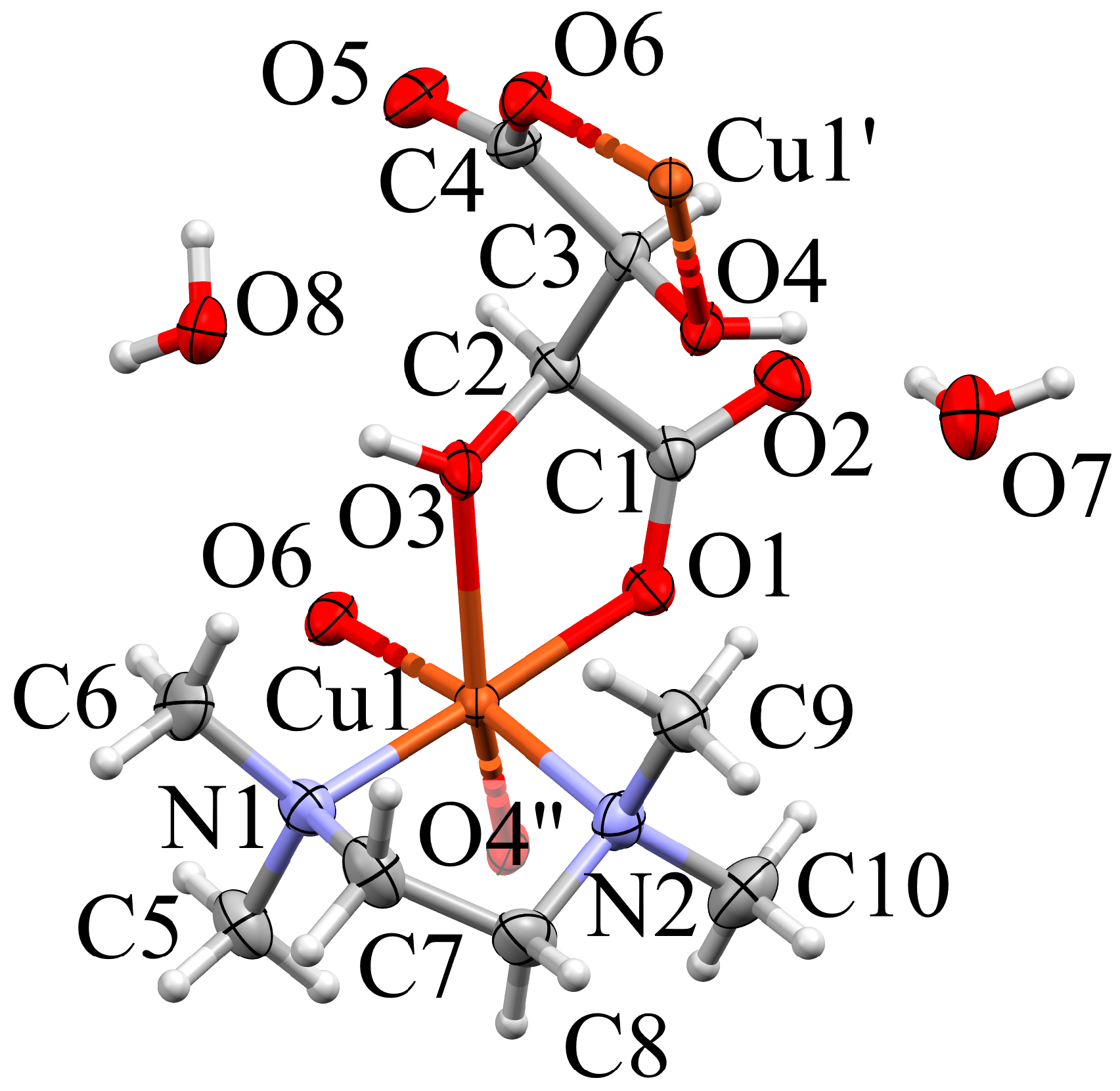
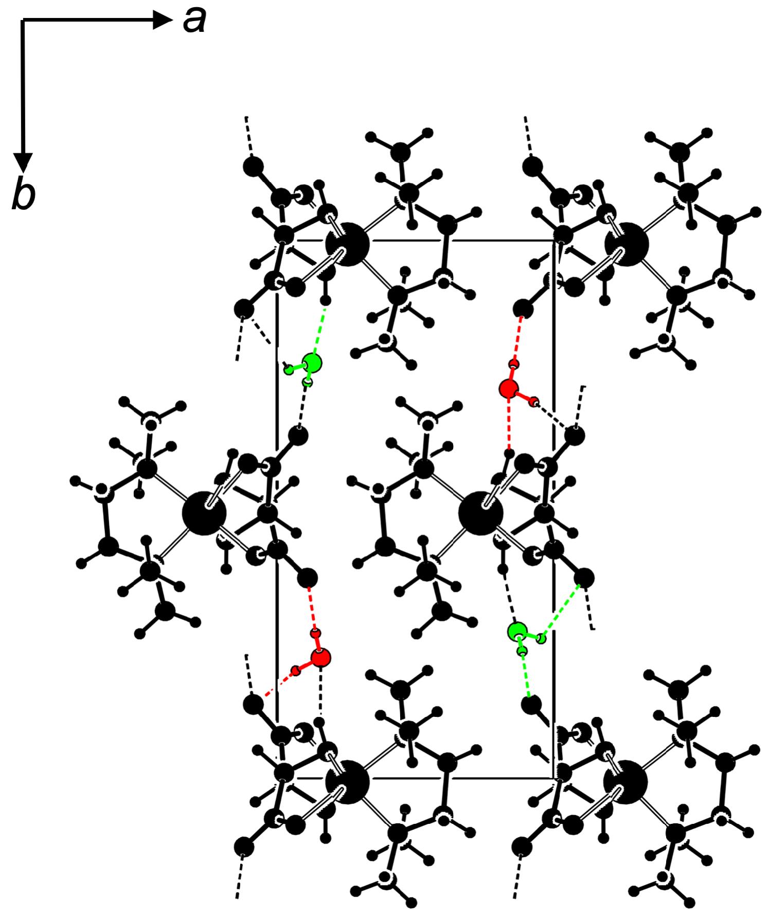
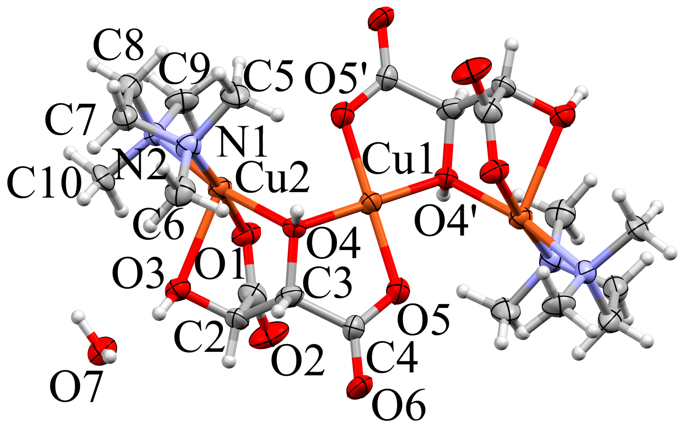
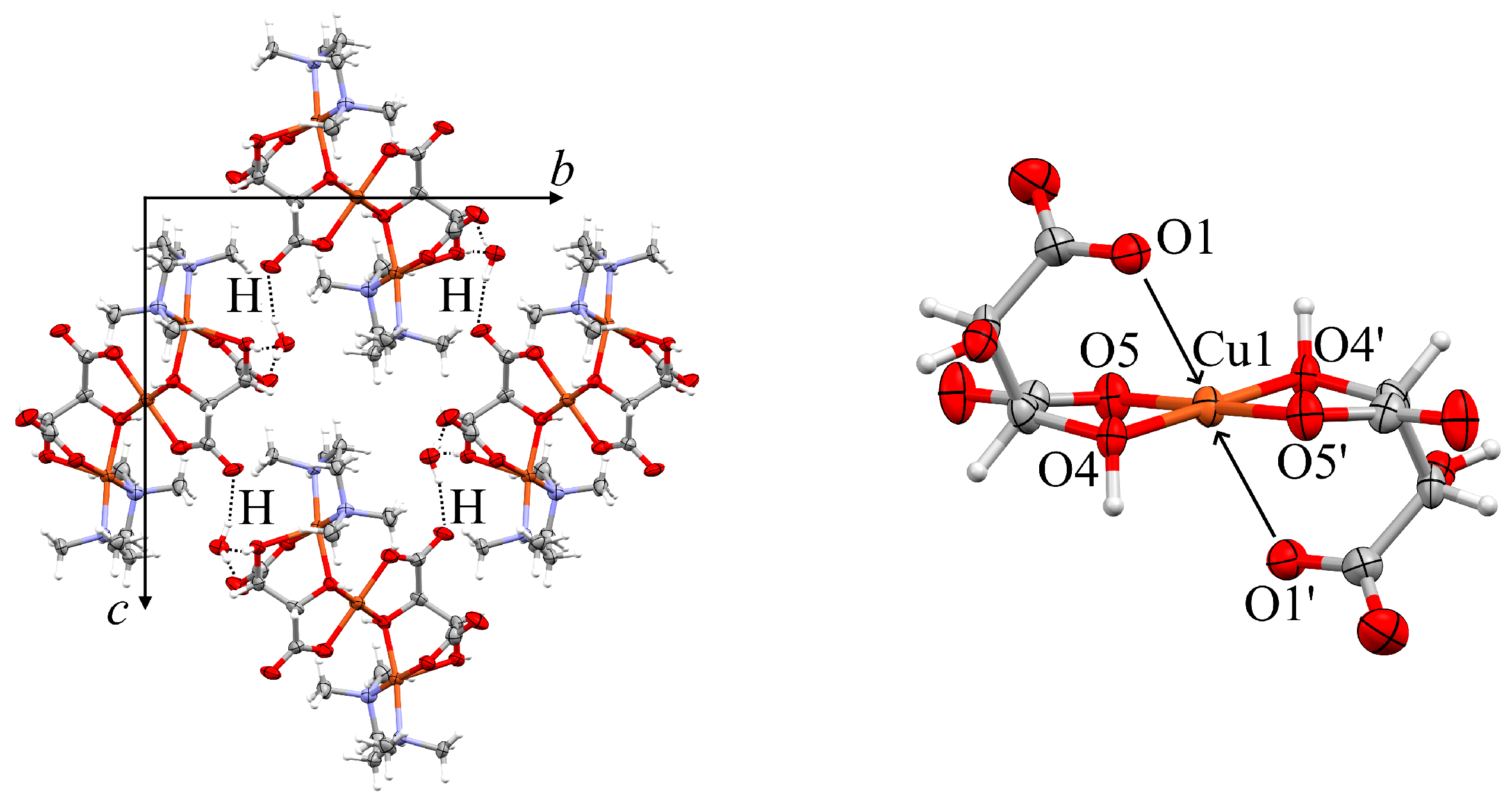
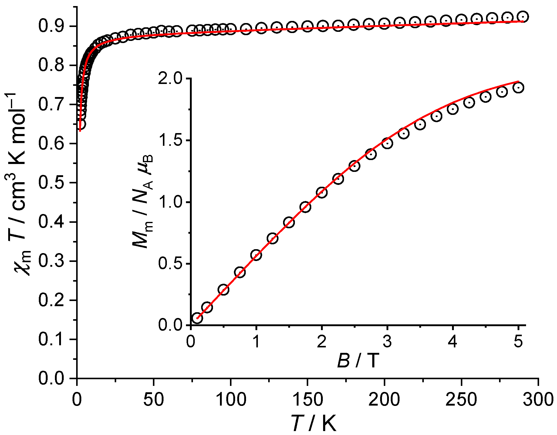
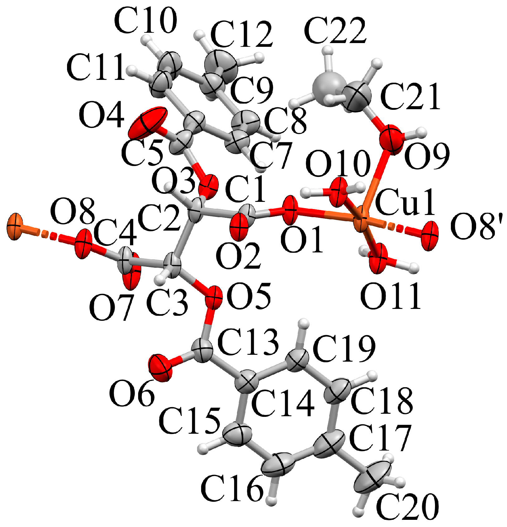
2.2. Compound 2a
2.3. Compound 2b
2.4. Compound 2c
2.5. Compound 3a
2.6. Compound 3b
3. Conlusions: Structural Trends and Construction Principles
4. Materials and Methods
4.1. Synthesis and Crystallization
4.1.1. Compound 1
4.1.2. Compounds 2a/b/c
4.1.3. Compounds 3a/3b
4.2. Single Crystal X-ray Diffraction
4.2.1. Data Collection
4.2.2. Structure Solution and Refinement
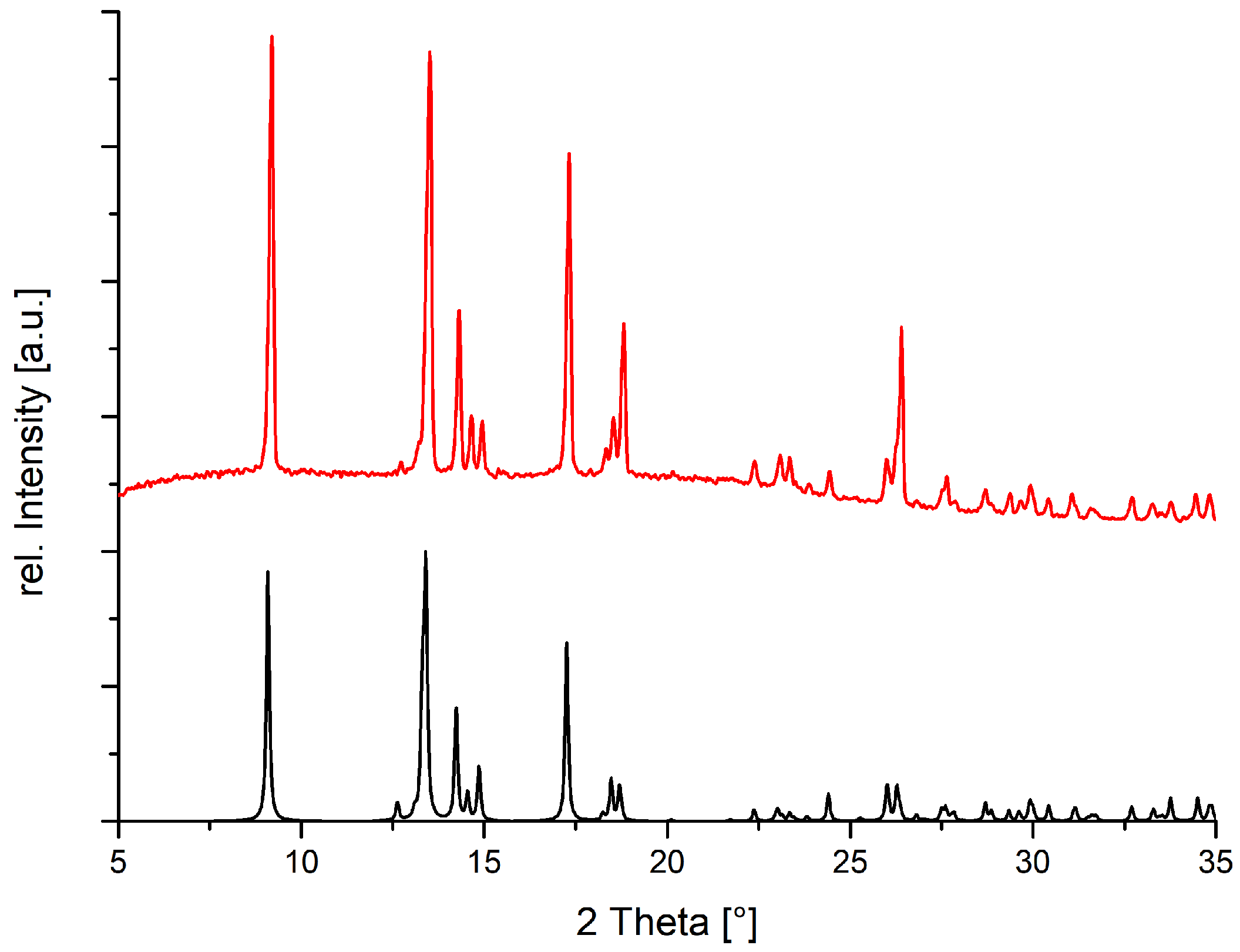
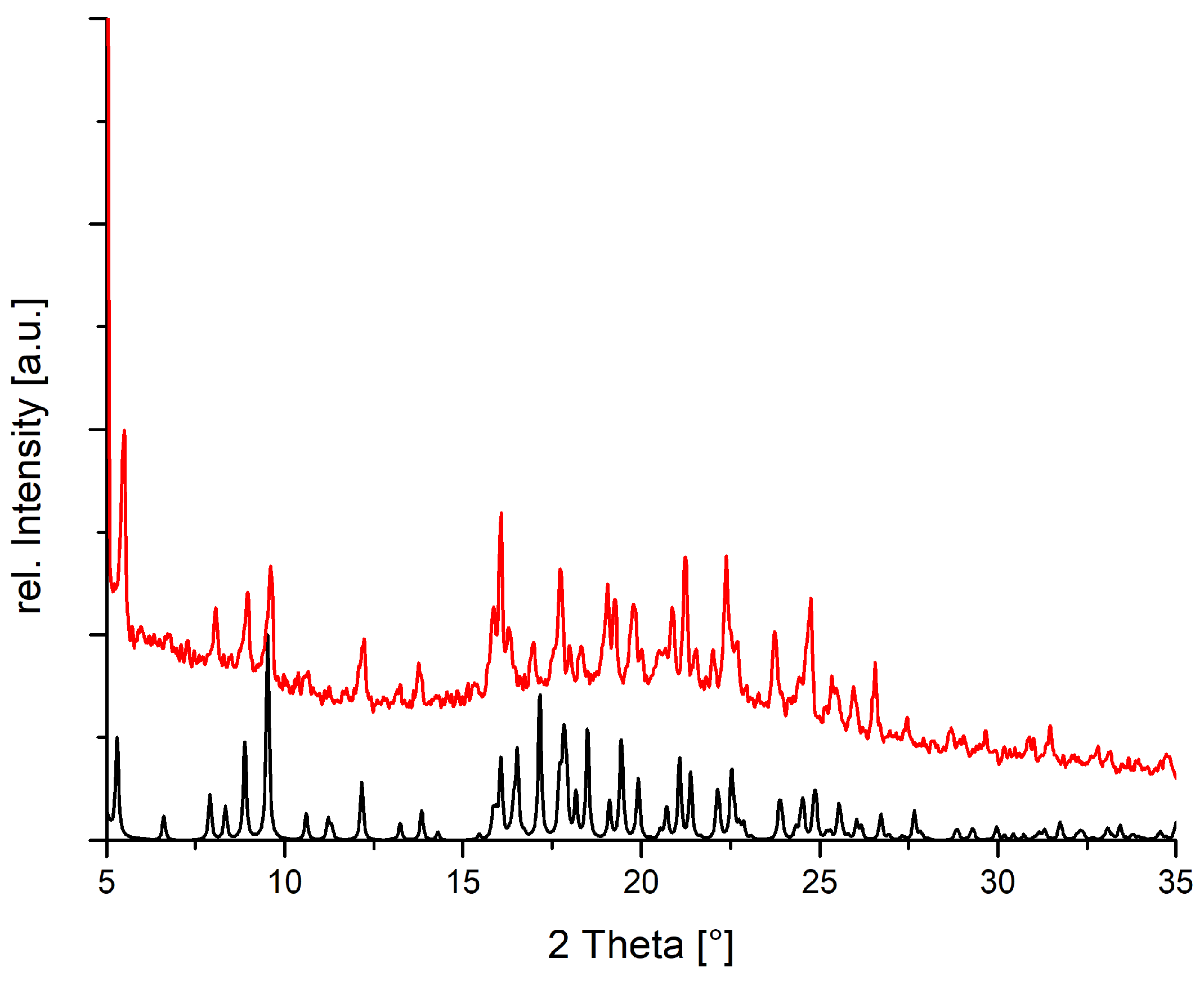
4.3. Powder Diffraction
4.4. Magnetic Measurements
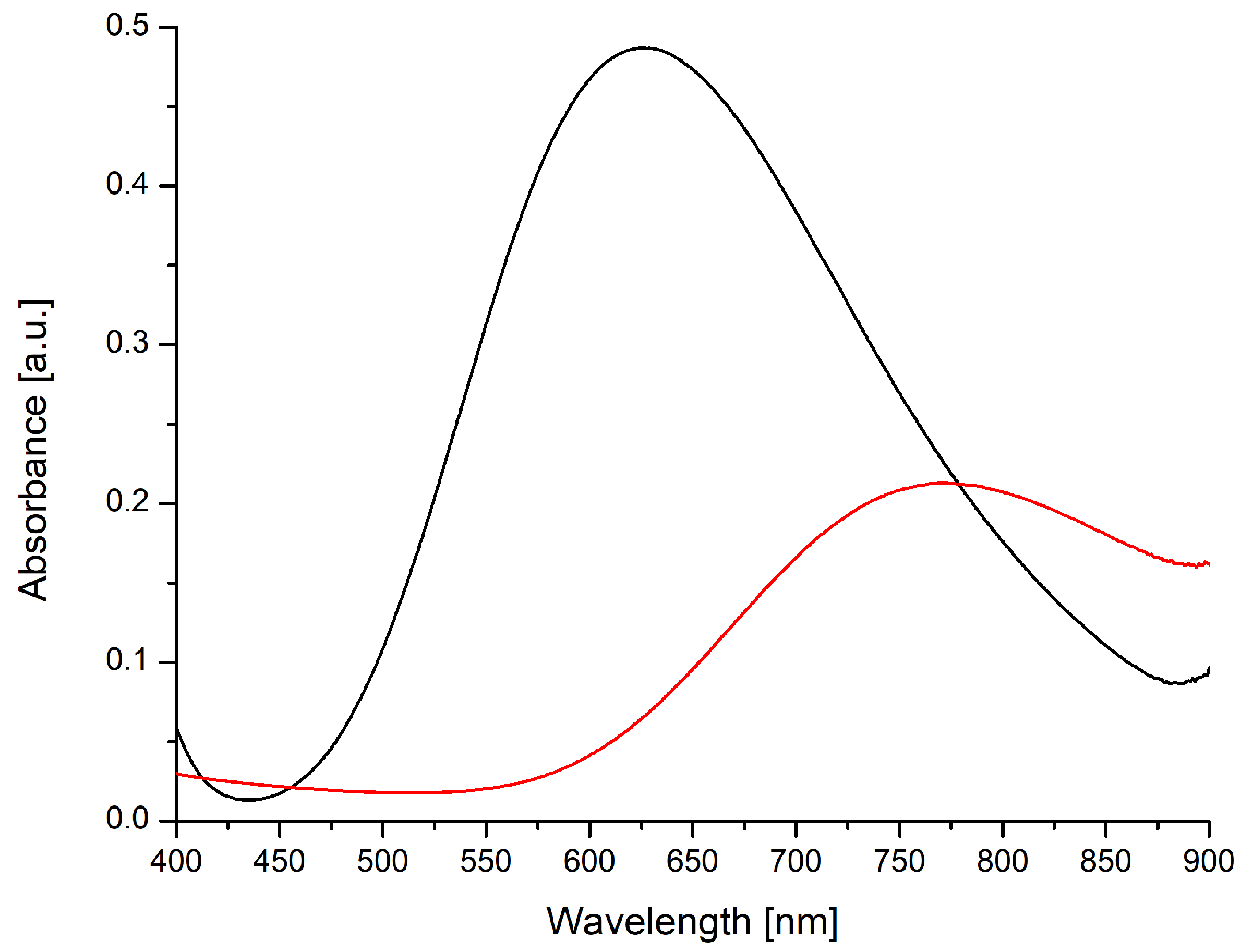
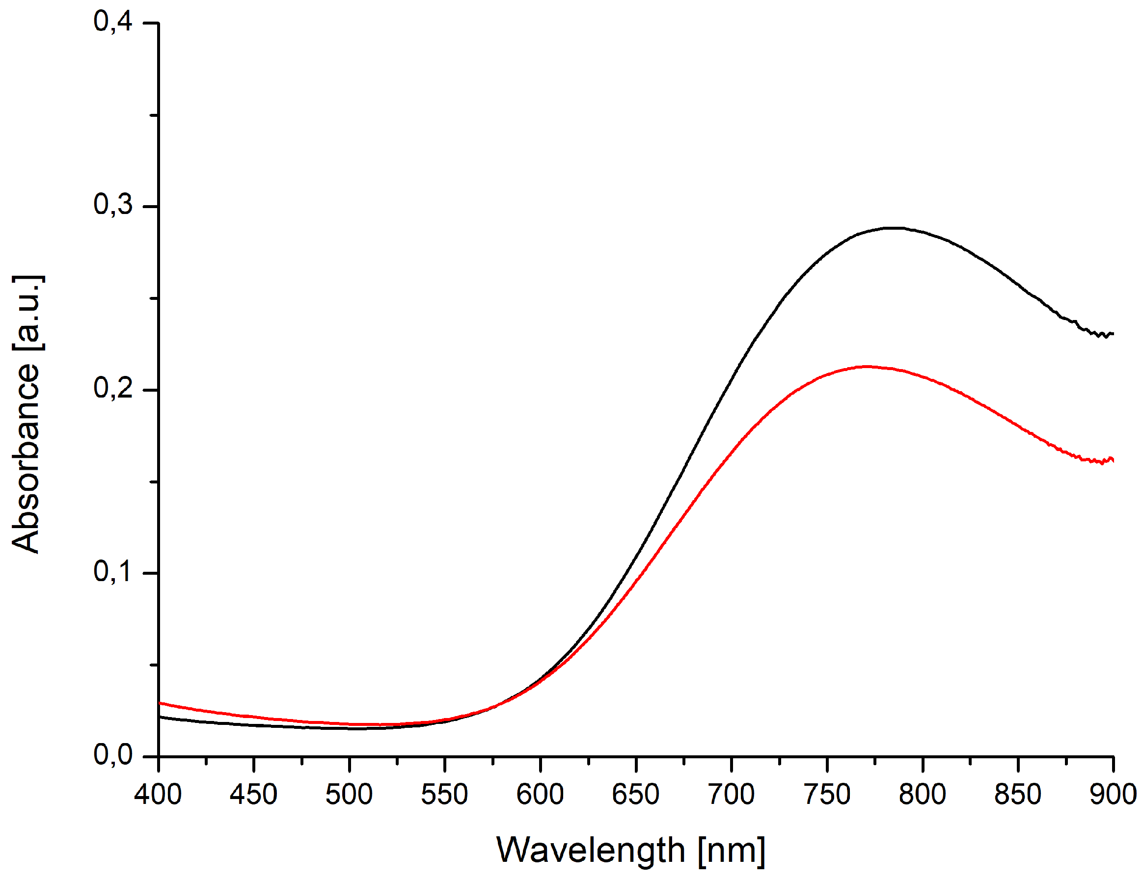
4.5. Absorption Spectroscopy
4.6. Database Survey
Supplementary Materials
Author Contributions
Funding
Acknowledgments
Conflicts of Interest
Abbreviations
| TMEDA | tetramethylethylenediamine |
| AMPY | 2-aminomethylpyridine |
| H2dptta | di-para-toluoyltartaric acid |
| dptta | di-para-toluoyltartrate |
| H2dbta | dibenzoyltartaric acid |
| HDBTA | mono protonated dibenzoyltartrate |
| H4tart | tartaric acid |
| H2tart | tartrate |
| H2asp | aspartic acid |
| asp | aspartate |
| EtOH | Ethanol |
References
- van Niekerk, J.N.; Schoening, F.R.L. The crystal structures of nickel acetate, Ni(CH3COO)2 · 4H2O, and cobalt acetate, Co(CH3COO)2 · 4H2O. Acta Crystallogr. 1953, 6, 609–612. [Google Scholar] [CrossRef]
- van Niekerk, J.N.; Schoening, F.R.L.; Talbot, J.H. The crystal structure of zinc acetate dihydrate, Zn(CH3COO)2 · 2H2O. Acta Crystallogr. 1953, 6, 720–723. [Google Scholar] [CrossRef]
- van Niekerk, J.N.; Schoening, F.R.L. A new type of copper complex as found in the crystal structure of cupric acetate, Cu2(CH3COO)4 · 2H2O. Acta Crystallogr. 1953, 6, 227–232. [Google Scholar] [CrossRef]
- Gu, J.; Wen, M.; Liang, X.; Shi, Z.; Kirillova, M.V.; Kirillov, A.M. Multifunctional Aromatic Carboxylic Acids as Versatile Building Blocks for Hydrothermal Design of Coordination Polymers. Crystals 2018, 8, 83. [Google Scholar] [CrossRef]
- Hu, C.; Englert, U. Crystal-to-Crystal Transformation from a Chain Polymer to a Two-Dimensional Network at Low Temperatures. Angew. Chem. Int. Ed. 2005, 44, 2281–2283. [Google Scholar] [CrossRef] [PubMed]
- Hu, C.; Englert, U. Space Filling Versus Symmetry: Two Consecutive Crystal-to-Crystal Phase Transitions in a 2D Network. Angew. Chem. Int. Ed. 2006, 45, 3457–3459. [Google Scholar] [CrossRef] [PubMed]
- Lamberts, K.; Kalf, I.; Ramadan, A.; Müller, P.; Dronskowski, R.; Englert, U. Tunable Crystal-to-Crystal Phase Transition in a Cadmium Halide Chain Polymer. Polymers 2011, 3, 1151–1161. [Google Scholar] [CrossRef]
- Guo, Q.; Merkens, C.; Si, R.; Englert, U. Crosslinking of the Pd(acacCN)2 building unit with Ag(I) salts: Dynamic 1D polymers and an extended 3D network. CrystEngComm 2015, 17, 4383–4393. [Google Scholar] [CrossRef]
- Kalf, I.; Mathieu, P.; Englert, U. From crystal to crystal: A new polymorph of (4-carboxylatopyridine)silver(i) by topotactic dehydration of its monohydrate. New J. Chem. 2010, 34, 2491–2495. [Google Scholar] [CrossRef]
- Truong, K.N.; Müller, P.; Dronskowski, R.; Englert, U. Dynamic Uptake and Release of Water in the Mixed-Metal EDTA Complex M3[Yb(EDTA)(CO3)] (M = K, Rb, Cs). Cryst. Growth Des. 2017, 17, 80–88. [Google Scholar] [CrossRef]
- Generally Recognized as Safe (GRAS). Available online: https://www.fda.gov/food/food-ingredients-packaging/generally-recognized-safe-gras (accessed on 5 June 2020).
- Głowiak, Z.; Ciunik, Z. The crysral structure of bis(DL-valine) calcium chloride dihydrate and bis(DL-2-Aminobutyric Acid) calcium chloride dihydrate. Bull. Acad. Pol. Sci. Ser. Sci. Chim. 1978, 26, 43–51. [Google Scholar]
- Ciunik, Z.; Głowiak, T. Glycinemanganese(II) dichloride dihydrate [catena-diaquadichloro-µ-glycinemanganese(II)]. Acta Crystallogr. B 1980, 36, 1212–1213. [Google Scholar] [CrossRef]
- Ciunik, Z.; Glowiak, Z. Conformational analysis of double carboxylic bridges in bis(dl-α-alanine)manganese(II) dibromide dihydrate and related compounds. Inorg. Chim. Acta 1980, 44, L249–L250. [Google Scholar] [CrossRef]
- Bissinger, H.; Beck, W. Metal Complexes with Biologically Important Ligands, XXXIX [1] Platinum(IV) Complexes with a-Amino Acid Esters and Peptide Esters; l3N and 14r,Pt NMR Spectra of Platinum Complexes with a-Amino Acids. Z. Naturforsch. B 1985, 40, 507–511. [Google Scholar] [CrossRef]
- Glowiak, T.; Legendziewicz, J.; Dao, C.; Huskowska, E. Absorption, luminescence and crystal structure studies of dysprosium compound with l-α-Alanine: [Dy(l-α-AlaH)(H2O)6]Cl3. J. Less Common Met. 1991, 168, 237–248. [Google Scholar] [CrossRef]
- Nolan, K.B.; Soudi, A.A.; Hay, R.W. Metal complexes of amino acids and peptides. In Amino Acids, Peptides and Proteins; Davies, J.S., Ed.; The Royal Society of Chemistry: London, UK, 1994; Volume 25, pp. 359–410. [Google Scholar]
- Severin, K.; Bergs, R.; Beck, W. Bioorganometallic Chemistry - Transition Metal Complexes with α-Amino Acids and Peptides. Angew. Chem. Int. Ed. 1998, 37, 1634–1654. [Google Scholar] [CrossRef]
- Mrozek, R.; Rzaczynska, Z.; Sikorska-Iwan, M.; Jaroniec, M.; Głowiak, T. A new complex of manganese(II) with l-α-alanine: Structure, spectroscopy and thermal study. Polyhedron 1999, 18, 2321–2326. [Google Scholar] [CrossRef]
- Wang, R.; Liu, H.; Carducci, M.D.; Jin, T.; Zheng, C.; Zheng, Z. Lanthanide Coordination with α-Amino Acids under Near Physiological pH Conditions: Polymetallic Complexes Containing the Cubane-Like [Ln4(µ3-OH)4]8+ Cluster Core. Inorg. Chem. 2001, 40, 2743–2750. [Google Scholar] [CrossRef]
- Slyudkin, O.; Tulupov, A. Chiral complexes of Pt with amino acids: Synthesis, structure, properties. Russ. J. Coord. Chem. 2005, 31, 77–85. [Google Scholar] [CrossRef]
- Farkas, E.; Sovago, I. Metal complexes of amino acids and peptides. Amino Acids Peptides Proteins 2007, 36, 287–345. [Google Scholar]
- Fleck, M. Compounds of glycine with halogen or metal halogenides: Review and comparison. Z. Kristallogr. Cryst. Mater. 2008, 223, 222–232. [Google Scholar] [CrossRef]
- Fleck, M.; Held, P.; Schwendtner, K.; Bohatý, L. New compounds of glycine with metal halogenides. Z. Kristallogr. Cryst. Mater. 2008, 223, 212–221. [Google Scholar] [CrossRef]
- Weng, Z.; Chen, Z.; Liang, F. Glycine-Templated Manganese Sulfate with New Topology and Canted Antiferromagnetism. Inorg. Chem. 2009, 48, 8703–8708. [Google Scholar] [CrossRef] [PubMed]
- Beck, W. Metal complexes of biologically important ligands, CLXXII [1]. metal ions and metal complexes as protective groups of amino acids and peptides–reactions at coordinated amino acids. Z. Naturforsch. B 2009, 64, 1221–1245. [Google Scholar] [CrossRef]
- Zheng, X.D.; Lu, T.B. Constructions of helical coordination compounds. CrystEngComm 2010, 12, 324–336. [Google Scholar] [CrossRef]
- Lamberts, K.; Englert, U. Structures from MnX2 and proline: Isomorphous racemic compounds and a series of chiral non-isomorphous chain polymers. Acta Crystallogr. B 2012, 68, 610–618. [Google Scholar] [CrossRef]
- Lamberts, K.; Porsche, S.; Hentschel, B.; Kuhlen, T.; Englert, U. An unusual linker and an unexpected node: CaCl2 dumbbells linked by proline to form square lattice networks. CrystEngComm 2014, 16, 3305–3311. [Google Scholar] [CrossRef]
- Lamberts, K.; Möller, A.; Englert, U. Enantiopure and racemic alanine as bridging ligands in Ca and Mn chain polymers. Acta Crystallogr. B 2014, 70, 989–998. [Google Scholar] [CrossRef]
- Fleck, M.; Petrosyan, A. Salts of Amino Acids: Crystallization, Structure and Properties; Springer International Publishing: Cham, Switzerland, 2014; pp. 1–574. [Google Scholar]
- Lamberts, K.; Şerb, M.D.; Englert, U. Unexpected proline coordination in the copper chain polymer [Cu(µ-Cl)2(µ-dl-proline-ϰ2O:O’)]1∞. Acta Crystallogr. C 2015, 71, 271–275. [Google Scholar] [CrossRef]
- Lamberts, K.; Englert, U. Crystal structures of coordination polymers from CaI2 and proline. Acta Crystallogr. E 2015, 71, 675–680. [Google Scholar] [CrossRef]
- Lamberts, K.; Englert, U. Incidental Polymorphism, Non-Isomorphic and Isomorphic Substitution in Calcium-Valine Coordination Polymers. Crystals 2015, 5, 261–272. [Google Scholar] [CrossRef]
- Lamberts, K.; Tegoni, M.; Jiang, X.; Kou, H.Z.; Englert, U. Silver complexation by metallacryptates. Dalton Trans. 2016, 45, 284–295. [Google Scholar] [CrossRef] [PubMed]
- Yoshinari, N.; Konno, T. Chiral Phenomena in Multinuclear and Metallosupramolecular Coordination Systems Derived from Metalloligands with Thiol-Containing Amino Acids. B Chem. Soc. Jpn. 2018, 91, 790–812. [Google Scholar] [CrossRef]
- Subhashini, R.; Arjunan, S.; Gunasekaran, B. Synthesis of a metal coordinated amino acid based nonlinear single crystal, Bis(l-threonine)zinc(II) using the solution growth technique and its physicochemical properties. J. Phys. Chem. Solids 2019, 135, 109077. [Google Scholar] [CrossRef]
- Kumary, V.A.; Abraham, P.; S, R.; Swamy, B.K.; Nancy, T.M.; Sreevalsan, A. A novel heterogeneous catalyst based on reduced graphene oxide supported copper coordinated amino acid—A platform for morphine sensing. J. Electroanal. Chem. 2019, 850, 113367. [Google Scholar] [CrossRef]
- Castinñeiras, A.; Balboa, S.; Carballo, R.; Niclós, J. The Crystal Structures of two Linearly Polymeric Copper(II) Complexes of 2,2′-Bipyridine with threefold Glycolato and Oxalato Bridges. Z. Anorg. Allg. Chem. 2002, 628, 2353–2359. [Google Scholar] [CrossRef]
- Castiñeiras, A.; Balboa, S.; Carballo, R.; González-Pérez, J.M.; Niclós-Gutiérrez, J. Structural Evidences for the Oxidation of α-Hydroxycarboxilic Acids to Oxalate Assisted by Copper(II) Ions. Z. Anorg. Allg. Chem. 2007, 633, 717–723. [Google Scholar] [CrossRef]
- Dybtsev, D.; Samsonenko, D.; Fedin, V. Porous coordination polymers based on carboxylate complexes of 3d metals. Russ. J. Coord. Chem. 2016, 42, 557–573. [Google Scholar] [CrossRef]
- Macrae, C.F.; Edgington, P.R.; McCabe, P.; Pidcock, E.; Shields, G.P.; Taylor, R.; Towler, M.; van de Streek, J. Mercury: Visualization and analysis of crystal structures. J. Appl. Cryst. 2006, 39, 453–457. [Google Scholar] [CrossRef]
- Ray, M.S.; Ghosh, A.; Das, A.; Drew, M.G.B.; Ribas-Ariño, J.; Novoa, J.; Ribas, J. A new tetrameric CuII cluster with square topology exhibiting ferro- and antiferromagnetic magnetic pathways: Which is which? Chem. Commun. 2004, 9, 1102–1103. [Google Scholar] [CrossRef]
- Groom, C.R.; Bruno, I.J.; Lightfoot, M.P.; Ward, S.C. The Cambridge Structural Database. Acta Crystallogr. B 2016, 72, 171–179. [Google Scholar] [CrossRef] [PubMed]
- Kathalikkattil, A.C.; Bisht, K.K.; Aliaga-Alcalde, N.; Suresh, E. Synthesis, Magnetic Properties, and Structural Investigation of Mixed-Ligand Cu(II) Helical Coordination Polymers with an Amino Acid Backbone and N-Donor Propping: 1-D Helical, 2-D Hexagonal Net (hcb), and 3-D ins Topologies. Cryst. Growth Des. 2011, 11, 1631–1641. [Google Scholar] [CrossRef]
- Ingleson, M.J.; Barrio, J.P.; Bacsa, J.; Dickinson, C.; Park, H.; Rosseinsky, M.J. Generation of a solid Brønsted acid site in a chiral framework. Chem. Commun. 2008, 1287–1289. [Google Scholar] [CrossRef] [PubMed]
- Antolini, L.; Battaglia, L.P.; Bonamartini Corradi, A.; Marcotrigiano, G.; Menabue, L.; Pellacani, G.C.; Saladini, M.; Sola, M. Tridentate facially coordinated L-aspartate ion complexation with the copper(II) ion: Spectroscopic and structural properties of aqua(L-aspartato)(1,10-phenanthroline)copper(II) tetrahydrate. Inorg. Chem. 1986, 25, 2901–2904. [Google Scholar] [CrossRef]
- Ma, L.F.; Liang, F.P.; Qin, H.C.; Hu, R.X.; Zhang, M.B.; Yu, K.B. Synthesis and Crystal Structure of Binuclear Complex [Cu2(L-Asp)(phen)3(NO3)](NO3)·4H2O. Chin. J. Inorg. Chem. 2004, 20, 1429–1432. [Google Scholar]
- Baggio, R.F.; Calvo, R.; Brondino, C.; Garland, M.T.; Atria, A.M.; Spodine, E. A Novel Structure of (L-Aspartato)(1,10-phenanthroline)copper(II) Hydrate. Acta Crystallogr. C 1995, 51, 382–385. [Google Scholar] [CrossRef]
- Spek, A.L. Structure validation in chemical crystallography. Acta Crystallogr. D 2009, 65, 148–155. [Google Scholar] [CrossRef]
- Hörner, T.G.; Klüfers, P. The Species of Fehling’s Solution. Eur. J. Inorg. Chem. 2016, 2016, 1798–1807. [Google Scholar] [CrossRef]
- Lueken, H. Magnetochemie; B.G. Teubner: Stuttgart, Germany, 1999. [Google Scholar]
- Calvo, R.; Steren, C.A.; Piro, O.E.; Rojo, T.; Zuniga, F.J.; Castellano, E.E. Crystal structure and magnetic properties of diaqua(L-aspartato)copper(II). Inorg. Chem. 1993, 32, 6016–6022. [Google Scholar] [CrossRef]
- Tulsky, E.G.; Long, J.R. Dimensional reduction: A practical formalism for manipulating solid structures. Chem. Mater. 2001, 13, 1149–1166. [Google Scholar] [CrossRef]
- Wu, W.Y.; Zhai, L.X. Poly[diaqua-µ-oxalato-copper(II) monohydrate]. Acta Cryst. E 2007, 63, m429–m430. [Google Scholar] [CrossRef]
- Yeşilel, O.; Erer, H.; Odabasoglu, M.; Büyükgüngör, O. A Novel Copper(II)–Hydrogen Oxalate Coordination Polymer Showing a New Coordination Mode. J. Inorg. Organomet. Polym. Mater. 2010, 20, 78–82. [Google Scholar] [CrossRef]
- Zhang, B. CCDC 1559673. In Experimental Crystal Structure Determination; The Cambridge Crystallographic Data Centre (CCDC): Cambridge, UK, 2017; Volume 20. [Google Scholar]
- Ghoshal, D.; Ghosh, A.K.; Mostafa, G.; Ribas, J.; Chaudhuri, N.R. Succinato-bridged copper(II) supramolecular 3D framework: Synthesis, crystal structure and magnetic property. Inorg. Chim. Acta 2007, 360, 1771–1775. [Google Scholar] [CrossRef]
- O’Connor, B.H.; Maslen, E.N. The crystal structure of Cu(II) succinate dihydrate. Acta Crystallogr. 1966, 20, 824–835. [Google Scholar] [CrossRef]
- Rastsvetaeva, R.K.; Pushcharovsky, D.Y.; Furmanova, N.G.; Sharp, H. Crystal and molecular structure of Cu(II) succinate monohydrate or “Never wash copper minerals with detergents”. Z. Kristallogr. Cryst. Mater. 1996, 211, 808–811. [Google Scholar] [CrossRef]
- Zheng, Y.Q.; Lin, J.L. New 1D and 2D metal oxygen connectivities in Cu(II) succinato and glutarato coordination polymers: [Cu3(H2O)2(OH)2(C4H4O4) 2] · 4H2O, [Cu4(H2O)2(OH) 4(C4H4O4)2] · 5H2O and [Cu5(OH)6(C5H6O4)2] · 4H2O. J. Coord. Chem. 2008, 61, 3420–3437. [Google Scholar] [CrossRef]
- Prout, C.K.; Carruthers, J.R.; Rossotti, F.J.C. Structure and stability of carboxylate complexes. Part VII. Crystal and molecular structures of copper(II)meso-tartrate trihydrate and copper(II)d-tartrate trihydrate. J. Chem. Soc. A 1971, 3336–3342. [Google Scholar] [CrossRef]
- Bruker. SAINT+; Bruker AXS Inc.: Madison, WI, USA, 2009. [Google Scholar]
- Bruker. SADABS; Bruker AXS Inc.: Madison, WI, USA, 2001. [Google Scholar]
- Kabsch, W. XDS. Acta Crystallogr. D 2010, 66, 125–132. [Google Scholar] [CrossRef]
- Kabsch, W. Integration, scaling, space-group assignment and post-refinement. Acta Crystallogr. D 2010, 66, 133–144. [Google Scholar] [CrossRef]
- Sheldrick, G.M. SHELXT–Integrated space-group and crystal-structure determination. Acta Crystallogr. A 2015, 71, 3–8. [Google Scholar] [CrossRef]
- Sheldrick, G.M. A short history of SHELX. Acta Crystallogr. A 2008, 64, 112–122. [Google Scholar] [CrossRef] [PubMed]


© 2020 by the authors. Licensee MDPI, Basel, Switzerland. This article is an open access article distributed under the terms and conditions of the Creative Commons Attribution (CC BY) license (http://creativecommons.org/licenses/by/4.0/).
Share and Cite
Kremer, M.; van Leusen, J.; Englert, U. Coordination Compounds Featuring Non-Toxic Chiral 1,4-Dicarboxylic Acids and Copper(II). Crystals 2020, 10, 485. https://doi.org/10.3390/cryst10060485
Kremer M, van Leusen J, Englert U. Coordination Compounds Featuring Non-Toxic Chiral 1,4-Dicarboxylic Acids and Copper(II). Crystals. 2020; 10(6):485. https://doi.org/10.3390/cryst10060485
Chicago/Turabian StyleKremer, Marius, Jan van Leusen, and Ulli Englert. 2020. "Coordination Compounds Featuring Non-Toxic Chiral 1,4-Dicarboxylic Acids and Copper(II)" Crystals 10, no. 6: 485. https://doi.org/10.3390/cryst10060485
APA StyleKremer, M., van Leusen, J., & Englert, U. (2020). Coordination Compounds Featuring Non-Toxic Chiral 1,4-Dicarboxylic Acids and Copper(II). Crystals, 10(6), 485. https://doi.org/10.3390/cryst10060485