Article

Identification and Characterization of New Laccase Biocatalysts from Pseudomonas Species Suitable for Degradation of Synthetic Textile Dyes

by
Mina Mandic
1,
Lidija Djokic
1,
Efstratios Nikolaivits
2,
Radivoje Prodanovic
3,
Kevin O’Connor
4,
Sanja Jeremic
1,*,
Evangelos Topakas
2,5,* and
Jasmina Nikodinovic-Runic
1,*
1
Institute of Molecular Genetics and Genetic Engineering, University of Belgrade, Vojvode Stepe 444a, 11000 Belgrade, Serbia
2
School of Chemical Engineering, National Technical University of Athens, 9 Iroon Polytechniou Str, Zoografou Campus, 15780 Athens, Greece
3
Faculty of Chemistry, University of Belgrade, Studentski Trg 12-16, 11000 Belgrade, Serbia
4
BEACON SFI Bioeconomy Research Centre and School of Biomolecular and Biomedical Science, University College Dublin, Belfield, Dublin 4, D4 Dublin, Ireland
5
Biochemical and Chemical Process Engineering, Division of Sustainable Process Engineering, Department of Civil, Environmental and Natural Resources Engineering, Luleå University of Technology, SE-97187 Luleå, Sweden
*
Authors to whom correspondence should be addressed.
Catalysts 2019, 9(7), 629; https://doi.org/10.3390/catal9070629
Submission received: 3 July 2019 / Revised: 15 July 2019 / Accepted: 17 July 2019 / Published: 23 July 2019
(This article belongs to the Special Issue Environmental Biocatalysis: From Remediation to Waste Valorization)

Abstract

:
Laccases are multicopper-oxidases with variety of biotechnological applications. While predominantly used, fungal laccases have limitations such as narrow pH and temperature range and their production via heterologous protein expression is more complex due to posttranslational modifications. In comparison, bacterial enzymes, including laccases, usually possess higher thermal and pH stability, and are more suitable for expression and genetic manipulations in bacterial expression hosts. Therefore, the aim of this study was to identify, recombinantly express, and characterize novel laccases from Pseudomonas spp. A combination of approaches including DNA sequence analysis, N-terminal protein sequencing, and genome sequencing data analysis for laccase amplification, cloning, and overexpression have been used. Four active recombinant laccases were obtained, one each from P. putida KT2440 and P. putida CA-3, and two from P. putida F6. The new laccases exhibited broad temperature and pH range and high thermal stability, as well as the potential to degrade selection of synthetic textile dyes. The best performing laccase was CopA from P. putida F6 which degraded five out of seven tested dyes, including Amido Black 10B, Brom Cresol Purple, Evans Blue, Reactive Black 5, and Remazol Brilliant Blue. This work highlighted species of Pseudomonas genus as still being good sources of biocatalytically relevant enzymes.

1. Introduction

Laccases are blue multicopper enzymes (EC 1.10.3.2) that oxidize a broad range of both phenolic and non-phenolic substrates, via a four-electron reduction of molecular oxygen to water [1,2]. These enzymes are widely distributed in nature and have been isolated from bacteria [3,4,5], fungi [6,7] and plants [8,9], as well as from lichens [10], and sponges [11]. Although laccases are heterogeneous in different species, implying diversity in their function, four copper-binding motifs are conserved in the most laccases, especially bacterial forms [12]. The presence of different metal ions can affect laccase activity, either inducing or suppressing it. Metal ions such as Cu+2, Ca+2, Ni+2, Co+2, and Mn+2 are generally known to accelerate laccase activity at a remarkable level [13].
Due to broad substrate spectrum, laccases have become very attractive for a variety of biotechnological and industrial applications such as organic synthesis; lignin degradation; and bio-product formation for the food, textile, and pharmaceutical industries; remediation of contaminated environments; as well as construction of biosensors and biofuel cells [14,15,16]. During the last decade laccases have been used in decolorization and detoxification of textile effluents [17]. Effluents from the textile industry are usually complex, containing a wide variety of synthetic dyes [18], among which the most common are azo dyes, anthraquinone, triphenylmethane, and indigo dyes [19].
In the recent years, development of high-performance recombinant bacterial strains and the possibility of increasing the production of recombinant proteins created new opportunities for the commercial use of laccases, since the production from wild type strains has limitations in growth and product yield, which are not suitable for standard industrial fermentations [20]. The majority of studies on laccases are focused on laccases originated from fungi. Having in mind that industrial processes often require high temperature and pressure, or extremely acidic or alkaline pH, fungal laccases are not the best candidates for such industrial applications since they operate in a temperature range from 30 °C to 55 °C and a slightly acidic pH range [2,21]. In addition, their heterologous expression is hindered by post-translational modifications [22], making their production cost-ineffective. Consequently, bacterial laccases are increasingly being sought for use in the industry due to the advantage of higher growth rates and better suitability for improvement of enzyme activity and expression level [23,24].
A number of bacterial laccases have been identified, heterologously expressed and studied at the molecular level. The first and the most studied bacterial laccase is CotA from Bacillus subtilis [25], followed by laccases from B. coagulans, B. clausii [26], and B. licheniformis [3]. The other significant group of bacterial laccases are from Streptomyces species, e.g., S. coelicolor [27], S. cyaneus [28], S. bikiniensis [29], and S. ipomoea [30].
Laccases from Pseudomonas species are mostly identified and purified from wild type producer strains [31,32,33] and until now, only one laccase from P. putida KT2440 was heterologously expressed and characterized [5]. Having in mind the importance of Pseudomonas strains, as a biotechnological platform for various industrial applications [34,35], as well as wealth and applicability of other enzymes from Pseudomonas species in biocatalysis, we set out to identify, recombinantly express, and characterize novel laccases from a number of Pseudomonas species, and assess their ability to degrade synthetic dyes widely used in textile industry.

2. Results and Discussion

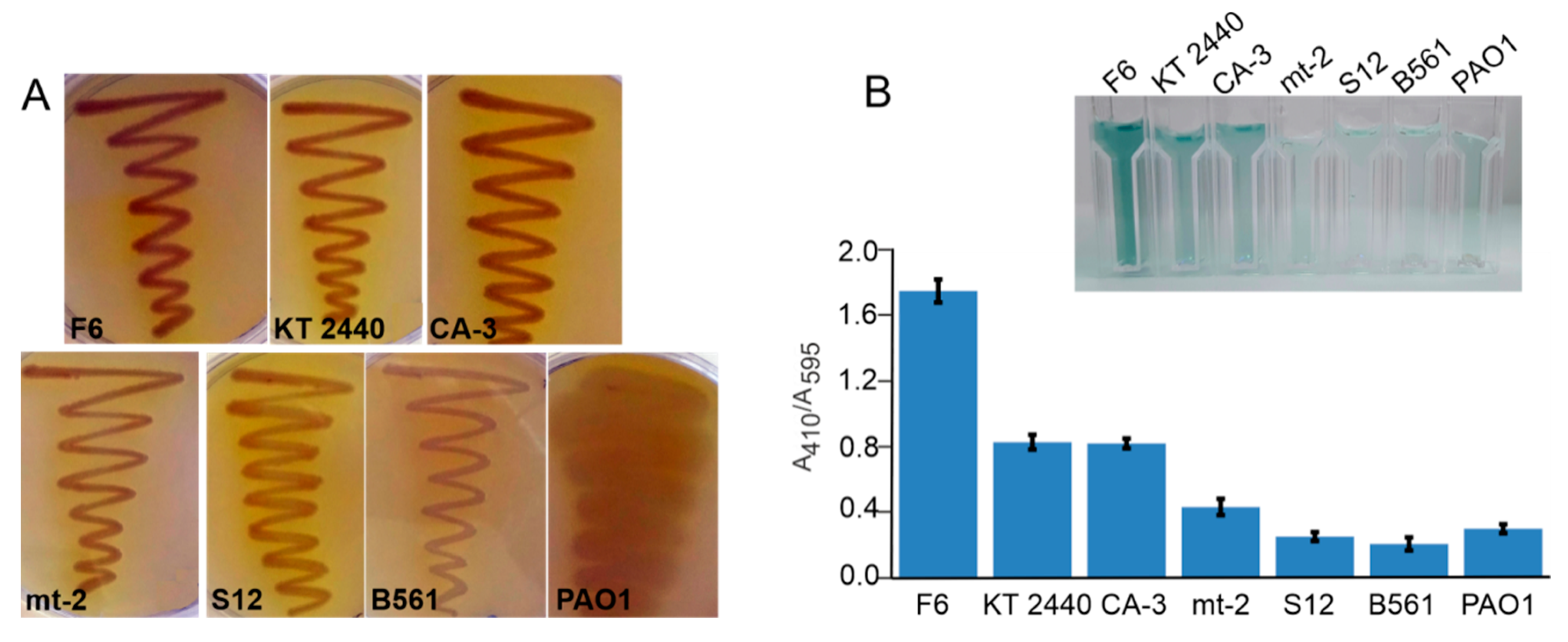
2.1. Screening for Laccase Activity Using Guaiacol Agar Plates and ABTS Assay

Seven different Pseudomonas strains (P. putida F6, P. putida KT2440, P. putida CA-3, P. putida mt-2, P. putida S12, P. chlororaphis B561 and P. aeruginosa PAO1) from our laboratory collection were tested for laccase-like activity on agar plates containing standard laccase substrates guaiacol or syringaldazine 0.01% (w/v) and the development of dark colors due to the oxidation of these substrates was monitored [36]. In addition, supernatants and cell-free extracts of these strains previously grown in mineral MSM liquid medium supplemented with 5 mM phenylacetic acid to induce expression of enzymes from the aerobic catabolism of aromatic compounds [37] were tested in the ABTS assay (Section 3.4). Significantly higher enzyme activities were detected in the cell-free extracts than in supernatants of all tested Pseudomonas sp. cultures (data not shown), indicating the intracellular location of these enzymes, which is in line with previous literature reports on intracellular bacterial laccases [38,39].
Based on the fast reddish-brown color formation, indicating oxidation of guaiacol (Figure 1A), as well as the best activity of cell-free extracts in ABTS assay (Figure 1B), three strains, namely P. putida F6, P. putida KT2440, and P. putida CA-3, were chosen for further study. Notably, cell-free extracts of P. putida F6 exhibited two times higher laccase activity (ABTS assay) compared to cell free extracts from P. putida KT2440 and P. putida CA-3, respectively, which showed a similar activity level (Figure 1B).

2.2. Primer Design Based on UniProt Data Analysis

Based on data from UniProt database 15 laccase and laccase-like multicopper oxidase (MCO) protein sequences from Pseudomonas strains were aligned using ClustalΩ tool (https://www.ebi.ac.uk/Tools/msa/clustalo/) and degenerate primers were designed (Table 1). Genes coding for enzymes with laccase activity from P. putida KT2440 and P. putida CA-3 were successfully amplified using designed primers and identified as MCO by sequencing. On the other side, amplification products obtained for P. putida F6 were also sequenced, but did not show any similarity with any known laccases (data not shown). Based on the obtained sequences for laccase genes from P. putida KT2440 and P. putida CA-3, specific primers for amplification and cloning were designed (Table 1). Amplicons obtained from P. putida KT2440 and P. putida CA-3 with specific primers were sequenced and aligned (https://www.ebi.ac.uk/Tools/psa/emboss_needle/nucleotide) [40], and showed 96.8% identity. Given that presence of laccase-like protein in P. putida F6 has been indicated previously in the literature [41] and that P. putida F6 in this study showed at least two-times higher activity in ABTS assay (Figure 1B), further pursuit for this particular enzyme was undertaken, involving protein purification from the wild type strain, followed by protein and genome sequencing.

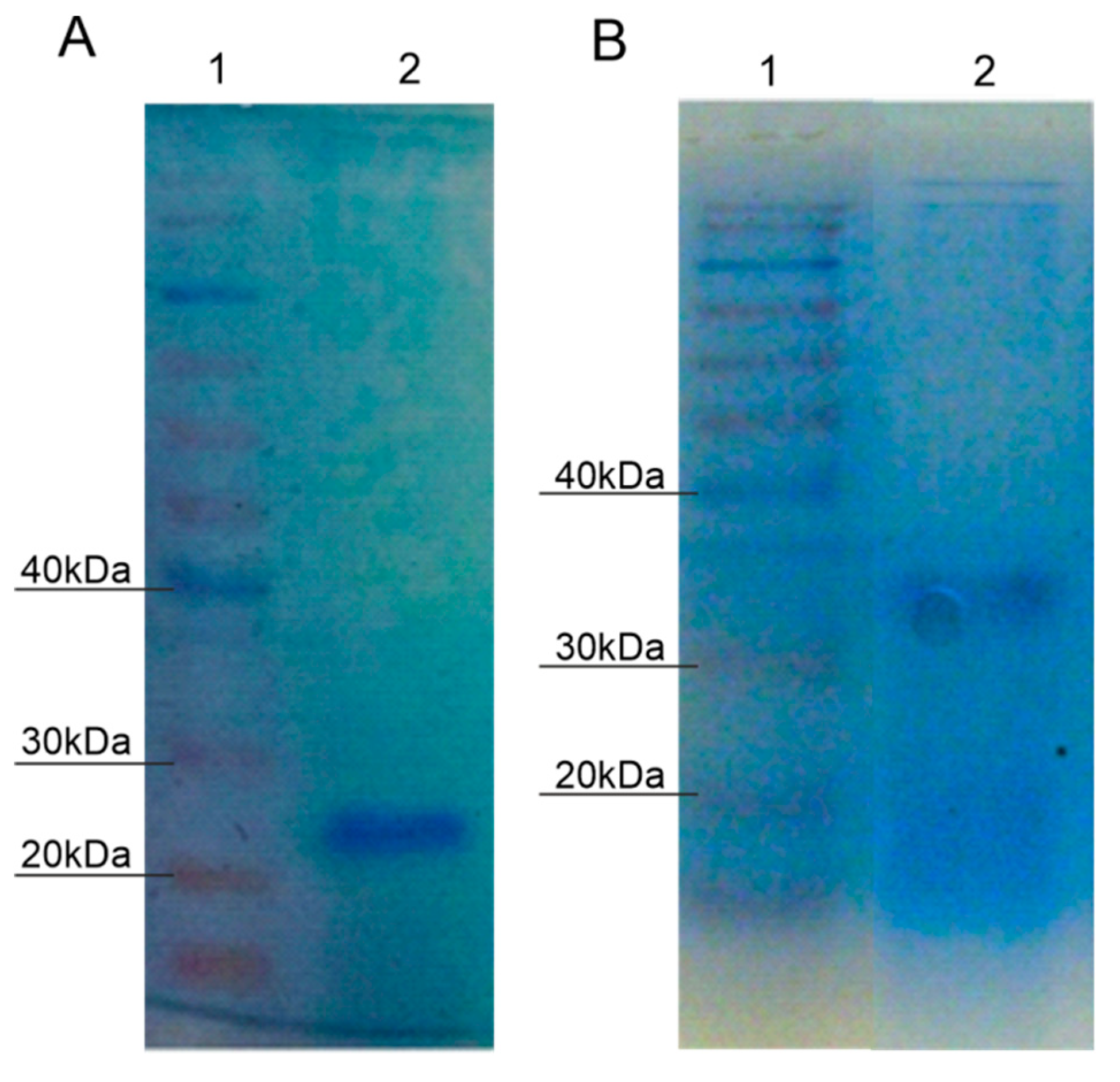
2.3. Purification of Protein with Laccase Activity from P. putida F6

Proteins from P. putida F6 with laccase activity were purified using ion-exchange chromatography. Fractions obtained were tested for enzyme activity towards ABTS in the presence of CuSO4 (2 mM) with activity detected in two fractions (Figure 2). Active fractions were pooled and named Lacc1 and Lacc2, implying that they exhibit a laccase-like activity and their activity was improved drastically in the presence of CuSO4. Both fractions were additionally purified, concentrated, and checked for homogeneity by SDS-PAGE. Lacc1 showed one homogenous band on ~24 kDa (Figure 2A), while the lacc2 fraction was non-homogeneous and lost most of its activity after two gel filtration steps (Figure 2B). Therefore, the Lacc1 fraction was used for further studies.

2.4. N-Terminal Lacc1 Protein and P. putida F6 Genome Sequencing

Lacc1 fraction from P. putida F6 was used for N-terminal sequencing that revealed MTHHSED motif on the N terminus. In addition, the whole genome of P. putida F6 was sequenced and annotated using the PROKKA software tool [42]. Analysis of the whole genome by a sequence-based search for proteins with laccase-like activity and a structure-based search for conserved protein domains using Conserved Domains tool (http://www.ncbi.nlm.nih.gov/Structure//cdd/wrpsb.cgi) [43,44] revealed eight different laccase-like gene sequences (Table 2). Amongst them, only one contained MTHHSED motif, namely gene coding for a four-helix bundle copper-binding protein (cbp). The Conserved Domains tool detected multicopper oxidase (copA) which was also of high interest due to the fact that both purified fractions showed a certain size and CuSO4 dependant activity (Figure 2). Based on the copA and cbp sequences, specific primers for cloning and heterologous expression were designed (Table 1). It is worth mentioning that partially purified Lacc1 protein fraction (Figure 2A) from wild type P. putida F6 has a molecular weight of approximately 24 kDa judging from the SDS-PAGE analysis (Figure 2A). The identified cbp gene was 348 bp in length, which predictably corresponds to approximately 12.7 kDa. This gene is followed by copC gene that is 381 bp in length, which predicts to a protein of approximately 13.9 kDa. The close proximity of these two genes (separated by 11 bp) could result in possible transcription of both genes (cbp and copC) and translation to a single peptide which in turn may explain the discrepancy in predicted size of Cbp protein (Table 2) and Lacc1 protein fraction (Figure 2A).

2.5. Cloning and Expression of Enzymes with Laccase Activity from Pseudomonas Species

Based on the sequence analysis and activity data, two amplicons from P. putida F6 (cbp—348 bp and copA—1869 bp) and amplicons from P. putida KT2440 (mcoKT—741 bp) and P. putida CA-3 (mcoCA3—738 bp) were cloned into pRSET B expression vector. Sequences of four amplicons are deposited in GenBank under accession numbers MN075141, MN075142, MN075139, and MN075140. The recombinant proteins were expressed as N-terminal His6 fusion proteins in E. coli Rosetta (DE3) after 72 h induction at 17 °C with 1 mM IPTG. Activity of overexpressed proteins was determined in ABTS assay using cell-free extracts. Proteins of interest were purified with Ni-NTA affinity chromatography, and their activity was confirmed using zymography assay (Figure 3).
Although most of the laccases studied thus far are of fungal origin, they have limitations including difficulties in heterologous protein expression due to the need for posttranslational modifications, as well as narrow temperature and pH range [4,14]. During the last decade bacterial laccases gained more attention [31,45] due to the number of advantages over fungal laccases, one of which is being more amenable for recombinant expression and directed evolution studies in an E. coli host. Despite wide use of Pseudomonas enzymes in different industrial applications, to date only one laccase-like enzyme, namely CopA from P. putida KT2440, was recombinantly expressed and characterized [5]. The 247 amino acid multicopper oxidase enzyme identified from P. putida KT2440 in the current study is much smaller than the 669 amino acid enzyme identified by Granja-Travez and co-workers [5].

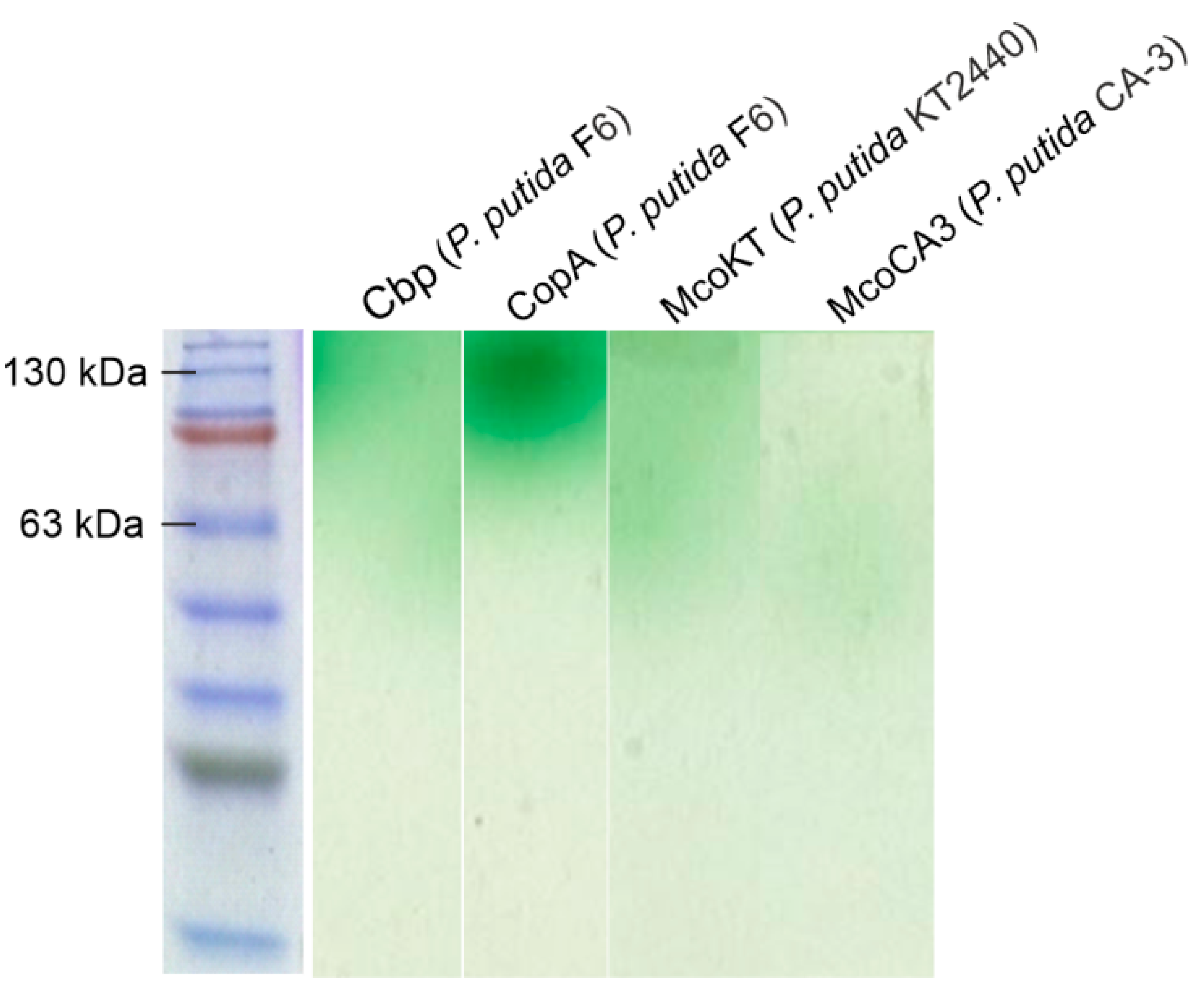
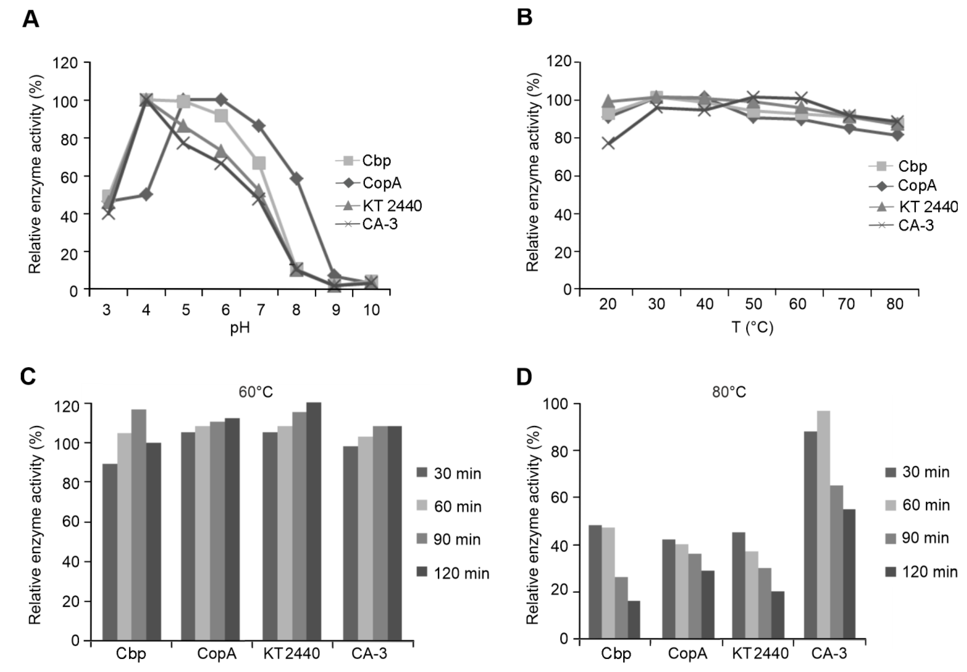
2.6. Characterization of Recombinantly Expressed Laccases from Pseudomonas species

Activity profiles of four purified laccases on different pH values were determined using ABTS as the substrate and buffers of different pHs ranging from 3 to 10. All tested enzymes exhibited a broad pH range, pH 3 to pH 8 for Cbp and CopA, and pH 3 to pH 7 for McoKT and McoCA3, all showing maximum activity at pH 4 (Figure 4A). Obtained results are in accordance with available literature data: CopA from P. putida KT2440, laccase from Bacillus coagulans, and B. clausii laccase [26] exhibited maximum activity on ABTS as a substrate on pH 4 [5]. Furthermore, laccase from B. licheniformis showed maximal activity at pH 3 [3] while laccase from Streptomyces cyaneus had the maximum activity at pH 5 [4].
Temperature optimum was determined by incubating the enzymes at temperatures ranging from 20 to 80 °C under previously determined optimal pH. All tested laccases showed a broad temperature range from 20 to 80 °C with optimal range from 30 to 50 °C (Figure 4B). High enzyme activity even at elevated temperatures correlates with other bacterial laccases and laccase-like multi-copper oxidases [26,27].
Temperature stability of purified laccases from Pseudomonas species was assessed by incubation of enzymes at 60 °C and 80 °C for a total of 120 min, while taking enzyme aliquots for activity tests with ABTS every 30 min. All tested enzymes exhibited high temperature stability at 60 °C, since their activities were slightly elevated in comparison to control (enzyme activity measured before incubation at 60 °C and 80 °C) that was arbitrarily set to 100%, even after 2h of incubation (Figure 4C). Temperature activation of enzyme is also previously shown by Ece and coworkers [4]. McoCA3 enzyme exhibited the best temperature stability overall, as it retained 55% of its initial activity after 120 min at 80 °C, while Cbp, CopA, and McoKT retained 16%, 29%, and 20% of activity, respectively (Figure 4D).
Considering that Pseudomonas strains are mesophilic microorganisms, enzymes tested in this study showed high thermotolerance, which is in accordance with literature data on other enzymes from mesophilic microorganisms. Lončar and coworkers tested the activity of purified recombinant laccase from Bacillus licheniformis and retained 50% of activity after 100 min incubation at 60 °C, and 60 min incubation at 70 °C [3]. Cell-free extracts of heterologously expressed laccase-like MCO from Bacillus strains retained their activities after 30 min of incubation at 70 °C [26]. Ece and coworkers tested the activity of purified recombinant laccase from Streptomyces strain on DMP (2,6-dimethoxyphenol) substrate and showed retention of 50% of activity after 1 h incubation at 60 °C, while almost all activity was lost at 90 °C [4].

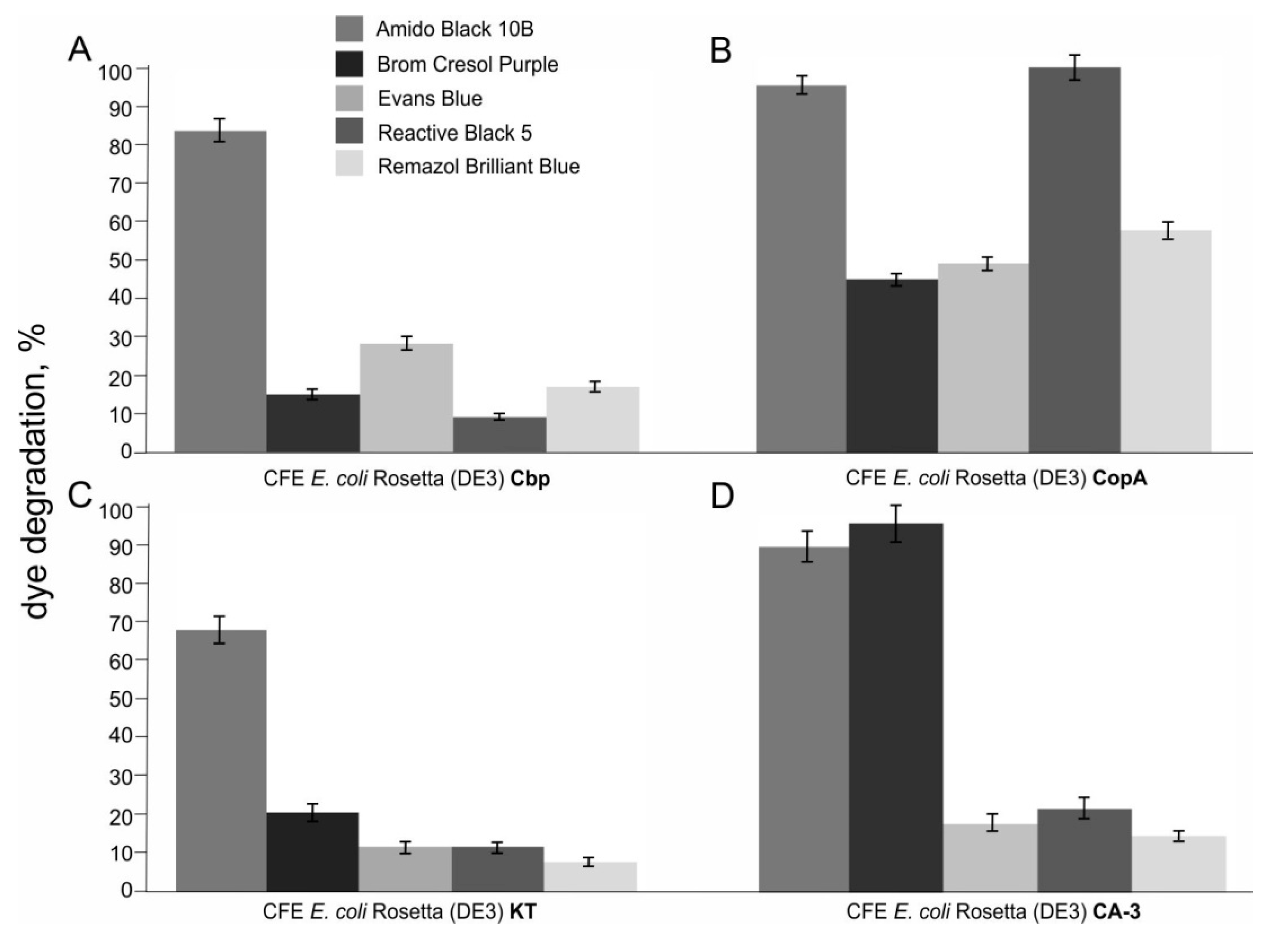
2.7. Degradation of Synthetic Textile Dyes Using Laccases from Pseudomonas Species

Potential of recombinant laccases from Pseudomonas strains to degrade seven synthetic dyes was followed spectrophotometrically for four days and results are presented as relative dye degradation in percents (Figure 5, only degraded dyes are shown). All dyes selected for degradation experiments are widely used in textile and dyeing industries. Among the seven tested dyes, five belong to azo dyes, which make up to 70% of all textile dyestuff produced [46]. Although, degradation experiments were first performed without the addition of a redox mediator (data not shown), the addition of ABTS (0.05 mM) substantially improved degradation of synthetic dyes by tested laccases. Two dyes, namely Erythrosin B and Orange G, were only dyes that were not degraded by tested laccases from Pseudomonas species. Among tested samples the best performing was cell-free extract containing CopA laccase from P. putida F6. CopA laccase degraded five out of seven tested dyes, namely Amido Black 10B, Brom Cresol Purple, Evans Blue, Reactive Black 5, and Remazol Brilliant Blue (Figure 5B). This laccase completely degraded azo dye Reactive Black 5 within four days, while 86% of dye degradation was detected after 48 h (Figure 5B). Pereira and coworkers [47] tested the ability of purified recombinant bacterial CotA-laccase from Bacillus subtilis to degrade Reactive Black 5 and obtained 85% of degradation after 24 h. Among seven tested dyes, Amido Black 5 was the only dye significantly degraded by all used laccases, with a degradation range from 70 to 95% within four days. Mishra and coworkers tested three Bacillus soil isolates and obtained 50–84% of azo dye Amido Black 5 degradation after four days incubation with bacterial culture [48]. Ninety-five percent of triphenylmethane dye Brom Cresol Purple was degraded within four days incubation with cell-free extract containing laccase from P. putida CA-3 (Figure 5D). The majority of studies on textile dyes degradation were performed using fungal laccases. All laccases tested in this study showed promising potential for the application in textile dye degradation, having in mind that presented results were obtained using cell-free extracts of recombinant strains expressing laccases, not purified enzymes to make the process scalable and to avoid costly enzyme purification procedures.

3. Materials and Methods

3.1. Reagents

All reagents and solvents used in this work were purchased either from Merck (Darmstadt, Germany) or Sigma-Aldrich (St. Louis, MO, USA). Dyes were purchased from Carl Roth (Karlsruhe, Germany), except Reactive Black 5 which was purchased from Sigma-Aldrich (St. Louis, MO, USA). GeneJet PCR purification, GeneJet gel extraction kit, and GeneJet plasmid purification kits were purchased from Thermo Fisher Scientific (Waltham, MA, USA).

3.2. Bacterial Strains

All strains used in this study as sources of laccases as well expression strains are presented in Table 3.

3.3. Media

Mineral Salts Medium (MSM) containing 9 g/L Na2HPO4 × 12H2O, 1.5 g/L KH2PO4, 0.2 g/L MgSO4 × 7H2O, 0.002 g/L CaCl2, 1 g/L NH4Cl, 1 mL salt solution [49], supplemented with 0.7% casamino acids, 1% glucose, and 5mM phenylacetic acid was used for growth of wild type Pseudomonas strains and induction of laccases.
Luria–Bertani (LB) broth containing 10 g/L tryptone, 10 g/L NaCl, and 5 g/L yeast extract was used for the growth. Laccase activity was tested on agar plates containing 10 g/L tryptone, 10 g/L NaCl, 5 g/L yeast extract, and 15 g/L agar with 0.01% guaiacol and 0.35 mM CuSO4. Plates were incubated for 7 days at 30 °C.
For selection of clones Luria Agar (LA) plates supplemented with 100 µg/mL of ampicillin. For expression of recombinant laccase terrific broth (TB) was used containing 24 g/L yeast extract, 20 g/L tryptone, 0.4% glycerol, 0.017 M KH2PO4, and 0.072M K2HPO4 with 100 µg/mL ampicillin.

3.4. Screen for Laccase Activity on Guaiacol Agar and in ABTS Assay

The plate method assay for laccase activity was performed on LA agar supplemented with 0.01%(w/v) guaiacol of syringaldazine as laccase substrates and 0.35 mM CuSO4. Strains were seeded and grown overnight at 37 °C and observed for the formation of brown colored zones around colonies that form when laccase is produced by the tested strain.
2,2′-azino-bis(3-ethylbenzothiazoline-6-sulphonic acid) (ABTS) assay was performed either with supernatants and cell-free extracts of wild type strains or with heterologously expressed purified proteins. The reaction was performed in 100 mM Na-acetate buffer pH 4.5 with the addition of 1 mM ABTS and 2 mM CuSO4. Absorption of formed product was followed spectrophotometrically at 410 nm.

3.5. Primer Design, Gene Amplification and Cloning

Based on the data retrieved from UniProt database search for laccase from Pseudomonas, primers for laccase amplification from selected Pseudomonas strains were designed. All primers are presented in Table 1, where restriction sites for positional cloning in pRSETB vector are designated in bold.
PCR amplification of genes coding for proteins with laccase activity was carried out based on the manufacturer’s instructions of GeneJet PCR kit (Thermo Fisher Scientific) on corresponding primer specific annealing temperature (Table 1). Amplified and restriction digested PCR fragments were positionally cloned in pRSET B vector and E. coli Rosetta (DE3) competent cells (both from Thermo Fisher Scientific) were transformed.

3.6. Protein Purification from P. putida F6

A functional isolation of protein with laccase (ABTS oxidizing) activity was performed using anion-exchange (Q-Sepharose) columns were purchased from Amersham (Amersham Biosciences AB, Uppsala, Sweden). Cell-free extracts of phenylacetic acid-induced (5 mM) P. putida F6 cells were obtained using following procedure: cell culture was centrifuged for 20 min at 3000× g (Eppendorf centrifuge 5804, Hamburg, Germany). Pellet was resuspended in lysis buffer (20 mM Tris-HCl, pH 8, and 1 mg/mL lysozyme) and incubated 30 min at 37 °C. Mixture was sonicated at 20 KHz, 3 pulses of 15 s (Soniprep 150, MSE (UK) Ltd., Lindon, UK). Cell-free extracts were clarified by centrifugation for 40 min at 20,817× g, 4 °C (Eppendorf Centrifuge 5417 R, Hamburg, Germany). Obtained cell-free extracts were dialyzed against 20 mM Tris-HCl buffer pH 8 and loaded on a manually packed (5 cm × 25 cm) strong anion-exchange Q-Sepharose column pre-equilibrated at the same buffer. Elution was performed applying a linear gradient of 0–0.5 M NaCl at a flow rate of 5 mL/min. Partially purified enzymes were equilibrated in 20 mM Tris-HCl pH 7 buffer, applied on pre-equilibrated column separately and eluted as previously described. Concentrated fractions were checked for homogeneity by SDS-PAGE, stained with Coomassie Brilliant Blue. Partially purified enzymes were loaded onto a HiPrep 16/60 Sephacryl S-200 HR (1.6 cm × 60 cm) and HiPrep Sephacryl S-300 HR 26/60 (2.6 cm × 60 cm) gel filtration columns, previously equilibrated with 20 mM Tris-HCl pH 7 with 150 mM NaCl at a flow rate 60 mL/h. Obtained fractions were tested for enzyme activity using ABTS assay (Section 3.4).

3.7. N-Terminal and Genome Sequencing

Purified laccase from Pseudomonas putida F6 was sequenced by Cambridge Peptides Ltd., West Midlands, UK. A sequence of 7 amino acids at N-terminus was identified by the Edman degradation method.
Genome of Pseudomonas putida F6 was sequenced in MicrobesNG company (Birmingham, UK). Obtained raw genome sequences were annotated using PROKKA software tool and analyzed using Conserved Domains tool [42,43,44]. Genes of interest were discovered by motif, sequence and domain-based search and primers for cloning in pRSETB were designed (Table 1).

3.8. Recombinant Protein Expression and Purification

Host cells carrying pRETB plasmid with laccases from different Pseudomonas strains were grown overnight at 37 °C in TB medium containing 100 g/mL ampicillin. Overnight culture was diluted (1%) into TB medium containing ampicillin (100 g/mL) and CuSO4 at final concentration of 2 mM and after reaching OD600 = 0.5 expression was induced with 1 mM IPTG and carried out for 48 h at 17 °C with shaking at 180 rpm. Cells were pelleted for 10 min at 3000× g (Eppendorf centrifuge 5804, Hamburg, Germany). Lysis buffer containing (50 mM NaH2PO4, pH 8, 300 mM NaCl, 10 mM imidazole, and 1 mg/mL lysozyme) was used for the preparation of cell-free extracts. The mixture was incubated for 30 min at 37 °C followed by sonication of 3 pulses of 15 s at 20 KHz (Soniprep 150, MSE (UK) Ltd., England). Cell-free extracts were clarified by centrifugation for 40 min at 20 817× g, 4 °C (Eppendorf Centrifuge 5417 R, Hamburg, Germany). Clear supernatant was loaded on a NiNTA agarose (Qiagen, Hilden, Germany) equilibrated with 50 mM NaH2PO4 pH8, 300 mM NaCl, 10 mM imidazole. Non-specifically bound proteins were washed with one volume of 50 mM imidazole in starting buffer followed by elution with 250 mM and 500 mM imidazole in the same buffer. Total protein concentration in collected fractions was determined according to the Bradford method [50] using Quick StartTM Bradford 1× Reagent (BioRad Laboratories, California, USA). Collected fractions were analyzed by activity assay on ABTS and SDS-PAGE.

3.9. Temperature and pH Optimum of Purified Laccases

The optimum pH of the laccase enzymes was determined within a pH range of 3 to 10 using ABTS (2,2′-azino-bis(3-ethylbenzothiazoline-6-sulphonic acid) as a substrate, and purified proteins with adjusted concentrations. For pH range from 3 to 5, acetate buffer was used; for pH 6 and 7, 20 mM phosphate buffer was used; while for pH 8 and pH 9 and 10, 20 mM Tris-HCl and 20 mM glycine-NaOH were used, respectively. Temperature optimum of enzyme activity was measured in the range of 20 to 80 °C at previously determined optimal pH and buffer and with ABTS as a substrate. ABTS assay contained 20 mM Na-acetate buffer pH 4.5, 100 µL of sample, 1 mM ABTS, and 2 mM CuSO4 and after incubation of 15 min at 37 °C reactions were followed spectrophotometrically for product formation at 420 nm.
Thermal stability of purified laccase was determined by measuring residual activity upon incubating aliquots of purified laccases at 60 °C and 80 °C. Samples were withdrawn at 30, 60, 90, and 120 min, placed on ice, and enzyme activity was determined in ABTS assay as described in Section 3.4. For testing thermal stability 100 mM Na-acetate buffer of pH 4.0—pH 5.0 was used.

3.10. Textile Dyes Degradation

Seven different dyes were selected to study degradation ability of the laccases from Pseudomonas species. Selected dyes belong to three different groups: Amido Black 10B, Evans Blue, Reactive Black 5, Remazol Briliant Blue, and Orange G are azo dyes, Brom Cresol Purple is triphenylmethane dye, while Erythrosin B is xanthene dye. Stock solutions of the dyes in 100 mM Na-acetate buffer pH 4.5 were stored in the dark at 4 °C. For dyes degradation experiments cell-free extracts of four recombinant strains expressing laccases from Pseudomonas species were used. Reaction consisted of cell-free extracts of adjusted protein concentrations in 100 mM Na-acetate buffer pH 4.5, 0.05 mM ABTS, and 2 mM CuSO4 and corresponding dye. Concentration of each dye was set to correspond to 0.6 absorbance units at the maximum wavelength of a specific dye. In this experiment two controls were set up. One control reaction consisted of appropriate dye, 100 mM Na-acetate buffer pH 4.5, 0.05 mM ABTS, and 2 mM CuSO4, and was used to monitor dye degradation over time. Second control reaction, used as blank, was prepared for each laccase, and consisted of cell-free extract in 100 mM Na-acetate buffer pH 4.5, 0.05 mM ABTS, and 2 mM CuSO4. Reactions were incubated for four days at 30 °C, and dye concentrations were spectrophotometrically measured at maximum wavelength determined for each dye. Dye degradation (%) was calculated using following equation:
i n i t i a l   a b s o r b a n c e f i n a l   a b s o r b a n c e i n i t i a l   a b s o r b a n c e × 100 %

4. Conclusions

Four active recombinant laccases from three Pseudomonas sp. (P. putida KT2440 and P. putida CA-3, and P. putida F6) were discovered using different molecular approaches including DNA sequence analysis, N-terminal protein sequencing, and genome sequencing data analysis. These new laccases exhibited good stability at elevated temperatures, and were active over a broad temperature and pH range. All tested laccases showed good potential in degradation of synthetic textile dyes, with CopA from P. putida F6 being the most promising, as this laccase was active on five out of seven tested dyes, namely Amido Black 10B, Brom Cresol Purple, Evans Blue, Reactive Black 5, and Remazol Brilliant Blue. This research confirmed that Pseudomonas genus is still a source of biocatalytically relevant enzymes, since all tested enzymes showed promising potential for the degradation of textile dyes which makes them good candidates for the application in bioremediation.

Author Contributions

E.T., S.J., and J.N.-R. designed and supervised this study. M.M. performed experimental work. E.N, R.P., and K.O. were involved in protein purification and characterization from wild type strain. L.D. performed protein and genome data analysis. All authors contributed in the preparation of the manuscript.

Funding

This work was supported by the Ministry of Education, Science and Technological Development, Republic of Serbia, under Grant No. 173048. FEMS Research Grant (FEMS-RG-2016-0088) to Mina Mandic is acknowledged.

Conflicts of Interest

The authors declare no conflict of interest.

References

  1. Yaropolov, A.I.; Skorobogat’Ko, O.V.; Vartanov, S.S.; Varfolomeyev, S.D. Laccase: Properties, catalytic mechanism, and applicability. Appl. Biochem. Biotechnol. 1994, 49, 257–280. [Google Scholar] [CrossRef]
  2. Morozova, O.V.; Shumakovich, G.P.; Gorbacheva, M.A.; Shleev, S.V.; Yaropolov, A.I. “Blue” laccases. Biochemistry (Moscow) 2007, 72, 1136–1150. [Google Scholar] [CrossRef] [PubMed]
  3. Lončar, N.; Božić, N.; Vujičić, Z. Expression and characterization of a thermostable organic solvent-tolerant laccase from Bacillus licheniformis ATCC 9945a. J. Mol. Catal. B Enzym. 2016, 134, 390–395. [Google Scholar] [CrossRef]
  4. Ece, S.; Lambertz, C.; Fischer, R.; Commandeur, U. Heterologous expression of a Streptomyces cyaneus laccase for biomass modification applications. AMB Express 2017, 7, 86. [Google Scholar] [CrossRef] [PubMed]
  5. Granja-Travez, R.S.; Bugg, T.D.H. Characterisation of multicopper oxidase CopA from Pseudomonas putida KT2440 and Pseudomonas fluorescens Pf-5: Involvement in bacterial lignin oxidation. Arch. Biochem. Biophys. 2018, 660, 97–107. [Google Scholar] [CrossRef] [PubMed]
  6. Brijwani, K.; Rigdon, A.; Vadlani, P.V. Fungal laccases: Production, function, and applications in food processing. Enzyme. Res. 2010, 2010, 149748–149758. [Google Scholar] [CrossRef] [PubMed]
  7. Zerva, A.; Koutroufini, E.; Kostopoulou, I.; Detsi, A.; Topakas, E. A novel thermophilic laccase-like multicopper oxidase from Thermothelomyces thermophila and its application in the oxidative cyclization of 2′,3,4-trihydroxychalcone. New Biotechnol. 2019, 49, 10–18. [Google Scholar] [CrossRef]
  8. Bryan, A.C.; Jawdy, S.; Gunter, L.; Gjersing, E.; Sykes, R.; Hinchee, M.A.; Winkeler, K.A.; Collins, C.M.; Engle, N.; Tschaplinski, T.J.; et al. Knockdown of a laccase in Populus deltoides confers altered cell wall chemistry and increased sugar release. Plant Biotechnol. J. 2016, 14, 2010–2020. [Google Scholar] [CrossRef]
  9. Liu, Q.; Luo, L.; Wang, X.; Shen, Z.; Zheng, L. Comprehensive analysis of rice laccase gene (OsLAC) family and ectopic expression of OsLAC10 enhances tolerance to copper stress in Arabidopsis. Int. J. Mol. Sci. 2017, 18, 209. [Google Scholar] [CrossRef]
  10. Laufer, Z.; Beckett, R.P.; Minibayeva, F.V.; Lüthje, S.; Böttger, M. Diversity of laccases from lichens in suborder Peltigerineae. Bryologist 2009, 112, 418–426. [Google Scholar] [CrossRef]
  11. Li, Q.; Wang, X.; Korzhev, M.; Schröder, H.C.; Link, T.; Tahir, M.N.; Diehl-Seifert, B.; Müller, W.E. Potential biological role of laccase from the sponge Suberites domuncula as an antibacterial defense component. Biochim. Biophys. Acta Gen. Subj. 2015, 1850, 118–128. [Google Scholar] [CrossRef] [PubMed]
  12. Jin, L.; Yang, X.; Sheng, Y.; Cao, H.; Ni, A.; Zhang, Y. The second conserved motif in bacterial laccase regulates catalysis and robustness. Appl. Microbiol. Biotechnol. 2018, 102, 4039–4048. [Google Scholar] [CrossRef] [PubMed]
  13. Muthukumarasamy, N.P.; Jackson, B.; Raj, A.J.; Sevanan, M. Production of extracellular laccase from Bacillus subtilis MTCC 2414 using agroresidues as a potential substrate. Biochem. Res. Int. 2015, 2015, 765190. [Google Scholar] [CrossRef] [PubMed]
  14. Piscitelli, A.; Pezzella, C.; Giardina, P.; Faraco, V.; Giovanni, S. Heterologous laccase production and its role in industrial applications. Bioengineered 2010, 1, 252–262. [Google Scholar] [CrossRef] [PubMed]
  15. Zouraris, D.; Zerva, A.; Topakas, E.; Karantonis, A. Kinetic and amperometric study of the MtPerII peroxidase isolated from the ascomycete fungus Myceliophthora thermophila. Bioelectrochemistry 2017, 118, 19–24. [Google Scholar] [CrossRef] [PubMed]
  16. Romero-Guido, C.; Baez, A.; Torres, E. Dioxygen activation by laccases: Green chemistry for fine chemical synthesis. Catalysts 2018, 8, 223. [Google Scholar] [CrossRef]
  17. Blánquez, A.; Rodríguez, J.; Brissos, V.; Mendes, S.; Martins, L.O.; Ball, A.S.; Arias, M.E.; Hernández, M. Decolorization and detoxification of textile dyes using a versatile Streptomyces laccase-natural mediator system. Saudi J. Biol. Sci. 2018, 26, 913–920. [Google Scholar] [CrossRef]
  18. Rodríguez Couto, S.; Toca-Herrera, J.L. Lacasses in the textile industry. Biotechnol. Mol. Biol. Rev. 2006, 1, 115–120. [Google Scholar]
  19. Legerska, B.; Chmelová, D.; Ondrejovič, M. Degradation of synthetic dyes by laccases—A mini-review. Nova Biotechnol. Chim. 2016, 15, 90–106. [Google Scholar] [CrossRef]
  20. Ferrer-Miralles, N.; Domingo-Espín, J.; Corchero, J.L.; Vázquez, E.; Villaverde, A. Microbial factories for recombinant pharmaceuticals. Microb. Cell Fact. 2009, 24, 8–17. [Google Scholar] [CrossRef]
  21. Couto, S.R.; Herrera, J.L.T. Industrial and biotechnological applications of laccases: A review. Biotechnol. Adv. 2006, 24, 500–513. [Google Scholar] [CrossRef]
  22. Rodgers, C.J.; Blanford, C.F.; Giddens, S.R.; Skamnioti, P.; Armstrong, F.A.; Gurr, S.J. Designer laccases: A vogue for high-potential fungal enzymes? Trends Biotechnol. 2010, 28, 63–72. [Google Scholar] [CrossRef] [PubMed]
  23. Fernandes, T.A.R.; Silveira, W.B.; Passos, F.M.L.; Zucchi, T.D. Laccases from actinobacteria—What we have and what to expect. Postepy Mikrobiol. 2014, 4, 285–296. [Google Scholar]
  24. Guan, Z.B.; Shui, Y.; Song, C.M.; Zhang, N.; Cai, Y.J.; Liao, X.R. Efficient secretory production of CotA-laccase and its application in the decolorization and detoxification of industrial textile wastewater. Environ. Sci. Pollut. Res. 2015, 22, 9515–9523. [Google Scholar] [CrossRef] [PubMed]
  25. Martins, L.O.; Soares, C.M.; Pereira, M.M.; Teixeira, M.; Costa, T.; Jones, G.H.; Henriques, A.O. Molecular and biochemical characterization of a highly stable bacterial laccase that occurs as a structural component of the Bacillus subtilis endospore coat. J. Biol. Chem. 2002, 277, 18849–18859. [Google Scholar] [CrossRef] [PubMed]
  26. Ihssen, J.; Reiss, R.; Luchsinger, R.; Thöny-Meyer, L.; Richter, M. Biochemical properties and yields of diverse bacterial laccase-like multicopper oxidases expressed in Escherichia coli. Sci. Rep. 2015, 5, 10465. [Google Scholar] [CrossRef] [PubMed] [Green Version]
  27. Sherif, M.; Waung, D.; Korbeci, B.; Mavisakalyan, V.; Flick, R.; Brown, G.; Abouzaid, M.; Yakunin, A.F.; Master, E.R. Biochemical studies of the multicopper oxidase (small laccase) from Streptomyces coelicolor using bioactive phytochemicals and site directed mutagenesis. Microb. Biotechnol. 2013, 6, 588–597. [Google Scholar] [CrossRef] [PubMed]
  28. Arias, M.E.; Arenas, M.; Rodríguez, J.; Soliveri, J.; Ball, A.S.; Hernández, M. Kraft pulp biobleaching and mediated oxidation of a nonphenolic substrate by laccase from Streptomyces cyaneus CECT 3335. Appl. Environ. Microbiol. 2003, 69, 1953–1958. [Google Scholar] [CrossRef]
  29. Kandasamy, S.; Devi, P.; Chendrayan Uthandi, S. Laccase producing Streptomyces bikiniensis CSC12 isolated from compost. J. Microbiol. Biotechnol. Food. Sci. 2017, 6, 794–798. [Google Scholar]
  30. Eugenio, M.E.; Hrenández, M.; Moya, R.; Martínsampedro, R. Evaluation of a new laccase produced by Streptomyces ipomoea on biobleaching and ageing of kraft pulps. BioResources 2011, 6, 3231–3241. [Google Scholar]
  31. Kuddus, M.; Joseph, B.; Ramteke, P.V. Production of laccase from newly isolated Pseudomonas putida and its application in bioremediation of synthetic dyes and industrial effluents. Biocatal. Agric. Biotechnol. 2013, 2, 333–338. [Google Scholar] [CrossRef]
  32. Peter, J.K.; Vandana, P. Congo red dye decolourization by partially purified laccases from Pseudomonas aeruginosa. Int. J. Curr. Microbiol. App. Sci. 2014, 3, 105–115. [Google Scholar]
  33. Arunkumar, T.; Anand, A.D.; Narendrakumar, G. Production and partial purification of laccase from Pseudomonas aeruginosa ADN04. J. Pure Appl. Microbiol. 2014, 8, 727–731. [Google Scholar]
  34. Bugg, T.D.; Ahmad, M.; Hardiman, E.H.; Singh, R. The emerging role for bacteria in lignin degradation and bio-product formation. Curr. Opin. Biotechnol. 2011, 3, 394–400. [Google Scholar] [CrossRef] [PubMed]
  35. Su, J.; Fu, J.; Wang, Q.; Silva, C.; Cavaco-Paulo, A. Laccase: A green catalyst for the biosynthesis of poly-phenols. Crit. Rev. Biotechnol. 2018, 38, 294–307. [Google Scholar] [CrossRef] [PubMed]
  36. Sakurai, T.; Kataoka, K. Basic and applied features of multicopper oxidases, CueO, bilirubin oxidase, and laccase. Chem. Rec. 2007, 7, 220–229. [Google Scholar] [CrossRef] [PubMed] [Green Version]
  37. Miñambres, B.; Martínez-Blanco, H.; Olivera, E.R.; García, B.; Díez, B.; Barredo, J.L.; Moreno, M.A.; Schleissner, C.; Salto, F.; Luengo, J.M. Molecular cloning and expression in different microbes of the DNA encoding Pseudomonas putida U phenylacetyl-CoA ligase. Use of this gene to improve the rate of benzylpenicillin biosynthesis in Penicillium chrysogenum. J. Biol. Chem. 1996, 271, 33531–33538. [Google Scholar] [CrossRef]
  38. Sharma, P.; Goel, R.; Capalash, N. Bacterial laccases. World J. Microbiol. Biotechnol. 2007, 23, 823–832. [Google Scholar] [CrossRef]
  39. Margot, J.; Bennati-Granier, C.; Maillard, J.; Blánquez, P.; Barry, D.A.; Holliger, C. Bacterial versus fungal laccase: Potential for micropollutant degradation. AMB Express 2013, 3, 63. [Google Scholar] [CrossRef]
  40. Chojnacki, S.; Cowley, A.; Lee, J.; Foix, A.; Lopez, R. Programmatic access to bioinformatics tools from EMBL-EBI update: 2017. Nucleic Acids Res. 2017, 45, 550–553. [Google Scholar] [CrossRef]
  41. McMahon, A.M.; Doyle, E.M.; Brooksa, S.; O’Connor, K.E. Biochemical characterization of the coexisting tyrosinase and laccase in the soil bacterium Pseudomonas putida F6. Enzm. Microb. Technol. 2007, 40, 1435–1441. [Google Scholar] [CrossRef]
  42. Seeman, T. PROKKA: Rapid prokaryotic genome annotation. Bioinformatics. 2014, 30, 2068–2069. [Google Scholar] [CrossRef] [PubMed]
  43. Marchler-Bauer, A.; Derbyshire, M.K.; Gonzales, N.R.; Lu, S.; Chitsaz, F.; Geer, L.Y.; Geer, R.C.; He, J.; Gwadz, M.; Hurwitz, D.I.; et al. CDD: NCBI’s conserved domain database. Nucleic Acids Res. 2015, 43, 222–226. [Google Scholar] [CrossRef] [PubMed]
  44. Marchler-Bauer, A.; Bo, Y.; Han, L.; He, J.; Lanczycki, C.J.; Lu, S.; Chitsaz, F.; Derbyshire, M.K.; Geer, R.C.; Gonzales, N.R.; et al. CDD/SPARCLE: Functional classification of proteins via subfamily domain architecture. Nucleic Acid Res. 2017, 45, 200–203. [Google Scholar] [CrossRef]
  45. Santhanam, N.; Vivanco, J.M.; Decker, S.R.; Reardon, K.F. Expression of industrially relevant laccases: Prokaryotic style. Trends. Biotechnol. 2011, 29, 480–489. [Google Scholar] [CrossRef] [PubMed]
  46. Tony, B.D.; Goyal, D.; Khanna, S. Decolorization of textile azo dyes by aerobic bacterial consortium. Int. Biodeterior. Biodegrad. 2009, 63, 462–469. [Google Scholar] [CrossRef]
  47. Pereira, L.; Coelho, A.V.; Viegas, C.A.; Correia dos Santos, M.M.; Robalo, M.P.; Martins, L.O. Enzymatic biotransformation of the azo dye Sudan Orange G with bacterial CotA-laccase. J. Biotechnol. 2009, 139, 68–77. [Google Scholar] [CrossRef]
  48. Mishra, V.K.; Sharma, H. Decolourization of textile azo dyes by Bacillus spp. In Proceedings of the International Conference on Emerging Trends in Traditional and Technical Textiles, Jalandhar, India, 11–12 April 2014. [Google Scholar]
  49. Schlegel, H.G.; Kaltwasser, H.; Gottschalk, G. A submersion method for culture of hydrogen-oxidizing bacteria: Growth physiological studies. Arch. Mikrobiol. 1961, 38, 209–222. [Google Scholar] [CrossRef]
  50. Bradford, M.M. A rapid and sensitive method for the quantitation of microgram quantities of protein utilizing the principle of protein-dye binding. Anal. Biochem. 1976, 72, 248–254. [Google Scholar] [CrossRef]
Figure 1. Laccase activity assay on guaiacol-containing plates (A) and in ABTS assay (B). Abbreviations F6, KT2440, CA-3, mt-2, S12, B561, and PAO1 refer to wild type strains P. putida F6, P. putida KT2440, P. putida CA-3, P. putida mt-2, P. putida S12, P. chlororaphis B561 and P. aeruginosa PAO1 (A) and appropriate cell-free extracts (B). Enzyme activity in ABTS assay was expressed as a ratio of product formation to the total protein concentration (A410/A595). Inlet in (B) represents color change in ABTS assay due to laccase activity from cell-free extracts.
Catalysts 09 00629 g001
Figure 2. SDS-PAGE of partially purified (enriched fractions) enzymes from P. putida F6. (A) lane 1—protein standard, lane 2—Lacc1 fraction (~24 kDa); (B) lane 1—protein standard, lane 2—Lacc2 fraction.
Catalysts 09 00629 g002
Figure 3. Zymography assay of purified laccases from Pseudomonas species using native PAGE analysis. The first lane—BlueStar prestained protein marker (Nippon genetics). Lanes from left to right: Cbp (P. putida F6), CopA (P. putida F6), McoKT (P. putida KT2440), and McoCA3 (P. putida CA-3).
Catalysts 09 00629 g003
Figure 4. pH, temperature optimum and temperature stability of expressed laccases from Pseudomonas species: (A) relative enzymes activity at different pH values, (B) relative enzymes activity at different temperatures, (C) temperature stability at 60 °C, (D) temperature stability at 80 °C.
Catalysts 09 00629 g004
Figure 5. Degradation of five synthetic textile dyes using cell-free extracts (CFE) of E. coli Rosetta (DE3) expressing laccases: (A) Cbp from P. putida F6; (B) CopA from P. putida F6; (C) KT from P. putida KT2440; and (D) CA-3 from P. putida CA-3.
Catalysts 09 00629 g005
Table 1. Oligonucleotide sequences and the annealing temperaturesused for the amplification of laccases from Pseudomonas species.
Primers with Restriction Enzyme SitesAnnealing TStrain
PS_LACFATGAGTGRCCTGRCBCAG50 °CPseudomonas spp.
PS_LACRGCGGNTCCAGCCASACCARSGA
CA3FTAACAGGATCCGAGTGGCCTGACTCAGG (Bam HI)55 °CP. putida CA-3
CA3RTAATTAAGCTTTTGCGGTTCCAGCCAGAC (Hind III)
KTFTAACAGGATCCGAGTGACCTGACGCAGG (Bam HI)59 °CP. putida KT2440
KTRTAATTAAGCTTGCGCGGGTCCAGCCAGAC (Hind III)
CopAFTAACAGCTAGCATGTCGCATGATGATTTTCGT (Nhe I) 55 °CP. putida F6
CopARTAATTAAGCTTTTCGTCGACCCTCACCGTGCG (Hind III)
CbpFTAACAGCTAGCATGACTCACCATTCCGAAGAC (Nhe I) 58 °CP. putida F6
CbpRTAATTAAGCTTAGCCGCCATGGCGCTGCAGCT (Hind III)
Table 2. Genes coding for proteins with laccase-like activity discovered in genome of P. putida F6.
Gene NameProtein Size (AA)GeneBank MatchGeneBank Acc NoIdentity (%)Discovered with
Putative cysteine-rich protein YhjQ116Four-helix bundle copper-binding protein (Cbp) from Pseudomonas putidaWP_026070601100MTHHSED motif search
Not annotated623Copper resistance system multicopper oxidase (CopA) from XantomonadaceaeWP_017354985100Conserved Domains tool
Copper resistance protein B425Copper resistance protein B (CopB) from XantomonadaceaeWP_017354984100Genome annotation
Copper resistance protein C127Copper homeostasis periplasmic binding protein (CopC) from XanthomonadaceaeWP_017354979100Genome annotation
Laccase domain protein YfiH246Multi-copper polyphenol oxidoreductase laccase from Pseudomonas putidaWP_07580445796.3Genome annotation
Laccase domain protein YfiH256Multi-copper polyphenol oxidoreductase laccase from Stenotrophomonas maltophiliaWP_09956024499.2Genome annotation
Multicopper oxidase mco460Multicopper oxidase from Pseudomonas putidaWP_07580669897.4Genome annotation
Blue copper oxidase CueO675Multicopper oxidase protein from PseudomonasWP_07580445579.9Genome annotation
Table 3. Bacterial strains used in this study.
Bacterial StrainReference
Pseudomonas aeruginosa PAO1ATCC 15,692
Pseudomonas putida CA-3NCIMB 41,162
Pseudomonas putida F6[33]
Pseudomonas putida KT2440ATCC 47,054
Pseudomonas putida mt-2NCIMB 10,432
Pseudomonas putida S12ATCC 70,0801
Pseudomonas chlororaphis B561ATCC 19,523
Escherichia coli Rosetta (DE3) Merck, Darmstadt, Germany

