Article

Alcoholysis Products by a GH53 Fungal Galactanase

by
Marco Zanon
,
Theo Tonne Hønning Lyholm
,
Yusuf Theibich
,
Sara Jonsdottir Glaser
and
Leila Lo Leggio
*
Department of Chemistry, University of Copenhagen, Universitetsparken 5, DK-2100 Copenhagen, Denmark
*
Author to whom correspondence should be addressed.
Current address: Department of Pharmaceutical and Pharmacological Sciences, University of Padua, Via F. Marzolo 5, 35131 Padua, Italy.
Catalysts 2026, 16(5), 421; https://doi.org/10.3390/catal16050421
Submission received: 27 February 2026 / Revised: 15 April 2026 / Accepted: 18 April 2026 / Published: 3 May 2026

Abstract

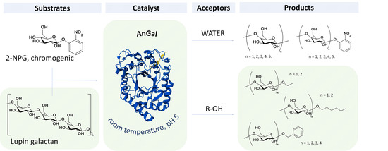
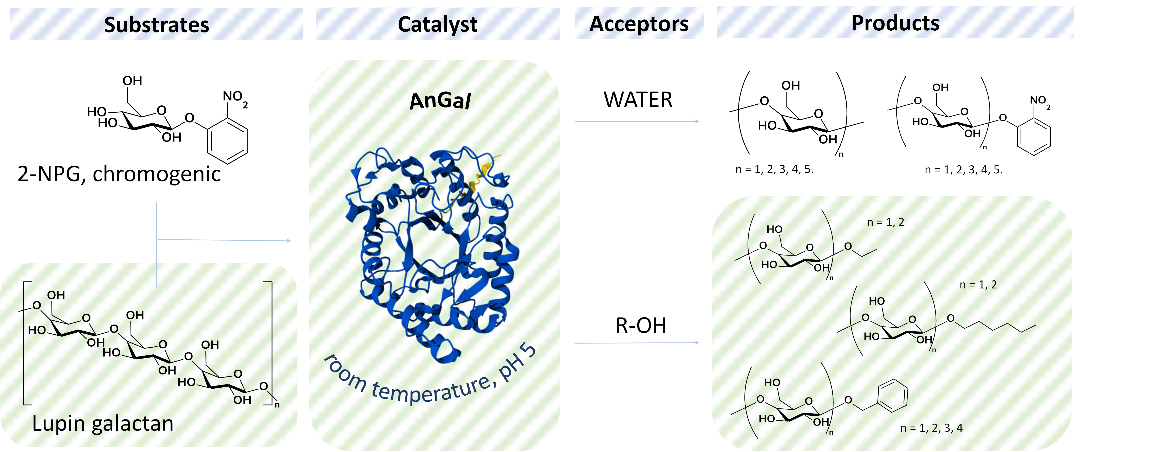
In this study, the native activity of an arabinogalactan endo-β-1,4-galactanase from Aspergillus niger (AnGal) was evaluated under different reaction conditions, and in the presence of various acceptor molecules during the cleavage of the β-1,4-glycosidic linkage of a chromogenic compound and lupin galactan. A combination of spectrophotometric assays, mass spectrometry and chromatography techniques provided insights into the reaction mechanism of the enzyme and its use in the synthesis of galactosides and galactooligosaccharide derivatives. In reactions containing 2-nitrophenol galactopyranoside, AnGal promoted transglycosylation, generating longer galactooligosaccharide derivatives of 2-nitrophenol that have not previously been reported for GH53 enzymes. Furthermore, new alcoholysis products have been detected when AnGal acted on lupin galactan in the presence of benzyl alcohol. To the best of our knowledge, we are first to report the synthesis of galactotriose and galactotetraose derivatives formed by endo-β-1,4-galactanase alcoholysis. This work showcases the potential of utilizing galactanases in the synthesis of valuable galactosides and galactooligosaccharides, under mild conditions from sustainable biomass sources. Potential beneficial applications may be found in several industrial fields such as in the preparation of prodrugs and prebiotics.

Graphical Abstract

1. Introduction

Glycoside hydrolase 53 (GH53) is a family of enzymes in the carbohydrate-active enzyme database (CAZy) with primarily arabinogalactan endo-β-1,4-galactanase activity (EC 3.2.1.89) [1,2]. These enzymes are capable of hydrolyzing β-1,4-galactan found in type I arabinogalactan of pectin, which is a major component of the plant cell wall [3]. Recent work has demonstrated that GH53 enzymes can also catalyze the hydrolysis of β-1,6-galactan branches from Type II arabinogalactans present in both plant and algal cell walls [4,5]. Endo-β-1,4-galactanases have also recently been identified as one of the key enzymes necessary for complex polysaccharide degradation, a finding that underscores their potential in diverse biotechnological applications [6].
Pectin is responsible for many properties of plant cells including cell growth and differentiation as well as cell wall rigidity and hydration, but changes in pectin are also essential in natural processes such as fruit ripening [2,3,7]. Despite this, GH53 have only been identified in 2 species of plants on CAZy [1]; instead, β-galactosidases and exo-galactanases are utilized by plants to modify galactan [8]. GH53 enzymes are often found in fungal and bacterial plant pathogens [1]. GH53 is the only CAZy family known to have endo-β-1,4-galactanase activity, except for 2 bacterial enzymes in the family GH147. In CAZy, there are currently crystal structures available for 8 different β-1,4-galactanases: 3 enzymes from the bacteria Bacillus licheniformis (BlGal), Bacteroides thetaiotaomicron (BtGal) and Geobacillus stearothermophilus (GanA), 4 from the fungi Aspergillus aculeatus (AaGal), Aspergillus nidulans (EnGal), Humicola insolens (HiGal) and Thermothelomyces thermophilus (MtGal) and 1 from the archaeon Ignisphaera aggregans (IaGal) [1].
Notably, these enzymes differ by the number of—subsites. The 4 structurally characterized fungal enzymes, along with the bacterial BtGal, contain −1 and −2 subsites, while the bacterial BlGal and GanA contain −1 to −4 subsites and the archaeal IaGal has an intermediate structure with −1 to −3 subsites. The difference in the number of −subsites is reflected in the hydrolysis profiles of these enzymes. A previous study showed that AaGal was able to hydrolyze galactotriose (gal3) and galactotetraose (gal4) completely to galactose (gal1) and galactobiose (gal2), while BlGal was inactive on galactotriose, but hydrolyzed galactotetraose completely to galactose, galactobiose and galactotriose [9]. Furthermore, the fungal enzymes MtGal and HiGal accumulated mostly galactose and galactobiose at the end of lupin galactan hydrolysis, whereas IaGal and BlGal mostly accumulated galactose, galactobiose and galactotriose [10].
These findings suggested that enzymes with 2—subsites bind galactobiose unproductively, while enzymes with 3 and 4 −subsites bind both galactobiose and galactotriose unproductively [2]. This is further corroborated by crystal structures of BlGal showing galactotriose bound to subsites −2 to −4 (1UR0, 2CCR, 2GFT and 2J74) [9,11] and galactobiose bound to subsites −2 to −3 (1UR4) [9]. In contrast the crystal structure of AaGal shows galactobiose bound to subsites −1 to −2 (6Q3R) [12]. Besides hydrolysis, some galactanases are also able to catalyze transglycosylation and alcoholysis [2,10]. While water is the acceptor in hydrolysis, carbohydrates and alcohols are the acceptors in transglycosylation and alcoholysis, respectively. This has been observed when a GH53 enzyme from the fungus Meripilus giganteus (MGGal) and AaGal were exposed to the chromogenic substrate 2-nitrophenol β-d-galactopyranoside (2-NPG), after which 2-nitrophenol 1,4-β-d-galactobioside (2-NPGB) was formed through transglycosylation [13]. Transglycosylation has also been observed at the end of lupin galactan hydrolysis by the fungal AaGal, MtGal and HiGal, where β-1,3-galactobiose started being formed. Furthermore, a subsite-deleted variant of IaGal, designed to resemble the fungal GH53 enzymes, also obtained the ability to form β-1,3-galactobiose through transglycosylation [10].
A galactanase from Penicillium citrinum has been utilized in transglycosylation reactions, utilizing arabinogalactan as a donor and various mono- and oligosaccharide acceptors such as lactose, to which galactose and galactobiose units could be transferred [13]. This enzyme has also been utilized in alcoholysis reactions with various phenols and benzyl alcohols (BAL) as acceptors, thus yielding galactoside and galactobioside derivatives [14].
The resulting galactooligosaccharides (GOS) and galactosides are compounds of interest: GOS for their prebiotic applications [15], and galactosides for their emerging roles in drug delivery, diagnostics and theranostics, where conjugation of a galactose unit to a drug may enhance solubility and reduce toxicity [16,17]. These compounds are exceedingly difficult to synthesize chemically. Enzyme-based production operating under mild and environmentally benign conditions offers a more efficient alternative [18].
The majority of work on GOS synthesis, however, has been based on lactose as a primary substrate owing to its abundant availability in whey, a major by-product in the milk industry [15,19,20]. A more recent paper used plant-based gum arabic as galactose source for GOS production via the transglycosylation activity of a β-galactosidase, yielding up to three galactose units [21]. β-galactosidases have traditionally been used to produce galactosides and GOS, mainly via transglycosylation and lactose hydrolysis [15,20,22]; however, the main drawback is that if the acceptor contains a β-galactosyl linkage at the non-reducing end, it will also be hydrolyzed by β-galactosidase. This problem is circumvented by using a β-galactanase instead, leading to the possibility of appending more sugar units [13]. Furthermore, the natural substrate of galactanases is galactan, which can be obtained from agro-industrial waste streams [23,24,25], providing a potential additional valorization stream in biorefinery processes. In this article, AnGal—a fungal GH53 enzyme from Aspergillus niger, obtained from a commercial source—is investigated. It has a sequence identity of 81%, 77%, 60% and 60% with the fungal galactanases AaGal, EnGal, HiGal and MtGal, respectively. An AlphaFold model of AnGal (A0A117E321) [26,27] has been generated, and was aligned with a galactobiose-complexed AaGal structure (Supplementary Figure S1). This shows that the structure of AnGal is predicted to be very similar to AaGal [2,12]. Furthermore, AnGal has −1 and −2 subsites, with corresponding tryptophan aromatic platforms positioned similarly. The catalytic acid/base and nucleophile are also predicted to be placed very similarly to AaGal.
These points suggested that AnGal would have functions similar to AaGal and other fungal GH53 enzymes and could be employed in a variety of reactions to continue the investigation of the alcoholysis and transglycosylation mode of GH53 enzymes.

2. Results

2.1. pH-Dependent Activity and Stability

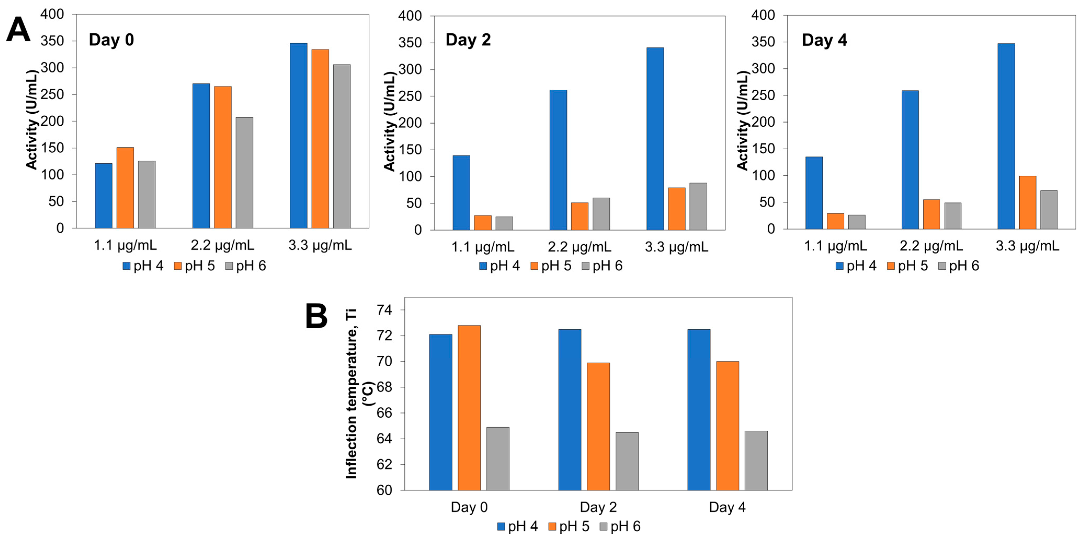
AnGal was purchased (see Section 4.1) as a suspension in 3.2 M ammonium sulphate in which it has high long-term stability. Initial characterization (Figure 1) showed slightly higher activity at pH 5.0 in dilute solution and very similar activities at pH 4.0 and 5.0 in more concentrated solution, while long term stability was impaired at pH 5.0 and pH 6.0. Subsequent experiments were carried out at pH 5.0 by taking fresh solution from the ammonium sulphate stock. Thermal stability was assessed using the inflection temperature, defined as the peak of the first derivative of the thermal unfolding curve. At Day 0, the enzyme exhibited slightly higher stability at pH 5.0, whereas a marked reduction in stability was observed at pH 6.0. By Day 2, a progressive overall decrease in thermal stability became evident, with the highest stability detected at pH 4.0 and the lowest at pH 6.0. A comparable pH-dependent pattern was maintained at Day 4, indicating a continued decline in stability across conditions.

2.2. Reaction with 2-Nitrophenyl-d-galactopyranoside (2-NPG) in Aqueous Solutions Support a Combination of Hydrolysis and Transglycosylation by AnGal

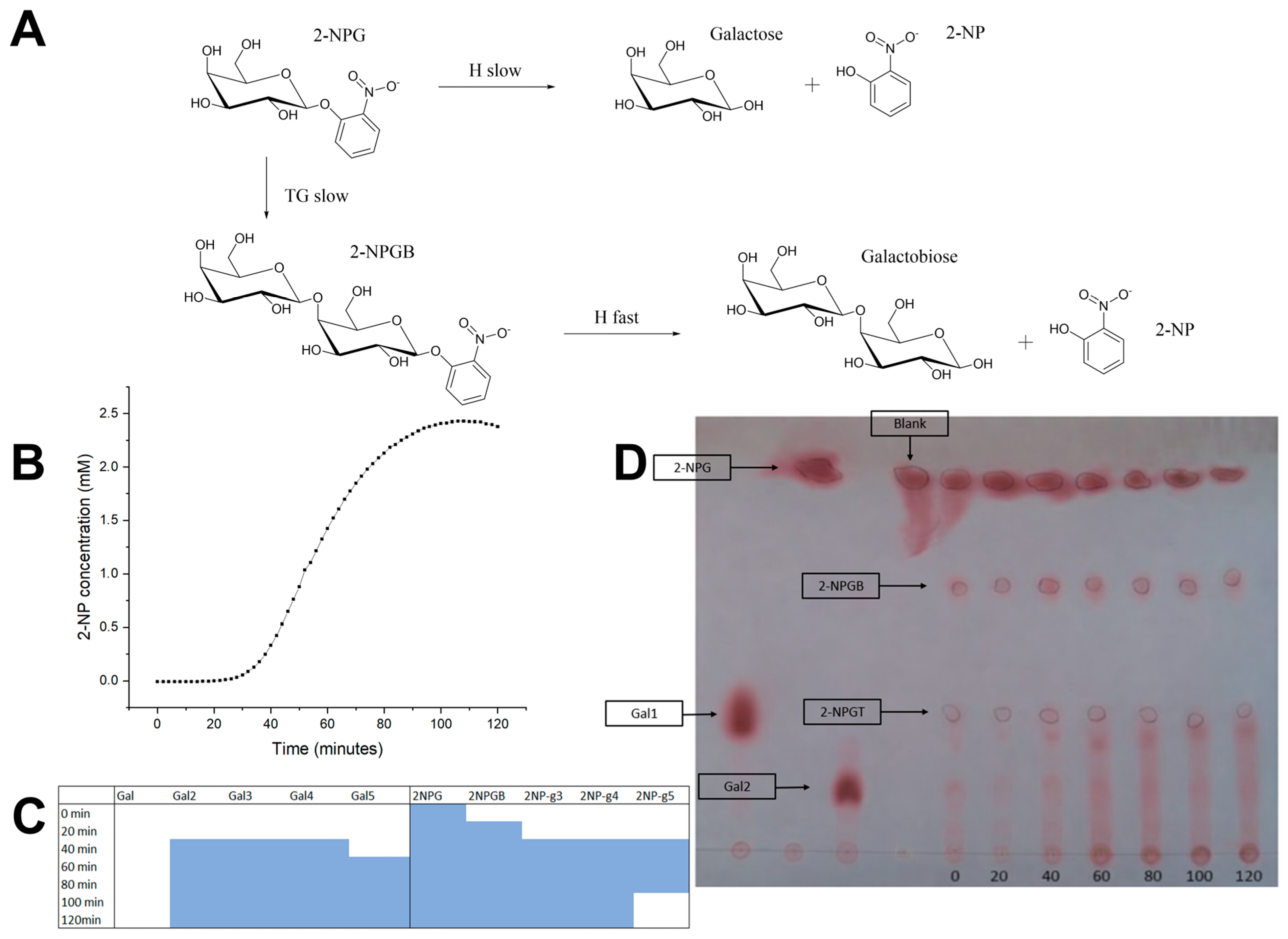
Initial hydrolysis reactions at pH 50 with high-concentration (16 mM) 2-NPG as substrate monitored spectrophotometrically at 400 nm, show the same characteristic profiles shown previously for other fungal galactanases [28], i.e., an initial lag phase where little hydrolysis product is observed, followed by a rapid increase in A400 and finally a decrease. We previously interpreted these curves as shown in Figure 2A, where hydrolytic activity on 2-NPG is minimal, but in the lag phase 2-nitrophenyl-galactobioside (2-NPGB) is slowly formed by transglycosylation, followed by rapid hydrolysis into 2-nitrophenyl (2-NP) and galactobiose (Gal2), as indicated by yellow color formation (Figure 2B). Interestingly, the color disappears after some time, which could indicate that 2-NP is incorporated into products at a later stage, but this was not investigated further.
Thin-Layer Chromatography (TLC) analysis of a repeated reaction supports the formation of UV-active species, one of which we assume to be 2-NPGB, while another was initially unknown. Galactose and galactobiose were identified in addition to possibly longer oligosaccharides towards the origin of the TLC plate, which could arise from various combinations of transglycosylation and hydrolysis (Figure 2C). Matrix-assisted laser desorption/ionization time-of-flight mass spectrometry (MALDI-TOF MS) in a similarly run reaction confirmed the presence of species with matching masses to several 2-NP derivatives and various galactooligosaccharides (Figure 2D and Supplementary Figure S2). Longer products than galactopentaose (gal5) and 2-NP-gal5 could not be detected. This may be because products are hydrolyzed before longer products can be formed or due to limitations of the technique. Note that for TLC analysis, the initial first sample, though drawn on addition of enzyme, is not a true time 0, since the reaction was not stopped and the enzyme can be expected to act for several minutes during drying on TLC. There were indications in the MALDI-TOF MS reactions that addition of matrix could quickly inactivate the enzyme. Generally, boiling and strong acid/bases are often used to stop enzyme reactions; in the current investigation, this type of quenching was not performed except during AnGal incubation in the presence of benzyl alcohol, to prevent undesired modification of products.

2.3. Alcoholysis Reactions with 2-NPG Result in Formation of Alkyl-Adducts

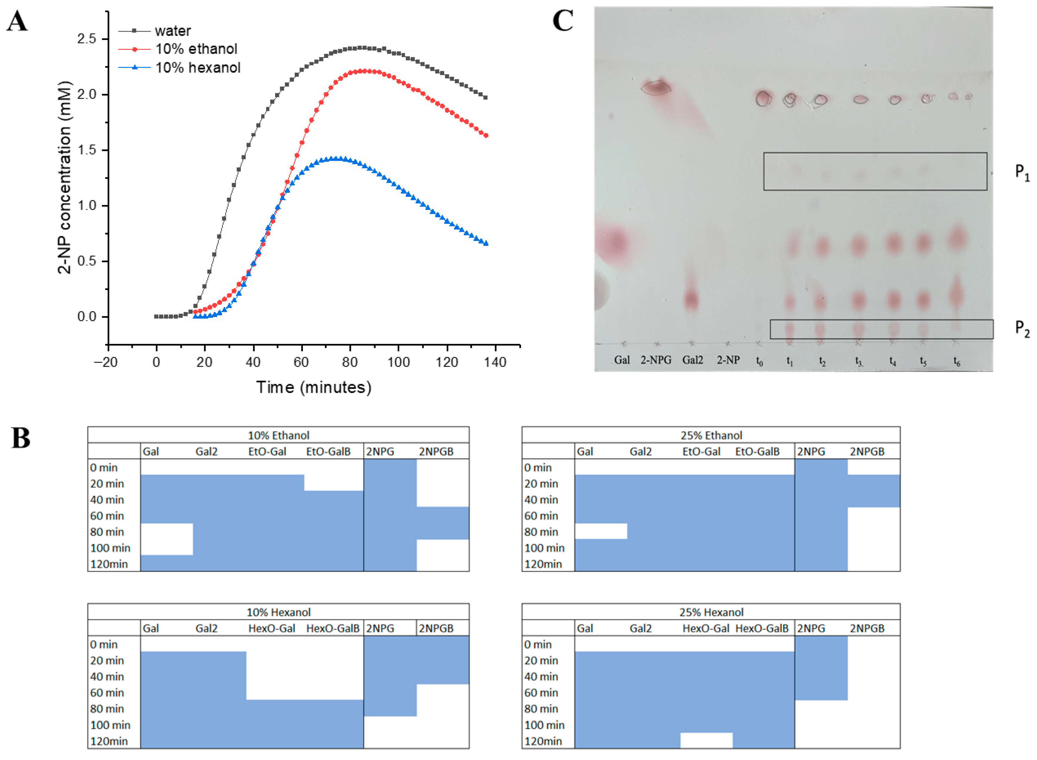
Transglycosylation can be considered an alcoholysis reaction, since the alcohol group of a carbohydrate carries out the nucleophilic attack on the covalent glycosyl-enzyme intermediate instead of water. Since AnGal can transglycosylate, we were curious to see if it could carry out other alcoholysis reactions to form alkyl-galactosides and alkyl-galactooligosaccharides. Initial attempts in 50% ethanol showed that AnGal was inactivated under these conditions. However, it was possible to carry out reactions with 2-NPG in 10–25% v/v primary alcohols (ethanol and 1-hexanol). The reaction was followed spectrophotometrically, showing a profile similar as in water, though with different kinetics as shown, for example, for 10% v/v alcohols vs. aqueous buffer alone (Figure 3A). TLC analysis of reactions in 10% v/v alcohol showed also very similar profiles as in water, but no additional product likely corresponding to alkyl-derivatives could be visualized. However, ethyl and hexyl adducts of galactose and galactobiose could clearly be observed by MALDI-TOF MS (Figure 3B and Supplementary Figure S3). Furthermore, TLC analysis of reactions carried out in the presence of 25% v/v 1-hexanol clearly show formation of an additional product which likely represents a hexyl adduct (Figure 3C).

2.4. Alcoholysis Reactions with Lupin Galactan Show Formation of Alkyl Glycosides

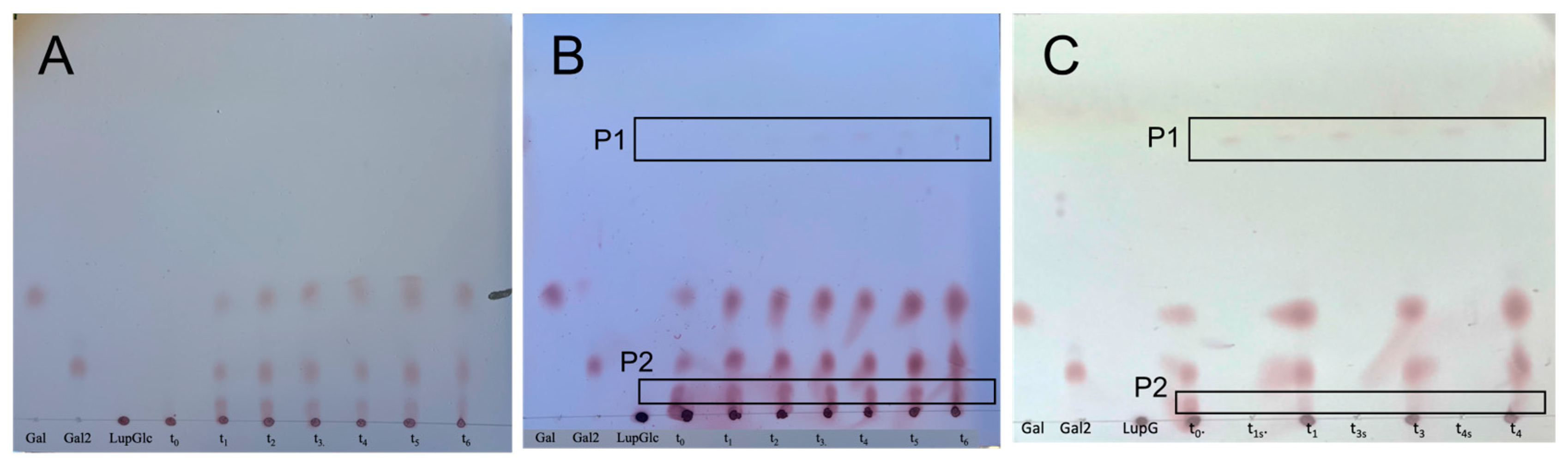
Given the formation of hexyl adducts using 2-NPG as substrate, we wondered if alkyl glycosides could be formed directly from galactan by AnGal. Figure 4A shows a hydrolysis reaction of lupin galactan by AnGal, compared to a parallel reaction in the presence of 25% v/v 1-hexanol (Figure 4B). In the presence of hexanol, two phases formed with time, and a longer reaction where the phases were sampled individually showed that the new product, presumed to be hexyl-galactobiose adduct (P1), accumulated in the organic phase (hexanol), whereas the galactotriose product (P2) was detected in the aqueous phase (Figure 4C).

2.5. Aromatic Alcohols Can Also Be Used as Acceptors by AnGal

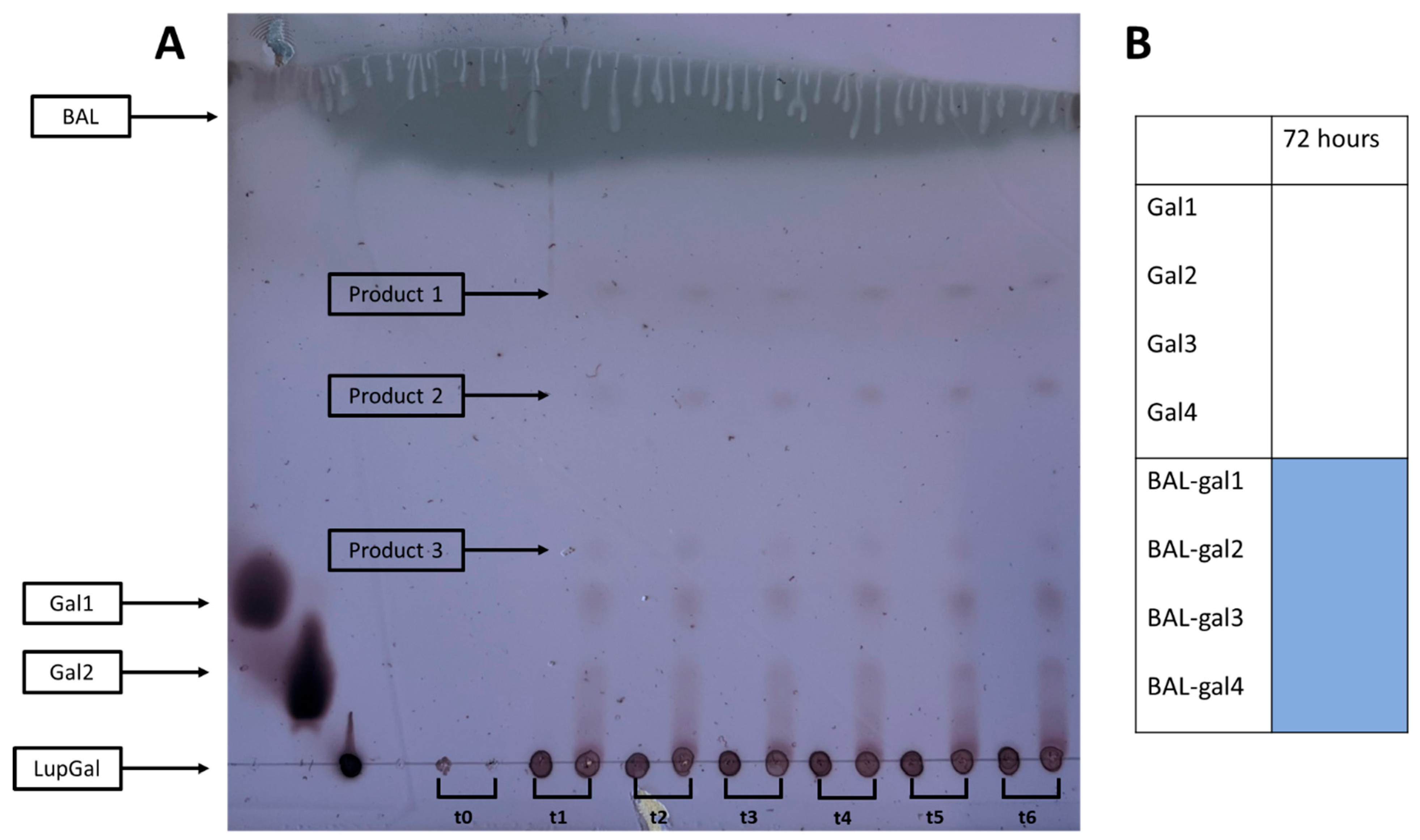
Next, we wanted to see if an alcohol containing an aromatic group—benzyl alcohol (BAL)—could also be used as an acceptor. As shown in the TLC in Figure 5A, BAL at 25% v/v could function as an acceptor with lupin galactan as a donor (Figure 5A), giving rise to three new products. Note that the reaction was inactivated by boiling and stored by freezing before analysis by TLC and MALDI-TOF MS. The presence of BAL derivatives was confirmed by MALDI-TOF MS (Figure 5B and Supplementary Figure S4). In parallel, high-performance liquid chromatography (HPLC) analysis revealed the appearance of six additional peaks for UV-absorbing compounds, when the reaction was carried out in the presence of benzyl alcohol. No corresponding peaks were detected in the hydrolysis (control) reaction without benzyl alcohol. (Supplementary Figure S5). These newly observed peaks are consistent with the formation of BAL derivatives of galactose and its oligosaccharides, although further structural confirmation of each peak is required. At the same time, a fourth product was detected by MALDI-TOF MS, but not detectable by TLC. No hydrolysis products were detectable by MALDI-TOF MS. The conditions and settings at the time of taking this measurement may have interfered with the detection of these products. The absence of a signal in a MALDI-TOF spectrum is not proof that a product has not been formed, as hydrolysis products are seen in the TLC, thus the results have to be analyzed holistically.
Interestingly, no reaction occurred with 2-NPG as donor in the presence of BAL, as the enzyme was inactivated under these reaction conditions.

3. Discussion

AnGal has been characterized and its ability to catalyze hydrolysis, transglycosylation and alcoholysis has been investigated by spectrophotometric assays, TLC, MALDI-TOF MS and HPLC. The activity of fungal galactanases on 2-NPG has previously been investigated [28], where an initial lag phase was found followed by rapid formation of 2-NP and then a decrease at the end of the reaction. It was hypothesized that 2-NPGB is slowly formed in a slow lag phase, after which this is rapidly hydrolyzed, releasing 2-NP and galactobiose; however, the presence of 2-NPGB was not verified at that time.
In this study, AnGal was found to start rapidly releasing 2-NP after 30 min from 2-NPG, which was further supported by MALDI-TOF MS results under similar conditions, where 2-NPGB was detected after 20 min. After 40 min, 2-NPGB—as well as not previously detected species 2-NP-gal3, 2-NP-gal4 and 2-NP-gal5—were detected by MALDI-TOF MS. These products may be better substrates for AnGal as illustrated by the appearance of hydrolyzed products gal2, gal3 and gal4 at the same time as the longer 2-NP derivatives followed by gal5 after 60 min. The reaction was also followed by TLC, which showed formation of 2-NPGB and galactobiose already at the 0 time sample, which may be due to the reaction taking longer to stop while drying on the TLC plate. In contrast, the sample was mixed with matrix before analysis by MALDI-TOF MS, which may rapidly inactivate the enzyme.
This is, to our knowledge, the first time that the formation of 2-NP-gal3, 2-NP-gal4 and 2-NP-gal5 from 2-NPG by a GH53 enzyme has been reported. These results support a mechanism in which the enzyme slowly forms 2-NPGB and longer derivatives, which can participate in further transglycosylation and hydrolysis reactions, resulting in the non-linear rate of formation of 2-NP (with respect to time) as seen in the spectrophotometric assays. The reason for the decrease in absorbance at the end of the reaction has not been confirmed and may be investigated further in the future to give a full picture of the details of the reaction mechanism.
When ethanol or hexanol was added at appropriate concentrations to the reaction with 2-NPG, the spectrophotometric assay yielded a graph similar to the hydrolysis reaction, suggesting that alcoholysis and/or transglycosylation takes place. The formation of EtO-Gal, EtO-GalB, HexO-Gal and HexO-GalB was confirmed by MALDI-TOF MS and TLC, illustrating that AnGal can catalyze alcoholysis in the presence of acceptors with varying degrees of polarity such as ethanol and hexanol.
The native substrate of AnGal is galactan, which represents a promising feedstock for alcoholysis because it can be readily obtained from waste biomass streams, such as spent sulfite liquor from the paper industry, wheat bran from cereal processing and sugar beet pulp from sugar production [23,24,25].
Three products were detected by TLC in a reaction between lupin galactan and BAL in the presence of AnGal. Furthermore, products determined to be BAL-gal1 to BAL-gal4 were detected by MALDI-TOF MS. AnGal could be utilized in the synthesis of longer derivative in alcoholysis with BAL. A P. citrinum galactanase has previously been utilized in the synthesis of galactosides and galactobiosides from various benzyl alcohols and phenols in the presence of soybean arabinogalactan. This galactanase showed a wide specificity compared to galactosidases, but the authors did not detect longer derivatives than the galactobiose derivatives [14]. Similarly, an exo-galactanase (from B. subtilis) has been utilized for galactoside and galactobioside synthesis in the presence of soybean arabinogalactan using glycerol as acceptor at various concentrations [29]. The ability of AnGal to synthesize longer derivatives may hint at a potential to utilize GH53 enzymes in the synthesis of promising galactosides and longer galactooligosaccharide derivatives of various alcohols. To the best of our knowledge, we are the first to report on the formation of gal3 and gal4 derivatives of an alcohol through the alcoholysis mode of a GH53 enzyme. Another similar type of reaction where a β-galactosidase was employed yielded benzyl galactosides, exclusively [30]. Galactosidases are commonly utilized in alcoholysis reactions, but whereas investigations of those reactions only yielded galactosides [31,32], galactanases however have more tendency to produce longer derivatives, since unlike the galactosidases, they are not as susceptible to hydrolysis due to substrate-binding subsites that could potentially stabilize the oligosaccharide donors/acceptors, thereby influencing the length of the products of alcoholysis [9,10].
Additionally, although AnGal was fully inactivated when incubated in the presence of 25% v/v BAL using 2-NPG as the substrate, the enzyme remained active under the same solvent conditions when lupin galactan was substrate instead. One possible explanation is that the polymeric nature of lupin galactan may provide a protective effect, stabilizing AnGal and enabling catalysis under conditions that would otherwise lead to deactivation. The substrate-binding subsites on galactanases could help stabilize the enzymes, for instance [9]. This presents an interesting area of research aimed at elucidating how polysaccharides may enhance enzyme stability and catalytic efficiency, particularly under conditions where organic solvents themselves act as acceptor molecules, such as in alcoholysis reactions used to produce promising galactooligosaccharide derivatives.
Furthermore, the sequence identity of AnGal with other fungal GH53 enzymes and its AlphaFold predicted structure suggested that AnGal is very similar to other previously characterized fungal GH53 enzymes. This structural similarity suggests that the alcoholysis and transglycosylation reactions demonstrated in this article may be reproducible using other fungal GH53, thereby providing a broader platform for extending mechanistic and applied research within this enzyme family.

4. Materials and Methods

4.1. Enzyme Source

AnGal (endo-1,4-β-galactanase, EC 3.2.1.89) from Aspergillus niger was purchased from Megazymes (Product number: E-EGALN, now discontinued; specific activity 150 U/mg at pH 4.0 on potato galactan). One unit (U) corresponds to the release of 1 µmol galactose min-1 in 100 mM sodium acetate buffer, pH 4.0 at 40 °C. The measured concentration (see below) was 3.6 mg/mL.

4.2. Biochemical Characterizations

4.2.1. Protein Analysis and Stability

Enzyme concentration was determined using a NanoDrop 1000 Spectrophotometer (Thermo Fisher Scientific, Wilmington, DE, USA). The molar extinction coefficient (ε = 87,445 M−1cm−1) and the molar mass (38,734 Da) were calculated with the ProtParam tool (ExPASy) based on the AnGal sequence (ATCC64974_107070; UniProt A0A3F3RY75). Thermal stability was assessed by nano differential scanning fluorimetry (nDSF) using a Tycho NT.6 instrument (NanoTemper Technologies, Munich, Germany), at enzyme concentration of 0.5 mg/mL in 500 mM sodium acetate buffer (pH 4.0, 5.0 or 6.0), on days 0, 2 and 4. Samples were subjected to a temperature ramp from 35 °C to 95 °C over 3 min while monitoring intrinsic fluorescence (350/330 nm ratio). The inflection temperature (Ti), obtained from the first derivative of the fluorescence ratio, was used as stability metric.

4.2.2. Spectrophotometric Assays

Azo-Galactan Assay
The specific activity of AnGal was determined using a colorimetric technique azo-galactan assay, product ID: S-AGALP (Megazymes, Wicklow, Ireland; discontinued) consists of a dyed azo galactan substrate. Enzymatic hydrolysis was quantified by monitoring the UV absorbance of the released soluble dye-labeled fragments at 590 nm (A590). The substrate contains Remazol Brilliant Blue R covalently bound to the galactan backbone. A modified version of the Megazymes azo-galactan assay protocol was used. Briefly, 100 µL of substrate solution (2% w/v azo-galactan prepared in 200 mM sodium acetate buffer, pH 4.0, 5.0 and 6.0) was mixed with 100 µL of enzyme solution containing AnGal at three concentrations (1.1, 2.2 and 3.3 µg/mL in 100 mM sodium acetate buffer, pH 4.0, 5.0 and 6.0). Reaction mixtures were incubated at 40 °C for 10 min. The reactions were terminated by adding 500 µL of 95% (v/v) ethanol. Samples were centrifuged using Heraeus Megafuge 8R (Thermo Scientific, Bremen, Germany) at 10,000× g for 10 min. The absorbance of resulting supernatants was measured at 590 nm using a microplate spectrophotometer (PowerWave X, BioTek Instruments, Winooski, VT, USA). Enzyme activity was calculated according to Equation (1) provided by the manufacturer.
m i l l i U n i t s a s s a y = 18 + 377 × A 590 41 × A 590 2
1 Unit = amount of enzyme required to release 1 µmol galactose-reducing sugar equivalents from galactan per minute at 100 mM sodium acetate buffer (pH 4.0), 40 °C and 10 mg/mL potato galactan substrate.
2-NP Colorimetric Assay for Activity on 2-NPG
A colorimetric assay was used to quantify AnGal activity by monitoring the release of 2-NP at UV wavelength of 400 nm. A calibration curve of 0.48–2.4 mM 2-NP in 500 mM sodium citrate gave an extinction coefficient of (2.16 ± 0.03) × 103 M−1cm−1 and was found to be very similar in other buffers. In the assay, AnGal hydrolyzes 2-NPG and higher galactooligosaccharide derivatives to yield 2-NP and the corresponding galactose or galactooligosaccharide products. Reactions were conducted in a microplate spectrophotometer) using the following standard conditions: 0.05 mg/mL AnGal, 16 mM 2-NPG, and 100 mM sodium acetate, citrate, or phosphate buffer at pH 5.0 (the specific buffer used is indicated in the corresponding figure legends). For alcoholysis reactions, the assay mixtures were supplemented with 10–25% v/v ethanol, 1-butanol or 1-hexanol. UV absorbance was recorded every 2 min over a total reaction time of 2 h.

4.3. Other Hydrolysis and Alcoholysis Reactions: 2-NPG and Galactan as Substrates

Hydrolysis and alcoholysis reactions were also followed by TLC, MALDI-TOF MS and HPLC, in the case of alcoholysis with addition 10% v/v–25% v/v alcohol (ethanol, 1-hexanol or benzyl alcohol). 2-NPG and lupin galactan substrate, product ID; P-GALLU (Megazymes, Wicklow, Ireland, discontinued) were used as donor compounds. Specific conditions are given in Figure legends. The total reaction duration was performed in the range of 3–72 h. Sampling at time 0 h for the hydrolysis reactions was directly after enzyme was added to the reaction mixture, whereas for alcoholysis reactions, time 0 h was sampled after alcohol was added about 15 min after enzyme was added. Analysis on products performed by TLC and MALDI-TOF MS.

4.4. Analytical Methods

4.4.1. TLC

Analytical TLC was performed using a mobile phase composed of 70% v/v ethyl acetate, 20% v/v methanol and 10% v/v milli-Q water. After development, plates were examined under UV lamp at 254 nm to visualize UV-active species, including 2-NPG and benzyl alcohol. Non-UV active compounds were visualized by staining. For this purpose, plates were immersed in a staining solution containing 0.5% w/v thymol, 10% v/v sulfuric acid and 90% v/v ethanol, followed by heating at 100 °C for 10 min to induce color development.

4.4.2. MALDI-TOF Mass Spectrometry

Reaction species were analyzed using MALDI-TOF mass spectrometer (Autoflex Max, Bruker, Bremen, Germany). The matrix sDHB, consisting of 2,5-dihydroxybenzoic acid and 2-hydroxy-5-methoxybenzoic acid in a 9:1 (w/w) ratio, was prepared at 10–50 mg/mL in 30:70% v/v acetonitrile/water containing 0.1% v/v trifluoroacetic acid. Samples were mixed at 1:1 ratio by combining 1.5 µL hydrolysate with 1.5 µL matrix solution. Subsequently, 1.5 µL of the mixture was spotted onto the MALDI target plate and allowed to dry before analysis. Spectra were measured on Bruker Autoflex Speed MALDI-TOF MS by FlexControl Version 3.4 and analyzed using FlexAnalysis Version 3.4.

4.4.3. HPLC

Analysis was performed using Vanquish Core HPLC system (Thermo Fisher Scientific, Waltham, MA, USA) equipped with Hypersil Gold C18 column (250 × 4.6 mm, 5 µm particle size; Thermo Fisher Scientific, Waltham, MA, USA). Using a 20 µL injection volume, the mobile phase comprised 0–100% acetonitrile gradient containing 0.1% (v/v) trifluoroacetic acid at a flow rate of 1 mL/min. A Vanquish Multiple Wavelength detector (Thermo Fisher Scientific, Waltham, MA, USA) was used as a UV detector.

Supplementary Materials

The following supporting information can be downloaded at: https://www.mdpi.com/article/10.3390/catal16050421/s1, Figure S1: AlphaFold predicted structure of AnGal; Figure S2: MALDI-TOF MS spectrum for the reaction of AnGal in the presence of 2-NPG and water; Figure S3: MALDI-TOF MS spectrum for the reaction of AnGal in the presence of 2-NPG and (A) ethanol and (B) hexanol; Figure S4: MALDI-TOF MS spectrum for the reaction of AnGal in the presence oflupin galactan andbenzyl alcohol (BAL); Figure S5: HPLC chromatogram showing the appearance of additional UV-absorbing peaks; Figure S6: Time-course samples of control reactions analyzed by TLC. Refs. [33,34] are cited in the Supplementary Materials.

Author Contributions

Conceptualization, L.L.L.; methodology, M.Z., T.T.H.L., Y.T., S.J.G. and L.L.L.; validation, M.Z., T.T.H.L., Y.T., S.J.G. and L.L.L.; formal analysis, M.Z., T.T.H.L., Y.T., S.J.G. and L.L.L.; investigation, M.Z., T.T.H.L., Y.T., S.J.G. and L.L.L.; resources, L.L.L.; data curation, M.Z., T.T.H.L., Y.T., S.J.G. and L.L.L.; writing—original draft preparation, M.Z., T.T.H.L., S.J.G. and L.L.L.; writing—review and editing, M.Z., T.T.H.L., S.J.G. and L.L.L.; visualization, M.Z., T.T.H.L., S.J.G. and L.L.L.; supervision, S.J.G. and L.L.L.; project administration, L.L.L.; funding acquisition, L.L.L. All authors have read and agreed to the published version of the manuscript.

Funding

This work was funded by the Independent Research Fund Denmark DFF-Research Project 1 (Thematic)—Green Research (UFS) 2102-00266B and Research Project 1-FTP 4286-00360B.

Data Availability Statement

The data generated and analyzed in this scientific investigation are available from the corresponding author upon reasonable request.

Acknowledgments

The authors gratefully acknowledge Theis Brock-Nannestad for his valuable guidance with MALDI-TOF MS. The authors used Microsoft 365 CoPilot Version 2.0 for the purposes of enhancing the text during the preparation of this manuscript/study. The authors have reviewed and edited the output and take full responsibility for the content of this publication.

Conflicts of Interest

The authors declare no conflicts of interest.

References

  1. Lombard, V.; Golaconda Ramulu, H.; Drula, E.; Coutinho, P.M.; Henrissat, B. The carbohydrate-active enzymes database (CAZy) in 2013. Nucleic Acids Res. 2014, 42, D490–D495. [Google Scholar] [CrossRef] [Scilit]
  2. Muderspach, S.J.; Jensen, K.; Krogh, K.B.R.M.; Lo Leggio, L. Structure, function, and protein engineering of GH53 β-1,4-galactanases. In Glycoside Hydrolases; Elsevier: Amsterdam, The Netherlands, 2023; pp. 295–322. [Google Scholar] [CrossRef] [Scilit]
  3. Voragen, A.G.J.; Coenen, G.-J.; Verhoef, R.P.; Schols, H.A. Pectin, a versatile polysaccharide present in plant cell walls. Struct. Chem. 2009, 20, 263–275. [Google Scholar] [CrossRef] [Scilit]
  4. Gistelinck, K.; Yu, X.; Leyder, A.; Osterne, V.J.S.; Desmet, T.; Tryfona, T.; Van Damme, E.J.M. Expression and characterization of an endo-β-1,6-galactanase from Arabidopsis thaliana. Biochem J. 2025, 482, 1935–1957. [Google Scholar] [CrossRef] [Scilit] [PubMed]
  5. Biswas, S.; Mandal, A.; Fontes, C.M.G.A.; Goyal, A. A unique highly efficient, thermostable and multi-substrate specific galactanase (AtGH53) from Acetivibrio thermocellus cleaving both β (1,4)and β (1,6)-linked galactans. Enzym. Microb. Technol. 2026, 194, 110801. [Google Scholar] [CrossRef] [Scilit] [PubMed]
  6. Plouhinec, L.; Zhang, L.; Pillon, A.; Haon, M.; Grisel, S.; Navarro, D.; Black, I.; Neugnot, V.; Azadi, P.; Urbanowicz, B.; et al. Unlocking soybean meal pectin recalcitrance using a multi-enzyme cocktail approach. Sci. Rep. 2025, 15, 1716. [Google Scholar] [CrossRef] [Scilit]
  7. Barrera-Chamorro, L.; Fernandez-Prior, A.; Rivero-Pino, F.; Montserrat-de la Paz, S. A comprehensive review on the functionality and biological relevance of pectin and the use in the food industry. Carbohydr. Polym. 2025, 348, 122794. [Google Scholar] [CrossRef] [Scilit]
  8. Smith, D.L.; Abbott, J.A.; Gross, K.C. Down-regulation of tomato β-galactosidase 4 results in decreased fruit softening. Plant Physiol. 2002, 129, 1755–1762. [Google Scholar] [CrossRef] [Scilit]
  9. Ryttersgaard, C.; Le Nours, J.; Lo Leggio, L.; Jorgensen, C.T.; Christensen, L.L.; Bjornvad, M.; Larsen, S. The structure of endo-β-1,4-galactanase from Bacillus licheniformis in complex with two oligosaccharide products. J. Mol. Biol. 2004, 341, 107–117. [Google Scholar] [CrossRef] [Scilit] [PubMed]
  10. Muderspach, S.J.; Fredslund, F.; Volf, V.; Poulsen, J.N.; Blicher, T.H.; Clausen, M.H.; Rasmussen, K.K.; Krogh, K.; Jensen, K.; Lo Leggio, L. Engineering the substrate binding site of the hyperthermostable archaeal endo-β-1,4-galactanase from Ignisphaera aggregans. Biotechnol. Biofuels 2021, 14, 183. [Google Scholar] [CrossRef] [Scilit]
  11. Le Nours, J.; De Maria, L.; Welner, D.; Jorgensen, C.T.; Christensen, L.L.; Borchert, T.V.; Larsen, S.; Lo Leggio, L. Investigating the binding of β-1,4-galactan to Bacillus licheniformis β-1,4-galactanase by crystallography and computational modeling. Proteins 2009, 75, 977–989. [Google Scholar] [CrossRef] [Scilit]
  12. Torpenholt, S.; Poulsen, J.-C.N.; Muderspach, S.J.; De Maria, L.; Lo Leggio, L. Structure of Aspergillus aculeatus β-1,4-galactanase in complex with galactobiose. Acta Cryst. 2019, F75, 399–404. [Google Scholar] [CrossRef] [Scilit]
  13. Fujimoto, H.; Nakano, H.; Isomura, M.; Kitahata, S.; Ajisaka, K. Enzymatic synthesis of oligosaccharides containing Galβ->4Gal-disaccharide at the non-reducing end using β-galactanase from Penicillium citrinum. Biosci. Biotech. Biochem. 1997, 61, 1258–1261. [Google Scholar] [CrossRef] [Scilit] [PubMed]
  14. Nakano, H.; Kitahata, S.; Ohgaki, H.; Takenishi, S. Transglycosylation of phenols by endo-1,4-β-galactanase from Penicillium citrinum and several β-galactosidases. Denpun Kagaku 1992, 39, 1–6. [Google Scholar] [CrossRef] [Scilit]
  15. Iqbal, M.W.; Riaz, T.; Mahmood, S.; Liaqat, H.; Mushtaq, A.; Khan, S.; Amin, S.; Qi, X. Recent advances in the production, analysis, and application of galacto-oligosaccharides. Food Rev. Int. 2022, 39, 5814–5843. [Google Scholar] [CrossRef] [Scilit]
  16. Battisegola, C.; Billi, C.; Molaro, M.C.; Schiano, M.E.; Nieddu, M.; Failla, M.; Marini, E.; Albrizio, S.; Sodano, F.; Rimoli, M.G. Galactose: A versatile vector unveiling the potentials in drug delivery, diagnostics, and theranostics. Pharmaceuticals 2024, 17, 308. [Google Scholar] [CrossRef] [Scilit]
  17. Sodano, F.; Lazzarato, L.; Rolando, B.; Spyrakis, F.; De Caro, C.; Magliocca, S.; Marabello, D.; Chegaev, K.; Gazzano, E.; Riganti, C.; et al. Paracetamol-galactose conjugate: A novel prodrug for an old analgesic drug. Mol. Pharm. 2019, 16, 4181–4189. [Google Scholar] [CrossRef] [Scilit]
  18. Mejia-Otalvaro, F.; Lax, B.M.; Kirtel, O.; Welner, D.H. Sustainable natural product glycosylation: A critical evaluation of biocatalytic and chemical approaches. ChemSusChem 2025, 18, e202501094. [Google Scholar] [CrossRef] [Scilit]
  19. Botvynko, A.; Synytsya, A.; Curda, L. Synthesis of galactooligosaccharides with four β-galactosidases: Structural comparison of the products by HPLC, ESI-MS and NMR. Biochem Biophys. Res. Commun. 2025, 744, 151204. [Google Scholar] [CrossRef] [Scilit]
  20. Zeng, M.; Mohapatra, S.; Oh, J.-H.; Astmann, T.; van Pijkeren, J.-P.; Pan, X. Novel galacto-oligosaccharides from lactose: Chemical synthesis, structural characterization, and in vitro assessment of prebiotic activity. ACS Sustain. Chem. Eng. 2023, 11, 14031–14045. [Google Scholar] [CrossRef] [Scilit]
  21. Wang, K.; Xu, Y.; Xuan, Z.; Xiao, X.; Gu, G.; Lu, L. Enzymatic synthesis of prebiotic galactooligosaccharides from galactose derived from gum arabic. Food Chem. 2023, 429, 136987. [Google Scholar] [CrossRef] [Scilit] [PubMed]
  22. Dumortier, V.; Montreuil, J.; Bouquelett, S. Primary structure of ten galactosides formed by transglycosylation during lactose hydrolysis by Bifidobacterium bifidum. Carbohydr. Res. 1990, 201, 115–123. [Google Scholar] [CrossRef] [Scilit]
  23. Rueda, C.; Calvo, P.A.; Moncalián, G.; Ruiz, G.; Coz, A. Biorefinery options to valorize the spent liquor from sulfite pulping. J. Chem. Technol. Biotechnol. 2014, 90, 2218–2226. [Google Scholar] [CrossRef] [Scilit]
  24. Glaser, S.J.; Al-Rudainy, B.; Hatti-Kaul, R.; Galbe, M. Wheat bran fractionation: Effect of steam explosion and hydrotropic extraction conditions on the recovery of sugars and lignin. Ind. Crops Prod. 2023, 195, 116405. [Google Scholar] [CrossRef] [Scilit]
  25. Glaser, S.J.; Abdelaziz, O.Y.; Demoitié, C.; Galbe, M.; Pyo, S.-H.; Jensen, J.P.; Hatti-Kaul, R. Fractionation of sugar beet pulp polysaccharides into component sugars and pre-feasibility analysis for further valorisation. Biomass Convers. Biorefinery 2022, 14, 3575–3588. [Google Scholar] [CrossRef] [Scilit]
  26. Fleming, J.; Magana, P.; Nair, S.; Tsenkov, M.; Bertoni, D.; Pidruchna, I.; Lima Afonso, M.Q.; Midlik, A.; Paramval, U.; Zidek, A.; et al. AlphaFold Protein Structure Database and 3D-Beacons: New Data and Capabilities. J. Mol. Biol. 2025, 437, 168967. [Google Scholar] [CrossRef] [Scilit]
  27. Jumper, J.; Evans, R.; Pritzel, A.; Green, T.; Figurnov, M.; Ronneberger, O.; Tunyasuvunakool, K.; Bates, R.; Zidek, A.; Potapenko, A.; et al. Highly accurate protein structure prediction with AlphaFold. Nature 2021, 596, 583–589. [Google Scholar] [CrossRef] [Scilit]
  28. Torpenholt, S.; Le Nours, J.; Christensen, U.; Jahn, M.; Withers, S.; Ostergaard, P.R.; Borchert, T.V.; Poulsen, J.C.; Lo Leggio, L. Activity of three β-1,4-galactanases on small chromogenic substrates. Carbohydr. Res. 2011, 346, 2028–2033. [Google Scholar] [CrossRef] [Scilit]
  29. Nakano, H.; Kitahata, S.; Fujimoto, H.; Kinugasa, H.; Watanabe, Y.; Ajisaka, K.; Takenishi, S. Transfer reaction catalyzed by exo-β-1,4-galactanase from Bacillus subtilis. Agric. Biol. Chem. 1991, 55, 2075–2082. [Google Scholar] [CrossRef] [Scilit] [PubMed]
  30. Porciuncula Gonzalez, C.; Cagnoni, A.J.; Marino, K.V.; Fontana, C.; Saenz-Mendez, P.; Irazoqui, G.; Giacomini, C. Enzymatic synthesis of non-natural trisaccharides and galactosides; Insights of their interaction with galectins as a function of their structure. Carbohydr. Res. 2019, 472, 1–15. [Google Scholar] [CrossRef] [Scilit] [PubMed]
  31. Garcia-Garibay, M.; Lopez-Munguia, A.; Barzana, E. Effect of β-galactosidase hydration on alcoholysis reaction in organic one-phase liquid systems. Biotechnol. Bioeng. 2000, 70, 647–653. [Google Scholar] [CrossRef] [Scilit]
  32. Scheckermann, C.; Wagner, F.; Fischer, L. Galactosylation of antibiotics using the β-galactosidase from Aspergillus oryzae. Enzym. Microb. Technol. 1997, 20, 629–634. [Google Scholar] [CrossRef] [Scilit]
  33. SignalP-5.0. Available online: https://services.healthtech.dtu.dk/services/SignalP-5.0/ (accessed on 18 March 2026).
  34. Schrödinger, L.D.W. PyMOL. Available online: http://www.pymol.org/pymol (accessed on 18 March 2026).
Figure 1. pH-dependent activity and stability: (A) Galactanase activity (U/mL) at enzyme concentrations of 1.1, 2.2 and 3.3 µg/mL at pH values 4.0, 5.0 and 6.0 on Days 0, 2 and 4. (B) Thermal stability expressed as inflection temperature (Ti) determined by nano differential scanning fluorimetry (nDSF).
Catalysts 16 00421 g001
Figure 2. Products of AnGal activity on 2-NPG in aqueous buffer. (A) Proposed reaction scheme, where 2-NPGB slowly accumulates from transglycosylation activity and then gets rapidly hydrolyzed. (B) Reaction as monitored by A400 in a 96-well plate reader. The reaction was carried out at room temperature in a volume of 100 µL in 100 mM citrate buffer pH 5.0, with 0.05 mg/mL AnGal and 16 mM 2-NPG. (C) Reaction as monitored by TLC. The reaction was carried out at room temperature in a volume of 200 µL in 100 mM citrate buffer pH 5.0, with 0.05 mg/mL AnGal and 16 mM 2-NPG. 2-NPGT in the figure refers to 2-NP-gal3, with 10 nmol galactose and β-1,4-galactobiose used as standard and 2 × 2 µL applied at each position with drying in between. Samples from the enzyme reaction were taken at 0–120 min, with the Blank representing the reaction mixture prior to enzyme addition. UV-active spots were penciled before thymol staining. (D) Reaction as monitored by MALDI-TOF MS (shading indicates presence of a species, see Supplementary Figure S2). The reaction was carried out at room temperature without shaking in a volume of 100 µL in 100 mM phosphate buffer pH 5.0, with 0.05 mg/mL AnGal and 16 mM 2-NPG. AnGal was added last, after which the time 0 sample was taken. Next, 1.5 µL of 20 mg/mL matrix was mixed with 1.5 µL of sample and from this mix 1.5 µL was added to the target plate and allowed to dry before measuring. Note that as the reaction was not quenched, the enzyme likely remained partly active during drying on TLC plate thus time 0 in (C,D) does not correspond to time 0 in A. However, for the MALDI-TOF MS, the sample matrix seemed to have inactivated the enzyme since no products were observed at time 0.
Catalysts 16 00421 g002
Figure 3. Products of AnGal activity on 2-NPG in aqueous buffers in the presence of primary aliphatic alcohols. (A) Reaction as monitored by A400 in a 96-well plate reader. The reaction was carried out at room temperature in a volume of 100 µL in 100 mM citrate buffer pH 5.0, with 0.05 mg/mL AnGal and 16 mM 2-NPG; 15 min after addition of enzyme, 10% v/v ethanol or 1-hexanol were added to the enzyme reaction. (B) Reactions as monitored by MALDI-TOF MS. The reactions were carried out at room temperature in a volume of 100 μL in 100 mM citrate buffer pH 5.0, with 0.05 mg/mL AnGal and 16 mM 2-NPG; 20 min after addition of enzyme, 10% or 25% v/v ethanol or 1-hexanol were added to the enzyme reaction, and 1.5 µL of 50 mg/mL matrix was mixed with 1.5 µL of sample and from this mix 1.5 µL was added to the target plate and allowed to dry before measuring. Shading indicates the presence of a species, see Supplementary Figure S3. (C) Reaction as monitored by TLC. The reaction was carried out at room temperature in a volume of 100 µL in 100 mM phosphate buffer pH 5.0, with 0.05 mg/mL AnGal and 16 mM 2-NPG and 25% v/v 1-hexanol. 10 nmol galactose (Gal), 2-NPG and β-1,4-galactobiose were used as standards, and 2 × 1 µL were applied at each position with drying in between. Hexanol was added to the reaction mixture at t0, 15 min after addition of enzyme to the buffered 2-NPG solution. Thereafter, samples from the enzyme reaction were taken every 20 min (t1–t6). UV-active spots were penciled prior to thymol staining. P1 is presumed to be a hexyl-galactobiose adduct, while P2 is likely to galactotriose. Note that as the reaction was not quenched, the enzyme likely remained partly active during drying on TLC plate thus time 0 in (B,C) does not correspond to time 0 in (A). Unlike the TLC, MALDI-TOF MS spectra showed no products at time 0 indicating that the sample matrix had quenched the reaction when time 0 sample was taken and analyzed.
Catalysts 16 00421 g003
Figure 4. Products of AnGal (0.05 mg/mL) acting on lupin galactan (1.16 mg/mL) in hydrolysis (A) or alcoholysis (B,C) reactions in 50 mM phosphate buffer pH 5.0 at room temperature in a volume of 100 µL without shaking. (A) The hydrolysis reaction was sampled (2 × 1 µL) at time of addition of enzyme (t0) and then after 30, 60, 90, 120, 150, and 180 min (t1–t6). In the presence of 25% v/v 1-hexanol (B,C), two phases formed. For (B), the reaction was sampled after mixing of the resulting phases, at the time of addition of 25% hexanol (t0, 15 min into hydrolysis) and then after 30, 60, 90, 120, 150, and 180 min (t1–t6). In (C), a sample was taken on addition of 25% hexanol (t0, 15 min into hydrolysis) and then from each phase (s denotes the organic phase) after 24 (t1), 48 (t3), 70 h (t4). 2 × 1 µL were applied at each position with drying in between. P1 is presumed to be the hexyl-galactobiose adduct, while P2 is likely to galactotriose. Standards were galactose (Gal), β-1,4-galactobiose at 10 nmol each. LupG denotes the reaction mixture prior to enzyme (and alcohol) addition. Note that as the reaction was not quenched, the enzyme likely remained partly active during drying on TLC plate.
Catalysts 16 00421 g004
Figure 5. Products of AnGal in the presence of BAL and lupin galactan. It was carried out in a 300 µL volume of 0.05 mg/mL AnGal, 27.5 mM phosphate buffer pH 5.0, 1.6 mg/mL lupin galactan and 25% v/v BAL. The time 0 (t0) was taken right before addition of AnGal, which was the last reactant added. Subsequently, samples were taken at 1, 2, 3, 6, 24 and 72 h (t1–t6). Samples were boiled to inactivate the enzyme and frozen for storage. (A) Reaction as monitored by TLC. 2 × 2 µL was spotted for each sample with drying in between. The samples for the reaction are shown on the right for each time point alongside a control without enzyme on the left. (B) Detection of products after 72 h as monitored by MALDI-TOF MS. 1 µL of 10 mg/mL matrix was added directly to the MALDI-TOF MS target plate followed by addition of 1 µL sample on top of the matrix drop. Shading indicates the presence of species, see Supplementary Figure S4.
Catalysts 16 00421 g005
Disclaimer/Publisher’s Note: The statements, opinions and data contained in all publications are solely those of the individual author(s) and contributor(s) and not of MDPI and/or the editor(s). MDPI and/or the editor(s) disclaim responsibility for any injury to people or property resulting from any ideas, methods, instructions or products referred to in the content.