


Abstract
This study reports the sustainable synthesis and thermal, morphological, and structural characterization of multifunctional silver/hydroxyapatite nanocomposite prepared from recycled caprine bone. The organic extract from caprine bone was characterized using Fourier Transform Infrared (FTIR) and Ultraviolet–Visible Spectroscopy (UV-Vis). The biogenic hydroxyapatite (CHAP) and its silver composite (Ag@CHAP) were characterized using thermal gravimetric analysis (TGA), Raman spectra, X-ray diffraction (XRD), scanning electron microscopy (SEM), energy-dispersive X-ray (EDX), and transmission electron microscope (TEM). The photocatalytic activity of Ag@CHAP was quantitatively confirmed through the degradation of Crystal Violet (5 ppm) under sunlight, achieving a high removal efficiency of 99.8% under optimum conditions, demonstrating significant potential for wastewater remediation. Ag@CHAP also demonstrated enhanced antimicrobial activity compared with CHAP and showed broad-spectrum efficacy against clinical human isolates P. aeruginosa ATCC 10145, E. coli ATCC 35218, S. aureus ATCC 25923, and C. albicans (human isolate). The in vitro hemolytic-activity assays revealed that both CHAP and Ag@CHAP had no hemolytic activity after 24 h of red blood cells incubation and effectively reduced lead-induced hemolysis from 86.73% to 39.35% and 49.13%, respectively. These findings confirm CHAP and Ag@CHAP as stable, biocompatible, and high-performance materials with promising applications in the sustainable water-treatment and biomedical fields.
1. Introduction
Food waste has become a worldwide problem due to the huge quantities produced, which has resulted in environmental pollution [1]. It is highly encouraged that food waste be recycled into ecofriendly products in view of the increasing pressure on ecosystems’ risks to human health. The slaughter industry generates about 130 tons of animal-bone waste annually [2]. As a result, there has been an increase attention on sustainability, innovative strategies to use such materials, and approaches to reduce not only waste but also the negative impact on the environment. One of these promising approaches is using food waste as a precursor for the green synthesis of nanoparticles [3]. The green synthesis method of nanometals and nanometal-oxides via food waste is sustainable, economic, safe, and ecofriendly because it does not consume hazardous chemicals [4].
Biogenic hydroxyapatite Ca10(PO4)6(OH)2, which is synthesized from animal-bone waste through physical or chemical treatment, is a well-known bioceramic, thus providing a low-cost and ecofriendly alternative to synthetic routes [5]. The bone-derived hydroxyapatite retains its natural calcium-to-phosphate ratio close to that of human bone, which makes it suitable for biocompatibility and bioactivity [6]. Therefore, it is deemed an ecofriendly component that is highly suitable for environmental and biomedical applications [7]. In addition, biogenic hydroxyapatite from bone waste contributes toward waste valorization and reduces environmental pollution [8]. Compared with the chemically synthesized hydroxyapatite, the biogenic hydroxyapatite produced from bone waste is more sustainable and uses environmentally friendly alternatives to hazardous chemicals [9].
Organic dyes in excessive amounts may pose an environmental hazard from the textile industry [10]. These dyes can be dangerous and cause harm to human health, which mandates their removal from wastewater [11]. One of these organic dyes is Crystal Violet dye, which is also known as gentian violet, methylrosaniline chloride, and methyl violet, is widely used in the textile, microbiology laboratory, pharmaceutical, and biomedical industries [12,13]. Crystal Violet is considered to be a hazardous dye due to its poisonous, genotoxic, and carcinogenic effects [14]. Several techniques are used for Crystal Violet removal from wastewater such as adsorption [15], photocatalysis [16], Fenton-like oxidation [17], and coagulation–flocculation methods [18].
Human pathogenic bacteria and fungi are clinically significant microorganisms that naturally inhabit and adapt within the human body [19]. Due to their continuous exposure to the body’s immune defense mechanisms and antimicrobial agents, they often tend to develop enhanced virulence and antibiotic resistance compared to other microbes found in water, soil, and air. Therefore, testing any new chemical compounds against human pathogenic bacteria provides a clearer and more realistic assessment of their potential medicinal value [20].
Nanoparticles have also attracted increasing attention due to their ability to protect red blood cells from oxidation destruction, a property known as antihemolytic activity [21,22]. The reduction in oxidative stress in stabilizing the fragile membranes of red blood cells can prevent them from breaking down under adverse conditions [23]. This protective effect highlights their promise in biomedical applications that require close interaction with blood products and in drug delivery systems [24].
Because of the high surface energy and localized surface plasmon resonance, incorporating Ag NPs into composite materials is an effective method to enhance their features. Ag NPs have great potential to increase the base material’s photocatalytic activity as an electron capture, stop electron recombination, and have excellent biological qualities when used as a suitable additional component [25]. The combination of hydroxyapatite and Ag NPs may create an ideal nanocomposite for use in environmental treatment and medical applications, making it an ideal candidate for multifunctional therapeutic and remedial uses [26].
In this study, both uncoated and silver-coated caprine bone-derived hydroxyapatite were investigated for their efficiency in removing Crystal Violet from wastewater, their antimicrobial activity against medically relevant human pathogenic microbes, and their antihemolytic performance on red blood cells.
2. Results and Discussions
2.1. Analysis of Caprine Bone Extract
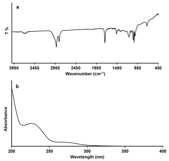
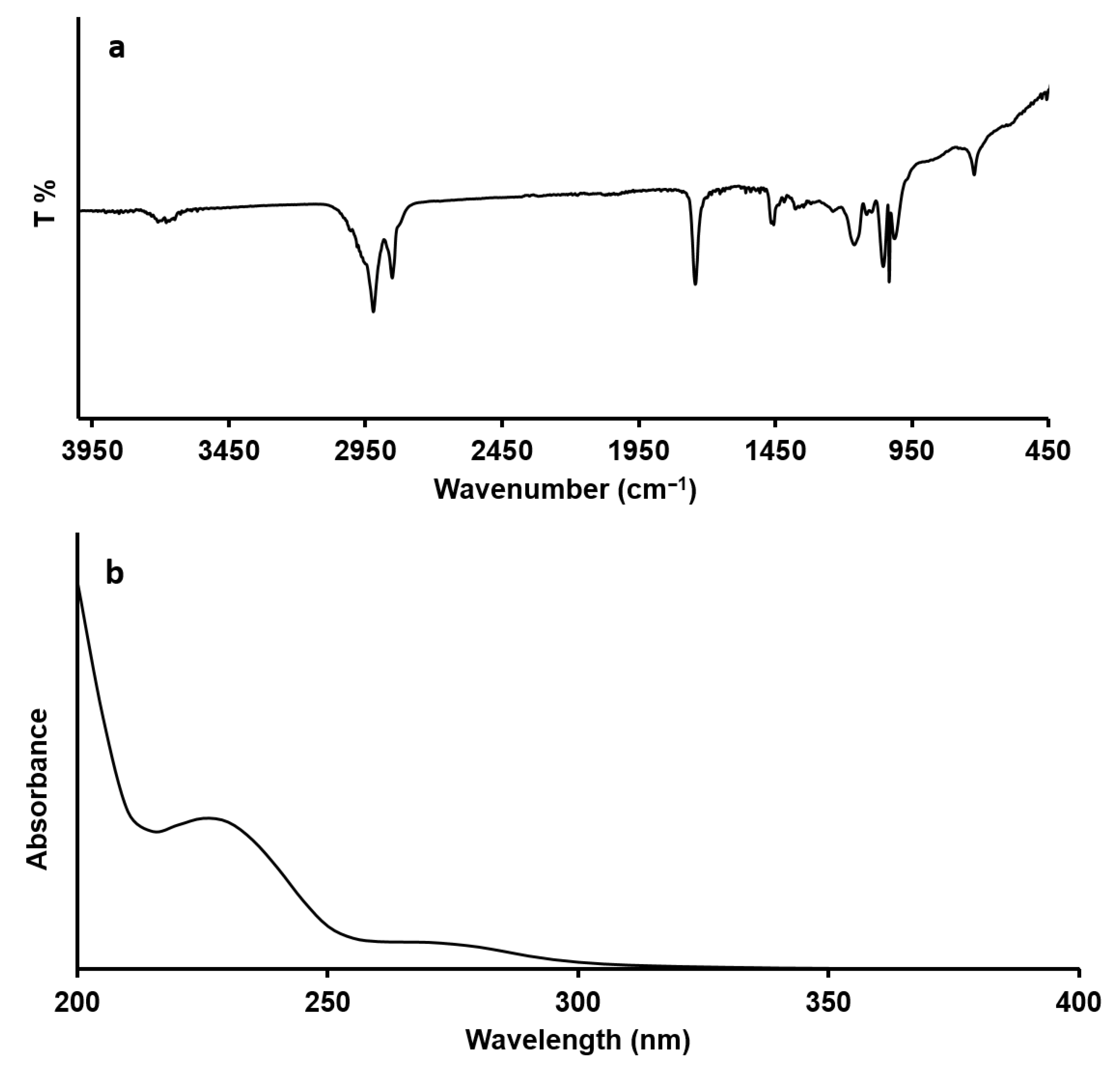
The FTIR and UV-Vis spectra of caprine bone extract confirm the presence of aliphatic organic compounds originating from the caprine bone during extraction (Figure 1). The weak bands in the FTIR spectrum around 3600–3700 cm−1 are attributed to ν OH groups commonly found in collagen and lipids [27]. Bands near 2800 and 2900 cm−1 indicate the presence of aliphatic C–H bonds in aliphatic amino-acid units and lipid molecules [28]. The bands around 1620 and 1350 cm−1 correspond to ν C=O and ν C-N, respectively, highlighting the contribution of collagen-derived peptides [29]. Additional bands between 1100 and 900 cm−1 match well with ν C–O and bending C–H vibrations from amino acid-based molecules [30].
Figure 1.
FTIR (a) and electronic (b) spectra of caprine bone extract.
The UV–Vis spectrum exhibits strong absorption around 230–240 nm, typically associated with n → π* transitions in carbonyl, carboxylic, and peptide bonds, as well as unsaturated functional groups in collagen-derived fragments. A small shoulder at 265–285 nm is related to tryptophan and tyrosine in the amide groups of proteins [31]. The absence of absorption above 300 nm indicates a lack of conjugated organic chromophores.
Overall, these spectral observations suggest that the extract primarily contains aliphatic organic molecules, including collagen fragments, protein-related compounds, and lipids that are naturally released from the bone matrix [32].
2.2. Characterization of CHAP and Ag@CHAP
2.2.1. TGA
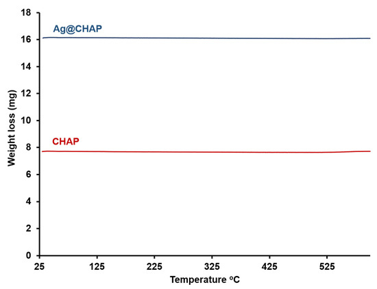
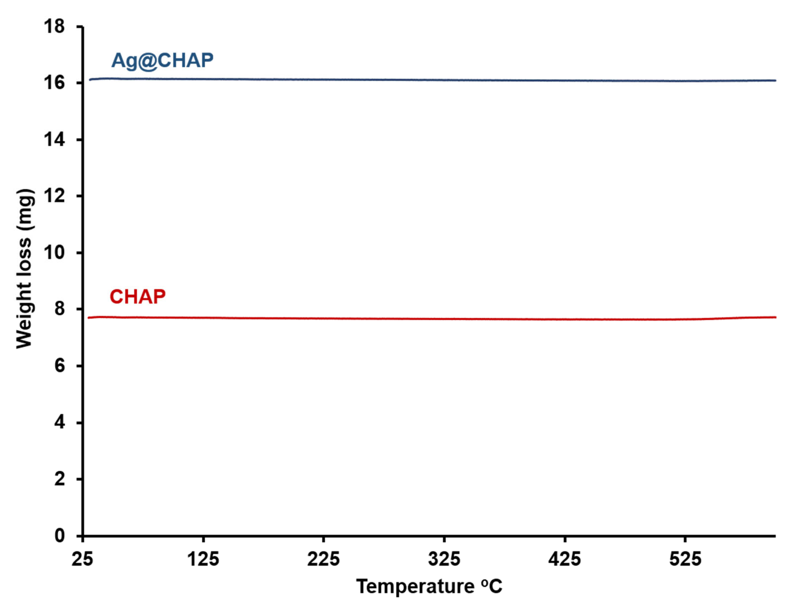
The TGA curves for CHAP and Ag@CHAP (Figure 2) display a nearly flat-line profile, with no distinct stage of thermal decomposition within the 25–600 °C range. This demonstrates high purity and excellent thermal stability for both of the compounds across room and elevated temperature ranges [33].
Figure 2.
TGA of CHAP and Ag@CHAP.
2.2.2. XRD
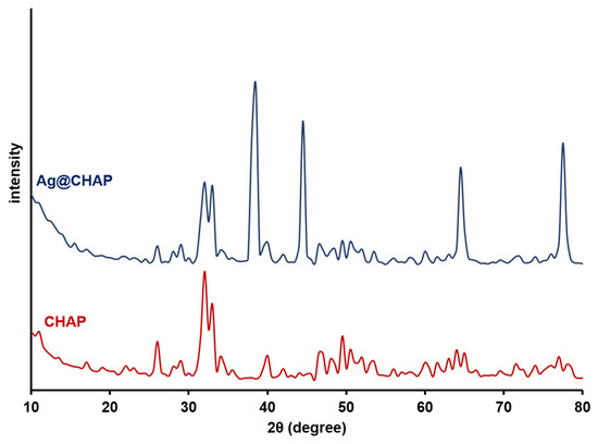
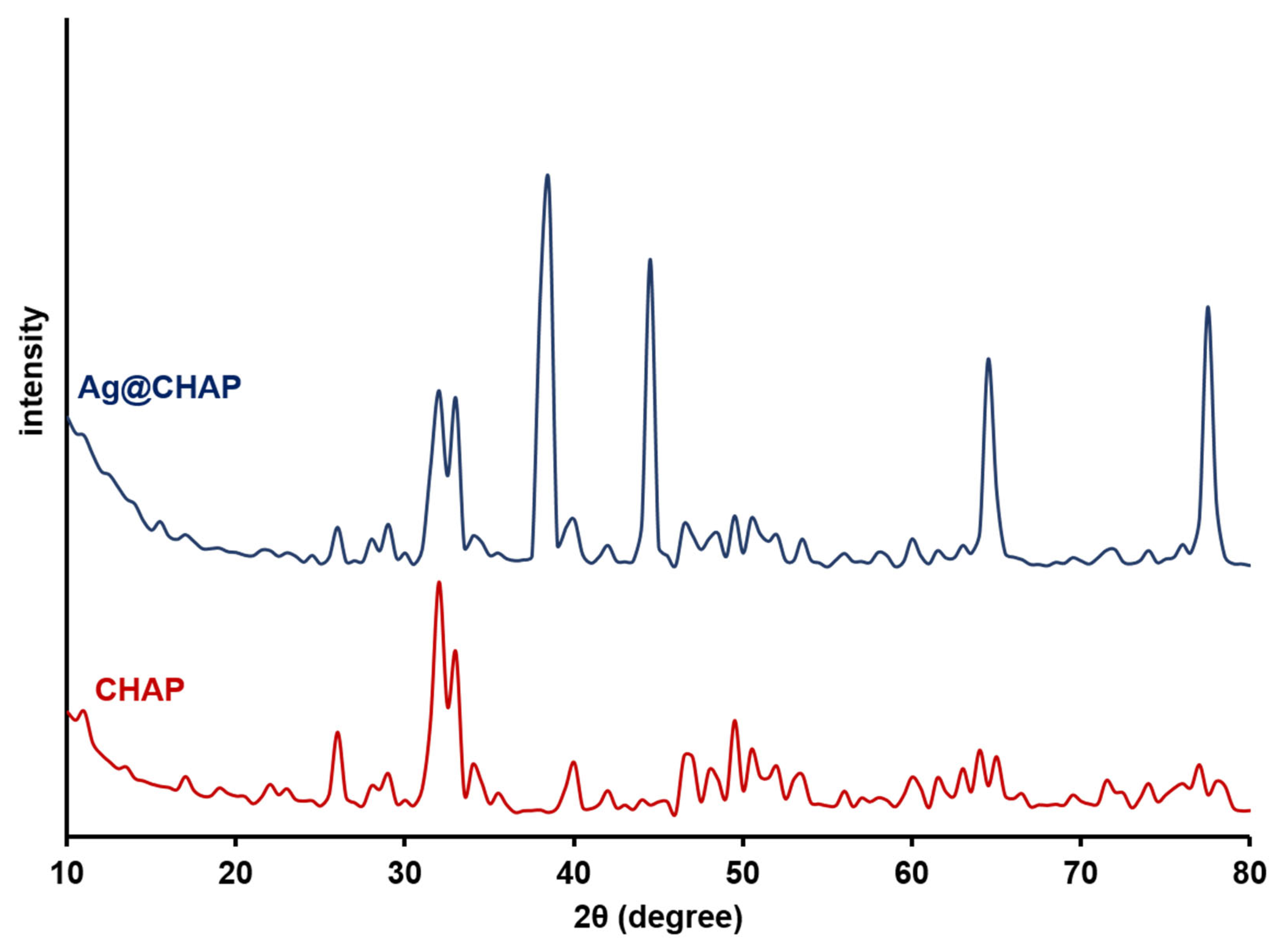
Figure 3 represents the XRD of CHAP and Ag@CHAP. The XRD of CHAP shows characteristic reflections corresponding to the (002), (211), (300), (202), (310), (222), and (213) planes at 2θ values of 26°, 32.1°, 33.2°, 34.3°, 40.1°, 47°, and 49.6°. These strong peaks emphasize the hexagonal phase of CHAP and indicate a high degree of crystallinity (JCPDS no. 09-0432) [34]. In the event where Ag@CHAP are displaced and less strong, the CHAP peaks still remain visible in the XRD of Ag@CHAP, likely due to surface interactions during composite formations. Additionally, the Ag@CHAP composite shows a new silver peaks at (111), (200), (220), and (311) around 2θ of 38.2°, 44.1°, 64.3°, and 77.4° (JCPDS no. 04-0783) [35], showing that the Ag Face-Centered Cubic (FCC) crystal structure was successfully loaded in CHAP. The crystalline size of CHAP and Ag@CHAP is calculated using the Scherrer formula, Equation (1) [36].
where D, β, θ, and λ denote crystalline size, full width at half maximum (FWHM) of the peak, the Brage angle, and wavelength (0.154 Å), respectively. The average crystalline sizes are 3.94 and 7.95 for CHAP and Ag@CHAP, respectively.
Figure 3.
XRD of CHAP and Ag@CHAP.
2.2.3. Raman Spectra
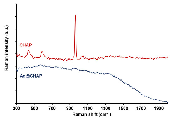
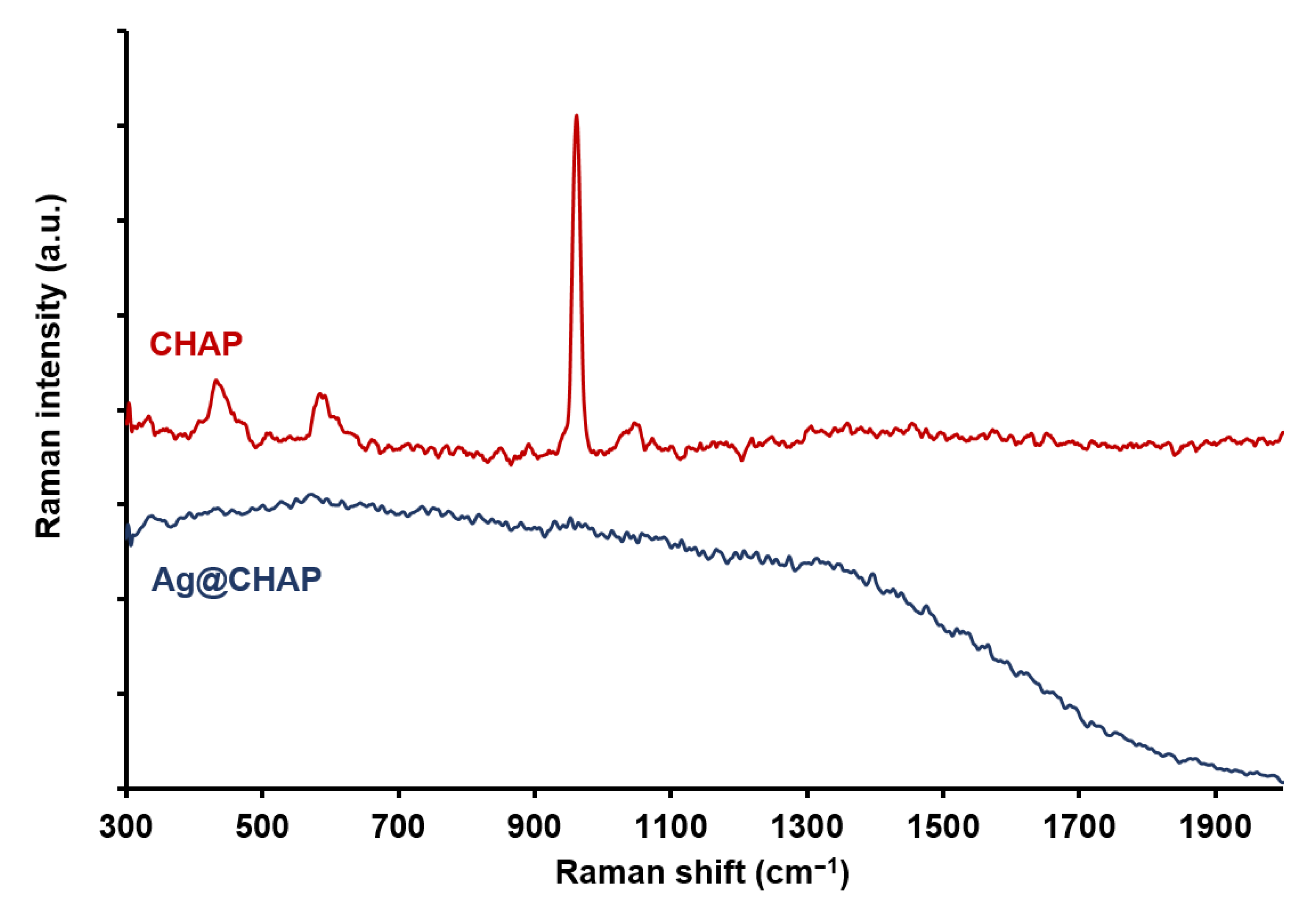
Figure 4 displays the Raman spectra of CHAP and Ag@CHAP. The spectrum of CHAP shows the characteristic vibrations of the phosphate group. The vibrations at 959 cm−1, 458 cm−1, 1150 cm−1, and 650 cm−1 are assignable to ν1 (symmetric P–O stretching vibration), ν2 (O–P–O bending motion), ν3 (asymmetric P–O stretching vibration), and ν4 (asymmetric O–P–O bending vibration) [37]. These bands are correlated with the tetrahedral structure of the phosphate group and matched with other recordings for hydroxyapatite nanoparticles [38].
Figure 4.
Raman spectra of CHAP and Ag@CHAP.
The vibration at 365 cm−1 is attributed to the OH vibration [39]. The spectrum of Ag@CHAP shows the disappearance of PO43− and OH− indicates the successful loading of Ag-NPs on the CHAP surface [40].
2.2.4. Morphological Structure
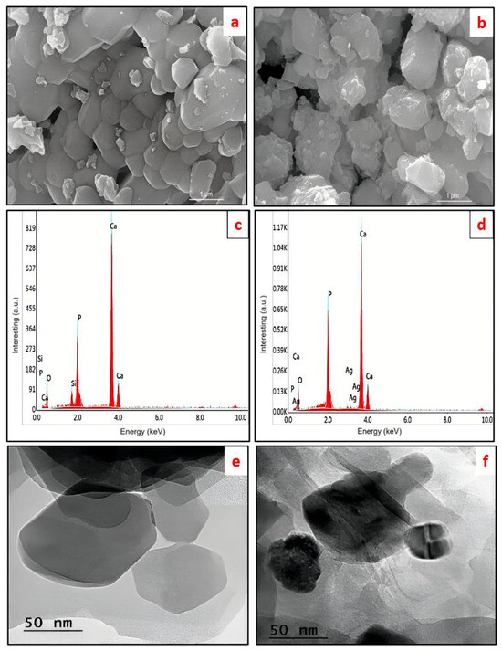
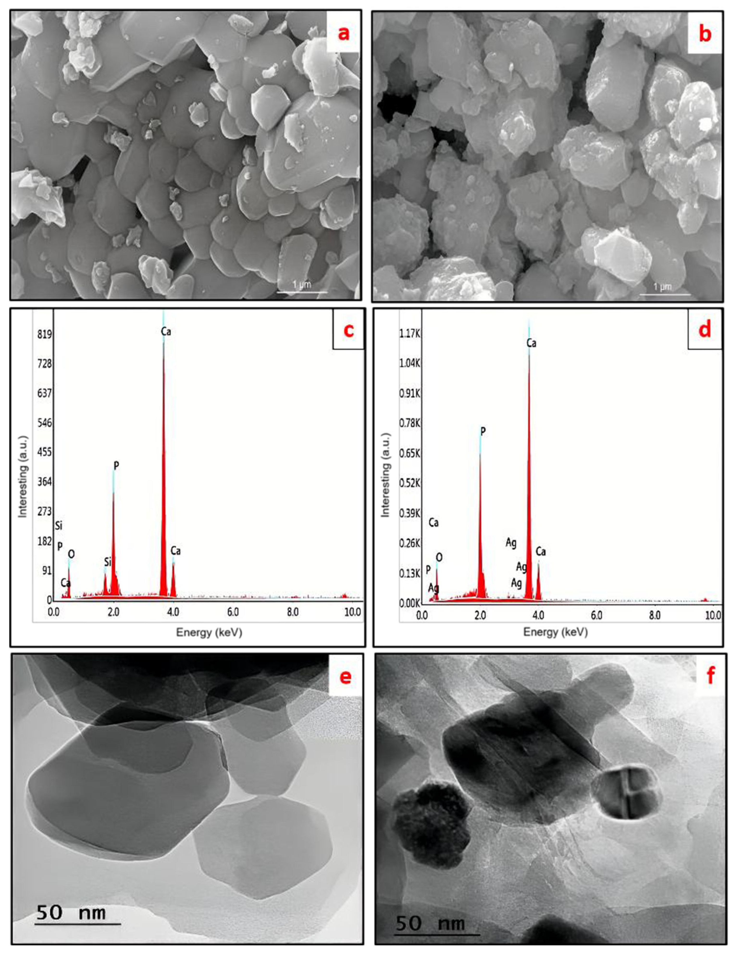
The scanning electron microscopy (SEM) analysis shows that there are obvious differences in morphology between the pristine CHAP and Ag@CHAP samples. As can be seen from Figure 5a, pristine CHAP has a plate-like shape with very smooth surfaces and clear boundaries, thereby showing high crystallinity and equal growth of particles. On the other hand, it can be noted from Figure 5b that Ag@CHAP has a very rough surface and irregularly spaced nanoscale particles evenly distributed on a CHAP matrix, which have been proven to be biogenically synthesized silver nanoparticles. The formation of Ag nanoparticles leads to some level of agglomeration and surface irregularities without affecting the basic CHAP structure, thereby adding another success to its surface decoration. Energy-dispersive X-ray (EDX) spectroscopy was utilized for analyzing the elemental composition of pure CHAP and Ag@CHAP, as represented in Figure 5c,d, respectively. The EDX spectrum of CHAP displays the characteristic peaks of calcium, phosphorus, and oxygen atoms, ensuring the formation of hydroxyapatite phases within the matrix. However, the EDX spectrum of Ag@CHAP reveals clear distinct peaks that represent the deposition of Ag-NPs on the surface of the hydroxyapatite phases within the CHAP matrix. The lack of any sharp peaks of other elements in both EDX spectra ensures that the synthesized samples are of pure compositional quality without any contaminants. The transmission electron microscope (TEM) analysis was conducted on CHAP and Ag@CHAP. From Figure 5e, it can be observed that CHAP has semi-transparent nanoparticles with smooth surfaces and nanoscale lateral dimensions, specifying a uniform dispersion of hydroxyapatite. However, a clear indication from the TEM image shown in Figure 5f of Ag@CHAP is the superimposed nanoparticles with a dark background representing CHAP; these are the silver nanoparticles with greater electron density [41]. From the observation, the Ag-NPs are homogeneously distributed with minimal aggregation while maintaining an intact CHAP matrix, thereby demonstrating a direct interface with the CHAP surface specifying a CHAP nanocomposite formation with silver nanoparticles.
Figure 5.
SEM, EDX, and TEM images of CHAP and Ag@CHAP. (a) SEM of CHAP; (b) SEM of Ag@CHAP; (c) EDX of CHAP; (d) EDX of Ag@CHAP; (e) TEM of CHAP; (f) TEM of Ag@CHAP.
2.3. Photocatalytic Activity of CHAP and Ag@CHAP
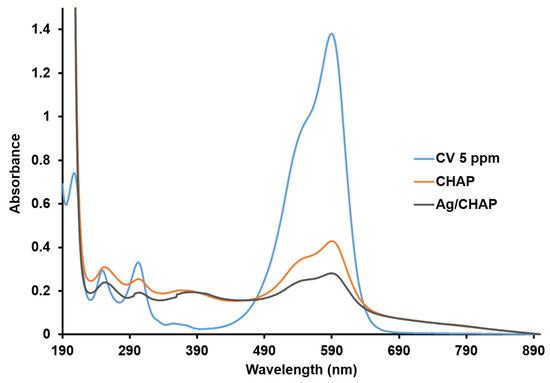
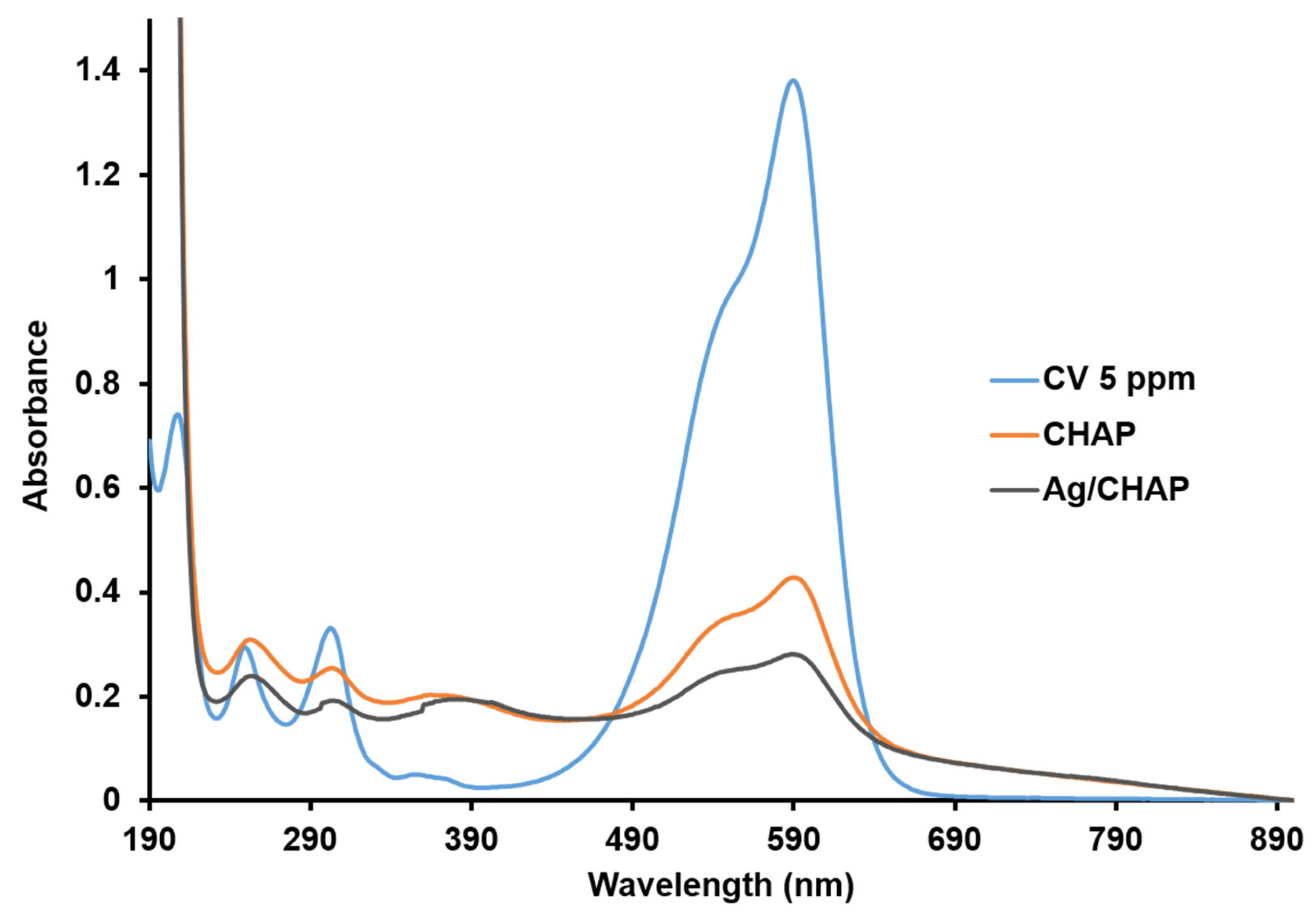
The photocatalytic activity of CHAP and Ag@CHAP was studied comparatively by mixing (1.6 g/L) both with 50 mL of 5 ppm CV dye under sunlight irradiation at the physiological pH. After 100 min, the removal percentages were determined from the absorption spectra (λmax 590 nm) [42] of both treated solutions using Equation (2) [43].
where A0 and At are the absorbance of initial CV concentration and absorbance after photocatalytic reaction period. The percentages of dye removal were 71.42% and 82.15% in the presence of CHAP and Ag@CHAP, respectively, which indicate the superior photocatalytic activity of the silver-loaded composite (Figure 6). Three control tests were performed: photolysis without Ag@CHAP, adsorption capability in the absence of light, and photocatalysis in the presence of Ag@CHAP. The photolysis experiment showed that no reduction in the chromophore band existed in the absence of Ag@CHAP. The results of the adsorption experiment revealed a reduction in CV absorbance after 100 min in the absence of light with a removal efficiency of ≈68.85%, indicating that there are active sites on the surface of Ag@CHAP, which allow the dye to be adsorbed. In the case of the presence of Ag@CHAP and irradiation with sunlight, an approximately 82.15% degradation of dye was obtained after a 90 min exposure. These findings confirm that Ag@CHAP is a suitable photocatalyst, effectively degrading CV upon sunlight irradiation. To establish the optimum conditions for the photocatalytic degradation of CV under irradiation in the presence of Ag@CHAP, different experiments have been conducted to investigate the influence of contact time, catalyst mass, and solution pH.
Figure 6.
UV-Vis spectra of CV dye after 100 min of solar irradiation in the presence of CHAP and Ag@CHAP as photocatalysts at natural pH.
2.3.1. Effect of Ag@CHAP Concentration
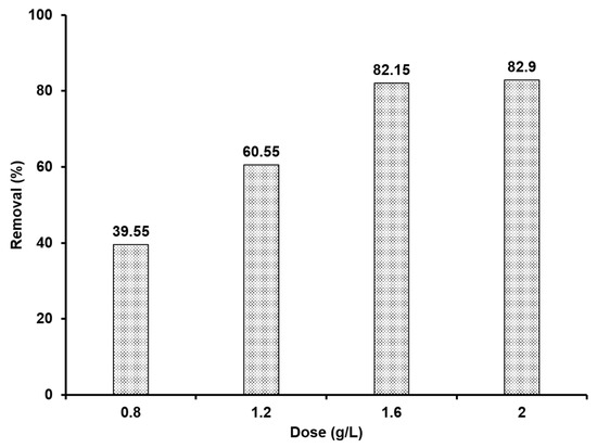
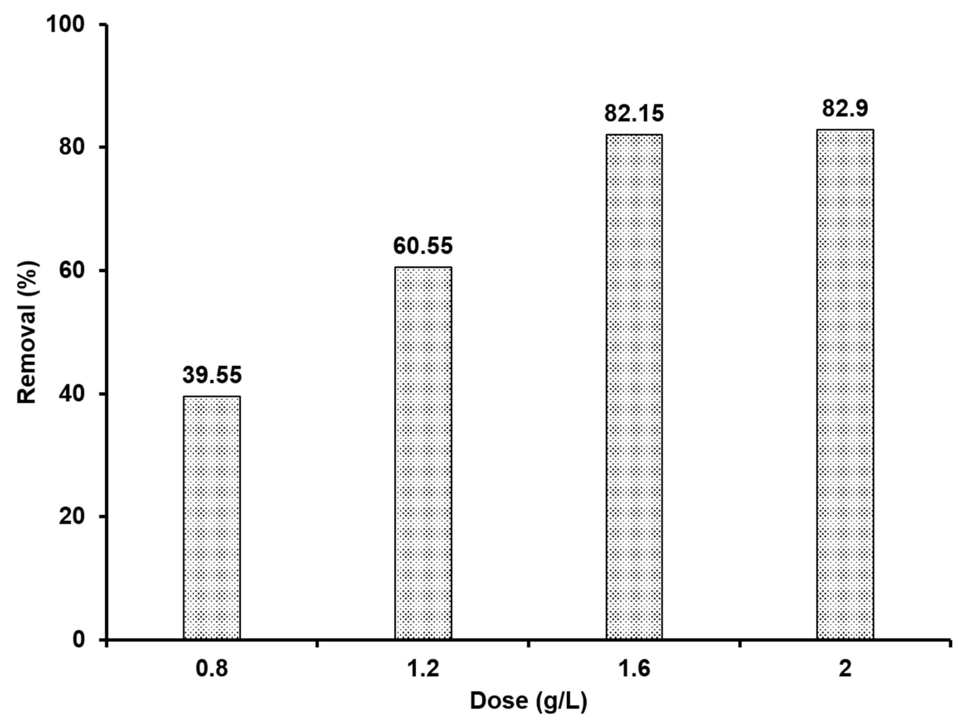
Figure 7 shows the percentages of photocatalytic removal efficiencies of 5 ppm CV dye using different mass concentrations (0.8, 1.2, 1.6, and 2 g/L) of Ag@CHAP after 100 min at pH (7.4). The removal percentage rose from 39.55% to 82.15% with increasing the doses from 0.8 to 1.6 g/L. This is assignable to the increase in the number of active sites suitable for CV adsorption with increasing the dose concentration [44]. No variation in removal efficiency appeared at a concentration of 2 g/L, which indicated that 1.6 g/L is the optimum concentration, which lead to its use in further experiments.
Figure 7.
Effect of Ag@CHAP dose on removal (%) of CV dye after 100 min of solar irradiation at natural pH.
2.3.2. Effect of pH
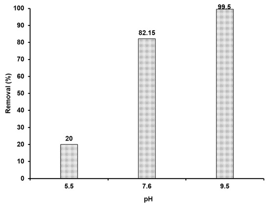
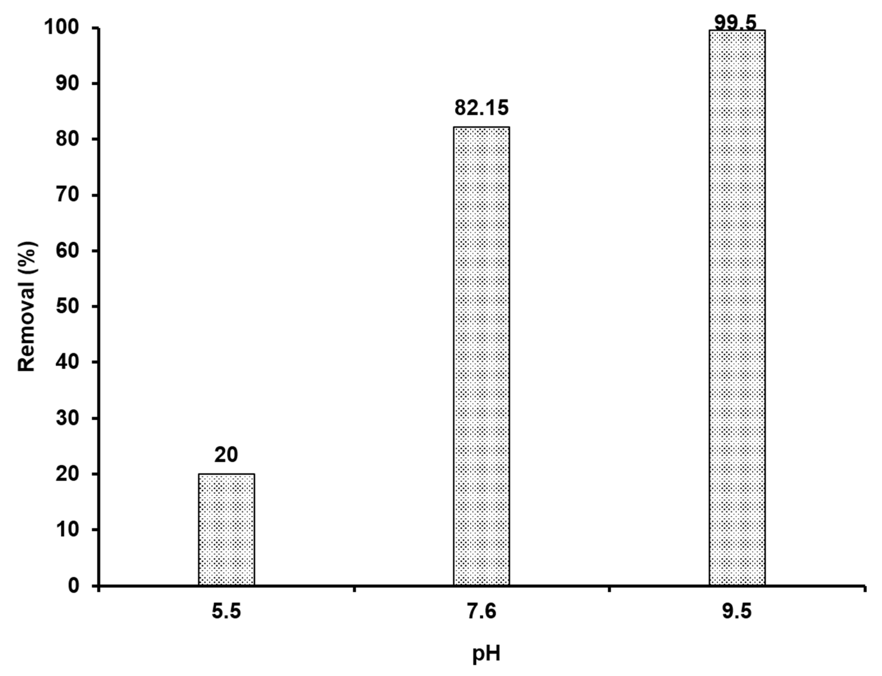
The impact of pH (5.5, 7.6, and 9.5) on the photocatalytic degradation of CV 5 ppm was investigated with a constant concentration of 1.6 g/L Ag@GHAP for 100 min (Figure 8). The percentage removal followed the order pH 9.5 > 7.6 > 5.5 which indicated that pH 9.5 is the optimal pH for CV degradation and the lowest at acidic pH. This can be explained by the increase in the negatively charged surfaces of the CV dye leading to enhanced cationic adsorption of CV dye. On the other hand, in the acidic solution, the catalyst surface was satisfied with a positive charge, which led to the repulsion of CV [45].
Figure 8.
Effect of pH on removal (%) of CV dye after 100 min of solar irradiation.
2.3.3. Effect of Illumination Time and Kinetic Investigation
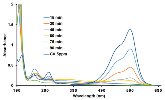
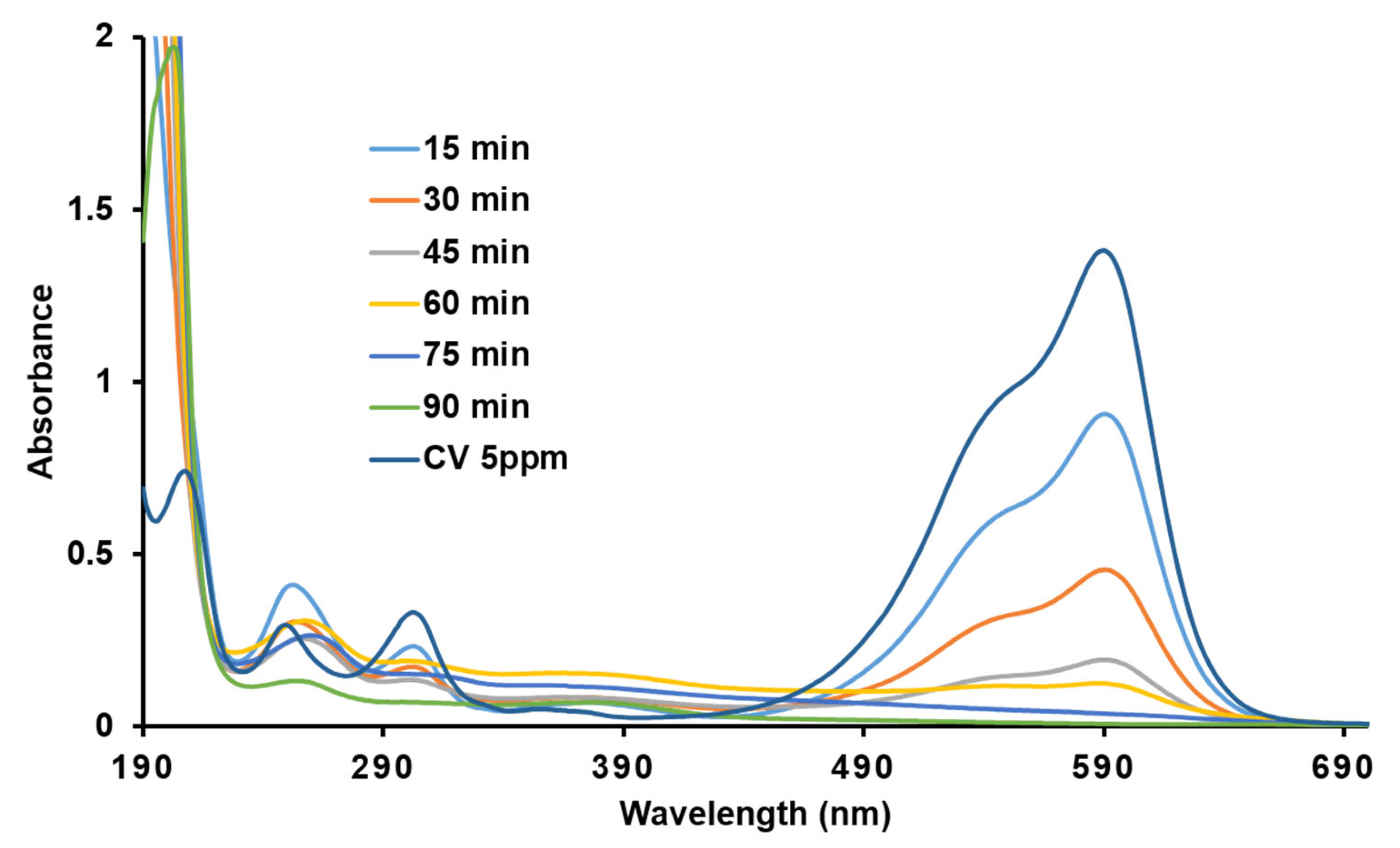
The study of the effect of time on the photocatalytic degradation of CV (5 ppm) using 1.6 g/L of Ag@CHAP at pH 9.5 is exhibited in Figure 9. As shown, increasing the illumination time led to an increase in the removal efficiency of CV. The maximum removal efficiency ≈ 99.8% was verified after 90 min. The reaction kinetic was investigated using the Langmuir–Hinshelwood model [46], as shown in Equation (3).
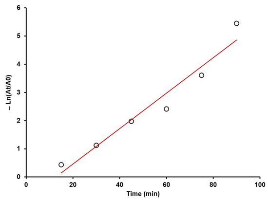
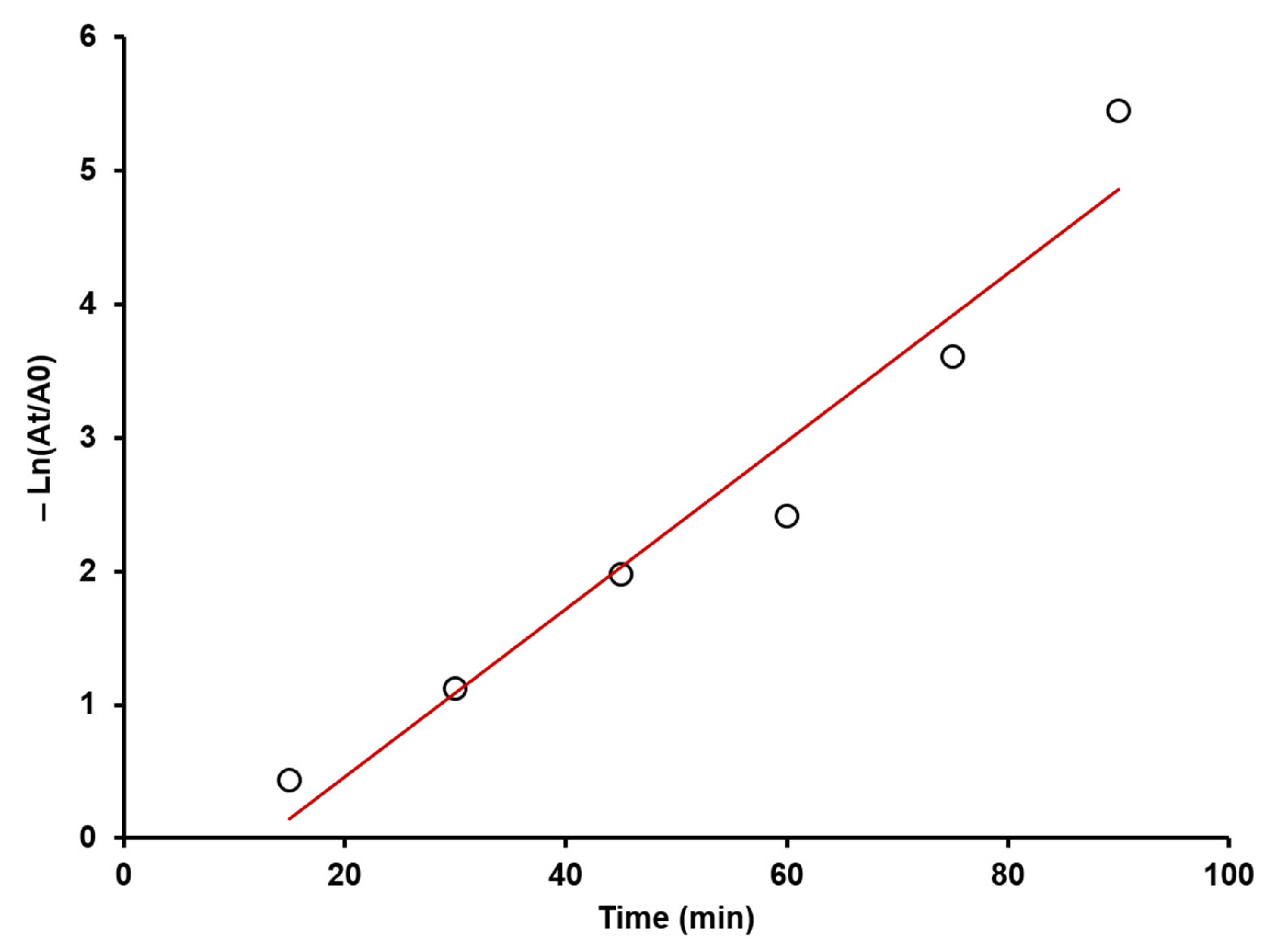
where A0, t, At, and k are the initial dye absorbance, reaction time, absorbance after contact time, and rate constant, respectively. By plotting and (t), the linear relationship indicates the pseudo-first-order model is suitable for reaction kinetics (Figure 10). The correlation coefficient (R2 = 0.95) and computed rate constant (k) of 0.0628 min−1 support the pseudo-first-order model’s ability to match the data. This implies that the catalyst provides enough active sites for dye adsorption, but the reaction is dependent on the CV concentration [47].
Figure 9.
Effect of contact time on removal (%) of CV dye under solar irradiation.
Figure 10.
Pseudo-first-order plot of CV degradation under sunlight in presence Ag@CHAP.
2.3.4. Recyclability Tests of Ag@CHAP
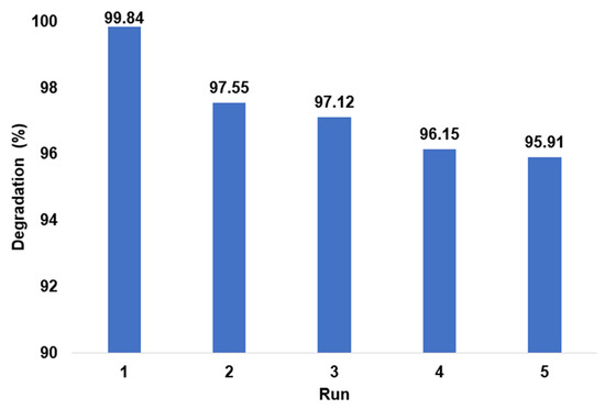
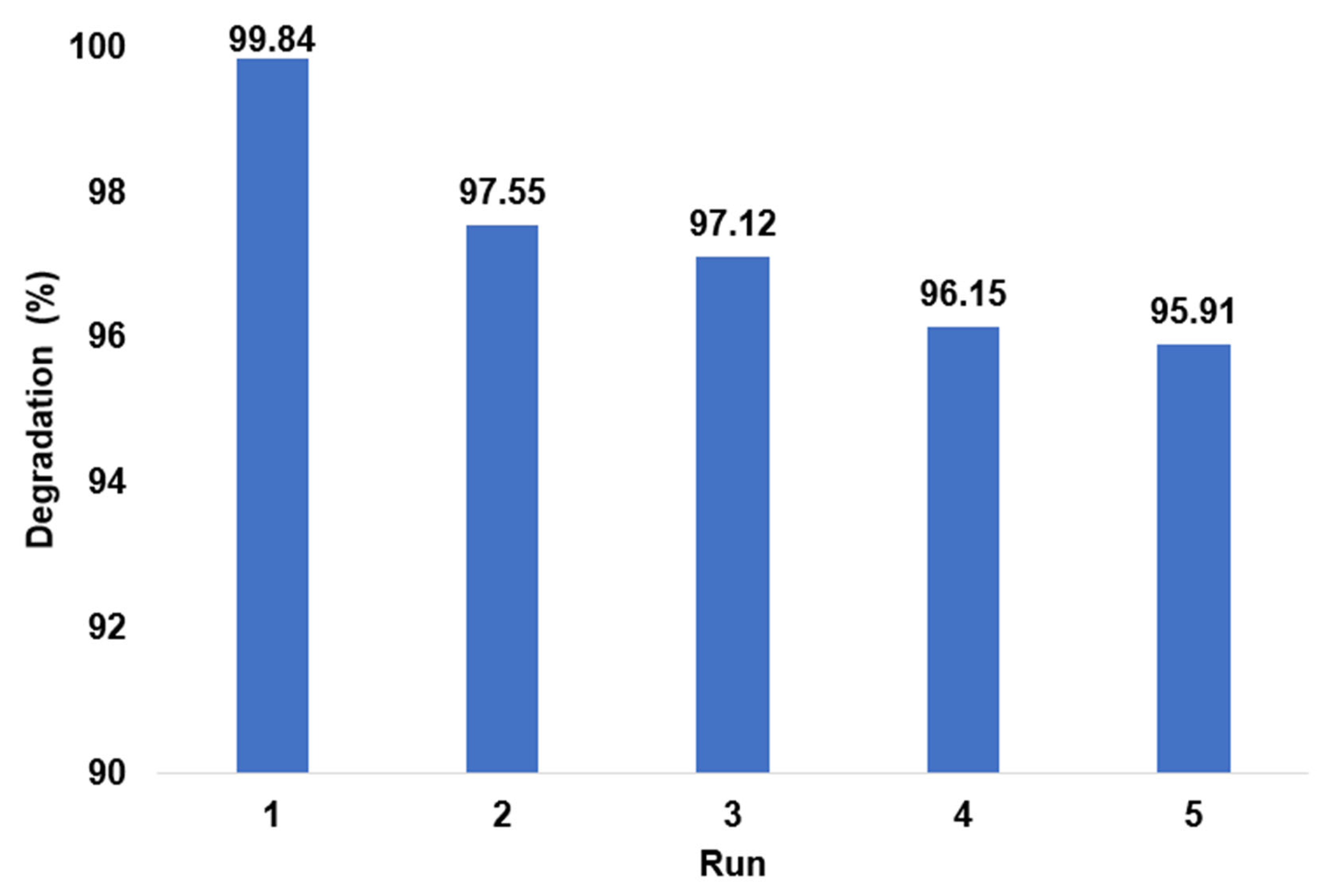
The recycling experiment investigates the reusability potential of Ag@CHAP and validates that they are useful for multiple practical applications. For this purpose, recycling experiments were carried out for Ag@CHAP nanocomposites in five cycles with optimal parameters (catalyst concentration of 1.6 g/L, pH value of 9.5, and illumination time of 90 min). As shown in Figure 11, there is a slight decrease in degradation efficiency from 99.84% in cycle one to 95.91% in cycle five.
Figure 11.
Reusability and decomposition percentages of Ag@CHAP photocatalyst during five runs.
2.4. Antimicrobial Activity of CHAP and Ag@CHAP
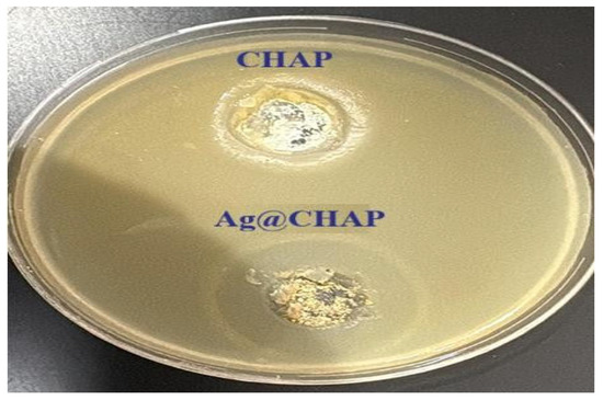
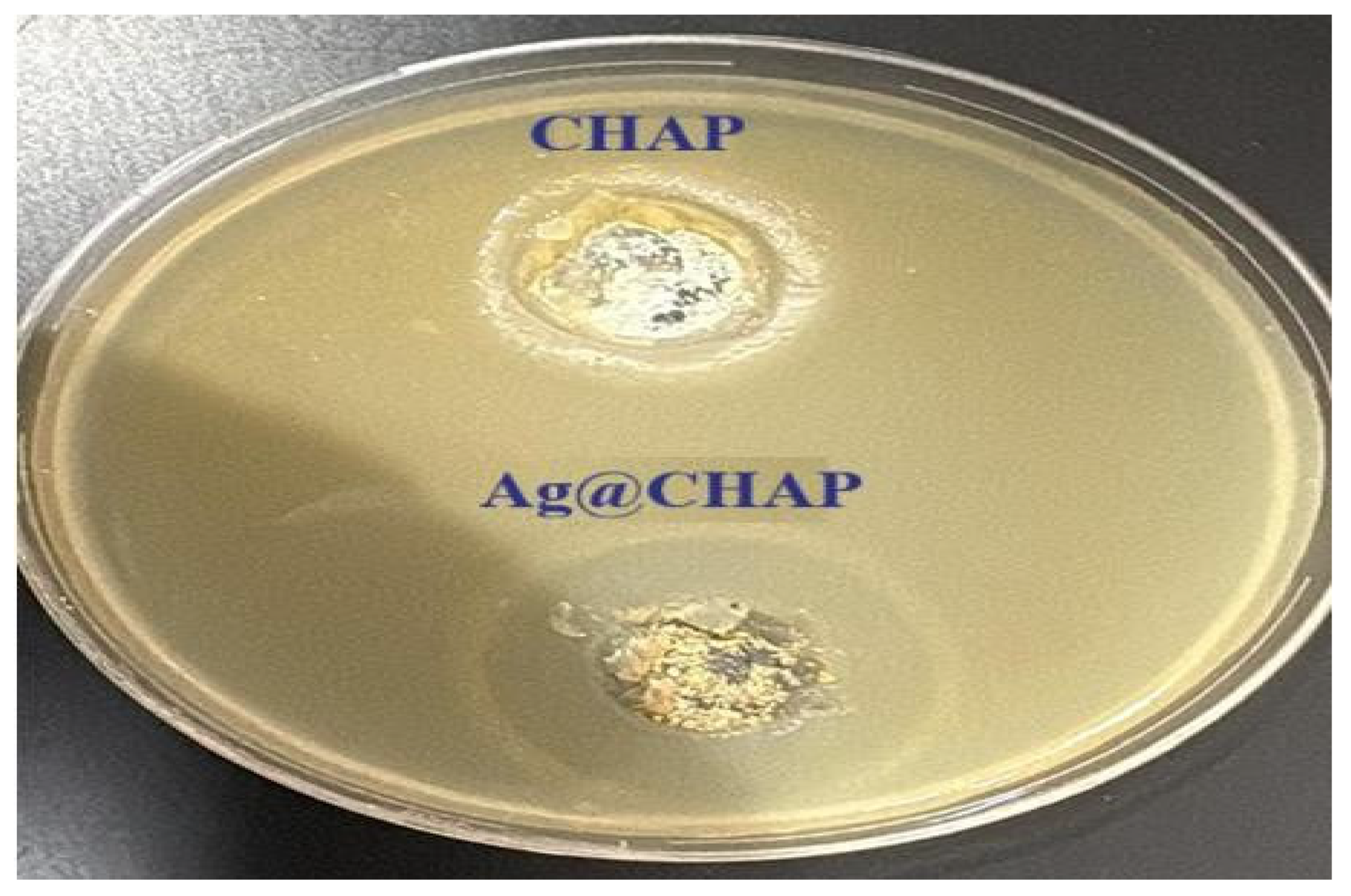
Both CHAP and Ag@CHAP were tested for their antimicrobial activity against E. coli at a concentration of 0.37 µg/m (Figure 12). The Ag@CHAP exhibited a significantly larger inhibition zone than CHAP, indicating an enhanced antimicrobial effect. This improvement could be explained by the presence of silver, which is well-known for its broad-spectrum antibacterial activities. Additionally, the MIC tests were carried out using Ag@CHAP to determine the optimal concentration for the antimicrobial activity by assessing its effects across different concentrations.
Figure 12.
Comparative antimicrobial effect of CHAP and Ag@CHAP at 0.37 µg/mL against. E. Coli.
A set of Ag@CHAP dilutions (0.012, 0.023, 0.04, 0.093, 0.187, 0.37, and 0.75 µg/mL) were prepared and tested against C. albicans, E. coli, S. aureus, and P. aeruginosa to determine the MIC and Minimum Bactericidal/Fungicidal Concentration (MBC/MFC) values [48]. Table 1 summarizes the results indicating that the MIC was consistently at 0.093 µg/mL for all tested species. This suggests that this concentration is sufficient to inhibit visible microbial growth across both Gram-positive and Gram-negative bacteria as well as the fungal strain, reflecting the broad-spectrum antimicrobial potential of Ag@CHAP.
Table 1.
MIC, MBC, and MFC Values of Ag@CHAP against different microbial species.
For bactericidal activity, E. coli and S. aureus exhibited relatively low MBC values of 0.187 µg/mL, demonstrating potent bactericidal effects at concentrations just above the MIC. C. albicans and P. aeruginosa, which are known for their higher resistance to antimicrobial agents, required a higher concentration of 0.37 µg/mL to achieve complete inhibition (MFC/MBC).
Table 2 summarizes the trend of antimicrobial activity with increasing concentrations of Ag@CHAP. A pronounced inhibitory effect against E. coli and S. aureus, with inhibition zones expanding significantly at higher concentrations was clearly shown. A moderate activity was observed against P. aeruginosa, while Candida albicans exhibited comparatively weaker inhibition zones, though some improvement was noted at higher concentrations. These results demonstrate the dose-dependent nature of Ag@CHAP and its stronger impact on bacterial strains compared to the fungal strain as an antimicrobial agent.
Table 2.
Antimicrobial activity of the Ag@CHAP against different pathogenic bacterial and fungal strains.
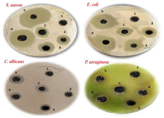
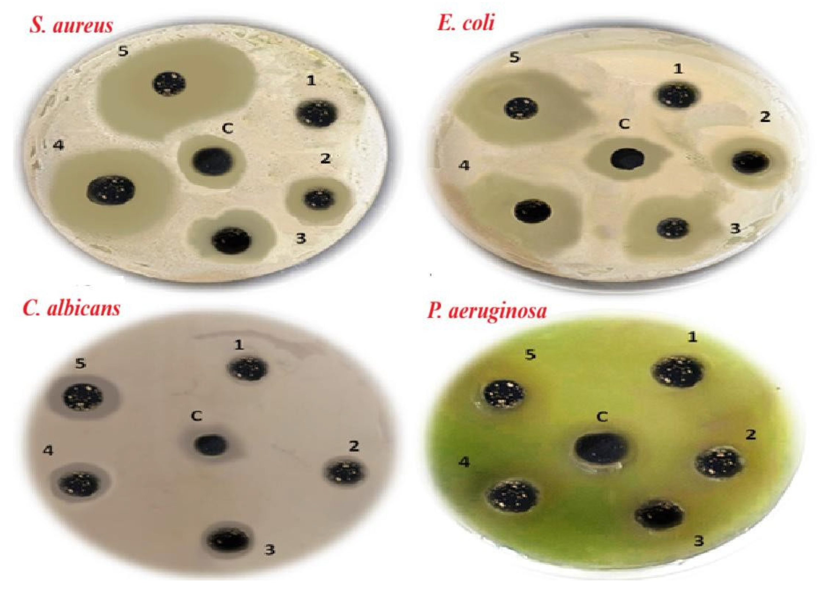
Figure 13 shows an increase in the inhibition zones with an increase in the Ag@CHAP concentration. The results highlight a concentration-dependent antimicrobial effect of the Ag@CHAP against all of the tested microorganisms.
Figure 13.
The effectiveness of Ag@CHAP against the studied microorganisms at the following concentrations: (1) 0.04 µg/mL, (2) 0.093 µg/mL, (3) 0.187 µg/mL, (4) 0.37 µg/mL, and (5) 0.75 µg/mL, in the dark at 30 °C. (c) Control: amoxicillin for bacteria and miconazole for fungus.
The enhanced bioactivity of Ag@CHAP can be attributed to the synergistic effect of Ag-NPs and CHAP combined together promoting their antimicrobial activity. It has been reported that the nanoparticles of both hydroxyapatite and silver improve the penetration of the microbial cell-wall, damaging it and preventing the microbe’s growth [49].
2.5. Antihemolytic Activity of CHAP and Ag@CHAP
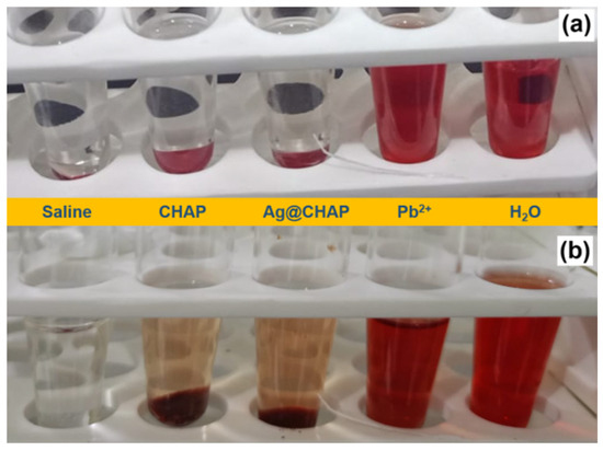
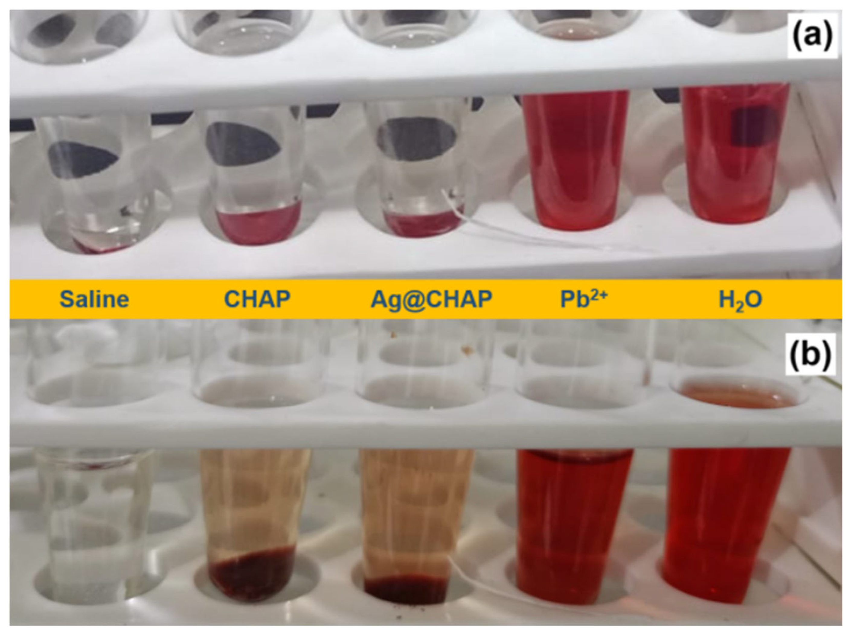
The hemolytic effects of CHAP, Ag@CHAP, and Pb2+ against RBCs is shown in Figure 14a. Water was used as a hemolytic positive control, and saline solution was used as a hemolytic negative control [50]. The calculation of hemolytic activity was assessed using Equation (4) [51].
% Hemolytic activity = absorbance of sample − absorbance of saline/absorbance of dis water × 100
Figure 14.
Hemolytic effect of CHAP, Ag@CHAP, saline, water, and Pb2+ (a) and effect of CHAP and Ag@CHAP on the hemolytic effect of Pb2+ (b).
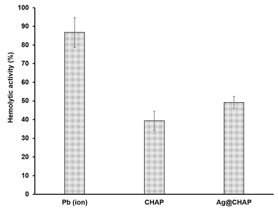
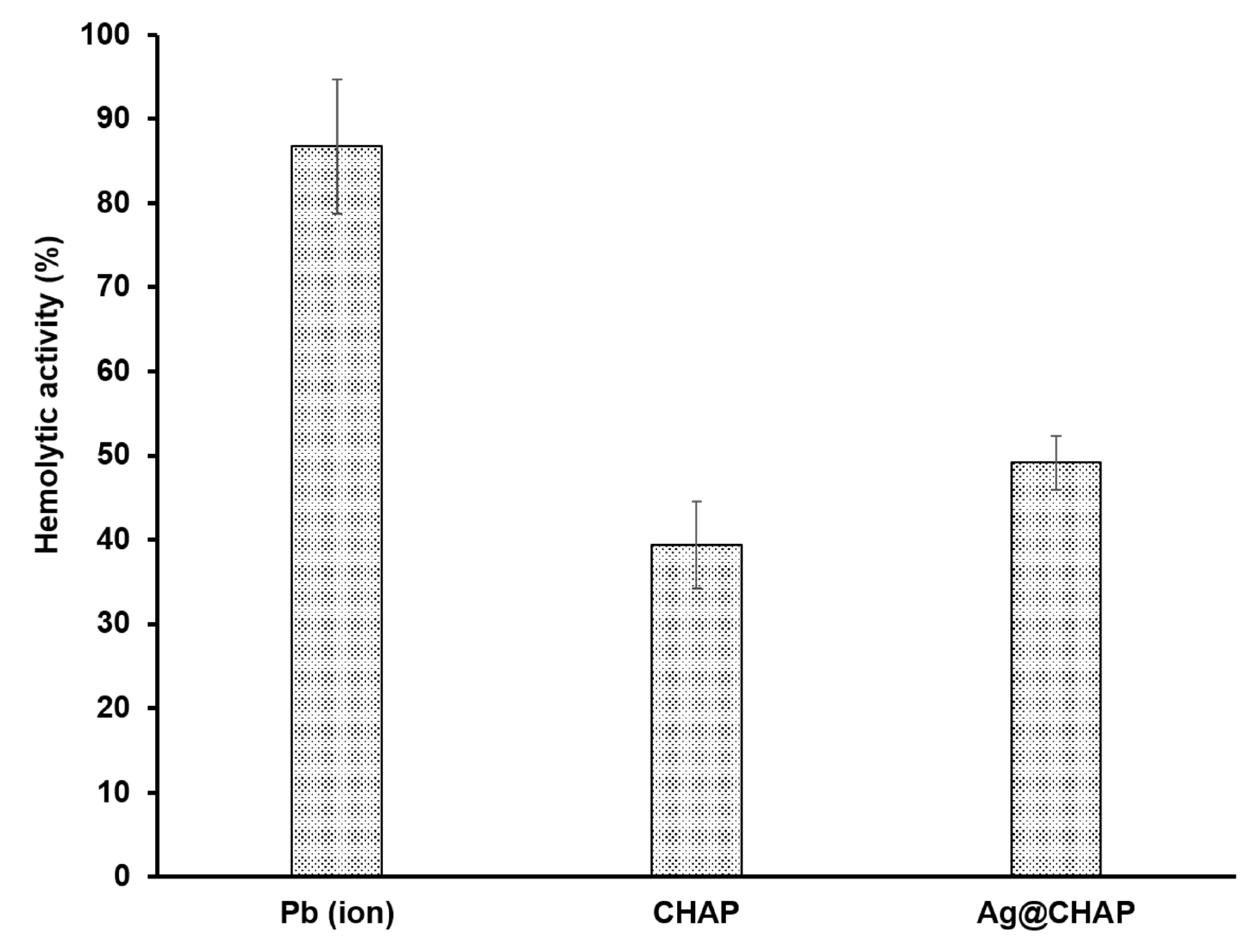
The application of both CHAP and Ag@CHAP had no hemolytic activity after 24 h against RBCs as compared to samples of Pb2+ solution and water. It was reported that lead is known to induce a significant cytotoxic effect on RBCs at a low concentration, which could be due to its ability to alter osmotic fragility, the interaction of lead with the membrane protein, and the induction of oxidative stress [52]. Employing Pb2 resulted in a 86.73% hemolytic effect. As shown in Figure 14b, pretreatment of RBCs with CHAP and Ag@CHAP significantly reduced RBC hemolysis induced by Pb2+ to 39.35% and 49.13%, respectively (p ≤ 0.01). These findings of the antihemolytic activity of CHAP and Ag@CHAP indicate that this compound possesses antihemolytic properties, as shown in Figure 15. This can be interpreted as their antioxidant efficacy [53], which protects the RBCs’ plasma membrane through contracting lipid peroxidation mediated by lead acetate [54].
Figure 15.
Percentage of hemolysis effect of Pb2+ in the absence and presence of CHAP and Ag@CHAP.
3. Experimental
3.1. Materials
The caprine bone peace (≈5 g) was obtained from a voucher market in Sakaka, Saudi Arabia. Silver nitrate, Crystal Violet, hydrochloric acid, sodium hydroxide, and DMSO were obtained from Sigma-Aldrich (St. Louis, MO, USA) and were used without further purifications. All aqueous solutions were prepared using Milli-Q water.
3.2. Instruments
The X-ray diffractions (XRD) patterns were measured using XRD-7000 SHIMADZU (Shimadzu, Kyoto, Japan) with a copper detector. To detect the thermal gravimetric analysis, TGA; TGA-51 Shimadzu (Shimadzu, Kyoto, Japan) was used. Raman spectra were determined using Shimadzu IR–Tracer 100 FTIR ((Shimadzu, Kyoto, Japan). Scanning electron microscope (SEM) photos were obtained using SEM Zeiss 1530 (JSM-5410, JEOL, Tokyo, Japan). A double beam LABOMED Spectro 99 (Labomed, Los Angeles, CA, USA) was used to record the electronic spectra, and a PeakTech digital multitester (PeakTech, Ahrensburg, Germany) was used to detect the sunlight intensity.
3.3. Synthesis of Caprine-Hydroxyapatite (CHAP) and Silver Nanoparticles Loaded Caprine-Hydroxyapatite (Ag@CHAP)
A 3 g piece of caprine bone was washed several times with distilled water, dried, crushed, and calcined in a muffle furnace at 1050 °C for 2 h. The resulting CHAP material was ground into a fine powder to be suitable for subsegment experiments. Ag@CHAP was prepared according to our previous method with some modifications [53]. An extract of 2 g of caprine bone was obtained by boiling a 100 mL mixture made of equal volumes of water, ethanol, acetone, and ethyl acetate, which was used as the solvent, for 1 h. This mixture was then filtered and mixed with an aqueous solution of silver nitrate (2% w/v). The CHAP was added to the mixture and stirred at 100 °C for 1 h. The solid formed-product was filtered, washed several times with hot water, air-dried, and then calcined in a furnace at 300 °C. The formed Ag@CHAP composite was stored in a desiccator for further experimental use.
3.4. Antimicrobial Experiments
3.4.1. Evaluation of Minimum Inhibitory Concentration (MIC): Microbroth Dilution Method (on Liquid Media)
Four microorganisms, P. aeruginosa ATCC 10145, E. coli ATCC 35218, S. aureus ATCC 25923, and C. albicans (human isolate), were used to evaluate the antimicrobial activity of CHAP and Ag@CHAP. The standard broth microdilution method was used to determine the minimum inhibitory concentration (MIC) of CHAP and Ag@CHAP. Serial dilutions of CHAP and Ag@CHAP at concentrations of 0.012, 0.023, 0.04, 0.093, 0.187, 0.37, and 0.75 µg/mL were prepared using dimethyl sulfoxide (DMSO) as a solvent. A total of 2 mL of Nutrient Broth (NB; Oxoid Ltd., Basingstoke, UK) was used for testing bacterial strains, while 2 mL of Potato Dextrose Broth (PDB, Oxoid Ltd., Basingstoke, UK) was used for fungal strains. All tubes were inoculated with 1 μL of the microbial suspension (~104 CFU/mL), and a fresh culture was used to create the microbial solution, which was adjusted to around 102 CFU/mL (0.5 McFarland standard). To achieve a final concentration of 10 CFU/mL, serial tenfold dilutions were carried out in sterile phosphate-buffered saline by moving 1 mL of the suspension into 9 mL of diluent four times. The final inoculum concentration was verified by counting colony-forming units after incubation and plating suitable dilutions on agar medium, followed by adding 1 μL of CHAP and Ag@CHAP dilution. Depending on the type of the organism, the tubes were incubated either at 25 °C or 30 °C for 16–24 h. Both positive (miconazole for fungus and ampicillin for bacteria) and negative (non-inoculated broth) controls were used.
The lowest concentration of CHAP and Ag@CHAP that completely inhibited visible microbial growth as indicated by the absence of visible turbidity was considered as the minimum inhibitory concentration (MIC). To demonstrate the absence of viable cells, 100 μL from the tubes (or wells) that did not exhibit any visible growth were sub-cultured onto fresh agar plates to determine the Minimum Bactericidal Concentration (MBC) and Minimum Fungicidal Concentration (MFC) [55].
3.4.2. Agar Well-Diffusion Method (Agar Media)
The agar well-diffusion technique was used to further assess the antimicrobial activity. A standardized microbial suspension (108 CFU/mL) was added to 15 mL of Nutrient Agar (NA, Oxoid Ltd., Basingstoke, UK) for bacterial isolates or Potato Dextrose Agar (PDA, Oxoid Ltd., Basingstoke, UK) for fungal isolates. A sterile cork borer was used to create wells that were about 3 mm in diameter once the agar plates were solidified. A volume of 100 µL of CHAP or Ag@CHAP at concentrations of 0.04, 0.093, 0.187, 0.37, and 0.75 µg/mL was added to each well. In order to determine the inhibition zone around each well as a measure to assess antimicrobial activity, agar plates with bacteria were incubated at 30 °C for 48 h, whereas incubation at 25°6 days was used for fungal plates [56].
3.5. Antihemolytic Experiments
The hemolytic activity of lead acetate was assessed in vitro using fresh human red blood cells (RBCs) [57]. Briefly, 100 µL of lead acetate solution (0.625 ppm) was incubated with 100 µL of erythrocytes that had been washed three times [58]. Furthermore, the antihemolytic activity of CHAP and Ag@CHAP was determined by using 100 µL of washed RBCs that were incubated with 100 µL/mL CHAP and Ag@CHAP (500 µg/mL) for 1 h. Additionally, 100 µL of lead acetate solution (0.625 ppm) was added to the mixture. After incubation at 37 °C for 24 h, the mixture was later centrifuged at 1500× g rpm for 10 min. The absorbance of the supernatants was measured at 540 nm (A540) using saline and distilled as the minimum and maximum hemolytic controls, respectively. All experiments were run in triplicate; statistical analysis of CHAP and Ag@CHAP was performed using GraphPad Prism version 3 software using the t-test. Results were presented as mean ± standard error (SE).
3.6. Photocatalysis Experiments
Crystal Violet dye (CV) was employed as a model to evaluate the solar semiconducting activity of CHAP and Ag@CHAP. A known amount of the photocatalyst was added to 50 mL of a 5 ppm Crystal Violet (CV) solution and magnetically stirred at 300 rpm in the dark to allow the dye to adsorb onto the catalyst surface. The suspension was then exposed to 40 × 103 lux of natural sunlight intensity at ambient conditions. To extract the photocatalyst, at 15-min intervals, approximately 3 mL of the treated CV solution mixture was withdrawn and centrifuged for 2 min. The absorbance (A) of the supernatant was measured at λmax = 590 nm, and the decomposition efficiency of CV was calculated.
4. Conclusions
A sustainable recycling strategy for caprine bone waste toward the synthesis of the biogenic hydroxyapatite (CHAP) and its silver nanoparticle composite (Ag@CHAP) has been developed and further characterized, offering an environmentally responsible route to the development of sustainable materials. The materials showed a significant multi-functionality with a high practical value. Ag@CHAP’s outstanding photocatalytic activity, achieving 99.8% degradation of Crystal Violet under natural sunlight, highlighted its great potential in ecofriendly wastewater treatments. Furthermore, Ag@CHAP showed a broad spectrum of antimicrobial activity against clinically isolated human pathogens. CHAP showed a higher ability to reduce lead-induced hemolysis position. These findings support CHAP and Ag@CHAP derived from caprine bone waste as stable, promising candidates for high-performance, nontoxic materials able to meaningfully contribute to both environmental protection and public health for safe biomedical and detoxification applications. The future studies should focus on the stability and biosafety of the CHAP and Ag@CHAP nanoparticles, in addition to the optimization of the loading capacity of Ag nanoparticles in CHAP for the simultaneous improvement of antimicrobial performance and environmental safety. The scaling-up of the methodologies could also help in testing their effectiveness in different environmental and biomedical applications.
Author Contributions
Conceptualization, A.H.A.; methodology, A.H.A., S.M.N.M., S.H.A., N.F.A., I.A.T., R.F.H. and A.M.N.; software, A.S.A.Z. and A.A.; validation, A.H.A. and A.S.A.Z.; formal analysis, A.S.A.Z. and N.F.A.; investigation, A.M.N.; resources, A.A., S.M.N.M. and S.H.A.; data curation, A.A. and R.F.H.; writing—original draft preparation, A.A., S.M.N.M., S.H.A. and I.A.T.; writing—review and editing, R.F.H. and A.M.N.; visualization, A.H.A., A.S.A.Z. and A.M.N.; supervision, A.H.A. and A.M.N.; project administration, A.M.N.; funding acquisition, A.H.A. All authors have read and agreed to the published version of the manuscript.
Funding
This work was funded by the Deanship of Graduate Studies and Scientific Research at Jouf University under grant No. DGSSR-2024–02-02182.
Data Availability Statement
Data is contained within the article.
Acknowledgments
The authors are appreciative of the central laboratory at Jouf University for support during the instrumental analysis.
Conflicts of Interest
The authors declare no conflict of interest.
References
- Nassar, A.M.; Arafa, W.A.; Ashammari, K.; Moustafa, S.M.; Alsirhani, A.M.; Hasaneen, M.F. A new green catalyst and antimicrobial agent derived from eco-friendly products of camel bones: Synthesis and physicochemical characterization. Biomass Convers. Biorefinery 2025, 15, 13589–13607. [Google Scholar] [CrossRef] [Scilit]
- Alanazi, A.H.; Arafa, W.A.; Moustafa, S.M.; Alsohaimi, I.H.; Elnasr, T.A.S.; Halawani, R.F.; Al Zbedy, A.S.; Nassar, A.M. Green extraction of biomass from waste goat bones for applications in catalysis, wastewater treatment, and water disinfection. J. Hazard. Mater. Adv. 2025, 18, 100645. [Google Scholar] [CrossRef] [Scilit]
- Osman, A.I.; Zhang, Y.; Farghali, M.; Rashwan, A.K.; Eltaweil, A.S.; Abd El-Monaem, E.M.; Mohamed, I.M.; Badr, M.M.; Ihara, I.; Rooney, D.W.; et al. Synthesis of green nanoparticles for energy, biomedical, environmental, agricultural, and food applications: A review. Environ. Chem. Lett. 2024, 22, 841–887. [Google Scholar] [CrossRef] [Scilit]
- Noor, U.; Soni, S.; Purwar, S.; Gupta, E. Sustainable valorization of food waste for the biogeneration of nanomaterials. In Green and Sustainable Approaches Using Wastes for the Production of Multifunctional Nanomaterials; Elsevier: Amsterdam, The Netherlands, 2024; pp. 91–101. [Google Scholar]
- Francis, A.A. Ecologic and economic motives for transforming calcium-based food wastes into sustainable value-added products: A review. Environ. Sci. Pollut. Res. 2025, 32, 428–451. [Google Scholar] [CrossRef] [Scilit] [PubMed]
- Osuchukwu, O.A.; Salihi, A.; Abdullahi, I.; Abdulkareem, B.; Nwannenna, C.S. Synthesis techniques, characterization and mechanical properties of natural derived hydroxyapatite scaffolds for bone implants: A review. SN Appl. Sci. 2021, 3, 822. [Google Scholar] [CrossRef] [Scilit]
- Saleem, M.; Rasheed, S.; Yougen, C. Silk fibroin/hydroxyapatite scaffold: A highly compatible material for bone regeneration. Sci. Technol. Adv. Mater. 2020, 21, 242–266. [Google Scholar] [CrossRef] [Scilit]
- Mahmoud, E.M.; Sayed, M.; Awaad, M.; El-Zomor, S.T.; Blum, M.; Killinger, A.; Gadow, R.; Naga, S.M. Evaluation of Ti/Al alloy coated with biogenic hydroxyapatite as an implant device in dogs’ femur bones. J. Mater. Sci. Mater. Med. 2021, 32, 119. [Google Scholar] [CrossRef] [Scilit]
- Vinayagam, R.; Pai, S.; Murugesan, G.; Varadavenkatesan, T.; Kaviyarasu, K.; Selvaraj, R. Green synthesized hydroxyapatite nanoadsorbent for the adsorptive removal of AB113 dye for environmental applications. Environ. Res. 2022, 212, 113274. [Google Scholar] [CrossRef] [Scilit]
- Kamal, A.B.; Hassane, A.M.; An, C.; Deng, Q.; Hu, N.; Abolibda, T.Z.; Altaleb, H.A.; Gomha, S.M.; Selim, M.M.; Shenashen, M.A.; et al. Developing a cost-effective and eco-friendly adsorbent/photocatalyst using biomass and urban waste for crystal violet removal and antimicrobial applications. Biomass Convers. Biorefinery 2025, 15, 10089–10107. [Google Scholar] [CrossRef] [Scilit]
- Shabna, S.; Singh, C.J.C.; Dhas, S.D.S.J.; Jeyakumar, S.C.; Biju, C.S. An overview of prominent factors influencing the photocatalytic degradation of cationic crystal violet dye employing diverse nanostructured materials. J. Chem. Technol. Biotechnol. 2024, 99, 1027–1055. [Google Scholar] [CrossRef] [Scilit]
- Prabha, N.; Arora, R.D.; Ganguly, S.; Chhabra, N. Gentian violet: Revisited. Indian J. Dermatol. Venereol. Leprol. 2020, 86, 600. [Google Scholar] [CrossRef] [Scilit]
- Sharma, P.; Kar, S.; Sahu, M.; Ganguly, M. Copper-based nanoparticles for the removal of the crystal violet dye via degradation and adsorption: A comparative account. RSC Adv. 2025, 15, 27995–28020. [Google Scholar] [CrossRef] [Scilit] [PubMed]
- Ilyas, A.; Rafiq, K.; Abid, M.Z.; Rauf, A.; Hussain, E. Growth of villi-microstructured bismuth vanadate (Vm-BiVO4) for photocatalytic degradation of crystal violet dye. RSC Adv. 2023, 13, 2379–2391. [Google Scholar] [CrossRef] [Scilit] [PubMed]
- Radoor, S.; Jayakumar, A.; Shivanna, J.M.; Karayil, J.; Kim, J.T.; Siengchin, S. Adsorptive removal of crystal violet from aqueous solution by bioadsorbent. Biomass Convers. Biorefinery 2025, 15, 2431–2442. [Google Scholar] [CrossRef] [Scilit]
- Singh, N.; Kumar, U.; Jatav, N.; Sinha, I. Photocatalytic degradation of crystal violet on Cu, Zn doped BiVO4 particles. Langmuir 2024, 40, 8450–8462. [Google Scholar] [CrossRef] [Scilit]
- Tran-Nguyen, P.L.; Ly, K.P.; Santoso, S.P.; Tran, N.P.D.; Angkawijaya, A.E.; Nguyen, H.N.; Huynh, Q.K.; Mai, N.T.N.; Nguyen, M.N.; Dang, H.G.; et al. Iron oxides/zeolite X composite derived from rice husk ash: Fabrication and physicochemical properties for superior heterogeneous Fenton-like oxidation of crystal violet. J. Chem. Technol. Biotechnol. 2025, 100, 1222–1237. [Google Scholar] [CrossRef] [Scilit]
- Fosso-Kankeu, E.; Webster, A.; Ntwampe, I.O.; Waanders, F.B. Coagulation/flocculation potential of polyaluminium chloride and bentonite clay tested in the removal of methyl red and crystal violet. Arab. J. Sci. Eng. 2017, 42, 1389–1397. [Google Scholar] [CrossRef] [Scilit]
- Divya, M.; Chen, J.; Durán-Lara, E.F.; Kim, K.S.; Vijayakumar, S. Revolutionizing healthcare: Harnessing nano biotechnology with zinc oxide nanoparticles to combat biofilm and bacterial infections-A short review. Microb. Pathog. 2024, 191, 106679. [Google Scholar] [CrossRef] [Scilit]
- Murugan, S.; Senthilvelan, T.; Govindasamy, M.; Thangavel, K. A Comprehensive Review on Exploring the Potential of Phytochemicals and Biogenic Nanoparticles for the Treatment of Antimicrobial-Resistant Pathogenic Bacteria. Curr. Microbiol. 2025, 82, 90. [Google Scholar] [CrossRef] [Scilit]
- Morán, M.D.C.; Porredon, C.; Gibert, C. Insight into the Antioxidant Activity of Ascorbic Acid-Containing Gelatin Nanoparticles in Simulated Chronic Wound Conditions. Antioxidants 2024, 13, 299. [Google Scholar] [CrossRef] [Scilit]
- Vijayakumar, N.; Venkatraman, S.K.; Abraham, J.; Hamdan, H.F.; Genasan, K.; Shukor, M.H.A.; Swamiappan, S. Biowaste-Derived Rankinite: A Study on Its Hemocompatibility, Antimicrobial, and Osteogenic Properties for Hard Tissue Regeneration. ChemistrySelect 2025, 10, e05912. [Google Scholar] [CrossRef] [Scilit]
- Laranjeira, M.S.; Moco, A.; Ferreira, J.; Coimbra, S.; Costa, E.; Santos-Silva, A.; Ferreira, P.J.; Monteiro, F.J. Different hydroxyapatite magnetic nanoparticles for medical imaging: Its effects on hemostatic, hemolytic activity and cellular cytotoxicity. Colloids Surf. B Biointerfaces 2016, 146, 363–374. [Google Scholar] [CrossRef] [Scilit]
- Yedgar, S.; Barshtein, G.; Gural, A. Hemolytic activity of nanoparticles as a marker of their hemocompatibility. Micromachines 2022, 13, 2091. [Google Scholar] [CrossRef] [Scilit] [PubMed]
- Nassar, A.M.; Alrowaili, Z.A.; Ahmed, A.A.; Cheba, B.A.; Akhtar, S. Facile synthesis of new composite, Ag-Nps-loaded core/shell CdO/Co3O4 NPs, characterization and excellent performance in antibacterial activity. Appl. Nanosci. 2021, 11, 419–428. [Google Scholar] [CrossRef] [Scilit]
- Prakash, M.; Rajan, H.K.; Chandraprabha, M.N.; Shetty, S.; Mukherjee, T.; Girish Kumar, S. Recent developments in green synthesis of hydroxyapatite nanocomposites: Relevance to biomedical and environmental applications. Green Chem. Lett. Rev. 2024, 17, 2422409. [Google Scholar] [CrossRef] [Scilit]
- Riaz, T.; Zeeshan, R.; Zarif, F.; Ilyas, K.; Muhammad, N.; Safi, S.Z.; Rahim, A.; Rizvi, S.A.; Rehman, I.U. FTIR analysis of natural and synthetic collagen. Appl. Spectrosc. Rev. 2018, 53, 703–746. [Google Scholar] [CrossRef] [Scilit]
- Howell, N.K.; Arteaga, G.; Nakai, S.; Li-Chan, E.C. Raman spectral analysis in the C−H stretching region of proteins and amino acids for investigation of hydrophobic interactions. J. Agric. Food Chem. 1999, 47, 924–933. [Google Scholar] [CrossRef] [Scilit]
- Stodolak-Zych, E.; Jeleń, P.; Dzierzkowska, E.; Krok-Borkowicz, M.; Zych, Ł.; Boguń, M.; Rapacz-Kmita, A.; Kolesińska, B. Modification of chitosan fibers with short peptides as a model of synthetic extracellular matrix. J. Mol. Struct. 2020, 1211, 128061. [Google Scholar] [CrossRef] [Scilit]
- Rudzki, A.; Chruściel, J.; Zalewski, S.; Zając, W. Thermal analysis and FTIR study of 4-n-hexadecyloxybenzoic acid (16OB). J. Therm. Anal. Calorim. 2023, 148, 10663–10677. [Google Scholar] [CrossRef] [Scilit]
- Zhang, X.; Wang, J.; Zhang, Q.; Fan, Y.; Zhang, H.; Ahmad, K.; Hou, H. Distribution, typical structure and self-assembly properties of collagen from fish skin and bone. Molecules 2023, 28, 6529. [Google Scholar] [CrossRef] [Scilit]
- Algehainy, N.A.; Mohamed, E.M.; Aly, H.F.; Younis, E.A.; Altemani, F.H.; Alanazi, M.A.; Bringmann, G.; Abdelmohsen, U.R.; Elmaidomy, A.H. Nutritional composition and anti-type 2 diabetes mellitus potential of femur bone extracts from bovine, chicken, sheep, and goat: Phytochemical and in vivo studies. Nutrients 2023, 15, 4037. [Google Scholar] [CrossRef] [Scilit] [PubMed]
- Alshahrani, A.A.; Alqarni, L.S.; Alghamdi, M.D.; Alotaibi, N.F.; Moustafa, S.M.; Nassar, A.M. Phytosynthesis via wasted onion peel extract of samarium oxide/silver core/shell nanoparticles for excellent inhibition of microbes. Heliyon 2024, 10, e24815. [Google Scholar] [CrossRef] [Scilit] [PubMed]
- Ningrum, E.O.; Altway, S.; Azhar, I.S.; Widiyanto, S.; Tiwikrama, A.H. Ultrasonic-Assisted Synthesis of Hydroxyapatite from Crustacean Waste: Effects of Amplitude and H3PO4 Concentration. J. Chem. Technol. Biotechnol. 2025, 100, 2650–2660. [Google Scholar] [CrossRef] [Scilit]
- Hossain, M.K.; Al-Saadi, A.A.; Khan, F. Silver nanoparticles-embedded zinc oxide microbars as SERS-active substrates. Opt. Laser Technol. 2025, 189, 113062. [Google Scholar] [CrossRef] [Scilit]
- Hassan, A.M.; Nassar, A.M.; Ibrahim, N.M.; Elsaman, A.M.; Rashad, M.M. An easy synthesis of nanostructured magnetite-loaded functionalized carbon spheres and cobalt ferrite. J. Coord. Chem. 2013, 66, 4387–4398. [Google Scholar] [CrossRef] [Scilit]
- Nosenko, V.V.; Yaremko, A.M.; Dzhagan, V.M.; Vorona, I.P.; Romanyuk, Y.A.; Zatovsky, I.V. Nature of some features in Raman spectra of hydroxyapatite-containing materials. J. Raman Spectrosc. 2016, 47, 726–730. [Google Scholar] [CrossRef] [Scilit]
- Relva, M.; Benzarti, Z.; Faia, P.; Carvalho, S.; Devesa, S. Biogenic synthesis of hydroxyapatite: A sustainable approach using Hylocereus undatus. Ceram. Int. 2025, 51, 44218–44230. [Google Scholar] [CrossRef] [Scilit]
- Marques, M.P.M.; Mamede, A.P.; Vassalo, A.R.; Makhoul, C.; Cunha, E.; Gonçalves, D.; Parker, S.F.; Batista de Carvalho, L.A.E. Heat-induced bone diagenesis probed by vibrational spectroscopy. Sci. Rep. 2018, 8, 15935. [Google Scholar] [CrossRef] [Scilit]
- Beiranvand, M.; Farhadi, S.; Mohammadi-Gholami, A. Ag NPs decorated on the magnetic rod-like hydroxyapatite/MIL-101 (Fe) nanocomposite as an efficient catalyst for the reduction of some nitroaromatic compounds and as an effective antimicrobial agent. RSC Adv. 2023, 13, 13683–13697. [Google Scholar] [CrossRef] [Scilit]
- Xie, C.M.; Lu, X.; Wang, K.F.; Meng, F.Z.; Jiang, O.; Zhang, H.P.; Zhi, W.; Fang, L.M. Silver nanoparticles and growth factors incorporated hydroxyapatite coatings on metallic implant surfaces for enhancement of osteoinductivity and antibacterial properties. ACS Appl. Mater. Interfaces 2014, 6, 8580–8589. [Google Scholar] [CrossRef] [Scilit]
- Zheng, G.K.; Rozi, S.K.M.; Ang, Q.Y.; Rahamathullah, R.; Yaakub, A.R.W.; Anuar, A.; Rasdi, F.L.M.; Taha, M.F.; Hussein, N.M.; Aburub, F.; et al. Hydrophobic deep eutectic solvent with antibacterial activity for remediation of crystal violet dye: Experimental and theoretical investigations. Int. J. Environ. Sci. Technol. 2025, 22, 13731–13747. [Google Scholar] [CrossRef] [Scilit]
- Alotaibi, N.F.; ALqarni, L.S.; Alghamdi, S.Q.; Al-Ghamdi, S.N.; Amna, T.; Alzahrani, S.S.; Moustafa, S.M.; Hasanin, T.H.; Nassar, A.M. Green synthesis of uncoated and olive leaf extract-coated silver nanoparticles: Sunlight photocatalytic, antiparasitic, and antifungal activities. Int. J. Mol. Sci. 2024, 25, 3082. [Google Scholar] [CrossRef] [Scilit] [PubMed]
- Puneetha, J.; Kottam, N.; Rajendrachari, S.; Swarna, S.; Shivaram, S.J.; Sriariyanun, M. Efficient Removal of Crystal Violet Dye Using Visible-Light-Active Mixed Phase Titanium Dioxide Nanoparticles: Synthesis, Degradation Kinetics, and Reusability Assessment. Top. Catal. 2025, 1–25. [Google Scholar] [CrossRef] [Scilit]
- Shabna, S.; Shaji, J.E.; Dhas, S.S.J.; Suresh, S.; Aravind, A.; Thomas, S.A.; Vinita, V.S.; Samuel, J.; Biju, C.S. Photocatalytic degradation of crystal violet using SnO2/ZnO nanocomposite synthesized by facile sol-gel method. J. Clust. Sci. 2024, 35, 597–606. [Google Scholar] [CrossRef] [Scilit]
- Nedelkovski, V.; Radovanović, M.; Antonijević, M. Advances in Photocatalytic Degradation of Crystal Violet Using ZnO-Based Nanomaterials and Optimization Possibilities: A Review. ChemEngineering 2025, 9, 120. [Google Scholar] [CrossRef] [Scilit]
- Senthilkumaar, S.; Porkodi, K. Heterogeneous photocatalytic decomposition of crystal violet in UV-illuminated sol–gel derived nanocrystalline TiO2 suspensions. J. Colloid Interface Sci. 2005, 288, 184–189. [Google Scholar] [CrossRef] [Scilit]
- Elbasuney, S.; El-Khawaga, A.M.; Elsayed, M.A.; Elsaidy, A.; Correa-Duarte, M.A. Enhanced photocatalytic and antibacterial activities of novel Ag-HA bioceramic nanocatalyst for waste-water treatment. Sci. Rep. 2023, 13, 13819. [Google Scholar] [CrossRef] [Scilit]
- Elbasuney, S.; El-Sayyad, G.S.; Radwan, S.M.; Correa-Duarte, M.A. Antimicrobial, and antibiofilm activities of silver doped hydroxyapatite: A novel bioceramic material for dental filling. J. Inorg. Organomet. Polym. Mater. 2022, 32, 4559–4575. [Google Scholar] [CrossRef] [Scilit]
- Kobatake, T.; Miyamoto, H.; Hashimoto, A.; Ueno, M.; Nakashima, T.; Murakami, T.; Noda, I.; Shobuike, T.; Sonohata, M.; Mawatari, M. Antibacterial Activity of Ag-Hydroxyapatite Coating Against Hematogenous Infection by Methicillin-Resistant Staphylococcus aureus in the Rat Femur. J. Orthop. Res. 2019, 37, 2655–2660. [Google Scholar] [CrossRef] [Scilit]
- Chen, H.P.; Yang, K.; You, C.X.; Lei, N.; Sun, R.Q.; Geng, Z.F.; Ma, P.; Cai, Q.; Du, S.S.; Deng, Z.W. Chemical constituents and insecticidal activities of the essential oil of Cinnamomum camphora leaves against Lasioderma serricorne. J. Chem. 2014, 2014, 963729. [Google Scholar] [CrossRef] [Scilit]
- Kim, M.J.; Lee, K.M.; Hur, S.P.; Choi, C.Y.; Kim, J.H. Toxic Effects of Waterborne Pb Exposure on Hematological Parameters and Plasma Components in Starry Flounder, Platichthys stellatus. Animals 2025, 15, 932. [Google Scholar] [CrossRef] [Scilit]
- Alanazi, A.H.; Atta, A.; Bilel, H.; Halawani, R.F.; Aloufi, F.A.; Al Zbedy, A.S.; Nassar, A.M. Biogenic Fabrication of Ag-NPs@ Hydroxyapatite from Goat Bone Waste: A Sustainable Route for Photocatalytic and Antioxidant Applications. Inorganics 2025, 14, 2. [Google Scholar] [CrossRef] [Scilit]
- Anjaneyulu, U.; Swaroop, V.K.; Vijayalakshmi, U. Preparation and characterization of novel Ag doped hydroxyapatite–Fe3O4–chitosan hybrid composites and in vitro biological evaluations for orthopaedic applications. RSC Adv. 2016, 6, 10997–11007. [Google Scholar] [CrossRef] [Scilit]
- Sadeghi, E.; Taghavi, R.; Hasanzadeh, A.; Rostamnia, S. Bactericidal behavior of silver nanoparticle decorated nano-sized magnetic hydroxyapatite. Nanoscale Adv. 2024, 6, 6166–6172. [Google Scholar] [CrossRef] [Scilit]
- Nassar, A.M.; Hassan, A.M.; Alabd, S.S. Antitumor and antimicrobial activities of novel palladacycles with abnormal aliphatic CH activation of Schiff Base 2-[(3-phenylallylidene) amino] phenol. Synth. React. Inorg. Met.-Org. Nano-Met. Chem. 2015, 45, 256–270. [Google Scholar] [CrossRef] [Scilit]
- Mukherjee, A.; Rajasekaran, C. In-vitro hemolytic activity of Allium stracheyi Baker. J. Pharm. Res. 2010, 3, 1160–1162. [Google Scholar]
- Mrugesh, T.; Dipa, L.; Manishika, G. Effect of lead on human erythrocytes: An in vitro study. Acta Pol. Pharm. 2011, 68, 653–656. [Google Scholar] [PubMed]

Disclaimer/Publisher’s Note: The statements, opinions and data contained in all publications are solely those of the individual author(s) and contributor(s) and not of MDPI and/or the editor(s). MDPI and/or the editor(s) disclaim responsibility for any injury to people or property resulting from any ideas, methods, instructions or products referred to in the content. |
© 2026 by the authors. Licensee MDPI, Basel, Switzerland. This article is an open access article distributed under the terms and conditions of the Creative Commons Attribution (CC BY) license.