Article

Enhanced Peroxydisulfate Activation via Fe-Doped BiOBr for Visible-Light Photocatalytic Degradation of Paracetamol

by
Zhigang Wang
1,
Mengxi Cheng
2,
Qiong Liu
2 and
Rong Chen
2,3,*
1
School of Chemistry and Chemical Engineering, Hubei Polytechnic University, Huangshi 435003, China
2
School of Chemistry and Environmental Engineering, Wuhan Institute of Technology, Wuhan 430205, China
3
State Key Laboratory of New Textile Materials and Advanced Processing, Wuhan Textile University, Wuhan 430200, China
*
Author to whom correspondence should be addressed.
Catalysts 2025, 15(6), 594; https://doi.org/10.3390/catal15060594
Submission received: 9 May 2025 / Revised: 28 May 2025 / Accepted: 13 June 2025 / Published: 16 June 2025

Abstract

:
Fe-doped BiOBr nanomaterials with varying Fe concentrations were synthesized using a solvothermal method. Paracetamol (APAP) was selected as the target pollutant to evaluate the visible-light-driven peroxydisulfate (PDS) activation performance of the prepared catalysts. Among all samples, 5% Fe-doped BiOBr (5% Fe-BOB) exhibited the highest catalytic efficiency, which can completely degrade APAP in 30 min under visible light irradiation. The degradation kinetics of APAP, PDS consumption, and the dominant reactive species in the 5% Fe-BOB/PDS/visible light system were systematically investigated. Results revealed that both photocatalyst dosage and PDS concentration significantly influenced activation efficiency. The primary active species responsible for APAP degradation were identified as photogenerated holes (h+) and singlet oxygen (1O2). Furthermore, cycling tests and control experiments confirmed that the 5% Fe-BOB/PDS/visible light system maintained high stability and effectively degraded APAP across a wide pH range. This work provides an efficient and stable photocatalytic system for pharmaceutical wastewater treatment through PDS-based advanced oxidation processes.

1. Introduction

In recent years, the advancement of the pharmaceutical industry has significantly contributed to enhancing medical standards and addressing human diseases [1]. However, this progress has also resulted in water pollution due to the presence of pharmaceutical molecules. Currently, commonly used medications encompass a range of substances including hormones, antibiotics, antifungal agents, antidepressants, anti-epileptic drugs, hypoglycemic agents, analgesics, and non-steroidal anti-inflammatory drugs [2]. These compounds exhibit considerable diversity in structure and complexity and are often resistant to complete degradation. Persulfate-based oxidation technology represents an effective approach for the deep degradation of organic pollutants and offers substantial advantages for breaking down complex pharmaceutical molecules [3,4,5]. Nevertheless, most pharmaceutical compounds possess intricate cyclic structures and functional groups that complicate their degradation. Additionally, the inherent complexity of aquatic environments further exacerbates these challenges. Traditional methods for persulfate activation can efficiently generate highly oxidative species; however, they frequently require significant energy input or introduce heavy metal ions into water systems, resulting in secondary pollution such as iron sludge [6,7]. To address these issues effectively while minimizing environmental impact, researchers have begun exploring the combination of semiconductor photocatalysis with persulfate activation [8]. In this integrated approach, semiconductor photocatalysis is employed to provide electrons that activate persulfate to produce various reactive species capable of targeting pollutants. This method holds promise for achieving efficient energy utilization alongside effective pollutant degradation while simultaneously mitigating water pollution [9,10,11,12,13]. However, identifying suitable heterogeneous catalysts remains a critical challenge within this field of research [14].
Persulfate activation by photocatalytic materials primarily occurs through two mechanisms: (1) Direct electron transfer, wherein photogenerated electrons from irradiated catalysts (e.g., TiO2/g-C3N4 [15], CuWO4 [16], and CuFe2O4 [17]) directly reduce persulfate (PDS) to generate •SO4 and •OH. This process facilitates the degradation of pollutants such as acetaminophen, sulfamethoxazole, and rhodamine B. (2) Indirect electron transfer, in which photogenerated electrons are initially captured by transition metal ions within catalysts (e.g., α-FeOOH@g-C3N4 [18], MIL-101 (Fe) [19], and Fe/Ti-MOF-NH2 [20]). These high-valent metal ions are reduced to low-valent states. Subsequently, these low-valent ions transfer electrons to persulfate, thereby activating it to produce radicals while being reoxidized back to their original states. The redox cycling of transition metal ions ensures continuous activation of persulfate. Consequently, the incorporation of transition metals into heterogeneous catalysts promotes sustained electron transfer for effective persulfate activation.
Among numerous semiconductor materials, BiOBr has garnered significant attention in the field of photocatalysis due to its distinctive electronic structure and environmentally friendly properties [21,22]. BiOBr is a layered semiconductor composed of alternating [Bi2O2]2+ layers and double Br layers. This unique architecture generates an internal electrostatic field that enhances the separation of photogenerated electron–hole pairs, thereby endowing BiOBr with remarkable photocatalytic performance [23]. At present, the research reports on the use of BiOBr for persulfate activation were divided into two categories. The first type is the direct activation of persulfate by BiOBr in the dark. For instance, Zhang et al. [24] demonstrated that BiOBr activates peroxymonosulfate (PMS) under dark conditions to degrade carbamazepine, suggesting that the conversion of surface Bi (III) to Bi (V) promotes PMS activation. The second type is the activation of persulfate under light. Bao et al. [25] reported visible-light-driven PMS activation by BiOBr for sulfamethoxazole degradation. The sustained PMS activation was also attributed to the redox cycling between Bi (III) and Bi (V). However, pure BiOBr exhibits limited visible-light absorption and restricted electron transfer, which impede its overall efficiency in activating persulfates. In recent years, researchers have adopted some method to modify BiOBr to enhance their activity in activating persulfate, such as constructing heterojunctions and transition metal doping. For instance, the WO3/BiOBr composite material can generate more photogenerated electrons under visible light excitation to activate PMS to produce •SO4 and •OH, and effectively degrade tetracycline [26]. The S-shaped heterojunctions in the 2D CuBi2O4/BiOBr composites can effectively promote the separation of photogenerated carriers and the Cu2+/Cu+ cycle during the reaction process, thereby promoting the activation of PMS and the degradation of tetracycline [27]. Fe doping significantly improves the separation efficiency of photoelectrons and holes in BiOBr, thereby endowing it with excellent activity for activating PDS to degrade bisphenol A (BPA) under ultraviolet (UV) sources [28]. However, systematic studies on modifying BiOBr through transition metal doping to enhance its catalytic performance in activating PDS to degrade drug molecules, related catalytic mechanisms, as well as the kinetic analysis of the degradation process, have rarely been reported.
Building upon this foundation, the present study aims to enhance the efficiency and stability of BiOBr in visible-light-activated persulfate systems for pharmaceutical degradation through the modification of BiOBr via iron (Fe) doping. Initially, BiOBr samples with varying levels of Fe doping were synthesized by adjusting the iron nitrate precursor. Then, the influence of Fe doping on the intrinsic structure of BiOBr and its performance in activating peroxydisulfate (PDS) to degrade paracetamol (APAP) under visible light were systematically studied. Kinetic models were employed to analyze factors influencing APAP degradation within the Fe-doped BiOBr/PDS/visible light system. Scavenger experiments and electron paramagnetic resonance (EPR) tests were conducted to elucidate the degradation mechanism. Furthermore, an evaluation of the stability of Fe-doped BiOBr was performed to assess its practical applicability in real-world scenarios.

2. Results and Discussion

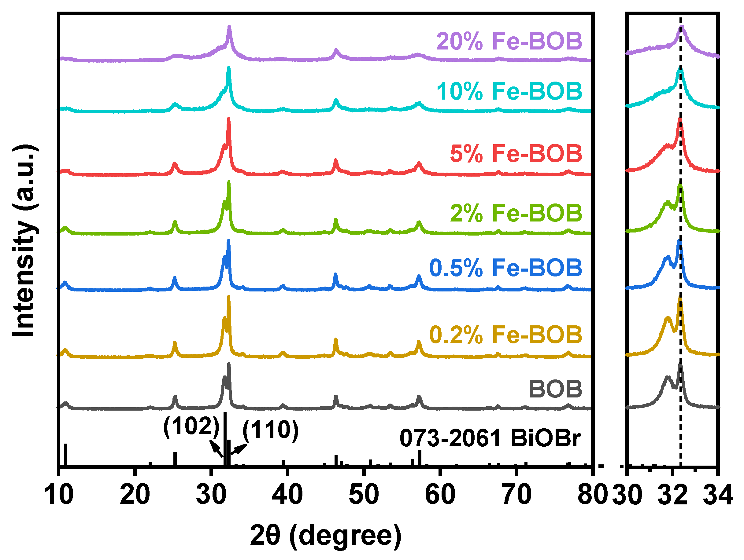
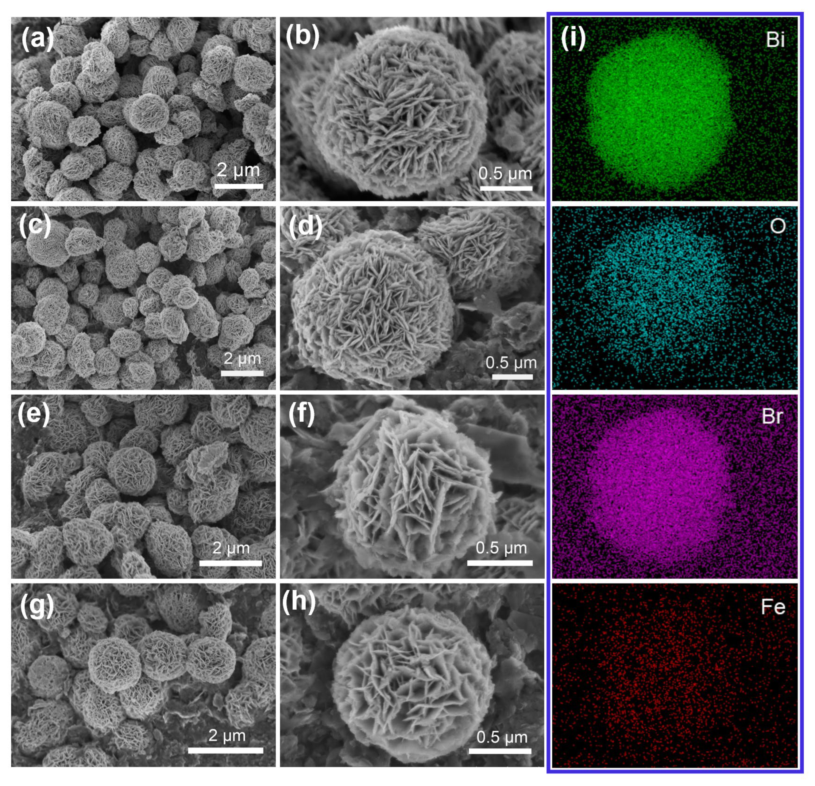
2.1. Characterization of the Fe-BOB Catalysts

Figure 1 shows the powder X-ray diffraction (XRD) patterns of BOB and Fe-BO samples doped with Fe in different molar percentages (0.2%, 0.5%, 2%, 5%, 10%, and 20%). The results showed that all the diffraction peaks of BOB were in good agreement with the standard XRD patterns of tetragonal BiOBr (JCPDF No. 01-073-2061). The characteristic diffraction peaks at 10.9°, 31.8°, and 32.3° correspond to the (001), (102), and (110) crystal planes, respectively. Compared with the pure BiOBr (BOB) sample, no other diffraction peaks were detected in the Fe-BOB patterns, and there was no shift in the magnified XRD pattern either, indicating that Fe doping has almost no effect on the phase structure of the BiOBr sample. However, the intensity ratio of the (102)/(110) crystal plane of Fe-BOB samples gradually decreases with the increase in Fe doping amount, and the characteristic diffraction peak becomes wider and intensity decreases at the same time, indicating that the crystallinity of the samples gradually decreases. The Fe weight percentage of Fe-BOB samples was measured by atomic absorption spectroscopy (AAS), which was 0.18%, 0.30%, 1.67%, 4.01%, 7.76%, and 15.28% for 0.5%, 2%, 5%, 10%, and 20% of Fe-doped BOB samples, respectively (Table S1 and Figure S1). Figure S2 presents the ultraviolet–visible diffuse reflection spectrum (UV–vis DRS) of BOB and Fe-BOB samples. The results indicate that Fe doping enhances the visible light absorption capacity of BOB sample.
As illustrated in the scanning electron microscope (SEM) images (Figure 2), both the BOB and Fe-BOB samples exhibited a microspherical morphology, with an average diameter of approximately 1.45 μm. These microspheres demonstrated three-dimensional architectures composed of irregular nanosheets, each with a thickness ranging from 10 to 20 nm. With the increase in Fe doping, the arrangement of nanosheets showed a more disordered and loose state (Figure 2c–h). The energy-dispersive X-ray spectroscopy (EDS) of the 5% Fe-BOB sample evidenced the uniform distribution of Bi, Br, O, and Fe (Figure 2i). As displayed in Figure S3 (Supporting Information), the weight percentage of Fe was measured to be around 0.65% (converted to a mass fraction is 3.63%), which is close to the result measured by AAS. The nitrogen isothermal adsorption–desorption curve and pore size distribution of the synthesized sample were shown in Figure S4. The measurement results showed typical type IV isotherm and type H3 hysteresis loop, indicating that these materials have obvious mesoporous structures [29]. The specific surface areas of the BOB, 0.5% Fe-BOB, 5% Fe-BOB, and 20% Fe-BOB samples were 26.88, 29.69, 34.95, and 43.55 g/m2, respectively, and the average pore diameters were 18.03 nm, 17.45 nm, 15.57 nm, and 10.48 nm, respectively. It indicates that Fe doping can increase the specific surface area of BiOBr and reduce the pore size of BiOBr.
The chemical states of the surface elements (Bi, O, Br, Fe) of BOB and Fe-BOB were analyzed by X-ray photoelectron spectroscopy (XPS). Figure 3a showed the fine spectrum of Bi 4f of different samples. The peaks with binding energies of 159.4 and 164.7 eV belong to the characteristic peaks of the Bi 4f 7/2 and Bi 4f 5/2 orbitals for Bi3+ [30]. The peaks at the positions of binding energy around 530.2 and 531.8 eV in the O 1s fine spectrum correspond to lattice oxygen atoms (Bi-O) and surface oxygen vacancies (OVs) in BiOBr, respectively [31]. And the peak with a binding energy of 533.2 eV is attributed to the surface hydroxyl group (Figure 3b) [32]. Furthermore, the peaks at 68.5 and 69.5 eV in Figure 3c are characteristic peaks of the Br 3d 5/2 and Br 3d 3/2 orbitals, respectively [30]. In the Fe 2p fine spectrum (Figure 3d), 5% of the Fe-BOB sample contains two valence states of Fe elements, in which the peak with binding energy of 710.3 and 723.9 eV is attributed to Fe (II), and the peak with 714.5 and 728.7 eV is attributed to Fe (III) [33,34].

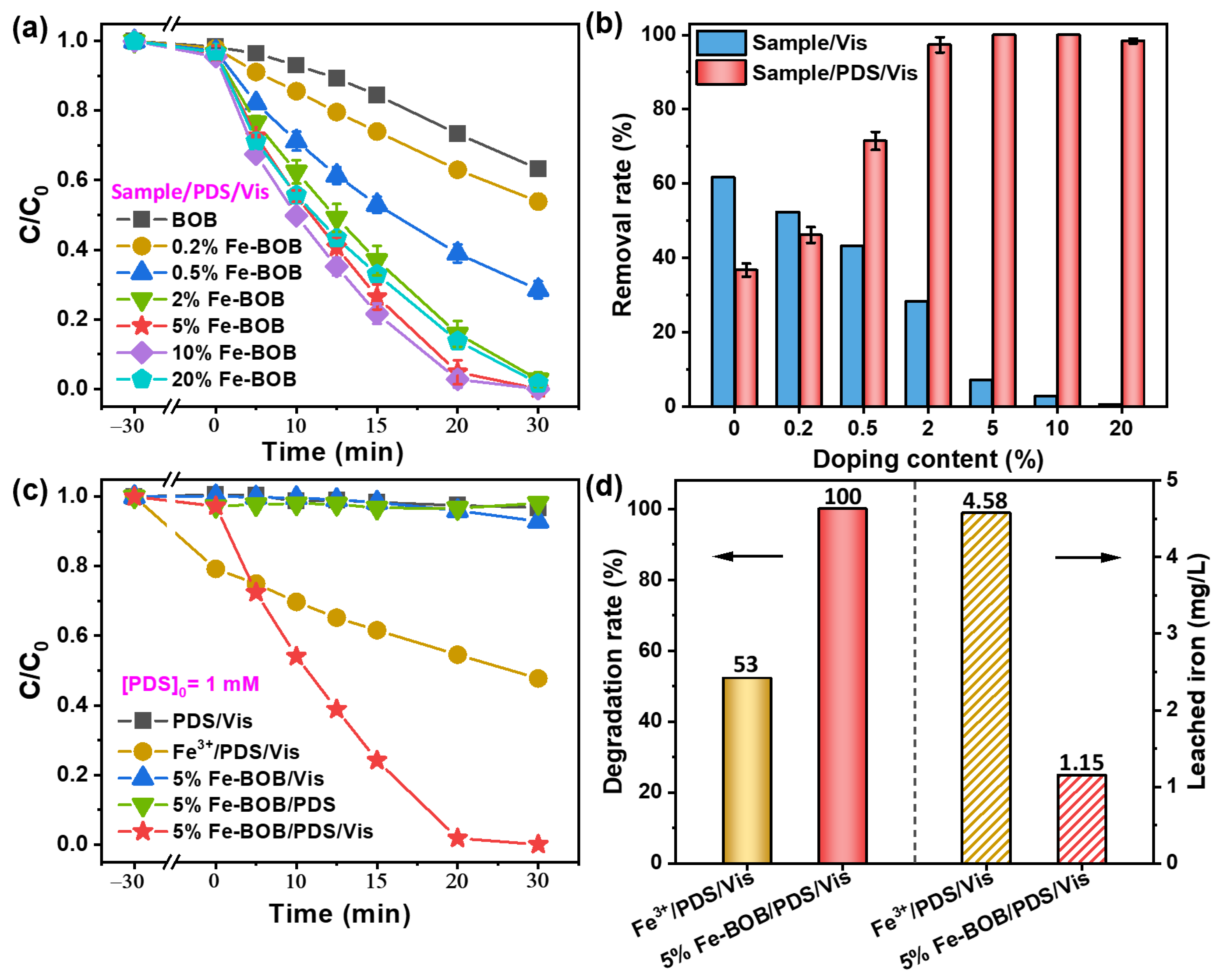
2.2. Evaluation of PDS Activation Performance of Fe-BOB Under Visible Light

The PDS activation activity of Fe-BOB samples with different Fe doping amounts (0.2%, 0.5%, 2%, 5%, 10%, and 20%) was evaluated by the degradation of APAP in visible light, in which the initial concentration of PDS in the reaction system was all 1 mmol/L. As shown in Figure 4a, with the increase in Fe content, the activity of Fe-BOB activating PDS to degrade APAP was first increased and then decreased. Both 5% and 10% Fe-BOB exhibited superior photocatalytic activity compared to the other samples, demonstrating the ability to completely degrade APAP within a duration of 30 min. It is worth noting that the 5% Fe-BOB/PDS system demonstrates a significant advantage in APAP degradation efficiency compared with other systems reported in the existing literature (Table S2). For comparison, the photocatalytic properties of BOB and Fe-BOB samples were tested without adding PDS. As shown in Figure 4b, within 40 min of light exposure, the photocatalytic degradation rate of APAP by BOB is about 60%. With the increase in Fe doping, the photocatalytic degradation activity of APAP over Fe-BOB gradually decreased. When the doping amount of Fe is 0.5~20%, the degradation rate of APAP over Fe-BOC/PDS was much higher than that of Fe-BOC. Furthermore, it was observed that there was only a minimal decrease in APAP levels under the PDS/Vis and 5% Fe-BOB/PDS systems (Figure 4c), indicating the exceptionally high catalytic activity of Fe-BOB for PDS activation when subjected to visible light irradiation. As a control of 5% Fe-BOB/PDS/Vis, an equal amount of Fe3+ (4.58 mg/L) was input into the system of PDS/Vis, whereas the degradation rate of APAP was only 53% under the same experimental conditions. Subsequently, the concentration of the released Fe3+ in aqueous solution after degradation of APAP in the system of 5% Fe-BOB/PDS/Vis was determined by AAS, which was only 1.15 mg/L (Figure 4d). The results demonstrated that the excellent activity of 5% Fe-BOB/PDS/Vis was highly attributed to the activation of PDS over Fe-BOB sample, although Fe3+ in solution has certain activation ability to PDS under visible light. In addition, the photocatalytic degradation rates of 5% Fe-BOB/PDS/Vis on carbamazepine (CBZ), sulfamethoxazole (SMX), bisphenol A (BPA), 2,4-dichlorophenol (2,4-DCP), and atrazine (ATZ) was 100%, 49%, 100%, 84%, and 68%, while it was only 63%, 22%, 29%, 18%, and 4% for that of 5% Fe-BOB/Vis without PDS, respectively (Figure S5). This result indicated that the activation of PDS by 5% Fe-BOB samples under visible light can promote the degradation of many organic pollutants.

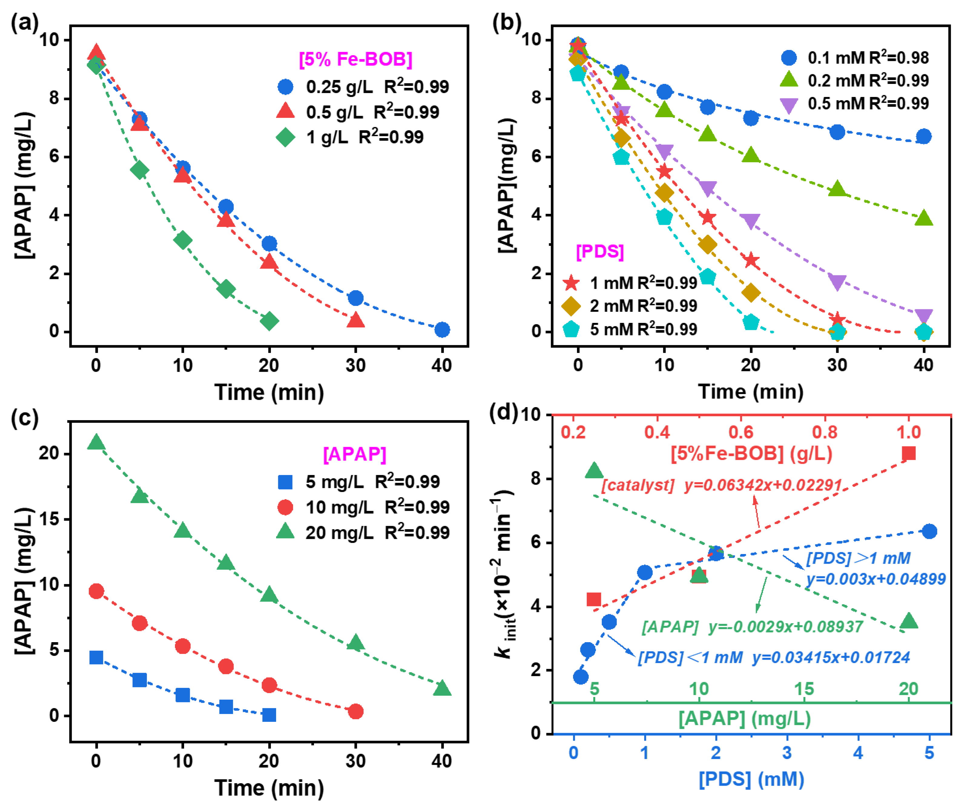
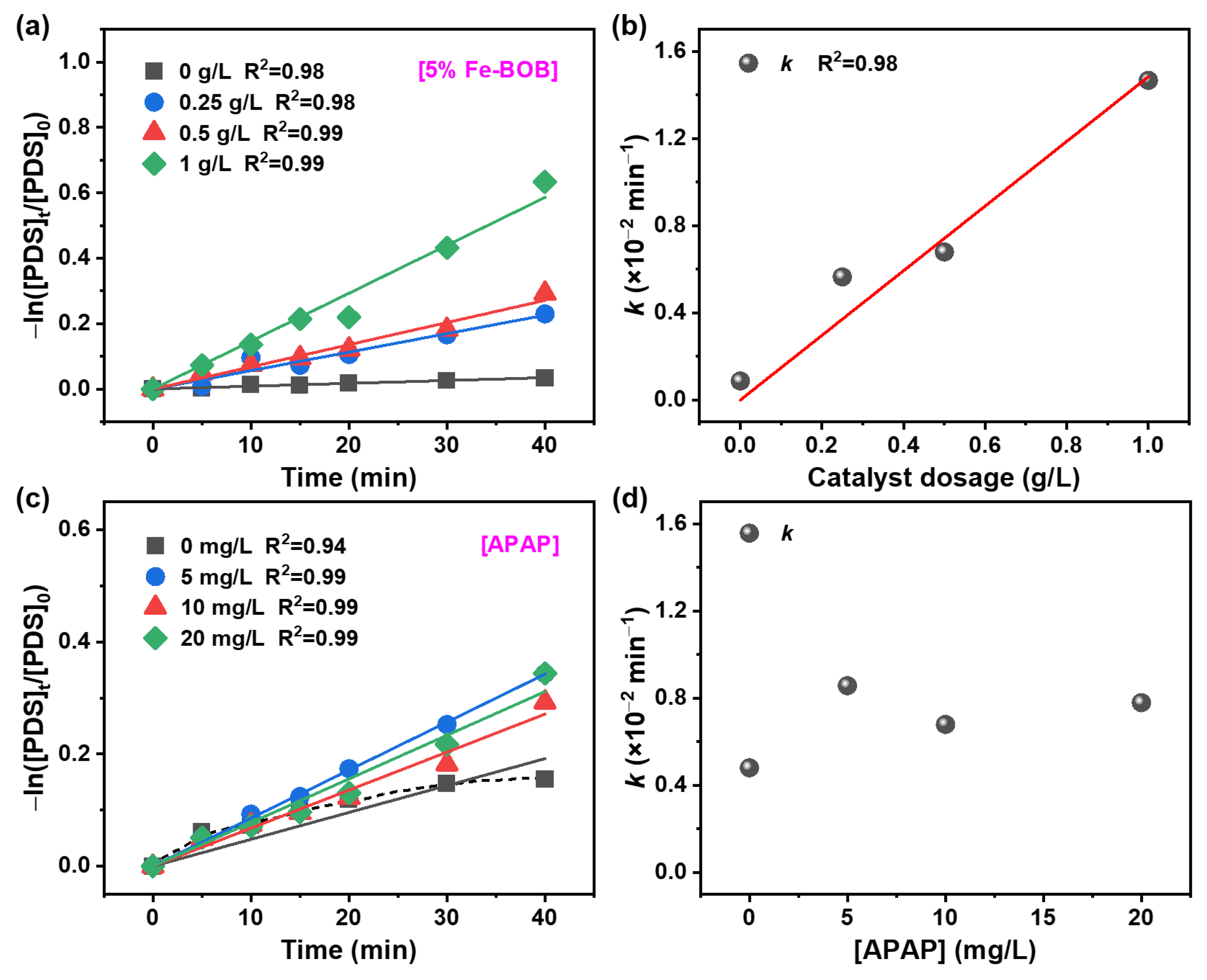
2.3. Degradation Kinetics of APAP over Fe-BOB/PDS

Firstly, the pseudo-first-order kinetic equation (Equation (S1), Supporting Information) was used to fit the process of activating PDS to degrade APAP over different Fe-BOB samples under visible light. As the results displayed in Figure S6a, when the doping amount of Fe exceeds 0.5%, the correlation coefficients of the fitted equations were relatively low, indicating a poor fit and potential deviations from an ideal pseudo-first-order rate model. As detailed in Equations (S2)–(S4), the retarded first-order model incorporates a retardation coefficient (α) to account for temporal declines in apparent reaction rate constants (kt), which may arise due to factors such as accumulation of degradation intermediates, catalyst surface saturation, or mass transfer limitations [35,36]. These considerations are particularly pertinent in heterogeneous systems like ours, where dynamic changes at catalyst surfaces and within the solution phase can impede degradation rates over time [37]. As illustrated in Figure S6b and Table S3, the retarded first-order model yielded excellent linear fits with R2 > 0.99 for all Fe-BOB/PDS/Vis degradation processes, indicating the appropriateness of this kinetic model in our system. With the increase in iron doping amount, the reaction rate constant of Fe-BOB samples activated PDS to degrade APAP initially increased and then decreased (Table S3), indicating that appropriate iron doping amount can improve the activity of BiOBr0activated PDS to degrade APAP. Figure 5 shows the delayed first-order kinetic fitting results of the degradation process of APAP over 5% Fe-BOB/PDS/Vis system under different catalysts, PDS dosages, and APAP concentrations, respectively. As shown in the corresponding kinetic parameters in Table S4, the correlation coefficients of all fitted equations are above 0.98, indicating the degradation process of APAP in the above-mentioned reactions can be fitted with delayed first-order kinetics. The results in Figure 5a,b showed that the increase of catalyst or PDS concentration was conducive to the degradation of APAP in a certain concentration range. In addition, the higher the concentration of APAP, the longer it takes for APAP to completely degrade (Figure 5c). As shown in Figure 5d, the catalyst concentration and the APAP concentration have a linear relationship with the corresponding k value, and the slopes were 0.06342 and −0.0029, respectively. The larger slope value of the former indicated that the 5% Fe-BOB sample concentration has a greater effect on the reaction rate. When the concentration of PDS was below 1 mM and above 1 mM, there was a good linear relationship with the corresponding k value, and the slopes were 0.03415 and 0.003, respectively. This result indicated that the change in the initial PDS concentration within 1 mM (including 1 mM) has a great impact on the degradation of APAP in the system.
To explore the role of PDS in the degradation process of APAP, the concentration of PDS in the degradation process of APAP by 5% Fe-BOB/PDS/Vis under different reaction conditions was measured. The pseudo-first-order kinetic model was used to fit the consumption process of PDS. As shown in Figure 6a, the self-decomposition degree of PDS was extremely low under visible light. While the consumption of PDS increased gradually with the extension of illumination time over different concentrations of 5% Fe-BOB sample, and all the linear correlation coefficients of PDS consumption reached above 0.98. In addition, the value of k of the consumption rate constant of PDS was positively correlated with the concentration of 5% Fe-BOB (Figure 6b), demonstrating that the Fe-BOB sample in the system was the key factor promoting the decomposition of PDS. The influence of different APAP concentrations on PDS consumption were shown in Figure 6c,d. The results showed that the consumption rate constant k value of PDS has no significant relationship with the initial concentration of APAP. Therefore, the presence of Fe-BOB catalyst was a direct factor affecting the activation and decomposition of PDS.

2.4. Identifying and Distributions of Reactive Oxygen Species

To identify the active species during the process of 5% Fe-BOB activating PDS to degrade APAP under visible light irradiation, trapping experiments were carried out. Methanol (MeOH) and tertiary butanol (TBA) with a concentration of 100 mM (100 times the concentration of PDS) were selected as quenching agents of •SO4 and •OH, respectively, and L-histidine and sodium oxalate (SO) with a concentration of 50 mM were selected as trapping agents of 1O2 and h+, respectively. As shown in Figure 7a,b, the addition of TBA and MeOH have little effect on the degradation of APAP, while the photodegradation rate of APAP was significantly inhibited after the addition of L-histidine and SO. These results illustrated that 1O2 and h+ were the main active species for APAP degradation in the Fe-BOB/PDS system. Then, the active species in the reaction process are further determined through the electron paramagnetic resonance (EPR) experiments. As illustrated in Figure 7c, no obvious •SO4 signal was detected in the EPR spectra of the 5% Fe-BOB/PDS system, regardless of whether under dark or light conditions. This indicates that no •SO4 was generated. The characteristic signal of DMPO-•OH with an intensity of 1:2:2:1 was observed in Fe-BOB/PDS system under dark conditions. In contrast, the intensity of the •OH signal remained unchanged for the Fe-BOC/PDS system after exposure to light, indicating that light could not promote the production of •OH. The detected •OH might be attributed to a direct reaction between PDS and surface Fe species on BiOBr, as well as the hydrolysis of •SO4, as illustrated in Equations (1) and (2) [38,39,40]. From Figure 7d, it was found that PDS itself did not produce 1O2 under light, while 5% Fe-BOB/PDS has obvious characteristic tetramethylpiperidine-1O2 (TEMP-1O2) signals under dark, and the signal intensity becomes stronger under light irradiation. The 1O2 might generate form the energy transfer between materials and molecular oxygen [41].
≡Fe2+ + S2O82− → •SO4 + ≡Fe3+ + SO42−
•SO4 + H2O → •OH + SO42− + H+

2.5. Stability Analysis and Effects of Solution pH on APAP Photodegradation over Fe-BOB/PDS System

In addition, the stability of photocatalysts is an important indicator for evaluating their practical application prospects. The cyclic experiment results are shown in Figure 8a; after ten cycles, the 5% Fe-BOB sample still remained high activity for activating PDS to degrade APAP under visible light irradiation, demonstrating its good stability. The slight decline in degradation efficiency might be due to the mass loss of the photocatalyst during the cycling process. Furthermore, there was no significant difference between the XRD pattern of the recovered sample after 10 cycles of experiment and the 5% Fe-BOB sample before reaction, which further demonstrated its good stability (Figure 8b). Considering the complexity of practical application, the effects of pH values on APAP photodegradation over 5% Fe-BOB/PDS were also investigated (Figure 8c). At pH 3, 5, 7, 9, and 11, the degradation rate of APAP all can reach more than 85% at 40 min, which demonstrated that 5% Fe-BOB can activate PDS to degrade APAP in a wide range of pH values, indicative its good practical application potential. The reaction rate of APAP in the 5% Fe-BOB/PDS/Vis system varies under different pH conditions. This variation may be attributed to the influence of pH value on the adsorption capacity of the catalyst surface for APAP. For instance, at a pH of 9, 5% Fe-BOB exhibits the lowest adsorption capacity for APAP, resulting in a correspondingly lowest degradation rate of APAP.

3. Conclusions

In this study, BiOBr samples with varying amounts of Fe doping were prepared by solvothermal method, and their efficacy in activating peroxydisulfate (PDS) for the degradation of APAP under visible light was evaluated. The results indicate that as the Fe doping concentration increases, the degradation efficiency of PDS activated by BiOBr on APAP under visible light initially improves and then slightly declines, with the 5% Fe-BOB sample exhibiting the highest photocatalytic activity. Moreover, the 5% Fe-BOB/PDS/Vis system can also effectively degrade drugs such as CBZ and SMX, as well as organic pollutants such as BPA, 2,4-DCP, and ATZ. The study of Fe-BOB/PDS degradation kinetics of APAP shows that the amount of catalyst has the greatest influence on the reaction. Furthermore, the consumption rate constant k value of PDS was positively correlated with the catalyst dose, but not with the APAP concentration, indicating that the activation of PDS in this process was mainly related to the catalyst dosage. The active species responsible for degrading APAP via PDS activation under visible light were predominantly h+ and 1O2. The Fe-BOC samples demonstrate good stability for PDS activated photocatalysis, and they can effectively activate PDS to degrade APAP across a broad pH range in solution, highlighting their potential for practical applications.

Supplementary Materials

The following supporting information can be downloaded at: https://www.mdpi.com/article/10.3390/catal15060594/s1, Figure S1. The Fe weight percentage of Fe-BOB samples was measured by atomic absorption spectroscopy (AAS) and the theoretical Fe content of Fe-BOB samples. Figure S2. UV–Vis diffuse reflectance spectra (a), and plots of (ahv)1/2 vs the photon energy (hv) of (b) of BOB and Fe-BOB samples. Figure S3. N2 adsorption–desorption isotherm and the corresponding pore size distribution plots of BOB (a), 0.5% Fe-BOB (b), 5% Fe-BOB (c), and 20% Fe-BOB (d). Figure S4. EDX spectrum of 5% Fe-BOB sample. Figure S5. The degradation of different organic pollutants over 5% Fe-BOB/PDS/Vis system (a) and its comparison with 5% Fe-BOB/Vis system (b). Figure S6. Pseudo first-order (a) and retarded first-order kinetic fitting curves (b) of APAP degradation by PDS activation with different Fe-BOB samples under visible light. Table S1. Fe content in different Fe-BOB samples determined by AAS. Table S2. Comparison of APAP degradation over various catalysts via PMS or PDS activation. Table S3. Retarded rate model fitting parameters for APAP degradation by Fe-BOB. Table S4. Retarded first-order kinetic parameters of APAP degradation by 5% Fe-BOB/PDS/Vis under different reaction conditions. References [35,36,37,42,43,44,45] are cited in the Supplementary Materials.

Author Contributions

Z.W.: Resources, Formal Analysis, Writing—Original Draft. M.C.: Investigation, Methodology, Validation. Q.L.: Conceptualization, Writing—Original Draft. R.C.: Funding Acquisition, Writing—Review and Editing. All authors have read and agreed to the published version of the manuscript.

Funding

This work was supported by the National Natural Science Foundation of China (22076149), the Innovative Team Program of Natural Science Foundation of Hubei Province (2023AFA027), and Special Project of State Key Laboratory of New Textile Materials & Advanced Processing Technologies from Wuhan Science and Technology Bureau (2022013988065204).

Data Availability Statement

The data presented in this study are openly available.

Conflicts of Interest

The authors declare no conflicts of interest.

References

  1. He, Z.X.; Cheng, X.R.; Kyzas, G.Z.; Fu, J. Pharmaceuticals pollution of aquaculture and its management in China. J. Mol. Liq. 2016, 223, 781–789. [Google Scholar] [CrossRef]
  2. Peng, Q.C.; Song, J.M.; Li, X.G.; Yuan, H.M.; Liu, M.T.; Duan, L.Q.; Zuo, J.L. Pharmaceutically active compounds (PhACs) in surface sediments of the Jiaozhou Bay, north China. Environ. Pollut. 2020, 266, 115245–115257. [Google Scholar] [CrossRef]
  3. Hu, X.; Zhu, M. Were persulfate-based advanced oxidation processes really understood? Basic concepts, cognitive biases, and experimental details. Environ. Sci. Technol. 2024, 58, 10415–10444. [Google Scholar] [CrossRef] [PubMed]
  4. Nie, C.Y.; Wang, J.L.; Cai, B.H.; Lai, B.; Wang, S.B.; Ao, Z.M. Multifunctional roles of MoS2 in persulfate-based advanced oxidation processes for eliminating aqueous organic pollutants: A review. Appl. Catal. B Environ. Energy 2024, 340, 123173. [Google Scholar] [CrossRef]
  5. Ming, H.B.; Bian, X.Q.; Cheng, J.J.; Yang, C.; Hou, Y.D.; Ding, K.N.; Zhang, J.S.; Anpo, M.; Wang, X.C. Carbon nitride with a tailored electronic structure toward peroxymonosulfate activation: A direct electron transfer mechanism for organic pollutant degradation. Appl. Catal. B Environ. Energy 2024, 341, 123314. [Google Scholar] [CrossRef]
  6. Li, Y.F.; Wang, D.B.; Yang, G.J.; Yuan, X.Z.; Xu, Q.X.; Liu, X.R.; Wang, Y.L.; Guan, R.P.; Fu, Q.Z.; Chen, F. The novel pretreatment of Co2+ activating peroxymonosulfate under acidic condition for dewatering waste activated sludge. J. Taiwan Inst. Chem. E 2019, 102, 259–267. [Google Scholar] [CrossRef]
  7. Wang, J.Q.; Hasaer, B.; Yang, M.; Liu, R.; Hu, C.Z.; Liu, H.J.; Qu, J.H. Anaerobically-digested sludge disintegration by transition metal ions-activated peroxymonosulfate (PMS): Comparison between Co2+, Cu2+, Fe2+and Mn2+. Sci. Total Environ. 2020, 713, 136530–136540. [Google Scholar] [CrossRef]
  8. Tang, W.J.; Wang, Z.J.; Guo, S.; Chen, R.; Chen, F.X. Efficient generation of 1O2 by activating peroxymonosulfate on graphitic carbon nanoribbons for water remediation. npj Clean Water 2024, 7, 92. [Google Scholar] [CrossRef]
  9. Tang, H.; Dai, Z.; Xie, X.D.; Wen, Z.P.; Chen, R. Promotion of peroxydisulfate activation over Cu0.84Bi2.08O4 for visible light induced photodegradation of ciprofloxacin in water matrix. Chem. Eng. J. 2019, 356, 472–482. [Google Scholar] [CrossRef]
  10. Liu, Y.P.; Gong, Y.H.; Cui, X.; Lu, Y.; Yu, H.B.; Qin, W.C.; Huo, M.X. Visible-light activation of persulfate by a Z-scheme photocatalyst Fe-C3N4/Bi2Sn2O7 for tetracycline degradation. Appl. Catal. A Gen. 2023, 666, 119422. [Google Scholar] [CrossRef]
  11. Xin, Y.; Wang, Y.J.; Jiang, Z.Q.; Deng, B.L.; Jiang, Z.J. Advances in the removal of organic pollutants from water by photocatalytic activation of persulfate: Photocatalyst modification strategy and reaction mechanism. Chemsuschem 2024, 17, e202400254. [Google Scholar] [CrossRef] [PubMed]
  12. Yang, C.; Zhang, G.H.; Meng, Y.; Pan, G.X.; Ni, Z.M.; Xia, S.J. Direct Z-scheme CeO2/@LDH core-shell heterostructure for photodegradation of Rhodamine B by synergistic persulfate activation. J. Hazard. Mater. 2021, 408, 124908. [Google Scholar] [CrossRef] [PubMed]
  13. Hu, Y.; Wang, X.; Wang, Y.; Li, S.; Cui, B.; Du, Y. Persulfate activation of photocatalysts based on S-scheme Bi3NbO7/BiOBr0.75I0.25 nanosheet heterojunctions for degradation of organic pollutants. ACS Appl. Nano Mater. 2023, 6, 10768–10778. [Google Scholar] [CrossRef]
  14. Guo, S.; Liu, Y.; Li, J. Nanocatalysts for the degradation of refractory pollutants. Catalysts 2024, 14, 444. [Google Scholar] [CrossRef]
  15. Du, X.; Bai, X.; Xu, L.; Yang, L.; Jin, P.K. Visible-light activation of persulfate by TiO2/g-C3N4 photocatalyst toward efficient degradation of micropollutants. Chem. Eng. J. 2020, 384, 123245–123258. [Google Scholar] [CrossRef]
  16. Zhang, M.X.; He, J.; Chen, Y.B.; Liao, P.Y.; Liu, Z.Q.; Zhu, M.S. Visible light-assisted peroxydisulfate activation via hollow copper tungstate spheres for removal of antibiotic sulfamethoxazole. Chinese Chem. Lett. 2020, 31, 2721–2724. [Google Scholar] [CrossRef]
  17. Zhong, X.; Ye, X.Y.; Wu, D.; Zhang, K.X.; Huang, W. A facile heterogeneous system for persulfate activation by CuFe2O4 under LED light irradiation. RSC Adv. 2019, 9, 32328–32337. [Google Scholar] [CrossRef]
  18. Zhang, G.; Wu, Z.; Liu, H.J.; Ji, Q.H.; Qu, J.H.; Li, J.H. Photoactuation healing of α-FeOOH@g-C3N4 catalyst for efficient and stable activation of persulfate. Small 2017, 13, 1702225–1702232. [Google Scholar] [CrossRef]
  19. Hu, H.; Zhang, H.X.; Chen, Y.; Chen, Y.J.; Zhuang, L.; Ou, H.S. Enhanced photocatalysis degradation of organophosphorus flame retardant using MIL-101(Fe)/persulfate: Effect of irradiation wavelength and real water matrixes. Chem. Eng. J. 2019, 368, 273–284. [Google Scholar] [CrossRef]
  20. Wang, M.H.; Yang, L.Y.; Guo, C.P.; Liu, X.F.; He, L.H.; Song, Y.P.; Zhang, Q.X.; Qu, X.W.; Zhang, H.Z.; Zhang, Z.H.; et al. Bimetallic Fe/Ti-based metal-organic framework for persulfate-assisted visible light photocatalytic degradation of orange II. Chemistryselect 2018, 3, 3664–3674. [Google Scholar] [CrossRef]
  21. Shen, Z.R.; Luo, Z.H.; Chen, J.Y.; Li, Y.W. Oxygen vacancy-mediated exciton effect in hierarchical BiOBr enables dichotomy of energy transfer and electron transfer in photocatalysis. Adv. Funct. Mater. 2023, 33, 2213935. [Google Scholar] [CrossRef]
  22. Wang, X.S.; Gao, R.; Fan, G.L.; Guo, Y.; Han, C.H.; Gao, Y.L.; Shen, A.; Wu, L.M.; Gu, X.J. Dual defects-induced iron single atoms immobilized in metal-organic framework-derived hollow BiOBr microtubes for low-barrier photocatalytic nitrogen reduction. Angew. Chem. Int. Ed. 2025, 64, e202501297. [Google Scholar] [CrossRef]
  23. Cui, D.D.; Wang, L.; Du, Y.; Hao, W.C.; Chen, J. Photocatalytic reduction on bismuth-based p-block semiconductors. ACS Sustain. Chem. Eng. 2018, 6, 15936–15953. [Google Scholar] [CrossRef]
  24. Zhang, T.Q.; Chu, S.P.; Li, J.; Wang, L.L.; Chen, R.; Shao, Y.; Liu, X.W.; Ye, M.M. Efficient degradation of aqueous carbamazepine by bismuth oxybromide-activated peroxide oxidation. Catalysts 2017, 7, 315. [Google Scholar] [CrossRef]
  25. Bao, Y.P.; Lee, W.J.; Guan, C.T.; Liang, Y.N.; Lim, T.T.; Hu, X. Highly efficient activation of peroxymonosulfate by bismuth oxybromide for sulfamethoxazole degradation under ambient conditions: Synthesis, performance, kinetics and mechanisms. Sep. Purif. Technol. 2021, 276, 119203–119210. [Google Scholar] [CrossRef]
  26. Liu, C.; Mao, S.; Wang, H.; Wu, Y.; Wang, F.; Xia, M.; Chen, Q. Peroxymonosulfate-assisted for facilitating photocatalytic degradation performance of 2D/2D WO3/BiOBr S-scheme heterojunction. Chem. Eng. J. 2022, 430, 132806. [Google Scholar] [CrossRef]
  27. Dou, X.; Chen, Y.; Shi, H. CuBi2O4/BiOBr composites promoted PMS activation for the degradation of tetracycline: S-scheme mechanism boosted Cu2+/Cu+ cycle. Chem. Eng. J. 2022, 431, 134054. [Google Scholar] [CrossRef]
  28. Lin, Z.; Guo, Y.; Qu, X.; Xiang, Y.; Shen, S.; Zhu, X. Degradation of bisphenol A by Fe-doped BiOBr enhanced UV/persulfate system: Significant role of superoxide radicals. Process Saf. Environ. 2023, 180, 148–159. [Google Scholar] [CrossRef]
  29. Liu, C.; Mao, S.; Shi, M.X.; Wang, F.Y.; Xia, M.Z.; Chen, Q.; Ju, X.H. Peroxymonosulfate activation through 2D/2D Z-scheme CoAl-LDH/BiOBr photocatalyst under visible light for ciprofloxacin degradation. J. Hazard. Mater. 2021, 420, 126613–126626. [Google Scholar] [CrossRef]
  30. Liu, Q.B.; Bai, C.; He, D.; Wu, X.T.; Wu, Y.P.; Chen, R. Oxygen vacancy mediation of BiOX (X=Cl, Br) towards enhanced photocatalytic CO2 reduction activity. J. Environ. Chem. Eng. 2024, 12, 112449. [Google Scholar] [CrossRef]
  31. Wang, Q.; Liu, Z.Q.; Liu, D.M.; Liu, G.S.; Yang, M.; Cui, F.Y.; Wang, W. Ultrathin two-dimensional BiOBrxI1-x solid solution with rich oxygen vacancies for enhanced visible-light-driven photoactivity in environmental remediation. Appl. Catal. B Environ. 2018, 236, 222–232. [Google Scholar] [CrossRef]
  32. Li, X.B.; Liu, Q.; Deng, F.; Huang, J.T.; Han, L.; He, C.Z.; Chen, Z.; Luo, Y.D.; Zhu, Y.F. Double-defect-induced polarization enhanced OV-BiOBr/Cu2-xS high-low junction for boosted photoelectrochemical hydrogen evolution. Appl. Catal. B Environ. Energy 2022, 314, 121502–121512. [Google Scholar] [CrossRef]
  33. Liu, Y.; Hu, Z.F.; Yu, J.C. Fe enhanced visible-light-driven nitrogen fixation on BiOBr nanosheets. Chem. Mater. 2020, 32, 1488–1494. [Google Scholar] [CrossRef]
  34. Bai, C.B.; Zhang, Y.H.; Liu, Q.; Zhu, C.X.; Li, J.; Chen, R. Interfacial complexation between Fe3+ and Bi2MoO6 promote efficient persulfate activation via Fe3+/Fe2+ cycle for organic contaminates degradation upon visible light irradiation. J. Colloid Interface Sci. 2024, 664, 238–250. [Google Scholar] [CrossRef]
  35. Wang, L.L.; Cheng, H.F. Birnessite (δ-MnO2) mediated degradation of organoarsenic feed additive p-arsanilic acid. Environ. Sci. Technol. 2015, 49, 3473–3481. [Google Scholar] [CrossRef]
  36. Yu, L.; Wang, C.P.; Ren, X.H.; Sun, H.W. Catalytic oxidative degradation of bisphenol A using an ultrasonic-assisted tourmaline-based system: Influence factors and mechanism study. Chem. Eng. J. 2014, 252, 346–354. [Google Scholar] [CrossRef]
  37. Huang, S.N.; Tian, F.; Dai, J.W.; Tian, X.S.; Li, G.F.; Liu, Y.L.; Chen, Z.Q.; Chen, R. Highly efficient degradation of chlorophenol over bismuth oxides upon near-infrared irradiation: Unraveling the effect of Bi-O-Bi-O defects cluster and 1O2 involved process. Appl. Catal. B Environ. 2021, 298, 120576. [Google Scholar] [CrossRef]
  38. Jawad, A.; Zhan, K.; Wang, H.; Shahzad, A.; Zeng, Z.; Wang, J.; Zhou, X.; Ullah, H.; Chen, Z.; Chen, Z. Tuning of persulfate activation from a free radical to a nonradical pathway through the incorporation of non-redox magnesium oxide. Environ. Sci. Technol. 2020, 54, 2476–2488. [Google Scholar] [CrossRef]
  39. Kim, H.-H.; Lee, H.; Lee, D.; Ko, Y.-J.; Woo, H.; Lee, J.; Lee, C.; Pham, A.L.-T. Activation of hydrogen peroxide by a titanium oxide-supported iron catalyst: Evidence for surface Fe(IV) and its selectivity. Environ. Sci. Technol. 2020, 54, 15424–15432. [Google Scholar] [CrossRef]
  40. Bai, C.; Guo, W.; Liu, Q.; Li, G.; Guo, S.; Chen, R. Cu2O/BiVO4 heterostructure controllably triggers radical and non-radical persulfate activation via light “on-off” for efficient organic contaminants degradation. Appl. Catal. B Environ. Energy 2024, 344, 123606. [Google Scholar] [CrossRef]
  41. Ding, J.; Dai, Z.; Tian, F.; Zhou, B.; Zhao, B.; Zhao, H.; Chen, Z.; Liu, Y.; Chen, R. Generation of defect clusters for 1O2 production for molecular oxygen activation in photocatalysis. J. Mater. Chem. A 2017, 5, 23453–23459. [Google Scholar] [CrossRef]
  42. Tan, C.; Xu, Q.; Sheng, T.; Cui, X.; Wu, Z.; Gao, H.; Li, H. Reactive oxygen species generation in FeOCl nanosheets activated peroxymonosulfate system: Radicals and non-radical pathways. J. Hazard. Mater. 2020, 398, 123084. [Google Scholar] [CrossRef] [PubMed]
  43. Sin, A.; Machala, L.; Kim, M.; Baďura, Z.; Petr, M.; Polaskova, M.; Novak, P.; Nadagouda, M.N.; Dionysiou, D.D.; Han, C. Development of tungsten-modified iron oxides to decompose an over-the-counter painkiller, Acetaminophen by activating peroxymonosulfate. Sci. Total. Environ. 2024, 951, 175472. [Google Scholar] [CrossRef] [PubMed]
  44. Chen, L.; Maqbool, T.; Fu, W.; Yang, Y.; Hou, C.; Guo, J.; Zhang, X. Highly efficient manganese (III) oxide submerged catalytic ceramic membrane for nonradical degradation of emerging organic compounds. Sep. Purif. Technol. 2022, 295, 121272. [Google Scholar] [CrossRef]
  45. Xue, Y.; Gao, R.; Lin, S.; Zhong, Q.; Zhang, Q.; Hong, J. Regulating the interface electron distribution of iron-based MOFs through ligand functionalization enables efficient peroxymonosulfate utilization and catalytic performance. J. Colloid Interface Sci. 2024, 663, 358–368. [Google Scholar] [CrossRef]
Figure 1. XRD and magnification patterns of BiOB and Fe-doped BiOBr.
Catalysts 15 00594 g001
Figure 2. SEM images of BOB (a,b), 0.5% Fe-BOB (c,d), 5% Fe-BOB (e,f), and 20% Fe-BOB (g,h); EDX elemental mapping images of 5% Fe-BOB sample (i).
Catalysts 15 00594 g002
Figure 3. High-resolution XPS spectra of Bi 4f (a), O 1s (b), Br 3d (c), and Fe 2p (d) of BOB and 5% Fe-BOB sample.
Catalysts 15 00594 g003
Figure 4. Degradation of APAP with PDS activated by Fe-BOB samples with different Fe contents under visible light (a) and its comparison with Fe-BOB without PDS (b); degradation of APAP by 5% Fe-BOB under different conditions (c); degradation rate of APAP by Fe3+/PDS/Vis and 5% Fe-BOB/PDS/Vis systems and leached iron in solutions (d).
Catalysts 15 00594 g004
Figure 5. The retarded first-order kinetic fitting of APAP degradation by 5% Fe-BOB/PDS/Vis with different 5% Fe-BOB concentrations (a), PDS dosages (b), and APAP concentrations (c). Relationship between each variable and the corresponding k value (d).
Catalysts 15 00594 g005
Figure 6. Effects of 5% Fe-BOB concentration on PDS consumption (a); relationship between PDS consumption rate constant k and catalyst concentration (b); effects of APAP concentration on PDS consumption; (c) relationship between PDS consumption rate constant k and APAP concentration (d) under visible light irradiation.
Catalysts 15 00594 g006
Figure 7. Trapping experiments (a) and removal rate (b) of APAP over 5% Fe-BOB/PDS system; EPR spectra of DMPO-•OH (c) and TEMP-1O2 (d) over 5% Fe-BOB/PDS system in dark and upon light irradiation, respectively.
Catalysts 15 00594 g007
Figure 8. Removal rate of APAP during ten cycles in the activation of PDS over 5% Fe-BOB under visible light (a); XRD patterns of 5% Fe-BOB before and after ten cycles (b); degradation of APAP in 5% Fe-BOB/PDS/Vis system at different pH (c).
Catalysts 15 00594 g008
Disclaimer/Publisher’s Note: The statements, opinions and data contained in all publications are solely those of the individual author(s) and contributor(s) and not of MDPI and/or the editor(s). MDPI and/or the editor(s) disclaim responsibility for any injury to people or property resulting from any ideas, methods, instructions or products referred to in the content.

