Article

Improving the Thermal Stability of Xylanase XynASP from Aspergillus Saccharolyticus JOP 1030-1 Through Modular Assembly

1
College of Biological and Food Engineering, Huanghuai University, Zhumadian 463000, China
2
CAS Key Laboratory of Environmental and Applied Microbiology & Environmental Microbiology Key Laboratory of Sichuan Province, Chengdu Institute of Biology, Chinese Academy of Sciences, Chengdu 610041, China
3
Shanghai Beimoo Biotechnology Co., Ltd., Shanghai 201400, China
*
Author to whom correspondence should be addressed.
Catalysts 2025, 15(6), 563; https://doi.org/10.3390/catal15060563
Submission received: 30 April 2025 / Revised: 27 May 2025 / Accepted: 29 May 2025 / Published: 5 June 2025
(This article belongs to the Section Biocatalysis)

Abstract

Xylanases, important enzymes in the food industry, have severely limited use in industrial applications due to insufficient thermal stability. This study focused on improving the thermostability of XynASP, a glycoside hydrolase family 11 (GH11) xylanase from Aspergillus saccharolyticus JOP 1030-1, through modular assembly and rational mutagenesis. By aligning XynASP with nine thermostable GH11 homologs, six variable structural modules (β1, β3, β6, β7, α1, β14) and eight non-conserved residues were identified. Six chimeras (Z1, Z2, Z3, Z4, Z5, Z6) and eight single mutants (S131T, Y133T, A137G, A144T, T147Y, A156R, V198M, and Y204Q) were constructed. Among these, the β3-module-substituted chimera Z2 exhibited a 15.4-fold extended half-life at 45 °C compared to wild-type XynASP. Single-point mutagenesis revealed that V198M showed the highest residual activity after thermal treatment. To further optimize stability, combinatorial mutagenesis was performed: the double mutant A144T/V198M demonstrated a 4.3-fold longer half-life at 50 °C. Combining Z2 with the A144T/V198M mutations yielded the chimeric ZM, which demonstrated a 26.5-fold increase in half-life at 50 °C and a 5.5-fold improvement in catalytic efficiency (197.4 U/mg) compared to wild-type XynASP. Structural analysis and molecular dynamics simulations showed that increased hydrophobic interactions at both the N- and C-termini improved the structural stability of chimeric ZM, while increasing the flexibility of the thumb can offset the negative impact on catalytic activity during thermal stability modification of GH11 xylanase. This study further confirmed that modular assembly is an effective approach for obtaining high-activity, heat-resistant xylanases. This study also notably deepened our understanding of the thermal stability mechanisms of xylanases.

Graphical Abstract

1. Introduction

Xylanase (EC 3.2.1.8), an important enzyme in food preparation, belongs to the glycoside hydrolase (GH) family, mainly the GH10 and GH11 families [1,2]. GH10 xylanases exhibit a β/α barrel structure containing multiple domains such as cellulose-binding, thermal-stable, and catalytic domains [3,4]. The GH11 family xylanases exhibit a right-handed, semi-closed structure with two anti-parallel β-sheets and one α-helix. GH11 xylanase has a simpler structure and higher catalytic activity compared to GH10 xylanase [5,6,7]. Numerous studies have shown the important role of xylanases in the food industry, such as enhancing the quality of flour products and facilitating the production of functional foods like xylooligosaccharides and dietary fiber in flour processing [8,9,10]. With the food industry becoming more complex and rapidly evolving, the demands for thermal stability and catalytic activity of xylanases have also risen. However, mesophilic xylanases’ poor thermal stability limits their industrial application and development [11,12,13,14].
Computational protein design optimizes enzyme performance via gene mining, molecular simulations, and iterative experimental validation [15,16]. Recent research has shown that computer-aided design based on structural simulation and energy calculation has significantly improved the accuracy of rational design for enzyme thermal stability [17,18]. Dotsenko et al. optimized Penicillium canescens xylanase E using PremPS and mCSM computational methods, designing four mutants (A45V, S104M, E177P, A341P). These mutants increased Tm values (1.1–3.1 °C) and had 1.3–1.7 times longer half-lives at 70 °C than the wild type [19]. Similarly, Wu et al. applied the computational method FRESCO to boost a xylanase’s melting temperature by 14 °C, resulting in a 10-fold increase in product yield after 5 h at 70 °C, enabling industrial application [20]. These research results illustrate the outstanding ability of computer-aided design to improve the thermal stability of xylanases.
The traditional approach of amino acid substitution has limitations in achieving significant enzyme performance improvements, whereas the principles of quantitative, standardized, and modular synthetic biology enable the enhancement of thermal stability in food enzymes through modular assembly [21]. Proteins exhibit a certain degree of modularity at different levels, including sequence, structure, function, and interaction network levels [22]. By dissecting, analyzing, and reassembling the natural enzyme scaffold in a modular manner, one can swiftly improve the enzymatic performance of the enzyme [23]. By combining modules from enzymes of different origins, this chimeric enzyme exhibits superior performance compared to the original enzyme by harnessing the strong characteristics from multiple sources. Zheng et al. disassembled the (β/α)8 barrel fold of GH5 cellulases. They treated each βα motif as a module to construct chimeric enzymes from two of the cellulases with significantly different thermal stabilities. Compared with the wild type, the optimal temperature of the chimeric enzyme increased by 20 °C, the Tm value increased by 22.9 °C, and the half-life at 55 °C increased by 650 times [24]. Similarly, Liu et al. identified N- and C-terminal modules affecting β-mannanases’ thermal stability. Mutations H112Y, F113Y, L375H, and A408P improved thermal stability. The quadruple mutant increased Tm by 13.8 °C and half-life at 75 °C by 89-fold compared to the wild type [25]. These studies designed chimeric enzymes with enhanced thermal stability, offering a rational approach for xylanase engineering [16,22].
The GH11 xylanase XynASP derived from Aspergillus saccharolyticus JOP 1030-1 has good specific activity (51.0 U/mg) but poor thermal stability (t1/250 °C: 5.3 min), which limits its industrial potential [26]. In previous studies, through computer simulation design and site-directed mutagenesis, we molecularly modified the xylanase XynASP to improve its thermal stability [18,27]. Although several mutants with improved thermal stability were obtained, they were still unsuitable for industrial applications. Using a modular assembly strategy, we aimed to identify key modules and sites conferring thermal stability through multiple sequence alignments and conservation analysis of each module. By screening for thermal stability, rationally combining beneficial mutations, and performing structural and functional characterizations, we obtained a chimeric enzyme with significantly improved thermal stability and catalytic efficiency. This systematic research strategy will help deepen the understanding of the thermal stability mechanisms of xylanases and provide a reference for the development of more high-performance industrial enzymes.

2. Results

2.1. Mutation Design Based on Consensus Sequences

Using BmGH11 (PDB: 8B8E) from B. mokoenaii as a template, XynASP was modeled with 73.2% similarity (Figure S1A). The XynASP model was evaluated using Verify 3D and PROCHECK from the SAVES server. The model demonstrated excellent quality and a reasonable distribution of amino acid dihedral angles (Figure S1B,C) [28]. Therefore, the model was used for the rational design of the thermal stability of XynASP. The XynASP sequence was compared with nine heat-resistant GH11 xylanases of known crystal structures (Figure S1). The amino acid residues in the β-sheets and α-helix of the nine heat-resistant xylanases were highly conserved (Figure 1 and Figure S2). By conducting a conserved sequence analysis, we identified six sequence segments (β1, β3, β6, β7, α1, and β14) in XynASP that showed notable variations in amino acid residues when compared to the corresponding segments in the nine heat-resistant xylanases. The length of the sequence in the β1 region of XynASP is noticeably longer than that of nine thermostable xylanases (Figure S1). Furthermore, residues 21–33 in the β1 region of XynASP show high conservation among the nine thermostable enzyme species, with the conserved residue sequence identified as 21DTTITQNQTGYDN33 (Figure 1). Therefore, the sequence in the β1 region (1–33 residues) of XynASP was replaced with the conserved sequence (21DTTITQNQTGYDN33) to create the chimeric Z1 (Table 1). Using a similar approach, residues in the β3, β6, β7, α1, and β14 regions of XynASP were replaced with the corresponding conserved residues from nine heat-resistant enzymes (β3: 46GTVSMTLH53; β6: 80TVTYNASFNP90; β7: 94GNAYLTLYGWYRN106; α1: 175TIGNHFDAWARA186; β14: 206SGSSTVSIS214), and the chimeric Z2, Z3, Z4, Z5, and Z6 were sequentially generated (Table 1 and Figure 1). In addition, comparison with other segments revealed that the residues Ser131, Tyr133, Ala137, Ala144, Thr147, Ala156, Val198, and Tyr204 in XynASP are highly conserved among the nine thermostable enzymes, corresponding to Thr, Thr, Gly, Thr, Tyr, Arg, Met and Gln, respectively (Figure S1). Based on the conserved sequences, we designed eight single mutants (S131T, Y133T, A137G, A144T, T147Y, A156R, V198M, and Y204Q).

2.2. Thermal Stability Characteristics of the Chimerics

Using whole-gene synthesis technology, the gene sequences of six chimerics (Z1, Z2, Z3, Z4, Z5, and Z6) were synthesized and expressed in E. coli BL21(DE3). The residual activity of wild-type XynASP and the chimerics was measured after incubation at 45 °C for 30 min. Only the chimeric Z2 showed significantly improved thermal stability compared to the wild-type XynASP, as it is composed of the β3 module replacement (Figure 2A,B). The residual activity of chimeric Z2 remained as high as 94.3%, which was 58.6% higher than that of the wild-type XynASP (35.7%) (Figure 2C). Yet, the residual activity of other chimerics (Z1, Z3, Z4, Z5, and Z6) did not show an enhancement compared to the wild type (Figure 2C). Notably, the replacement of the β7 region in Z4 caused a decrease in the stability of the catalytic pocket, resulting in a lower thermal stability [29]. In chimeric Z6, segment β14 in the C-terminus was replaced (Figure 2A). Several studies have demonstrated a close relationship between the C-terminus and thermal stability. Therefore, the substitution of β14 in XynASP may have decreased its structural stability, resulting in lower thermal stability [30,31].
The chimeric Z2 was purified to further investigate its characteristics (Figure S3A). The optimum temperature of chimeric Z2 was 60 °C, which was 15 °C higher than that of the wild-type XynASP (45 °C) (Figure 3A). The thermal stability of wild-type XynASP and chimeric Z2 was assessed at 45–60 °C. After heat treatment at 45 °C for 1 h, the activity of chimeric Z2 showed no significant decrease. In contrast, the activity of wild-type XynASP dramatically decreased to 24.4% (Figure 3B). After heat treatment at 50 °C for 1 h, the activity of chimeric Z2 decreased quickly, but Z2 still retained 34.2% residual activity, which was 22.6% higher than that of the wild type (Figure 3C). After heat treatment at 55 °C for 1 h, chimeric Z2 still retained 10.5% of its activity, while wild-type XynASP lost all activity after incubation for 30 min at 55 °C (Figure 3D). Chimeric Z2 had a notably longer half-life at 45 °C, lasting 339.8 min, which was 15.4 times higher than the wild-type XynASP (22 min). Additionally, the half-lives of chimeric Z2 at 50 °C and 55 °C were 45.1 min and 11.9 min, respectively, which were 8.5 and 2.8 times longer than those of wild-type XynASP (Table 2). The specific activity of chimeric Z2 was 222.1 U/mg, which was 4.4 times higher than that of the wild type. These results indicated that the thermal stability and specific activity of chimeric Z2 were significantly improved compared to those of wild-type XynASP.

2.3. Thermal Stability Characteristics of the Single Mutants

Based on site-directed mutagenesis, eight single mutants (S131T, Y133T, A137G, A144T, T147Y, A156R, V198M, and Y204Q) were generated (Figure 2D), and their thermal stability was measured after incubating at 45 °C for 30 min. The residual activities of mutants S131T, Y133T, A137G, T147Y, and Y204Q were similar to or lower than that of wild-type XynASP. The residual activities of mutants A144T, A156R, and V198M were 47.7%, 63.9%, and 72.9%, respectively, which were 12%, 28.2%, and 37.2% higher than that of the wild type. Among these mutants, V198M showed the highest thermal stability (Figure 2C,E). Using mutant V198M as the parent, combinatorial mutagenesis was conducted, and two double mutants (A144T/V198M and A156R/V198M) and a triple mutant (A144T/A156R/V198M) were generated (Figure 2E). After incubating at 50 °C for 30 min, the residual activities of the mutants A144T/V198M and A156R/V198M were 28% and 8.1% higher, respectively, compared to that of the parent V198M. This indicated that the double mutations based on V198M had a synergistic effect that improved the thermal stability of XynASP. However, the triple mutant A144T/A156R/V198M exhibited significantly lower residual activity compared to the parent V198M (Figure 2F).
Among all mutants, the A144T/V198M variant exhibited the highest thermal stability. Mutant A144T/V198M was purified by Ni-affinity chromatography and showed electrophoretic purity by SDS-PAGE. (Figure S3B). The optimum temperature of the mutant A144T/V198M was 55 °C, which was 10 °C higher than that of wild-type XynASP (Figure 3A). The residual activity of mutant A144T/V198M at 45 °C, 50 °C, 55 °C, and 60 °C after 1 h of incubation was higher than that of the wild type (Figure 3B–E). The half-lives (t1/2) of the mutant A144T/V198M at 45 °C, 50 °C, and 55 °C were 44.7 min, 22.7 min, and 8.2 min, respectively, which were 2.0, 4.3, and 1.9 times higher than that of the wild-type XynASP (Table 2). However, the specific activity of mutant A144T/V198M decreased by 18.2 U/mg relative to the wild-type XynASP (Table 2).

2.4. Integration of Chimeric Z2 with Mutant A144T/V198M and Their Enzymatic Characterization

To further improve the thermal stability of XynASP, the chimeric Z2 was integrated with the mutant A144T/V198M through site-directed mutagenesis to generate the chimeric ZM. The optimum temperature of chimeric ZM was 60 °C, consistent with chimeric Z2 (Figure 3A). After heat treatment at 45 °C for 1 h, chimeric ZM retained a similar level of residual activity to that of the chimeric Z2 (Figure 3B). However, after heat treatment at 50 °C and 55 °C for 1 h, the residual activity of chimeric ZM was 72.4% and 36.5%, respectively, which was 48.2% and 26% higher than that of the chimeric Z2 (Figure 3C,D). After heat treatment at 60 °C for 1 h, the residual activity of chimeric ZM was 9.2%, whereas chimeric Z2 completely lost all activity after incubating for 40 min (Figure 3E). Notably, by integrating chimeric Z2 with mutant A144T/V198M, chimeric ZM exhibited a 15.6- and 26.5-fold higher half-life at 45 °C and 50 °C, respectively, than the wild-type XynASP (Table 2). Additionally, the Tm of chimeric ZM was 64.5 °C, which was 19.5 °C higher than that of the wild type (46 °C) (Figure 3F). Unfortunately, the specific activity of chimeric ZM (197.4 U/mg) was slightly lower than that of chimeric Z2. Nevertheless, the specific activity of chimeric ZM was 3.9 times higher than wild-type XynASP. Compared to the wild type, the chimeric Z2 and ZM showed an increased Km value, indicating a reduced substrate-binding affinity. Compared to the wild-type XynASP (0.18 mg/mL), the chimeric Z2 (0.34 mg/mL) and ZM (0.48 mg/mL) exhibited higher Km values, indicating decreased substrate-binding affinity. However, the significant increase in kcat of Z2 and ZM compared to wild-type XynASP suggests a faster catalytic turnover, leading to a notable improvement in catalytic efficiency (kcat/Km). Thus, Z2 and ZM showed a 6.0- and 5.5-fold increase in catalytic efficiency, respectively, compared to wild-type XynASP (Table 3). In addition, although the mutant A144T/V198M showed improved thermal stability compared to the wild type, it exhibited similar catalytic efficiency as wild-type XynASP (Table 3).

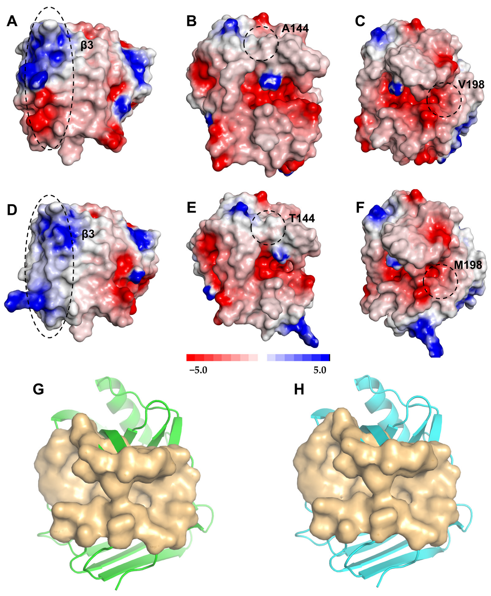
2.5. Structural Analysis of XynASP and ZM

To elucidate the mechanism of thermal stability, the chimeric ZM was modeled using XynASP as a template. Significant changes were observed in the interactions in the mutated region of chimeric ZM compared to those of wild-type XynASP (Figure 4). The number of hydrogen bonds interacting between residues in β3 and β1 was consistent in both the chimeric ZM and the wild-type XynASP, but the electrostatic interaction (R23 vs. T47) in chimeric ZM disappeared due to the D47T substitution (Figure 4A vs. Figure 4E). The interactions between residues in β3 and β2 and in β3 and β5 changed; two hydrogen bonds present in wild-type XynASP (Y37 vs. N52; N52 vs. K72) were lost in chimeric ZM, and were replaced by two newly formed hydrophobic interactions (Y37 vs. L52; M50 vs. L52). This formed a hydrophobic interaction cluster between β3-β2 and β3-β5 in chimeric ZM (Figure 4B vs. Figure 4F). The interactions between residues in β3 and β4 also changed due to the substitutions D47T, E49S, Y50M, N52L, and G53H in chimeric ZM; some hydrogen bond positions were altered compared to the wild-type XynASP, but the overall number of hydrogen bonds remained the same as the wild-type XynASP (Figure 4C vs. Figure 4G). Analysis of the interaction between the β1 of chimeric ZM and its adjacent region reveals a significantly increased hydrophobic effect compared to the wild-type XynASP. Hydrophobic interactions play a crucial role in protein folding, and enhancing the hydrophobic interactions between β-strands can improve the stability of protein structures.
Furthermore, residue A144 is located near the β7 sheet of XynASP (residues 94–106), close to the catalytic cleft in the “thumb region” (Figure 2D). This region is typically involved in substrate binding and conformational regulation in GH11 xylanases. The wild-type XynASP has a shorter and less hydrophobic side chain for Ala144, forming weaker hydrophobic interactions with the neighboring residue P162 (Figure 4D). The A144T mutation in the chimeric ZM leads to the formation of a new hydrogen bond between Thr144 and Q160 (Figure 4H), which compensates for the lack of hydrophobic interactions between the original Ala144 and P162. This interaction enhances the connection between the β7 strand and adjacent structural domains, decreases local conformational variability, and ultimately stabilizes the protein structure. Val198 is located in the C-terminal β14 strand (residues 206–214) of XynASP (Figure 4D). This region undergoes conformational changes during heat-induced unfolding, affecting overall stability. The substitution of Val198 with Met introduces a new hydrophobic interaction with Tyr84, forming a hydrophobic cluster (Y84-L98-V100-M198) that enhances the tight packing between the C-terminal β14 strand and the core structural domain, reducing the solvent-accessible surface area (SASA decreased by 72.5 Å2, Table 4), thereby stabilizing the protein conformation.
To further interpret the mechanism by which thermal stability was improved, the electrostatic properties of chimeric ZM were calculated using Discovery Studio 3.0. The surface electrostatic potential of chimeric ZM showed significant changes compared to wild-type XynASP. In chimeric ZM, the substituted β3 and nearby local surface displayed a prominent blue color in the electrostatic potential diagram, indicating a strong positive charge in this region (Figure 5A vs. Figure 5D). In contrast, the surface electrostatic potential of Ala144 in the wild type is slightly negative (Figure 5B), while the polar hydroxyl group of Thr144 in the chimeric ZM shifts the local electrostatic potential to slightly positive (Figure 5E). The surface electrostatic potential of Met198 in chimeric ZM was similar to that of the corresponding region in the wild-type XynASP (Figure 5C vs. Figure 5F). Based on the analysis above, it can be inferred that the ZM may stabilize local structures by enhancing charge complementarity, as opposed to the wild type. However, the catalytic pocket did not change significantly in ZM compared to the wild type (Figure 5G vs. Figure 5H). The surface area and catalytic volume of ZM remain largely unchanged from the wild type (Table 4).
A 100 ns molecular dynamics simulation was performed at 300 K for both the wild-type XynASP and the chimeric ZM. The results showed that both wild-type XynASP and chimeric ZM reached a stable state after 25 ns of simulation. The average (0.9047 Å) and final RMSD values (0.9838 Å) of chimeric ZM were lower than those of wild-type XynASP (average value: 1.0722 Å; final value: 1.1471 Å) (Figure 6A). Compared to wild-type XynASP, the RMSF value of chimeric ZM was significantly reduced (Figure 6B). The RMSD serves as an important indicator for assessing thermal fluctuations in protein conformation, with protein stability showing a negative correlation to RMSD fluctuation. Lower RMSF values indicate reduced residue mobility [18]. These results suggested that ZM exhibits a significantly enhanced overall rigidity in comparison to the wild type. It is worth noting that, despite the rigidity enhancement of ZM, the flexibility in the thumb region of ZM is significantly increased compared to the wild type (Figure 6B). Improved flexibility in the thumb region may offset the negative effects of rigid replacement in the β3 region on catalytic activity.

3. Discussion

At present, a variety of protein engineering methods, such as rational design and directed evolution, have been utilized to enhance the thermal stability of mesophilic xylanases [32,33,34,35]. Among these, the SCHEMA-based computational design can effectively construct functional chimeric enzymes by screening sequence fragments to reduce unfavorable residue interactions [23,36]. Similar strategies have been successfully applied in modifying enzymes like β-lactamases and β-glucosidases, significantly improving their thermal stability and catalytic activity [23,37]. The unique “jelly roll” structure of GH11 xylanases, composed of antiparallel β-sheets and α-helices, offers an ideal framework for modular recombination. For instance, extending the N-terminus of Neocallimastix patriciarum xylanase Np-Xyn boosts catalytic efficiency [38], while rigidizing the N-terminal flexible loop of Streptomyces rameus L2001 xylanase XynA enhances thermal stability [28]. These studies demonstrate that the performance of GH11 xylanases can be directionally optimized through modular recombination.
Changes, replacements, duplications, insertions, and deletions in sequences are the main ways in which protein structure and function evolve, as in this study [39]. Based on structural and conserved sequence analyses, multiple segments of XynASP were modularly recombined with heat-resistant enzymes. After screening the thermal stability and combining mutations, the chimeric ZM was ultimately obtained with significantly higher stability and specific activity than the wild type. The substituted region (N-terminus) of the chimeric ZM had the same number of hydrogen bonds as the wild type, but it exhibited an increase in hydrophobic interactions compared to the wild-type XynASP. The critical role of hydrophobic interactions in thermostability has been widely recognized in GH11 xylanases. For instance, Paës et al. [5] highlighted that the hydrophobic core in GH11 xylanases is a key characteristic of thermal adaptation, which aligns with our observation that the Y84-L98-V100-M198 hydrophobic cluster (introduced via V198M) reinforces the C-terminal packing density. Unlike studies relying on single mutations or module replacements [27], the chimera ZM combined N- and C-terminal hydrophobic clusters (Figure 4F–H), resulting in a remarkable 19.5 °C increase in Tm (Table 2). This synergy suggests that distributed hydrophobic networks can amplify thermal stability beyond incremental improvements from isolated modifications. Furthermore, although excessive hydrophobic packing could theoretically reduce enzyme solubility or induce aggregation, the hydrophobic clusters introduced in XynASP (e.g., Y84-L98-V100-M198) were strategically localized to regions with low solvent accessibility and did not form exposed hydrophobic patches, thereby avoiding reductions in enzyme solubility and molecular aggregation.
In molecular dynamics simulations, RMSD and RMSF characterize the overall conformational changes and local residue flexibility of proteins, respectively [9,15]. Compared to the wild-type XynASP, ZM exhibited significantly lower RMSD (0.90 Å vs. 1.07 Å) and RMSF values, indicating enhanced global rigidity and a more stable conformation. Additionally, ZM showed a reduced solvent-accessible surface area (SASA) of 72.5 Å2 (Table 4), which further decreased conformational entropy and improved thermal stability [40]. In addition, an enzyme’s surface net charge significantly impacts its thermal stability and refolding behavior. Optimizing the surface charge distribution to increase the net charge can enhance thermal stability [16,41]. In ZM, the surface electrostatic potential is significantly enhanced in the β3 domain and at position 144 (Figure 5), indicating that optimizing charge complementarity further stabilizes the structure of protein. According to research, a higher net surface charge can support reversible refolding by hindering xylanase aggregation [42]. In summary, the more compact xylanase structure and the increase in net surface charges all contributed to the improved thermal stability of ZM. Rigidification of the flexible sites in the enzyme may enhance its thermal stability, but it can also decrease catalytic activity [15].
Interestingly, the chimeric ZM demonstrated significant improvements in catalytic activity and catalytic efficiency. The catalytic cleft of GH11 xylanase consists of the palm, fingers, thumb, and spine regions. The interactions during opening and closing of the cleft directly affect substrate entry and product release, and therefore strongly regulate the catalytic performance of xylanase [43]. The thumb domain refers to the loop connecting the β-sheets B8 and B7. The flexibility of the thumb region affects the extent to which the xylanase catalytic cleft opens and closes, thus affecting the ability of a substrate to bind and thereby influencing the catalytic activity and catalytic efficiency of xylanase [44]. The thumb region (residues 150–155) of ZM showed a higher RMSF value compared to that of wild-type XynASP, indicating increased flexibility in this region for ZM (Figure 4B). This increased flexibility may facilitate substrate access to the catalytic cleft, ultimately resulting in improved catalytic efficiency for ZM. Moreover, a simulation analysis revealed no significant changes in the catalytic pocket of ZM compared to that of the wild-type XynASP (Figure 5G vs. Figure 5H), and both the wild type and ZM had similar active pocket surface areas and volumes (Table 4). This further supported the idea that the higher flexibility of the thumb region in ZM was a major factor contributing to its improved catalytic activity.
The challenge in enzyme engineering lies in finding the balance between rigidity and flexibility in protein structure to simultaneously enhance both thermal stability and catalytic activity of an enzyme [45]. A feasible design strategy to increase the thermal stability of these two regions includes introducing additional interactions, but also carries the risk of reducing the enzyme’s flexibility, thereby lowering its catalytic activity [46,47]. Our study revealed that enhanced hydrophobic interactions at the N- and C-termini of GH11 xynlanase stabilized the overall structure, while increased flexibility in the thumb region preserved catalytic activity. A phenomenon aligning with Tsou’s seminal work on the necessity of maintaining active-site flexibility despite global rigidification for optimal enzyme function [48]. These results demonstrated potential strategies for comprehensively improving the catalytic performance of GH11 xylanases.

4. Materials and Methods

4.1. Materials

By using Thermomonospora fusca GH11 family xylanase gene TfxA (GenBank accession number: KF927166.1) as the probe, the gene xynASP (GenBank accession number: XM_025579666.1) was analyzed and obtained by sequence alignment using the BLAST program (https://blast.ncbi.nlm.nih.gov/Blast.cgi (6 March 6 2021)) [26]. The plasmid pET-28a and E. coli strains BL21(DE3) and DH5α were purchased from Novagen (Madison, WI, USA) (6 January 2019). The pET-28a-xynASP was synthesized at Sangon Biotech (Shanghai, China). A quick mutagenesis kit was purchased from Vazyme Biotech (Nanjing, China). Beechwood xylan was purchased from Shanghai Yuanye Biotechnology (Shanghai, China).

4.2. Site-Directed Mutagenesis

Site-directed mutagenesis was performed using a quick mutagenesis kit, following the manufacturer’s instructions [27]. The recombinant plasmid pET-28a-xynASP was used as the template. Information on the primers can be found in Table S1. After amplification, the PCR products were incubated with DpnI to remove the original DNA template. The digested PCR products were then transformed into E. coli DH5α. Positive clones were sequenced by Sangon Biotech (Shanghai) to confirm the accuracy of the constructed plasmids.

4.3. Protein Expression and Purification

After confirming the correct sequences of the positive clones, the plasmids were extracted and chemically transformed into E. coli BL21(DE3) competent cells for expression induction. A single colony was inoculated into 2 mL of LB medium containing kanamycin (final concentration 50 μg/mL) and cultured overnight at 37 °C with shaking at 180 rpm. The culture was then transferred to 100 mL of TB medium (kanamycin final concentration 50 μg/mL) at a 1/100 ratio and incubated at 37 °C and 180 rpm until the OD reached 0.6–0.8. IPTG was added to a final concentration of 2 mM, and the culture was incubated at 30 °C and 180 rpm for 2 h. After induction, the culture was centrifuged at 6000 rpm for 10 min to collect the cells. The cell pellet was resuspended in sodium dihydrogen phosphate-citrate buffer (pH 4.6), and the cells were disrupted using ultrasonic sonication. The mixture was then centrifuged at 10,000 rpm for 15 min at 4 °C, and the supernatant was collected as a crude enzyme solution.
After the crude enzyme solution was filtered through a 0.45-μm membrane, the enzyme was purified using nickel affinity chromatography on Ni-NTA Sefinose™ resin 6FF (Sangon Biotech, Shanghai, China). The recombinant xylanase was eluted with an elution buffer (20 mM Tris-HCl, pH 8.0, 500 mM NaCl, and 300 mM imidazole) and desalted using a Superdex G25 column (GE Healthcare, New York, USA) with a Na2HPO4-citrate buffer (50 mM, pH 6.0). The purity of the enzyme was determined by sodium dodecyl sulfate-polyacrylamide gel electrophoresis (SDS-PAGE). The protein concentration was measured using a BCA protein assay kit (Sangon Biotech, Shanghai, China).

4.4. Recombinant Xylanase Activity Assay

The activity of xylanase was determined using the DNS method. Briefly, 1.5 mL of a solution of 0.5% beechwood xylan was mixed with 1 mL of an appropriately diluted enzyme solution, and the reaction was carried out at 40 °C for 15 min. Then, 2.5 mL of DNS reagent was added to immediately stop the reaction, and the mixture was boiled in a water bath for 7 min to develop the color. After cooling, 5 mL of distilled water was added and mixed thoroughly. The absorbance was measured at λ = 540 nm. One unit (U) of xylanase activity was defined as the amount of enzyme required to release 1 μmol of reducing sugar per minute from beechwood xylan [18].

4.5. Enzymatic Property and Kinetic Parameter Measurements

The optimum temperature of xylanase was determined in a Na2HPO4-citrate buffer (50 mM, pH 6.0) at temperatures ranging from 35 °C to 70 °C. The thermal stability of wild-type XynASP and its mutants was evaluated by measuring residual activity after incubating at different temperatures for varying periods of time. The residual activity after the heat treatment was determined by comparing the specific activity of the enzyme after a certain time and temperature treatment with the specific activity of the untreated enzyme. The enzyme’s half-life was calculated using exponential fitting. The melting temperature (Tm) of the enzyme was determined by differential scanning fluorimetry using a CFX Touch 96-well system (Bio-Rad Laboratories, Inc., CA, USA). Purified enzyme solution (20 μL) was mixed with buffer (50 mM Na2HPO4-citrate buffer, pH 6.0) at a final concentration of 0.4–0.8 mg/mL and 2 μL of 10× SYPRO Orange dye. Fluorescence was measured at temperatures ranging from 10 °C to 95 °C with a 0.5 °C/s gradient (λex = 480 nm, λem = 580 nm) to observe the relationship between temperature and protein unfolding [46]. Under the optimal reaction conditions for each enzyme, the kinetic parameters (Km, Vmax, and kcat) of the purified enzyme were measured using beechwood xylan at concentrations ranging from 0.1 to 0.8 mg/mL. Kinetic constants were evaluated by fitting the experimental data using GraphPad Prism 8.0 (GraphPad Software, Inc., San Diego, CA, USA). All experiments in this study were performed in triplicate.

4.6. Structural Analysis and Molecular Dynamics Simulations

The amino acid sequence of XynASP was submitted to SWISS-MODEL for a sequence homology analysis. The crystal structure of the GH11 family xylanase BmGH11 (PDB No. 8B8E) showed the highest homology (73.2%) and was, therefore, used as a template for homology modeling of XynASP. The quality of the model was assessed using the SAVES v6.1 online server (https://saves.mbi.ucla.edu/ (17 January 2025)). Amino acid sequence alignment of the homologous proteins was performed using ClustalX and ESPript 3.0. The surface electrostatic potential of XynASP was calculated using Discovery Studio 2020 software. The surface area and volume of the active pocket of the enzyme were calculated using the CavityPlus online server (http://www.pkumdl.cn:8000/cavityplus/index.php#/computation (25 January 2025)). Intermolecular interactions and protein structure visualization were performed using Discovery Studio 3.0 and PyMOL.
All molecular dynamics simulations were conducted using GROMACS 5.1. The protein topology was generated using PDB2GMX, with the SPC/E water model used as the solvent model. Sodium ions were added to neutralize the system’s charge to ensure that the system was electrically neutral. Periodic boundary conditions were applied. The protein and small molecule ligand complex was energy-minimized for 1000 ps at 300 K. Then, the optimized system underwent 100 ps NVT ensemble equilibration and 100 ps NPT ensemble equilibration, during which the positions in the system were restricted. Finally, the entire system underwent a 100 ns molecular dynamics simulation at 300 K with position restraints removed.

5. Conclusions

This study significantly enhanced the thermal stability and catalytic activity of the GH11 xylanase XynASP from Aspergillus saccharolyticus JOP 1030-1 through computer-aided design and modular assembly strategies. Structural analysis and MD simulations revealed that enhanced hydrophobic interactions at the N- and C-termini improved structural rigidity, while increased flexibility in the thumb region counterbalanced potential negative effects of mutations on catalytic activity. These findings enrich the strategy of synergizing thermal stability and catalytic activity. This study confirms that modular design effectively addresses the core challenge of insufficient stability in xylanases for high-temperature industrial applications.

Supplementary Materials

The following supporting information can be downloaded at: https://www.mdpi.com/article/10.3390/catal15060563/s1, Table S1. sequences of mutated primers; Figure S1. 3D model of XynASP and model analysis. (A) 3D model of XynASP. (B) Ramachandran plot of the XynASP model. (C) Verification of the 3D plot of the XynASP model; Figure S2. Sequence alignment of XynASP with nine known crystal structures of heatresistant GH11 xylanases. Nine xylanases are named according to their PDB code; Figure S3. SDS-PAGE analysis of purified XynASP and its mutants. (A) M: marker; lane 1: XynASP; lane 2: Z2. (B) M: marker; lane 1: XynASP; lane 2: A144T/V198M; lane 3: ZM.

Author Contributions

Conceptualization, T.L. and J.Z. (Jinjin Zhu); methodology, J.Z. (Jinjin Zhu), Q.Z., J.Z. (Jiaxin Zhao), and X.F.; investigation, J.Z. (Jinjin Zhu) and M.W.; structural analysis, T.L., H.W., and Y.L.; visualization, J.Z. (Jinjin Zhu) and H.X.; project administration, T.L.; writing—original draft preparation, J.Z. (Jinjin Zhu); writing—review and editing, T.L. and Y.L. All authors have read and agreed to the published version of the manuscript.

Funding

This study was financially supported by the Foundation of Science and Technology Development Project of Henan Province (No. 242102111041; 232102310189; 222102110180) and the Natural Science Foundation of Henan (No. 242300421582).

Data Availability Statement

The original contributions presented in this study are included in the article and Supplementary Material. Further inquiries can be directed to the corresponding author.

Conflicts of Interest

Yan Liu was employed by the Name of company Co., Ltd. The remaining authors declare that the research was conducted in the absence of any commercial or financial relationships that could be construed as a potential conflict of interest.

References

  1. Nezhad, N.G.; Rahman, R.N.Z.R.; Normi, Y.M.; Oslan, S.N.; Shariff, F.M.; Leow, T.C. Recent advances in simultaneous thermostability-activity improvement of industrial enzymes through structure modification. Int. J. Biol. Macromol. 2023, 232, 123440. [Google Scholar] [CrossRef] [PubMed]
  2. Wu, X.; Shi, Z.; Tian, W.; Liu, M.; Huang, S.; Liu, X.; Yin, H.; Wang, L. A thermostable and CBM2-linked GH10 xylanase from Thermobifida fusca for paper bleaching. Front. Bioeng. Biotechnol. 2022, 10, 939550. [Google Scholar] [CrossRef]
  3. Nakamura, T.; Takita, T.; Kuwata, K.; Mizutani, K.; Mikami, B.; Nakamura, S.; Yasukawa, K. Activity-stability trade-off observed in variants at position 315 of the gh10 xylanase xynr. Sci. Rep. 2024, 14, 7767. [Google Scholar] [CrossRef]
  4. Talens-Perales, D.; Jimenez-Ortega, E.; Sanchez-Torres, P.; Sanz-Aparicio, J.; Polaina, J. Phylogenetic, functional and structural characterization of a gh10 xylanase active at extreme conditions of temperature and alkalinity. Comput. Struct. Biotechnol. J. 2021, 19, 2676–2686. [Google Scholar] [CrossRef]
  5. Paës, G.; Berrin, J.; Beaugrand, J. Gh11 xylanases: Structure/function/properties relationships and applications. Biotechnol. Adv. 2012, 30, 564–592. [Google Scholar] [CrossRef]
  6. Boonyapakron, K.; Keiser, B.; Prabmark, K.; Aiewviriyasakul, K.; Arunrattanamook, N.; Jaruwat, A.; Chitnumsub, P.; Li, J.Y.; Wong, T.S.; Zhao, X.Q.; et al. Hyperthermophilic xylanase and thermophilicity analysis by molecular dynamic simulation with quantum mechanics. Appl. Microbiol. Biotechnol. 2024, 108, 526. [Google Scholar] [CrossRef]
  7. Wu, Q.; Zhang, C.; Dong, W.; Lu, H.; Yang, Y.; Li, W.; Xu, Y.; Li, X. Enhanced thermostability of xylanase xyna via computationally designed assembly of multiple n-terminal disulfide bridges. Process Biochem. 2024, 138, 67–78. [Google Scholar] [CrossRef]
  8. Phuyal, M.; Budhathoki, U.; Bista, D.; Shakya, S.; Shrestha, R.; Shrestha, A.K. Xylanase-producing microbes and their real-world application. Int. J. Chem. Eng. 2023, 2023, 3593035. [Google Scholar] [CrossRef]
  9. Zhang, D.; Huang, J.; Liu, Y.; Chen, X.; Gao, T.; Li, N.; Huang, W.; Wu, M. Directed modification of a ghf11 thermostable xylanase ausm for enhancing inhibitory resistance towards syxip-i and application of ausmpkk in bread making. Foods 2023, 12, 3574. [Google Scholar] [CrossRef]
  10. Zhu, W.; Qin, L.; Xu, Y.; Lu, H.; Wu, Q.; Li, W.; Zhang, C.; Li, X. Three molecular modification strategies to improve the thermostability of xylanase xyna from streptomyces rameus l2001. Foods 2023, 12, 879. [Google Scholar] [CrossRef]
  11. Glekas, P.D.; Kalantzi, S.; Dalios, A.; Hatzinikolaou, D.G.; Mamma, D. Biochemical and Thermodynamic Studies on a Novel Thermotolerant GH10 Xylanase from Bacillus safensis. Biomolecules 2022, 12, 790. [Google Scholar] [CrossRef]
  12. Wang, Y.; Wang, J.; Zhang, Z.; Yang, J.; Turunen, O.; Xiong, H. High-temperature behavior of hyperthermostable thermotoga maritima xylanase xyn10b after designed and evolved mutations. Appl. Microbiol. Biotechnol. 2022, 106, 2017–2027. [Google Scholar] [CrossRef]
  13. You, S.; Zha, Z.; Jing, L.; Zhang, W.; Bai, Z.; Hu, Y.; Wang, X.; Chen, Y.; Chen, Z.; Wang, J.; et al. Improvement of xyl10c_ Δ n catalytic performance through loop engineering for lignocellulosic biomass utilization in feed and fuel industries. Biotechnol. Biofuels 2021, 14, 195. [Google Scholar] [CrossRef]
  14. Yin, X.; Li, J.F.; Wang, J.Q.; Tang, C.D.; Wu, M.C. Enhanced thermostability of a mesophilic xylanase by N-terminal replacement designed by molecular dynamics simulation. J. Sci. Food Agric. 2013, 93, 3016–3023. [Google Scholar] [CrossRef] [PubMed]
  15. Luo, J.; Song, C.; Cui, W.; Han, L.; Zhou, Z. Counteraction of stability-activity trade-off of nattokinase through flexible region shifting. Food Chem. 2023, 423, 136241. [Google Scholar] [CrossRef]
  16. Pinto, G.P.; Corbella, M.; Demkiv, A.O.; Kamerlin, S.C.L. Exploiting enzyme evolution for computational protein design. Trends Biochem. Sci. 2022, 47, 375–389. [Google Scholar] [CrossRef]
  17. Sun, Z.; Liu, Q.; Qu, G.; Feng, Y.; Reetz, M.T. Utility of b-factors in protein science: Interpreting rigidity, flexibility, and internal motion and engineering thermostability. Chem. Rev. 2019, 119, 1626–1665. [Google Scholar] [CrossRef]
  18. Li, T.; Wang, R.; Hua, B.; Cao, L.; Zhang, Q.; Zhai, Y.; Ling, S.; Wang, M.; Li, E. Improving the Thermal Stability of GH11 Xylanase XynASP through Cord Region Engineering. J. Agric. Food Chem. 2025, 73, 1516–1528. [Google Scholar] [CrossRef]
  19. Dotsenko, A.S.; Denisenko, Y.A.; Rozhkova, A.M.; Zorov, I.N.; Korotkova, O.G.; Sinitsyn, A.P. Enhancement of thermostability of gh10 xylanase e penicillium canescens directed by δδg calculations and structure analysis. Enzyme Microb. Technol. 2021, 152, 109938. [Google Scholar] [CrossRef]
  20. Bu, Y.; Cui, Y.; Peng, Y.; Hu, M.; Tian, Y.E.; Tao, Y.; Wu, B. Engineering improved thermostability of the gh11 xylanase from neocallimastix patriciarum via computational library design. Appl. Microbiol. Biotechnol. 2018, 102, 3675–3685. [Google Scholar] [CrossRef]
  21. Lai, Z.; Zhou, C.; Ma, X.; Xue, Y.; Ma, Y. Enzymatic characterization of a novel thermostable and alkaline tolerant GH10 xylanase and activity improvement by multiple rational mutagenesis strategies. Int. J. Biol. Macromol. 2021, 170, 164–177. [Google Scholar] [CrossRef] [PubMed]
  22. Crean, R.M.; Biler, M.; van der Kamp, M.W.; Hengge, A.C.; Kamerlin, S.C.L. Loop Dynamics and Enzyme Catalysis in Protein Tyrosine Phosphatases. J. Am. Chem. Soc. 2021, 143, 3830–3845. [Google Scholar] [CrossRef] [PubMed]
  23. Chen, Q.; Wu, J.; Wu, Y.; Wang, Z.; Zeng, M.; He, Z.; Chen, J.; Mu, W. Rational Design of Loop Dynamics for a Barrel-Shaped Enzyme by Introducing Disulfide Bonds. J. Agric. Food Chem. 2024, 72, 16541–16542. [Google Scholar] [CrossRef]
  24. Zheng, F.; Vermaas, J.V.; Zheng, J.; Wang, Y.; Tu, T.; Wang, X.; Xie, X.; Yao, B.; Beckham, G.T.; Luo, H. Activity and thermostability of gh5 endoglucanase chimeras from mesophilic and thermophilic parents. Appl. Environ. Microbiol. 2019, 85, e02079-18. [Google Scholar] [CrossRef]
  25. Liu, W.; Tu, T.; Gu, Y.; Wang, Y.; Zheng, F.; Zheng, J.; Wang, Y.; Su, X.; Yao, B.; Luo, H. Insight into the Thermophilic Mechanism of a Glycoside Hydrolase Family 5 β-Mannanase. J. Agric. Food Chem. 2019, 67, 473–483. [Google Scholar] [CrossRef]
  26. Li, T.; Zhu, X.; Ye, M.; Wang, M.; Zhu, J.; Shen, F.; Li, E. Rapid improvement in thermostability of gh11 xylanase (xynasp) from aspergillus saccharolyticus jop 1030-1 by tryptophan residue substitution. J. Biotech Res. 2022, 13, 26–39. [Google Scholar]
  27. Li, T.; Yang, S.; Wang, X.; Cai, H.; Wang, Y.; Li, C.; Li, E. Improving thermostability of gh11 xylanase xynasp by the design of loop region. Crystals 2022, 12, 1228. [Google Scholar] [CrossRef]
  28. Wu, Q.; Zhang, C.; Dong, W.; Lu, H.; Yang, Y.; Li, W.; Xu, Y.; Li, X. Simultaneously enhanced thermostability and catalytic activity of xylanase from streptomyces rameus l2001 by rigidifying flexible regions in loop regions of the n-terminus. J. Agric. Food. Chem. 2023, 71, 12785–12796. [Google Scholar] [CrossRef]
  29. Vucinic, J.; Novikov, G.; Montanier, C.Y.; Dumon, C.; Schiex, T.; Barbe, S. A Comparative Study to Decipher the Structural and Dynamics Determinants Underlying the Activity and Thermal Stability of GH-11 Xylanases. Int. J. Mol. Sci. 2021, 22, 5961. [Google Scholar] [CrossRef]
  30. Yang, Y.; Zhang, C.; Lu, H.; Wu, Q.; Wu, Y.; Li, W.; Li, X. Improvement of thermostability and catalytic efficiency of xylanase from myceliophthora thermophilar by n-terminal and c-terminal truncation. Front. Microbiol. 2024, 15, 1385329. [Google Scholar] [CrossRef]
  31. Ajeje, S.B.; Hu, Y.; Song, G.; Peter, S.B.; Afful, R.G.; Sun, F.; Asadollahi, M.A.; Amiri, H.; Abdulkhani, A.; Sun, H. Thermostable Cellulases/Xylanases from Thermophilic and Hyperthermophilic Microorganisms: Current Perspective. Front. Bioeng. Biotechnol. 2021, 9, 794304. [Google Scholar] [CrossRef] [PubMed]
  32. Marneth, K.; van den Elst, H.; Cramer-Blok, A.; Codee, J.; Overkleeft, H.S.; Aerts, J.M.F.G.; Ubbink, M.; Ben Bdira, F. Tuning the Transglycosylation Reaction of a GH11 Xylanase by a Delicate Enhancement of its Thumb Flexibility. Chembiochem 2021, 22, 1743–1749. [Google Scholar] [CrossRef] [PubMed]
  33. Huang, J.; Dai, S.; Chen, X.; Xu, L.; Yan, J.; Yang, M.; Yan, Y. Alteration of chain-length selectivity and thermostability ofrhizopus oryzae lipase via virtual saturation mutagenesis coupled with disulfide bond design. Appl. Environ. Microbiol. 2023, 89, e01878-22. [Google Scholar] [CrossRef] [PubMed]
  34. Dong, W.; Zhu, W.; Wu, Q.; Li, W.; Li, X. Improvement the thermostability and specific activity of acidic xylanase PjxA from Penicillium janthinellum via rigid flexible sites. Int. J. Biol. Macromol. 2024, 279, 135399. [Google Scholar] [CrossRef]
  35. Wang, L.; Wang, Y.Y.; Chen, Z.L.; Li, Y.H. Enzymatic characterization and thermostability improvement of an acidophilic endoxylanase PphXyn11 from Paenibacillus physcomitrellae XB. Protein Expr. Purif. 2024, 219, 106482. [Google Scholar] [CrossRef]
  36. Meyer, M.M.; Hochrein, L.; Arnold, F.H. Structure-guided schema recombination of distantly related β-lactamases. Protein Eng. Des. Sel. 2006, 19, 563–570. [Google Scholar] [CrossRef]
  37. Heinzelman, P.; Snow, C.D.; Wu, I.; Nguyen, C.; Villalobos, A.; Govindarajan, S.; Minshull, J.; Arnold, F.H. A family of thermostable fungal cellulases created by structure-guided recombination. Proc. Natl. Acad. Sci. USA 2009, 106, 5610–5615. [Google Scholar] [CrossRef]
  38. Song, L.; Dumon, C.; Siguier, B.; André, I.; Eneyskaya, E.; Kulminskaya, A.; Bozonnet, S.; O Donohue, M.J. Impact of an n-terminal extension on the stability and activity of the gh11 xylanase from thermobacillus xylanilyticus. J. Biotechnol. 2014, 174, 64–72. [Google Scholar] [CrossRef]
  39. Corbella, M.; Pinto, G.P.; Kamerlin, S.C.L. Loop dynamics and the evolution of enzyme activity. Nat. Rev. Chem. 2023, 7, 536–547. [Google Scholar] [CrossRef]
  40. Yasuda, S.; Kazama, K.; Akiyama, T.; Kinoshita, M.; Murata, T. Elucidation of cosolvent effects thermostabilizing water-soluble and membrane proteins. J. Mol. Liq. 2020, 301, 112403. [Google Scholar] [CrossRef]
  41. Briganti, L.; Capetti, C.; Pellegrini, V.O.A.; Ghio, S.; Campos, E.; Nascimento, A.S.; Polikarpov, I. Structural and molecular dynamics investigations of ligand stabilization via secondary binding site interactions in Paenibacillus xylanivorans GH11 xylanase. Comput. Struct. Biotechnol. J. 2021, 19, 1557–1566. [Google Scholar] [CrossRef] [PubMed]
  42. Chong, W.; Zhang, Z.; Li, Z.; Meng, S.; Nian, B.; Hu, Y. Hook loop dynamics engineering transcended the barrier of activity-stability trade-off and boosted the thermostability of enzymes. Int. J. Biol. Macromol. 2024, 278, 134953. [Google Scholar] [CrossRef] [PubMed]
  43. Li, Y.; Li, C.; Huang, H.; Rao, S.; Zhang, Q.; Zhou, J.; Li, J.; Du, G.; Liu, S. Significantly enhanced thermostability of aspergillus niger xylanase by modifying its highly flexible regions. J. Agric. Food Chem. 2022, 70, 4620–4630. [Google Scholar] [CrossRef] [PubMed]
  44. Ling, S.; Xing, J.; Li, S.; Zhang, L.; Shen, C.; Hong, J.; Huang, S.; Li, T.; Wei, L.; Ding, R. Enhancing the catalytic performance of xylanase XynASP through semi-rational design in the cord region to promote its application in juice clarification. Int. J. Biol. Macromol. 2025, 305, 141138. [Google Scholar] [CrossRef]
  45. Wang, L.; Cao, K.; Pedroso, M.M.; Wu, B.; Gao, Z.; He, B.; Schenk, G. Sequence- and structure-guided improvement of the catalytic performance of a gh11 family xylanase from bacillus subtilis. J. Biol. Chem. 2021, 297, 101262. [Google Scholar] [CrossRef]
  46. Dong, R.; Liu, X.; Wang, Y.; Qin, X.; Wang, X.; Zhang, H.; Wang, Y.; Luo, H.; Yao, B.; Bai, Y.; et al. Fusion of a proline-rich oligopeptide to the C-terminus of a ruminal xylanase improves catalytic efficiency. Bioengineered 2022, 13, 10482–10492. [Google Scholar] [CrossRef]
  47. Sinha, R.; Shukla, P. Current trends in protein engineering: Updates and progress. Curr. Protein Pept. Sci. 2019, 20, 398–407. [Google Scholar] [CrossRef]
  48. Tsou, C.L. Conformational flexibility of enzyme active sites. Science 1993, 262, 380–381. [Google Scholar] [CrossRef]
Figure 1. Sequence alignment of the xylanase XynASP with nine heat-resistant GH11 xylanases. The dashed box indicates the target region replaced in the wild type.
Catalysts 15 00563 g001
Figure 2. Effects of region substitution and site-directed mutations on XynASP. (A) The regions in XynASP targeted for substitution. (B) The results of the region substitutions. (C) After incubation at 45 °C for 30 min, the residual activity of wild-type XynASP and its mutants was evaluated at 45 °C. (D) The mutated residues in XynASP. (E) Beneficial point mutations and their combinations. (F) After incubation at 50 °C for 30 min, the residual activity of the mutants was evaluated at 45 °C. The values in parentheses represent the residual activity of the enzyme after heat treatment.
Catalysts 15 00563 g002
Figure 3. Effect of temperature on the wild type and its mutants. (A) Optimal reaction temperature. (BE) Thermal stability of each enzyme determined by monitoring residual activity for 1 h at 45 °C (B), 50 °C (C), 55 °C (D), and 60 °C (E). (F) Melting temperature (Tm).
Catalysts 15 00563 g003
Figure 4. Interaction analysis of the wild type and ZM. (A) Changes in the interactions between β3 and β1 in the wild type. (B) Changes in the interactions between β3 and β2/β5 in the wild type. (C) Changes in the interactions between β3 and β4 in XynASP. (D) Changes in the interactions at positions 144 and 198 in the wild type. (E) Changes in the interactions between β3 and β1 in ZM. (F) Changes in the interactions between β3 and β2/β5 in ZM. (G) Changes in the interactions between β3 and β4 in ZM. (H) Changes in the interactions around positions 144 and 198 in ZM. Hydrogen bonds, hydrophobic interactions, and electrostatic interactions are represented by green, purple, and red, respectively.
Catalysts 15 00563 g004
Figure 5. Electrostatic potential analysis of the wild type and ZM and the catalytic pocket. (A) Electrostatic potential of β3 in the wild type; (B) electrostatic potential of position 144 in the wild type; (C) electrostatic potential of position 198 in the wild type; (D) electrostatic potential of β3 in ZM; (E) electrostatic potential of position 144 in ZM; (F) electrostatic potential of position 198 in ZM; (G) wild type; (H) chimeric enzyme ZM. The positive, negative, and neutral electrostatic potentials are represented by blue, red, and white shading, respectively. The mutated regions of the enzyme are marked with a black border. The yellow surface indicates the shape of the catalytic pocket.
Catalysts 15 00563 g005
Figure 6. MD simulation analysis. (A) RMSD analysis. (B) RMSF analysis. Hydrogen bonds, hydrophobic interactions, and electrostatic interactions are represented by green, purple, and red, respectively.
Catalysts 15 00563 g006
Table 1. Structural and sequence characteristics of the chimeric enzymes.
EnzymeSegmentOriginal SequenceSubstituted Sequence
Z1β11APVASLEEEEERASNFSSALAARSTASSTGWSN3321DTTITQNQTGYDN33
Z2β346GDVEYTNG5346GTVSMTLH53
Z3β680TITYSGSWTS9080TVTYNASFNP90
Z4β794SNSYLSVYGWTTS10694GNAYLTLYGWYRN106
Z5α1175TTANHFNAWAAL186175TIGNHFDAWARA186
Z6β14206SGSASITVS214206SGSSTVSIS214
Table 2. Characteristics of XynASP and its mutants.
EnzymeOptimal Temperature (°C)Specific Activity (U/mg)t1/245 °C (min)t1/250 °C (min)t1/255 °C (min)Tm (°C)
Wild type4551.0 ± 2.422.05.34.346.0
Z260222.1 ± 1.3339.845.111.9
A144T/V198M5532.8 ± 1.944.722.78.2
ZM60197.4 ± 1.4343.5140.337.664.5
Table 3. Kinetic parameters of wild-type XynASP and its mutants.
EnzymeVmax (µmol·min−1 mg−1)Km (mg/mL)kcat (s−1)kcat/Km (mL·mg−1 s−1)
Wild type75.7 ± 3.50.18 ± 0.0211.5 ± 0.563.8 ± 7.7
Z2361.6 ± 16.80.34 ± 0.03131.0 ± 6.1385.3 ± 38.5
A144T/V198M91.4 ± 9.60.84 ± 0.1555.6 ± 0.166.1 ± 14.9
ZM399.1 ± 23.30.48 ± 0.06167.4 ± 9.8349.2 ± 46.3
Table 4. Structural characteristics of wild-type XynASP and its mutants.
EnzymeSASA (Å2)Active Pocket Region Characteristics
Surface Area (Å2)Volume (Å3)
Wild type8370.2643.0794.3
ZM8297.7643.5793.5
Disclaimer/Publisher’s Note: The statements, opinions and data contained in all publications are solely those of the individual author(s) and contributor(s) and not of MDPI and/or the editor(s). MDPI and/or the editor(s) disclaim responsibility for any injury to people or property resulting from any ideas, methods, instructions or products referred to in the content.

