Article

Green Innovation: Multifunctional Zinc Oxide Nanoparticles Synthesized Using Quercus robur for Photocatalytic Performance, Environmental, and Antimicrobial Applications

1
Laboratory of Physics for Matter and Radiation, Faculty of Science and Technology, Souk Ahras University, P.O. Box 1553, Souk-Ahras 41000, Algeria
2
Laboratory of Biomaterials and Transport Phenomena, University of Médéa, Médéa 26000, Algeria
3
Ecole Nationale Supérieure de Chimie de Rennes, CNRS, ISCR—UMR6226, University Rennes, 35000 Rennes, France
4
Laboratoire de Sécurité des Procédés Chimiques—LSPC EA 4704, Normandie Univ, INSA Rouen, UNIROUEN, 76000 Rouen, France
5
Laboratory of Chemical Process Engineering, Department of Process Engineering, Faculty of Technology, University of Ferhat Abbas Setif-1, Setif 19000, Algeria
6
Department of Biochemistry, College of Science, King Saud University, Riyadh 11451, Saudi Arabia
7
College of Engineering, Imam Mohammad Ibn Saud Islamic University, IMSIU, Riyadh 11432, Saudi Arabia
8
Research Scientific and Technical Center on Physico-Chemical Analysis (CRAPC), BP 384, Industrial Zone Bou-Ismail, Tipaza 42000, Algeria
9
School of Engineering, Merz Court, Newcastle University, Newcastle upon Tyne NE1 7RU, UK
10
Biotechnology, Water, Environment and Health Laboratory, Abbes Laghrour University, Khenchela 40000, Algeria
*
Authors to whom correspondence should be addressed.
Catalysts 2025, 15(3), 256; https://doi.org/10.3390/catal15030256
Submission received: 5 January 2025 / Revised: 17 February 2025 / Accepted: 27 February 2025 / Published: 7 March 2025

Abstract

:
This study investigates the green synthesis of zinc oxide nanoparticles (ZnO NPs) using leaf extract as a natural reducing agent, evaluating their antimicrobial and photocatalytic properties. The nanoparticles were annealed at 320 °C and 500 °C, and the effects of leaf extract concentration and annealing temperature on their structural, morphological, and electronic properties were systematically explored. X-ray diffraction (XRD) analysis confirmed the hexagonal wurtzite structure of ZnO, with crystallite size and defect density being influenced by the concentration of the extract. Scanning electron microscopy (SEM) revealed the formation of smaller, spherical particles, with increased aggregation observed at higher extract concentrations. Fourier-transform infrared spectroscopy (FTIR) identified key functional groups, such as hydroxyl groups, C–O bonds, and metal–oxygen vibrations. UV–Vis spectroscopy showed a reduction in band gap energy and an increase in Urbach energy as the extract concentration and annealing temperature were increased. The antimicrobial activity of the ZnO NPs was evaluated against Gram-positive and Gram-negative bacteria as well as Candida albicans, demonstrating significant antibacterial efficacy. Photocatalytic degradation studies of methylene blue dye revealed a superior efficiency of up to 74% for the annealed samples, particularly at 500 °C. This research highlights the potential of green-synthesized ZnO NPs for a wide range of applications, including antimicrobial agents, water purification, and environmental catalysis. It contributes to the advancement of sustainable nanotechnology, offering promising solutions for both technological and ecological challenges.

1. Introduction

The growing population, combined with rising industrialization and economic demands, has necessitated the extensive use of synthetic chemicals and organic dyes, particularly in industries that produce by-products during their processes [1,2]. Despite the economic benefits provided by sectors such as textiles and chemicals, the untreated discharge of industrial wastewater leads to significant challenges, including health hazards, ecosystem imbalances, and reduced biodiversity due to its toxicity and persistence [3,4,5]. To address these issues, various treatment methods have been developed to eliminate contaminants before they are released into the environment [6,7]. These approaches include biological [8], chemical [9,10,11,12], physicochemical [13,14], and hybrid techniques for water purification [15,16].
Nanoremediation is an advanced remediation strategy that leverages nanomaterials to break down or remove pollutants efficiently [17]. This technique is recognized for its superior effectiveness in managing environmental contamination and enabling pollutant detection, monitoring, and treatment [18]. Nanoremediation methods are cost-efficient and significantly reduce the time needed to degrade pollutants, often lowering their concentration to near-zero levels [19,20]. This technology has been successfully applied to address various pollutants in water, such as chlorinated compounds, heavy metals, organic chemicals, hydrocarbons, pesticides, and inorganic ions [18,21,22].
A broad spectrum of nanomaterials, including zeolites, metal oxides, carbon nanostructures, and bimetallic nanoparticles, is employed in nanoremediation to transform or degrade environmental contaminants [23,24,25]. Conventional wastewater treatment techniques, such as filtration, coagulation, reverse osmosis, sedimentation, flotation, oxidation, precipitation, evaporation, and adsorption, remain widely used [26,27]. Among these, advanced oxidation processes (AOPs) are particularly effective for treating industrial wastewater [28,29]. Photocatalysis, a subset of AOPs, is especially advantageous because it does not generate secondary pollutants during the treatment process [29,30].
Heterogeneous photocatalysis is considered a highly promising method for breaking down organic pollutants due to its simplicity, high efficiency, and environmentally friendly characteristics. This technique is further valued for its non-toxic nature, low energy consumption, ease of use, and reproducibility [31,32]. It involves generating reactive oxygen species (ROS), such as hydroxyl radicals (OH•), superoxide ions (•O2), and hydrogen peroxide (H2O2), to oxidize a wide range of organic contaminants. Semiconductor materials play a central role in photocatalysis, with important attributes including optimal band gap values, electronic structure, light absorption efficiency, charge transport, stability, and reusability [33,34]. For decades, metal oxides such as TiO2, ZnO, SnO2, and CeO2 have been extensively utilized as photocatalysts due to their desirable properties [35,36,37,38,39,40]. More specifically, they have played a crucial role as heterogeneous photocatalysts in various applications [41].
Among these, ZnO is a promising alternative to TiO2 due to its affordability, accessibility, and environmental compatibility. ZnO nanoparticles exhibit superior photocatalytic efficiency because of their high surface reactivity, which promotes the formation of numerous defect sites due to oxygen non-stoichiometry, making them more effective than many other metal oxides [42]. Additionally, ZnO nanoparticles have shown potential for enhancing the performance of perovskite-based photodetectors [43,44].
Several nanoparticle synthesis methods have been developed, including hydrothermal synthesis, sol–gel techniques, gas-phase processes, thermolysis, and hydrolysis. However, these approaches often face challenges such as high costs, environmental risks, and difficulties in controlling nanoparticle size and surface characteristics [45]. To overcome these issues, green synthesis methods using biological systems, such as plants and fungi, have gained traction [46,47]. These environmentally friendly methods are increasingly being explored as alternatives to conventional physical and chemical techniques [48].
Plants are particularly suitable for large-scale nanoparticle production due to their accessibility, safety, and ability to function as both reducing and stabilizing agents. They facilitate the synthesis of stable nanoparticles with diverse sizes and shapes [47,49,50,51,52]. Several plants, including Hibiscus sabdariffa [7], Carica papaya [53], Aloe barbadensis [54], Solanum nigrum [55], Agathosma betulina [56], Ruellia tuberosa [57], Foeniculum vulgare [58], have been employed to synthesize ZnO nanoparticles for various applications.
In this work, a green synthesis method was developed to produce zinc oxide nanoparticles (ZnO NPs) using an extract from Quercus robur leaves. This extract, rich in bioactive compounds such as flavonoids and polyphenols, was utilized as both a reducing and stabilizing agent to replace conventional synthesis methods, which are often costly and environmentally damaging. The synthesis parameters were carefully optimized to ensure precise control over the properties of the nanoparticles. The samples were dried at 25 °C and then calcined at 320 °C and 500 °C. The concentrations of the leaf extract varied between 0.03 g/L and 10 g/L. These adjustments allowed for the study of the impact of these parameters on the physical, chemical, and functional characteristics of the ZnO NPs.
Characterization techniques were employed to analyze the synthesized nanoparticles. X-ray diffraction (XRD) was used to determine the crystalline structure and crystallite size while scanning electron microscopy (SEM) provided information on the morphology, size, and distribution of the particles. Fourier transform infrared spectroscopy (FTIR) revealed the functional groups present on the nanoparticles, indicating interactions between the extract compounds and the ZnO NPs. Additionally, UV–Vis spectroscopy was used to study the optical properties of the nanoparticles, including their band gap energy and Urbach energy. The photocatalytic activity of the ZnO NPs was evaluated using methylene blue (MB) as a model organic pollutant. Tests were conducted under UV irradiation to track the degradation kinetics of the dye. This evaluation allowed for an assessment of the nanoparticle efficiency as a function of synthesis conditions, such as extract concentration and calcination temperature. The antimicrobial properties of the ZnO NPs were tested against several microbial strains, including Gram-positive and Gram-negative bacteria, as well as a fungal strain. The minimum inhibitory concentration (MIC) method was used to determine the lowest concentration of nanoparticles required to inhibit microbial growth. The tests evaluated the efficacy of the ZnO NPs in relation to calcination conditions and extract concentrations. Finally, this study explored the potential applications of these nanoparticles in environmental and biomedical fields, highlighting their potential as photocatalysts for pollutant degradation and as antimicrobial agents. This work represents a significant contribution to the development of sustainable and environmentally friendly nanomaterials.
The novelty of this work lies in the use of an extract from Quercus robur leaves as a reducing and stabilizing agent for the green synthesis of zinc oxide nanoparticles (ZnO NPs), offering an environmentally friendly and innovative approach to producing multifunctional nanomaterials. Unlike conventional methods that require costly and sometimes toxic chemicals, this approach harnesses natural bioactive compounds, such as flavonoids and polyphenols, to simultaneously control the size, crystalline structure, and optical properties of the ZnO NPs. Furthermore, this study introduces a systematic optimization of synthesis parameters, including calcination temperature and extract concentration, to tailor the properties of the ZnO NPs. This research stands out by combining evaluations of the photocatalytic and antimicrobial performance of the nanoparticles, paving the way for dual applications in environmental remediation and bacterial infection control. Finally, this research makes a significant contribution by integrating green synthesis strategies with thorough characterizations, positioning ZnO NPs as sustainable and versatile materials suited to address contemporary environmental and biomedical challenges.

2. Results

2.1. Characterisation

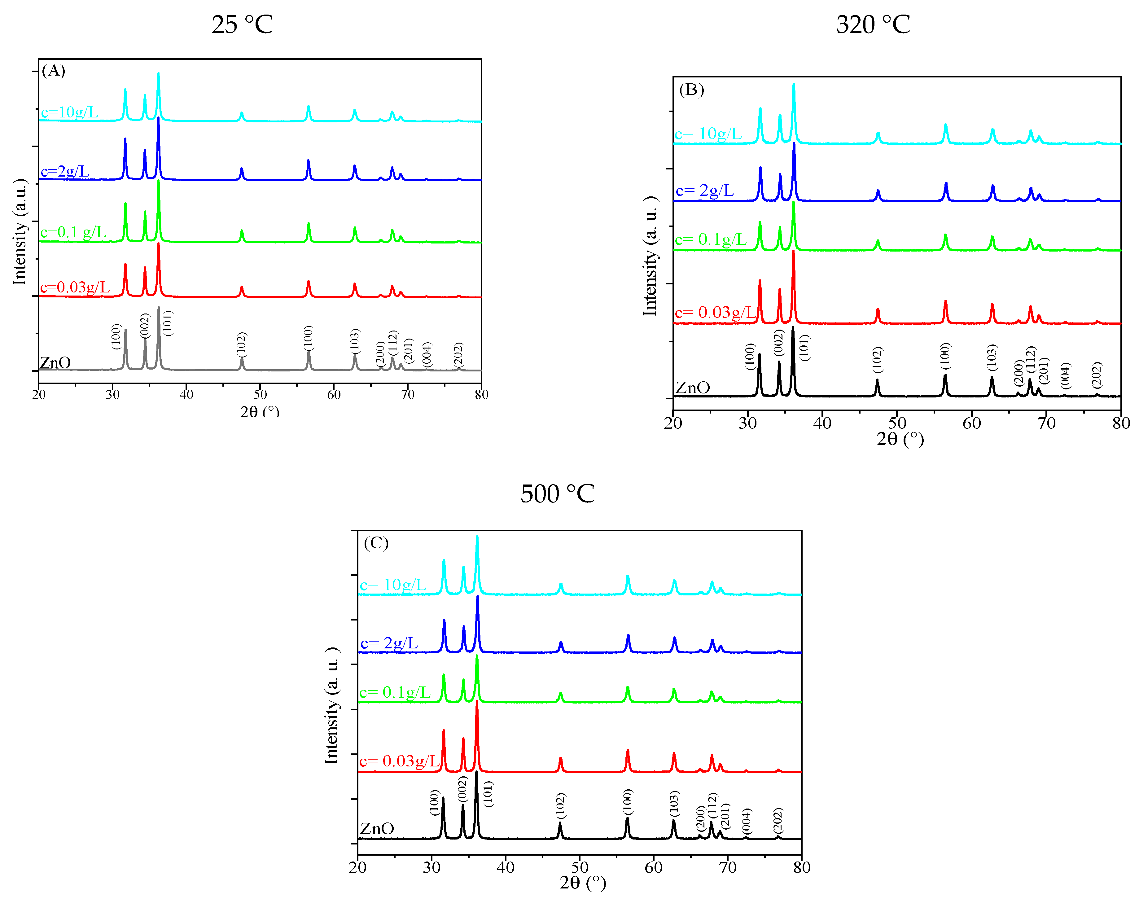
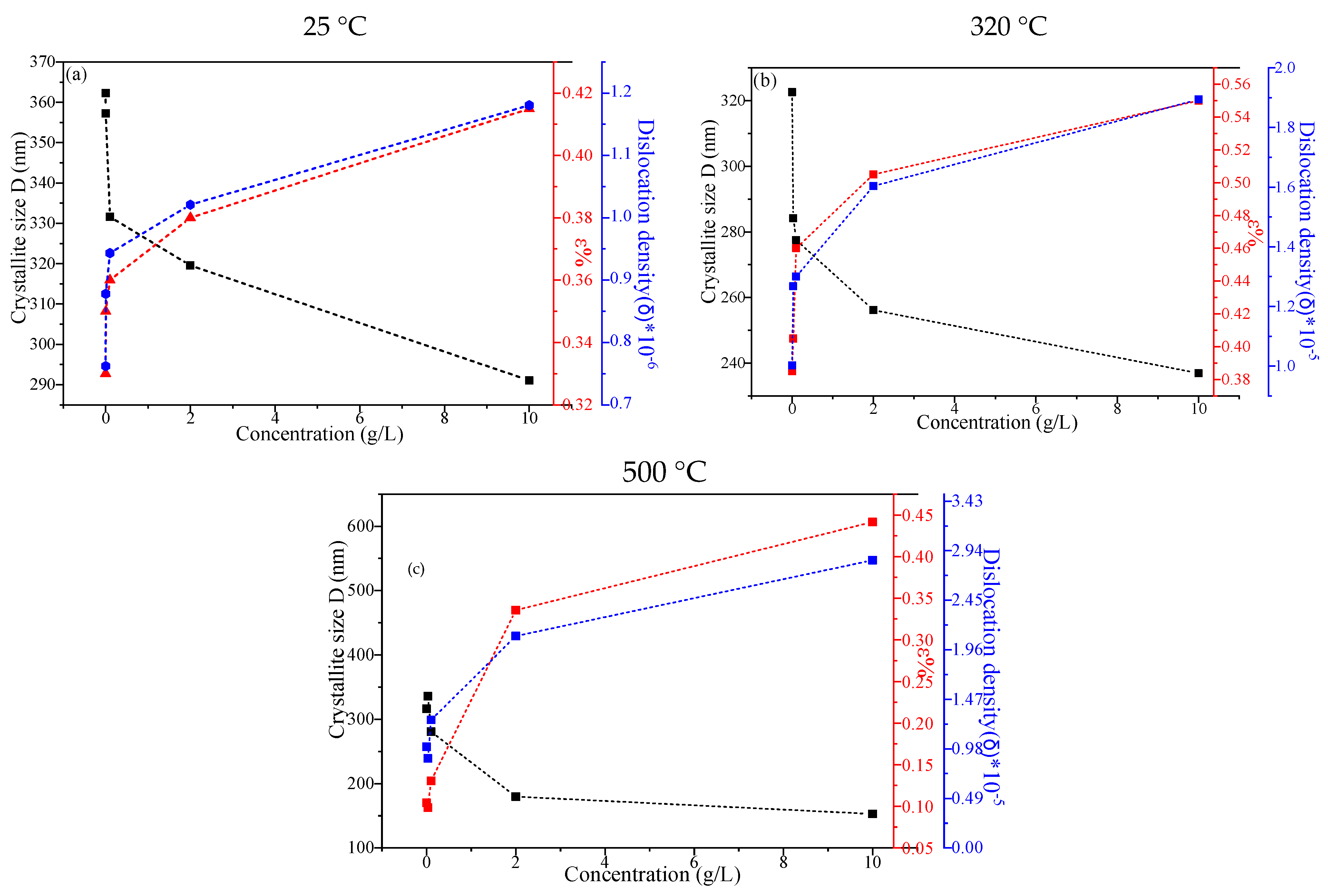
2.1.1. X-Ray Diffraction

Figure 1 illustrates the XRD patterns of both the as-synthesized and calcined ZnO nanoparticles, which were synthesized using varying concentrations of leaf extract (c = 0, 0.03, 0.1, 2, 10 g/L). The observed diffraction peaks correspond to the hexagonal wurtzite crystal structure of ZnO, in alignment with the data from the JCPDS card No. 36-1451 [59]. These peaks are present at specific angular positions (2θ) of 31.77°, 34.42°, 36.26°, 47.53°, 56.58°, 62.84°, 66.34°, 67.92°, 69.06°, 72.52°, and 76.92°. These positions are attributed to the (100), (002), (101), (102), (110), (103), (200), (112), (201), (004), and (202) Bragg planes, respectively. The diffraction patterns do not display any additional peaks that would suggest the presence of other phases, thereby confirming the high purity of the synthesized ZnO nanoparticles. The crystallite size of the nanoparticles was estimated using Scherrer Equation (1), providing insights into the material’s structural properties [42].
D = k λ β cos θ
where D is the crystallite size in Å; k is the correction factor that accounts for the shape of the particles (k = 0.94); λ is the wavelength of the XRD beam in nm (λ = 0.15406 nm); β is the full-width at half maximum (FWHM) of the peak in radians, and θ is half of the Bragg’s diffraction angle [59].
The microstrain (ε%) and the density of the dislocations (δ) were computed based on relations (2) (3), respectively [60]:
ε % = β 4 t a n θ
δ = 1 D 2
  • D denotes the mean crystallite size;
  • β represents the full width at half-maximum (FWHM) of the XRD peak, quantified in radians;
  • θ represents the Bragg angle, defined as half the angle formed between the incident X-ray beam and the dispersed X-ray beam.
Figure 2 reveals that for both the as-synthesized and the annealed green-synthesized ZnO nanoparticles, increases in lattice parameters and microstrain were observed at the nanometric scale when high concentrations of plant extract were used. This increase in lattice spacing can be attributed to the higher presence of plant-derived molecules that interact with the ZnO structure, potentially causing a modification of its crystallographic arrangement. At the macroscopic scale, however, the crystallite size was found to decrease, which, in turn, led to the creation of structural defects. These defects were reflected in a noticeable increase in dislocation density, which indicates the presence of imperfections in the crystal structure.
The results suggest that as the concentration of plant extract increased, the size of the crystalline domains of the green-synthesized ZnO nanoparticles decreased. The decrease in the size of the crystalline domains of the green-synthesized ZnO nanoparticles with an increase in the concentration of plant extract is primarily influenced by the interaction between the plant extract and the ZnO precursor during the synthesis process. As the concentration of the plant extract increases, the functional groups in the extract can better control the nucleation and growth of ZnO nanoparticles, leading to smaller crystalline domains. While thermal energy during the calcination process may also contribute, particularly in influencing crystallization and particle size, it is the concentration of plant extract that more directly drives the observed trend. The reduction in the size of the crystalline domains was primarily attributed to the increased concentration of plant extract, with thermal energy playing a secondary role during the calcination process [61].
These findings emphasize the importance of both the quantity of plant extract and the annealing temperature, as they have a significant impact on the crystallinity, structural defects, and the overall size of the ZnO nanoparticles. This highlights the delicate balance required to optimize the properties of the nanoparticles, ensuring their effectiveness for various applications [62,63].

2.1.2. Scanning Electron Microscopy (SEM)

Figure 3 presents the SEM images of ZnO nanoparticles synthesized via a green method under different annealing temperatures. These images offer valuable insights into the morphology, distribution, and degree of aggregation of the nanoparticles, which predominantly exhibit a spherical shape. Additionally, the particle size distribution of all samples was determined using SEM images and quantitatively analyzed with ImageJ software 1.51p, providing a comprehensive assessment of nanoparticle dimensions [64].
The results reveal a spherical shape and that both the leaf extract concentration and the annealing temperature significantly influence the nanoparticle size. Specifically, in the absence of leaf extract, the average nanoparticle size ranges from 10 to 35 nm. However, increasing the leaf extract concentration results in a noticeable reduction in particle size, albeit irregularly, as shown in Figure 3. This suggests that the leaf extract acts as a capping and stabilizing agent, crucial in controlling nanoparticle size by affecting nucleation and growth processes.
Furthermore, higher concentrations of leaf extract combined with elevated annealing temperatures reduce nanoparticle aggregation and lead to new surface morphologies. These changes, visible in Figure 3, reveal more distinct surface features than nanoparticles synthesized under lower extract concentrations and annealing temperatures. This enhancement in morphology can be attributed to the stabilizing effect of the leaf extract and the increased thermal energy during annealing, which prevent excessive aggregation and promote controlled growth.
In summary, the amount of plant extract used in the synthesis of ZnO nanoparticles (ZnO-NPs) plays a crucial role in controlling the nucleation, growth, and stabilization of these particles, ultimately influencing their size and morphology. As the concentration of the extract increases, the abundance of active phytochemical compounds, such as phenolics and flavonoid groups, also rises, enhancing their role as reducing and capping agents. This increased availability of reducing agents promotes faster nucleation of ZnO and inhibits particle growth by preventing agglomeration and uncontrolled crystallization. Studies have shown that when the extract volume is increased, the particle size decreases significantly. For example, the use of higher volumes of Azadirachta indica extract reduced ZnO-NP size from 25–60 nm (at 20 mL) to 8.2–11.9 nm (at 40 mL) due to more uniform nucleation and stabilization [65]. Similarly, varying extract concentrations with Moringa leaf and Dysphania ambrosioides demonstrated a clear trend: higher extract amounts yielded smaller, more stable particles with controlled morphology. The organic functional groups from the extract bind to Zn ions and modulate growth by capping the particles, which inhibits further growth and leads to uniform nanoscale structures [65]. However, insufficient extract levels may result in larger particles and agglomeration due to incomplete capping of ZnO surfaces. Therefore, optimizing the extract amount is key to controlling particle size, which is critical for applications where surface area and reactivity are important [64].

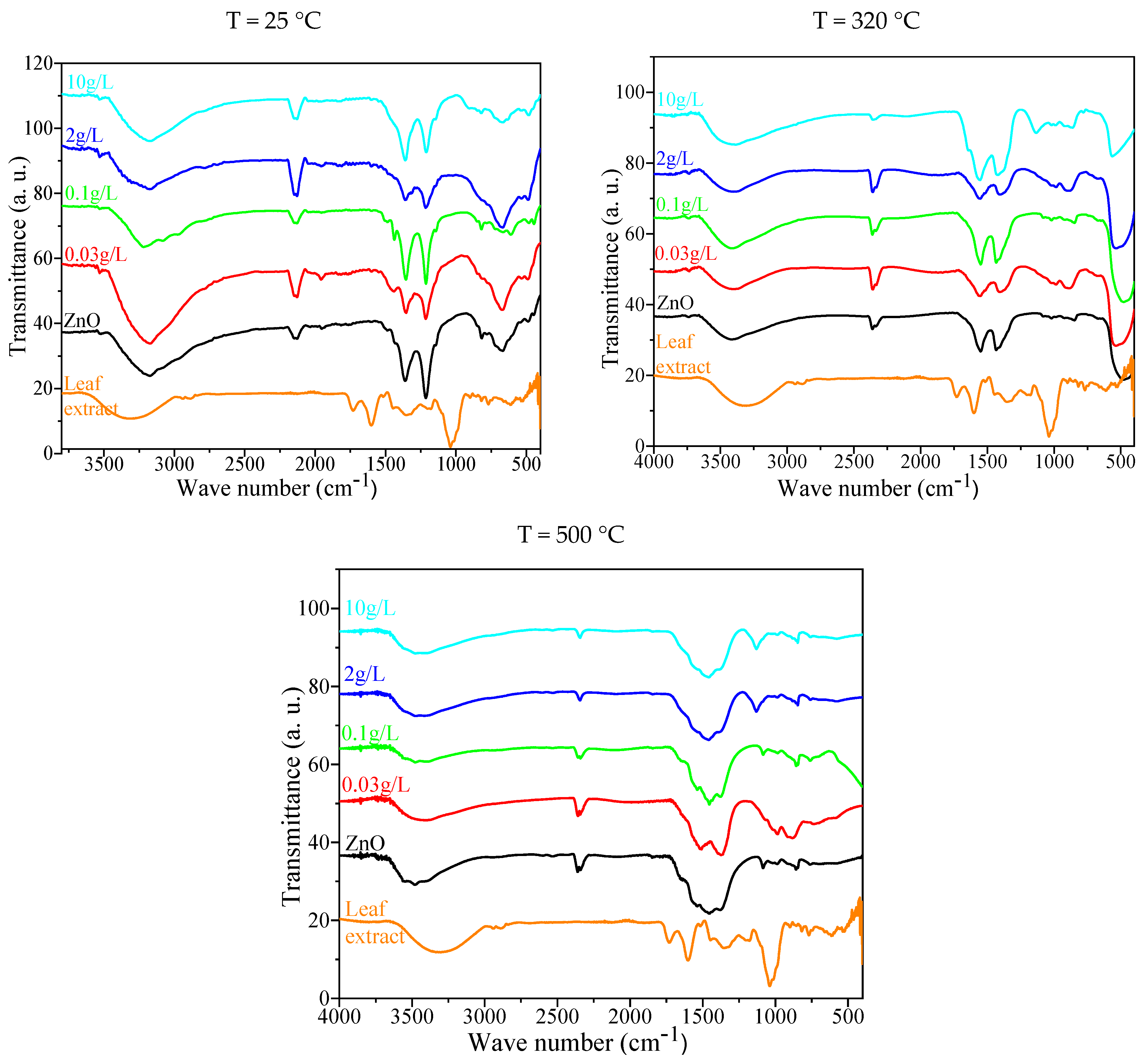
2.1.3. Fourier Transformation Infrared Spectroscopy (FTIR)

Spectroscopic analysis serves as a powerful tool for tracking the chemical and structural transformations that occur during heat treatment processes. Figure 4 presents the FTIR spectra of both as-synthesized and annealed green-synthesized ZnO nanoparticles, providing insight into their molecular characteristics. The Zn–O stretching vibration, which is indicative of the Zn–O bond, is confirmed by a distinct absorption band between 400–600 cm−1 [66]. This region is associated with the primary lattice vibration in ZnO, which is an essential feature of its crystal structure. The band observed between 800 and 900 cm−1 is attributed to the presence of Zn–OH bonds, suggesting the existence of hydroxyl groups that might be part of the nanoparticle surface or the surrounding environment [67].
Furthermore, vibrations in the 1500–1300 cm−1 region are likely due to the presence of hydroxyl (O–H) or carboxylate groups that are bound to the ZnO surface, confirming the interaction between the nanoparticles and these functional groups [68]. The range of 1600–1500 cm−1 is particularly significant as it corresponds to the carbonyl (C=O) stretch and other functional groups, such as C=C and −CO, originating from organic residues that are adsorbed onto the surface of the ZnO nanoparticles during synthesis [69]. Additionally, the broad absorption band between 3600–3200 cm−1 indicates the stretching vibrations of (OH) groups, which could either be due to adsorbed water molecules or hydroxyl groups that are chemically bound to the surface of the nanoparticles [70].
When ZnO nanoparticles are annealed at 320 °C and 500 °C, a significant reduction in the intensity of the infrared bands is observed. This reduction is likely caused by structural modifications occurring during the annealing process, such as the formation of ZnO-based composites or changes to the nanoparticle surface. These structural changes can disrupt or weaken the interactions between the functional groups and the nanoparticles, leading to the observed decline in band intensity [62].
The decrease in infrared intensity could also suggest a reduction in the number of surface-bound organic residues or hydroxyl groups, further supporting the idea that thermal treatment is influencing nanoparticle surface chemistry.
The FTIR spectrum of the leaf extract powder, which was used in the green synthesis of ZnO nanoparticles, also reveals distinct absorption bands corresponding to various functional groups in the plant material. One such peak, located at 1732 cm−1, is indicative of the carbonyl (C=O) groups present in the leaf extract, which are likely involved in the reduction and stabilization of the nanoparticles [71]. Another prominent peak around 1600 cm−1, within the 1600–1670 cm−1 range, reflects the stretching vibrations of carbon–carbon double bonds (C=C), which are common in aromatic compounds and may play a role in stabilizing the nanoparticles [71]. The peak at 1365 cm−1 is attributed to the in-plane bending vibration of O–H groups, further confirming the presence of hydroxyl groups in the leaf extract that contribute to the reduction in metal ions during synthesis [72]. Finally, the absorption feature at 1176 cm−1 is related to the symmetric stretching of C–O bonds, which could be involved in the coordination between the organic molecules and the ZnO surface [73]. These findings not only provide valuable information about the chemical composition and interactions within the green-synthesized ZnO nanoparticles but also underscore the important role that both the leaf extract and the annealing temperature play in determining the final properties of the nanoparticles. The FTIR analysis offers a comprehensive understanding of how the functional groups in the plant extract interact with the metal oxide, helping to stabilize the nanoparticles and influence their structural characteristics.

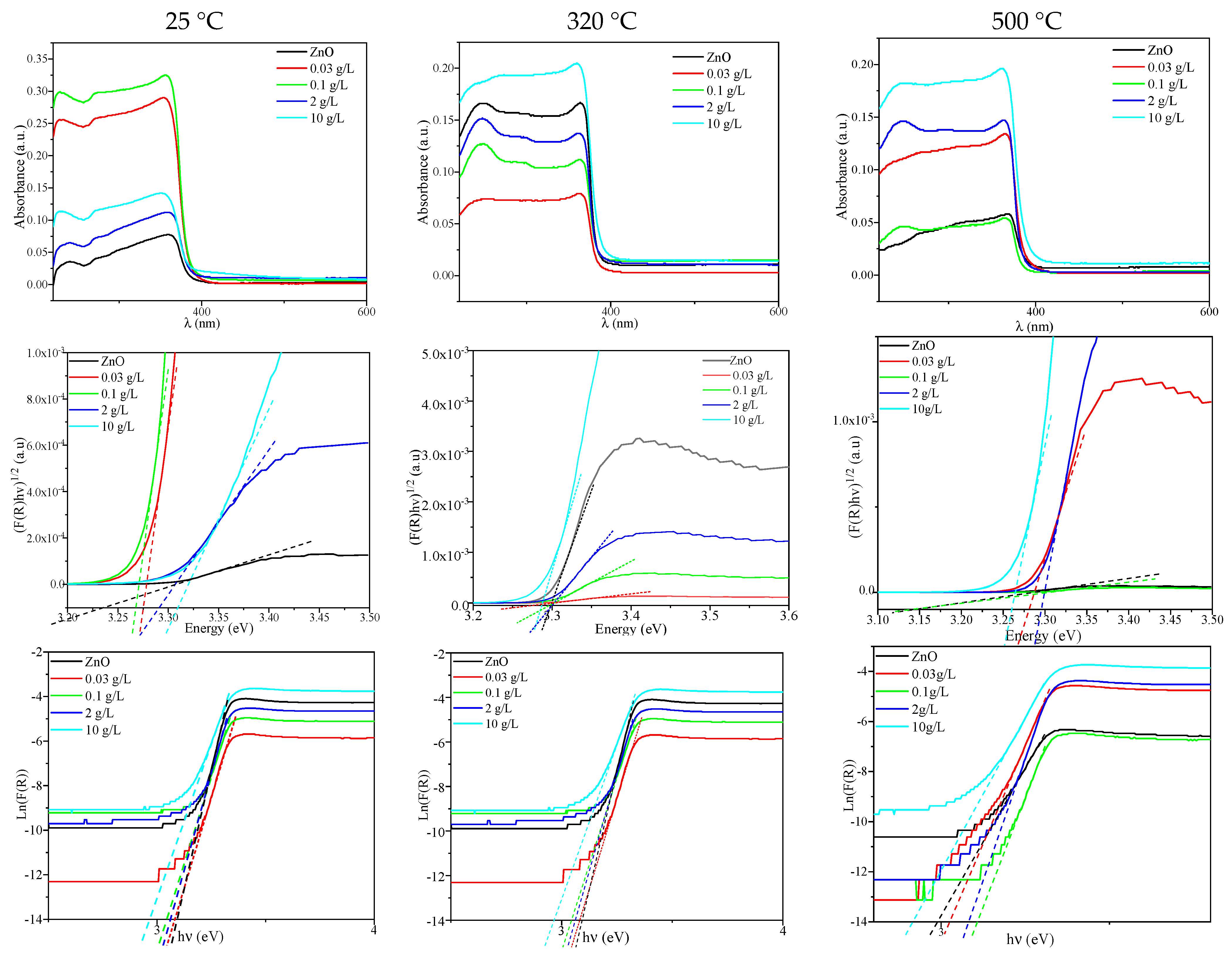
2.1.4. UV–Vis Spectroscopy
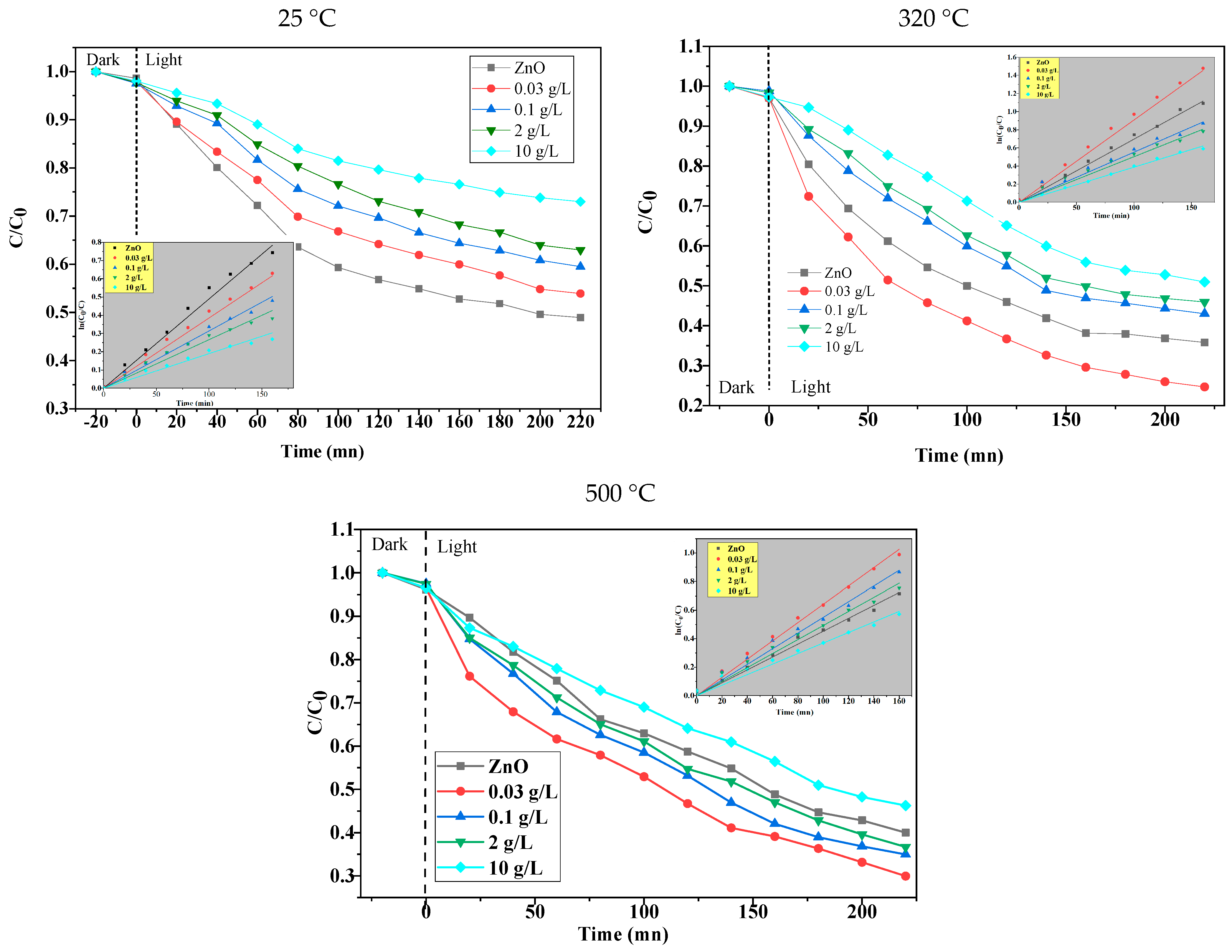
UV–Vis–DRS (Ultraviolet–Visible Diffuse Reflectance Spectroscopy) is a highly effective technique for analyzing the electronic properties of materials, particularly their band gap and Urbach energy, which are crucial for understanding their optical characteristics. Figure 5 presents the diffuse reflectance spectra of ZnO nanoparticles synthesized through a green method. The analysis of these spectra allows for the estimation of the band gap (“Eg”) and Urbach energy (“Eu”) of the nanoparticles. To quantify these properties, the absorption coefficients, denoted by F(R), were derived from the reflectance data using the Kubelka–Munk Equation (4) [74]:
F ( R ) = ( 1 R ) 2 2 R
In Figure 5 (insert), the variations in both the Urbach energy (“Eu”) and the band gap (“Eg”) are illustrated according to the Kubelka–Munk method. The band gap (“Eg”) is computed using the Tauc plot, which is a graphical method for determining the optical band gap of semiconductors. In the case of ZnO, the Tauc plot’s linear portion of the Equation (5) is as follows:
(αhν)n αhνEg
For direct band gap semiconductors, such as ZnO, the parameter ‘n’ is taken as 2. The photon energy, , is used, and the corresponding energy values are extracted accordingly.
The results of the band gap measurements are presented in Table 1, where the estimated band gap values for each sample are listed.
The Urbach energy (“Eu”) is an important parameter that gives insight into the tailing of the density of states near the conduction band edge. It is calculated using the following Equation (6), where E0 and α0 are constants that depend on the specific material being studied [74]:
α = α 0 e x p ( h υ E 0 ) E u
The Urbach energy (“Eu”) can be extracted from the slope of the linear portion of the plot of ln[F(R)] vs. , where is the photon energy. The Urbach energy reflects the degree of disorder in the material’s electronic structure, which is influenced by various defects such as vacancies, interstitial sites, lattice strain, and dislocations. These structural features contribute to the broadening of the band tail and affect the material’s optical properties.
UV–Vis–DRS spectroscopy, by directly analyzing the absorption and reflection spectra, provides valuable experimental data for determining key optical properties like the band gap and Urbach energy. Figure 3 showcases the spectra of ZnO nanoparticles that were synthesized using green methods at room temperature (25 °C), as well as those that were annealed at higher temperatures of 320 °C and 500 °C.
At the ambient temperature of 25 °C, the values for Ec (conduction band energy) and Eg (band gap energy) remain constant across all leaf extract concentrations (show Table 1). However, when the nanoparticles are annealed at 320 °C, the Eg values remain relatively uniform for all concentrations of leaf extract, while the Urbach energy decreases as the concentration of leaf extract increases (show Table 2). This suggests that higher concentrations of leaf extract may promote the formation of a more ordered crystal structure with fewer defects, thereby lowering the Urbach energy.
At an annealing temperature of 500 °C, both the Ec and Eg values exhibit an increase with higher concentrations of leaf extract (show Table 3). This trend is indicative of changes in the crystallinity and electronic structure of the nanoparticles as the temperature and extract concentration are varied. By comparing the calcined ZnO nanoparticles with those that were synthesized without annealing, a significant reduction in the band gap (Eg) is observed for the annealed nanoparticles. This decrease in the band gap is associated with an increase in the crystallite size of the nanoparticles, which tends to occur at higher calcination temperatures. As the crystallite size increases, the energy levels in the material become more defined, leading to a narrowing of the band gap.
Essentially, the increase in calcination temperature promotes the growth of ZnO nanoparticles, which, in turn, leads to the reduction in the band gap. This phenomenon is consistent with the general principle that larger particles typically exhibit a smaller band gap due to the quantum size effects becoming less pronounced as the particle size increases. Therefore, a narrower band gap results when the nanoparticles grow larger as the calcination temperature is increased [42]. This observation highlights the critical role of both the leaf extract concentration and the annealing temperature in modulating the size, electronic properties, and optical behavior of ZnO nanoparticles.

2.2. Photocatalysis

2.2.1. Photocatalytic Activity

A series of ZnO nanoparticles were synthesized using a green synthesis method with varying calcination temperatures to examine the influence of these temperatures on the photocatalytic performance of the ZnO semiconductors. The synthesis utilized different concentrations of leaf extract (c = 0, 0.03, 0.2, 2, and 10 g/L) to test the ability of the ZnO nanoparticles to degrade MB under UV light irradiation. The degradation process was monitored using UV–visible spectroscopy, and the decay of the normalized concentration (C(t)/C0) of MB was recorded at a wavelength of λ = 664 nm. The kinetic data were then fitted to an exponential decay model described by Equation (7):
C ( t ) C 0 = A 0 + B 0   e k ap t
In this equation, A0 represents the integrated baseline, and A0 + B0 corresponds to the initial concentration of MB at t = 0 (after adsorption). The term kap denotes the apparent photocatalytic rate constant, which reflects the rate at which the ZnO nanoparticles break down the MB under UV light exposure.
The findings showed that the photocatalytic degradation of MB was more efficient for ZnO nanoparticles calcined at 320 °C and 500 °C compared to those that were not calcined. This improvement in photocatalytic efficiency can be attributed to several factors related to changes in the morphology and optical properties of the nanoparticles during calcination. Higher calcination temperatures likely increased the surface area of the ZnO nanoparticles, which is beneficial for photocatalytic reactions, as a larger surface area provides more active sites for the adsorption and degradation of the dye molecules. Additionally, the calcination process may have improved the crystallinity of the ZnO nanoparticles, making them more efficient in promoting photocatalytic reactions [75,76]. Using the pseudo-first-order reaction rate equation, the following equation is used:
Ln ( C 0 C t ) = kt
This equation allows for the determination of the rate constant kt, where C0 is the initial concentration of MB, and Ct is the concentration of MB at time t. By plotting ln(C0/Ct) against reaction time, the rate constant can be extracted, providing insight into the efficiency of the photocatalytic degradation process. Table 4, Table 5 and Table 6 show the calculated rate constants for each experimental condition.
The results indicated that both the concentration of leaf extract used in the synthesis of the ZnO nanoparticles and the calcination temperature significantly impacted the degradation rates. The calcination temperature plays a pivotal role in determining the photocatalytic performance of materials, particularly in the context of materials calcined at 320 °C and 500 °C. Research suggests that this temperature range often leads to an optimal balance between crystallite size, surface area, and the material’s optical properties, which are crucial for photocatalysis. The calcining ZnO materials at 500 °C results in an increased crystallinity and surface area, facilitating the efficient separation of photogenerated electron–hole pairs, a critical factor in photocatalytic degradation. Additionally, the calcination process at these intermediate temperatures is ideal for maintaining a high surface-to-volume ratio while preventing excessive sintering, which would otherwise hinder the material’s photocatalytic capabilities. Moreover, the calcination temperature of 320 °C plays an important role in the green synthesis of ZnO nanoparticles, especially in terms of their photocatalytic performance. In the context of green synthesis, this temperature range is typically used to avoid high-energy consumption while ensuring the formation of crystalline and pure ZnO. At 320 °C, the precursor material undergoes decomposition, which promotes the development of ZnO with optimal crystallinity while preserving the eco-friendly aspects of the synthesis method. Research indicates that calcination at this moderate temperature helps achieve the right balance between particle size and surface area, both of which are crucial for photocatalytic efficiency. For instance, studies have demonstrated that ZnO nanoparticles calcined at lower temperatures (around 320 °C) tend to have a high surface area, which is beneficial for photocatalytic degradation due to an increased number of active sites available for reactions. Additionally, green synthesis methods, such as using plant extracts, are employed to minimize harmful chemical residues, contributing to both environmental sustainability and enhanced photocatalytic properties. The low-temperature calcination ensures the stability of these materials, which is vital for their application in long-term photocatalytic processes. Thus, the 320 °C calcination temperature is often chosen for its ability to optimize the physical properties of ZnO while maintaining an energy-efficient and environmentally friendly process [42].
Furthermore, the impact of the plant extract on the ZnO NPs and calcination temperature deserves attention. The plant extract reduces the particle size of ZnO, increasing the specific surface area accessible for catalytic reactions. This expanded surface area exposes more active sites, allowing for better adsorption of MB dye molecules and more effective catalytic turnover. The plant extract may add catalytic sites to the surface of ZnO NPs, increasing their activity. Adding more active sites to the photocatalyst improves its overall efficiency by providing extra catalyst centers [74].
The calcination temperature plays a crucial role in enhancing the photocatalytic performance of ZnO nanoparticles. We studied the photodegradation of BM dye using green-synthesized ZnO NPs (c = 0, 0.03, 0.5, 1, 2 g/L) at different calcination temperatures, as illustrated in Figure 6. Higher temperatures result in larger particles with better crystallinity, which are more effective at degrading pollutants like MB. Furthermore, the concentration of leaf extract used during synthesis also affects the photocatalytic efficiency, with higher concentrations generally leading to improved degradation performance. These findings highlight the importance of optimizing both the synthesis conditions and calcination parameters to maximize the photocatalytic potential of ZnO nanoparticles for environmental applications. In fact, the key factors influencing photocatalytic performance, such as crystallinity, surface area, and the presence of active sites, may stabilize at moderate calcination temperatures. Temperatures like 320 °C and 500 °C provide sufficient enhancement without introducing detrimental effects. ZnO synthesized through green methods has shown that both higher and lower calcination temperatures could lead to similar photocatalytic outcomes when specific material properties, such as particle size and surface defects, are optimized. For instance, ZnO calcined at 500 °C has been found to perform similarly to materials calcined at 320 °C in terms of photocatalytic efficiency despite differences in crystallinity. This suggests that the optimal temperature for photocatalytic performance may depend more on the specific properties of the synthesized material rather than solely on a higher calcination temperature. The recommendation to reconsider the assertion that the material calcined at 500 °C is the best is, therefore, well-supported, as both temperatures offer favorable outcomes for photocatalytic degradation, particularly under UV irradiation.

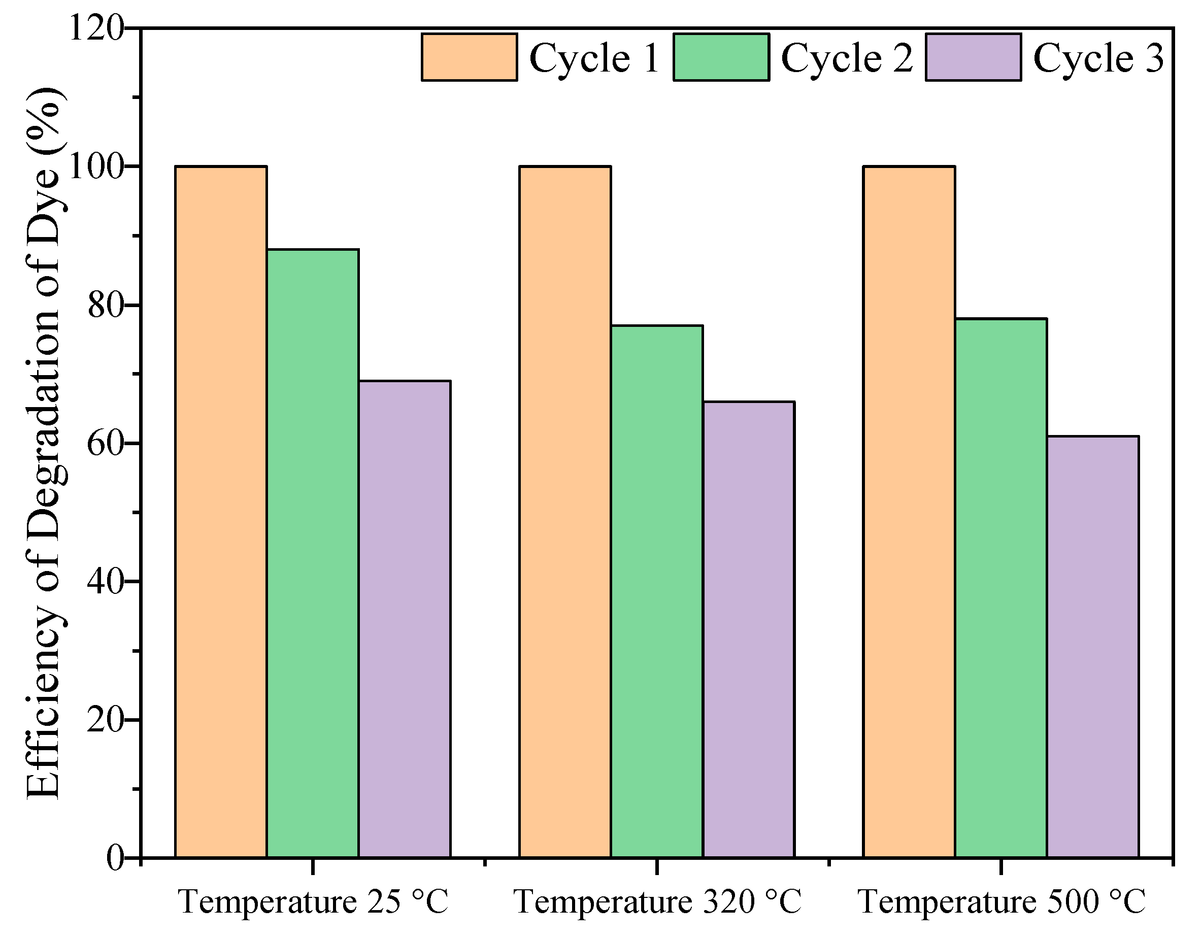
2.2.2. Photocatalytic Cycling Test for Methylene Blue Degradation Under UV Light

Photostability plays a vital role in determining the long-term functionality and practicality of a photocatalyst. To comprehensively evaluate the structural integrity and catalytic performance of the ZnO nanocomposite, a series of three consecutive photocatalytic degradation tests of MB were carried out. This approach not only provides insight into the material’s catalytic efficiency but also examines how well it can sustain its activity over multiple cycles of degradation.
Each photocatalytic test involved the degradation of MB under UV light, followed by a precise recycling process. After the completion of each test, the nanocomposite was separated from the reaction mixture through centrifugation, a key step to isolate the photocatalyst. To ensure the removal of any residual contaminants and maintain the material’s catalytic potential, the nanocomposite underwent a thorough three-step washing procedure. This involved washing with distilled water, followed by a mixture of distilled water and ethanol, and finally, a wash with double-distilled water. These steps helped eliminate any adsorbed organic molecules or by-products that could affect the photocatalyst’s performance in subsequent cycles. After the washing procedure, the ZnO material was carefully collected and dried at 60 °C for 24 h. The drying step was essential to ensure that the photocatalyst was completely free of any solvents and ready for reuse in the next cycle.
Figure 7 presents the results of the three consecutive photocatalytic degradation cycles, with the data showing consistent photocatalytic activity across all cycles. This stability indicates that the ZnO nanocomposite retained its efficiency in degrading MB under UV light despite undergoing multiple testing cycles. The results highlight the intrinsic stability and resilience of the pre-synthesized and annealed ZnO nanocomposite. This suggests that the material has a robust structure capable of maintaining its catalytic properties over extended use, a crucial factor for any practical application of photocatalysts in environmental remediation processes.
  • Surface Degradation: Prolonged exposure to reactive species or harsh conditions may lead to surface degradation of ZnO, such as oxidation, reduction, or the formation of defects that diminish its active sites;
  • Structural Changes: High temperatures or repeated cycles of catalysis can induce morphological or structural changes in ZnO, such as sintering or particle agglomeration, reducing the surface area available for catalysis;
  • Contamination or Fouling: The active surface of ZnO may become contaminated by impurities, byproducts, or undesired species, leading to a reduction in catalytic efficiency over time;
  • Leaching of Active Sites: In liquid-phase catalysis, ZnO may experience leaching, where catalytic components are gradually dissolved into the reaction medium, leading to performance decay;
  • Formation of Inactive Phases: Chemical interactions between ZnO and the reaction medium could result in the formation of secondary, less active, or inactive phases, altering the material’s catalytic behavior;
  • Photo-Induced Changes: For photocatalytic applications, prolonged exposure to UV or visible light can induce photocorrosion or electron–hole recombination, which can degrade the material over time.
The decline in photocatalytic performance of ZnO over successive reuse cycles is generally attributed to several factors. First, the surface deactivation of ZnO can occur due to the accumulation of by-products or contaminants from the reactions, which block the active sites necessary for photocatalysis. Additionally, the crystallinity of ZnO nanoparticles may degrade over time, especially under harsh conditions during photocatalytic reactions (e.g., UV light exposure). This degradation can lead to a reduction in the available surface area, further diminishing the material’s efficiency. Another key factor is the leaching of ZnO components during the photocatalytic process, which can result in a loss of catalytic activity with repeated cycles. Finally, structural changes, such as particle agglomeration or morphological changes, could also hinder the photocatalytic process by reducing the material’s surface-to-volume ratio and available active sites.

2.2.3. Effect of Catalyst Dosage

Figure 8 illustrates the relationship between catalyst dose and photocatalytic activity, measured by the C/C0 ratio over time, for varying catalyst concentrations of 0.25, 0.5, 1, and 2 g/L. At lower concentrations of 0.25 g/L and 0.5 g/L, the degradation of MB is moderate, with efficiency levels that are significantly lower compared to the 1 g/L concentration Table 7. This suggests that lower concentrations do not provide enough active surface area for efficient photocatalytic reactions, leading to incomplete degradation of the MB dye.
When the concentration is increased to 1 g/L, the degradation efficiency reaches an optimal value of 74%. This improvement can be attributed to a sufficient specific surface area and effective light transmission, which minimizes shading or saturation effects. At this concentration, the catalyst particles are dispersed enough to allow light to reach all active sites, promoting enhanced photon absorption. This, in turn, leads to the generation of more electron–hole (e, h+) pairs, which are crucial for the photocatalytic process, and increases the production of reactive radicals that facilitate the breakdown of organic contaminants such as MB [77].
However, at the highest concentration of 2 g/L, the photocatalytic activity drops dramatically, with only 22% degradation observed. This decrease in efficiency is primarily due to two factors: shading and saturation. As the concentration increases beyond 1 g/L, the catalyst particles tend to agglomerate or overlap, blocking light from reaching the active sites on the particles. This shading effect prevents efficient photon absorption and hampers the photocatalytic process. Furthermore, the saturation effect occurs when the number of active sites becomes limited, meaning that even though the catalyst concentration is higher, there are not enough available sites to accommodate the increased amount of particles. This leads to a decrease in the overall catalytic performance.
The results from this study underline the importance of selecting the optimal catalyst concentration for photocatalytic applications. Too low concentrations result in inefficient use of the available surface area, while too high concentrations lead to inefficiencies due to shading and saturation effects. The 1 g/L concentration strikes a balance, offering the best combination of surface area and light transmission, ensuring maximum degradation of MB. This emphasizes that for efficient photocatalytic degradation, careful control over the catalyst concentration is necessary to avoid diminishing returns. Thus, 1 g/L is identified as the optimal concentration, where the photocatalytic activity is maximized, demonstrating the critical role of catalyst dosage in photocatalytic processes.

2.2.4. Effect of Reaction pH

Figure 9 provides a detailed overview of the effect of pH on the photocatalytic degradation of MB, with results measured by the C/C0 ratio as a function of time across a range of pH values (4, natural pH, 8, and 10). The data reveals that the natural pH condition (pH 7) is optimal for photocatalytic activity, achieving a degradation rate of 74%. This suggests that the natural pH maintains the stability of the ZnO photocatalyst surface and allows for efficient interaction between the photocatalyst and the pollutant, promoting the generation of active species required for degradation.
At pH 8, a slight decrease in degradation efficiency was observed, with a reduction of 58%. This result still indicates considerable photocatalytic activity, although it is not as efficient as at natural pH. The slightly basic environment may slightly alter the surface charge or the ability of the photocatalyst to interact effectively with the pollutant molecules, thus reducing the overall efficiency of the degradation process.
In contrast, the lowest pH value tested, pH 4, resulted in only 42% degradation, while at pH 10, the degradation was 51% (Table 8). These findings suggest that both acidic and basic environments are less ideal for photocatalytic activity. At acidic pH, the surface of the photocatalyst may develop a highly positive charge due to protonation, which could reduce its interaction with certain negatively charged pollutant ions. Additionally, ZnO is known to be sensitive to weak acids like acetic acid, leading to corrosion of the material and a subsequent decrease in catalytic performance. This degradation effect might explain the observed reduction in activity. Studies on the functionalization of ZnO powders for applications such as gas sensors may provide insights into strategies for improving its stability under acidic conditions.
This study underscores the importance of maintaining a neutral or slightly acidic pH range for optimal photocatalytic performance. The results suggest that pH significantly influences the photocatalytic degradation of MB, with neutral to slightly acidic conditions (pH 7–8) being the most favorable for achieving the highest degradation efficiency. On the other hand, very acidic (pH 4) or basic (pH 10) conditions diminish the photocatalytic efficiency, likely due to destabilization of the catalyst’s surface or unfavorable interactions with reactive species.

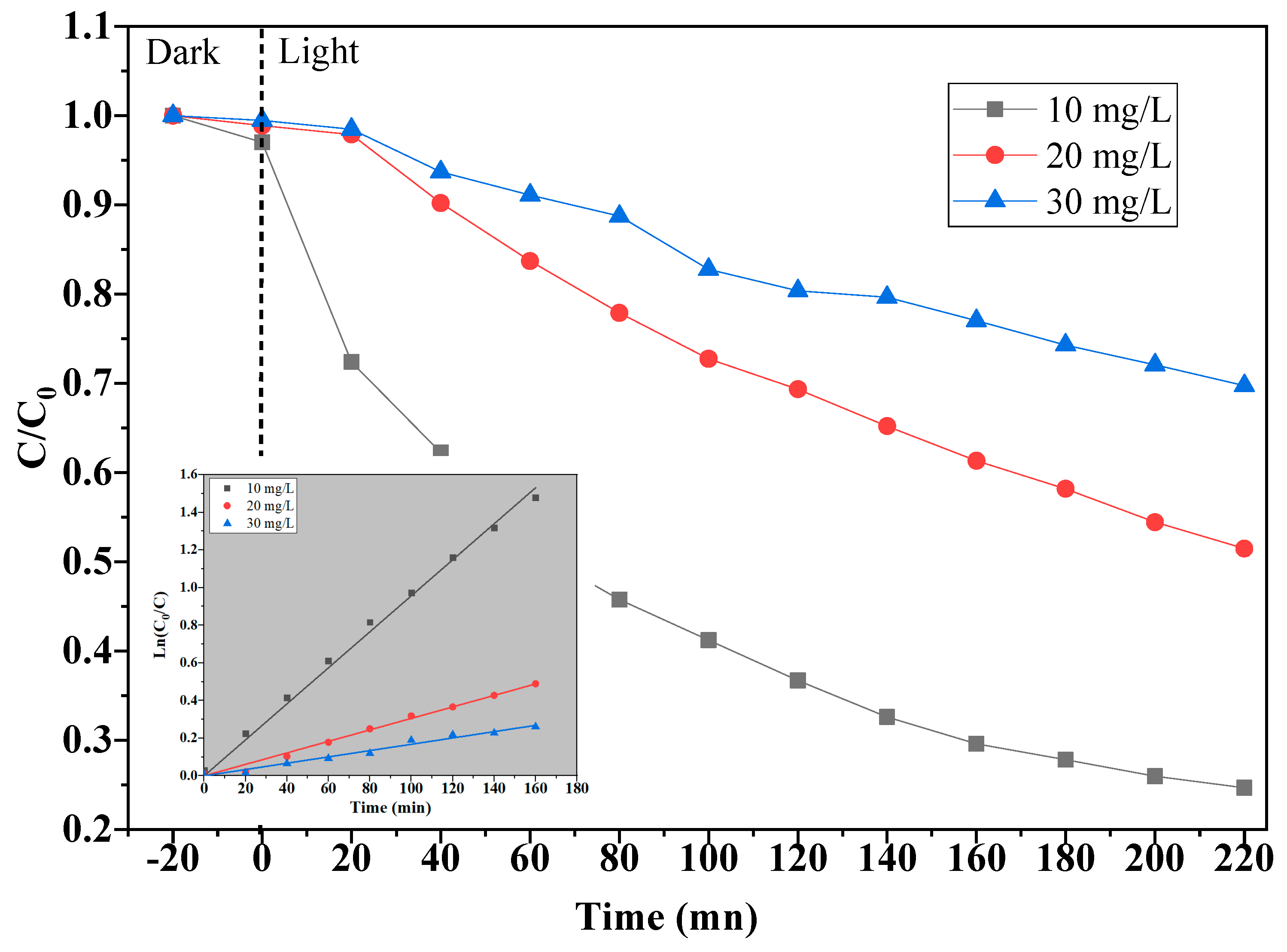
2.2.5. Effect of Initial Concentrations of MB

To investigate the impact of initial dye concentration on the photocatalytic degradation of MB, a series of experiments were conducted by varying the concentration of MB from 10 to 30 mg/L. The photocatalytic degradation process was monitored for a reaction time of 220 min, and the dye removal efficiency was calculated for each concentration. The results, shown in Figure 10, indicated that the removal efficiency decreased as the dye concentration increased. Specifically, at 10 mg/L MB, a high removal rate of 74% was achieved, with a corresponding rate constant (k) of 9.57 × 10⁻³ min−1. In contrast, for 20 mg/L, the removal efficiency dropped to 48% (k = 3.05 × 10⁻³ min−1), and at the highest concentration of 30 mg/L, the removal efficiency further decreased to 29% (k = 1.68 × 10⁻³ min−1) as it shown in Table 9.
This decline in removal efficiency with increasing MB concentration can be attributed to several factors. As the dye concentration increases, more MB molecules adsorb onto the surface of the photocatalyst, occupying the active sites required for the adsorption of reactive species such as oxygen and hydroxide ions. This reduces the available surface area for the formation of reactive radicals (such as hydroxyl and superoxide anion radicals), which are essential for the photocatalytic degradation process. As a result, the efficiency of the photocatalytic reaction is compromised.
Furthermore, at higher dye concentrations, the increased absorption of light by the dye molecules reduces the amount of light available for the photocatalyst, leading to lower photon absorption. This phenomenon, known as light shielding or light blocking, further reduces the efficiency of the photocatalytic process. As the concentration of MB increases, the dye molecules form a layer on the surface of the photocatalyst, obstructing the effective interaction between the catalyst and the light energy [78,79,80].
Overall, these results demonstrate that the photocatalytic degradation of MB is less effective at higher initial dye concentrations. The key factors contributing to this decrease in efficiency include the competition for active sites on the catalyst surface, reduced production of reactive radicals, and diminished light absorption due to photon shielding effects. Therefore, to optimize the photocatalytic degradation process, a moderate dye concentration is preferable, as it ensures maximum active site availability and efficient photon absorption by the photocatalyst.

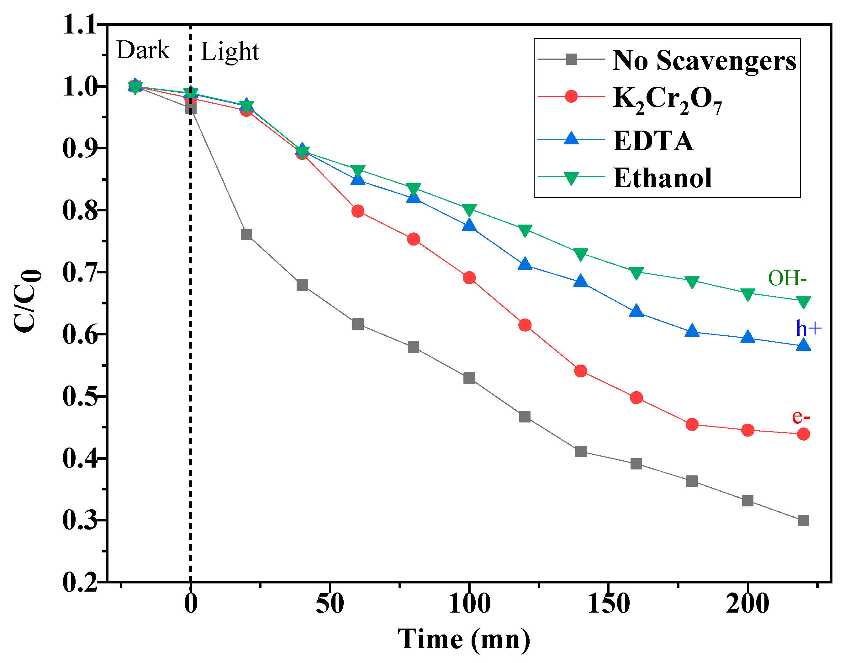
2.2.6. Effect of Scavengers

In the photocatalytic degradation process, reactive species such as hydroxyl radicals (•OH), superoxide anions (•O2) and electron–hole pairs (e/h+) are generated and play a crucial role in the breakdown of pollutants like MB. To identify which specific reactive species are responsible for the degradation, selective scavengers or inhibitors are used in a test. These scavengers are chemical compounds that can effectively capture or neutralize a specific reactive species, preventing it from participating in the reaction. By introducing these scavengers into the reaction system, it is possible to determine the role of each reactive species in the photocatalytic process.
Typically, the experiment is set up by maintaining the same experimental conditions, including the photocatalyst concentration, plant extract concentration, and UV light intensity. A series of tests are carried out by adding different types of scavengers to the solution. These scavengers are chosen based on their ability to specifically react with and neutralize a particular reactive species. For example, t-butanol is commonly used as a scavenger for hydroxyl radicals, while potassium bromide (KBr) is used to capture superoxide anions.
After the scavenger is added, the photodegradation efficiency of the substrate is measured, usually by tracking the decrease in the concentration of the pollutant (e.g., MB) over time, often using UV–Vis spectroscopy. The comparison of photodegradation efficiency in the presence of each scavenger helps to pinpoint which reactive species are most responsible for the catalytic breakdown of the substrate.
In this way, the experiment allows for a clearer understanding of the mechanisms driving photocatalysis and can help in optimizing the photocatalytic process by highlighting which species need to be generated in larger quantities for more efficient pollutant removal. Additionally, this approach helps to identify potential limitations in the photocatalytic reaction, guiding further improvements in catalyst design or reaction conditions.
This study aimed to understand the roles of various reactive species involved in the photocatalytic degradation of MB dye by selectively trapping specific species such as •OH, e, and h+. Ethanol was employed to capture hydroxyl radicals; K2Cr2O7 was used to trap electrons, and ethylenediaminetetraacetic acid (Na2-EDTA) was utilized to sequester holes. These scavengers were added to the reaction mixture under optimized experimental conditions, with the irradiation duration maintained at a fixed period of 220 min. The results, as illustrated in Figure 11, demonstrate significant variations in photocatalytic efficiency depending on the type of scavenger used.
The purpose of using selective trapping agents was to isolate and quantify the individual contributions of these reactive species to the overall photocatalytic degradation of MB. Figure 12 clearly shows the impact of these scavengers on the degradation process. When ethanol was used to trap •OH, the photocatalytic efficiency decreased significantly, with a degradation rate of only 34%. This is considerably lower than the degradation rate of 67% observed when no scavengers were used, indicating that •OH radicals are primarily responsible for the majority of the degradation process.
In contrast, when h+ ions were trapped using EDTA, the degradation rate was 41%. This result suggests that the photogenerated holes also play an essential role, although their contribution is less significant than that of the hydroxyl radicals. The presence of K2Cr2O7 to trap e led to a degradation rate of 54%, showing that electrons also contribute substantially to the photocatalytic activity.
Overall, these findings demonstrate that multiple reactive species, including •OH radicals, h+ ions, and e, are involved in the photocatalytic degradation of MB dye, with each species contributing to varying extents. The primary role of hydroxyl radicals in the degradation process is evident from the significant decrease in degradation efficiency when they were trapped. The results also suggest that optimizing the photocatalytic process could involve enhancing the production or efficiency of these key reactive species to improve degradation rates further.

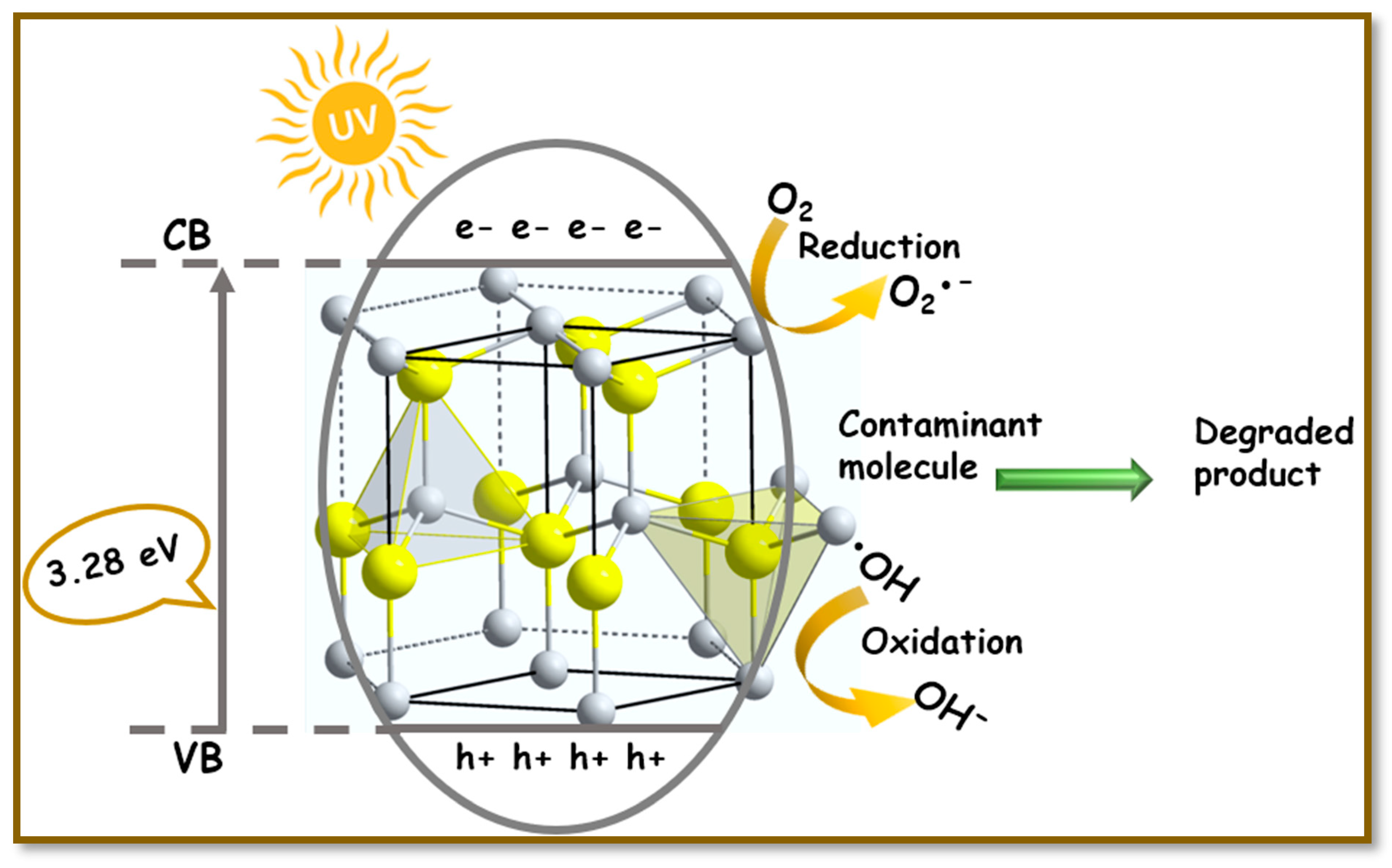
2.3. Photocatalytic Mechanism

ZnO functions as an efficient photocatalyst, leveraging light to accelerate various chemical reactions, making it a crucial material in numerous environmental and energy-related applications. Its exceptional photocatalytic properties stem from its wide bandgap and the ability to form electron–hole pairs when exposed to light. These electron–hole pairs are essential for initiating the photocatalytic process, as they participate in redox reactions that degrade pollutants and harness energy from light.
The principle behind ZnO’s photocatalytic activity is that when it absorbs ultraviolet (UV) light, the photonic energy matches or exceeds its excitation energy (Eg), electrons in the valence band of ZnO are excited to the conduction band (CB). This excitation creates an electron–hole pair. These electron–hole pairs migrate to the surface of ZnO, where they interact with surrounding molecules, contributing to a series of redox reactions. The h+ ions are highly reactive and can react with water and hydroxide ions to form •OH, one of the most potent oxidizing agents in photocatalysis. The e ions react with molecular O2 to form •O2, which are also reactive and involved in the degradation process.
In addition to these fundamental reactions, H2O2 is formed as a byproduct of electron interaction with oxygen, which further contributes to the generation of •OH. These hydroxyl radicals, superoxide anions, and other reactive species collectively degrade pollutants on the surface of the ZnO photocatalyst. This process not only purifies the surrounding environment by breaking down harmful substances but also releases oxygen as a byproduct [81,82,83]. The consisted response equations are given below as follows:
ZnO + hυ→ h+ + e
h+ + H2O → OH + H+
h+ + OHOH
h+ + pollutant → (pollutant)+
e + O2 → •O2
•O2 + H+OOH
2OOH+ → O2 + H2O2
H2O2 + O2OH + OH + O2
H2O2 + hυ → 2OH
Pollutant + (OH, h+, OOH/O2) → degraded product
Despite its advantages, ZnO photocatalysts face challenges related to photostability and photocorrosion. Photocorrosion refers to the tendency of ZnO to degrade or dissolve when exposed to UV light, releasing Zn2+ ions into the environment. This contamination of the surface not only reduces the catalyst’s efficiency but also limits its effectiveness over prolonged use. The geometry and structure of ZnO play a significant role in mitigating photocorrosion. Higher crystallinity, the presence of crystalline planes, and optimized surface structure help prevent the breakdown of ZnO under UV exposure. Additionally, surface oxygen vacancies have been shown to prevent photocorrosion by stabilizing the catalyst surface [62]. To address these limitations and enhance the stability of ZnO photocatalysts, various strategies can be employed. One common method is post-synthesis heat treatment, such as annealing, which improves the crystallinity of ZnO and optimizes the Zn:O ratio. This can significantly enhance the photocatalyst’s stability and overall performance. Furthermore, combining ZnO with other materials, particularly carbon-based compounds like graphene or carbon nanotubes, has been shown to reduce photocorrosion. These materials can help improve the electronic properties of ZnO, facilitate electron transfer, and prevent electron–hole recombination, thereby enhancing photocatalytic efficiency [84]. In addition to these conventional strategies, green synthesis approaches are gaining attention. Using plant extracts as capping agents during the synthesis of ZnO nanoparticles can improve their stability and reduce photocorrosion. The phytochemicals present in the plant extracts act as protective agents that stabilize the ZnO structure, reducing the likelihood of degradation during photocatalytic reactions. These green synthesis methods are not only environmentally friendly but also cost-effective, making them an attractive option for the production of stable ZnO photocatalysts.

2.4. Comparison of the Photocatalytic Efficacy of ZnO Nanoparticles Synthetized by Different Methods

Photodegradation of dyes or organic contaminants using ZnO synthesized by “green” methods has attracted increasing interest in recent years due to its ecological and economic benefits. In this study, the photocatalytic performance of our ZnO green synthesized was evaluated by degrading methylene blue under UV light irradiation.
The results show that our green ZnO allowed a photodegradation of 67% of MB after 220 min. This rate is comparable to or higher than some works reported in the literature presented in Table 10.
The results obtained in this study confirm the effectiveness of green synthesis methods to produce ZnO-based photocatalysts. Differences in the literature can be attributed to variations in the synthesis method (nature of biomass used, calcination conditions), crystal structure, and experimental parameters. These variations directly influence the light absorption capacity and the generation of electron–hole pairs, essential for photocatalytic degradation.

2.4.1. Antibacterial Activity

This study investigates the antimicrobial properties of biosynthesized ZnO nanoparticles against a broad spectrum of pathogens, providing crucial insights into their potential as antimicrobial agents. The ZnO NPs were synthesized using a sustainable and environmentally friendly method, with varying concentrations ranging from 0 to 10 g/L. The antimicrobial efficacy of these nanoparticles was evaluated using the MIC method, a standard technique in microbiology. This method assesses the lowest concentration of an antimicrobial agent needed to inhibit the growth of microorganisms, providing quantitative data essential for evaluating the effectiveness of antimicrobial substances.
In this study, the antimicrobial activity of ZnO NPs was tested against both Gram-positive bacteria (Staphylococcus aureus and Bacillus subtilis), Gram-negative bacteria (Escherichia coli and Salmonella enterica), and the fungal species Candida albicans. The ability to inhibit the growth of these diverse microorganisms suggests that ZnO nanoparticles could be used as versatile antimicrobial agents. The application of the MIC method offers a reliable means of determining the potency of these nanoparticles, which is crucial for assessing their potential as therapeutic or disinfectant agents.
The microtiter plate technique, a high-throughput method used in this study, enables the simultaneous screening of multiple samples. This technique involves exposing microbial cultures to various concentrations of ZnO nanoparticles and observing growth inhibition, typically marked by the absence of turbidity in the wells. The use of this method allows for an efficient and detailed assessment of the nanoparticles’ antimicrobial activity against different pathogens.
The results shown in Table 11 indicate that the biosynthesized ZnO nanoparticles, particularly the ones that were annealed, exhibited significantly better antimicrobial performance compared to those produced by conventional methods. The green synthesis approach, which utilizes plant extracts or other eco-friendly methods, appears to preserve and even enhance the antimicrobial properties of ZnO nanoparticles. This suggests that the green synthesis method not only contributes to environmental sustainability but also plays a role in preserving the antimicrobial properties of the nanoparticles.
However, it is important to note that while the ZnO nanoparticles displayed consistent antibacterial effects, they did not exhibit significant antifungal activity against Candida albicans. This limitation suggests that while these nanoparticles are effective against bacterial pathogens, they may not be as effective against fungal infections. This finding underscores the need for further research to explore potential modifications to the ZnO nanoparticle formulations or to investigate alternative methods for improving their antifungal activity. Such research could help expand the range of applications of these nanoparticles in the treatment of fungal infections.
Despite the absence of significant antifungal effects, this study highlights the considerable promise of biosynthesized ZnO nanoparticles as effective antimicrobial agents for controlling bacterial infections. Their ability to inhibit both Gram-positive and Gram-negative bacteria suggests their potential use in various biomedical and environmental applications. For example, these nanoparticles could be integrated into wound dressings to prevent infections, incorporated into water purification systems to eliminate harmful pathogens, or used as coatings for medical devices to reduce the risk of bacterial contamination.
Moreover, the antibacterial properties of biosynthesized ZnO nanoparticles could be particularly valuable in healthcare settings, where bacterial resistance to conventional antibiotics is an emerging concern. The use of green-synthesized ZnO nanoparticles may offer a safer and more sustainable alternative to synthetic antibiotics, providing a novel approach to combating bacterial infections.
While the biosynthesized ZnO nanoparticles exhibit strong antibacterial activity, further investigations are needed to optimize their formulations for enhanced antifungal performance. This study paves the way for the potential use of ZnO nanoparticles in various applications, including biomedical treatments, environmental remediation, and disinfectant products.

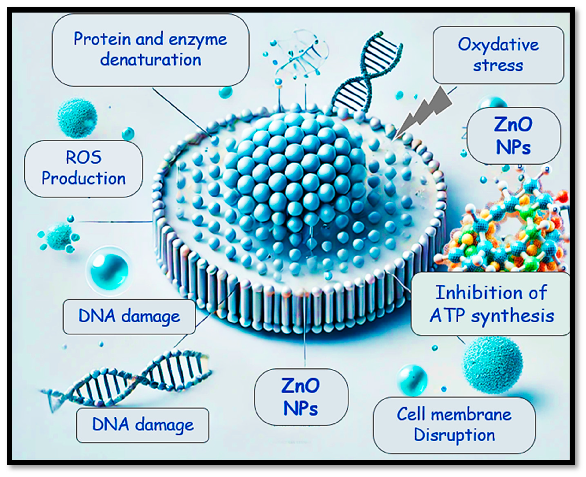
2.4.2. Antibacterial Mechanism

Recent studies have extensively explored the advancements in the field of metal oxide and modified metal oxide nanoparticles, particularly focusing on their antibacterial properties and the eco-friendly synthesis methods used to produce them [88,89,90,91]. Among the various metal oxide nanoparticles, ZnO has attracted considerable attention due to its remarkable antibacterial potential. This is primarily because ZnO nanoparticles can release zinc ions (Zn2+) upon interacting with microbial cells, which can penetrate the cell membrane and disrupt crucial cellular functions, leading to the death of the microorganisms [92]. Similarly, ZnO nanoparticles have emerged as promising candidates for antibacterial applications, owing to their capacity to modulate the electrical properties of bacterial membranes. This modulation enhances the reactivity of the nanoparticles’ surfaces, facilitating interactions with microbial cells and ultimately resulting in bactericidal effects [93].
In-depth research on the mechanisms behind the antibacterial activity of ZnO nanoparticles has uncovered fascinating details about their mode of action Figure 13. These nanoparticles are believed to form strong interactions with essential biomolecules inside bacterial cells, including RNA and DNA [94]. By interfering with processes like DNA replication, these nanoparticles can induce cell cycle arrest, leading to the death of the bacteria. Besides direct damage to DNA, ZnO nanoparticles can generate oxidative stress within bacterial cells, which disrupts ATP synthesis and leads to the production of reactive oxygen species (ROS). These ROS then initiate a cascade of events that ultimately cause apoptosis in the bacterial cells, further contributing to the antibacterial effects of ZnO nanoparticles [95].
With the increasing concerns surrounding the environmental impact of synthetic processes, researchers have shifted their focus toward green synthesis approaches for nanoparticle production [96,97]. These methods utilize renewable natural resources such as plant extracts, bacteria, fungi, algae, and organic products to synthesize nanoparticles. This not only makes the process more environmentally friendly but also enhances the antibacterial properties of the nanoparticles, offering a sustainable alternative to traditional chemical synthesis methods [98]. Plant leaf extracts are particularly valuable for green synthesis, as they contain a wealth of pharmacologically active compounds that help facilitate the interactions between the nanoparticles and bacterial membranes, thus improving their antibacterial efficacy [99,100]. Among these, the green synthesis of ZnO nanoparticles using Quercus robur leaf extract has shown promising results, with the nanoparticles demonstrating substantial antimicrobial activity. This aligns with the traditional use of oak bark and leaf extracts in folk medicine to treat microbial infections. The use of Quercus robur extract underscores the potential of green synthesis in leveraging the therapeutic properties of natural resources, thus paving the way for environmentally sustainable and effective antimicrobial agents [101,102]. The incorporation of plant-derived compounds in the synthesis process not only enhances the antibacterial properties of the nanoparticles but also reduces the environmental footprint of nanoparticle production. These green synthesis methods offer a practical and sustainable route to producing antibacterial nanoparticles that can be applied in various fields, including medicine, environmental remediation, and material science.
Moreover, the continued research into the green synthesis of ZnO nanoparticles highlights the importance of exploring natural resources to reduce the reliance on synthetic chemicals in the production process. This has significant implications for reducing chemical waste, lowering costs, and improving the efficiency of nanoparticle production. By tapping into the power of natural materials, scientists are working toward developing antimicrobial agents that are both effective and sustainable, ensuring that the benefits of advanced nanotechnology can be enjoyed without compromising the environment.

3. Materials and Methods

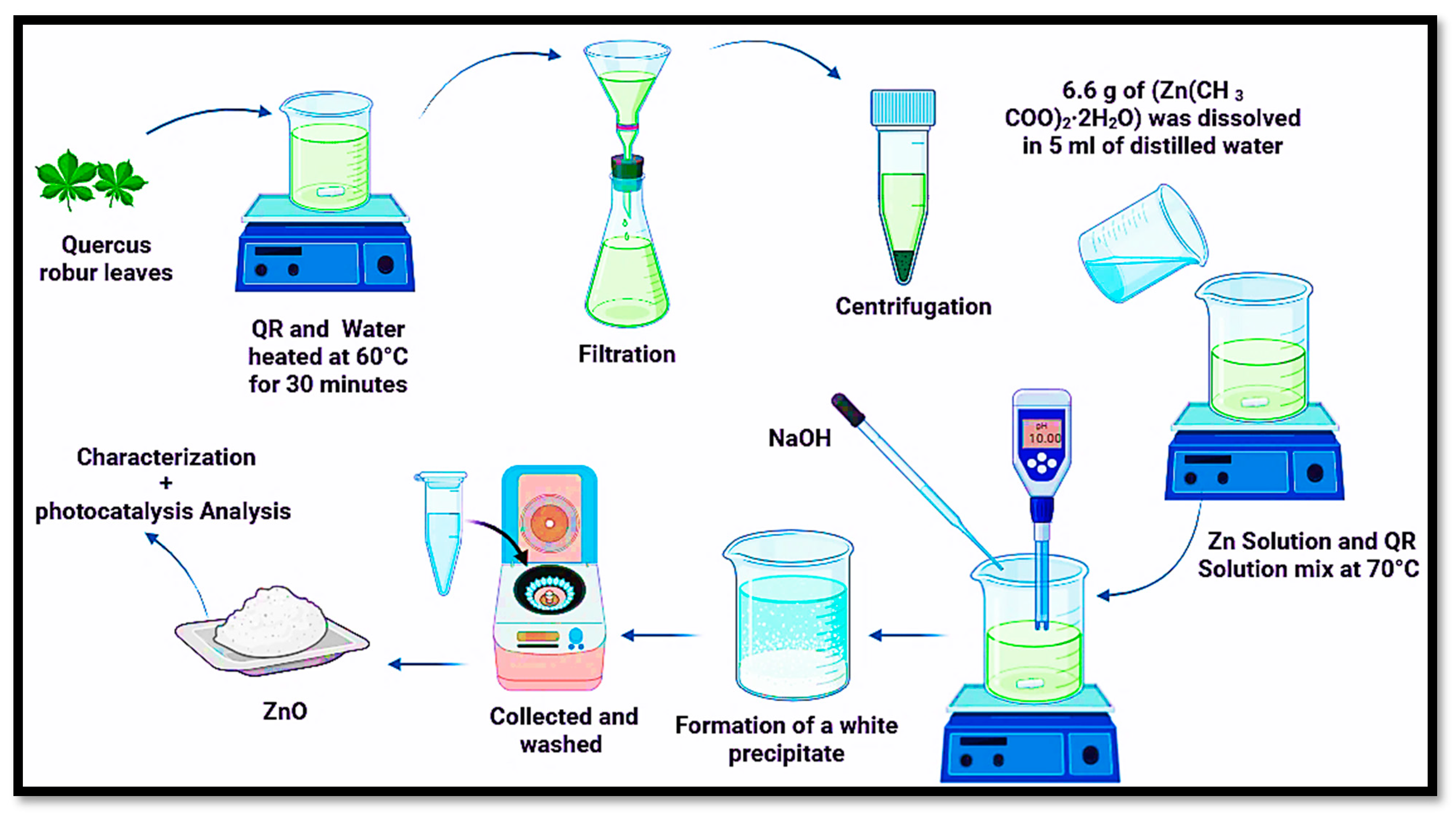
3.1. Preparation of Leaf Extract

The selection of Quercus robur leaves for this study was based on their abundance and year-round availability, which makes them a practical and sustainable resource for experimental purposes. The leaves used in this research were harvested from the Mashrouha forest region, located in Souk Ahras, Algeria, during the month of January. This timing ensured the collection of healthy, mature leaves that could be effectively used in the synthesis of the extract.
Once collected, the leaves were thoroughly washed with deionized water to eliminate any dust, dirt, or other potential contaminants that might interfere with the quality and purity of the extract. This cleaning step was essential to ensure that only the compounds naturally present in the leaves were involved in the subsequent extraction process.
After the cleaning process, the leaves were dried under controlled conditions—specifically, in the shade at room temperature—for a period of one week. The drying process was performed slowly to prevent the degradation of bioactive compounds within the leaves due to excessive heat or direct sunlight exposure. The leaves were then ground using a blender to increase the surface area and enhance the extraction efficiency when the material was mixed with deionized water.
To prepare the Quercus robur extract, varying amounts of ground leaves (0.03, 0.1, 2, and 10 g) were dissolved in 1 L of deionized water. The water was heated to 60 °C and continuously stirred with a magnetic stirrer for 30 min. This heating process helped release the bioactive compounds from the leaf material into the solution, facilitating a more efficient extraction.
After the extraction, the solution was subjected to centrifugation to separate the solid plant matter from the liquid extract. The liquid extract was then filtered to remove any remaining particulates, ensuring a clear solution. Finally, the extract was stored in the refrigerator to preserve its bioactive properties until it was required for further experiments. This methodical preparation of the Quercus robur extract allowed for a controlled and reproducible extraction process, enabling the investigation of its potential antimicrobial properties and other relevant bioactivities.

3.2. Green Synthesis of ZnO NPs

To biosynthesize ZnO nanoparticles Figure 14, a precise method involving the combination of zinc acetate dihydrate and Quercus robur leaf extract was used. Initially, 6.6 g of zinc acetate dihydrate (Zn(CH3COO)2·2H2O) was dissolved in 5 mL of distilled water at room temperature while stirring magnetically. This solution was carefully prepared to ensure a uniform distribution of the zinc salt in the solvent, creating the necessary precursor solution for nanoparticle formation. Similarly, 2.85 g of sodium hydroxide (NaOH) was dissolved in water following the same procedure to create the alkaline solution required to adjust the pH during the synthesis process.
Next, the zinc acetate solution was mixed with 45 mL of Quercus robur leaf extract, which serves as a green reducing agent in the biosynthesis of nanoparticles. The leaf extract contains various organic compounds, such as polyphenols, flavonoids, and other phytochemicals, which aid in the transformation of zinc ions (Zn2+) into ZnO nanoparticles through precipitation and subsequent oxidation processes. Once dried, the ZnO nanoparticles were stored in an appropriate container, ready for further characterization and analysis, such as examining their morphology, size distribution, and potential applications in various fields.
The temperatures chosen in the study were based on the previous literature and our experimental optimization process to achieve the desired morphological, structural, and optical properties of ZnO. For instance, research has shown that low-temperature synthesis (~60–100 °C) using hydrothermal or sol–gel methods produces ZnO nanoparticles with controlled morphology and good dispersibility. The mixture was then heated to 70 °C using a magnetic heating stirrer. This temperature was selected to enhance the reaction rate while ensuring that the leaf extract maintained its reducing properties. Once the temperature reached a stable 70 °C, the sodium hydroxide solution was added dropwise over 15 min to adjust the pH of the mixture to the desired level, ensuring that it reached an optimal pH for nanoparticle synthesis.
The solution was kept under continuous stirring and heating for an additional 30 min. During this time, a white precipitate formed, indicating the successful synthesis of ZnO nanoparticles. This precipitate was then separated from the solution by centrifugation at 2500 rpm for 10 min. The solid nanoparticles were carefully washed multiple times with deionized water to remove any residual impurities. After the water wash, the nanoparticles were further cleaned with ethanol to ensure the complete removal of organic substances from the leaf extract. Finally, the obtained ZnO nanoparticles were dried in a laboratory oven at 45 °C for 48 h to remove any remaining solvents and moisture. Once dried, the ZnO nanoparticles were stored in an appropriate container, ready for further characterization and analysis, such as examining their morphology, size distribution, and potential applications in various fields. The percentage yields in this work range from 70% to 85%. The percentage yield can be calculated using the following formula (Equation (19)):
Percentage   Yield = ( M a s s   o f   Z n O   o b t a i n e d T h e o r e t i c a l   m a s s   o f   Z n O ) × 100
This green synthesis method not only provides an eco-friendly approach to producing ZnO nanoparticles but also integrates the beneficial properties of plant extracts, which can enhance the stability and functionality of the nanoparticles.

3.3. Photocatalytic Tests

The degradation process of MB using ZnO NPs, synthesized through an eco-friendly green method, was systematically studied by employing UV–Vis spectroscopy. This analytical technique allowed for the tracking of changes in the absorption spectrum, particularly at the wavelength of 664 nm, which is the characteristic λmax for MB. This method enables a precise monitoring of the degradation kinetics over time.
The photocatalytic reactions were carried out under controlled laboratory conditions, where a 100 mL double-walled beaker with a surface area of 32.17 cm2 was utilized. The beaker was equipped with a cooling water jacket, ensuring that the reaction temperature was consistently maintained at 20 ± 2 °C. This was important to minimize any temperature-related interference in the photocatalytic process.
To provide the necessary UV irradiation, a Haichao T8 15W UV lamp, emitting at 365 nm UVA, was employed. The lamp was positioned 10 cm from the solution, ensuring uniform exposure of the photocatalyst to the UV light. The controlled environment, including the precise regulation of temperature and UV light exposure, helped ensure that the experiment was conducted under optimal and reproducible conditions, thereby allowing for accurate evaluation of the ZnO NPs’ photocatalytic degradation efficiency MB. This approach not only ensured effective monitoring of the degradation process but also contributed to a better understanding of the photocatalytic properties of green-synthesized ZnO NPs in the degradation of organic pollutants like MB.

3.4. Antimicrobial Study

The MIC of ZnO was determined through a stepwise two-fold serial dilution technique. Initially, the ZnO NPs were introduced into wells 1 to 10 of row A in a 96-well microplate, where a series of 100 µL dilutions were systematically made across the plate. Each dilution progressively reduced the concentration of the nanoparticles, yielding concentrations of 200, 100, 50, 25, 12.5, 6.25, 3.13, and 1.56 µg/mL. This allowed for the establishment of a concentration gradient, which was essential for identifying the minimum concentration at which the ZnO NPs could effectively inhibit bacterial growth. After setting up the dilutions, the microplate was incubated at 37 °C for a period of 24 h, ensuring that the bacterial strains were exposed to the nanoparticles for sufficient time to allow any potential antibacterial effects to take place.
To evaluate bacterial viability and determine the MIC, 2,3,5-triphenyl tetrazolium chloride (TTC) was used as a viability indicator. TTC is a colorimetric agent that stains living bacterial cells pink due to the reduction in the tetrazolium salt by metabolically active cells. On the other hand, if bacterial growth was inhibited, the TTC remained colorless, indicating a lack of bacterial metabolism. The extent of the color change in each well directly correlates to the degree of bacterial growth inhibition, with the shift from pink to transparent providing clear visual evidence of the antibacterial activity of the ZnO NPs.
The lowest concentration of ZnO NPs at which the bacterial growth was significantly suppressed, as indicated by the absence of pink color in the wells, was identified as the MIC. The results showed that the biosynthesized ZnO NPs exhibited substantial antibacterial activity, demonstrating their potential as effective antimicrobial agents against the tested bacterial strains. The precise correlation between the concentration of ZnO NPs and bacterial inhibition provides critical information for optimizing the use of these nanoparticles in various applications, such as in the development of antimicrobial coatings or medical treatments aimed at combating bacterial infections.

4. Conclusions

This study provides a comprehensive analysis of zinc oxide nanoparticle (ZnO NPs) synthesis and characterization using environmentally friendly methods, highlighting their potential as both antimicrobial agents and photocatalytic catalysts. The research focused on optimizing calcination temperature and examining the effect of plant extract concentration on the structural properties of the nanoparticles. X-ray diffraction (XRD) confirmed the presence of characteristic diffraction peaks corresponding to the hexagonal wurtzite structure of ZnO, with crystallite sizes ranging from 10 to 29 nm, influenced by both extract concentration and annealing temperature. Scanning electron microscopy (SEM) revealed that increasing the leaf extract concentration led to smaller particle sizes; however, higher concentrations also resulted in greater nanoparticle aggregation. Fourier-transform infrared spectroscopy (FTIR) and UV–visible spectroscopy analyses identified functional groups on both the ZnO nanoparticles and the leaf extract, and variations in band gap energy and Urbach energy were observed as a function of extract concentration and calcination temperature. Antimicrobial studies demonstrated that the environmentally synthesized ZnO nanoparticles effectively inhibited the growth of both Gram-positive and G ram-negative bacteria, with minimum inhibitory concentrations ranging from 6.25 to 100 μg/mL depending on the concentration of the leaf extract. However, the antifungal activity against Candida albicans was minimal across all tested concentrations. Photocatalytic degradation tests showed that green-synthesized nanoparticles achieved optimal removal of methylene blue (MB) compared to their chemically synthesized counterparts, with efficiency further enhanced at higher calcination temperatures (320 and 500 °C). In conclusion, the environmentally friendly synthesis of ZnO nanoparticles using leaf extract holds promise for the development of antimicrobial agents and photocatalytic catalysts. While the nanoparticles exhibited notable antibacterial activity, further optimization is required to enhance their antifungal efficacy. Additionally, exploring strategies to improve photocatalytic efficiency—considering factors such as particle size, aggregation, and surface functionalization—should be prioritized. These findings contribute significantly to the advancement of nanoparticle technology for biomedical, environmental, and catalytic applications.

Author Contributions

Conceptualization, S.R., H.G., H.T. and L.A.; data curation, S.R. and H.G.; formal analysis, S.R. and H.G.; funding acquisition, M.S.O. and J.Z.; investigation, S.R., H.G., H.T., L.A., D.C., A.A.A., M.K., J.Z., A.A. and S.L.; methodology, S.R., H.G., H.T., L.A., D.C., M.S.O., J.Z. and A.A.; project administration, H.G., H.T., M.S.O., J.Z. and A.A.; resources, S.R., H.G., H.T., A.A.A., M.K., A.A. and S.L.; supervision, H.G; validation, S.R., H.G. and A.A.; visualization, H.G., H.T., L.A., A.A. and S.L.; writing—original draft, S.R. and H.G.; writing—review and editing, H.G. All authors have read and agreed to the published version of the manuscript.

Funding

Researchers Supporting Project Number (RSPD2025R710), King Saud University, Riyadh, Saudi Arabia.

Data Availability Statement

The data supporting the findings of this manuscript are available from the corresponding authors upon reasonable request.

Acknowledgments

The authors wish to express their thanks to the Algerian Ministry of Higher Education and Scientific Research (MESRS, DGRSDT) for research support. The authors also thank Researchers Supporting Project Number (RSPD2025R710) from King Saud University, Riyadh, Saudi Arabia, for financial support.

Conflicts of Interest

All authors declare that they do not have a conflict of interest.

References

  1. Palai, P.; Muduli, S.; Priyadarshini, B.; Sahoo, T.R. A Facile Green Synthesis of ZnO Nanoparticles and Its Adsorptive Removal of Congo Red Dye from Aqueous Solution. Mater. Today Proc. 2021, 38, 2445–2451. [Google Scholar] [CrossRef]
  2. Kaliraj, L.; Ahn, J.C.; Rupa, E.J.; Abid, S.; Lu, J.; Yang, D.C. Synthesis of Panos Extract Mediated ZnO Nano-Flowers as Photocatalyst for Industrial Dye Degradation by UV Illumination. J. Photochem. Photobiol. B Biol. 2019, 199, 111588. [Google Scholar] [CrossRef]
  3. Toumi, S.; Lekmine, S.; Touzout, N.; Moussa, H.; Elboughdiri, N.; Boudraa, R.; Benslama, O.; Kebir, M.; Danish, S.; Zhang, J. Harnessing Deep Learning for Real-Time Water Quality Assessment: A Sustainable Solution. Water 2024, 16, 3380. [Google Scholar] [CrossRef]
  4. Chung, K.T.; Fulk, G.E.; Andrews, A.W. Mutagenicity Testing of Some Commonly Used Dyes. Appl. Environ. Microbiol. 1981, 42, 641–648. [Google Scholar] [CrossRef]
  5. LaKind, J.S.; Anthony, L.G.; Goodman, M. Review of Reviews on Exposures to Synthetic Organic Chemicals and Children’s Neurodevelopment: Methodological and Interpretation Challenges. J. Toxicol. Environ. Health Part B 2017, 20, 390–422. [Google Scholar] [CrossRef]
  6. Ming, J.; Ni, S.-Q.; Guo, Z.; Wang, Z.-B.; Xie, L. Photocatalytic Material–Microorganism Hybrid Systems in Water Decontamination. Trends Biotechnol. 2024; online ahead of print. [Google Scholar] [CrossRef]
  7. Soto-Robles, C.A.; Luque, P.A.; Gómez-Gutiérrez, C.M.; Nava, O.; Vilchis-Nestor, A.R.; Lugo-Medina, E.; Ranjithkumar, R.; Castro-Beltrán, A. Study on the Effect of the Concentration of Hibiscus Sabdariffa Extract on the Green Synthesis of ZnO Nanoparticles. Results Phys. 2019, 15, 102807. [Google Scholar] [CrossRef]
  8. Tahraoui, H.; Belhadj, A.-E.; Amrane, A.; Toumi, S.; Jaouadi, B.; Zhang, J. Maximizing Diclofenac Bioremoval Efficiency Using Chlorella vulgaris Strain H1 and Chlorella sorokiniana Strain H2: Unveiling the Impact of Acetic Acid on Microalgae. J. Taiwan Inst. Chem. Eng. 2024, 165, 105783. [Google Scholar] [CrossRef]
  9. Smara, M.; Khalladi, R.; Moulai-Mostefa, N.; Madi, K.; Mansour, D.; Lekmine, S.; Benslama, O.; Tahraoui, H.; Zhang, J.; Amrane, A. Efficiency of Hydrogen Peroxide and Fenton Reagent for Polycyclic Aromatic Hydrocarbon Degradation in Contaminated Soil: Insights from Experimental and Predictive Modeling. Processes 2024, 12, 621. [Google Scholar] [CrossRef]
  10. Nedjhioui, M.; Nasrallah, N.; Kebir, M.; Tahraoui, H.; Bouallouche, R.; Assadi, A.A.; Amrane, A.; Jaouadi, B.; Zhang, J.; Mouni, L. Designing an Efficient Surfactant–Polymer–Oil–Electrolyte System: A Multi-Objective Optimization Study. Processes 2023, 11, 1314. [Google Scholar] [CrossRef]
  11. Kebir, M.; Benramdhan, I.-K.; Nasrallah, N.; Tahraoui, H.; Bait, N.; Benaissa, H.; Ameraoui, R.; Zhang, J.; Assadi, A.A.; Mouni, L. Surface Response Modeling of Homogeneous Photo Fenton Fe (III) and Fe (II) Complex for Sunlight Degradation and Mineralization of Food Dye. Catal. Commun. 2023, 183, 106780. [Google Scholar] [CrossRef]
  12. Mechati, S.; Zamouche, M.; Tahraoui, H.; Filali, O.; Mazouz, S.; Bouledjemer, I.N.E.; Toumi, S.; Triki, Z.; Amrane, A.; Kebir, M. Modeling and Optimization of Hybrid Fenton and Ultrasound Process for Crystal Violet Degradation Using AI Techniques. Water 2023, 15, 4274. [Google Scholar] [CrossRef]
  13. Moussa, H.; Hamid, S.; Mameri, A.; Lekmine, S.; Tahraoui, H.; Kebir, M.; Touzout, N.; Dahmoune, F.; Ola, M.S.; Zhang, J. From Green Chemistry to Healthy Environments: Silver Nanoparticles as a Dual Antioxidant and Antibacterial Agents for Advancing Biomedicine and Sustainable Wastewater Treatment. Bioengineering 2024, 11, 1205. [Google Scholar] [CrossRef] [PubMed]
  14. Tahraoui, H.; Toumi, S.; Boudoukhani, M.; Touzout, N.; Sid, A.N.E.H.; Amrane, A.; Belhadj, A.-E.; Hadjadj, M.; Laichi, Y.; Aboumustapha, M. Evaluating the Effectiveness of Coagulation–Flocculation Treatment Using Aluminum Sulfate on a Polluted Surface Water Source: A Year-Long Study. Water 2024, 16, 400. [Google Scholar] [CrossRef]
  15. Touahri, S.; Halimi, O.; Zaabat, M.; Mammeri, S.; Boudine, B.; Sebais, M.; Tahraoui, H.; Zhang, J.; Amrane, A. Exploring the Influence of Sr Concentration on the Structural and Catalytic Properties of CuO/SrSO4 Nanocomposites for Organic Dye Degradation. J. Phys. Chem. Solids 2024, 195, 112299. [Google Scholar] [CrossRef]
  16. Golmohammadi, M.; Honarmand, M.; Ghanbari, S. A Green Approach to Synthesis of ZnO Nanoparticles Using Jujube Fruit Extract and Their Application in Photocatalytic Degradation of Organic Dyes. Spectrochim. Acta Part A Mol. Biomol. Spectrosc. 2020, 229, 117961. [Google Scholar] [CrossRef] [PubMed]
  17. Farid, M.; Fayyaz, A.; Ahmed, E.; Arooj, M.; Ali, S.; Sarfraz, W.; Abbas, M.; Idrees, A.; Shahzadi, M.; Rasheed, Z. Increase in Food Scarcity, Agricultural Challenges, and Their Management: Pakistan Perspectives. In Managing Plant Production Under Changing Environment; Hasanuzzaman, M., Ahammed, G.J., Nahar, K., Eds.; Springer Nature Singapore: Singapore, 2022; pp. 437–458. ISBN 978-981-16-5058-1. [Google Scholar] [CrossRef]
  18. Rajan, C.S.R. Nanotechnology in Groundwater Remediation. Int. J. Environ. Sci. Dev. 2011, 2, 182–187. [Google Scholar] [CrossRef]
  19. Chelghoum, H.; Nasrallah, N.; Tahraoui, H.; Seleiman, M.F.; Bouhenna, M.M.; Belmeskine, H.; Zamouche, M.; Djema, S.; Zhang, J.; Mendil, A. Eco-Friendly Synthesis of ZnO Nanoparticles for Quinoline Dye Photodegradation and Antibacterial Applications Using Advanced Machine Learning Models. Catalysts 2024, 14, 831. [Google Scholar] [CrossRef]
  20. Kebir, M.; Bouallouche, R.; Nasrallah, N.; Tahraoui, H.; Elboughdiri, N.; Ait Merzeg, F.; Dergal, F.; Amirouche, S.; Assadi, A.A.; Amrane, A. Sustainable Photodegradation of Amoxicillin in Wastewater with a Nickel Aluminate and ZnO Heterosystem Oxides: Experimental and Gaussian Process Regression Modeling Studies. Catalysts 2024, 14, 875. [Google Scholar] [CrossRef]
  21. Liu, Y.; Zeng, X.; Hu, X.; Hu, J.; Zhang, X. Two-dimensional Nanomaterials for Photocatalytic Water Disinfection: Recent Progress and Future Challenges. J. Chem. Technol. Biotechnol. 2019, 94, 22–37. [Google Scholar] [CrossRef]
  22. Teow, Y.H.; Mohammad, A.W. New Generation Nanomaterials for Water Desalination: A Review. Desalination 2019, 451, 2–17. [Google Scholar] [CrossRef]
  23. Liu, X.; Ma, R.; Wang, X.; Ma, Y.; Yang, Y.; Zhuang, L.; Zhang, S.; Jehan, R.; Chen, J.; Wang, X. Graphene Oxide-Based Materials for Efficient Removal of Heavy Metal Ions from Aqueous Solution: A Review. Environ. Pollut. 2019, 252, 62–73. [Google Scholar] [CrossRef] [PubMed]
  24. Shan, Z.; Yang, Y.; Shi, H.; Zhu, J.; Tan, X.; Luan, Y.; Jiang, Z.; Wang, P.; Qin, J. Hollow Dodecahedra Graphene Oxide-Cuprous Oxide Nanocomposites with Effective Photocatalytic and Bactericidal Activity. Front. Chem. 2021, 9, 755836. [Google Scholar] [CrossRef] [PubMed]
  25. Anbarasu, K.; Devarajan, Y. Nanomaterials-Based Wastewater Treatment: Addressing Challenges and Advancing Sustainable Solutions. BioNanoScience 2025, 15, 149. [Google Scholar] [CrossRef]
  26. Crini, G.; Lichtfouse, E. Advantages and Disadvantages of Techniques Used for Wastewater Treatment. Environ. Chem. Lett. 2019, 17, 145–155. [Google Scholar] [CrossRef]
  27. Zhou, C.; Lai, C.; Xu, P.; Zeng, G.; Huang, D.; Zhang, C.; Cheng, M.; Hu, L.; Wan, J.; Liu, Y.; et al. In Situ Grown AgI/Bi12O17Cl2 Heterojunction Photocatalysts for Visible Light Degradation of Sulfamethazine: Efficiency, Pathway and Mechanism. ACS Sustain. Chem. Eng. 2018, 6, 4174–4184. [Google Scholar] [CrossRef]
  28. Lu, K.; Qin, J.; Hu, M.; Hu, L.; Mao, M.; Li, X.; Lin, Z.; Liu, W. High-Efficiency Nickel Recovery from Spent Electroless Nickel Plating Solution: Effective Degradation of High-Concentration Nickel Complexes to Form a Nickel Ferrite Nanomaterial via Fe3O4 Catalytic Oxidation. Environ. Sci. Nano 2024, 11, 900–910. [Google Scholar] [CrossRef]
  29. Ikram, M.; Rashid, M.; Haider, A.; Naz, S.; Haider, J.; Raza, A.; Ansar, M.T.; Uddin, M.K.; Ali, N.M.; Ahmed, S.S.; et al. A Review of Photocatalytic Characterization, and Environmental Cleaning, of Metal Oxide Nanostructured Materials. Sustain. Mater. Technol. 2021, 30, e00343. [Google Scholar] [CrossRef]
  30. Zhuang, Q.; Li, X.; Lian, X.; Hu, H.; Wang, N.; Wu, J.; Miao, K.; Feng, G.; Luo, X. Catalysis Enhancement of Co3O4 through the Epitaxial Growth of Inert ZnO in Peroxymonosulfate Activation: The Catalytic Mechanism of Surface Hydroxyls in Singlet Oxygen Generation. Cryst. Growth Des. 2025, 25, 319–329. [Google Scholar] [CrossRef]
  31. Ali, T.; Tripathi, P.; Azam, A.; Raza, W.; Ahmed, A.S.; Ahmed, A.; Muneer, M. Photocatalytic Performance of Fe-Doped TiO2 Nanoparticles under Visible-Light Irradiation. Mater. Res. Express 2017, 4, 015022. [Google Scholar] [CrossRef]
  32. Hasija, V.; Raizada, P.; Sudhaik, A.; Sharma, K.; Kumar, A.; Singh, P.; Jonnalagadda, S.B.; Thakur, V.K. Recent Advances in Noble Metal Free Doped Graphitic Carbon Nitride Based Nanohybrids for Photocatalysis of Organic Contaminants in Water: A Review. Appl. Mater. Today 2019, 15, 494–524. [Google Scholar] [CrossRef]
  33. Anpo, M.; Ichihashi, Y.; Takeuchi, M.; Yamashita, H. Design of Unique Titanium Oxide Photocatalysts by an Advanced Metal Ion-Implantation Method and Photocatalytic Reactions under Visible Light Irradiation. Res. Chem. Intermed. 1998, 24, 143–149. [Google Scholar] [CrossRef]
  34. Guediri, M.K.; Chebli, D.; Bouguettoucha, A.; Bourzami, R.; Amrane, A. Novel Fe2TiO5/Reduced Graphene Oxide Heterojunction Photocatalyst with Improved Adsorption Capacity and Visible Light Photoactivity: Experimental and DFT Approach. Environ. Sci. Pollut. Res. 2021, 28, 8507–8519. [Google Scholar] [CrossRef]
  35. Muthuvel, A.; Jothibas, M.; Mohana, V.; Manoharan, C. Green Synthesis of Cerium Oxide Nanoparticles Using Calotropis Procera Flower Extract and Their Photocatalytic Degradation and Antibacterial Activity. Inorg. Chem. Commun. 2020, 119, 108086. [Google Scholar] [CrossRef]
  36. Xing, Z.; Zhang, J.; Cui, J.; Yin, J.; Zhao, T.; Kuang, J.; Xiu, Z.; Wan, N.; Zhou, W. Recent Advances in Floating TiO2-Based Photocatalysts for Environmental Application. Appl. Catal. B Environ. 2018, 225, 452–467. [Google Scholar] [CrossRef]
  37. Ong, C.B.; Ng, L.Y.; Mohammad, A.W. A Review of ZnO Nanoparticles as Solar Photocatalysts: Synthesis, Mechanisms and Applications. Renew. Sustain. Energy Rev. 2018, 81, 536–551. [Google Scholar] [CrossRef]
  38. Li, X.; Peng, T.; Zhang, Y.; Wen, Y.; Nan, Z. A New Efficient Visible-Light Photocatalyst Made of SnO2 and Cyclized Polyacrylonitrile. Mater. Res. Bull. 2018, 97, 517–522. [Google Scholar] [CrossRef]
  39. Rasheed, H.M.; Aroosh, K.; Meng, D.; Ruan, X.; Akhter, M.; Cui, X. A Review on Modified ZnO to Address Environmental Challenges through Photocatalysis: Photodegradation of Organic Pollutants. Mater. Today Energy 2025, 48, 101774. [Google Scholar] [CrossRef]
  40. Chikhi, B.; Gouasmi, M.; Mounia, A.; Gasem, L.; Saadi, A.; Mekaoui, N.; Bachari, K.; Boudjemaa, A. Propyl Paraben Removal Using Cu2O/ZnO-NPs Photocatalyst Elaborated via Green Method. Environ. Sci. Pollut. Res. 2025, 32, 2107–2122. [Google Scholar] [CrossRef] [PubMed]
  41. Khan, M.M.; Adil, S.F.; Al-Mayouf, A. Metal Oxides as Photocatalysts. J. Saudi Chem. Soc. 2015, 19, 462–464. [Google Scholar] [CrossRef]
  42. Madi, K.; Chebli, D.; Ait Youcef, H.; Tahraoui, H.; Bouguettoucha, A.; Kebir, M.; Zhang, J.; Amrane, A. Green Fabrication of ZnO Nanoparticles and ZnO/rGO Nanocomposites from Algerian Date Syrup Extract: Synthesis, Characterization, and Augmented Photocatalytic Efficiency in Methylene Blue Degradation. Catalysts 2024, 14, 62. [Google Scholar] [CrossRef]
  43. Wang, H.; Zhang, P.; Zang, Z. High Performance CsPbBr3 Quantum Dots Photodetectors by Using Zinc Oxide Nanorods Arrays as an Electron-Transport Layer. Appl. Phys. Lett. 2020, 116, 162103. [Google Scholar] [CrossRef]
  44. Li, C.; Han, C.; Zhang, Y.; Zang, Z.; Wang, M.; Tang, X.; Du, J. Enhanced Photoresponse of Self-Powered Perovskite Photodetector Based on ZnO Nanoparticles Decorated CsPbBr3 Films. Sol. Energy Mater. Sol. Cells 2017, 172, 341–346. [Google Scholar] [CrossRef]
  45. Dejen, K.D.; Sabir, F.K.; Ananda Murthy, H.C.; Ayanie, G.T.; Shume, M.S.; Bekele, E.T. Green Synthesis of Nanomaterials for Environmental Remediation. In Green Nanoremediation; Policarpo Tonelli, F.M., Roy, A., Ananda Murthy, H.C., Eds.; Springer International Publishing: Cham, Switzerland, 2023; pp. 27–65. ISBN 978-3-031-30557-3. [Google Scholar]
  46. Duan, H.; Wang, D.; Li, Y. Green Chemistry for Nanoparticle Synthesis. Chem. Soc. Rev. 2015, 44, 5778–5792. [Google Scholar] [CrossRef]
  47. Iravani, S. Green Synthesis of Metal Nanoparticles Using Plants. Green Chem. 2011, 13, 2638. [Google Scholar] [CrossRef]
  48. Bandeira, M.; Giovanela, M.; Roesch-Ely, M.; Devine, D.M.; Da Silva Crespo, J. Green Synthesis of Zinc Oxide Nanoparticles: A Review of the Synthesis Methodology and Mechanism of Formation. Sustain. Chem. Pharm. 2020, 15, 100223. [Google Scholar] [CrossRef]
  49. Jena, S.K.; Sadasivam, R.; Packirisamy, G.; Saravanan, P. Nanoremediation: Sunlight Mediated Dye Degradation Using Electrospun PAN/CuO–ZnO Nanofibrous Composites. Environ. Pollut. 2021, 280, 116964. [Google Scholar] [CrossRef]
  50. Alavi-Tabari, S.A.R.; Khalilzadeh, M.A.; Karimi-Maleh, H. Simultaneous Determination of Doxorubicin and Dasatinib as Two Breast Anticancer Drugs Uses an Amplified Sensor with Ionic Liquid and ZnO Nanoparticle. J. Electroanal. Chem. 2018, 811, 84–88. [Google Scholar] [CrossRef]
  51. Chandran, S.P.; Chaudhary, M.; Pasricha, R.; Ahmad, A.; Sastry, M. Synthesis of Gold Nanotriangles and Silver Nanoparticles Using Aloe Vera Plant Extract. Biotechnol. Prog. 2006, 22, 577–583. [Google Scholar] [CrossRef]
  52. Song, J.Y.; Kim, B.S. Rapid Biological Synthesis of Silver Nanoparticles Using Plant Leaf Extracts. Bioprocess Biosyst. Eng. 2009, 32, 79–84. [Google Scholar] [CrossRef]
  53. Rathnasamy, R.; Thangasamy, P.; Thangamuthu, R.; Sampath, S.; Alagan, V. Green Synthesis of ZnO Nanoparticles Using Carica Papaya Leaf Extracts for Photocatalytic and Photovoltaic Applications. J. Mater. Sci. Mater. Electron. 2017, 28, 10374–10381. [Google Scholar] [CrossRef]
  54. Sangeetha, G.; Rajeshwari, S.; Venckatesh, R. Green Synthesis of Zinc Oxide Nanoparticles by Aloe Barbadensis Miller Leaf Extract: Structure and Optical Properties. Mater. Res. Bull. 2011, 46, 2560–2566. [Google Scholar] [CrossRef]
  55. Ramesh, M.; Anbuvannan, M.; Viruthagiri, G. Green Synthesis of ZnO Nanoparticles Using Solanum Nigrum Leaf Extract and Their Antibacterial Activity. Spectrochim. Acta Part A Mol. Biomol. Spectrosc. 2015, 136, 864–870. [Google Scholar] [CrossRef]
  56. Thema, F.T.; Manikandan, E.; Dhlamini, M.S.; Maaza, M. Green Synthesis of ZnO Nanoparticles via Agathosma Betulina Natural Extract. Mater. Lett. 2015, 161, 124–127. [Google Scholar] [CrossRef]
  57. Pugazhendhi, A.; Mathimani, T.; Alharbi, S.A.; Chinnathambi, A.; Karuppusamy, I.; Brindhadevi, K.; Kheawfu, K.; Pikulkaew, S. In Vitro Efficacy of Green Synthesized ZnO Nanoparticles against Biofilm and Virulence of Serratia Marcescens. Prog. Org. Coat. 2022, 166, 106781. [Google Scholar] [CrossRef]
  58. Khoso, S.; Mehar, S.; Anam; Naheed, N.; Saeed, F.; Khan, N.; Abbasi, B.H. Green synthesis of ZnO nanoparticles from foeniculum vulgare, its characterization and biological potential against bacteria. J. Anim. Plant Sci. 2021, 32, 229–237. [Google Scholar] [CrossRef]
  59. Etape, E.P.; Foba-Tendo, J.; Ngolui, L.J.; Namondo, B.V.; Yollande, F.C.; Nguimezong, M.B.N. Structural Characterization and Magnetic Properties of Undoped and Ti-Doped ZnO Nanoparticles Prepared by Modified Oxalate Route. J. Nanomater. 2018, 2018, 9072325. [Google Scholar] [CrossRef]
  60. Bourzami, R.; Guediri, M.K.; Chebli, D.; Bouguettoucha, A.; Amrane, A. Bottom-up Construction of Reduced-Graphene-Oxide-Anchored Spinel Magnet Fe2.02Ni1.01O3.22, Anatase TiO2 and Metallic Ag Nanoparticles and Their Synergy in Photocatalytic Water Reduction. J. Environ. Chem. Eng. 2021, 9, 105307. [Google Scholar] [CrossRef]
  61. Demissie, M.G.; Sabir, F.K.; Edossa, G.D.; Gonfa, B.A. Synthesis of Zinc Oxide Nanoparticles Using Leaf Extract of Lippia adoensis (Koseret) and Evaluation of Its Antibacterial Activity. J. Chem. 2020, 2020, 7459042. [Google Scholar] [CrossRef]
  62. Mallika, A.N.; Reddy, A.R.; Reddy, K.V. Annealing Effects on the Structural and Optical Properties of ZnO Nanoparticles with PVA and CA as Chelating Agents. J. Adv. Ceram. 2015, 4, 123–129. [Google Scholar] [CrossRef]
  63. Chen, Y.-S.; Liao, C.-H.; Chueh, Y.-L.; Lai, C.-C.; Chen, L.-Y.; Chu, A.-K.; Kuo, C.-T.; Wang, H.-C. High Performance Cu2O/ZnO Core-Shell Nanorod Arrays Synthesized Using a Nanoimprint GaN Template by the Hydrothermal Growth Technique. Opt. Mater. Express 2014, 4, 1473. [Google Scholar] [CrossRef]
  64. Caliman, F.A.; Robu, B.M.; Smaranda, C.; Pavel, V.L.; Gavrilescu, M. Soil and Groundwater Cleanup: Benefits and Limits of Emerging Technologies. Clean Technol. Environ. Policy 2011, 13, 241–268. [Google Scholar] [CrossRef]
  65. Oniya, E.O.; Olubi, O.E.; Ibitoye, A.; Agbi, J.I.; Agbeni, S.K.; Faweya, E.B. Effect of Milling Equipment on the Level of Heavy Metal Content of Foodstuff. Phys. Sci. Int. J. 2018, 20, 1–8. [Google Scholar] [CrossRef]
  66. Naiel, B.; Fawzy, M.; Halmy, M.W.A.; Mahmoud, A.E.D. Green synthesis of zinc oxide nanoparticles using Sea Lavender (Limonium pruinosum L. Chaz.) extract: Characterization, evaluation of anti-skin cancer, antimicrobial and antioxidant potentials. Sci. Rep. 2022, 12, 20370. [Google Scholar] [CrossRef] [PubMed]
  67. Sharma, V.; Sharma, J.K.; Kansay, V.; Sharma, V.D.; Sharma, A.; Kumar, S.; Sharma, A.K.; Bera, M.K. The Effect of Calcination Temperatures on the Structural and Optical Properties of Zinc Oxide Nanoparticles and Their Influence on the Photocatalytic Degradation of Leather Dye. Chem. Phys. Impact 2023, 6, 100196. [Google Scholar] [CrossRef]
  68. Agnieszka, K.-R.; Ewa, M.; Teofil, J. Structural Characterisation of ZnO Particles Obtained by the Emulsion Precipitation Method. J. Nanomater. 2012, 656353. [Google Scholar] [CrossRef]
  69. Khan, S.H.; R, S.; Pathak, B.; Fulekar, M. Photocatalytic Degradation of Organophosphate Pesticides (Chlorpyrifos) Using Synthesized Zinc Oxide Nanoparticle by Membrane Filtration Reactor under UV Irradiation. Front. Nanosci. Nanotechnol. 2015, 1, 23–27. [Google Scholar] [CrossRef]
  70. Christobel, G.J. Vibrational Spectroscopy of ZnO-ZnS Nanoparticles. Int. J. Sci. Res. 2013, 5, 2228–2230. [Google Scholar]
  71. Veiga, A.; Toledo, M.D.G.T.; Rossa, L.S.; Mengarda, M.; Stofella, N.C.F.; Oliveira, L.J.; Gonçalves, A.G.; Murakami, F.S. Colorimetric Microdilution Assay: Validation of a Standard Method for Determination of MIC, IC50%, and IC90% of Antimicrobial Compounds. J. Microbiol. Methods 2019, 162, 50–61. [Google Scholar] [CrossRef]
  72. Bourzami, R.; AitYoucef, H.C.; Hamdouni, N.; Sebais, M. Synthesis, Crystal Structure, Vibrational Spectra and Thermal Properties of Novel Ionic Organic-Inorganic Hybrid Material. Chem. Phys. Lett. 2018, 711, 220–226. [Google Scholar] [CrossRef]
  73. Ouksel, L.; Chafaa, S.; Bourzami, R.; Hamdouni, N.; Sebais, M.; Chafai, N. Crystal Structure, Vibrational, Spectral Investigation, Quantum Chemical DFT Calculations and Thermal Behavior of Diethyl [Hydroxy (Phenyl) Methyl] Phosphonate. J. Mol. Struct. 2017, 1144, 389–395. [Google Scholar] [CrossRef]
  74. Redjili, S.; Ghodbane, H.; Bourzami, R.; Mayouf, F.; Chebli, D.; Boudechicha, A.; Djelloul, C. Green Synthesis of Silver Oxide Nanoparticles: Eco-Friendly Approach for Sustainable Solutions. Ind. Crops Prod. 2025, 223, 120168. [Google Scholar] [CrossRef]
  75. Boudiaf, M.; Messai, Y.; Bentouhami, E.; Schmutz, M.; Blanck, C.; Ruhlmann, L.; Bezzi, H.; Tairi, L.; Eddine Mekki, D. Green Synthesis of NiO Nanoparticles Using Nigella Sativa Extract and Their Enhanced Electro-Catalytic Activity for the 4-Nitrophenol Degradation. J. Phys. Chem. Solids 2021, 153, 110020. [Google Scholar] [CrossRef]
  76. Liu, H.R.; Shao, G.X.; Zhao, J.F.; Zhang, Z.X.; Zhang, Y.; Liang, J.; Liu, X.G.; Jia, H.S.; Xu, B.S. Worm-Like Ag/ZnO Core–Shell Heterostructural Composites: Fabrication, Characterization, and Photocatalysis. J. Phys. Chem. C 2012, 116, 16182–16190. [Google Scholar] [CrossRef]
  77. Garg, R. Characterization and Performance Evaluation of Synthesized ZnO Nanoflowers, Nanorods, and Their Hybrid Nanocomposites with Graphene Oxide for Degradation of Orange G. Environ. Sci. Pollut. Res. 2021, 28, 57009–57029. [Google Scholar] [CrossRef] [PubMed]
  78. Chen, X.; Wu, Z.; Liu, D.; Gao, Z. Preparation of ZnO Photocatalyst for the Efficient and Rapid Photocatalytic Degradation of Azo Dyes. Nanoscale Res. Lett. 2017, 12, 143. [Google Scholar] [CrossRef] [PubMed]
  79. Bagheri, M.; Mahjoub, A.R.; Mehri, B. Enhanced Photocatalytic Degradation of Congo Red by Solvothermally Synthesized CuInSe2–ZnO Nanocomposites. RSC Adv. 2014, 4, 21757. [Google Scholar] [CrossRef]
  80. Kumar, R.; Kumar, G.; Umar, A. ZnO Nano-Mushrooms for Photocatalytic Degradation of Methyl Orange. Mater. Lett. 2013, 97, 100–103. [Google Scholar] [CrossRef]
  81. Mutalib, A.A.A. ZnO Photocatalysts Applications in Abating the Organic Pollutant Contamination: A Mini Review. Total. Environ. Res. Themes 2022, 3, 100013. [Google Scholar] [CrossRef]
  82. Naha, U. Rapid Precipitation-Free Synthesis of Zinc Oxide Nanowire Arrays. Mater. Today Proc. 2023, 79, 132–138. [Google Scholar] [CrossRef]
  83. Yashni, G.; Al-Gheethi, A.; Al-Kahtani, A.A.; Al-Sahari, M.; Hazhar, N.J.N.; Noman, E.; Alkhadher, S. Bio-Inspired ZnO NPs Synthesized from Citrus Sinensis Peels Extract for Congo Red Removal from Textile Wastewater via Photocatalysis: Optimization, Mechanisms, Techno-Economic Analysis. Chemosphere 2021, 281, 130661. [Google Scholar] [CrossRef]
  84. Gawade, V.V.; Gavade, N.L.; Shinde, H.M.; Babar, S.B.; Kadam, A.N.; Garadkar, K.M. Green Synthesis of ZnO Nanoparticles by Using Calotropis Procera Leaves for the Photodegradation of Methyl Orange. J. Mater. Sci. Mater. Electron. 2017, 28, 14033–14039. [Google Scholar] [CrossRef]
  85. Madhumitha, G.; Fowsiya, J.; Gupta, N.; Kumar, A.; Singh, M. Green Synthesis, Characterization and Antifungal and Photocatalytic Activity of Pithecellobium Dulce Peel–Mediated ZnO Nanoparticles. J. Phys. Chem. Solids 2019, 127, 43–51. [Google Scholar] [CrossRef]
  86. Sherly, E.D.; Vijaya, J.J.; Selvam, N.C.S.; Kennedy, L.J. Microwave Assisted Combustion Synthesis of Coupled ZnO–ZrO2 Nanoparticles and Their Role in the Photocatalytic Degradation of 2,4-Dichlorophenol. Ceram. Int. 2014, 40, 5681–5691. [Google Scholar] [CrossRef]
  87. Shore, A. Expression of Concern: One-Pot Sol–Gel Synthesis of Reduced Graphene Oxide Uniformly Decorated Zinc Oxide Nanoparticles in Starch Environment for Highly Efficient Photodegradation of Methylene Blue. RSC Adv. 2018, 8, 10332. [Google Scholar] [CrossRef]
  88. Kumar Sur, U.; Ankamwar, B.; Karmakar, S.; Halder, A.; Das, P. Green Synthesis of Silver Nanoparticles Using the Plant Extract of Shikakai and Reetha. Mater. Today Proc. 2018, 5, 2321–2329. [Google Scholar] [CrossRef]
  89. Suppiah, D.D.; Julkapli, N.M.; Sagadevan, S.; Johan, M.R. Eco-Friendly Green Synthesis Approach and Evaluation of Environmental and Biological Applications of Iron Oxide Nanoparticles. Inorg. Chem. Commun. 2023, 152, 110700. [Google Scholar] [CrossRef]
  90. Kavitha, A.; Doss, A.; Praveen Pole, R.P.; Pushpa Rani, T.P.K.; Prasad, R.; Satheesh, S. A Mini Review on Plant-Mediated Zinc Oxide Nanoparticles and Their Antibacterial Potency. Biocatal. Agric. Biotechnol. 2023, 48, 102654. [Google Scholar] [CrossRef]
  91. Gudkov, S.V.; Burmistrov, D.E.; Serov, D.A.; Rebezov, M.B.; Semenova, A.A.; Lisitsyn, A.B. Do Iron Oxide Nanoparticles Have Significant Antibacterial Properties? Antibiotics 2021, 10, 884. [Google Scholar] [CrossRef]
  92. Manjamadha, V.P.; Muthukumar, K. Ultrasound Assisted Green Synthesis of Silver Nanoparticles Using Weed Plant. Bioprocess Biosyst. Eng. 2016, 39, 401–411. [Google Scholar] [CrossRef]
  93. Raffi, M.; Hussain, F.; Bhatti, T.M.; Akhter, J.I.; Hameed, A.; Hasan, M.M. Antibacterial Characterization of Silver Nanoparticles against E. coli ATCC-15224. J. Mater. Sci. Technol. 2008, 24, 192–196. [Google Scholar]
  94. Panáček, A.; Kvítek, L.; Prucek, R.; Kolář, M.; Večeřová, R.; Pizúrová, N.; Sharma, V.K.; Nevěčná, T.; Zbořil, R. Silver Colloid Nanoparticles: Synthesis, Characterization, and Their Antibacterial Activity. J. Phys. Chem. B 2006, 110, 16248–16253. [Google Scholar] [CrossRef] [PubMed]
  95. Xia, T.; Kovochich, M.; Brant, J.; Hotze, M.; Sempf, J.; Oberley, T.; Sioutas, C.; Yeh, J.I.; Wiesner, M.R.; Nel, A.E. Comparison of the Abilities of Ambient and Manufactured Nanoparticles to Induce Cellular Toxicity According to an Oxidative Stress Paradigm. Nano Lett. 2006, 6, 1794–1807. [Google Scholar] [CrossRef] [PubMed]
  96. Song, H.Y.; Ko, K.K.; Oh, I.H.; Lee, B.T. Fabrication of Silver Nanoparticles and Their Antimicrobial Mechanisms. Eur. Cells Mater. 2006, 11 (Suppl. S1), 58. [Google Scholar]
  97. Sambhy, V.; MacBride, M.M.; Peterson, B.R.; Sen, A. Silver Bromide Nanoparticle/Polymer Composites:  Dual Action Tunable Antimicrobial Materials. J. Am. Chem. Soc. 2006, 128, 9798–9808. [Google Scholar] [CrossRef] [PubMed]
  98. Menon, S.; Agarwal, H.; Kumar, S.R.; Kumar, S.V. Green synthesis of silver nanoparticles using medicinal plant acalypha indica leaf extracts and its application as an antioxidant and antimicrobial agent against foodborne pathogens. Int. J. Appl. Pharm. 2017, 9, 42–50. [Google Scholar] [CrossRef]
  99. Yadav, J.P.; Kumar, S.; Budhwar, L.; Yadav, A.; Yadav, M. Characterization and Antibacterial Activity of Synthesized Silver and Iron Nanoparticles Using Aloe Vera. J. Nanomed. Nanotechnol. 2016, 7, 3. [Google Scholar] [CrossRef]
  100. Agarwal, H.; Venkat Kumar, S.; Rajeshkumar, S. A Review on Green Synthesis of Zinc Oxide Nanoparticles—An Eco-Friendly Approach. Resour.-Effic. Technol. 2017, 3, 406–413. [Google Scholar] [CrossRef]
  101. Benyagoub, E.; Nabbou, N.; Boukhalkhel, S.; Dehini, I. The In Vitro Evaluation of the Antimicrobial Activity of Quercus robur L. Methanolic and Aqueous Leaves’ Extracts, from the Algerian High Plateaus Against Some Uropathogenic Microbial Strains. Phytothérapie 2020, 18, 262–274. [Google Scholar] [CrossRef]
  102. Šukele, R.; Skadiņš, I.; Koka, R.; Bandere, D. Antibacterial Effects of Oak Bark (Quercus robur) and Heather Herb (Calluna vulgaris L.) Extracts against the Causative Bacteria of Bovine Mastitis. Vet. World 2022, 15, 2315–2322. [Google Scholar] [CrossRef]
Figure 1. (AC) XRD patterns of ZnO at different calcination temperatures vs. extract concentration.
Catalysts 15 00256 g001
Figure 2. (ac) Crystallite size (D), microstrain (ε%), and dislocation density (δ) vs. extract concentration.
Catalysts 15 00256 g002
Figure 3. SEM images of green synthesized ZnO NPs. Top images 100 nm.
Catalysts 15 00256 g003
Figure 4. FTIR spectra of green synthesised ZnO NPs.
Catalysts 15 00256 g004
Figure 5. UV–visible spectra of green synthesized ZnO NPs. The inset figure shows a variation in the band gap, e.g., according to the Kubelka–Munk method, and a variation in Urbach energy. All spectra are normalized to one by the maximum absorbance.
Catalysts 15 00256 g005
Figure 6. Photodegradation of BM dye using green synthesized ZnO NPs (c = 0, 0.03, 0.5, 1, 2 g/L) at different calcination temperatures.
Catalysts 15 00256 g006
Figure 7. Comparative Performance of Recycled ZnO Photocatalyst.
Catalysts 15 00256 g007
Figure 8. Effect of catalyst dose on photodegradation of MB by green synthesized ZnO (neutral pH, [MB] = 10 mg/L).
Catalysts 15 00256 g008
Figure 9. Effect of reaction pH on photodegradation of MB by ZnO (catalyst one g/L, [MB] = 10 mg/L).
Catalysts 15 00256 g009
Figure 10. Effect of the initial concentration of methylene blue on photodegradation performance of ZnO NPs.
Catalysts 15 00256 g010
Figure 11. Effect of scavengers on the photocatalytic activity of ZnO NPs.
Catalysts 15 00256 g011
Figure 12. Schematic illustration of the photocatalytic mechanism of ZnO.
Catalysts 15 00256 g012
Figure 13. Schematic illustration of the antimicrobial mechanism of ZnO.
Catalysts 15 00256 g013
Figure 14. Green synthesis of ZnO NPs using Quercus robur leaves extract.
Catalysts 15 00256 g014
Table 1. Direct band gap energy and Urbach energy of green synthesized ZnO NPs at 25 °C (c = 0, 0.03, 1, 2, and 10 g/L).
Concentration (g/L)ZnO0.030.1210
Direct gap energy (eV)3.293.293.293.283.29
Urbach energy (eV)2.942.903.012.972.90
Table 2. Direct band gap energy and Urbach energy of green synthesized ZnO NPs annealed at 320 °C (c = 0, 0.03, 1, 2, and 10 g/L).
Concentration (g/L)ZnO0.030.1210
Direct gap energy (eV)3.283.273.253.293.28
Urbach energy (eV)3.053.032.993.012.90
Table 3. Direct band gap energy and Urbach energy of green synthesized ZnO NPs annealed at 500 °C (c = 0, 0.03, 0.1, 2 and 10 g/L).
Concentration (g/L)ZnO0.030.1210
Direct gap energy (eV)2.983.013.153.273.26
Urbach energy (eV)2.893.013.103.072.90
Table 4. Elimination efficiency and apparent rate constants of the photocatalysis (25 °C).
Concentration (g/L)00.030.1210
E%5044383526
k (10−3 min−1)4.903.853.142.661.90
Table 5. Elimination efficiency and apparent rate constants of the photocatalysis (320 °C).
Concentration (g/L)00.030.1210
E%6274555347
k (10−3 min−1)6.909.095.545.053.89
Table 6. Elimination efficiency and apparent rate constants of the photocatalysis (500 °C).
Concentration (g/L)00.030.1210
E%5674626050
k (10−3 min−1)4.526.415.494.933.70
Table 7. Rate constant K of degradation of MB with different dosages of ZnO.
Concentration (g/L)0.250.512
E%66447422
k (10−3 min−1)4.542.619.571.88
Table 8. Rate constant K of degradation of MB with different pH reactions.
pH4neutral810
E%42745851
k (10−3 min−1)2.526.415.243.56
Table 9. Rate constant K of degradation of MB with different initial concentration of dye.
Concentration (mg/L)102030
E%744829
k (10−3 min−1)9.573.051.68
Table 10. Comparison of the photocatalytic efficacy of ZnO nanoparticles.
CatalystPreparation Method/PlantDyeIrradiationCatalyst
Dose/g L−1
Dye
Conc/mg L−1
Degradation Efficiency/%Ref.
ZnOGreen synthesisMBUV-lamp1.51063%[85]
ZnOMicrowave-assisted urea-nitrate combustionMBUV-lamp0.3075>75%[86]
ZnOSol gelMBUV (Hg lamp 365 nm)0.331037%[87]
ZnOGreen synthesisMBUV-lamp11074%Present work
Table 11. Minimum inhibitory concentrations of green synthesised ZnO NPs.
Staphylococcus aureusBacillus subtilisEscherichia coliSalmonella enterica
ZnO NPs
Concentrations (g/L)
Annealing Temperature
°C
MIC
(μg/mL)
MIC
(μg/mL)
MIC
(μg/mL)
MIC
(μg/mL)
c = 0.0251002510050
32025252512.5
500256.252550
0.0325502510050
3206.255010012.5
5002512.510012.5
0.12510050100100
320256.2510025
500506.25No25
225No25100100
320No6.252525
500No50NoNo
102525252525
320No6.25100100
500No50No100
Disclaimer/Publisher’s Note: The statements, opinions and data contained in all publications are solely those of the individual author(s) and contributor(s) and not of MDPI and/or the editor(s). MDPI and/or the editor(s) disclaim responsibility for any injury to people or property resulting from any ideas, methods, instructions or products referred to in the content.

