Article

Photocatalytic Activity of Green-Synthesized Semiconductor CuO/ZnO Nanocomposites Against Organic Dye: An Assessment of Antimicrobial and Cytotoxicity Investigations

1
School of Nuclear Science and Technology, University of South China, Hengyang 421001, China
2
Department of Botany and Microbiology, Faculty of Science, Al-Azhar University, Cairo 11884, Egypt
3
Department of Biology, College of Science, Taibah University, Madinah 42353, Saudi Arabia
*
Author to whom correspondence should be addressed.
Catalysts 2025, 15(12), 1096; https://doi.org/10.3390/catal15121096
Submission received: 1 October 2025 / Revised: 10 November 2025 / Accepted: 13 November 2025 / Published: 21 November 2025
(This article belongs to the Special Issue Advanced Semiconductor Photocatalysts)

Abstract

In this paper, by employing an eco-friendly and green approach, semiconductor CuO/ZnO nanocomposite are synthesized using an aqueous extract of Urtica urens. FT-IR, XRD, TEM, SAED, EDX, TGA, and UV-Vis spectroscopy were used for semiconductor characterization. The data revealed the successful formation of crystalline spherical nanocomposites with sizes ranging from 5 to 45 nm. The main components of the synthesized nanocomposites were Cu, Zn, and O, which had different weights and atomic percentages. The maximum absorbance of nanocomposites was 358 nm, with a direct bandgap of 2.25 eV, which is suitable for photocatalysis under visible light. The maximum photocatalytic activity of the synthesized semiconductor nanocomposites for photodegradation of methylene blue dye was 95.8%, where it was 44.5% and 65.5% for monometallic CuO and ZnO, respectively. The optimum conditions for maximum photocatalytic activity were a pH of 9, a dye concentration of 5 mg L−1, and nanocomposite concentration of 1.0 mg mL−1 after 70 min. The reusability of the synthesized semiconductor was promising for the fourth cycle, with a reduced capacity of 5%. Complementary investigations, antimicrobial activity and cytotoxic activity, were performed to increase the application of semiconductor nanocomposites. The data revealed the promising activity of the nanocomposite against E. coli, P. aeruginosa, B. subtilis, S. aureus, C. parapsilosis, C. albicans, and C. tropicalis with low MICs ranging between 50 and 25 µg mL−1. Additionally, compared with normal cell line, the synthesized nanocomposite targeted the cancer cell line HepG2 with a low IC50 value of 69.9 µg mL−1 (vs. IC50 220 µg mL−1 of normal cell line HFB4). Overall, the green-synthesized semiconductor CuO/ZnO nanocomposite showed promising activity as environmental contaminant cleaner and was integrated with antimicrobial and in vitro cytotoxic activities.

Graphical Abstract

1. Introduction

The main challenge facing rapidly growing populations and some industries such as textiles, paper, and plastic, is the discharge of organic and inorganic contaminants [1]. These contaminants have adverse effects on the environment and human health. Dyes are the main contaminants produced by these industries and have dangerous effects on plants, humans, animals, aquatic systems, and environments [2]. Different methods such as ozonation, reverse osmosis, electrocoagulation, oxidation, adsorption, and flocculation are considered cost-effective and insufficient [3]. Therefore, the production of new active compounds by eco-friendly methods for the treatment of these contaminants is the main goal. Recently, nanomaterials have become highly appealing because of their multifunctional biological, chemical, and physical properties. Accordingly, nanomaterials have an extensive range of applications in different fields, such as energy, engineering, health care, textile, environmental, and water treatment [4,5]. Among these nanomaterials, semiconductors and oxide-semiconductors have versatile applications, such as sensing, solar cells, heavy metal removal, wastewater treatment, optoelectronics, medical applications, and photocatalysis [6]. Different metal oxides are promising for synthesizing semiconductors such as CuO, ZnO, TiO2, SnO2, NiO, and FeO because of their unique characteristics, cost-effectiveness, potential applications, high photocatalytic efficiency, and safe end products [7]. Photocatalysis is a promising method for eliminating environmental pollutants and improving the versatility of applications of nanomaterials that depend on the interaction between light and catalysts to accelerate chemical reaction [8].
Semiconductor nanocomposites are fabricated by various methods including chemical, physical, and biological methods. Biological or green approaches involve the fabrication of nanocomposites using biological entities, including plants or microorganisms. In this method, the active metabolites produced by these entities are used to reduce metal or metal oxides to form nanostructures, after which the final product is capped to improve its stability [9,10]. Moreover, biological methods are preferable to other approaches because of their scalability, biocompatibility, cost-effectiveness, environmental friendliness, and smoothness; moreover, biological methods do not require complicated conditions such as elevated temperature, and avoid the production of toxic end products [11,12]. Different semiconductor nanocomposites have been fabricated by green methods and used in various applications. For example, ZnO/SnO2/CuO semiconductors have been synthesized using the Physalis philadelphica extract, and their photocatalytic activity towards the degradation of organic dyes, such as rhodamine B, methyl orange methylene blue (MB), malachite green, and congo red, has been investigated [4]. Furthermore, semiconductor CoFe2O4/ZnS nanocomposites are produced from the aqueous extract of Moringa oleifera leaves and are used under photocatalytic conditions for the degradation of methylene blue dye [13]. Moreover, Avena fatua extract was used for the fabrication of semiconductor Co-Fe-ZnS nanocomposites, and used for degradation of amoxicillin, CO2 reduction, and the production of hydrogen under UV-light conditions [14]. The organic dye MB was degraded by 97% when green semiconductor CoFe2O4/TiO2 nanocomposites were fabricated from the aqueous leaf extract of Moringa oleifera under UV irradiation conditions [15].
In the current investigation, semiconductor CuO/ZnO nanocomposites are synthesized by aqueous extract of Urtica urens L. This plant belonging to family Urticaceae, is a herbaceous, flowering, annual plant, has medicinal importance, and produces a diverse range of active metabolites such as polyphenols, flavonoids, terpenoids, and organic acids [16]. These metabolites were used as reducing agents to produce a broad range of high stable nanomaterials [17]. CuO are characterized by p-type semiconductors with narrow bandgaps ranging from 1.3 eV to 2.1 eV. It has gained more interest due to unique properties, such as electrical conductivity, optical features, biomedical applications, high visible light absorption, and chemical stability [18], whereas, ZnO are n-type semiconductors with a wide bandgap of 3.37 eV and have promising photocatalytic activity, optical properties, biomedical applications, high chemical and thermal stability, UV adsorption, high exciton binding energy, strong photoluminescence, and high biocompatibility [19]. Methylene blue (MB) is a common organic dye, which used in different applications and industries, such as printing, textiles, medical sensors, foods, and aquaculture industry [20]. It is highly persistent in the environment, causing dangerous effects to ecosystem. Therefore, it is important to treat it before being discharged into environment.
Also, in this paper, the photocatalytic activity of green-synthesized semiconductor CuO/ZnO nanocomposites for the degradation of MB dye under visible light conditions was investigated. The synthesized semiconductor was characterized by FT-IR, XRD, TEM, SAED, EDX, TGA, and UV-Vis spectroscopy. Moreover, the activities against Gram-positive (G+) and Gram-negative (G−) bacteria, and unicellular Candida strains, as well as the cytotoxic activity of semiconductors, were assessed.

2. Results and Discussion

2.1. Characterization of CuO/ZnO Nanocomposite

2.1.1. Fourier Transform Infrared (FT-IR)

Different functional groups exist in the aqueous extract of Urtica urens leaves, and the role of the extract in the biofabrication of nanocomposites through the reduction in metals to form a nanoscale structure followed by capping and improving its stability was detected by FT-IR. The FT-IR spectrum of the leaf extract showed nine peaks at wavenumbers of 3425, 2920, 2845, 2356, 1652, 1420, 1240, 1050, and 530 cm−1. After nanocomposite fabrication, the intensity of these peaks decreased or increased, and some new peaks appeared. The FT-IR spectrum of the nanocomposite showed nine peaks at wavenumbers of 3395, 2070, 1618, 1145, 1095, 875, 770, 625, and 460 cm−1 (Figure 1). The peak at 3425 cm−1 corresponds to the stretching of the N–H group of primary amines or the O–H of the carboxylic group (overlapping) [21,22]. This peak shifted to a wavenumber of 3395 cm−1 after the nanocomposite produced. The weak peaks at 2920, 2845, and 2356 cm−1 in the plant extract indicate the stretching of the C–H of alkane, C≡C of alkyne, or CO2 adsorption from the environment [23,24]. The peak at 1652 cm−1, which shifted to 1618 cm−1 after the formation of CuO/ZnO nanocomposites, corresponds to polysaccharides in the plant extract, primary and secondary amides, and/or the stretching C=C of α,β-unsaturated ketone [23,25]. The medium peak at 1420 cm−1 could be attributed to the bending O–H of alcohol or C=C of aromatic compounds, whereas the weak peak at 1240 cm−1 was related to the stretching C–N of aromatic amines [26]. The broad peak at 1050 cm−1 in the plant extract, which was deconvoluted into two peaks at 1145 and 1095 cm−1 after the formation of nanocomposites, was related to the C–OH group of U. urens-proteins or the C–N of aliphatic amines [27,28]. The appearance of peaks at 875, 770, 625, and 460 cm−1 related to the binding of metal–oxygen (Cu-O-Zn) [8,29] indicates the successful fabrication of nanocomposites using U. urens aqueous leaf extract.

2.1.2. X-Ray Diffraction (XRD)

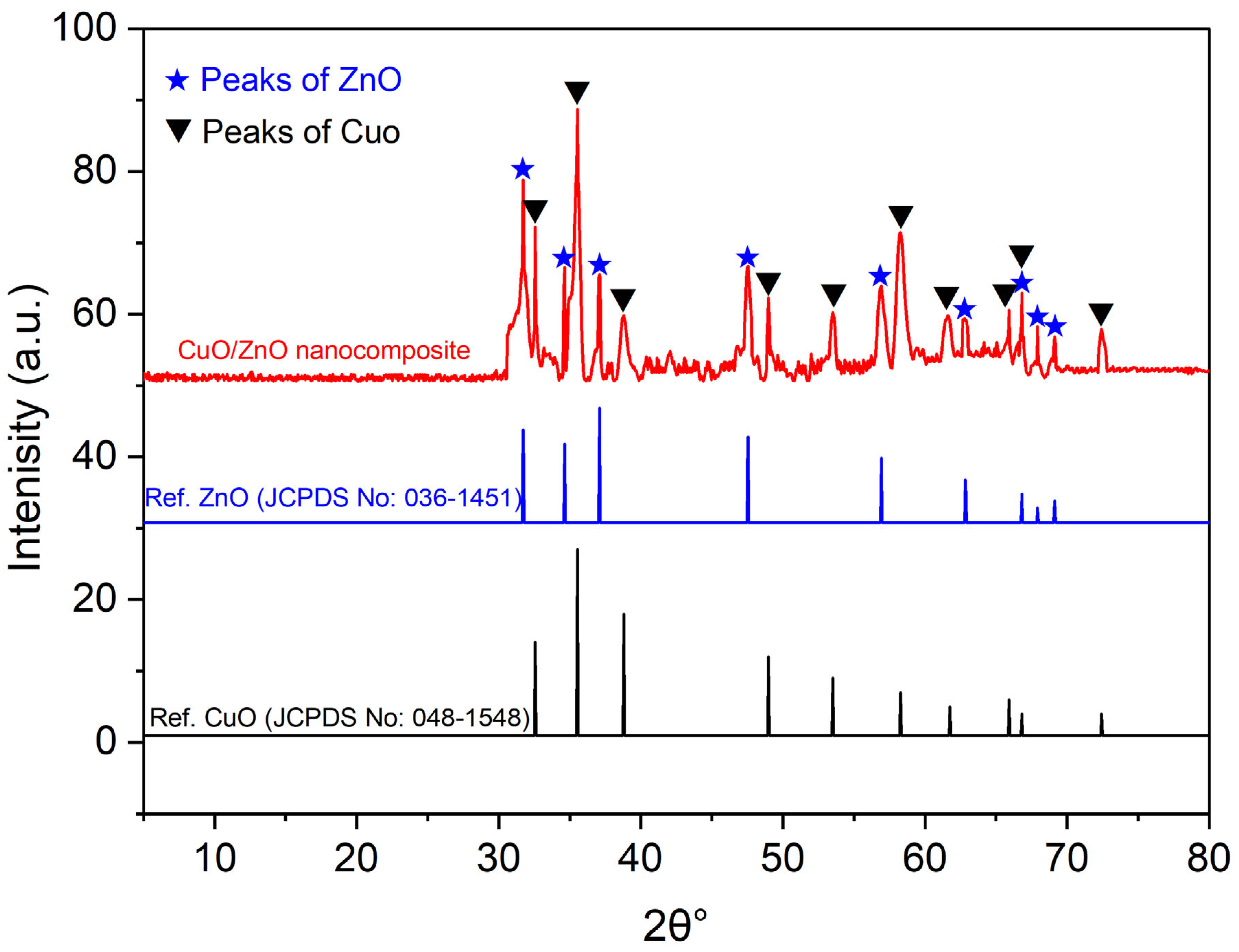
The crystallographic structure and phase of the green CuO/ZnO nanocomposites were assessed using X-ray diffraction analysis (Figure 2). The diffraction peaks at 32.48°, 35.62°, 38.78°, 48.94°, 53.51°, 58.29°, 61.72°, 65.91°, 66.42°, and 72.46°, which matched those of (110), (11-1), (111), (20-2), (020), (202), (11-3), (022), (31-1), and (311), respectively, confirmed the presence of the monoclinic phase of CuO (JCPDS No: 048-1548) (Figure 2). Moreover, the hexagonal phase of ZnO appeared at 2θ° values of 31.79°, 34.45°, 36.26°, 47.58°, 56.62°, 62.88°, 66.42°, 67.98°, and 69.11° and matched Bragg’s peaks of (100), (002), (101), (102), (110), (103), (200), (112), and (201), respectively (JCPDS No: 036-1451) (Figure 2). The presence of these diffraction peaks in the pattern of the synthesized sample confirmed the fabrication of the crystalline Cu-O-Zn nanocomposite. In a similar investigation, an XRD pattern of the green fabrication of CuO/ZnO nanocomposites using an aqueous extract of Verbascum sinaiticum containing the same diffraction peaks of CuO and ZnO was observed [30]. The authors reported that the XRD pattern of monoclinic CuO contain peaks at 32.5 (110), 35.6 (11–1), 38.8 (111), 48.9 (20–2), 53.5 (020), 58.3 (202), 61.7 (11–3), 65.9 (022), 66.4 (31–1), 68.1 (220), and 72.5 (311), whereas those of hexagonal ZnO are 31.8 (100), 34.5 (002), 36.3 (101), 47.6 (102), 56.6 (110), 62.9 (103), 66.4 (200), 67.9 (112), and 69.1 (201). On the other hand, the XRD pattern of the nanocomposite CuO/ZnO sample contains the diffraction peaks of two metal oxides. Additionally, the XRD results for the CuO/ZnO nanocomposite showed diffraction peaks for monoclinic CuO (2θ values of 35.4°, 38.8°, 48.8°, 53.5°, 58.3°, 61.6°, 66.3°, and 68.2°) and peaks for hexagonal ZnO (2θ values of 31.8°, 34.5°, 36.3°, 47.5°, 56.7°, 62.9°, and 69.0°) on the same chart [31]. Interestingly, the presence of sharp and distinct XRD peaks confirmed the crystalline structure of the green-synthesized nanocomposite. The average crystallite size of the green-synthesized CuO/ZnO nanocomposites was calculated using the Debye–Scherrer equation, and was 23.5 nm. The obtained finding were compatible with different published investigations. For instance, the crystallite size of CuO/ZnO fabricated by a water extract of Annona glabra with ratios 70:30, 50:50, and 30:70 was 27.2, 22.5, and 24.9 nm, respectively [32]. Also, the average crystallite size of Tragia involucrate-mediated biosynthesis of CuO/ZnO was 20.4 nm as detected by XRD analysis [31]. XRD chart showed the presence of some extra peaks that may be related to the scattering of plant metabolites (organic residues) which capped or coated the nanocomposite surface [31]. Also, these extra peaks could be related to sample preparations such as inadequate thickness and presence of moisture [33].

2.1.3. Morphological and Composition Analyses

The morphological features (shape and size) of the synthesized nanocomposites as well as their ion compositions were investigated through TEM and EDX analyses, respectively. TEM analysis confirmed the ability of U. urens aqueous extract to produce spheres with sizes ranging from 5 nm to 45 nm and an average particle size of 34.47 ± 1.81 nm (Figure 3A–D). Some particle aggregations were detected during TEM analysis, which could be related to sample preparation (such as drying, loading high NP concentration on the TEM grid, and a lack of or insufficient solvent), insufficient plant metabolites that act as stabilizing agents, and the tendency of Cu and Zn to aggregate during sample preparation because of high surface energy [34]. Similarly, the TEM size of Cu doped with ZnO nanostructures ranged from 19 to 30 nm and the size increased with increasing Zn concentration [8]. In contrast, spherical green-synthesized CuO/ZnO nanocomposite with sizes in the range of 20–130 nm were produced by an aqueous extract of Sambucus nigra, and this wide range of sizes attributed to the different shapes of ZnO after it was mixed with CuO nanostructures [35]. In some cases, the sizes of bimetallic NPs are larger than those of mono-nanostructure. For example, the sizes of bimetallic Cu/Zn nanostructures increase after they are mixed compared with those of Cu or Zn monostructures because of the Van der Waals mechanism [36]. In addition to the effects of morphological characteristics on nanocomposite application, the authors reported that different factors, such as pore diameter and surface charge, are also important.
The selected area electron diffraction (SAED) pattern of the green-fabricated CuO/ZnO nanocomposite is shown in Figure 3E. The presence of sharp and continuous well-defined rings indicates the polycrystallinity of the synthesized nanocomposite. Moreover, the orientation or texture degree of crystals was confirmed by the bright spots on the rings. Similarly, the polycrystallinity of the ZnO/CuO nanocomposite was detected by SAED analysis through bright spots and concentrated rings [37]. The compositions of synthesized nanocomposites play a vital role in different applications. Therefore, EDX analysis was used to detect the metal components of the green CuO/ZnO nanocomposite. The presence of Cu peaks at bending energies (KeV) of 0.96, 8.09, and 8.96 KeV; Zn peaks at 1.05, 8.64, and 7.53 KeV; and the O peak at 0.5 KeV confirmed the successful formation of CuO/ZnO (Figure 3F). EDX analysis confirmed that the main components (represented by weight % and atomic %) of the synthesized nanocomposite were Cu, Zn, and O, with weight percentages of 23.5, 17.7, and 35.4% and atomic percentages of 8.9, 6.6, and 53.5%, respectively. The high amount of O is due to the successful fabrication of CuO and ZnO, in addition to organic plant metabolites that cap the nanocomposite surface. Additionally, the high percentage of Cu compared to that of Zn indicates that Cu is more effective to reduce with plant metabolites [38]. Similarly, the weights of Cu, Zn, and O in plant-mediated green-synthesized CuO/ZnO nanocomposites were 14.3, 31.9, and 53.8%, respectively [39]. Some authors reported that the weight and atomic percentages of monometallic Cu and Zn nanoparticles are greater than those after they are mixed to form nanocomposites. For example, the weights of Cu and Zn in the nanocomposite structure (CuO/ZnO) were 34.7 and 36.5%, respectively, which were lower than the weights of Cu and Zn in the mono-nanostructure, which were 74.4 and 77.4%, respectively [36]. In the current EDX chart, some minor peaks, including C and S, are present, with weights of 10.6% and 12.9% and atomic percentages of 21.3% and 9.7%, respectively. These peaks could be related to the scattering of U. urens metabolites that coated the nanocomposite surface or originated from the handling sample [38,40]. On the other hand, the presence of C and S ions may be related to contaminants on the surface of nanocomposites. This finding was confirmed by TGA analysis, which leads to the weight loss in the range of 200–500 °C due to removal of impurities such as carbonates and salts from the nanocomposite surface. These impurities have some negative impacts on nanocomposite activity, such as blocking the active sites, which decrease the production of reactive oxygen species (ROS). Also, it can change the hydrophilicity and hydrophobicity which affect the adsorption of the dyes. Moreover, it can give false positive results in antimicrobial and cytotoxic activities because of toxic effects of these impurities.

2.1.4. Thermal Stability

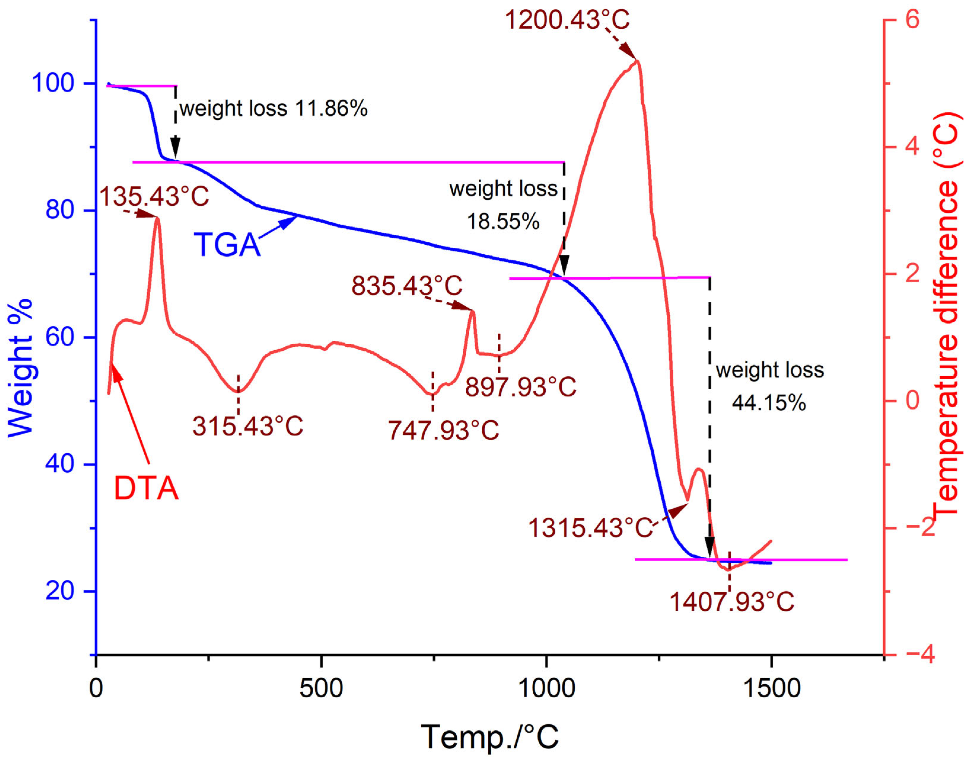
The thermal stability behavior of the CuO/ZnO green nanocomposites was assessed using TGA-DTA analysis at various temperatures from 30 °C to 1500 °C (Figure 4). The synthesized nanocomposite decomposes under heat treatment into three steps with varying weight loss. The first step occurs in the range of 30–177 °C with a weight loss percentage of 11.86%, which is related to the evaporation of adsorbed H2O and humidity [41]. This weight loss increased to 18.55% after the temperature increased to 1037 °C because of different factors, including the breakdown of coated organic molecules from plant metabolites, dihydroxylation of the nanocomposite surface, removal of some impurities such as carbonates and salts from the nanocomposite surface, and removal of remaining water and moisture contents [42]. The second weight loss step indicates the efficacy of different plant metabolites such as carbohydrates, proteins, amino acids, and other organic materials, in the fabrication of nanocomposites. The final and maximum weight loss occurred after the temperature increased to 1357 °C, with a percentage of 44.15%. This loss is related to the complete decomposition of the organic material coating, high calcination of the nanocomposite, and removal and oxidation of the remaining impurities [12]. With respect to the mass of the sample in mg, the mass losses due to temperature treatment were 2.17, 3.39, and 8.09 mg at the respective three steps compared with the mass at zero degree (18.3149 mg). The DTA curve revealed that the green CuO/ZnO nanocomposite exhibited three exothermic and five endothermic peaks. Exothermic peaks were observed at 135.43 °C (for the decomposition of plant organic metabolites), 835.43 °C (related to the crystallization of the nanocomposite), and 1200.43 °C (related to phase transformation and grain growth). The endothermic peaks were located at 315.43 °C (may be related to dehydration or removal or remaining organic residue), 747.93 °C (may be attributed to melting and removal of impurities), 897.93 °C (transition of the solid phase), 1315.43 °C, and 1407.93 °C (related to melting or partial fusion of the nanocomposite or thermal decomposition) (Figure 4).

2.1.5. Optical Features and Bandgap Detection

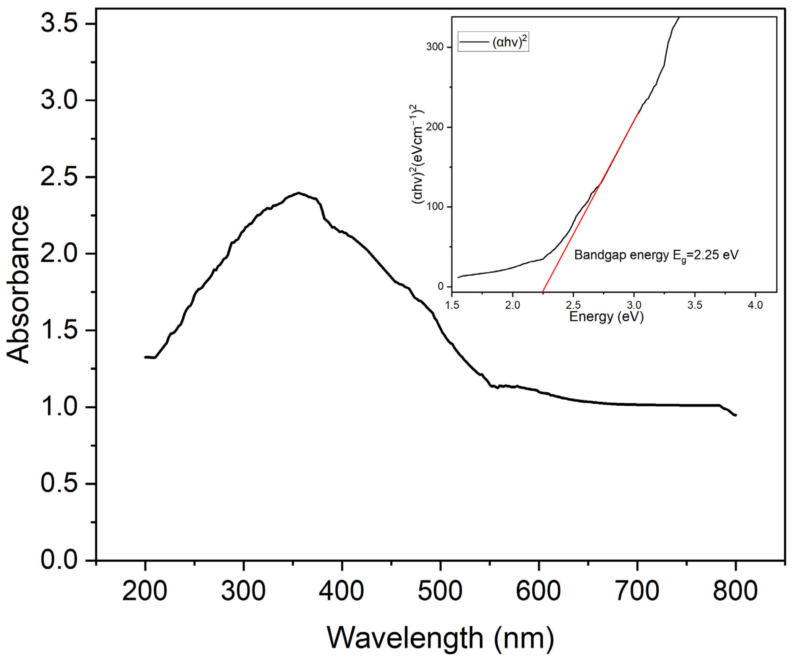
The UV absorption pattern of plant-fabricated CuO/ZnO nanocomposite was assessed at 200 nm to 800 nm (Figure 5). The synthesized nanocomposite showed maximum absorbance at a wavelength of 358 nm due to the presence of ZnO which is characterized by wide bandgap. Interestingly, the presence of broad spectra in the visible range related to the presence of CuO which is characterized by a narrower bandgap. Similarly, the optical properties of CuO-doped ZnO nanocomposite showed a maximum absorption peak at 355 nm [43]. The authors suggested that this peak is due to the presence of ZnO and the presence of a broad absorption peak related to the presence of CuO. In addition, the UV spectra of the plant-synthesized CuO/ZnO nanocomposite showed maximum absorption peak at 378 nm [39].
The direct energy bandgap (Eg) of the fabricated nanocomposites was determined by Tauc’s plot method as follows [44]:
α h v = A ( h v E g ) 1 / 2
where α represents the absorption coefficient of the fabricated nanocomposite, hv denotes the energy of the photon, A indicates a constant, and Eg represent the bandgap.
Tauc’s assay was plotted between (αhv)2 and the photon energy (eV) on the Y- and X-axes, respectively. Linear Tauc plot analysis revealed that the direct bandgap of the plant-synthesized nanocomposite is equal to 2.25 eV (Figure 5). This small bandgap increases the absorption of visible light by the nanocomposite. The synergistic interaction between semiconductor CuO (p-type) and ZnO (n-type) forms a p–n heterojunction, which improves the separation of charge and hinders the recombination of photogeneration electron–hole pairs to improve the photocatalysis process under visible light [45]. In this study, the bandgap of the synthesized CuO/ZnO nanocomposite is 2.25 eV, which is intermediate between bandgap of pure monometallic CuO (1.4 eV) and ZnO (3.08 eV) [31]. These findings confirm the formation of a heterojunction nanostructure rather than a physical mixture [45]. Additionally, the photocatalytic activity of nanocomposites in the visible light range is greater than that of pure monometallic compounds [32].

2.2. Photocatalytic Reaction

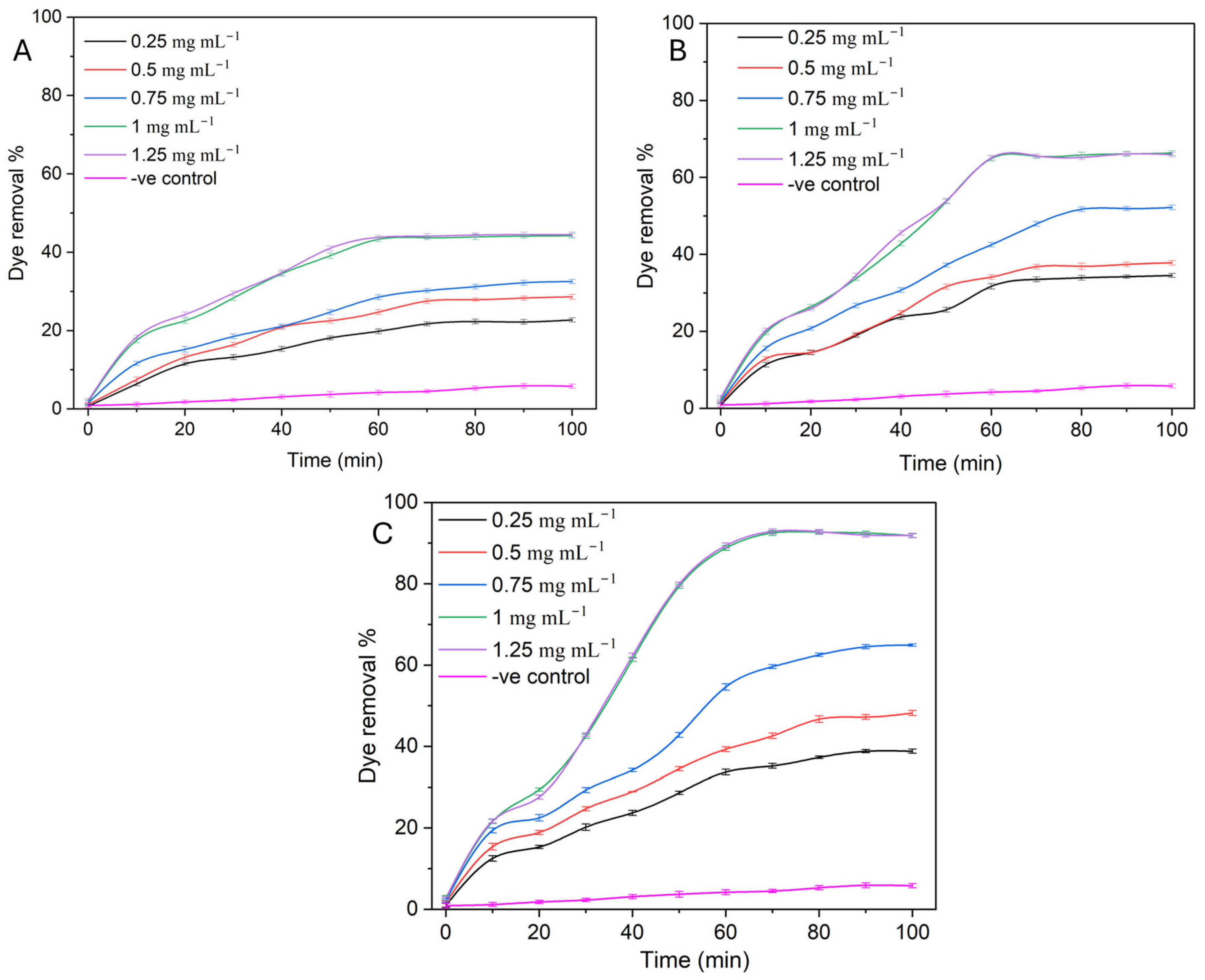
The activity of synthesized semiconductor nanocomposite compared to that of monometallic CuO and ZnO nanoparticles for methylene blue (MB) removal (as a model of dye, with a starting dye concentration of 10 mg L−1 and an adjusted solution pH of 8) was investigated under visible light conditions at various nanocomposite concentrations (0.25, 0.5, 0.75, 1.0, and 1.25 mg mL−1). The percentage of dye removal was assessed after different contact times (0–100 min with a time interval of 10 min). Data analysis revealed that the activity of monometallic and nanocomposite dye removal was concentration- and time-dependent. The removal percentage increased at high concentrations until it reached a point of equilibrium, at which point the removal percentage was stable even when the nanocomposite concentration increased. The findings obtained are compatible with those of different investigations published. For example, Sonkar et al. reported that Cu doped with ZnO-NPs showed promising MB and Rhodamine B removal under xenon light conditions in a time-dependent manner [8]. Similarly, the degradation of chlorpyrifos as a model for pesticides using CuO/ZnO nanocomposites was dependent on the concentration of the nanocatalyst and the incubation time [46]. In the current investigation, the activity of the nanocomposite for the removal of MB dye is promising because of the smaller nanocomposite sizes (5–45 nm) obtained. The maximum percentage of dye removal for the negative control (-ve control, MB solution under visible light conditions in the absence of semiconductor and monometallic) was 5.8 ± 0.5%, indicating that the photolytic degradation of MB dye under visible light is very low [43]. At the lowest concentration (0.25 mg mL−1), the percentage of dye removal increased from 6.4 ± 0.5%, 11.3 ± 0.6%, and 12.5 ± 0.7% for the CuO, ZnO, and semiconductor CuO/ZnO nanocomposites, respectively, after a 10 min incubation period to 22.5%, 34.2%, and 38.8%, respectively, after 90–100 min (Figure 6). With increasing concentration, the maximum dye removal percentages were obtained at 1.0 mg mL−1, with values of 44.1 ± 0.5%, 65.6 ± 0.6%, and 92.7 ± 0.5% after 70 min for the CuO, ZnO, and CuO/ZnO nanocomposites, respectively. The removal percentage after 80–100 min did not substantially differ from that after 60 min (Figure 6). Moreover, the removal percentage at the next concentration (1.25 mg mL−1) did not substantially differ from that at 1.0 mg mL−1 because the nanocomposite reached equilibrium sorption at this concentration. Therefore, the optimum conditions for MB dye removal using monometallic and CuO/ZnO nanocomposites were determined to be 1.0 mg mL−1 after 70 min. Recently, a green CuO/ZnO nanocomposite synthesized from Tragia involucrate aqueous extract showed maximum removal of Rhodamine B dye at 1.0 mg mL−1 with a percentage of 96.1% [31]. The authors reported that the percentage of dye removal decreased before and after the optimum concentration was reached to 64.1%, 76.5%, and 63.5% at concentrations of 0.25, 0.5, and 1.5 mg mL−1, respectively. Moreover, compared with monometallic ZnO-NPs and CuO-NPs, a nanocomposite of ZnO/CuO fabricated from the aqueous extract of Corriandrum sativum showed promising activity for the removal of MB dye [47]. The authors suggested that this activity was related to the high number of active sites of the nanocomposite that were more than those of the monometallic nanostructure. On the other hand, the weight proportional ratio between Cu and Zn during synthesis plays a critical role in the degradation and removal of dyes. For example, compared with nanocomposites with various weight ratios (2:8, 6:4, 1:1, and 8:2), the nanocomposite containing ZnO and CuO with a weight ratio of 4:6 showed promising MB dye removal under visible light with a percentage of 93% after 60 min [48]. Compared with nanocomposites with various weight ratios (99:1, 97:3, 90:10, and 50:50), the ZnO:CuO nanocomposite with a weight ratio of 95:5 showed maximum MB and methyl orange dyes removal with percentages of 97% and 87%, respectively, after 120 min under visible light irradiation [49]. The results revealed that compared with that of the monometallic CuO-NPs and ZnO-NPs, the photodegradation of MB by the semiconductor CuO/ZnO nanocomposite is superior. These findings are consistent with those of Azabi et al. [50], who reported that the photocatalytic activity of green CuO/ZnO nanocomposites synthesized from the water extract of Genista hispanica towards the degradation of MB degradation increased the activity of monometallic nanoparticles. After 120 min, the authors reported that nanocomposite degradation activity of the nanocomposites was 87%, whereas that of CuO and ZnO was 38% and 27%, respectively. Therefore, the current investigation was completed with CuO/ZnO nanocomposites.
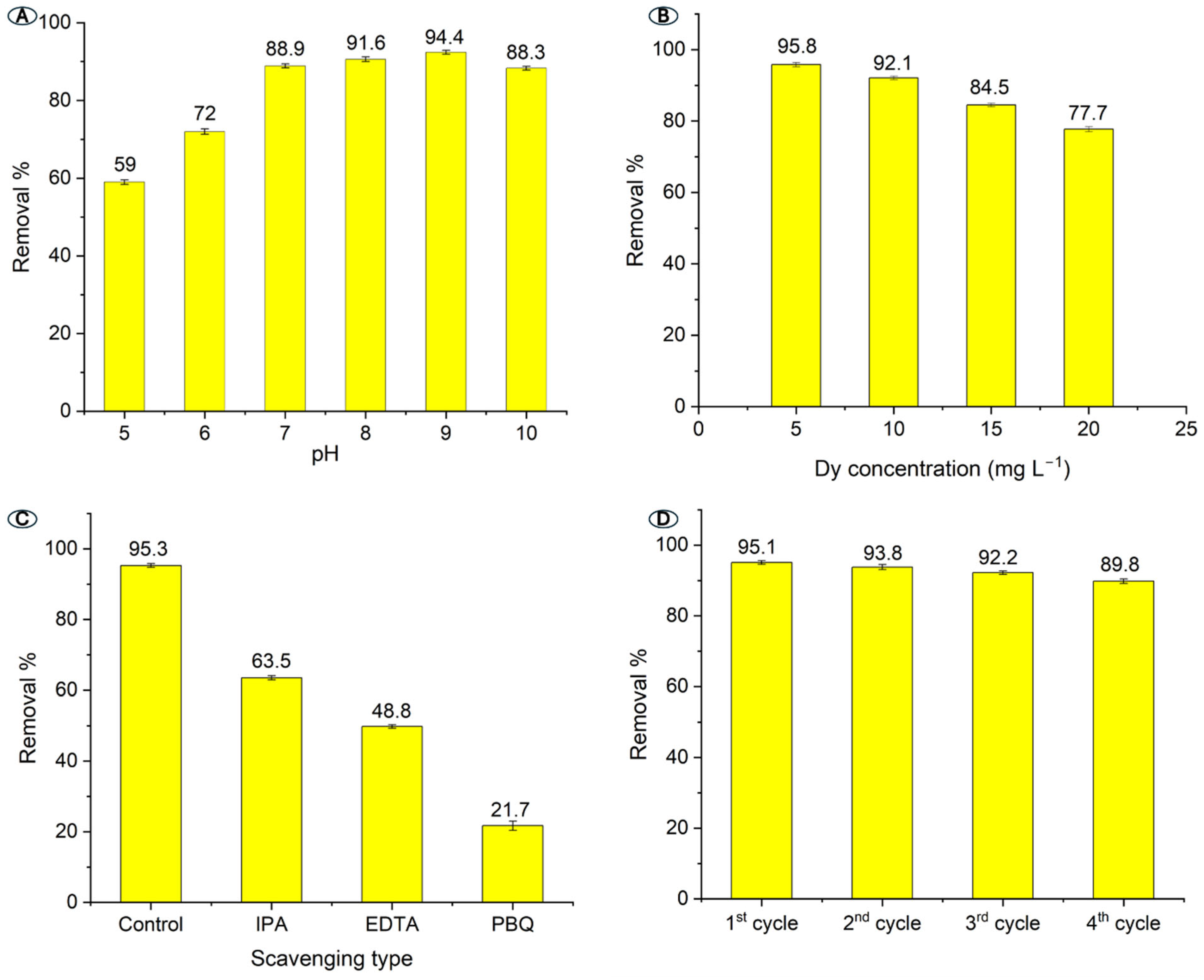
The wastewater effluents have varied pH values based on the type of dyes and other contaminants. As a result, investigating the activity of synthesized nanocomposites for dye removal at a wide range of solution pH is a critical point. The pH of the solutions has a positive or negative impact on the degradation percentages because of its involvement in catalyst charge and hydroxyl radical generation, and it plays an important role in catalyst sizes [51,52]. At MB dye and nanocomposite concentrations of 10 mg L−1 and 1.0 mg L−1, the removal percentages were calculated at various pH of 5–10 with value interval of 1. The maximum dye removal was obtained at pH 9 with a percentage of 94.4% (Figure 7A). The nanocomposite showed low activity at acidic conditions with removal percentages of 59 ± 0.6% and 72 ± 0.7% at pH 5 and 6, respectively. These findings could be attributed to the dissolving nanocomposites at acidic conditions forming Cu2+ and Zn2+ ions, which damage the active sites [53]. At alkaline conditions, the activity of CuO/ZnO nanocomposite increased to reach the maximum percentage at 9 and decreased at pH 10 (Figure 7A). Similarly, the maximum dye removal of MB by CuO/ZnO nanocomposite was attained at pH 9 with percentages of 97.4% and decreased at low pH values to be 60.7 at pH 3, 75.9 at pH 5, and 94.9 at pH 7 [8]. The green CuO/ZnO nanocomposite formed by aqueous extract of Tragia involucrata showed maximum MB removal percentage (96.1%) at pH 9 [31]. The authors reported that the removal percentage decreased before and above this value to be 47.2%, 82%, and 86.2%, at pH values of 5, 7, and 11, respectively.
With a constant solution pH of 9 and a nanocomposite concentration of 1.0 mg L−1, different dye concentrations (5, 10, 15, and 20 mg L−1) were used to investigate the activity of the synthesized catalyst. Data analysis revealed that the highest removal percentage (95.8%) occurred at a dye concentration (5 mg L−1). The removal decreased with increasing dye concentration to 92.1%, 84.5%, and 77.7% at dye concentrations of 10, 15, and 20 mg L−1, respectively (Figure 7B). This phenomenon may be attributed to the absorption of MB dye to light, resulting in decreased generation of hydroxyl radicals by inhibition of the photocatalyst active sites [54]. Furthermore, at high dye concentrations, MB is absorbed on the catalyst surface, resulting in reduced efficacy and a decrease in the number of surface-active sites. Similarly, the optimum MB dye concentration for 100% removal by the green CuO/ZnO nanocatalyst was 5 mg L−1, and the removal percentages decreased to 96.1% and 44.7% at 10 and 15 mg L−1, respectively [31]. The removal percentages of MB by Cu doped with ZnO were also 97%, 62%, 45%, and 25% at dye concentrations of 3, 10, 30, and 50 mg L−1, respectively [8].
Different scavengers, including p-benzoquinone, isopropyl alcohol, and EDTA, were used to investigate the role of reactive species O2, OH, and h+ in photodegradation. These scavengers are added to the photocatalytic solution under optimum conditions to inhibit the reactive species production as follows: PBQ, IPA, and EDTA inhibit the generation of O2, OH, and h+, respectively. At the end of the experiment, the degradation or removal of MB dye using nanocomposites were negatively affected as follows: 63.5 ± 0.6%, 48.8 ± 0.5%, and 21.7 ± 1.3% due to the presence of IPA, EDTA, and PBQ, respectively compared to 95.3 ± 0.5% removal for control (photocatalytic solution without any scavengers) (Figure 7C). The results obtained showed that although all reactive species were involved in the photodegradation of MB using CuO/ZnO nanocomposites, the photocatalytic reaction was mainly dependent on the O2 generation. In a similar research, plant-mediated synthesized CuO/ZnO nanocomposites revealed that the degradation of Rhodamine-B dye under visible light was dependent on different reactive species (O2, OH, and h+), but O2 is the mainly used during photocatalytic reaction [31]. The photocatalytic mechanism based on the generation of reactive species was described in Figure 8.
Reusability or recyclability of green nanocomposite is a critical point for detecting successful usage and stability of catalysts in the environmental contaminant’s treatment. After each cycle, the nanocomposite was collected and washed twice by distilled water followed by ethanol before being dried to add in the next cycle. Data analysis revealed that the removal percentages after the first cycle were 95.1 ± 0.5% and the percentage gradually decreased with cycling, reaching to 93.8 ± 0.7%, 92.2 ± 0.5%, and 89.8 ± 0.6% after second, third, and fourth cycles, respectively (Figure 7D). Interestingly, the reducing level in the efficacy of nanocomposite between the first and fourth cycles is approximately 6% and this percentage indicates the high efficacy and high stability of green-synthesized CuO/ZnO. In a similar investigation, the photodegradation of MB using biosynthesized CuO/ZnO fabricated with different weight ratios of 80:20, 50:50, and 20:80 showing promising recyclability for 10 cycles with decreasing percentages of 5%, 4%, and 7%, respectively [55]. Also, photocatalyst CuO/ZnO nanocomposite showed high activity for degradation of MB for fourth cycles with decreasing percentages of ~8% [56].

2.3. Antimicrobial Activity

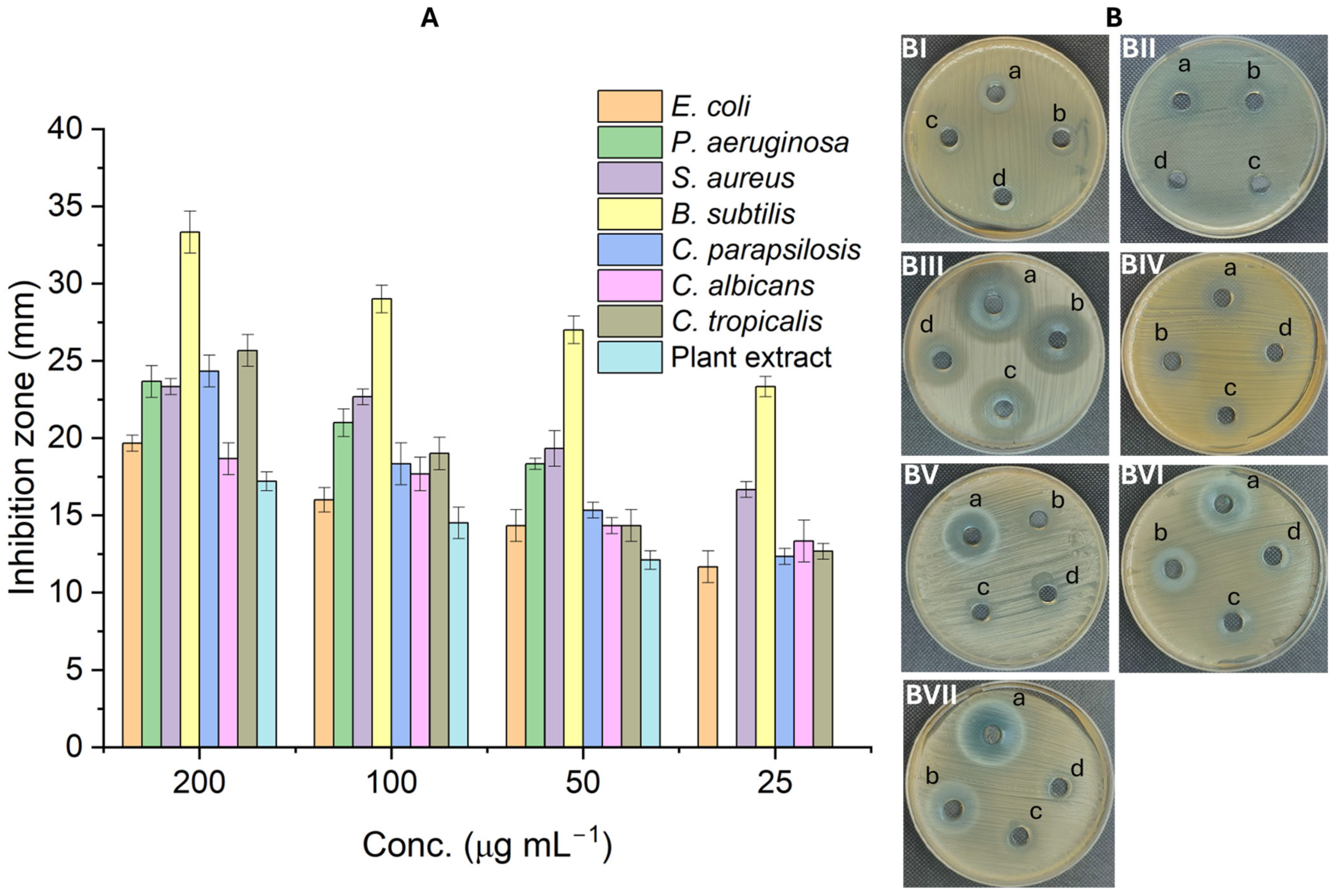
To increase the usability of the green CuO/ZnO nanocomposite, its antimicrobial activity against different microbial strains, including G-ve bacteria (E. coli and P. aeruginosa), G+ bacteria (B. subtilis and S. aureus), and unicellular fungi (C. parapsilosis, C. albicans, and C. tropicalis) was investigated using the agar well diffusion method. Data analysis revealed that the antimicrobial activity of the green nanocomposite against all tested strains was dependent on the concentration used. These findings are compatible with those of other investigations. For example, a rhizome aqueous extract of Zingiber officinale was used to fabricate CuO/ZnO (10% and 20%) nanocomposites, which showed promising concentration-dependent antibacterial activity against S. aureus (G+) and E. coli (G-ve) strains [57]. In addition, the antibacterial activity of Zn/Cu nanocomposite with weight ratios of 2:1, 1:1, and 1:2 was concentration-dependent against S. aureus, P. aeruginosa, E. coli, and Klebsiella pneumoniae; the maximum activity occurred at a concentration of 60 mg mL−1 and the lowest activity occurred at 20 mg mL−1 [58]. In the current investigation, the greatest inhibition zones were observed at a concentration of 200 µg mL−1 with diameters of 19.7 ± 0.5, 23.7 ± 1.03, 23.3 ± 0.5, 33.3 ± 1.4, 24.3 ± 1.1, 18.7 ± 1.02, 25.7 ± 1.03, and 17.2 ± 0.6 mm against E. coli, P. aeruginosa, B. subtilis, S. aureus, C. parapsilosis, C. albicans, C. tropicalis, and the plant extract, respectively (Figure 9A,B). The activity decreased to 16.0 ± 0.8, 21.0 ± 0.9, 22.7 ± 0.5, 29.0 ± 0.9, 18.3 ± 1.4, 17.7 ± 1.1, 19.0 ± 1.1, and 14.5 ± 1.02 mm and 14.3 ± 1.03, 18.3 ± 0.4, 19.3 ± 1.2, 27.0 ± 0.9, 15.3 ± 0.5, 14.3 ± 0.5, 14.3 ± 1.03, and 12.5 ± 0.6 mm for low concentrations of 100 and 50 µg mL−1, respectively, against the same sequence (Figure 9A,B). In a recent investigation, the antimicrobial activity of U. urens aqueous extract was higher than that in the current investigation [59]. The authors reported that the maximum antimicrobial activity of U. urens extract was attained at the highest concentration of 25,000 µg mL−1 with inhibition zones of 26.0 ± 1.3, 19.0 ± 1.1, 24.0 ± 0.9, 25.0 ± 0.3, and 25.0 ± 0.7 mm against E. coli, Salmonella typhi, B. subtilis, S. aureus, and C. albicans, respectively. As shown, compared with high concentrations of plant extract, low concentrations of synthesized nanocomposite have promising activity. Similarly, aqueous extract of Eryngium foetidum leaves was used to fabricate CuO/ZnO nanocomposites, which showed concentration-dependent antimicrobial activity with maximum clear zones of 20.7, 18.7, 21.1, and 15.4 mm at a concentration of 3000 µg mL−1 against E. coli, Enterobacter aerogens, S. aureus, and B. subtilis, respectively [60]. The authors reported that the lowest activity was attained at 1000 µg mL−1 with clear zones of 12.8, 10.1, 17.2, and 10.4 mm against the same previous test organism sequence.
MIC (minimum inhibition concentration) detection is important for evaluating the efficacy and potential of antimicrobial nanocomposite drugs. The MIC values for P. aeruginosa and the plant extract were 50 µg mL−1, with inhibition zones of 18.3 and 12.1 mm, respectively, whereas the MIC value for the other tested microorganisms was 25 µg mL−1, forming inhibition zones of 11.7, 16.7, 23.3, 12.3, 13.3, and 12.7 mm for E. coli, B. subtilis, S. aureus, C. parapsilosis, C. albicans, and C. tropicalis, respectively. The Zn/CuO nanostructure formed using the extract of Aspergillus niger showed antimicrobial activity, with MICs of 125 µg mL−1 for E. coli, 31.25 µg mL−1 for B. subtilis, 125 µg mL−1 for S. aureus, and 500 µg mL−1 for C. albicans, with inhibition zones of 19, 23, 16, and 11 mm, respectively [61].
Different mechanisms explain the antimicrobial activity of CuO/ZnO nanocomposites. For example, production of ROS, selective permeability damage of cell membrane, disruption of cell wall sterols in unicellular fungi, enzymes, amino acids, and proteins destroying upon nanocomposite entrance to microbial cells, accumulation of toxic ions within the cells, and genotoxicity. The oxidative stress as a result of ROS generation and accumulation toxic ions (Zn2+ and Cu2+) within cells cause dysfunction and peroxidation of lipid membrane, leading to leakage of essential components, such as proteins, sugars, amino acids, and nucleic acids, outside the cells [62,63]. Also, the integrity of the microbial cell wall due to their interaction with nanocomposites leads to cell damage [64]. The interaction of thiol group (-SH) with accumulated toxic ions within cells lead to protein inactivation and decrease or inhibition of the selective permeability function, ultimately leading to cell death [65,66]. Moreover, the accumulation of Zn2+ and Cu2+ within cells reacts with phosphorus in DNA and sulfur in proteins leading to inhibition of microbial cell metabolic processes. In addition, Zn2+ inhibits the ATP synthesis via interaction with the proton pump whereas Cu2+ stops the microbial respiratory system via the disrupting of electron transport chains [67,68]. In Candida spp., the generation of ROS, Zn2+, and Cu2+ can destroy the ergosterol via peroxidation, leading to integrity of Candida cell membrane [69]. Also, the ROS impairs the ATP production and cell division via dysfunction of mitochondria function and destroying DNA double strand [70]. Moreover, the inhibition of Candida spp. by nanocomposite could be related to its activity to suppress the formation of hyphae (responsible for invasion), decrease the hydrolytic enzymes production, and inhibit the biofilm activity [71,72].

2.4. In Vitro Cytotoxicity

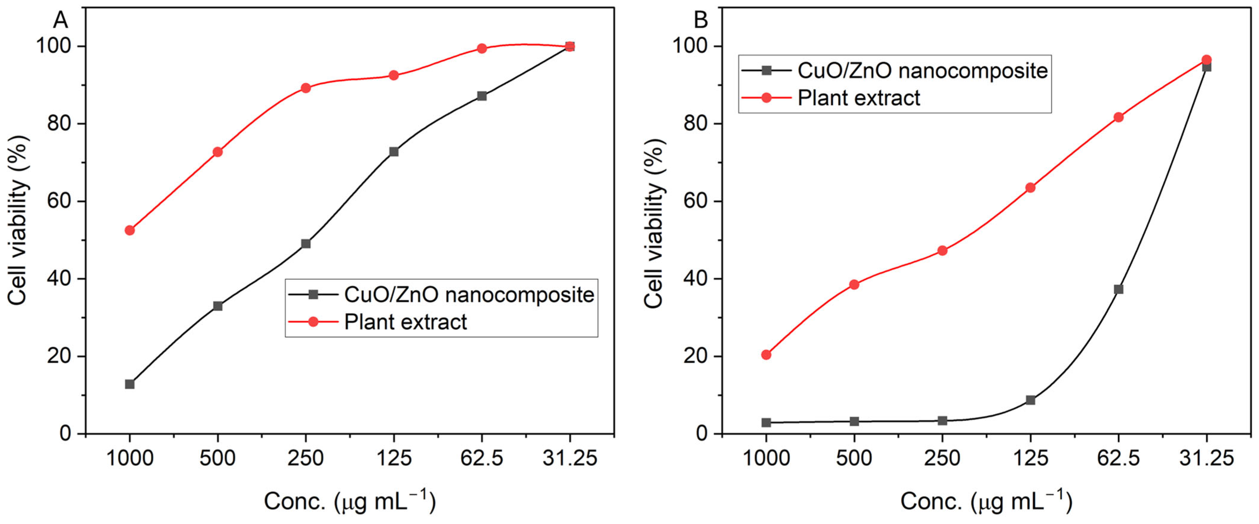
The toxicity of the green-synthesized CuO/ZnO nanocomposite against the normal human fibroblast skin cell line HFB4 and the human epithelial hepatocarcinoma cell line HepG2 was evaluated using an MTT assay at various concentrations of 1000–31.25 µg mL−1 after incubation for 48 h. The cytotoxicity was compared between the synthesized nanocomposite and the plant extract at the same concentrations. MTT is a sensitive spectrophotometer assay that depends on the reduction in MTT (yellow) to formazan (blue) via succinate dehydrogenase mitochondrial enzymes. Active compounds of nanomaterials react with MTT, which facilitates the breakdown of tetrazolium ring by the dehydrogenase enzyme [73]. The results represent the percentages of viable cells after the nanocomposite and plant extract treatments. The results of the data analysis revealed that the viability of normal and cancer cells is a concentration-dependent manner and that the viability decreased at high concentrations (Figure 10A,B). Similarly, the activity of CuO/ZnO nanocomposites against hepatocellular carcinoma cells, HuH7 and HepG2, and normal liver cells, BNL, was dose-dependent [74]. Analysis of variance revealed that compared with normal cell treatment, treatment with nanocomposites at low concentrations had greater effects on the viability of cancer cells. The cell viability percentages of HepG2 cells ranged from 3% to 9% at concentrations of 1000–125 µg mL−1, whereas the viability percentages ranged from 12% to 73% for normal cells at the same concentrations (Figure 10A). In another words, the nanocomposite showed cancer cell death with percentages of 91% at these concentrations compared to 27% cell death for normal cells. However, the death of cancer cells after treatment with 1000 µg mL−1 plant extract was 80% and decreased to 37% at 125 µg mL−1. In contrast, the HFB4 cells were deleterious, with percentages of 47.5% and 7.5% at 1000 and 125 µg mL−1 plant extract, respectively. At low concentrations, the cell viability percentages due to the nanocomposite treatment were 37.3 ± 0.5% and 94.7 ± 0.5%, respectively, for HepG2 at 62.5 and 31.25 µg mL−1 (vs. 81.8 ± 0.5% and 96.5 ± 0.6% for the plant extract) (Figure 10B). On the other hand, the HFB4 cells were 87.2 ± 0.4% and 99.9 ± 0.5%, viable at nanocomposite concentrations of 62.5 and 31.25 µg mL−1, respectively (vs. 99.4 ± 0.5% and 99.6 ± 0.6% for the plant extract) (Figure 10A). In a similar study, Cu/ZnO nanocomposites inhibited the growth of normal Vero cells by 28%, whereas they inhibited the growth of Hela and MCF7 cells by 70% and 59%, respectively, at a concentration of 300 µg mL−1 [75]. The authors reported that the growth inhibition of Vero, HeLa, and MCF7 cells decreased to (10%, 24%, and 13%) and (5%, 17%, and 7%) at Cu/ZnO concentrations of 37.5 and 18.75 µg mL−1, respectively. Additionally, a Pleurotus ostreatus aqueous extract was utilized for the fabrication of ZnO/CuO nanocomposite, which showed 23.5% cytotoxic activity for cancerous HeLa cell lines compared with 41.8% for normal HEK cell lines after treatment with 100 µg mL−1 of the nanocomposite. The percentage of viable cells gradually decreased to 62.2% for HeLa and 86.9% for HEK at a nanocomposite concentration of 10 µg mL−1 [76].
The detection of IC50 values of synthesized nanocomposite against normal and cancer cells is important. Data analysis revealed that the concentration of CuO/ZnO nanocomposite inhibits the growth of 50% of normal cells at 220 µg mL−1 compared to 69.9 µg mL−1 for cancer cells. The data obtained indicate the successful targeting of cancer cells at low concentration without affecting the viability of normal cells.
In the current study, the smaller nanocomposite sizes (ranging from 5 to 45 nm) penetrate easily through the mammalian cell wall followed by dissimulating into toxic ions (Cu2+ and Zn2+), leading to accumulation of these ions within the cell and interacting with its components such as protein, mitochondria, amino acids, and nucleic acids, ultimately causing dysfunction of cellular components [76,77]. Also, the presence of these toxic ions increases the ROS production within the cells which leads to more oxidative stress and finally dysfunction of major metabolic process, inhibiting the mitochondrial function, and stoping cell division [78].

3. Materials and Methods

3.1. Urtica Urens-Mediated Biosynthesis of CuO/ZnO Nanocomposites

The green leaves of Urtica urens were collected from Al-Munira, Al-Qanater Al-Khairiya, Qalyubia Governorate (30°14′01.2″ N 31°06′39.8″ E), rinsed with distilled water (dis. H2O) to remove any attached debris, and left to dry in air for 10 days. The dried leaves were collected and ground to a fine powder, after which 10 g of fine plant powder was mixed with 100 mL of dis. H2O under stirring conditions (150 rpm) for 1 h at 60 °C. Centrifugation of the plant mixture was performed to collect the supernatant (aqueous extract) free of any plant debris. The collected supernatant was used for the biofabrication of the CuO/ZnO nanocomposite as follows: 60 mg of Cu (CH3COO)2. H2O was dissolved in 30 mL of dis. H2O and mixed with 40 mL of plant extract for 1 h under stirring conditions (150 rpm) at 60 °C. Afterwards, 66 mg of Zn (CH3COO)2. 2H2O was dissolved in 30 mL of dis. H2O, after which it was mixed with a copper and plant extract mixture and stirred for another hour to obtain a final nanocomposite concentration of 3 mM. Under stirring conditions, the pH of the mixture was adjusted to 8 with the addition of 1M NaOH. At the end of the incubation conditions, the mixture was kept overnight at room temperature in the dark to confirm the complete reduction in metal precursors by plant metabolites. Afterwards, the liquid was evaporated using a rotary evaporator to collect the pelt (residue), which was subsequently washed twice with dis. H2O and ethanol for purification before being subjected to calcination at 200 °C for 2 h [12,39].

3.2. Nanocomposite Characterization

The functional groups in U. urens aqueous extract related to active metabolites and their role in the fabrication and formation of nanocomposites were investigated by Cary-660-Fourier transform infrared (FT-IR, Agilent system, Tokyo, Japan) spectroscopy. Approximately, 10 µg of nanocomposite powder or a few drops of plant extract were mixed well with KBr before being subjected to disc formation and scanning at wavenumbers ranging from 400 to 4000 cm−1 [79]. The crystallinity of the nanocomposite was evaluated using Panalytical X’PERT PRO X-ray diffraction (XRD, MPD, Philips, The Netherlands) at a voltage of 40 KV, current of 30 mA, and X-ray source of Cu-Kα in the ranges of 2θ° of 5–80° [31]. The crystallite size of the obtained CuO/ZnO nanocomposite was calculated using the Debye–Scherrer equation as follows:
Crystallite   size   = 0.9 × 1.54 β cos θ
where 0.9 represents the Debye–Scherrer constant, 1.54 denotes the λ of the X-ray source, β indicates the half-maximum intensity; and θ represents Bragg’s angle.
The morphological features of nanocomposite such as shapes, sizes, and agglomeration were detected by JEOL-1010-Transmission Electron Microscopy (JEOL, Tokyo, Japan). The TEM grid was covered by a few drops of synthesized nanocomposite by micropipette until complete adsorption and the excess solution was removed by blotting paper before TEM analysis. The elemental compositions of synthesized nanocomposite were assessed using JEOL-JSM-6360LA-energy-dispersive X-ray (EDX, Tokyo, Japan) analysis [80]. Thermogravimetric analysis (TGA) was performed by a NETZSCH STA 449F3 (NETZSCH-Gerätebau GmbH, Wittelsbacherstraße 42, 95100 Selb, Germany) to monitor the thermal decomposition of the CuO/ZnO nanocomposite. The analysis started with a temperature range of 30 °C to 1500 °C in the presence of N2 and a weight of 18.3149 mg. The absorbance of the synthesized nanocomposite was measured by a JENWAY-6305 (Jenway, Tokyo, Japan) to detect the maximum surface plasmon at wavenumbers in the ranges of 200–800 nm. Tauc’s plot method was used to measure the bandgap of the nanocomposite by plotting the linear graph between (αhv)2 as the Y-axis and the photon energy (eV) as the X-axis [8].

3.3. Photocatalytic Experiment

The photocatalytic activity of semiconductor CuO/ZnO formed from U. urens aqueous leaf extract was investigated. Different concentrations of the synthesized semiconductor, 0.25, 0.5, 0.75, 1.0, and 1.25 mg mL−1, were used for the photocatalytic removal of methylene blue (MB) dye (10 mg L−1 and pH 8) under visible light conditions (a xenon lamp, 350 W, λ > 420 nm to block UV-light, light intensity of 100 mW/cm2, and distance between the light source and the surface of the solution of 10 cm). The catalyst and the MB solution were kept in the dark for 30 min before the photocatalytic experiment to reach the adsorption equilibrium. Each concentration was mixed with 50 mL of MB solution under air bubble aeration to compare degradation efficacy to that of the control (MB solution under the same conditions without a semiconductor). At specific contact times (0–100 min with intervals of 10 min), 2 mL of MB solution was withdrawn and centrifuged at 10,000 rpm to collect the clear supernatant which was used to detect the degradation efficacy after its absorbance was measured at 664 nm using a spectrophotometer (M-ETCAL, Penang, Malaysia) as follows:
P h o t o d e g r a d a t i o n   e f f i c a c y % = I n i t i a l   a b s o r b a n c e f i n a l   a b s o r b a n c e I n i t i a l   a b s o r b a n c e × 100
For a comparative study, monometallic CuO-NPs and ZnO-NPs were synthesized by the same method as that used for the nanocomposites and used under the same conditions for photocatalytic activity.
On the basis of the results of the photocatalytic experiments, the materials that exhibited the greatest activity were used to investigate the effects of several factors, such as the pH values (5–10) and the concentration of MB (5 to 20 mg L−1 with an interval of 5 mg L−1), on the activity. At the end of each experiment, 2 mL of solution was withdrawn and centrifuged, and the photocatalytic efficacy was measured using Equation (3).
Under optimum conditions, the efficacy of reactive oxygen species (ROS) on photocatalytic experiments were investigated using trapping assay. In this method, 1 mL of neutralized chemicals including EDTA, p-benzoquinone (PBQ), and isopropyl alcohol (IPA) was added to the photocatalytic solution under optimum conditions to scavenging the ROS radicals including h+, O2, and OH, respectively [8].

3.4. Antimicrobial Activity

The antimicrobial activity of the synthesized semiconductor nanocomposite was investigated against different bacterial and Candida species including E. coli (ATCC8739), Pseudomonas aeruginosa (ATCC9027), Bacillus subtilis (ATCC6533), Staphylococcus aureus (ATCC6528), Candida albicans (ATCC10231), Candida parapsilosis, and Candida tropicalis. The assay was performed by the agar well diffusion method. All selected strains are coded except C. parapsilosis and C. tropicalis and are purchased from the Microbiological Lab, National Research Centre, Cairo, Egypt. The activity of the semiconductor CuO/ZnO was assessed at the following concentrations: 25, 50, 100, and 200 µg mL−1. DMSO (Dimethyl sulfoxide solvent system) served as a negative control, whereas the positive control is the plant extract dissolved in DMSO at the same concentrations. The bacterial strains and Candida spp. were cultivated on nutrient agar media and sabouraud dextrose media, respectively, for 24 h at 35 ± 2 °C. The bacterial suspension was adjusted at 0.5 McFarland standard (approximately 1–2 × 10⁸ CFU mL−1) prior to inoculation [81]. The surface of a Muller Hinton agar plate was streaked with bacterial suspension before being formed as a 4-well plate (the diameter of each well is 6.0 mm) by a sterilized cork borer and filled with 100 µL from each concentration. The plates were incubated in the refrigerator for one hour (to confirm spreading of active compound before bacterial growth) and transferred to an incubator at 35 ± 2 °C for 24 h. After incubation, the results were recorded as a diameter of inhibition zone around each well by mm [39]. The minimum inhibitory concentration (MIC) was detected as the lowest semiconductor concentration that inhibits microbial growth. The experiment was achieved in triplicate.

3.5. Cytotoxicity Assay

An MTT (3-(4,5-dimethylthiazol-2-yl)-2,5-diphenyltetrazolium bromide) assay was used to investigate the cytotoxic activity of the semiconductor CuO/ZnO nanocomposite against two human cell lines, HFB4 (normal skin fibroblast) and HepG2 (hepatocarcinoma cells). Different concentrations of synthesized nanocomposites ranging from 31.25 µg mL−1 to 1000 µg mL−1 (double-fold concentrations) were used, and their toxicity was assessed after 48 h of incubation. The selected cells were purchased from VACSERA (Holding Company for Biological Products and Vaccines), Cairo, Egypt. The tissue plate (96-well) was used to culture the cells (1 × 10⁶ per mL) in a CO2 incubator (5–6.5%) at 37 °C for 24 h until they reached 1 × 10⁵ cells/well and formed a monolayer sheet. The former sheet was subsequently washed and kept in RPMI media (ready-prepared, Sigma-Aldrich, Germany) with 2% serum for 48 h at the concentration of the nanocomposite. The control was RPMI media without synthesized nanocomposites. Afterward, the excess growth medium was discarded, and the cells were shaken with MTT solution (50 µL/well) and incubated for 4 h. DMSO (10%) was used to dissolve the formazan crystals (which formed as a result of the metabolized MTT solution), resulting in the formation of a new color whose optical density (OD) was measured at 570 nm using ELIZA plate reader (Thermo-Fisher Scientific Inc., Waltham, MA, USA) [82]. The percentages of cell viability were calculated using the following formula:
C e l l   v i a b i l i t y   % = O D   o f   t r e a t e d   c e l l s O D   o f   c o n t r o l   ×   100

3.6. Statistical Analysis

The SPSS (Version 17) statistical tool was used to analyze the three independent replicates collected during the current study. ANOVA one-way-analysis (analysis of variance) was used to compare the difference between treatments (p < 0.05).

4. Conclusions

Semiconductor CuO/ZnO nanocomposites were successfully produced by aqueous extract of U. urens leaves and exhibit promising environmental and biological applications. The synthesized nanocomposites are characterized by FT-IR which confirm the role of active metabolites in aqueous extract in biosynthesis. A TEM image exhibits the formation of spherical shapes with small sizes ranging from 5 nm to 45 nm and average of 34.47 ± 1.81 nm. XRD analysis confirms the crystalline structure whereas the EDX confirms the presence of Cu, Zn, and O as the main components. TGA and UV analysis exhibits the thermal stability of the synthesized nanocomposite with maximum absorbance at 358 nm and a direct bandgap of 2.25 eV. The synthesized semiconductor CuO/ZnO nanocomposites showed promising activity in MB degradation under visible light with removal percentages of 95.8% under optimum conditions (1.0 mg mL−1, pH 9, and dye concentration of 5 mg L−1 after 70 min). The trapping test revealed that the O2 radicals were mainly involved in the photocatalytic process followed by OH, and h+. Semiconductor CuO/ZnO nanocomposites promised antimicrobial activity against pathogenic bacterial and Candida strains at low concentrations. Additionally, the semiconductor CuO/ZnO nanocomposites targeted cancer cells at low concentrations compared to its activity against normal cells. The current study opens the way to fabricate semiconductor nanocomposites using plant extract for a wide range of applications including environmental and biomedicals.

Author Contributions

Conceptualization, A.F., A.M.E. and M.A.A.; methodology, A.F., S.M.A., A.M.E., M.A.A. and F.A.A.; software, A.M.A., D.A.B., N.A.A. and M.A.A.-R.; validation, S.M.A., A.M.A. and F.A.A., formal analysis, A.F., A.M.E., M.A.A. and M.A.A.-R. investigation, A.S.A., F.A.A., A.M.A. and D.A.B.; resources, A.F., S.M.A., A.M.E., M.A.A. and M.A.A.-R.; data curation, A.F., A.M.E. and M.A.A.-R.; writing—original draft preparation, A.M.E., A.S.A. and M.A.A.; writing—review and editing, A.F., S.M.A., F.A.A., A.M.A., D.A.B., N.A.A. and M.A.A.-R. visualization, S.M.A., A.S.A., F.A.A., A.M.A., D.A.B. and N.A.A.; supervision, A.F., A.M.E. and M.A.A.; project administration, A.F. and M.A.A.-R.; funding acquisition, A.F., S.M.A., A.M.E., A.S.A., F.A.A., A.M.A., D.A.B. and N.A.A. All authors have read and agreed to the published version of the manuscript.

Funding

This research received no external funding.

Data Availability Statement

The original contributions presented in this study are included in the article material. Further inquiries can be directed to the corresponding author.

Acknowledgments

The authors extend their appreciation to University of South China, Hunan, China, Al-Azhar University, Cairo, Egypt, and Taibah University, Saudi Arabia, Madinah for supporting this work.

Conflicts of Interest

The authors declare no conflicts of interest.

References

  1. Fouda, A.; Hassan, S.E.-D.; Saied, E.; Hamza, M.F. Photocatalytic degradation of real textile and tannery effluent using biosynthesized magnesium oxide nanoparticles (MgO-NPs), heavy metal adsorption, phytotoxicity, and antimicrobial activity. J. Environ. Chem. Eng. 2021, 9, 105346. [Google Scholar] [CrossRef]
  2. Mrabet, C.; Jaballah, R.; Mahdhi, N.; Boukhachem, A.; Amlouk, M. CuO-ZnO nanocomposites-based thin films: Characterization, physical properties and sunlight photocatalytic degradation of organic pollutants. J. Alloys Compd. 2023, 968, 172252. [Google Scholar] [CrossRef]
  3. Loganathan, P.; Vigneswaran, S.; Kandasamy, J.; Cuprys, A.K.; Maletskyi, Z.; Ratnaweera, H. Treatment Trends and Combined Methods in Removing Pharmaceuticals and Personal Care Products from Wastewater—A Review. Membranes 2023, 13, 158. [Google Scholar] [CrossRef] [PubMed]
  4. Magaña-Chavez, C.A.; Villegas-Fuentes, J.A.; Nava, O.J.; Vilchis-Nestor, A.R.; Luque, P.A. Green synthesis of semiconductor nanoparticles (ZnO, CuO, and SnO2) using Physalis philadelphica peel extract: Characterization and photocatalytic studies on five organic dyes. J. Mater. Sci. Mater. Electron. 2025, 36, 1728. [Google Scholar] [CrossRef]
  5. Salem, S.S.; Fouda, A. Green Synthesis of Metallic Nanoparticles and Their Prospective Biotechnological Applications: An Overview. Biol. Trace Elem. Res. 2021, 199, 344–370. [Google Scholar] [CrossRef] [PubMed]
  6. Khoshsang, H.; Abbasi, K.; Ghaffarinejad, A. Biosynthesis of ZnO and CuO nanoparticles using sunflower petal extract. Inorg. Chem. Commun. 2023, 155, 111083. [Google Scholar] [CrossRef]
  7. Zhigang, Z.; Wensi, C.; Yong, Z. Metal Oxide Semiconductors: State-of-the-Art and New Challenges. In Metal Oxide Semiconductors: Synthesis, Properties, and Devices; Wiley: Hoboken, NJ, USA, 2024; pp. 1–13. [Google Scholar] [CrossRef]
  8. Sonkar, R.; Mondal, N.J.; Boro, B.; Ghosh, M.P.; Chowdhury, D. Cu doped ZnO nanoparticles: Correlations between tuneable optoelectronic, antioxidant and photocatalytic activities. J. Phys. Chem. Solids 2024, 185, 111715. [Google Scholar] [CrossRef]
  9. Ghareeb, A.; Fouda, A.; Kishk, R.M.; El Kazzaz, W.M. Unlocking the potential of titanium dioxide nanoparticles: An insight into green synthesis, optimizations, characterizations, and multifunctional applications. Microb. Cell Fact. 2024, 23, 341. [Google Scholar] [CrossRef]
  10. Alsaiari, N.S.; Alzahrani, F.M.; Amari, A.; Osman, H.; Harharah, H.N.; Elboughdiri, N.; Tahoon, M.A. Plant and Microbial Approaches as Green Methods for the Synthesis of Nanomaterials: Synthesis, Applications, and Future Perspectives. Molecules 2023, 28, 463. [Google Scholar] [CrossRef]
  11. Pandit, C.; Roy, A.; Ghotekar, S.; Khusro, A.; Islam, M.N.; Emran, T.B.; Lam, S.E.; Khandaker, M.U.; Bradley, D.A. Biological agents for synthesis of nanoparticles and their applications. J. King Saud. Univ. Sci. 2022, 34, 101869. [Google Scholar] [CrossRef]
  12. El-Sawaf, A.K.; El-Moslamy, S.H.; Kamoun, E.A.; Hossain, K. Green synthesis of trimetallic CuO/Ag/ZnO nanocomposite using Ziziphus spina-christi plant extract: Characterization, statistically experimental designs, and antimicrobial assessment. Sci. Rep. 2024, 14, 19718. [Google Scholar] [CrossRef]
  13. Ayu Larasati, D.; Lismawenning Puspitarum, D.; Imani Istiqomah, N.; Partini, J.; Ali, H.; Ali, D.; Suharyadi, E. Novel green synthesis approach of eco-friendly magnetic/semiconductor nanocomposites as magnetically separable and reusable photocatalyst for organic dye degradation. Results Chem. 2024, 10, 101693. [Google Scholar] [CrossRef]
  14. Farooq, U.; Zahra, S.T.; Najeeb, J.; Naseem, K.; Khan, M.E.; Ali, W.; Alomar, M.S.; Ali, S.K.; Hassan, M.; Al Zoubi, W.; et al. Harnessing the potential of green synthesized Co-Fe-ZnS nanocomposites for successive solar-driven degradation of amoxicillin, hydrogen production, and CO2 reduction: Optimization of reaction parameters by response surface methodology. Sustain. Mater. Technol. 2025, 44, e01377. [Google Scholar] [CrossRef]
  15. Puspitarum, D.L.; Istiqomah, N.I.; Larasati, D.A.; Kusumaatmaja, A.; Aliah, H.; Suharyadi, E. Photocatalytic mechanism and properties of recyclable hybrid magnetic/semiconductor nanocomposites synthesized via green route for organic dye degradation. Results Mater. 2023, 19, 100439. [Google Scholar] [CrossRef]
  16. Grauso, L.; de Falco, B.; Lanzotti, V.; Motti, R. Stinging nettle, Urtica dioica L.: Botanical, phytochemical and pharmacological overview. Phytochem. Rev. 2020, 19, 1341–1377. [Google Scholar] [CrossRef]
  17. Carvalho, A.R.; Costa, G.; Figueirinha, A.; Liberal, J.; Prior, J.A.V.; Lopes, M.C.; Cruz, M.T.; Batista, M.T. Urtica spp.: Phenolic composition, safety, antioxidant and anti-inflammatory activities. Food Res. Int. 2017, 99, 485–494. [Google Scholar] [CrossRef]
  18. Prabu, P.; Losetty, V. Green synthesis of copper oxide nanoparticles using Macroptilium Lathyroides (L) leaf extract and their spectroscopic characterization, biological activity and photocatalytic dye degradation study. J. Mol. Struct. 2024, 1301, 137404. [Google Scholar] [CrossRef]
  19. Albo Hay Allah, M.A.; Ibrahim, H.K.; Alshamsi, H.A.; Radhi Saud, H. Eco-friendly synthesis of biochar supported with zinc oxide as a heterogeneous catalyst for photocatalytic decontamination of Rhodamine B under sunlight illumination. J. Photochem. Photobiol. A Chem. 2024, 449, 115413. [Google Scholar] [CrossRef]
  20. Oladoye, P.O.; Ajiboye, T.O.; Omotola, E.O.; Oyewola, O.J. Methylene blue dye: Toxicity and potential elimination technology from wastewater. Results Eng. 2022, 16, 100678. [Google Scholar] [CrossRef]
  21. Binsalah, M.; Devanesan, S.; AlSalhi, M.S.; Nooh, A.; Alghamdi, O.; Nooh, N. Biomimetic Synthesis of Silver Nanoparticles Using Ethyl Acetate Extract of Urtica diocia Leaves; Characterizations and Emerging Antimicrobial Activity. Microorganisms 2022, 10, 789. [Google Scholar] [CrossRef]
  22. Mohamed, A.E.; Elgammal, W.E.; Eid, A.M.; Dawaba, A.M.; Ibrahim, A.G.; Fouda, A.; Hassan, S.M. Synthesis and characterization of new functionalized chitosan and its antimicrobial and in-vitro release behavior from topical gel. Int. J. Biol. Macromol. 2022, 207, 242–253. [Google Scholar] [CrossRef] [PubMed]
  23. Coates, J. Interpretation of Infrared Spectra, A Practical Approach. In Encyclopedia of Analytical Chemistry; Meyers, R.A., Ed.; JohnWiley & Sons Ltd.: West Sussex, England, 2006; pp. 10815–10837. [Google Scholar]
  24. Hamza, M.F.; Mubark, A.E.; Wei, Y.; Vincent, T.; Guibal, E. Quaternization of Composite Algal/PEI Beads for Enhanced Uranium Sorption—Application to Ore Acidic Leachate. Gels 2020, 6, 12. [Google Scholar] [CrossRef] [PubMed]
  25. Nassar, A.A.; Eid, A.M.; Atta, H.M.; El Naghy, W.S.; Fouda, A. Exploring the antimicrobial, antioxidant, anticancer, biocompatibility, and larvicidal activities of selenium nanoparticles fabricated by endophytic fungal strain Penicillium verhagenii. Sci. Rep. 2023, 13, 9054. [Google Scholar] [CrossRef] [PubMed]
  26. Pasieczna-Patkowska, S.; Cichy, M.; Flieger, J. Application of Fourier Transform Infrared (FTIR) Spectroscopy in Characterization of Green Synthesized Nanoparticles. Molecules 2025, 30, 684. [Google Scholar] [CrossRef]
  27. Khalil, M.M.H.; Ismail, E.H.; El-Magdoub, F. Biosynthesis of Au nanoparticles using olive leaf extract: 1st Nano Updates. Arab. J. Chem. 2012, 5, 431–437. [Google Scholar] [CrossRef]
  28. Das, R.K.; Borthakur, B.B.; Bora, U. Green synthesis of gold nanoparticles using ethanolic leaf extract of Centella asiatica. Mater. Lett. 2010, 64, 1445–1447. [Google Scholar] [CrossRef]
  29. Karthik, K.V.; Raghu, A.V.; Reddy, K.R.; Ravishankar, R.; Sangeeta, M.; Shetti, N.P.; Reddy, C.V. Green synthesis of Cu-doped ZnO nanoparticles and its application for the photocatalytic degradation of hazardous organic pollutants. Chemosphere 2022, 287, 132081. [Google Scholar] [CrossRef]
  30. Bekru, A.G.; Tufa, L.T.; Zelekew, O.A.; Goddati, M.; Lee, J.; Sabir, F.K. Green Synthesis of a CuO–ZnO Nanocomposite for Efficient Photodegradation of Methylene Blue and Reduction of 4-Nitrophenol. ACS Omega 2022, 7, 30908–30919. [Google Scholar] [CrossRef]
  31. Jeevarathinam, M.; Asharani, I.V. Synthesis of CuO, ZnO nanoparticles, and CuO-ZnO nanocomposite for enhanced photocatalytic degradation of Rhodamine B: A comparative study. Sci. Rep. 2024, 14, 9718. [Google Scholar] [CrossRef]
  32. Nguyen, T.T.T.; Nguyen, Y.N.N.; Tran, X.T.; Nguyen, T.T.T.; Tran, T.V. Green synthesis of CuO, ZnO and CuO/ZnO nanoparticles using Annona glabra leaf extract for antioxidant, antibacterial and photocatalytic activities. J. Environ. Chem. Eng. 2023, 11, 111003. [Google Scholar] [CrossRef]
  33. Holder, C.F.; Schaak, R.E. Tutorial on Powder X-ray Diffraction for Characterizing Nanoscale Materials. ACS Nano 2019, 13, 7359–7365. [Google Scholar] [CrossRef]
  34. Filippov, S.K.; Khusnutdinov, R.; Murmiliuk, A.; Inam, W.; Zakharova, L.Y.; Zhang, H.; Khutoryanskiy, V.V. Dynamic light scattering and transmission electron microscopy in drug delivery: A roadmap for correct characterization of nanoparticles and interpretation of results. Mater. Horiz. 2023, 10, 5354–5370. [Google Scholar] [CrossRef] [PubMed]
  35. Cao, Y.; Dhahad, H.A.; El-Shorbagy, M.A.; Alijani, H.Q.; Zakeri, M.; Heydari, A.; Bahonar, E.; Slouf, M.; Khatami, M.; Naderifar, M.; et al. Green synthesis of bimetallic ZnO–CuO nanoparticles and their cytotoxicity properties. Sci. Rep. 2021, 11, 23479. [Google Scholar] [CrossRef] [PubMed]
  36. Turabik, M.; Özdemir, S.; Akinbingol, G.; Gonca, S.; Gecgel, C. Comparison of antioxidant, antimicrobial, DNA cleavage, cell viability, and biofilm inhibition activities of mono- and bimetallic copper and zinc nanoparticles. Inorg. Chem. Commun. 2023, 155, 111072. [Google Scholar] [CrossRef]
  37. Hitkari, G.; Chowdhary, P.; Kumar, V.; Singh, S.; Motghare, A. Potential of Copper-Zinc Oxide nanocomposite for photocatalytic degradation of congo red dye. Clean. Chem. Eng. 2022, 1, 100003. [Google Scholar] [CrossRef]
  38. Riaz, T.; Assey, N.; Javed, M.; Shahzadi, T.; Zaib, M.; Shahid, S.; Iqbal, S.; Elkaeed, E.B.; Alzhrani, R.M.; Alsaab, H.O.; et al. Biogenic plant mediated synthesis of monometallic zinc and bimetallic Copper/Zinc nanoparticles and their dye adsorption and antioxidant studies. Inorg. Chem. Commun. 2022, 140, 109449. [Google Scholar] [CrossRef]
  39. Madeshwaran, K.; Venkatachalam, R. Green synthesis of bimetallic ZnO–CuO nanoparticles using Annona muricata l. extract: Investigation of antimicrobial, antioxidant, and anticancer properties. J. Ind. Eng. Chem. 2024, 140, 454–467. [Google Scholar] [CrossRef]
  40. Fouda, A.; Saied, E.; Eid, A.M.; Kouadri, F.; Alemam, A.M.; Hamza, M.F.; Alharbi, M.; Elkelish, A.; Hassan, S.E. Green Synthesis of Zinc Oxide Nanoparticles Using an Aqueous Extract of Punica granatum for Antimicrobial and Catalytic Activity. J. Funct. Biomater. 2023, 14, 205. [Google Scholar] [CrossRef]
  41. Hamza, M.F.; Ibrahim, A.G.; Yin, X.; Salih, K.A.M.; Eid, A.M.; Wei, Y.; Abdel-Rahman, M.A.; Hassan, S.E.-D.; Fouda, A. Efficacy of green synthesized TiO2/ZnO nanocomposite for photocatalytic sorption and removal of uranium: A sustainable approach for environmental remediation. J. Water Process Eng. 2025, 74, 107870. [Google Scholar] [CrossRef]
  42. Rami, J.M.; Patel, C.D.; Patel, C.M.; Patel, M.V. Thermogravimetric analysis (TGA) of some synthesized metal oxide nanoparticles. Mater. Today Proc. 2021, 43, 655–659. [Google Scholar] [CrossRef]
  43. Toloman, D.; Popa, A.; Stan, M.; Stefan, M.; Vlad, G.; Ulinici, S.; Baisan, G.; Silipas, T.D.; Macavei, S.; Leostean, C.; et al. Visible-light-driven photocatalytic degradation of different organic pollutants using Cu doped ZnO-MWCNT nanocomposites. J. Alloys Compd. 2021, 866, 159010. [Google Scholar] [CrossRef]
  44. Tauc, J.; Grigorovici, R.; Vancu, A. Optical Properties and Electronic Structure of Amorphous Germanium. Phys. Status Solidi B 1966, 15, 627–637. [Google Scholar] [CrossRef]
  45. Bopape, D.A.; Hintsho-Mbita, N.C. Commelina benghalensis-mediated CuO–ZnO nanocomposite: Effect of the p-n heterojunction on the photocatalytic activity against Congo red and carbamazepine. Inorg. Chem. Commun. 2025, 178, 114529. [Google Scholar] [CrossRef]
  46. Pathania, D.; Sharma, A.; Kumar, S.; Srivastava, A.K.; Kumar, A.; Singh, L. Bio-synthesized Cu–ZnO hetro-nanostructure for catalytic degradation of organophosphate chlorpyrifos under solar illumination. Chemosphere 2021, 277, 130315. [Google Scholar] [CrossRef] [PubMed]
  47. Basit, R.A.; Abbasi, Z.; Hafeez, M.; Ahmad, P.; Khan, J.; Khandaker, M.U.; Al-Mugren, K.S.; Khalid, A. Successive Photocatalytic Degradation of Methylene Blue by ZnO, CuO and ZnO/CuO Synthesized from Coriandrum sativum Plant Extract via Green Synthesis Technique. Crystals 2023, 13, 281. [Google Scholar] [CrossRef]
  48. Dien, N.D.; Thu Ha, P.T.; Vu, X.H.; Trang, T.T.; Thanh Giang, T.D.; Dung, N.T. Developing efficient CuO nanoplate/ZnO nanoparticle hybrid photocatalysts for methylene blue degradation under visible light. RSC Adv. 2023, 13, 24505–24518. [Google Scholar] [CrossRef]
  49. Saravanan, R.; Karthikeyan, S.; Gupta, V.K.; Sekaran, G.; Narayanan, V.; Stephen, A. Enhanced photocatalytic activity of ZnO/CuO nanocomposite for the degradation of textile dye on visible light illumination. Mater. Sci. Eng. C 2013, 33, 91–98. [Google Scholar] [CrossRef]
  50. Azabi, W.; Gherraf, N.; Romero, A.; Abdullah, J.A.A. Synergetic green synthesis of CuO, ZnO, and CuO-ZnO nanocomposite nanoparticles using Genista hispanica L. extract for enhanced photocatalytic and antioxidant properties. Res. Chem. Intermed. 2025, 51, 4491–4517. [Google Scholar] [CrossRef]
  51. Yu, Q.; Su, Y.; Tursun, R.; Zhang, J. Synthesis and characterization of low density porous nickel zinc ferrites. RSC Adv. 2019, 9, 13173–13181. [Google Scholar] [CrossRef]
  52. Yin, X.; Eid, A.M.; Wei, Y.; Hamza, M.F.; Abdel-Rahman, M.A.; Zheng, N.; Fouda, A. Biogenic iron oxide nanoparticles with enhanced photocatalytic activity for effective removal of uranium and microbial decontamination. J. Water Process Eng. 2025, 70, 107093. [Google Scholar] [CrossRef]
  53. Xu, M.; Wang, Y.; Mu, Z.; Li, S.; Li, H. Dissolution of copper oxide nanoparticles is controlled by soil solution pH, dissolved organic matter, and particle specific surface area. Sci. Total Environ. 2021, 772, 145477. [Google Scholar] [CrossRef]
  54. Pawar, R.C.; Choi, D.-H.; Lee, J.-S.; Lee, C.S. Formation of polar surfaces in microstructured ZnO by doping with Cu and applications in photocatalysis using visible light. Mater. Chem. Phys. 2015, 151, 167–180. [Google Scholar] [CrossRef]
  55. Fouda, A.; Salem, S.S.; Wassel, A.R.; Hamza, M.F.; Shaheen, T.I. Optimization of green biosynthesized visible light active CuO/ZnO nano-photocatalysts for the degradation of organic methylene blue dye. Heliyon 2020, 6, e04896. [Google Scholar] [CrossRef] [PubMed]
  56. Yadav, R.; Chundawat, T.S.; Surolia, P.K.; Vaya, D. Photocatalytic degradation of textile dyes using β-CD-CuO/ZnO nanocomposite. J. Phys. Chem. Solids 2022, 165, 110691. [Google Scholar] [CrossRef]
  57. Takele, E.; Feyisa Bogale, R.; Shumi, G.; Kenasa, G. Green Synthesis, Characterization, and Antibacterial Activity of CuO/ZnO Nanocomposite Using Zingiber officinale Rhizome Extract. J. Chem. 2023, 2023, 3481389. [Google Scholar] [CrossRef]
  58. Jayanetti, M.; Thambiliyagodage, C.; Liyanaarachchi, H.; Ekanayake, G.; Mendis, A.; Usgodaarachchi, L. In vitro influence of PEG functionalized ZnO–CuO nanocomposites on bacterial growth. Sci. Rep. 2024, 14, 1293. [Google Scholar] [CrossRef]
  59. Abd-ElGawad, A.; Amin, M.A.-A.; Ismail, M.; Ismail, M.; Sarker, T.C.; Elnaggar, M.; Abdelkareem, E. Selenium/copper oxide nanoparticles prepared with Urtica urens extract: Their antimicrobial, antioxidant, antihemolytic, anticoagulant, and plant growth effects. BioResources 2025, 20, 2791–2810. [Google Scholar] [CrossRef]
  60. Daimari, J.; Deka, A.K. Anticancer, antimicrobial and antioxidant activity of CuO–ZnO bimetallic nanoparticles: Green synthesised from Eryngium foetidum leaf extract. Sci. Rep. 2024, 14, 19506. [Google Scholar] [CrossRef]
  61. Hasanin, M.S.; Hashem, A.H.; Al-Askar, A.A.; Haponiuk, J.; Saied, E. A novel nanocomposite based on mycosynthesized bimetallic zinc-copperoxide nanoparticles, nanocellulose and chitosan: Characterization, antimicrobial and photocatalytic activities. Electron. J. Biotechnol. 2023, 65, 45–55. [Google Scholar] [CrossRef]
  62. Dhage, S.B.; Sah, P.M.; Lakkakula, J.; Raut, R.W.; Malghe, Y.S.; Roy, A.; Verma, D.; Kaur, K. ZnO-CuO Nanocomposite Synthesized by Co-precipitation: Characterization and Antibacterial Properties. Biointerface Res. Appl. Chem. 2024, 14, 59. [Google Scholar] [CrossRef]
  63. Shaheen, T.I.; Salem, S.S.; Fouda, A. Current Advances in Fungal Nanobiotechnology: Mycofabrication and Applications. In Microbial Nanobiotechnology: Principles and Applications; Lateef, A., Gueguim-Kana, E.B., Dasgupta, N., Ranjan, S., Eds.; Springer: Singapore, 2021; pp. 113–143. [Google Scholar] [CrossRef]
  64. Jan, T.; Azmat, S.; Mansoor, Q.; Waqas, H.M.; Adil, M.; Ilyas, S.Z.; Ahmad, I.; Ismail, M. Superior antibacterial activity of ZnO-CuO nanocomposite synthesized by a chemical Co-precipitation approach. Microb. Pathog. 2019, 134, 103579. [Google Scholar] [CrossRef]
  65. Aziz, S.N.; Abdulwahab, A.M.; Aldeen, T.S.; Alqabili, D.M.A. Synthesis, characterization, and evaluation of antibacterial and antifungal activities of CuO-ZnO-Co3O4 nanocomposites. Heliyon 2024, 10, e37802. [Google Scholar] [CrossRef]
  66. Fouda, A.; Hassan, S.E.; Eid, A.M.; Awad, M.A.; Althumayri, K.; Badr, N.F.; Hamza, M.F. Endophytic bacterial strain, Brevibacillus brevis-mediated green synthesis of copper oxide nanoparticles, characterization, antifungal, in vitro cytotoxicity, and larvicidal activity. Green. Process. Synth. 2022, 11, 931–950. [Google Scholar] [CrossRef]
  67. Guerrero Correa, M.; Martínez, F.B.; Vidal, C.P.; Streitt, C.; Escrig, J.; de Dicastillo, C.L. Antimicrobial metal-based nanoparticles: A review on their synthesis, types and antimicrobial action. Beilstein J. Nanotechnol. 2020, 11, 1450–1469. [Google Scholar] [CrossRef] [PubMed]
  68. Nassar, A.-R.A.; Atta, H.M.; Abdel-Rahman, M.A.; El Naghy, W.S.; Fouda, A. Myco-synthesized copper oxide nanoparticles using harnessing metabolites of endophytic fungal strain Aspergillus terreus: An insight into antibacterial, anti-Candida, biocompatibility, anticancer, and antioxidant activities. BMC Complement. Med. Ther. 2023, 23, 261. [Google Scholar] [CrossRef] [PubMed]
  69. Lv, Q.Z.; Yan, L.; Jiang, Y.Y. The synthesis, regulation, and functions of sterols in Candida albicans: Well-known but still lots to learn. Virulence 2016, 7, 649–659. [Google Scholar] [CrossRef]
  70. Abdel-Maksoud, G.; Gaballah, S.; Youssef, A.M.; Eid, A.M.; Sultan, M.H.; Fouda, A. Eco-friendly approach for control of fungal deterioration of archaeological skeleton dated back to the Greco-Roman period. J. Cult. Herit. 2023, 59, 38–48. [Google Scholar] [CrossRef]
  71. Jalal, M.; Azam, A.M.; Ghazanfar, A.S.; Khan, H.M.; Rehman, S. Anticandidal activity of bioinspired ZnO NPs: Effect on growth, cell morphology and key virulence attributes of Candida species. Artif. Cells Nanomed. Biotechnol. 2018, 46, 912–925. [Google Scholar] [CrossRef]
  72. Amin, M.A.; Algamdi, N.A.; Waznah, M.S.; Bukhari, D.A.; Alsharif, S.M.; Alkhayri, F.; Abdel-Nasser, M.; Fouda, A. An Insight into Antimicrobial, Antioxidant, Anticancer, and Antidiabetic Activities of Trimetallic Se/ZnO/CuO Nanoalloys Fabricated by Aqueous Extract of Nitraria retusa. J. Clust. Sci. 2024, 36, 19. [Google Scholar] [CrossRef]
  73. Ghasemi, M.; Turnbull, T.; Sebastian, S.; Kempson, I. The MTT Assay: Utility, Limitations, Pitfalls, and Interpretation in Bulk and Single-Cell Analysis. Int. J. Mol. Sci. 2021, 22, 12827. [Google Scholar] [CrossRef]
  74. Allam, R.M.; El-Nasr, N.M.E.A.; Elbaset, M.A.; Saleh, D.O.; El-Seidy, A.M.A. Unveiling the potency of ZnO and CuO nanocomposites in combating hepatocellular carcinoma by inducing cell death and suppressing migration. Sci. Rep. 2025, 15, 15477. [Google Scholar] [CrossRef]
  75. Rishikesan, S.; Basha, M.A.M. Synthesis, Characterization and Evaluation of Antimicrobial, Antioxidant & Anticancer Activities of Copper Doped Zinc Oxide Nanoparticles. Acta Chim. Slov. 2020, 67, 235–245. [Google Scholar] [CrossRef]
  76. Ngwenya, S.; Sithole, N.J.; Ramachela, K.; Mthiyane, D.M.N.; Mwanza, M.; Singh, M.; Onwudiwe, D.C. Eco-friendly synthesis of ZnO, CuO, and ZnO/CuO nanoparticles using extract of spent Pleurotus ostreatus substrate, and their antioxidant and anticancer activities. Discov. Nano 2025, 20, 35. [Google Scholar] [CrossRef] [PubMed]
  77. Shiny, P.J.; Mukherjee, A.; Chandrasekaran, N. DNA damage and mitochondria-mediated apoptosis of A549 lung carcinoma cells induced by biosynthesised silver and platinum nanoparticles. RSC Adv. 2016, 6, 27775–27787. [Google Scholar] [CrossRef]
  78. Xuan, L.; Ju, Z.; Skonieczna, M.; Zhou, P.-K.; Huang, R. Nanoparticles-induced potential toxicity on human health: Applications, toxicity mechanisms, and evaluation models. MedComm 2023, 4, e327. [Google Scholar] [CrossRef] [PubMed]
  79. Fouda, A.; Eid, A.M.; Guibal, E.; Hamza, M.F.; Hassan, S.E.; Alkhalifah, D.H.M.; El-Hossary, D. Green Synthesis of Gold Nanoparticles by Aqueous Extract of Zingiber officinale: Characterization and Insight into Antimicrobial, Antioxidant, and In Vitro Cytotoxic Activities. Appl. Sci. 2022, 12, 2879. [Google Scholar] [CrossRef]
  80. Alsharif, S.M.; Eid, A.M.; Hassan, S.E.-D.; Abdel-Rahman, M.A.; Abdel-Maksoud, G.; Bukhari, D.A.; Sefrji, F.O.; Alqurashi, A.A.; Waznah, M.S.; Fouda, A. Unveiling the biodeterioration activity of microbial communities to the historical manuscript: Biocontrol using biosynthesized gold and zinc oxide nanoparticles. J. Cult. Herit. 2025, 71, 440–452. [Google Scholar] [CrossRef]
  81. McFarland, J. Nephelometer: An Instrument for Estimating the Number of Bacteria in Suspensions Used for Calculating the Opsonic Index and for Vaccines. J. Am. Med. Assoc. 1907, 14, 1176–1178. [Google Scholar] [CrossRef]
  82. Eid, A.M.; Hassan, S.E.; Hamza, M.F.; Selim, S.; Almuhayawi, M.S.; Alruhaili, M.H.; Tarabulsi, M.K.; Nagshabandi, M.K.; Fouda, A. Photocatalytic, Antimicrobial, and Cytotoxic Efficacy of Biogenic Silver Nanoparticles Fabricated by Bacillus amyloliquefaciens. Catalysts 2024, 14, 419. [Google Scholar] [CrossRef]
Figure 1. FT-IR for U. urens aqueous leaf extract and CuO/ZnO nanocomposites with different functional groups.
Catalysts 15 01096 g001
Figure 2. XRD pattern for the CuO/ZnO nanocomposite showing the Bragg peaks of CuO and ZnO.
Catalysts 15 01096 g002
Figure 3. (AC) TEM analysis showing the shape, size, and aggregation of plant-fabricated CuO/ZnO nanocomposite, (D) size distribution graph, (E) SAED pattern confirming the crystallinity, and (F) SEM-EDX analysis showing the ion components of the sample.
Catalysts 15 01096 g003
Figure 4. Thermal stability analysis using TGA and DTA of the green-synthesized CuO/ZnO nanocomposite.
Catalysts 15 01096 g004
Figure 5. UV-Vis spectroscopy chart of the CuO/ZnO nanocomposite and detection of the bandgap using Tauc’s plot method.
Catalysts 15 01096 g005
Figure 6. Photodegradation of MB dye using green-fabricated monometallic CuO-NPs (A) and ZnO-NPs (B) was compared with that of the CuO/ZnO nanocomposite (C) at various concentrations (0.25, 0.5, 0.75, 1.0, and 1.25 mg L−1) after different incubation times (10–100 min with a time interval of 10 min).
Catalysts 15 01096 g006
Figure 7. (A) Effect of different pH values, (B) effect of MB dye concentration, (C) role of different reactive species in photodegradation when different scavengers are used, and (D) reusability of green semiconductor CuO/ZnO nanocomposites for four cycles.
Catalysts 15 01096 g007
Figure 8. Photocatalytic mechanisms for the degradation of MB by green semiconductor CuO/ZnO nanocomposites.
Catalysts 15 01096 g008
Figure 9. Antimicrobial activity of green-synthesized semiconductor CuO/ZnO nanocomposites. (A) Statistical analysis showing the diameter of the clear zone (mm) and determination of the MIC value. (B) Photoplate showing the clear zone against various test organisms: (BI) E. coli, (BII) P. aeruginosa, (BIII) B. subtilis, (BIV) S. aureus, (BV) C. parapsilosis, (BVI) C. albicans, and (BVII) C. tropicalis. Small letters, a–d, refer to concentrations of 200, 100, 50, and 25 µg mL−1, respectively.
Catalysts 15 01096 g009
Figure 10. Comparisons between the percentages of cell viability in response to the nanocomposite and plant extract treatments for normal, HFB4, (A) and cancer, HepG2, (B) cells at different concentrations.
Catalysts 15 01096 g010
Disclaimer/Publisher’s Note: The statements, opinions and data contained in all publications are solely those of the individual author(s) and contributor(s) and not of MDPI and/or the editor(s). MDPI and/or the editor(s) disclaim responsibility for any injury to people or property resulting from any ideas, methods, instructions or products referred to in the content.