Article

Synthesis of Plant-Mediated Silver-Doped Metal Oxides Using Catharanthus roseus and Evaluation of Their Catalytic and Biological Activities

Chemistry Department, Faculty of Science, King Abdulaziz University, P. O. Box 80203, Jeddah 21589, Saudi Arabia
*
Author to whom correspondence should be addressed.
Catalysts 2025, 15(10), 1001; https://doi.org/10.3390/catal15101001
Submission received: 15 September 2025 / Revised: 8 October 2025 / Accepted: 10 October 2025 / Published: 21 October 2025
(This article belongs to the Special Issue Design and Synthesis of Nanostructured Catalysts, 3rd Edition)

Abstract

This study explores the green synthesis of silver-doped lanthanum oxide (La/Ag), silver-doped yttrium oxide (Y/Ag), and silver-doped lanthanum–yttrium oxide (La/Y/Ag) nanocomposites using Catharanthus roseus extract as a natural reducing and stabilizing agent. The nanocomposites were characterized using various spectroscopic techniques to confirm their morphology, composition, crystallinity, and functional groups. La/Ag, Y/Ag, and La/Y/Ag exhibited significant catalytic activity in the reduction and degradation of methylene blue (MB), methyl orange (MO), acridine orange (AO), and 4-nitrophenol (4-NP). Optimization studies showed that La/Ag achieved complete MB reduction within 3 min, while La/Y/Ag reduced MO in 90 s. Both catalysts maintained high activity over multiple cycles, with only slight efficiency loss. In real water media, La/Ag and La/Y/Ag achieved reduction efficiencies of 98% and 97%, respectively. La/Ag also demonstrated excellent photocatalytic degradation of AO under UV light, achieving complete degradation in 80 min, and 98% degradation in tap and seawater samples. Additionally, the nanocomposites demonstrated broad-spectrum antimicrobial activity against bacterial and fungal pathogens, with varying inhibition levels across species.

1. Introduction

Nanotechnology has rapidly expanded over the past two decades, encompassing various disciplines such as nanochemistry, nanophysics, nanomedicine, nanobiotechnology, and nanotoxicology. Nanomaterials are playing key roles in various fields, and their properties vary significantly at a larger scale [1,2,3,4,5,6].
One intriguing method in nanotechnology is the preparation of metal and metal oxides nanoparticles (NPs) from naturally occurring substances, including plant extracts. However, variety of inorganic NPs, especially metallic NPs have been produced through biological synthesis. Plant-based compounds are thought to be the most efficient and scalable choice among natural sources for NPs synthesis on a wide scale. Crude plant extracts contain secondary metabolites, including proteins, phenolic acids, terpenoids, flavonoids, and alkaloids, which are commonly used in NPs synthesis. These biomolecules frequently serve as both capping and reducing agents, encouraging the single-step synthesis of stable nanoparticles [7,8,9,10,11,12].
With an emphasis on creating dependable and environmentally acceptable techniques for NPs synthesis, plant extracts have proven to be an efficient matrix for metal-based NPs [13]. Numerous research has emphasized the use of plant extracts in the production of NPs [14]. One medicinal plant that is well-known for its therapeutic qualities, Catharanthus roseus, has been investigated as a possible candidate for the synthesis of palladium, iron, and silver nanoparticles, with a numerous use in the biomedical and catalytic domains [15,16,17]. Lanthanides, sometimes referred to as rare earth elements, are among the metals that have garnered a lot of interest because of their special optical [18], magnetic, and catalytic properties [19,20,21].
However, synthesis of rare earth-based nanomaterials can also be accomplished using traditional methods like laser ablation, mechanical milling, electrodeposition, hydrothermal and solvothermal methods [22,23,24,25]. Lanthanide NPs production has drawn enormous attention. Various plant extracts, such as Centella asiatica, Tridax plants, and Lantana camara L. have been used to synthesize La2O3 and Y2O3 NPs, respectively. These synthesized NPs have shown promising properties and applications. They demonstrated photocatalytic degradation and electrochemical capabilities. Additionally, the NPs exhibited good antimicrobial activity and anticancer properties [26,27].
In recent decades lanthanide-doped NPs have been recognized and extensively investigated by incorporating different metals for sophisticated applications [28]. Lanthanide-doped NPs are commonly used in photocatalytic, luminescent, and sensors application [29,30,31].
Numerous organic synthesis procedures are ecologically friendly according to the ideas of green chemistry. For instance, the objectives of green chemistry are well-aligned with the application of transition metal catalysts in the field of catalysts to improve reaction efficiency [32]. Recently numerous green techniques have been seen for energy production, electronics manufacture, water purification, and other purposes [33,34,35]. One interesting approach for the catalytic reduction of organic dyes is the green production of metal nanoparticles. Numerous studies have demonstrated how metal nanoparticles prepared by green synthesis using plant extracts can break down organic dye’s colors. Gold, iron, and silver nanoparticles have demonstrated effective catalytic degradation of organic dyes [36,37,38,39,40,41]. Because of their special qualities, silver nanoparticles made the green way, for example, have demonstrated strong catalytic activity in reducing 4-NP [40,42] and toxic organic azo dyes, including organic dyes like MB and malachite green [43]. Likewise, palladium nanoparticles made from plant extracts have shown exceptional catalytic activity in degrading dyes such as rhodamine B, MO, and MB [44]. Moreover, zerovalent iron nanoparticles produced using a green chemistry method have demonstrated effective catalytic performance in the reduction of 4-NP and the degradation of dyes such as malachite green, MO and MB [45]. Through electron transfer mechanisms, these nanoparticles have demonstrated efficacy in reducing organic dyes, offering eco-friendly dye removal techniques [36,38]. Furthermore, core–shell composite metal nanoparticles have demonstrated encouraging outcomes in applications involving hydrogenation and dye degradation, according to particular kinetics for the reduction of organic dyes [46]. Additionally, the catalytic capabilities of these metal nanoparticles have been studied for their ability to degrade azo dyes and reduce harmful chemicals, highlighting their function in environmental cleanup [47,48,49]. Through synergistic adsorption and degradation processes, these nanoparticles’ adsorption and catalytic properties have resulted in the very effective removal of dye pollutants [50]. Recent advances have emphasized the development of eco-friendly and recyclable photocatalyst systems derived from green sources for wastewater remediation. For instance, BiVO4/sodium alginate aerogel composite, synthesized via a green and sustainable route, achieved nearly complete degradation of rhodamine B under visible light, highlighting the importance of interfacial engineering and structural stability in designing high-performance photocatalytic materials [51].
The present study aims to the green synthesis of silver-doped lanthanum, yttrium, and lanthanum-yttrium oxide nanocomposites via an eco-friendly Catharanthus roseus plant extract mediated route. This sustainable method avoids toxic chemicals typical in conventional synthesis while enabling controlled incorporation of Y3+ ions into the La2O3 lattice alongside well-dispersed silver nanoparticles. The resulting nanocomposites exhibit synergistic lattice distortion and surface plasmon effects, significantly enhancing catalytic reduction and photocatalytic degradation of organic dyes. The investigation extends to real environmental water samples, demonstrating the practical applicability and recyclability of these novel bi-metal oxide/Ag catalysts in wastewater treatment scenarios. This work thus advances the frontier of multifunctional rare earth-based nanocatalysts synthesized by green chemistry approaches with promising environmental remediation capabilities.

2. Results and Discussion

2.1. FTIR Characterization of La/Ag and La/Y/Ag Nanocomposites

FTIR spectra was utilized to identify the phytochemical functional groups present in the plant extract, La/Ag and La/Y/Ag. The plant extract exhibited bands at 3314, 2940, 1589, 1396, 1259, and 1033 cm−1. The broad band at 3314 cm−1 corresponds to the OH stretching of alcohol, polyphenols, or flavonoid compounds. The band at 2940 cm−1 attributed to C–H stretching, while the sharp peak at 1589 cm−1 represents the carbonyl stretching group of polyphenolic flavonoids. The peak at 1396 cm−1 indicate C=C amine –NH stretching of the aromatic compound. The sharp band at 1033 cm−1 indicates amine stretching, and peaks at 842 and 778 cm−1 correspond to C–N stretching in amine groups.
The aforementioned functional groups have served as a reducing and stabilizing agent in NPs. The functional groups in the plant extract contribute to the bonding mechanism within the La/Ag and La/Y/Ag. The shift in the location or intensity of the bands in the spectrum can be correlated with the interaction of agents present in plant extract with the NPs. In Figure 1, the distinctive band of La–O stretching manifests at 723 cm−1 and 694 cm−1, thereby signifying the formation of lanthanum oxide. The FTIR spectrum comparison suggests that the plant extract retains many functional groups when synthesizing La/Y/Ag nanoparticles. The interaction between the plant extract and the metal oxides leads to new peaks and shifts, confirming the formation of the desired nanomaterials. The bands specific to Y2O3 (at 1055 cm−1 and 593 cm−1) in Figure 1 further validate the successful synthesis of yttrium oxide using Catharanthus roseus plant extract.

2.2. SEM–EDX Characterization of La/Ag and La/Y/Ag Nanocomposites

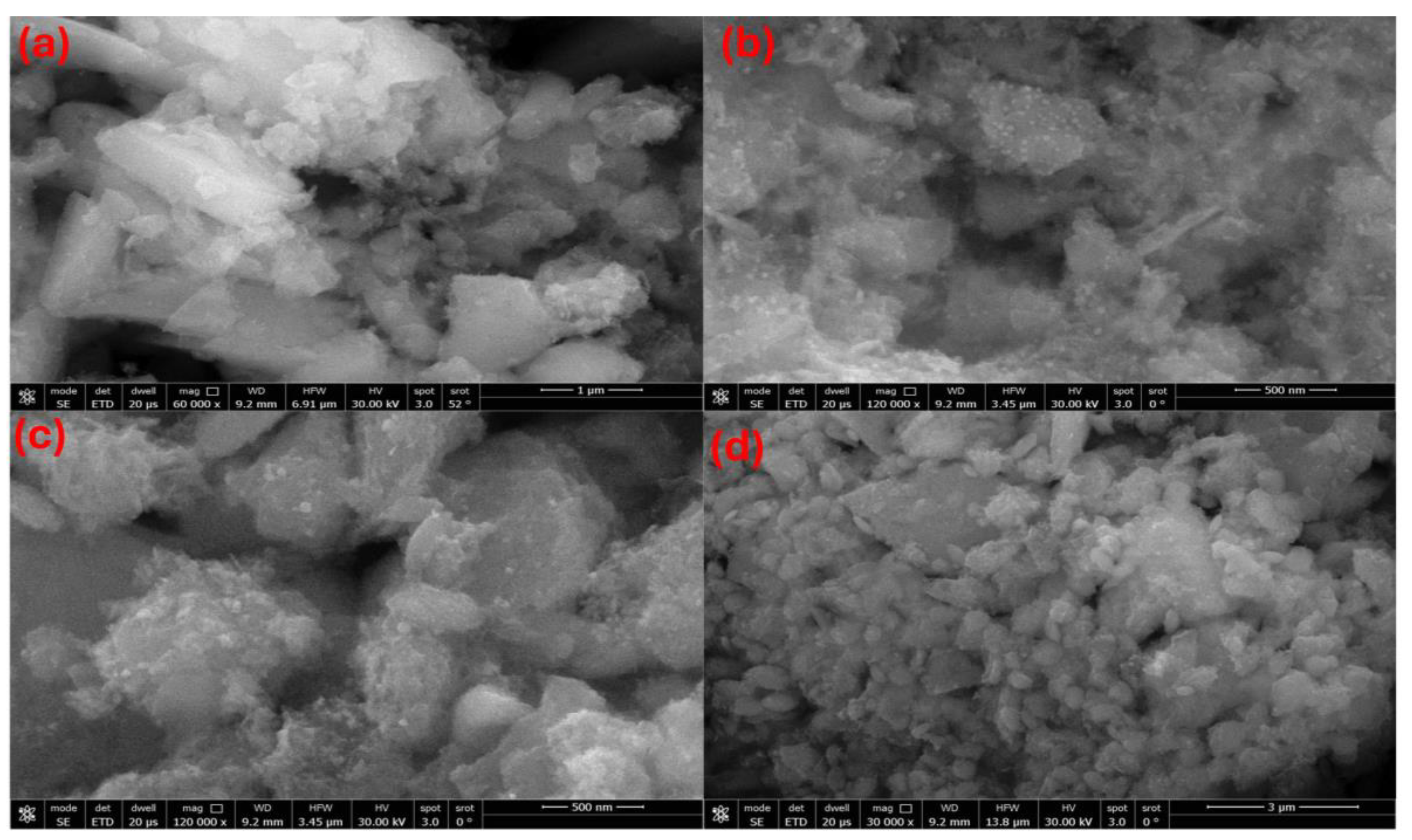
SEM micrographs (Figure 2) show that both composites recorded at a magnification scale of 20 µm possess highly agglomerated, porous morphologies composed of irregular, nanostructured particles. The La/Ag displays a mix of flake-like and plate-shaped structures embedded with brighter silver nanoparticles, while the La/Y/Ag sample exhibits a more granular texture, likely due to the mixed oxide matrix, with silver appearing as dispersed nanoscale dots. The finer particles (AgNPs) are consistent with SEM verified AgNPs distributions in hybrid systems, where nanoparticles appear as discrete, spherical domains [52,53]. Their dispersion within the La and La/Y matrix suggests successful anchoring, a feature critical for enhancing surface reactivity [54].
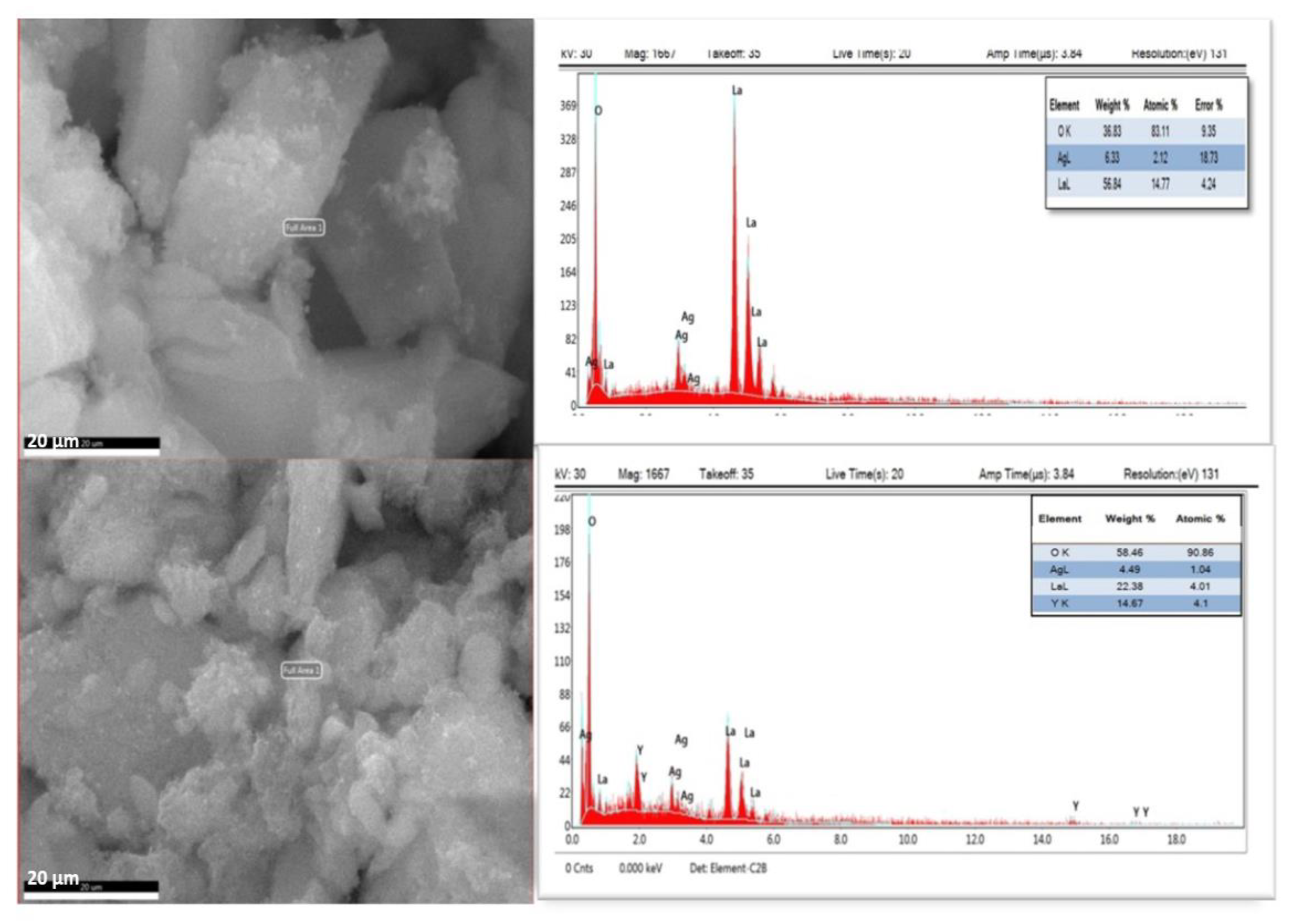
The SEM–EDX analysis (Figure 3) confirms the successful synthesis and structural features of La/Ag and La/Y/Ag nanocomposites. The EDX spectra reveals that both composites contain high levels of oxygen, indicative of predominant oxide phases such as La2O3 [55]. In the La/Ag nanocomposite, the atomic composition is O (84.69%), La (11.72%), and Ag (3.59%), confirming lanthanum as the primary element and silver as a secondary dopant, likely present as well-anchored nanoparticles within the La2O3 matrix. In contrast, the La/Y/Ag nanocomposite shows O (90.86%), La (4.01%), Y (4.10%), and Ag (1.04%) with nearly equal atomic percentages of La and Y suggesting the formation of La/Y/Ag nanocomposite, where yttrium is homogeneously incorporated into the La2O3 lattice [56]. The relatively low but distinct Ag content in both cases supports its presence as surface nanoclusters rather than as a separate crystalline phase. Structural and compositional characteristics confirm the successful formation of multifunctional La based nanocomposites with homogeneously distributed Ag, promising enhanced surface reactivity for catalytic applications.

2.3. XRD Characterization of La/Ag and La/Y/Ag Nanocomposites

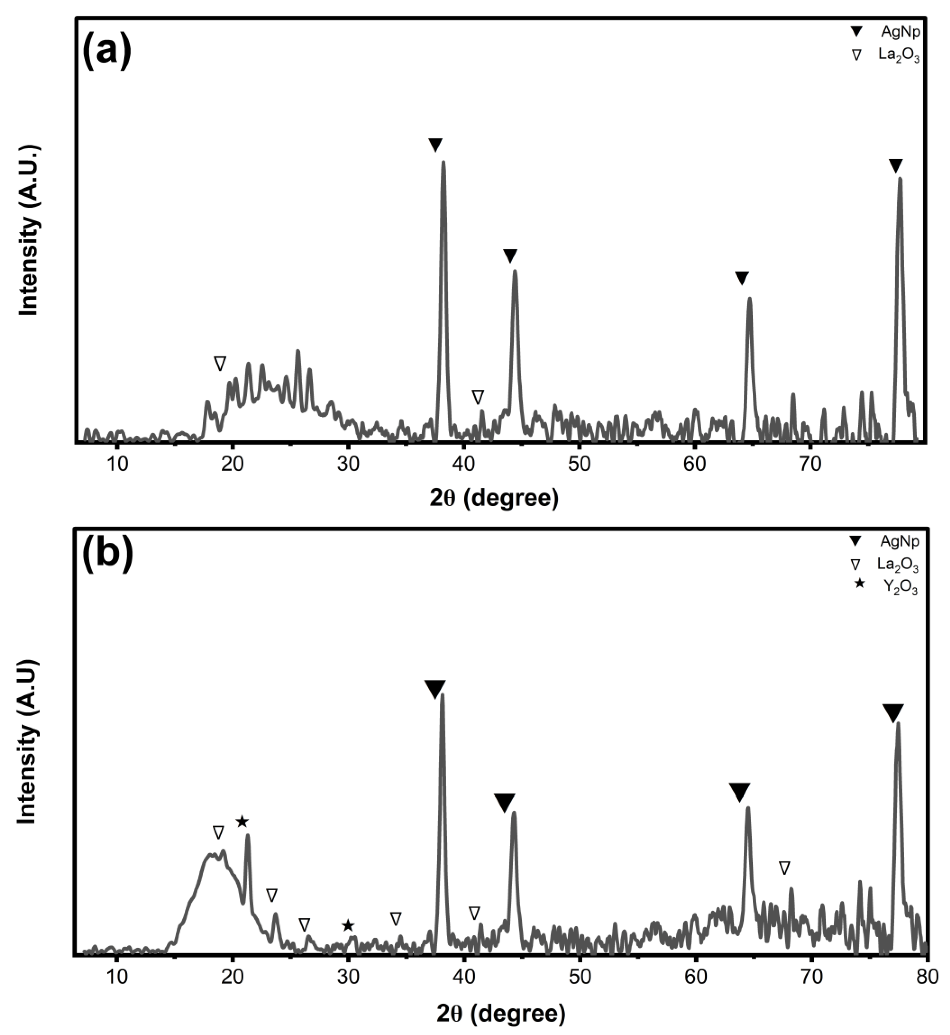
The X-ray diffraction (XRD) patterns of La/Y and La/Y/Ag nanocomposites are shown in Figure 4. The XRD profiles display broad, low-intensity peaks, especially in the 2θ range of 20–30°, indicating a poorly crystalline framework with high defect density. This broadening arises from small crystallite size, lattice disorder, and the presence of organic compounds derived from the plant extract used in synthesis, thereby reducing crystallinity [7,57]. For the La/Y nanocomposite, peaks corresponding to La2O3 (▽) are visible around 20°, 28°, and 55°, while weak Y2O3 (★) peaks appear in the low-angle region (15–30°), though they are poorly resolved. The absence of distinct Y2O3 peaks suggests that Y3+ ions are substitutionally incorporated into the La2O3 lattice, forming a La–Y–O composite with associated lattice distortion and minor peak shifts [58,59]. In the La/Y/Ag nanocomposite, additional sharp peaks at 2θ ≈ 38°, 44°, 64°, and 77°, corresponding to the (111), (200), (220), and (311) planes of fcc Ag nanoparticles (▼), confirm the presence of well-crystallized silver domains (JCPDS Card No. 04-0783) [60].
Functionally, these structural transformations have major implications for catalytic activity. The nanocomposites, particularly La/Y/Ag, show superior performance due to their higher surface area, greater defect density, and better nanoparticle dispersion, all of which stem from their amorphous, and organic-rich structure [57,60,61].

2.4. Application of Prepared Materials

2.4.1. Catalytic Reduction Study

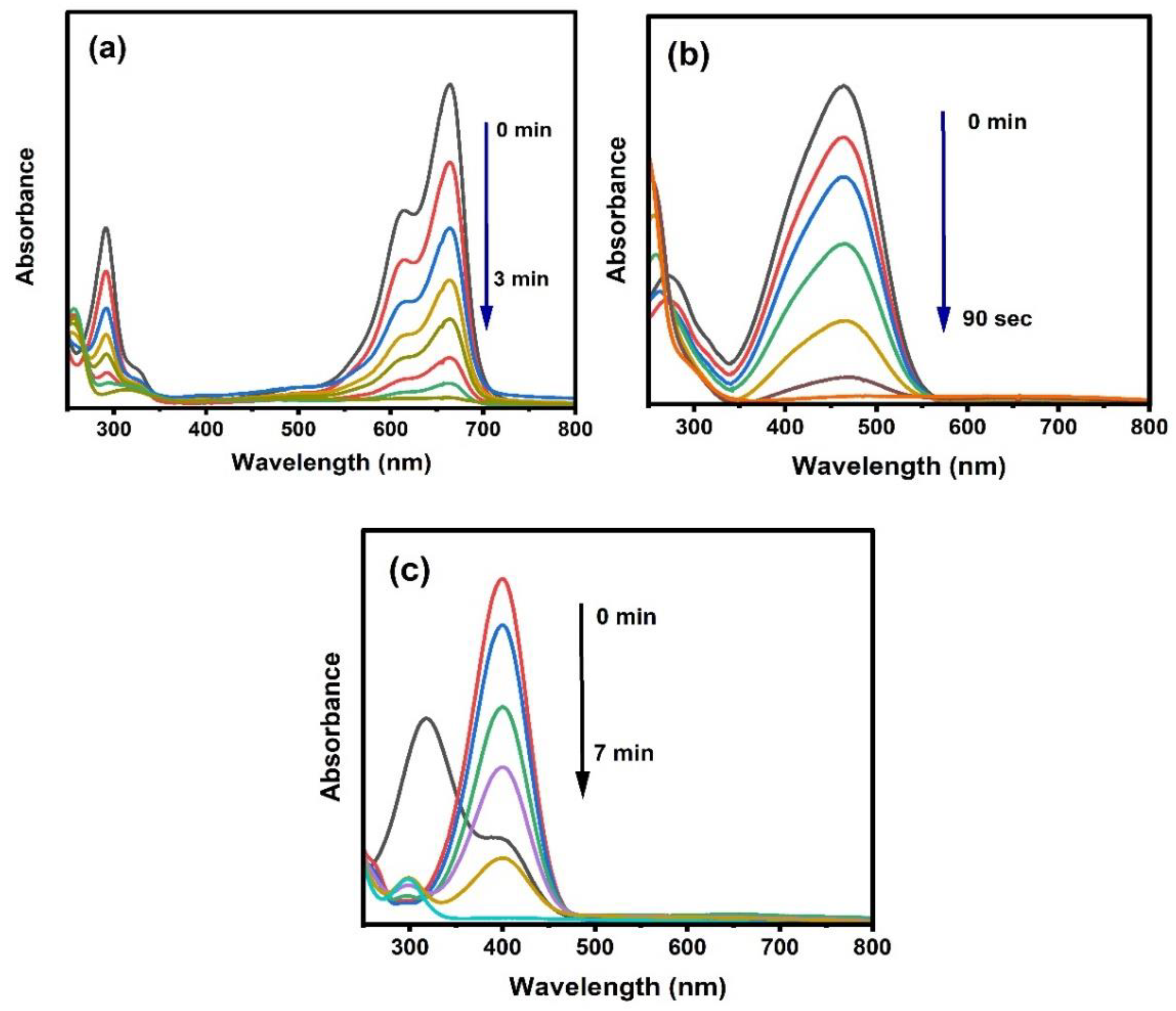
The catalytic activity of La/Ag, Y/Ag and La/Y/Ag were investigated using various dyes, including MB, MO, and AO, as well as 4-NP with NaBH4 serving as the reducing agent. Among these, the La/Ag nanoparticles exhibited better catalytic activity in the reduction of MB (Figure 5) while La/Y/Ag nanoparticles showed better catalytic performance in the reduction of MO (Figure 6).
The catalytic efficiency of La/Ag was tested in the reduction reaction of MB, MO, AO and 4-NP. The performance was monitored using UV-Vis spectrophotometry and visual inspection of the color change. The catalytic reduction of MO and AO was completed within 5 min, 4-NP was reduced in 4 min, while MB showed the highest catalytic reduction efficiency with complete reduction achieved in 3 min under optimal conditions. MB dye was studied under various conditions where different parameters were evaluated, including dye concentrations (0.03 mM, 0.05 mM, and 0.07 mM), catalyst amounts (3 mg, 1.5 mg, and 0.5 mg), and reducing agent (NaBH4) volumes (0.5 mL, 1 mL, and 1.5 mL). The optimal catalytic activity was achieved using 0.07 mM solution of MB which reduced in 3.0 min. MB (0.07 mM) was successfully reduced in 3.0 min by freshly prepared 0.5 mL of 0.1 M of NaBH4 solution with 3 mg of La/Ag. The catalytic reduction activities of La/Ag, Y/Ag, and La/Y/Ag nanocomposites demonstrated clear differences in performance, which attribute to their unique structural and electronic properties. The superior reduction efficiency of La/Ag toward MB can be associated with the effective electron relay facilitated by silver nanoparticles, which act as electron sinks enhancing charge transfer from NaBH4 to dye molecules. Silver well-known surface plasmon resonance effect also contributes to improved catalytic performance by increasing local electron density and generating active sites [62,63,64]. The pseudo–first-order kinetic analysis of the catalytic reduction of various dyes (MB, AO, 4-NP, and MO) using La/Ag nanocatalyst was performed by plotting ln(At/A0) versus time (seconds). The linear relationships confirmed pseudo–first-order behavior under excess NaBH4. The calculated apparent rate constants (k) were 0.02016 s−1 for MB, 0.01224 s−1 for AO, 0.00873 s−1 for 4-NP, and 0.00715 s−1 for MO, with corresponding half-lives of 34.4, 56.6, 79.4, and 96.9 s, respectively. Among the tested dyes, MB exhibited the highest apparent rate constant and the shortest half-life, indicating superior catalytic efficiency of La/Ag toward MB reduction. This enhanced activity can be attributed to the cationic nature of MB, which promotes strong electrostatic interactions with the negatively charged La/Ag surface, facilitating faster electron transfer. In contrast, the comparatively lower rate constants for AO, 4-NP, and MO suggest weaker adsorption and slower electron-transfer kinetics.
The catalytic efficiency of Y/Ag was tested with MB, MO, AO, and 4-NP. MO was reduced within 8 min, MB within 6 min, AO within 20 min, and 4-NP within 8 min. Similar parameters were evaluated, where a dye concentration of 0.07 mM was tested, along with catalyst quantities of 3.0 mg and NaBH4 (0.1 M) of 0.5 mL. The optimal catalytic performance of Y/Ag was achieved using 3 mg of catalyst, 0.5 mL of 0.1M NaBH4 solution as a reducing agent, and 2 mL of the dye at a concentration of 0.07 mM. All constituents were mixed directly in the cuvette during the catalytic reaction.
The catalytic efficiency of La/Y/Ag was also tested with the MB, MO, AO, and 4-NP. In contrast, MO dye showed the most efficient catalytic reduction within 90 s, whereas the MB dye required 3 min, and 4-NP required 7 min. However, AO showed no catalytic reduction. Similar parameters were evaluated as in the study with La/Ag. The examination of similar parameters was conducted in the investigation involving La/Ag nanoparticles, encompassing dye concentrations of 0.03 mM, 0.05 mM, and 0.07 mM, catalyst quantities of 3 mg, 1.5 mg, and 0.5 mg, and volumes of reducing agent (NaBH4) were 0.5 mL, 1 mL, and 1.5 mL. The optimal catalytic performance of the La/Y/Ag nanoparticles with MO was achieved using 3 mg of catalyst, 0.5 mL of 0.1 M NaBH4 solution as a reducing agent, and 2 mL of MO dye at a concentration of 0.07 mM. It is worth noting that all constituents were mixed directly in the cuvette. The incorporation of yttrium into the La2O3 lattice in La/Y/Ag creates lattice distortion and oxygen vacancies, which are crucial for enhanced catalytic activity. Y3+ substitution creates defect sites that improve electron mobility and facilitate adsorption of both reactants and intermediates, thus enable the rapid reduction of MO by La/Y/Ag within 90 s. These observations align with studies demonstrating that mixed rare earth oxides often improve catalytic efficiency via synergistic electronic interactions and increased surface defects that delay electron-hole recombination in photocatalysis [65,66]. Increasing the catalyst dosage and reducing agent concentration consistently improved reduction rates, revealing the importance of abundant active sites and hydride ion availability during catalytic reduction. Conversely, higher dye concentrations prolonged reduction times due to competitive surface site occupation, consistent with pseudo-first-order kinetics in dye reduction reactions [62,67]. The pseudo–first-order rate constants (k) for the catalytic reduction of different dyes using La/Y/Ag were determined from the slopes of ln(At/A0) versus time plots. The calculated k values were 0.04105 s−1 for MO, 0.01183 s−1 for MB, and 0.00643 s−1 for 4-NP, corresponding to half-lives of 16.9, 58.6, and 107.8 s, respectively. Among the tested dyes, MO exhibited the highest apparent rate constant, indicating the most rapid reduction. The superior catalytic activity of La/Y/Ag toward MO can be attributed to favorable adsorption of the anionic dye on La/Y/Ag surface, which promotes efficient electron transfer from the borohydride ions to the dye molecules. These results confirm that La/Y/Ag is a highly active and selective catalyst for the rapid reduction of azo dyes under mild conditions.
Effect of Different Amounts of La/Ag in the Reduction of MB and La/Yt/Ag with MO
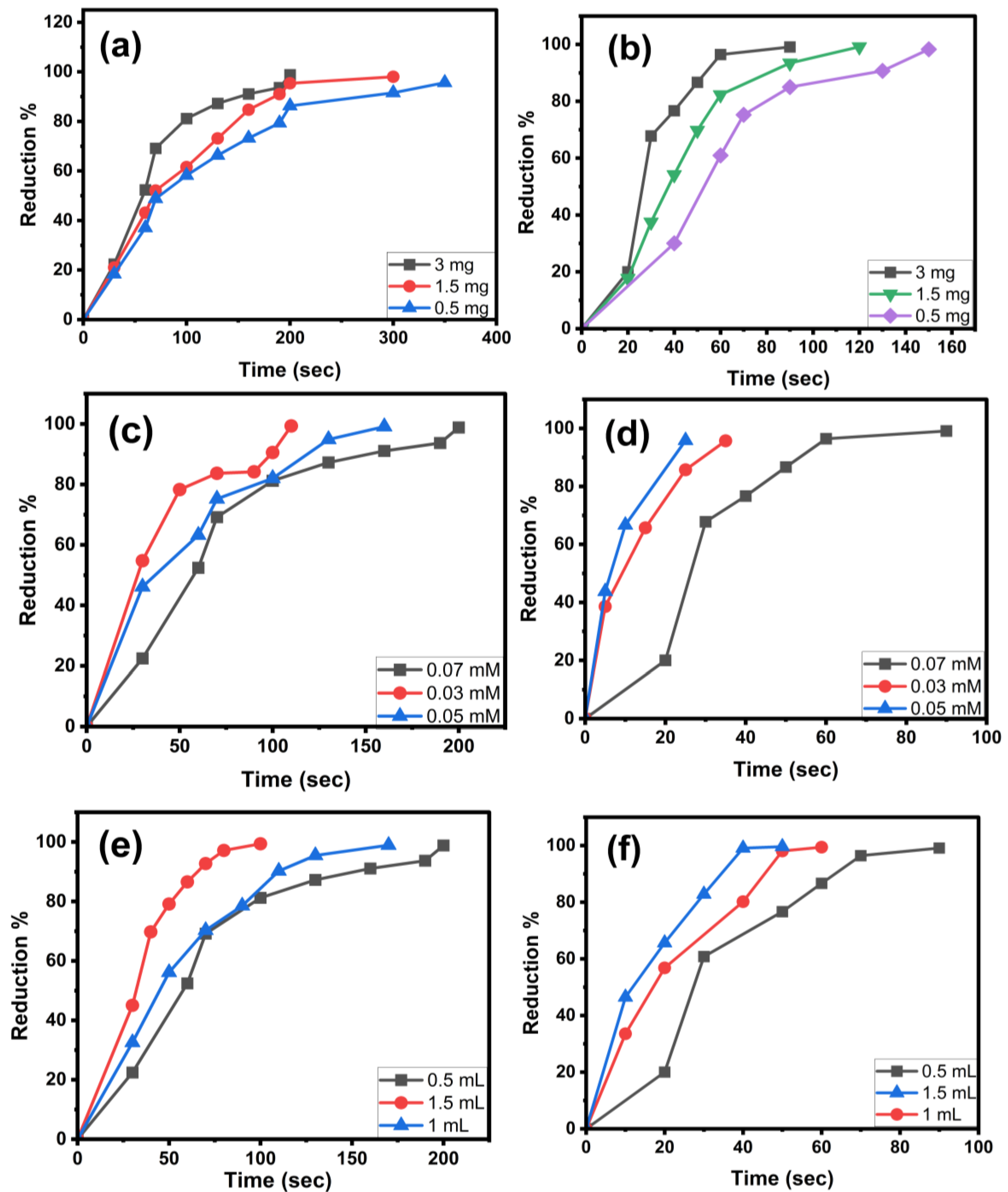
In this study, the catalytic activity was investigated using varying amounts of La/Ag (3 mg, 1.5 mg, and 0.5 mg) while maintaining concentrations of NaBH4 (0.5 mL, 0.1 M) and dye (2 mL, 0.07 mM). Among the tested amounts, the best result was achieved with 3 mg of La/Ag, which successfully reduced the dye in just 3 min. This was followed by 1.5 mg, which reduced the dye by 5 min, and 0.5 mg, which reduced the dye within 7 min (Figure 7a). The reduction percentage was calculated by using the following equation:
Reduction   ( % )   =   C 0 C e C 0   × 100
The catalytic activity of MO was assessed using different amounts of La/Y/Ag: 3 mg, 1.5 mg, and 0.5 mg while keeping the 0.5 mL NaBH4 (0.1 M) and 2 mL (0.07 mM) of MO dye constant. The optimal result was obtained with 3 mg of La/Y/Ag, which reduced the dye in just 90 s. The 1.5 mg sample followed, reducing the dye within 3 min and 20 s, and the 0.5 mg sample took 4 min to achieve the same reduction (Figure 7b).
Effect of Different Concentrations of Dyes
The catalytic reduction activity was examined using different concentrations of MB (0.07 mM, 0.05 mM, and 0.03 mM) while maintaining a constant amount of NaBH4 (0.5 mL) and a fixed amount of the catalyst (3 mg). As the dye concentration decreased, the reduction time improved: 3 min for 0.07 mM, 1 min and 50 s for 0.05 mM, and 1 min for 0.03 mM (Figure 7c).
The catalytic reduction activity was investigated using varying concentrations of MO dye (0.07 mM, 0.05 mM, and 0.03 mM) while maintaining a constant volume of NaBH4 solution (0.5 mL) and a fixed amount (3 mg) of La/Y/Ag catalyst. As the dye concentration decreased, the reduction time increased: 90 s for 0.07 mM, 40 s for 0.05 mM, and just 30 s for 0.03 mM (Figure 7d).
Effect of NaBH4 Concentrations
Additionally, the catalytic reduction activity was investigated using different volumes of NaBH4 (0.5 mL, 1 mL, and 1.5 mL) while maintaining a constant dye concentration (0.07 mM) and a fixed amount of the La/Ag catalyst (3 mg). As the concentration of NaBH4 increased, the reduction time improved: 3 min for 0.5 mL, 2 min and 40 s for 1 mL, and 2 min for 1.5 mL (Figure 7e).
The study explored catalytic reduction activity by adjusting the concentrations of NaBH4 (0.5 mL, 1 mL, and 1.5 mL) while keeping 0.07 mM concentration of dye and using a consistent La/Y/Ag catalyst amount of 3 mg. Results showed that higher NaBH4 concentration led to faster reduction times: 90 s for 0.5 mL, 1 min for 1 mL, and just 50 s for 1.5 mL (Figure 7f).
Recyclability of La/Ag and La/Y/Ag
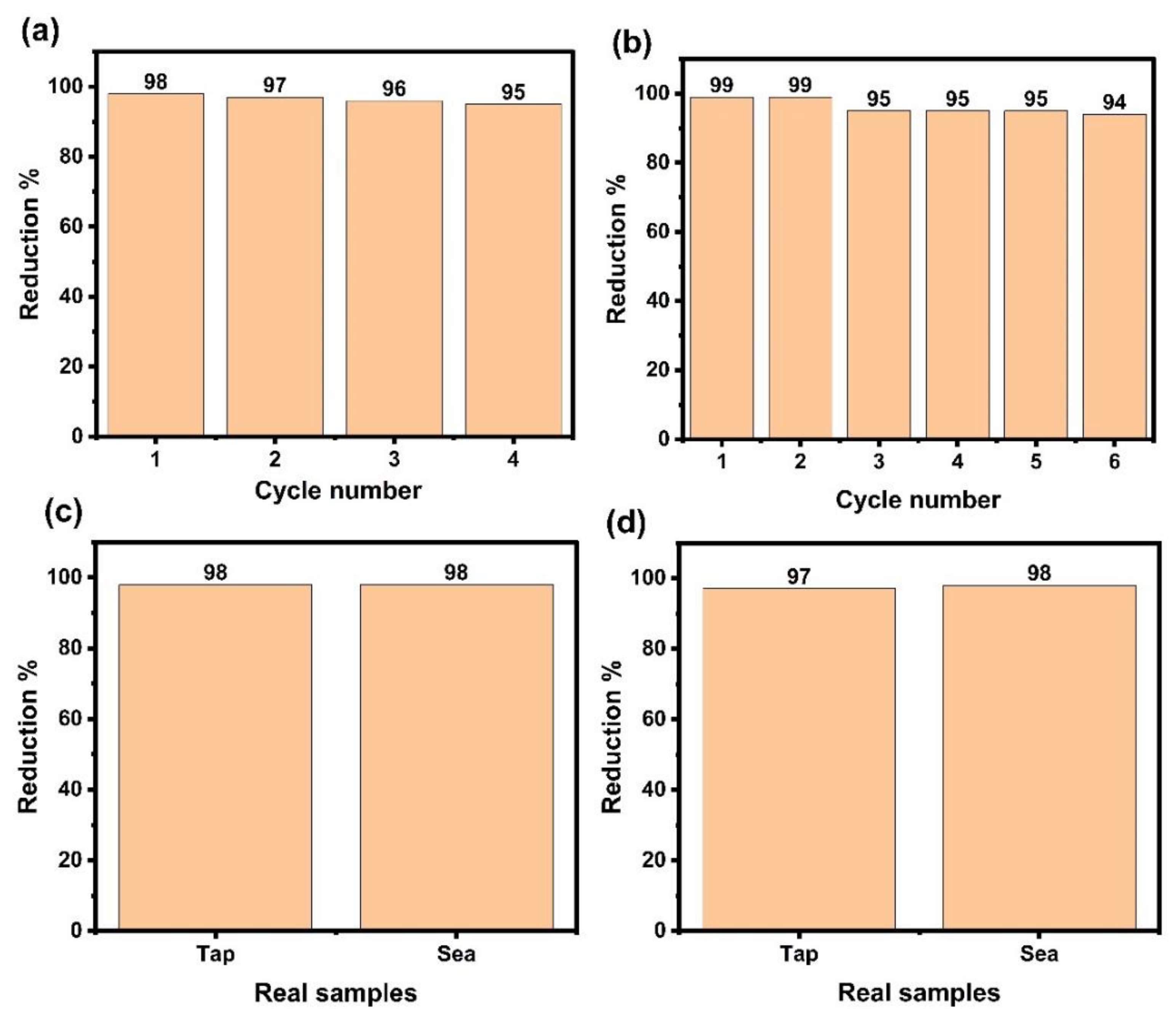
Studying recyclability and reusability is crucial in assessing the regeneration capability of a catalyst before its application on real samples. In this study, the reusability of La/Ag with MB was examined by using 3 mg of La/Ag, (0.5 mL) freshly prepared NaBH4, and 2 mL of (0.07 mM) MB dye. The process was repeated five times using the same 3 mg of catalyst. As shown in the results, the La/Ag catalyst decreased reduction percentage (%) from 98% to 95% over successive cycles. This decline suggests a reduction in the La/Ag catalyst efficiency with each reuse. Therefore, La/Ag can be reused, and its effectiveness slightly decreases after multiple cycles (Figure 8a).
The reusability with MO was examined by using 3 mg of La/Y/Ag, (0.5 mL) freshly prepared NaBH4, and 2 mL of (0.07 mM) MO dye. The process was repeated six times using the same 3 mg of catalyst. As shown in the results, the La/Y/Ag catalyst demonstrated a decrease in reduction percentage (%), decreasing from 99% to 94% over successive cycles. This decline suggests a reduction in the efficiency of the La/Y/Ag catalyst with each reuse. Therefore, La/Y/Ag can be reused, and its effectiveness slightly decreases after multiple cycles (Figure 8b).
The slight decline in catalytic efficiency over successive reuse cycles for La/Ag and La/Y/Ag, is commonly explained by factors like partial aggregation and surface oxidation of silver nanoparticles, which reduce active surface area and electron transfer capability. Additionally, minor catalyst loss during washing and recovery is also a routine contributor to decreased activity across cycles. These phenomena are typical in heterogeneous catalysis and are not indicative of major structural breakdown. Studies confirm that catalysts of this class often retain more than 94–95% of their original activity after multiple cycles, reflecting their high stability for sustainable catalysis [68,69,70].
Catalytic Efficiency of La/Ag and La/Y/Ag in Real Water Samples
In this study, 1 mL of tap water and 0.5 mL of seawater were separately added to 2 mL of MB dye solution (0.07 mM). 0.5 mL of NaBH4 and 3 mg of La/Ag were added to each mixture. All components were thoroughly mixed in a quartz cuvette, and the samples were measured directly using UV-Vis spectroscopy. The findings revealed that the reduction (%) of MB in both the tap water and the seawater samples reached 98%, indicating that the high reduction rate demonstrates the exceptional effectiveness of the current suggested approach by employing La/Ag as a catalyst (Figure 8c).
To investigate the catalytic reduction properties of La/Y/Ag in real samples, separate experiments were conducted using 1 mL of tap water and 0.5 mL of seawater. Each water sample was mixed with 2 mL of a 0.07 mM MO solution. Subsequently, 0.5 mL of NaBH4 and 3 mg of La/Y/Ag were added to each mixture. The components were thoroughly blended in a quartz cuvette, and the samples were analyzed using UV-Vis spectroscopy. The results showed that the reduction of the MO dye in the tap water and seawater samples reached 97% and 98%, respectively. This high reduction rate indicates remarkable effectiveness of La/Y/Ag as a catalyst in the proposed method (Figure 8d). The catalyst’s excellent performance in real water samples further confirms their practical potential. Stability and high efficiency in complex tap and seawater samples demonstrate resistance to ion interference and the robustness of the develop catalysts. Thus green-synthesized Ag-doped rare earth oxides are promising materials for sustainable environmental remediation [71,72,73].

2.4.2. Photocatalytic Degradation Study

The photocatalytic activity of La/Ag, Y/Ag, and La/Y/Ag nanocomposites was evaluated using three organic dyes: MB, MO, and AO. Preliminary tests indicated that AO exhibited the highest degradation efficiency, particularly with the La/Ag catalyst. Therefore, subsequent photocatalytic optimization experiments were focused on AO as the representative dye. Under optimal conditions dye concentration of 0.07 mM and catalyst loading of 20 mg La/Ag achieved complete degradation of AO within 120 min.
For comparison, AO adsorption by the La/Ag was tested till 160 min in dark without UV light irradiation. It is clear from the UV-Vis spectrum (Figure 9a) that the catalyst adsorbs limited amount of AO. The adsorption process takes place in 30 min and there is no further adsorption. Therefore, before each photocatalytic experiment, the reaction was kept in dark for 30 min and then proceeded under UV light. Therefore, the removal of AO is due to adsorption followed by photocatalysis i.e., adsorption assisted photocatalysis.
To determine the effect of experimental parameters, different catalyst amounts (20, 30, and 40 mg), dye concentrations (0.03–0.07 mM), The experiments were conducted at pH 7. The highest degradation efficiency was achieved under this condition, where the degradation rate increases with increase of the catalyst dosage and decrease of dye concentration. All photocatalytic reactions were performed under UV light irradiation, confirming the superior performance of La/Ag in degrading AO dye under the conditions studied.
It is important to note that no reducing agent (NaBH4) was used in these photocatalytic experiments. The degradation of dyes occurred solely through photoexcitation of the catalyst under UV irradiation (Philips UV lamp, λ = 365 nm), which generated electron–hole pairs responsible for oxidative degradation of dye molecules. All photocatalytic experiments were carried out under identical illumination and environmental conditions, confirming the superior performance of the La/Ag catalyst in degrading AO dye.
Effect of Different Amounts of Catalyst on Photocatalytic Degradation of AO
In a separate experiment, the photocatalytic degradation of AO was evaluated under UV light using different amounts of catalyst: 20 mg, 30 mg, and 40 mg. The dye concentration was kept constant i.e. 20 mL of AO (0.07 mM) solution. The time required for degradation decreased as the amount of catalyst increased. Specifically, 40 mg of catalyst completely degraded the dye in 80 min, 30 mg of catalyst achieved full degradation in 100 min, and 20 mg of catalyst took 120 min to fully degrade the dye under optimized conditions (Figure 9).
Effect of Different Concentrations of AO with La/Ag
The photocatalytic activity was investigated using varying concentrations of AO (0.07 mM, 0.05 mM, and 0.03 mM) under UV light irradiation, with a 40 mg of La/Ag. As AO concentration decreased, the reaction time improved: 80 min for 0.07 mM, 40 min for 0.05 mM, and 20 min for 0.03 mM (Figure 10a).
Recyclability of La/Ag with AO
The reusability of La/Ag for the photocatalytic degradation of AO was studied under optimal conditions: AO concentration of 0.07 mM, 40 mg of La/Ag, and exposure to UV light. The experiment was conducted over three cycles using the same La/Ag catalyst. The findings indicated a gradual decline in AO degradation efficiency with each successive cycle. In the first cycle, 97% of the AO was degraded, followed by 94% in the second cycle, and a notable drop to 44% in the third cycle. This decrease suggests a significant loss of La/Ag during washing and recovery and reduction in the efficiency of the La/Ag catalyst after repeated use. Thus, La/Ag can be recycled but need much care during washing and recovery to avoid the loss of La/Ag, maintain its effectiveness after multiple cycles (Figure 10b).
Photocatalytic Efficiency of La/Ag in Real Water Samples
The photocatalytic efficiency of La/Ag was assessed using real water samples. For this purpose, the AO (0.07 mM) was dissolved directly in 20 mL of tap water and 20 mL of seawater instead of distilled water to simulate real environmental conditions. A fixed amount of catalyst (40 mg) was then added to each solution, and the mixtures were exposed to UV light. The degradation of AO was monitored using UV–Vis spectroscopy. The results showed that AO degradation (%) in both tap water and seawater samples reached 98% (Figure 10c). This high degradation efficiency demonstrates the excellent photocatalytic performance of La/Ag in real water samples.
In photocatalytic degradation, La/Ag activity in AO removal is largely attributed to photoinduced electron-hole pair generation facilitated by Ag plasmonic enhancement and La2O3 wide bandgap stability. The photocatalytic mechanism activates reactive oxygen species such as –OH and –O2 radicals, leading to oxidative AO breakdown under UV irradiation. Increasing La/Ag loading enhanced degradation efficiency due to improved light absorption and more active sites, while higher AO concentration reduced degradation rates by impeding light penetration. The observed photocatalyst recyclability decline after multiple cycles can be linked to surface fouling and partial deactivation, typical in heterogeneous photocatalysis, but the initial high degradation percentages reflect commendable durability under operational conditions. Overall, these results validate the synergistic interplay between Ag nanoparticles and rare earth oxide support in improving charge separation, surface adsorption, and redox activity, consistent with prior reports on Ag-doped La2O3 and Y-doped oxide nanomaterials synthesized via plant extracts [64,65,66,67,74].

2.4.3. Antimicrobial Activity Study

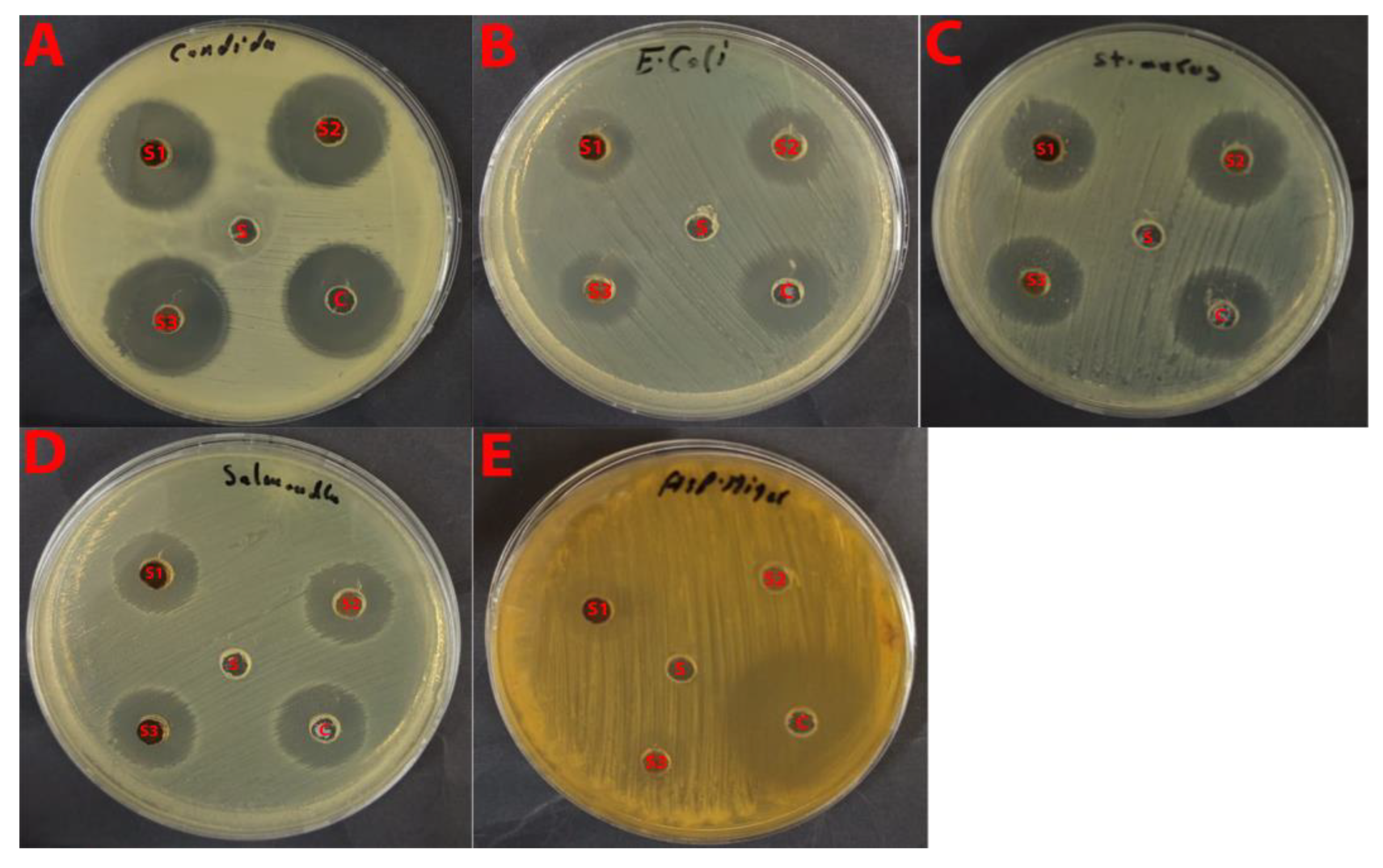
The antimicrobial activity of three nanocomposite samples, La/Y/Ag (S1), Y/Ag (S2), and La/Ag (S3), was evaluated against both bacterial and fungal pathogens using the agar well diffusion method. The study revealed varying degrees of inhibition against different microorganisms, demonstrating the broad-spectrum antimicrobial potential of these nanocomposites.
Antifungal Activity
Against A. niger, all three samples demonstrated significant antifungal activity, with inhibition zones ranging from 18 to 20 mm. Sample S1 showed the highest activity (20 ± 0.5 mm), followed by S3 (19 ± 0.5 mm) and S2 (18 ± 0.5 mm). However, these values were notably lower than the fluconazole control (39 ± 1 mm). The substantial inhibition zones suggest that the silver-containing nanocomposites effectively disrupt fungal growth, possibly through multiple mechanisms including cell membrane disruption and metabolic interference.
For C. albicans (ATCC 10221), the samples showed remarkably consistent activity, with inhibition zones of 20–21 mm. Samples S1 and S3 exhibited identical activity (21 ± 0.5 mm), while S2 showed slightly lower inhibition (20 ± 0.5 mm). The activity against C. albicans was particularly noteworthy, as it approached 70% of the effectiveness of the fluconazole control (30 ± 0.2 mm), suggesting these nanocomposites could be promising candidates for antifungal applications.
Antibacterial Activity
The nanocomposites demonstrated broad-spectrum antibacterial activity against both Gram-positive and Gram-negative bacteria. Against E. coli (ATCC 8739), the inhibition zones ranged from 17 to 19 mm, with S3 showing the highest activity (19 ± 0.5 mm). These results were comparable to the gentamycin control (22 ± 0.2 mm), indicating robust antibacterial efficacy.
For S. typhi (ATCC 6539), the samples showed consistent activity with inhibition zones of 18–20 mm, with S3 again demonstrating the highest activity (20 ± 0.5 mm). The activity was notably strong compared to the gentamycin control (27 ± 0.1 mm), achieving approximately 74% of the control’s effectiveness.
Against S. aureus (ATCC 6538), the nanocomposites showed enhanced activity compared to other bacterial species, with inhibition zones ranging from 19 to 21 mm. Sample S3 exhibited the strongest activity (21 ± 0.5 mm), approaching 78% of the gentamycin control’s effectiveness (27 ± 1 mm).
B. subtilis (ATCC 6633) showed similar susceptibility patterns, with inhibition zones ranging from 18 to 20 mm. Sample S3 again demonstrated the highest activity (20 ± 0.5 mm), showing comparable effectiveness to other tested bacteria.
Comparative Analysis
A notable pattern emerged across all tested microorganisms: Sample S3 (La/Ag) consistently demonstrated the highest antimicrobial activity, followed by S1 (La/Y/Ag), while S2 (Y/Ag) generally showed slightly lower activity. This trend suggests that the lanthanum-silver combination might play a crucial role in enhancing antimicrobial efficacy.
The effectiveness of these nanocomposites can be attributed to several potential mechanisms; The silver component likely contributes to cell membrane disruption and metabolic interference, the rare earth elements (La and Y) may enhance the delivery and stability of the active components, and the carbon matrix possibly provides improved dispersion and sustained release of the active components.
These results suggest that these nanocomposites, particularly S3, could serve as promising broad-spectrum antimicrobial agents.
The consistent activity against both fungal and bacterial pathogens, combined with their relatively stable performance compared to conventional antibiotics, makes them potential candidates for various applications in antimicrobial therapy.
It is also important to note that the DMSO-only control showed no measurable inhibition zones, confirming that the solvent used for sample dispersion did not interfere with the antimicrobial results. This finding is consistent with previous studies reporting that DMSO at concentrations below 2% has negligible antimicrobial activity [75,76,77,78]. Therefore, the inhibition zones observed in this study are attributed exclusively to the intrinsic activity of the La/Ag, Y/Ag, and La/Y/Ag nanocomposites.
Table 1 provides a comprehensive summary of the inhibition zones observed for all samples and controls against the tested microorganisms. The visual observation of the inhibition zones (Figure 11) reveals clear, well-defined zones with complete inhibition, indicating strong and consistent antimicrobial activity.
The results demonstrate that these nanocomposites possess significant antimicrobial potential, with activity levels approaching those of conventional antimicrobial agents in several cases.
In all plates: S1 = La/Y/Ag, S2 = Y/Ag, S3 = La/Ag C = Control (Gentamycin for bacteria and Fluconazole for fungi at 1.0 mg/mL). Wells were loaded with 100 μL of sample solutions (50 mg/mL in DMSO). Clear zones around the wells indicate antimicrobial activity, with larger zones representing stronger activity. Scale bars represent the actual size of the Petri dishes (90 mm diameter).

3. Experimental

3.1. Materials

Methanol (absolute and 99%), lanthanum (III) nitrate hexahydrate (La(NO3)3·6H2O, 99.999% trace metals basis), yttrium (III) nitrate hexahydrate (Y(NO3)3·6H2O 99.8% trace metals basis), silver nitrate (AgNO3, 99.8%), and sodium borohydride (NaBH4, 99.9%) were obtained from Sigma-Aldrich (St. Louis, MO, USA) and used without further purification.

3.2. Preparation Methods

3.2.1. Preparation of the Plant Extract

Catharanthus roseus plant was collected from a local source. After that, it was thoroughly rinsed with distilled water to eliminate any dust, The plant was cut into small pieces and left to dry at room temperature. Approximately 100 g was transferred into 250 mL beakers filled with 500 mL of 30% methanol solution and then subjected to ultrasonic for 60 min at 50 °C, followed by 3 days at room temperature for further extraction. The resulting extracts were filtered using a Buchner funnel to remove particulate matter and obtain clear solutions. Finally, the plant extracts were kept at 4 °C.

3.2.2. Preparation of La/Ag

La nanoparticles were synthesized by mixing 50 mL of lanthanum (III) nitrate hexahydrate solution (0.1 M) with 10 mL of Catharanthus roseus extract, followed by sonication for 60 min. 0.5 M NaOH solution was used to elevate the pH to 9.0, and the mixture was agitated until a yellow suspension formed, indicating metal oxide precipitation. After washing and centrifuging, the resultant precipitate was dried in an oven set at 50 °C. Subsequently, approximately 30 mg of the La based metal oxide (dried powder) was added to 10 mL of silver nitrate solution (0.1 M) stirred overnight at room temperature (25 ± 2 °C) for 12 h. A color transformation from light yellow to brownish designated the development of La/Ag nanoparticles. The mixture was then treated with freshly prepared NaBH4 solution freshly prepared NaBH4 solution (0.01 M, added dropwise at a rate of 1 mL per min, reaction continued for 30 min under constant stirring at 25 °C) to reduce Ag⁺ ions to silver nanoparticles.

3.2.3. Preparation of Y/Ag

The same procedure as for La/Ag was used to prepare Y/Ag nanoparticles. Approximately 30 mg of the Y based metal oxide was dipped in 10 mL of silver nitrate solution (0.1 M) and stirred overnight at room temperature (25 ± 2 °C) for 12 h. Gradual color change was observed, indicating the interaction between the Y based metal oxide and silver ions. The mixture was then treated with a freshly prepared NaBH4 solution (0.01 M, added dropwise at 1 mL per min, maintained under stirring for 30 min at 25 °C) solution to reduce Ag⁺ ions to silver nanoparticles.

3.2.4. Preparation La/Y/Ag

La/Y nanoparticles were synthesized by mixing 50 mL of lanthanum (III) nitrate hexahydrate solution (0.1 M) with 10 mL of yttrium (III) nitrate hexahydrate solution (0.1 M) and 12 mL of Catharanthus roseus extract. The mixed solution was subjected to sonication for 60 min. 0.5 M NaOH solution was used to elevate the pH to 9.0, and the mixture was agitated until a yellow suspension formed, indicating La/Y precipitation. After washing and centrifuging, the resultant precipitate was dried in an oven set at 50 °C. The dried powder was collected and weighed.
Subsequently, 50 mg of the prepared La/Y was dispersed in 10 mL of silver nitrate solution (0.1 M), and the mixture was stirred overnight at room temperature (25 ± 2 °C) for 12 h. A visible color change from pale yellow to gray indicated silver deposition. The mixture was then treated with freshly prepared NaBH4 solution (0.01 M, added dropwise at 1 mL per min, reaction maintained for 30 min at 25 °C) solution to reduce Ag⁺ ions to silver nanoparticles.

3.2.5. Characterization

XRD analysis was performed to identify and quantify the crystalline phases of the synthesized materials. The measurements were conducted using a Bruker D8 Discover diffractometer (Bruker AXS GmbH, Karlsruhe, Germany), equipped with a Cu Kα radiation source (λ = 1.5406 Å). The system operated at 40 kV and 40 mA, and data were collected over a 2θ range of 10–100°, with a step size of 0.02° and a scan speed of 1 s/step. Surface morphology and elemental composition were conducted using a JEOL JSM-7600F field emission scanning electron microscope (JEOL Ltd., Tokyo, Japan), coupled with an Oxford Instruments X-Max 80 mm2 silicon drift detector for EDX analysis. SEM imaging was conducted in secondary electron (SE) mode at 30 kV accelerating voltage, with a working distance of 9.3 mm and spot size of 3.0. High-resolution images were captured at 120,000× magnification. EDX spectra were acquired at a take-off angle of 35°, with a collection time of 100 s, and analyzed using the AZtec software v6.2 (Oxford Instruments Abingdon, Oxfordshire, UK) with ZAF correction for semi-quantitative elemental analysis. Image processing and particle size analysis were performed using v1.53 (National Institutes of Health, Bethesda, MD, USA) and JEOL SCUI v2.x (JEOL Ltd., Tokyo, Japan). Fourier Transform Infrared (FT-IR) Spectroscopy: FT-IR analysis was carried out to identify the functional groups present in the synthesized materials. The spectra were recorded using a PerkinElmer Spectrum 400 FT-IR/Far-IR spectrometer (PerkinElmer Inc., Waltham, MA, USA). The instrument was operated in the range of 4000–400 cm−1 with a resolution of 4 cm−1, and each spectrum was obtained by averaging 32 scans to ensure a high signal-to-noise ratio. Samples were prepared using the KBr pellet method (for the mid-IR region) and directly mounted (for the far-IR region), allowing precise characterization of the vibrational modes associated with the materials.
UV-Visible Spectroscopy (UV-Vis) absorption spectra were recorded using a UV–Vis spectrophotometer (MultiSpec 1501; Shimadzu Corporation, Kyoto, Japan). Data acquisition and analysis are conducted using LabSolutions UV–Vis software version 3.2. Measurements were carried out in the wavelength range of 200–800 nm using quartz cuvettes with a 1 cm path length.

3.2.6. Catalytic Reduction Study

The catalytic activities of La/Ag, Y/Ag, and La/Y/Ag were investigated for the reduction of MB, MO, AO, and 4-NP using NaBH4 as the reducing agent. The catalysts were tested using different amounts of 0.5 mg, 1.5 mg, and 3 mg. Dye solutions (2 mL) with concentrations of 0.03 mM, 0.05 mM, and 0.07 mM were prepared, and the reactions were initiated by adding 0.5, 1.0, or 1.5 mL of freshly prepared 0.1 M NaBH4 solution.
All catalytic reactions were conducted directly in a 1 cm quartz cuvette, which served simultaneously as the reaction vessel and measurement cell. Immediately after adding NaBH4, the cuvette was placed in the UV-Vis spectrophotometer (Shimadzu MultiSpec 1501), The absorbance was measured every 5–10 s until complete decolorization of the dye was observed. The progress of the reaction was monitored by recording the decrease in the characteristic absorption peaks of the dyes (MB ≈ 664 nm, MO ≈ 465 nm, AO ≈ 490 nm, and 4-NP ≈ 400 nm).
Control experiments conducted without the catalyst showed no significant reduction, confirming that the presence of the catalyst is essential for the reaction. Subsequently, the catalytic efficiencies of La/Ag, Y/Ag, and La/Y/Ag were compared under identical conditions to evaluate their relative performance.

3.2.7. Photocatalytic Degradation Study

The photocatalytic performance of La/Ag, Y/Ag, and La/Y/Ag nanocomposites were evaluated under UV irradiation using three organic dyes: MB, MO, and AO. Approximately 20 mL of dye solution (concentration 0.03–0.07 mM) was mixed with 30 mg of the catalyst and stirred in the dark for 30 min to establish adsorption–desorption equilibrium. Following this, the same beaker containing the dye–catalyst mixture was exposed to UV light from a Philips 15 W lamp (λ = 365 nm) positioned 10 cm above the reaction surface in an enclosed photocatalytic chamber to ensure uniform illumination and prevent external light interference. The degradation of the dyes was monitored at regular intervals of 20 min using UV-Vis spectroscopy by measuring the decrease in absorbance at their respective characteristic wavelengths. Among the tested nanocomposites, La/Ag exhibited the highest photocatalytic efficiency, completely degrading AO within 120 min at a dye concentration of 0.07 mM and a catalyst amount of 20 mg. The effects of catalyst dosage (20, 30, and 40 mg), dye concentration (0.03–0.07 mM), and pH (3–9) were systematically investigated. Optimal degradation occurred at pH 7 with a catalyst amount of 40 mg, and the degradation time was observed to decrease with lower dye concentrations.

3.2.8. Antimicrobial Activity

The antimicrobial properties of three nanocomposite materials, La/Ag, Y/Ag, and La/Y/Ag were evaluated using the agar well diffusion method following CLSI (Clinical and Laboratory Standards Institute) guidelines. The study assessed their efficacy against both bacterial and fungal pathogens of clinical significance.
Microbial Strains and Culture Conditions
The antimicrobial activity was tested against six different microorganisms: two fungal species (Aspergillus niger and Candida albicans ATCC 10221) and four bacterial species (Escherichia coli ATCC 8739, Salmonella typhi ATCC 6539, Staphylococcus aureus ATCC 6538, and Bacillus subtilis ATCC 6633). The bacterial strains were cultured on Mueller-Hinton agar medium (pH 7.2–7.4), while the fungal species were grown on appropriate mycological media. All test organisms were maintained at optimal growth conditions and standardized to match 0.5 McFarland standard (approximately 1.5 × 10⁸ CFU/mL).
Sample Preparation
The nanocomposite samples were prepared by dissolving 50 mg of each compound in 1.0 mL of DMSO to achieve a final concentration of 50 mg/mL. DMSO was selected as the solvent due to its ability to dissolve the nanocomposites effectively while maintaining minimal antimicrobial interference. Gentamycin (1.0 mg/mL) served as the positive control for bacterial strains, while fluconazole (1.0 mg/mL) was used as the positive control for fungal species.
Agar Well Diffusion Assay The antimicrobial assessment was conducted using the agar well diffusion method. The microbial suspensions were uniformly spread on the agar surface using sterile cotton swabs. Wells of 6 mm diameter were created in the inoculated agar plates using sterile cork borers. Each well received 100 μL of the respective nanocomposite solution. The plates were allowed to stand for 30 min at room temperature for proper diffusion of the compounds before incubation.
Incubation and Measurement
The inoculated plates were incubated under standardized conditions: bacterial cultures at 37 °C for 24 h, C. albicans at 30 °C for 24–48 h, and A. niger at 28 °C for 48–72 h. Following incubation, the zones of inhibition were measured in millimeters using a calibrated digital caliper. Each assay was performed in triplicate to ensure reproducibility, and the results were expressed as mean ± standard deviation.
Quality Control
To validate the reliability of the results, several quality control measures were implemented. These included verification of the purity of test organisms through microscopic examination and biochemical testing, inclusion of positive controls (Gentamycin for bacteria and fluconazole for fungi), regular monitoring of incubation conditions, standardization of inoculum density using McFarland standards, and testing of culture media sterility and performance. In addition, DMSO-only negative control was tested under identical experimental conditions to verify that the solvent itself exhibited no antimicrobial effect. Wells containing only DMSO (final concentration ≤ 1% v/v) were prepared and tested alongside the nanocomposite samples. The DMSO control showed no inhibition zones against any bacterial or fungal strains, confirming that the solvent served solely as a dispersing medium. This observation is consistent with previous reports indicating that DMSO concentrations below 2% show negligible antimicrobial activity and can be reliably used as solvent controls in antimicrobial assays [75,76].

4. Conclusions

This study successfully demonstrated the synthesis of La/Ag, Y/Ag, and La/Y/Ag nanocomposites using plant extracts, as confirmed by multiple analytical techniques. FTIR analysis revealed the presence of phytochemical functional groups in the plant extract, which played a crucial role in reducing and stabilizing the nanoparticles. The interaction between these functional groups and the metal precursors was evidenced by shifts and new peaks in the FTIR spectra, confirming the formation of the desired nanomaterials. XRD analysis further validated the successful synthesis and crystalline nature of the nanoparticles. The XRD patterns displayed characteristic diffraction peaks corresponding to La2O3, Y2O3, and Ag, indicating the presence of well-crystallized phases. The absence of extraneous peaks confirmed the high purity of the synthesized nanocomposites, while the peak broadening suggested the formation of nanoparticles with small crystallite sizes. SEM coupled with EDX provided insights into the morphology and elemental composition of the synthesized materials. EDX analysis confirmed the elemental composition, showing strong signals for lanthanum, yttrium, silver, and oxygen, with no significant impurities detected. This confirmed the successful incorporation of all intended elements and the effectiveness of the synthesis route. The synthesized nanocomposites exhibited excellent catalytic and photocatalytic activities, efficiently reducing and degrading various dyes, including MB, MO, AO, and 4-NP, under optimized conditions. La/Ag showed the fastest catalytic reduction of MB, while La/Y/Ag achieved rapid reduction of MO, with both materials maintaining high efficiency in real water samples. The catalysts also demonstrated moderate recyclability, although their efficiency gradually declined with repeated use. Additionally, the nanocomposites displayed significant antimicrobial activity against both bacterial and fungal pathogens, highlighting their broad-spectrum potential.

Author Contributions

Conceptualization, S.B.K.; methodology, S.M.A. and E.A.A.; validation, E.M.B. and K.A.; formal analysis, S.M.A. and S.B.K.; investigation, E.M.B. and K.A.; resources, S.B.K.; data curation, S.M.A.; writing—original draft preparation, S.M.A.; writing—review and editing, E.A.A., E.M.B., K.A. and S.B.K.; supervision, S.B.K.; funding acquisition, S.B.K. All authors have read and agreed to the published version of the manuscript.

Funding

This project was funded by KAU Endowment (WAQF) at king Abdulaziz University, Jeddah, Saudi Arabia.

Data Availability Statement

The data presented in this study are available on request from the corresponding author.

Acknowledgments

The project was funded by KAU Endowment (WAQF) at king Abdulaziz University, Jeddah, Saudi Arabia. The authors, therefore, acknowledge with thanks WAQF and the Deanship of Scientific Research (DSR) for technical and financial support.

Conflicts of Interest

The authors declare no conflict of interest.

References

  1. Bayda, S.; Adeel, M.; Tuccinardi, T.; Cordani, M.; Rizzolio, F. The history of nanoscience and nanotechnology: From chemical–physical applications to nanomedicine. Molecules 2019, 25, 112. [Google Scholar] [CrossRef]
  2. Nandre, V.; Jadhav, Y.; Das, D.K.; Ahire, R.; Ghosh, S.; Jadkar, S.; Kodam, K.; Waghmode, S. Nanomaterials for sensors: Synthesis and applications. In Advanced Nanomaterials for Point of Care Diagnosis and Therapy; Elsevier: Amsterdam, The Netherlands, 2022; pp. 477–492. [Google Scholar] [CrossRef]
  3. Ghorbanpour, M.; Fahimirad, S. Plant nanobionics a novel approach to overcome the environmental challenges. In Medicinal Plants and Environmental Challenges; Springer International Publishing: Cham, Switzerland, 2017; pp. 247–257. [Google Scholar] [CrossRef]
  4. Ahmed, S.; Ahmad, M.; Swami, B.L.; Ikram, S. A review on plants extract mediated synthesis of silver nanoparticles for antimicrobial applications: A green expertise. J. Adv. Res. 2016, 7, 17–28. [Google Scholar] [CrossRef]
  5. Beyene, H.D.; Werkneh, A.A.; Bezabh, H.K.; Ambaye, T.G. Synthesis paradigm and applications of silver nanoparticles (AgNPs), a review. Sustain. Mater. Technol. 2017, 13, 18–23. [Google Scholar] [CrossRef]
  6. Mekuye, B.; Abera, B. Nanomaterials: An overview of synthesis, classification, characterization, and applications. Nano Sel. 2023, 4, 486–501. [Google Scholar] [CrossRef]
  7. Iravani, S. Green synthesis of metal nanoparticles using plants. Green. Chem. 2011, 13, 2638. [Google Scholar] [CrossRef]
  8. Patel, V.; Berthold, D.; Puranik, P.; Gantar, M. Screening of cyanobacteria and microalgae for their ability to synthesize silver nanoparticles with antibacterial activity. Biotechnol. Rep. 2015, 5, 112–119. [Google Scholar] [CrossRef] [PubMed]
  9. Munir, H.; Bilal, M.; Mulla, S.I.; Khan, H.A.; Iqbal, H.M.N. Plant-mediated green synthesis of nanoparticles. In Advances in Green Synthesis: Avenues and Sustainability; Springer International Publishing: Cham, Switzerland, 2021; pp. 75–89. [Google Scholar] [CrossRef]
  10. Nayak, S.; Ghugare, P.; Vaidhun, B. Green-synthesis of silver nanoparticles by Hygrophila auriculata extract: Innovative technique and comprehensive evaluation. Indian J. Pharm. Educ. Res. 2021, 55, s510–s517. [Google Scholar] [CrossRef]
  11. Anandam, S.; Indira, K. Antibacterial activity of biosynthesized ferric oxide nanoparticles using Cissus Quadrangularis extract. Res. Sq. 2021. [Google Scholar] [CrossRef]
  12. Alshammari, S.O.; Mahmoud, S.Y.; Farrag, E.S. Synthesis of green copper nanoparticles using medicinal plant Krameria sp. root extract and its applications. Molecules 2023, 28, 4629. [Google Scholar] [CrossRef]
  13. Kerketta, A.; Sahu, B. Chapter 13 Nanomaterials synthesis from medicinal plant extract. In Phytochemicals in Medicinal Plants; De Gruyter: Berlin, Germany, 2023; pp. 291–314. [Google Scholar] [CrossRef]
  14. Bagheri, A.R.; Aramesh, N.; Hasnain, M.S.; Nayak, A.K.; Varma, R.S. Greener fabrication of metal nanoparticles using plant materials: A review. Chem. Phys. Impact 2023, 7, 100255. [Google Scholar] [CrossRef]
  15. Kalaiselvi, A.; Roopan, S.M.; Madhumitha, G.; Ramalingam, C.; Elango, G. Synthesis and characterization of palladium nanoparticles using Catharanthus roseus leaf extract and its application in the photo-catalytic degradation. Spectrochim. Acta A Mol. Biomol. Spectrosc. 2015, 135, 116–119. [Google Scholar] [CrossRef] [PubMed]
  16. Roy, A.; Singh, V.; Sharma, S.; Ali, D.; Azad, A.K.; Kumar, G.; Bin Emran, T. Antibacterial and dye degradation activity of green synthesized iron nanoparticles. J. Nanomater. 2022, 2022, 3636481. [Google Scholar] [CrossRef]
  17. Fouad, A.; Hegazy, A.E.; Azab, E.; Khojah, E.; Kapiel, T. Boosting of antioxidants and alkaloids in Catharanthus roseus suspension cultures using silver nanoparticles with expression of CrMPK3 and STR Genes. Plants 2021, 10, 2202. [Google Scholar] [CrossRef]
  18. Xian, T.; Meng, Q.; Gao, F.; Hu, M.; Wang, X. Functionalization of luminescent lanthanide complexes for biomedical applications. Coord. Chem. Rev. 2023, 474, 214866. [Google Scholar] [CrossRef]
  19. Balaram, V. Rare earth elements: A review of applications, occurrence, exploration, analysis, recycling, and environmental impact. Geosci. Front. 2019, 10, 1285–1303. [Google Scholar] [CrossRef]
  20. Gimaev, R.; Komlev, A.; Davydov, A.; Kovalev, B.; Zverev, V. Magnetic and electronic properties of heavy lanthanides (Gd, Tb, Dy, Er, Ho, Tm). Crystals 2021, 11, 82. [Google Scholar] [CrossRef]
  21. Hou, W.; Wang, G.; Wu, X.; Sun, S.; Zhao, C.; Liu, W.-S.; Pan, F. Lanthanide clusters as highly efficient catalysts regarding carbon dioxide activation. New J. Chem. 2020, 44, 5019–5022. [Google Scholar] [CrossRef]
  22. Patil, A.S.; Patil, A.V.; Dighavkar, C.G.; Adole, V.A.; Tupe, U.J. Synthesis techniques and applications of rare earth metal oxides semiconductors: A review. Chem. Phys. Lett. 2022, 796, 139555. [Google Scholar] [CrossRef]
  23. Wiglusz, R.J.; Grzyb, T.; Lis, S.; Strek, W. Hydrothermal preparation and photoluminescent properties of MgAl2O4: Eu3+ spinel nanocrystals. J. Lumin. 2010, 130, 434–441. [Google Scholar] [CrossRef]
  24. Liang, X.; Kuang, S.; Li, Y. Solvothermal synthesis and luminescence of nearly monodisperse LnVO4 nanoparticles. J. Mater. Res. 2011, 26, 1168–1173. [Google Scholar] [CrossRef]
  25. Rudnev, A.V. Electrodeposition of lanthanides from ionic liquids and deep eutectic solvents. Russ. Chem. Rev. 2020, 89, 1463–1482. [Google Scholar] [CrossRef]
  26. Govindasamy, R.; Govindarasu, M.; Alharthi, S.S.; Mani, P.; Bernaurdshaw, N.; Gomathi, T.; Ansari, M.A.; Alomary, M.N.; Atwah, B.; Malik, M.S.; et al. Sustainable green synthesis of yttrium oxide (Y2O3) nanoparticles using Lantana camara leaf extracts: Physicochemical characterization, photocatalytic degradation, antibacterial, and anticancer potency. Nanomaterials 2022, 12, 2393. [Google Scholar] [CrossRef]
  27. Rashmi, B.N.; Harlapur, S.F.; Gurushantha, K.; Ravikumar, C.; Kumar, M.; Santosh, M.; Kumar, V.; Kumar, A.; Azad, A.K.; Murthy, H.A. Facile green synthesis of lanthanum oxide nanoparticles using Centella Asiatica and Tridax plants: Photocatalytic, electrochemical sensor and antimicrobial studies. Appl. Surf. Sci. Adv. 2022, 7, 100210. [Google Scholar] [CrossRef]
  28. Cotruvo, J.A. The chemistry of lanthanides in biology: Recent discoveries, emerging principles, and technological applications. ACS Cent. Sci. 2019, 5, 1496–1506. [Google Scholar] [CrossRef]
  29. Teo, R.D.; Termini, J.; Gray, H.B. Lanthanides: Applications in cancer diagnosis and therapy. J. Med. Chem. 2016, 59, 6012–6024. [Google Scholar] [CrossRef]
  30. Dong, H.; Du, S.-R.; Zheng, X.-Y.; Lyu, G.-M.; Sun, L.-D.; Li, L.-D.; Zhang, P.-Z.; Zhang, C.; Yan, C.-H. Lanthanide nanoparticles: From design toward bioimaging and therapy. Chem. Rev. 2015, 115, 10725–10815. [Google Scholar] [CrossRef]
  31. Sarkar, D.; Ganguli, S.; Samanta, T.; Mahalingam, V. Design of lanthanide-doped colloidal nanocrystals: Applications as phosphors, sensors, and photocatalysts. Langmuir 2019, 35, 6211–6230. [Google Scholar] [CrossRef]
  32. Zhang, L.; Long, S.; Jiao, H.; Liu, Z.; Zhang, P.; Lei, A.; Gong, W.; Pei, X. Cellulose derived Pd nano-catalyst for efficient catalysis. RSC Adv. 2022, 12, 18676–18684. [Google Scholar] [CrossRef] [PubMed]
  33. Nishu, S.K. Smart and innovative nanotechnology applications for water purification. Hybrid. Adv. 2023, 3, 100044. [Google Scholar] [CrossRef]
  34. Yadav, M.; Gupta, R.; Sharma, R.K. Green and sustainable pathways for wastewater purification. In Advances in Water Purification Techniques; Elsevier: Amsterdam, The Netherlands, 2019; pp. 355–383. [Google Scholar] [CrossRef]
  35. Vrchota, J.; Pech, M.; Rolínek, L.; Bednář, J. Sustainability outcomes of green processes in relation to industry 4.0 in manufacturing: Systematic review. Sustainability 2020, 12, 5968. [Google Scholar] [CrossRef]
  36. Jara, Y.S.; Mekiso, T.T.; Washe, A.P. Highly efficient catalytic degradation of organic dyes using iron nanoparticles synthesized with Vernonia Amygdalina leaf extract. Sci. Rep. 2024, 14, 6997. [Google Scholar] [CrossRef]
  37. Blanckenberg, A.; Malgas-Enus, R. Raspberry-like gold-decorated silica (SSx–AMPS–Au) nanoparticles for the reductive discoloration of dyes. SN Appl. Sci. 2019, 1, 787. [Google Scholar] [CrossRef]
  38. Agha, M.K.; Maatouk, B.; Mhanna, R.; El-Dakdouki, M.H. Catalytic degradation efficacy of silver nanoparticles fabricated using Actinidia deliciosa peel extract. J. Nanomater. 2024, 2024, 8813109. [Google Scholar] [CrossRef]
  39. Muthu, K.; Thangapushbam, V.; Rama, P.; Sivakami, S.; Jothika, M. Green synthesis, characterization and photocatalytic reduction of Congo red using silver nanoparticles from Millingtonia hortensis leaf extract. Res. Sq. 2023. [Google Scholar] [CrossRef]
  40. Ismail, M.; Khan, M.I.; Khan, M.A.; Akhtar, K.; Asiri, A.M.; Khan, S.B. Plant-supported silver nanoparticles: Efficient, economically viable and easily recoverable catalyst for the reduction of organic pollutants. Appl. Organomet. Chem. 2019, 33, e4971. [Google Scholar] [CrossRef]
  41. Srivastava, V.; Choubey, A.K. Synthesis of nanostructured silver particles using Citrus limetta peel extract for catalytic degradation of azo dyes through electron relay effect. Adv. Nat. Sci. Nanosci. Nanotechnol. 2019, 10, 45015. [Google Scholar] [CrossRef]
  42. Bhakya, S.; Muthukrishnan, S.; Sukumaran, M.; Muthukumar, M.; Kumar, S.T.; Rao, M.V. Catalytic degradation of organic dyes using synthesized silver nanoparticles: A green approach. J. Bioremediat. Biodegrad. 2015, 6, 1. [Google Scholar] [CrossRef]
  43. Rajasekar, R.; Thanasamy, R.; Samuel, M.; Edison, T.N.J.I.; Raman, N. Eco-friendly green synthesis of silver nanoparticles using Luffa acutangula: Synthesis, characterisation and catalytic degradation of methylene blue and malachite green dyes. Int. J. Environ. Anal. Chem. 2024, 104, 2255–2267. [Google Scholar] [CrossRef]
  44. Velidandi, A.; Sarvepalli, M.; Gandam, P.K.; Pabbathi, N.P.P.; Baadhe, R.R. Characterization, catalytic, and recyclability studies of nano-sized spherical palladium particles synthesized using aqueous poly-extract (turmeric, neem, and tulasi). Environ. Res. 2023, 228, 115821. [Google Scholar] [CrossRef]
  45. Anasdass, J.R.; Kannaiyan, P.; Gopinath, S.C.B. Biosynthesis of zerovalent iron nanoparticles for catalytic reduction of 4-nitrophenol and decoloration of textile dyes. Biotechnol. Appl. Biochem. 2022, 69, 2780–2793. [Google Scholar] [CrossRef] [PubMed]
  46. Kulkarni, S.; Jadhav, M.; Raikar, P.; Raikar, S.; Raikar, U. Core–shell novel composite metal nanoparticles for hydrogenation and dye degradation applications. Ind. Eng. Chem. Res. 2019, 58, 3630–3639. [Google Scholar] [CrossRef]
  47. Dobrucka, R. Biofabrication of Au–Pt nanoparticles using Asarum europaeum extract and evaluation of their activity in degradation of organic dyes. J. Inorg. Organomet. Polym. Mater. 2018, 28, 1953–1961. [Google Scholar] [CrossRef]
  48. Malik, M.A.; Alshehri, A.A.; Abomuti, M.A.; Danish, E.Y.; Patel, R. Bioengineered Matricaria recutita extract-assisted palladium nanoparticles for the Congo red dye degradation and catalytic reduction of 4-nitrophenol to 4-aminophenol. Toxics 2021, 9, 103. [Google Scholar] [CrossRef] [PubMed]
  49. Prabhu, M.S.A.; Sheela, G.E.; Mohammad, A.M.; Hegazy, R.A.; Florence, S.S.; Sarojini, V. Biogenic synthesis of silver, gold, and palladium nanoparticles using moringa oleifera seeds: Exploring photocatalytic, catalytic, and antimicrobial activities. Adv. Nat. Sci. Nanosci. Nanotechnol. 2024, 15, 15007. [Google Scholar] [CrossRef]
  50. Wang, X.; Hou, C.; Qiu, W.; Ke, Y.; Xu, Q.; Liu, X.Y.; Lin, Y. Protein-directed synthesis of bifunctional adsorbent-catalytic hemin-graphene nanosheets for highly efficient removal of dye pollutants via synergistic adsorption and degradation. ACS Appl. Mater. Interfaces 2017, 9, 684–692. [Google Scholar] [CrossRef]
  51. Zhang, W.; Zhou, T.; Qiu, J. Interfacial engineering of BiVO4 immobilized on sodium alginate aerogels enable synergistic photocatalytic-peroxymonosulfate degradation of rhodamine B. Polymers 2025, 17, 2204. [Google Scholar] [CrossRef]
  52. Quintero-Quiroz, C.; Botero, L.E.; Zárate-Triviño, D.; Acevedo-Yepes, N.; Saldarriaga Escobar, J.; Pérez, V.Z.; Javier Cruz Riano, L. Synthesis and characterization of a silver nanoparticle-containing polymer composite with antimicrobial abilities for application in prosthetic and orthotic devices. Biomater Res 2020, 24, 13. [Google Scholar] [CrossRef]
  53. Assis, M.; Robeldo, T.; Foggi, C.C.; Kubo, A.M.; Mínguez-Vega, G.; Condoncillo, E.; Beltran-Mir, H.; Torres-Mendieta, R.; Andrés, J.; Oliva, M.; et al. Ag nanoparticles/α-Ag2WO4 composite formed by electron beam and femtosecond irradiation as potent antifungal and antitumor agents. Sci. Rep. 2019, 9, 9927. [Google Scholar] [CrossRef]
  54. Lamoth, M.; Plodinec, M.; Scharfenberg, L.; Wrabetz, S.; Girgsdies, F.; Jones, T.E.; Rosowski, F.; Horn, R.; Schlögl, R.; Frei, E. Supported Ag nanoparticles and clusters for CO oxidation: Size effects and influence of the silver–oxygen interactions. ACS Appl. Nano Mater. 2019, 2, 2909–2920. [Google Scholar] [CrossRef]
  55. Kim, S.J.; Han, W.K.; Kang, S.G.; Han, M.S.; Cheong, Y.H. Formation of lanthanum hydroxide and oxide via precipitation. Solid. State Phenom. 2008, 135, 23–26. [Google Scholar] [CrossRef]
  56. Fabrichnaya, O.; Savinykh, G.; Schreiber, G.; Seifert, H.J. Experimental study of phase relations in the ZrO2–La2O3–Y2O3 system. Int. J. Mater. Res. 2009, 100, 1521–1528. [Google Scholar] [CrossRef]
  57. Singh, J.; Dutta, T.; Kim, K.-H.; Rawat, M.; Samddar, P.; Kumar, P. ‘Green’ synthesis of metals and their oxide nanoparticles: Applications for environmental remediation. J. Nanobiotechnol. 2018, 16, 84. [Google Scholar] [CrossRef] [PubMed]
  58. Yang, J.; Han, Y.; Luo, J.; Leifer, K.; Strømme, M.; Welch, K. Synthesis and characterization of amorphous magnesium carbonate nanoparticles. Mater. Chem. Phys. 2019, 224, 301–307. [Google Scholar] [CrossRef]
  59. Zhang, X.; Wang, Y.; Cheng, F.; Zheng, Z.; Du, Y. Ultrathin lanthanide oxides nanomaterials: Synthesis, properties and applications. Sci. Bull. 2016, 61, 1422–1434. [Google Scholar] [CrossRef]
  60. Vadivel, S.; Madkour, M.; Rajendran, S.; Sengottaiyan, C. Effective degradation of aqueous bisphenol-A using novel Ag2C2O4/Ag@GNS photocatalyst under visible light. Int. J. Hydrogen Energy 2023, 48, 6510–6520. [Google Scholar] [CrossRef]
  61. Maou, A.; Gouitaa, N.; Lamcharfi, T.D.; Kandri, N.I. Effect of calcination temperature on the stability of the perovskite materials—Study of structural and morphological properties. Ecol. Eng. Environ. Technol. 2023, 24, 1–8. [Google Scholar] [CrossRef]
  62. Ravi, G.; Sarasija, M.; Ayodhya, D.; Kumari, L.S.; Ashok, D. Facile synthesis, characterization and enhanced catalytic reduction of 4-nitrophenol using NaBH4 by undoped and Sm3+, Gd3+, Hf3+ doped La2O3 nanoparticles. Nano Converg. 2019, 6, 12. [Google Scholar] [CrossRef]
  63. Zhang, L.; Zhu, S.; Li, R.; Deng, W.; Hong, C.; Liu, D.; Guo, L. Ag-doped δ-MnO2 nanosheets as robust catalysts for toluene combustion. ACS Appl. Nano Mater. 2020, 3, 11869–11880. [Google Scholar] [CrossRef]
  64. Saad, S.K.M.; Umar, A.A.; Umar, M.I.A.; Tomitori, M.; Rahman, M.Y.A.; Salleh, M.M.; Oyama, M. Two-dimensional, hierarchical Ag-doped TiO2 nanocatalysts: Effect of the metal oxidation state on the photocatalytic properties. ACS Omega 2018, 3, 2579–2587. [Google Scholar] [CrossRef]
  65. Rahul, S.; George, A.; Jayakumar, G. Unraveling the photocatalytic performance of La2O3 nanoparticles for the degradation of six organic dyes. Langmuir 2025, 41, 16378–16390. [Google Scholar] [CrossRef]
  66. Ayub, A.; Ikram, M.; Haider, A.; Shahzadi, I.; Ul-Hamid, A.; Shahzadi, A.; Algaradah, M.M.; Fouda, A.M.; Nabgan, W.; Imran, M. Enhanced industrial dye degradation and antibacterial activity supported by the molecular docking study of yttrium and carbon sphere-doped lanthanum oxide nanostructures. ACS Omega 2023, 8, 37564–37572. [Google Scholar] [CrossRef]
  67. Rooshde, M.S.; Abdullah, W.R.W.; Ibrahim, N.F.; Ghazali, M.S.M.; Nik, W.M.N.W. Photocatalytic efficiency of lanthanide-doped zinc oxide for degradation of methylene blue dye. J. Sustain. Sci. Manag. 2018, 13, 153–162. [Google Scholar]
  68. Wazir, W.; Ahmad, Z.; Zulfiqar, S.; Cochran, E.W.; Mubarik, S.; Kousar, T.; Somaily, H.; Shim, J.-J.; Alsalmah, H.A.; Aadil, M. Synergistic effects of noble metal doping and nanoengineering on boosting the azo dye mineralization activity of nickel oxide. Ceram. Int. 2023, 49, 38026–38035. [Google Scholar] [CrossRef]
  69. Ershov, V.A.; Ershov, B.G. Oxidative dissolution and the aggregation of silver nanoparticles in drinking and natural waters: The influence of the medium on the process development. Toxics 2024, 12, 757. [Google Scholar] [CrossRef] [PubMed]
  70. Jin, Q.; Ma, L.; Zhou, W.; Shen, Y.; Fernandez-Delgado, O.; Li, X. Smart paper transformer: New insight for enhanced catalytic efficiency and reusability of noble metal nanocatalysts. Chem. Sci. 2020, 11, 2915–2925. [Google Scholar] [CrossRef]
  71. Dhiman, P.; Rana, G.; Kumar, A.; Dawi, E.A.; Sharma, G. Rare earth doped ZnO nanoparticles as spintronics and photo catalyst for degradation of pollutants. Molecules 2023, 28, 2838. [Google Scholar] [CrossRef]
  72. Isacfranklin, M.; Ameen, F.; Ravi, G.; Yuvakkumar, R.; Hong, S.; Velauthapillai, D.; AlKahtani, M.D.; Thambidurai, M.; Dang, C. Y2O3 nanorods for cytotoxicity evaluation. Ceram. Int. 2020, 46, 20553–20557. [Google Scholar] [CrossRef]
  73. Singh, B. Lanthanide-based catalysts for electrochemical water splitting: Unraveling the interplay of structure, properties, and performance. Chem. Commun. 2025, 61, 9753–9779. [Google Scholar] [CrossRef] [PubMed]
  74. Prabhavathy, S.; Arivuoli, D. Visible light-induced Silver and Lanthanum co-doped BiVO4 nanoparticles for photocatalytic dye degradation of organic pollutants. Inorg. Chem. Commun. 2022, 141, 109483. [Google Scholar] [CrossRef]
  75. Zhang, C.-L.; Qiu, X.-Y.; Liu, S.-J. Synthesis, crystal structure and antimicrobial activity of a linear trinuclear nickel(II) complex with schiff base ligand. Acta Chim. Slov. 2019, 66, 484–489. [Google Scholar] [CrossRef]
  76. Weber, J.T. (Ed.) Antimicrobial Resistance; Karger: Basel, Switzerland, 2010; Volume 6. [Google Scholar] [CrossRef]
  77. Ansel, H.C.; Norred, W.P.; Roth, I.L. Antimicrobial activity of dimethyl sulfoxide against Escherichia coli, Pseudomonas aeruginosa, and Bacillus megaterium. J. Pharm. Sci. 1969, 58, 836–839. [Google Scholar] [CrossRef] [PubMed]
  78. Kirkwood, Z.; Millar, B.; Downey, D.; Moore, J. Antimicrobial effect of dimethyl sulfoxide and N, N-dimethylformamide on Mycobacterium abscessus: Implications for antimicrobial susceptibility testing. Int. J. Mycobacteriol. 2018, 7, 134. [Google Scholar] [CrossRef] [PubMed]
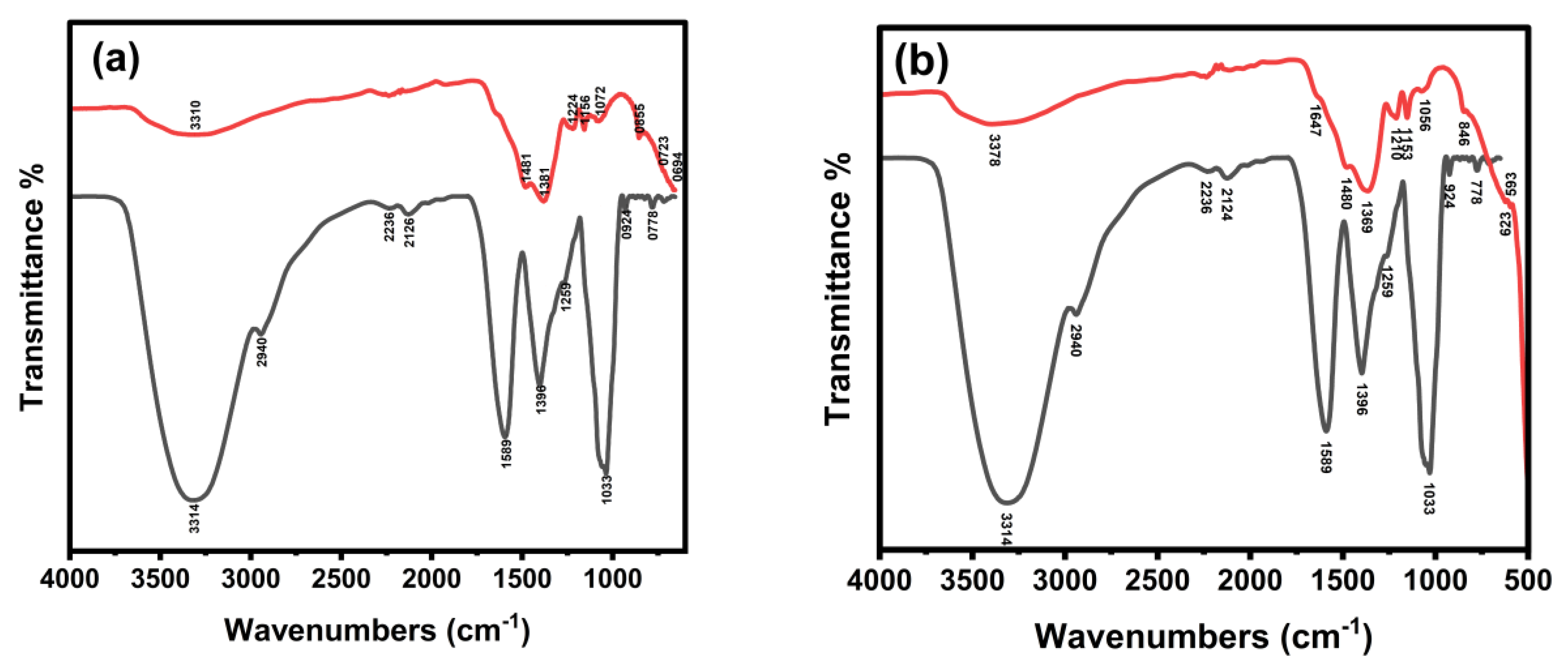
Figure 1. FTIR spectra comparing Catharanthus roseus plant extract (black) with synthesized La/Ag (a) and La/Y/Ag (b) nanocomposites.
Catalysts 15 01001 g001
Figure 2. SEM images of La/Ag (a,b) and La/Y/Ag (c,d) nanocomposites.
Catalysts 15 01001 g002
Figure 3. EDX characterization of La/Ag and La/Y/Ag nanocomposites.
Catalysts 15 01001 g003
Figure 4. XRD characterization of La/Ag (a) and La/Y/Ag (b) nanocomposites.
Catalysts 15 01001 g004
Figure 5. Reduction of MB (a), MO (b), AO (c) and 4-NP (d) in the presence of La/Ag.
Catalysts 15 01001 g005
Figure 6. Reduction of MB (a), MO (b), and 4-NP (c) in the presence of La/Y/Ag.
Catalysts 15 01001 g006
Figure 7. Effect of various amounts of (a) La/Ag in the reduction of MB and (b) La/Y/Ag in the reduction of MO. Effect of different concentrations of (c) MB and (d) MO on reduction activity. Effect of varying concentrations of NaBH4 on (e) MB and (f) MO reduction.
Catalysts 15 01001 g007
Figure 8. Recyclability of La/Ag with MB (a) and La/Y/Ag with MO (b). Catalytic efficiency of La/Ag (c) and La/Yt/Ag (d) in real samples.
Catalysts 15 01001 g008
Figure 9. Effect of Different Amounts of La/Ag on photocatalytic degradation of AO, 20 mg (a), 30 mg (b), 40 mg (c), and degradation (%) of AO by different amounts of La/Ag (d).
Catalysts 15 01001 g009
Figure 10. Effect of different concentrations of AO using La/Ag (a). Recyclability of La/Ag with AO (b). Photocatalytic degradation of AO in real water samples using La/Ag (c), and adsorption of AO in the dark for 160 min (d).
Catalysts 15 01001 g010
Figure 11. Antimicrobial activity of synthesized nanocomposites against various pathogenic microorganisms using agar well diffusion method. Antifungal activity against Candida albicans showing clear zones of inhibition around wells containing samples S1, S2, S3, and control C (A). Antibacterial activity against Escherichia coli (ATCC 8739) demonstrates inhibition zones for different nanocomposite samples (B). Antibacterial activity against Staphylococcus aureus (ATCC 6538) showing effectiveness of the tested compounds (C). Antibacterial activity against Salmonella typhi (ATCC 6539) indicating zones of growth inhibition (D). Antifungal activity against Aspergillus niger displayed on nutrient agar medium with characteristic yellow background (E).
Catalysts 15 01001 g011
Table 1. Antimicrobial activity of synthesized nanocomposites against various pathogens.
Control(La/Y/Ag) (Y/Ag)(La/Ag) Microorganism
39 ± 119 ± 0.518 ± 0.520 ± 0.5A. niger
30 ± 0.221 ± 0.520 ± 0.521 ± 0.5Candida albicans (ATCC 10221)
22 ± 0.219 ± 0.517 ± 0.518 ± 0.5E. coli (ATCC 8739)
27 ± 0.120 ± 0.518 ± 0.519 ± 0.5Salmonella typhi (ATCC 6539)
27 ± 121 ± 0.519 ± 0.520 ± 0.5Staph. aureus (ATCC 6538)
27 ± 120 ± 0.518 ± 0.519 ± 0.5Bacillus subtilis (ATCC 6633)
Disclaimer/Publisher’s Note: The statements, opinions and data contained in all publications are solely those of the individual author(s) and contributor(s) and not of MDPI and/or the editor(s). MDPI and/or the editor(s) disclaim responsibility for any injury to people or property resulting from any ideas, methods, instructions or products referred to in the content.