Article

Structural Distortion of g-C3N4 Induced by N-Defects for Enhanced Photocatalytic Hydrogen Evolution

1
Engineering Technology Research Center of Henan Province for Solar Catalysis, College of Chemistry and Pharmaceutical Engineering, Nanyang Normal University, Nanyang 473061, China
2
Key Laboratory of Inorganic Nonmetallic Crystalline and Energy Conversion Materials, College of Materials and Chemical Engineering, China Three Gorges University, Yichang 443002, China
*
Authors to whom correspondence should be addressed.
Catalysts 2022, 12(12), 1496; https://doi.org/10.3390/catal12121496
Submission received: 16 October 2022 / Revised: 11 November 2022 / Accepted: 16 November 2022 / Published: 23 November 2022
(This article belongs to the Section Catalysis for Sustainable Energy)

Abstract

:
Hydrogen evolution by photocatalytic technology has been one of the most promising and attractive solutions, and can harvest and convert the abundant solar energy into green, renewable hydrogen energy. As a new kind of photocatalytic material, graphitic carbon nitride (g-C3N4) has drawn much attention in photocataluytic H2 production due to its visible light response, ease of preparation and good stability. For a higher photocatalyic performance, N defects were introduced in to the traditional g-C3N4 in this work. The existence of N defects was proved by adequate material characterization. Significantly, a new absorption region at around 500 nm of N-deficient g-C3N4 appeared, revealing the exciting n-π* transition of lone pair electrons. The photocatalytic H2 production performance of N-deficient g-C3N4 was increased by 5.8 times. The enhanced photocatalytic performance of N-deficient g-C3N4 was attributed to the enhanced visible light absorption, as well as the promoted separation of photo-generated carries and increased specific surface area.

1. Introduction

The solar photocatalytic decomposition of water to produce hydrogen has become a research hotspot to deal with environmental pollution and the energy crisis. At present, the photocatalytic efficiency of traditional photocatalysts such as TiO2 and ZnO is still not ideal due to their low solar utilization [1,2]. The development of new kinds of semiconductor photocatalytic materials with a visible light response has been a research hotspot. In recent years, g-C3N4 has been widely used in photocatalysis due to its visible light response, low cost and high stability [3]. However, due to the limited absorption of visible light and fast recombination of carriers, the photocatalytic activity of g-C3N4 is very low. To overcome these shortcomings, numerous chemical modification approaches have been conducted [4]. Among various strategies to improve photocatalytic activity, such as defects’ engineering, doping [5,6,7], heat treatment [8], heterojunction constructing [9], carbon material modifying [10,11] and copolycondensation [12], constructing structural defects has been proved to be an efficient way to improve photocatalytic performance [13], as it can not only regulate the electronic structure of semiconductor, but also act as the active site of the photocatalytic reaction [14,15,16].
For traditional planar-constructed g-C3N4, only intrinsic π-π* electronic transitions can be excited by light, while n-π* transitions of N-lone pair electrons are forbidden [17], and the close relationship between the band structure and the structural distortion has been identified by both experiments and theoretical calculations [18]. In our previous works, non-planar g-C3N4 was prepared by atomic or multiatomic group grafting, which effectively activated the n-π* electronic transition and exhibited a new absorption of around 500 nm, showing enhanced photocatalytic performance at around 500 nm wavelength. [19]. Therefore, it is worth studying whether N defects have similar effects, as this will induce the structural distortion.
Here, N-deficient g-C3N4 was prepared by calcining the mixture of melamine and ammonium acetate. During the heating process, ammonium acetate firstly decomposed to release NH3, leaving CH3COOH around melamine. Then, acidic CH3COOH combined with the alkaline melamine by occupying some amino groups. Finally, CH3COOH decomposed, leaving the N-deficient g-C3N4. N defects destroy the planar structure of traditional g-C3N4, resulting in a distorted non-planar structure of the N-deficient g-C3N4. XPS and EDS spectra, combined with element composition analysis results and EPR spectra, confirmed the introduction of N defects. Significantly, a new absorption region of around 500 nm of N-deficient g-C3N4 appeared, revealing the exciting n-π* transition of N-lone pair electrons caused by the structural distortion. The photocatalytic H2 production performance of N-deficient g-C3N4 was enhanced by 5.8 times. In addition, N-deficient g-C3N4 exhibited a photocatalytic rate of 5.93 μmol/h for H2 production at 500 nm single-wavelength illumination, while no H2 was detected over g-C3N4 at 500 nm. Therefore, the light absorption at 500 nm, assigned to the n-π* electronic transition induced by N defects, is of value to improve the photocatalytic hydrogen evolution of g-C3N4. In addition to the enhanced light absorption, the promoted separation of photo-generated carriers and the increased specific surface area play important roles in enhancing photocatalytic performance for H2 production over N-deficient g-C3N4.

2. Experimental Procedures

2.1. Chemical Reagents

All chemical reagents in analytical grade were purchased from Aladdin Chemical Co., Ltd. (Shanghai, China) and used as received without any further purification. Deionized water with resistivity ≥ 18 MΩ cm was used in the experiments where specified.

2.2. Synthesis of Photocatalysts

A total of 2.0 g of melamine and 4.0 g of ammonium acetate were mixed by thoroughly grounding and heated in the muffle furnace at 550 °C for 4 h using a heating rate of 3 °C/min. The calcined sample was named ND-g-C3N4. As a comparison, g-C3N4 was prepared by direct calcination of melamine under the same conditions.

2.3. Characterization

X-ray diffraction (XRD) for crystallographic structure analysis was conducted on a D8 Advance X-ray diffractometer from Bruker, Karlsruhe, Germany at room temperature. A Fourier transform infrared (FTIR) spectrometer (NICOLET 5700 FT-IR Spectrometer, Thermo Co., Waltham, MA, USA) was used for molecular structure and functional group analysis. The specific surface area was acquired at 77 K by an automated gas absorption system. An X-ray photoelectron spectrometer (XPS) (Thermo ESCALAB 250XI, USA) was used for chemical composition analysis. Field emission scanning electron microscopy (SEM-Merlin, Zeiss, Oberkochen, Germany) was used for morphology observation. Electron paramagnetic resonance (EPR) spectra were acquired by a JESFA200 spectrometer from Germany. The Brunauer–Emmett–Teller (BET) specific surface area and pore size were tested at 77 k using a Quantachrome Autosorb-IQ from USA (Boynton Beach, FL) automated gas adsorption system. The UV-vis absorption spectra of the samples were acquired using a ultraviolet-visible spectrometer (Shimadz, Kyoto, Japan). Photoluminescence (PL) spectra were obtained using a FLS980 fluorescence spectrometer (Edinburgh instrument, Edinburgh, UK) with an excitation wavelength of 310 nm at room temperature. Surface photovoltage (SPV) was tested by self-assembled surface photovoltaic testing equipment.

2.4. Photoelectrochemical Measurements

The electrochemical properties of the prepared samples were revealed by a CHI660E electrochemical workstation. For the preparation of a working electrode, 50 mg sample was firstly dispersed into 10 mL ethanol with 5 mg ethyl cellulose. Then, the dispersed solution was coated uniformly onto a fluorine-doped tin oxide (FTO) conductive glass and dried at 120 °C. The analyses were performed in a conventional three-electrode quartz cell with Pt and Ag/AgCl served as the counter-electrode and reference electrode, respectively. The photocurrent response of the electrodes was measured at + 0.5 V versus an Ag/AgCl electrode in 0.50 mol/L Na2SO4 aqueous solution, irradiated by a 300 W xenon lamp (PLS-300/300UV) with a 420 nm cut-off filter. The integrated visible-light intensity was approximately 150 mW/cm2. Electrochemical impedance spectroscopy (EIS) plots tests were conducted at the corresponding open-circuit potentials over frequencies ranging from 100 kHz to 10 mHz in the dark using a 0.10 mol/L K3[Fe(CN)6]/ K4[Fe(CN)6] aqueous solution as the electrolyte.

2.5. Photocatalytic H2 Evolution

A total of 0.05 g of the sample was uniformly dispersed by ultrasound in a solution of 90 mL water and 10 mL triethanolamine (sacrificial reagent); then, 3 mL of H2PtCl6·6H2O aqueous solution was added. Under light conditions, H2PtCl6 was reduced to Pt (3%) loaded on the surface of the sample as a cocatalyst. The reactor was then vacuuming before illumination to remove all air. A 300 W Xenon lamp with a 420 nm cut-off filter was used to illuminate the reactor. H2 produced in the reactor was analyzed using a gas chromatograph equipped with a TCD detector.

2.6. Computational Details

The density functional theory (DFT) calculations were carried out by using the Gaussian 09 program (64-bit Gaussian 16W for Windows Multiprocessor, USA). The B3LYP functionals were employed and the 6–31G(d) basis set was used for all the atoms in the DFT calculations. A 2 × 2 supercell of monolayer g-C3N4 and N-deficient g-C3N4 were created, and the vacuum layer utilized in this work was set as 20 Å to avoid the effect of mirror interaction. The structural optimizations were performed without any symmetry constraints.

3. Results and Discussion

A schematic diagram of the generative process of ND-g-C3N4 is displayed in Figure 1. During the heating process, ammonium acetate first decomposed to release NH3, leaving CH3COOH around melamine. Then, acidic CH3COOH was combined with the alkaline melamine by occupying some amino groups. Finally, CH3COOH decomposed, leaving the N-deficient g-C3N4. N defects destroy the planar structure of traditional g-C3N4, resulting in a distorted, non-planar structure of ND-g-C3N4.
Figure 2A shows the XRD patterns of the prepared traditional g-C3N4 and N-deficient sample ND-g-C3N4, The two characteristic diffraction peaks, located at 27.4 and 13.1°, correspond to the (002) and (100) crystal faces of g-C3N4, which is consistent with the original report of g-C3N4 [20]. Both peaks show almost no changes in position and intensity, indicating the consistency of the chemical composition and crystal structure [21]. Figure 2B shows the FTIR spectra of the two samples, both of which show several apparent peaks in the range of 1200-1700 cm−1, proving the existence of C-N heterocycles [22]. The peak at 807 cm−1 is from the bending vibration of C6N7 ring [23]. The bands at 3000–3500 cm−1 come from the stretching vibration of N-H at the edge of the molecular skeleton of g-C3N4, which is formed due to the incomplete polycondensation of melamine [24]. FTIR spectra results show that the molecular structures of the two samples are basically the same, indicating that ammonium acetate was completely decomposed during calcination, and no oxygen-containing groups were doped into the molecular skeleton of g-C3N4. X-ray photoelectron spectra were recorded to disclose more information about the chemical compositions of g-C3N4 and ND-g-C3N4. The survey spectra in Figure 2C demonstrate that both g-C3N4 and ND-g-C3N4 are mainly composed of C and N elements. In Figure 2D, the O 1s peak located at 531.8 eV is identified as the O element in H2O molecules absorbed on the surface of the samples. Figure 2E is the high-resolution C 1s spectra of g-C3N4 and ND-g-C3N4. The C 1s peak at 284.1 eV for both g-C3N4 and ND-g-C3N4 is ascribed to the signal C–C bonds of absorbed graphitic carbon used as the calibration peak. The peaks located at 288.3 and 288.1 eV for g-C3N4 and ND-g-C3N4, respectively, are attributed to the sp2-hybridized carbon in the N-containing aromatic ring (N-C=N). The peak at 286.2 eV is identified as the OH group in H2O molecules absorbed on the surface of the samples. This peak is negligible for ND-g-C3N4, consistent with the reduced intensity of the O 1s peak in Figure 2D, demonstrating its reduced adsorption of H2O molecules. Figure 2F shows the high-resolution N 1s spectra of g-C3N4 and ND-g-C3N4. Four peaks at 398.5, 399.6, 401.1, and 404.4 eV of g-C3N4, are attributed to the sp2 hybridized aromatic nitrogen in the triazine units (C-N=C groups), the bridging N atoms in N-(C)3 or H-N-(C)2 groups, quaternary N of amino groups (N-H) and π electron excitations in g-C3N4 heterocycles, respectively. The position of these four peaks of ND-g-C3N4 is basically the same with g-C3N4. However, the percentage of the peak around 398.2 eV increased from 51.59% to 65.04%, whereas the percentage of the peak around 399.5 eV decreased from 37.13% to 24.08% for ND-g-C3N4 compared with g-C3N4. The loss of the bridging N atoms implies the introduction of N defects in ND-g-C3N4. In addition, the N/C atom ratio of g-C3N4 and ND-g-C3N4 is 1.518 and 1.455, respectively, calculated based on the XPS spectra, which was consistent with the results of elemental analysis in Table 1. Both illustrate the existence of N defects in ND-g-C3N4.
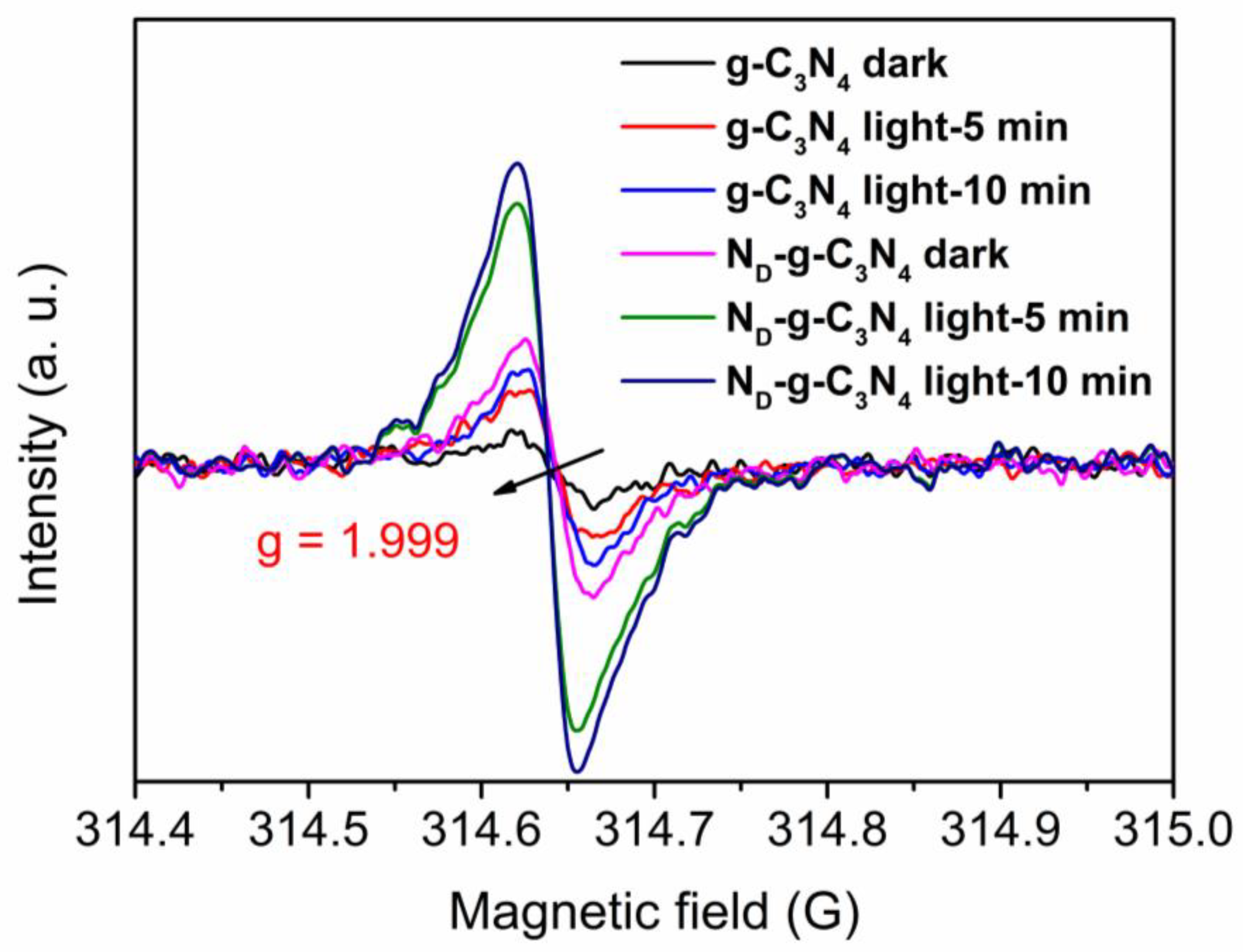
The generation of unpaired e- and h+ in the samples was studied using electron spin resonance (EPR). As shown in Figure 3, a Lorentz line centered at a g value of 1.999 was attributed to unpaired electrons in the aromatic ring of C-atoms and π-bonded nanoclusters on the surface of the samples [25]. The EPR signal of ND-g-C3N4 was enhanced in the dark compared with g-C3N4. In addition, it should be noted that the intensity of the EPR peak of both samples increased under the illumination condition, and the longer the illumination time, the stronger the peak. A higher EPR intensity indicates greater delocalization and mobility of spin, leading to a higher charge carrier density [26].
As shown in Figure 4, SEM was used to observe and analyze the morphology of the samples. Figure 4A,B show the SEM image of g-C3N4. It could be seen that g-C3N4 is a massive structure formed by the accumulation of uneven particles, and a typical lamellar graphite-like structure can be seen. Figure 4C,D are the SEM images of ND-g-C3N4. It is obvious that ND-g-C3N4 is composed of fluffy nanosheets, and the graphite-like lamellar structure becomes more obvious. The EDS spectra of the two samples demonstrate that they both are composed of C and N elements. The N/C atom ratio of g-C3N4 and ND-g-C3N4 is 1.70 and 1.39, respectively, confirming the N defects in ND-g-C3N4.
N2 adsorption/desorption isotherms of g-C3N4 and ND-C3N4 are recorded and the result is shown in Figure 5. The samples both displayed a type IV isotherm according to Figure 5A, and ND-g-C3N4 exhibited remarkably enhanced N2 adsorption capacity compared with g-C3N4. The measured specific surface areas of g-C3N4 and ND-C3N4 were 14.0 and 23.3 m2/g, respectively. Significantly, the enhanced specific surface area may provide more active sites for photocatalytic reactions. Figure 5B shows the pore size distribution curves of the samples, more pores were formed in ND-g-C3N4 than g-C3N4, which may be caused by the thinner nanosheet structure.
The UV-Vis diffuse reflectance spectra were shown in Figure 6A. Apparently, N defects enhance the light absorption of ND-g-C3N4 compared to g-C3N4 in the entire 200–800 nm region. Significantly, a new absorption region around 500 nm appears, which may be caused by the n-π* electronic transition. Figure 6B shows the (αhν)2 vs hν curves of the samples after conversion, and is used to estimate the bandgaps of the two samples. The obtained bandgaps of the two samples are equal to 2.68 eV. VBXPS was used to determine the VB position of the sample. According to Figure 6C, the VB position of ND-g-C3N4 is corrected by 0.2 eV compared with that of g-C3N4. As they have the same bandgap, the CB position of ND-g-C3N4 will shift 0.2 eV, negatively, compared with g-C3N4 (Figure 6D), which means that the photogenerated electrons produced by ND-g-C3N4 will be more easily and quickly transferred to the Pt.
The band structures of the samples are calculated by density functional calculation (DFT). The electronic band density of g-C3N4 in Figure 7A is sparser than that of ND-g-C3N4 in Figure 7B. ND-g-C3N4 has a dense electron band, which is attributed to the increase in the degree of atomic hybridization due to the absence of N atoms in g-C3N4 [27,28]. Notably, the bandgaps of g-C3N4 and ND-gC3N4 are almost the same, but all are narrower than the experimental value, due to the limitations of the DFT method itself [29]. Significantly, the VBM and CBM of ND-g-C3N4 were completely negatively shifted. In Figure 7C,D, the density of states (DOS) of ND-g-C3N4 VB edge are much higher than that of g-C3N4, suggesting the nitrogen defects can increase the charge density near the Fermi level [30]. The increase in DOS can promote more carriers for Nd-g-C3N4 to participate in the photocatalytic reaction of H2 production.
As shown in Figure 8A, the H2 production over both g-C3N4 and ND-g-C3N4 increased linearly within 4 h illumination. The average hydrogen production rate of ND-g-C3N4 (42.36 μmol/h) is 5.8 times that of g-C3N4 (7.25 μmol/h). Significantly, ND-g-C3N4 exhibited a photocatalytic rate of 5.93 μmol/h for H2 production at 500 nm single-wavelength illumination, while no H2 was detected over g-C3N4 at 500 nm. Therefore, the light absorption at 500 nm assigned to the n-π* electronic transition induced by N defects is of value to improve the photocatalytic hydrogen evolution of g-C3N4. For comparison, the ptotocatalytic performance for H2 evolution over other N-deficient g-C3N4 reported in the literature is provided in Table 2. Eliminating the effect of nanosheet morphology, the present ND-g-C3N4 exhibits a relatively high photocatalytic performance for H2 production. For comparison, the photocatalytic performance of g-C3N4 and ND-g-C3N4 without Pt was tested. According to Figure 8B, both g-C3N4 and ND-g-C3N4 showed decreased activity without Pt, indicating the importance of Pt, which is usually taken as a cocatalyst for many photocatalysts due to its appropriate work functions and binding energy with H atoms. Figure 8C shows the stability test of ND-g-C3N4 for four cycles (total 16 h). This proves that the photocatalytic activity of ND-g-C3N4 hardly decreased within 16 h tests, indicating the good stability.
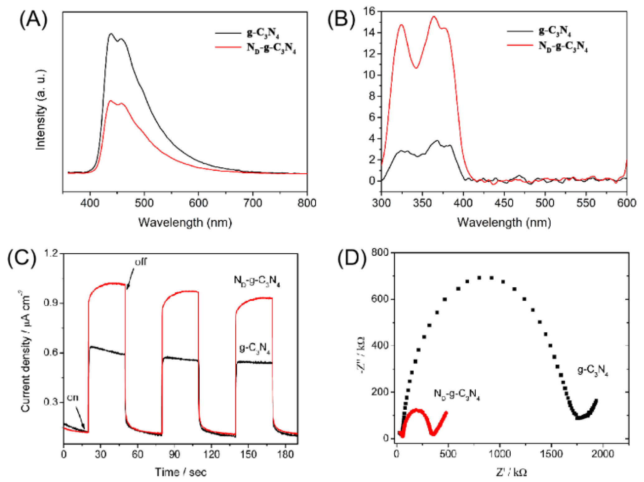
The electron and hole separation efficiencies of the samples were studied by photoluminescence (PL) spectroscopy at about 310 nm excitation wavelength, as shown in Figure 9A. It is evident that the PL intensity of ND-g-C3N4 is weaker than that of g-C3N4, indicating that nitrogen defects can improve the separation efficiency of electrons and holes [26]. The surface photovoltage (SPV) can explain the separation efficiency of photocarriers under illumination. The higher the surface photovoltage intensity, the higher the separation of photogenerated charge carriers. As shown in Figure 9B, the surface photovoltage intensity of ND-g-C3N4 is obviously higher than that of g-C3N4, indicating that the separation efficiency of ND-g-C3N4 photocarriers is higher [37,38]. In order to further explain the separation efficiency of electrons and holes, a photocurrent test was carried out, as shown in Figure 9C. Obviously, the photocurrent density of ND-g-C3N4 is higher than that of g-C3N4, which indicates that ND-g-C3N4 has a higher photoelectron and hole separation efficiency, and also has more photoelectrons [39]. To study the conductivity of the photocatalysts, the electrochemical impedance of the catalyst was tested. As shown in Figure 9D, a smaller ND-g-C3N4 arc indicates a higher conductivity, which is beneficial to the separation of photogenerated electrons and holes.

4. Conclusions

In this work, N-deficient g-C3N4 was obtained by calcining the mixture of melamine and ammonium acetate. N defects induce the molecular distortion of g-C3N4, which enhances the absorption in the entire visible region, especially around 500 nm. The prepared N-deficient g-C3N4 exhibits an excellent photocatalytic hydrogen production performance, 5.8 times that of g-C3N4. The enhanced photocatalytic performance is mainly attributed to the promoted light absorption, as well as the promoted separation of photo-generated carriers and increased specific surface area.

Author Contributions

Conceptualization, F.S. and L.Y.; Formal analysis, H.X.; Investigation, F.S. and Y.Z.; Data curation, Z.W. and C.D.; Writing–original draft, F.S.; Funding acquisition, F.S. All authors have read and agreed to the published version of the manuscript.

Funding

This work was financially supported by the Science and Technology Department of Henan Province (No. 222102320238), the Education Department of Henan Province (No. 21A150041), the National Natural Science Foundation of China (No. 51872147), the Science Foundation of Nanyang Normal University (No. 2018ZX006, 2022PY018), and the Innovation and Entrepreneurship Training Program for College Students in Henan Province (No. 202110481006).

Data Availability Statement

The data presented in this study are available in insert article here.

Conflicts of Interest

The authors declare no conflict of interest.

References

  1. Zhang, K.; Zhou, W.; Chi, L.; Zhang, X.; Hu, W.; Jiang, B.; Pan, K.; Tian, G.; Jiang, Z. Black N/H-TiO2 nanoplates with a flower-like hierarchical architecture for photocatalytic hydrogen evolution. ChemSusChem 2016, 9, 2841–2848. [Google Scholar] [CrossRef] [PubMed] [Green Version]
  2. Su, F.Y.; Zhang, W.D. Fabrication and photoelectrochemical property of In2O3/ZnO composite nanotube arrays using ZnO nanorods as self-sacrificing templates. Mater. Lett. 2018, 211, 65–68. [Google Scholar] [CrossRef]
  3. Ong, W.J.; Tan, L.L.; Ng, Y.H.; Yong, S.T.; Chai, S.P. Graphitic carbon nitride (g-C3N4)-based photocatalysts for artificial photosynthesis and environmental remediation: Are we a step closer to achieving sustainability. Chem. Rev. 2016, 116, 7159–7329. [Google Scholar] [CrossRef] [PubMed]
  4. Majdoub, M.; Anfar, Z.; Amedlou, A. Emerging Chemical Functionalization of g-C3N4: Covalent/Noncovalent Modifications and Applications. ACS Nano 2020, 14, 12390–12469. [Google Scholar] [CrossRef] [PubMed]
  5. Jiang, J.; Xiong, Z.; Wang, H.; Liao, G.; Bai, S.; Zou, J.; Wu, P.; Zhang, P.; Li, X. Sulfur-doped g-C3N4/g-C3N4 isotype step-scheme heterojunction for photocatalytic H2 evolution. J. Mater. Sci. Technol. 2022, 118, 15–24. [Google Scholar] [CrossRef]
  6. Zhang, Y.; Yuan, J.; Zhao, L.; Wu, B.; Zhang, B.; Zhang, P.; Zhang, S.; Dong, C. Boosting exciton dissociation and charge transfer in P-doped 2D porous g-C3N4 for enhanced H2 production and molecular oxygen activation. Ceram. Int. 2022, 48, 4031–4046. [Google Scholar] [CrossRef]
  7. Wang, X.; Wang, X.; Tian, W.; Meng, A.; Li, Z.; Li, S.; Wang, L.; Li, G. High-energy ball-milling constructing P-doped g-C3N4/MoP heterojunction with MoN bond bridged interface and Schottky barrier for enhanced photocatalytic H2 evolution. Appl. Catal. B Environ. 2022, 303, 120933. [Google Scholar] [CrossRef]
  8. Andryushina, N.; Shvalagin, V.; Korzhak, G.; Grodzyuk, G.; Kuchmiy, S.; Skoryk, M. Photocatalytic evolution of H2 from aqueous solutions of two-component electron-donor substrates in the presence of g-C3N4 activated by heat treatment in the KCl+ LiCl melt. Appl. Surf. Sci. 2019, 475, 348–354. [Google Scholar] [CrossRef]
  9. Hu, Z.; Shi, D.; Wang, G.; Gao, T.; Wang, J.; Lu, L.; Li, J. Carbon dots incorporated in hierarchical macro/mesoporous g-C3N4/TiO2 as an all-solid-state Z-scheme heterojunction for enhancement of photocatalytic H2 evolution under visible light. Appl. Surf. Sci. 2022, 601, 154167. [Google Scholar] [CrossRef]
  10. Yang, B.; Wang, Z.; Zhao, J.; Sun, X.; Wang, R.; Liao, G.; Jia, X. 1D/2D carbon-doped nanowire/ultra-thin nanosheet g-C3N4 isotype heterojunction for effective and durable photocatalytic H2 evolution. Int. J. Hydrog. Energy 2021, 46, 25436–25447. [Google Scholar] [CrossRef]
  11. Liang, J.; Yang, X.; Fu, H.; Ran, X.; Zhao, C.; An, X. Integrating optimal amount of carbon dots in g-C3N4 for enhanced visible light photocatalytic H2 evolution. Int. J. Hydrog. Energy 2022, 47, 18032–18043. [Google Scholar] [CrossRef]
  12. Ding, Y.; Tang, Y.; Yang, L.; Zeng, Y.; Yuan, J.; Liu, T.; Zhang, S.; Liu, C.; Luo, S. Porous nitrogen-rich carbon materials from carbon self-repairing g-C3N4 assembled with graphene for high-performance supercapacitor. J. Mater. Chem. A 2016, 4, 14307–14315. [Google Scholar] [CrossRef]
  13. Wang, J.; Pan, R.; Hao, Q.; Gao, Y.; Ye, J.; Wu, Y.; van Ree, T. Constructing Defect-Mediated CdS/g-C3N4 by an In-situ Interlocking Strategy for Cocatalyst-free Photocatalytic H2 Production. Appl. Surf. Sci. 2022, 599, 153875. [Google Scholar] [CrossRef]
  14. Ye, L.; Deng, Y.; Wang, L.; Xie, H.; Su, F. Bismuth-based photocatalysts for solar photocatalytic carbon dioxide conversion. ChemSusChem 2019, 12, 3671–3701. [Google Scholar] [CrossRef] [PubMed]
  15. Hong, Z.; Shen, B.; Chen, Y.; Lin, B.; Gao, B. Enhancement of photocatalytic H2 evolution over nitrogen-deficient graphitic carbon nitride. J. Mater. Chem. A 2013, 1, 11754–11761. [Google Scholar] [CrossRef]
  16. Su, K.; Liu, H.; Gao, Z.; Fornasiero, P.; Wang, F. Nb2O5-Based Photocatalysts. Adv. Sci. 2021, 8, 2003156. [Google Scholar] [CrossRef]
  17. Jorge, A.B.; Martin, D.J.; Dhanoa, M.T.; Rahman, A.S.; Makwana, N.; Tang, J.; Sella, A.; Cora, F.; Darr, J.A.; Firth, S.; et al. H2 and O2 Evolution from Water half-splitting reactions by graphitic carbon nitride materials. J. Phys. Chem. C 2013, 117, 7178–7185. [Google Scholar] [CrossRef]
  18. Chen, Y.; Wang, B.; Lin, S.; Zhang, Y.; Wang, X. Activation of n → π* transitions in two-dimensional conjugated polymers for visible light photocatalysis. J. Phys. Chem. C 2014, 118, 29981–29989. [Google Scholar] [CrossRef]
  19. Su, F.Y.; Xu, C.Q.; Yu, Y.X.; Zhang, W.D. Carbon self-doping induced activation of n–π* electronic transitions of g-C3N4 nanosheets for efficient photocatalytic H2 evolution. ChemCatChem 2016, 8, 3527–3535. [Google Scholar] [CrossRef]
  20. Wang, X.; Maeda, K.; Thomas, A.; Takanabe, K.; Xin, G.; Carlsson, J.M.; Domen, K.; Antonietti, M. A metal-free polymeric photocatalyst for hydrogen production from water under visible light. Nat. Mater. 2009, 8, 76–80. [Google Scholar] [CrossRef]
  21. Kong, L.; Dong, Y.; Jiang, P.; Wang, G.; Zhang, H.; Zhao, N. Light-assisted rapid preparation of a Ni/g-C3N4 magnetic composite for robust photocatalytic H2 evolution from water. J. Mater. Chem. A 2016, 4, 9998–10007. [Google Scholar] [CrossRef]
  22. Han, Q.; Wang, B.; Gao, J.; Cheng, Z.H.; Zhao, Y.; Zhang, Z.P.; Qu, L.T. Atomically thin mesoporous nanomesh of graphitic C3N4 for high-efficiency photocatalytic hydrogen evolution. ACS Nano 2016, 10, 2745–2751. [Google Scholar] [CrossRef] [PubMed]
  23. Mao, J.; Peng, T.Y.; Zhang, X.H.; Li, K.; Ye, L.Q.; Zan, L. Effect of graphitic carbon nitride microstructures on the activity and selectivity of photocatalytic CO2 reduction under visible light. Catal. Sci. Technol. 2013, 3, 1253–1260. [Google Scholar] [CrossRef]
  24. Su, F.Y.; Zhang, W.D. Creating distortion in g-C3N4 framework by incorporation of ethylenediaminetetramethylene for enhancing photocatalytic generation of hydrogen. Mol. Catal. 2017, 432, 64–75. [Google Scholar] [CrossRef]
  25. Su, F.Y.; Zhang, W.D. Carbonyl-Grafted g-C3N4 Porous Nanosheets for Efficient Photocatalytic Hydrogen Evolution. Chem.–Asian J. 2017, 12, 515–523. [Google Scholar] [CrossRef]
  26. Li, K.; Xie, X.; Zhang, W. Photocatalysts based on g-C3N4-encapsulating carbon spheres with high visible light activity for photocatalytic hydrogen evolution. Carbon 2016, 110, 356–366. [Google Scholar] [CrossRef]
  27. Qin, Z.X.; Xue, F.; Chen, Y.B.; Shen, S.H.; Guo, L.J. Spatial charge separation of one-dimensional Ni2P-Cd0.9Zn0.1S/g-C3N4 heterostructure for high-quantum-yield photocatalytic hydrogen production. Appl. Catal. B Environ. 2017, 217, 551–559. [Google Scholar] [CrossRef]
  28. Araña, J.; Doña-Rodríguez, J.M.; Cabo, C.G.; González-Díaz, O.; Herrera-Melián, J.A.; Pérez-Peña, J. FTIR study of gas-phase alcohols photocatalytic degradation with TiO2 and AC-TiO2. Appl. Catal. B Environ. 2004, 53, 221–232. [Google Scholar] [CrossRef]
  29. Miao, Z.; Wang, Q.; Zhang, Y.; Meng, L.; Wang, X. In situ construction of S-scheme AgBr/BiOBr heterojunction with surface oxygen vacancy for boosting photocatalytic CO2 reduction with H2O. Appl. Catal. B Environ. 2022, 301, 120802. [Google Scholar] [CrossRef]
  30. Kong, L.; Mu, X.; Fan, X.; Li, R.; Zhang, Y.; Song, P.; Ma, F.; Sun, M. Site-selected N vacancy of g-C3N4 for photocatalysis and physical mechanism. Appl. Mater. Today 2018, 13, 329–338. [Google Scholar] [CrossRef]
  31. Ruan, D.; Kim, S.; Fujitsuka, M.; Majima, T. Defects rich g-C3N4 with mesoporous structure for efficient photocatalytic H2 production under visible light irradiation. Appl. Catal. B Environ. 2018, 238, 638–646. [Google Scholar] [CrossRef]
  32. Yu, H.; Shi, R.; Zhao, Y.; Bian, T.; Zhao, Y.; Zhou, C.; Waterhouse, G.I.N.; Wu, L.; Tung, C.; Zhang, T. Alkali-assisted synthesis of nitrogen deficient graphitic carbon nitride with tunable band structures for efficient visible-light-driven hydrogen evolution. Adv. Mater. 2017, 29, 1605148. [Google Scholar] [CrossRef] [PubMed]
  33. Jiang, Y.; Sun, Z.; Tang, C.; Zhou, Y.; Zeng, L.; Huang, L. Enhancement of photocatalytic hydrogen evolution activity of porous oxygen doped g-C3N4 with nitrogen defects induced by changing electron transition. Appl. Catal. B Environ. 2019, 240, 30–38. [Google Scholar] [CrossRef]
  34. Wang, X.; Wu, L.; Wang, Z.; Wu, H.; Zhou, X.; Ma, H.; Zhong, H.; Xing, Z.; Cai, G.; Jiang, C.; et al. C/N vacancy co-enhanced visible-light-driven hydrogen evolution of g-C3N4 nanosheets through controlled He+ ion irradiation. Sol. RRL 2019, 3, 1800298. [Google Scholar] [CrossRef]
  35. Zeng, Y.; Li, H.; Luo, J.; Yuan, J.; Wang, L.; Liu, C.; Xia, Y.; Liu, M.; Luo, S.; Cai, T.; et al. Sea-urchin-structure g-C3N4 with narrow bandgap (~2.0 eV) for efficient overall water splitting under visible light irradiation. Appl. Catal. B Environ. 2019, 249, 275–281. [Google Scholar] [CrossRef]
  36. Yan, B.; Du, C.; Yang, G. Constructing Built-in Electric Field in Ultrathin Graphitic Carbon Nitride Nanosheets by N and O Codoping for Enhanced Photocatalytic Hydrogen Evolution Activity. Small 2020, 16, 1905700. [Google Scholar] [CrossRef]
  37. Cheng, C.; Shi, J.; Wen, L.; Dong, C.L.; Huang, Y.C.; Zhang, Y.; Zong, S.; Diao, Z.; Shen, S.; Guo, L. Disordered nitrogen-defect-rich porous carbon nitride photocatalyst for highly efficient H2 evolution under visible-light irradiation. Carbon 2021, 181, 193–203. [Google Scholar] [CrossRef]
  38. Ge, L.; Zuo, F.; Liu, J.K.; Ma, Q.; Wang, C.; Sun, D.Z.; Bartels, L.; Feng, P.Y. Synthesis and efficient visible light photocatalytic hydrogen evolution of polymeric g-C3N4 coupled with CdS quantum dots. J. Phys. Chem. C 2012, 116, 13708–13714. [Google Scholar] [CrossRef]
  39. Li, Y.; Yin, Q.; Zeng, Y.; Liu, Z. Hollow spherical biomass derived-carbon dotted with SnS2/g-C3N4 Z-scheme heterojunction for efficient CO2 photoreduction into CO. Chem. Eng. J. 2022, 438, 135652. [Google Scholar] [CrossRef]
Figure 1. Schematic diagram of the generative process of ND-g-C3N4.
Catalysts 12 01496 g001
Figure 2. (A) XRD patterns, (B) FTIR spectra and XPS spectra of g-C3N4 and ND-g-C3N4: (C) Survey spectra, (D) O 1s, (E) C 1s and (F) N 1s.
Catalysts 12 01496 g002
Figure 3. EPR spectra of the samples under dark and light irradiation for 5 min and 10 min at room temperature showed a g value of 1.999.
Catalysts 12 01496 g003
Figure 4. SEM images of (A), (B) g-C3N4 and (C), (D) ND-g-C3N4, EDS spectra of (E), (F) g-C3N4 and (G), (H) ND-g-C3N4.
Catalysts 12 01496 g004
Figure 5. (A) N2 adsorption/desorption isotherms and (B) BJH pore size distribution curves of g-C3N4 and ND-C3N4.
Catalysts 12 01496 g005
Figure 6. (A) UV-Vis DRS, (B) Tauc model to estimate bandgap, (C) VB-XPS and (D) electronic band structure of the samples.
Catalysts 12 01496 g006
Figure 7. Calculated band structure of (A) g-C3N4, (B) ND-g-C3N4; Calculated DOS of (C) g-C3N4, (D) ND-g-C3N4.
Catalysts 12 01496 g007
Figure 8. (A) The amount of H2 evolution and (B) H2 evolution rate of the samples, and (C) Stability of ND-g-C3N4 in 16 h photocatalytic reaction.
Catalysts 12 01496 g008
Figure 9. (A) PL spectra, (B) SPV spectra, (C) Photocurrent respons, and (D) Electrochemical impedance spectra of the samples.
Catalysts 12 01496 g009
Table 1. Element composition of g-C3N4 and ND-g-C3N4 samples.
SampleN (wt%)C (wt%)H (wt%)N/C (at.)
g-C3N460.0234.262.2421.502
ND-g-C3N459.7534.402.0621.489
Table 2. Comparison of photocatalytic activity with other N-deficient g-C3N4 reported in the literature.
Photocatalytic MaterialsCocatalystLight SourceReaction SolutionsHydrogen Evolution RateRef.
ND-C3N43% wt% Ptλ > 400 nm90 mL water and 10 mL triethanolamine865.2 μmol/g/hThis work
N-deficient g-C3N4 nanosheets3% wt% Ptλ > 400 nm50 mL of aqueous solution
containing 20 vol% triethanolamine
3.1 mmol/g/h[31]
N-deficient g-C3N4
nanosheets
1 wt% Ptλ > 420 nmLactic acid aqueous solution
(20 mL, 25 vol%)
6.9 mmol/g/h[32]
Porous O-doped C3N4
with N vacancies
3 wt% Ptλ > 420 nm50 mL of aqueous solution
(40 mL and
10 mL triethanolamine)
258.18 μmol/g/h[33]
N/C-deficient g-C3N4
nanosheets
3 wt% Ptλ > 420 nm72 mL water and 8 mL
triethanolamine
1271 μmol/g/h[34]
Sea-urchin-structure
N-deficient g-C3N4
3 wt% Ptλ > 420 nmTriethanolamine solution (15 vol%, 80 mL)41.5 μmol/g/h[35]
N, O-codoped C3N43 wt% Ptλ > 420 nm100 mL of aqueous solution containing
triethanolamine (10 vol%)
1284 μmol/g/h[36]
Publisher’s Note: MDPI stays neutral with regard to jurisdictional claims in published maps and institutional affiliations.