Article

Synthesis and Photocatalytic Activity of β-Ga2O3 Nanostructures for Decomposition of Formaldehyde under Deep Ultraviolet Irradiation

1
School of Electrical Engineering, Korea University, 145 Anam-ro, Seongbuk-gu, Seoul 02841, Korea
2
Light Convergence Research Team, Korea Institute of Lighting and ICT, 370 Dongseo-ro, Iksan-si, Jeollabuk-do 54630, Korea
3
VKTECH Research Center, Nguyen Tat Thanh University, 298-300A Nguyen Tat Thanh Street, District 4, Ho Chi Minh City 70000, Vietnam
4
Department of Solid State Physics, University of Science, Vietnam National University, Ho Chi Minh City (VNU-HCM) 227 Nguyen Van Cu Street, District 5, Ho Chi Minh City 70000, Vietnam
*
Authors to whom correspondence should be addressed.
These authors contributed equally to this work.
Catalysts 2020, 10(10), 1105; https://doi.org/10.3390/catal10101105
Submission received: 11 August 2020 / Revised: 11 September 2020 / Accepted: 22 September 2020 / Published: 24 September 2020
(This article belongs to the Section Nanostructured Catalysts)

Abstract

:
An attempt to degrade volatile organic compounds (VOCs) and sterilize air simultaneously is highly desirable to improve indoor air quality. With the help of deep ultraviolet (UVC) lighting, harmful bacteria that exists in the air can be destroyed. Thus, a new photocatalytic substance that can break down VOCs under UVC irradiation is required. Here, we demonstrate the photocatalytic activity of β-Ga2O3 nanostructures, synthesized via the solvothermal method for removing formaldehyde (HCHO) under deep ultraviolet irradiation. The Raman and XRD results indicated that as-synthesized nanostructures showed β-crystalline phase with a monoclinic structure. The photoluminescence spectrum exhibited a broad and strong green emission peak at 510 nm, which was likely attributed to a considerable amount of oxygen and gallium vacancies formed during the calcinating process. The photocatalytic efficiency for decomposing HCHO at room temperature under deep ultraviolet irradiation (278 nm) of the synthesized β-Ga2O3 nanoparticles is higher than that of the β-Ga2O3 nanorods. Both nanoparticles and nanorods obeyed the pseudo-first-order Langmuir-Hinshelwood kinetic model with a degradation rate constant of 0.057 and 0.033 min−1, corresponding to the efficiency of 82% and 62% in the formaldehyde removal, respectively.

1. Introduction

Indoor air pollution gives rise to more and more illnesses in developed and developing countries. A lot of efforts have thus been conducted to find sustainable ways to control the indoor air quality (IAQ) of residential units and workplaces since people usually spend more than 80% of their lifetime performing indoor activities. Indoor air pollution arises from particulate matters, nitrogen oxides, carbon monoxide, and volatile organic compounds (VOCs) [1]. Among those, formaldehyde (HCHO), a type of VOC emitted from interior decoration materials, adhesives, and paints, is representative of indoor pollutants [2]. It is strongly persistent and thus can slowly release from the materials over an extensive period. Additionally, it is more dangerous than other VOCs due to its higher polarity and reactivity characteristics. HCHO is a probable human carcinogen and has various adverse effects on human organs and metabolic systems. Therefore, the removal of formaldehyde from indoor air environments is necessary to improve the quality of life and human health. In the past decade, adsorbents with a large surface area medium for physical and chemical adsorption were used as the traditional technology for HCHO removal, both in the laboratory and in the field [3]. The ultraviolet-photocatalytic oxidation (UV-PCO) for indoor VOCs removal has received growing interest in recent years. The advantage of this method is that it operates at room temperature and is capable of degrading many organics under light irradiation without the production of secondary pollution [4]. However, a study on the performance and mechanism of the UV-PCO for indoor HCHO removal is still a challenge. Furthermore, it is highly desirable to replace artificial UVA light by UVC light in order to implement UVC-PCO technology. The main purpose of using a UVC configuration to operate air purification systems is to simultaneously activate the photocatalyst and germicidal function of the UVC irradiation in one reactor fashion. In this manner, both air pollutants and airborne micro-organisms that exist in indoor air can be efficiently eliminated. Therefore, it is important to seek a chemically stable photocatalyst with a predictable photocatalytic mechanism. Semiconductor nanostructures can be used as an efficient photocatalytic material. TiO2 has emerged as one of the more promising photocatalytic materials due to its excellent photocatalytic activity and nontoxic material [5]. However, the practical application of TiO2-based photocatalysts is limited, owing to the long-term instability that decreases the photocatalytic activity and cyclic stability. Gallium oxide (Ga2O3) has recently been considered as an attractive alternative due to its long stability and high redox capabilities [6,7]. Yoo et al. demonstrated a superior photocatalytic activity of Ga2O3 on VOCs compared to that of TiO2 [8]. In particular, monoclinic gallium oxide (β-Ga2O3) has emerged because of its wide bandgap (4.7–4.9 eV) and unique optical properties [9].
In this study, we demonstrate the photocatalytic performance of synthesized β-Ga2O3 nanostructures with different morphology for removing HCHO under UVC irradiation. β-Ga2O3 nanostructures have been synthesized by several methods such as hydrothermal [10,11], solvothermal [12], and dc arc-discharge [13] techniques. The nanostructures were successfully synthesized by solvothermal techniques. The structure and the optical property of the β-Ga2O3 nanostructures were investigated using an X-ray diffractometer (XRD, Philips, PW3710, Amsterdam, Netherlands), Raman spectroscopy, field-emission scanning electron microscopy (FE-SEM, JEOL, JSM-600F, Akishima, Japan) and photoluminescence (PL, He-Cd radiation, λ = 325 nm, 30 mW).

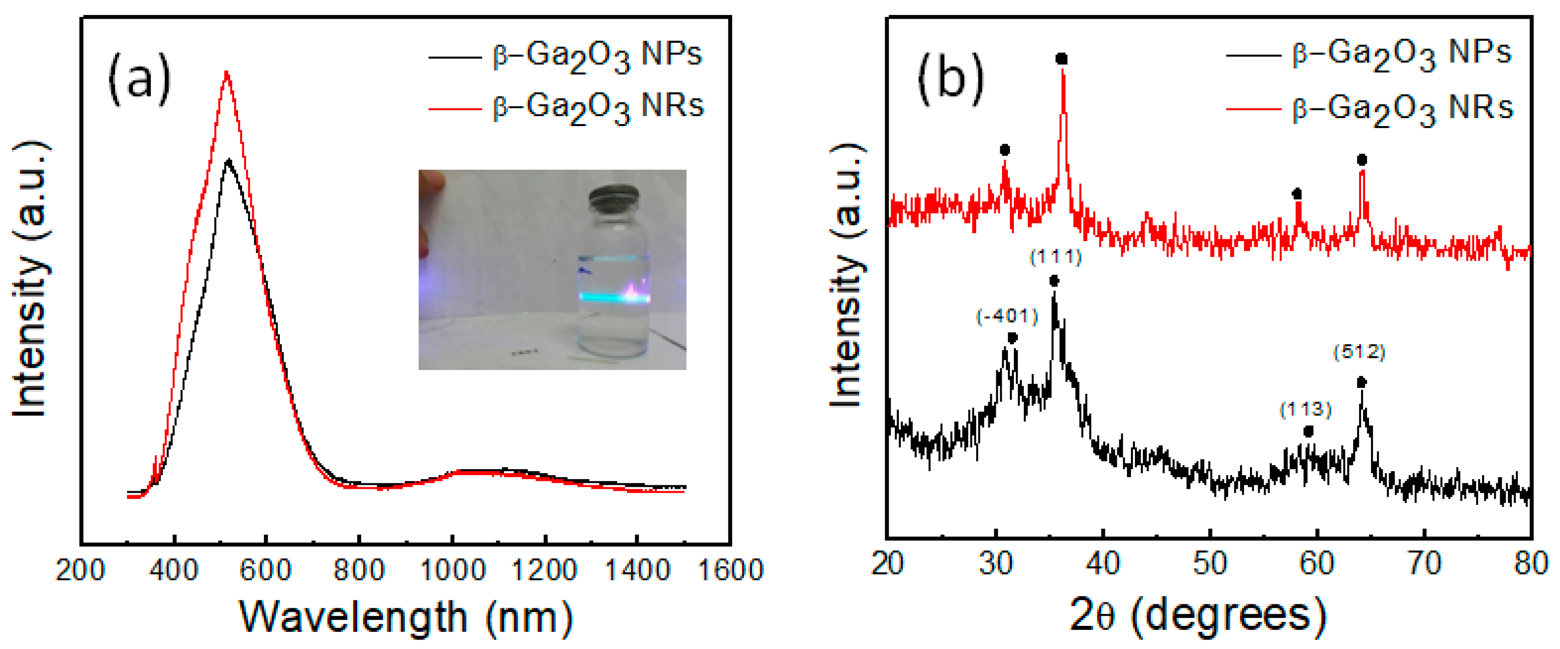
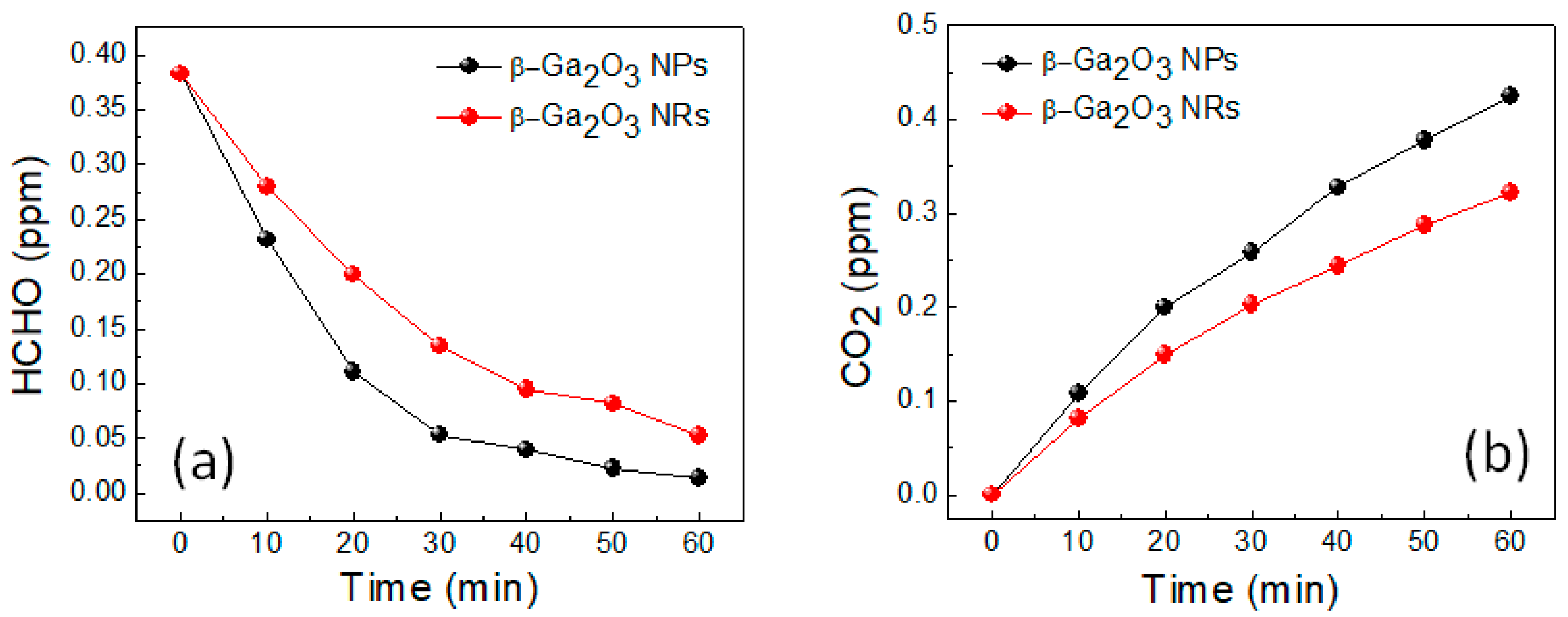
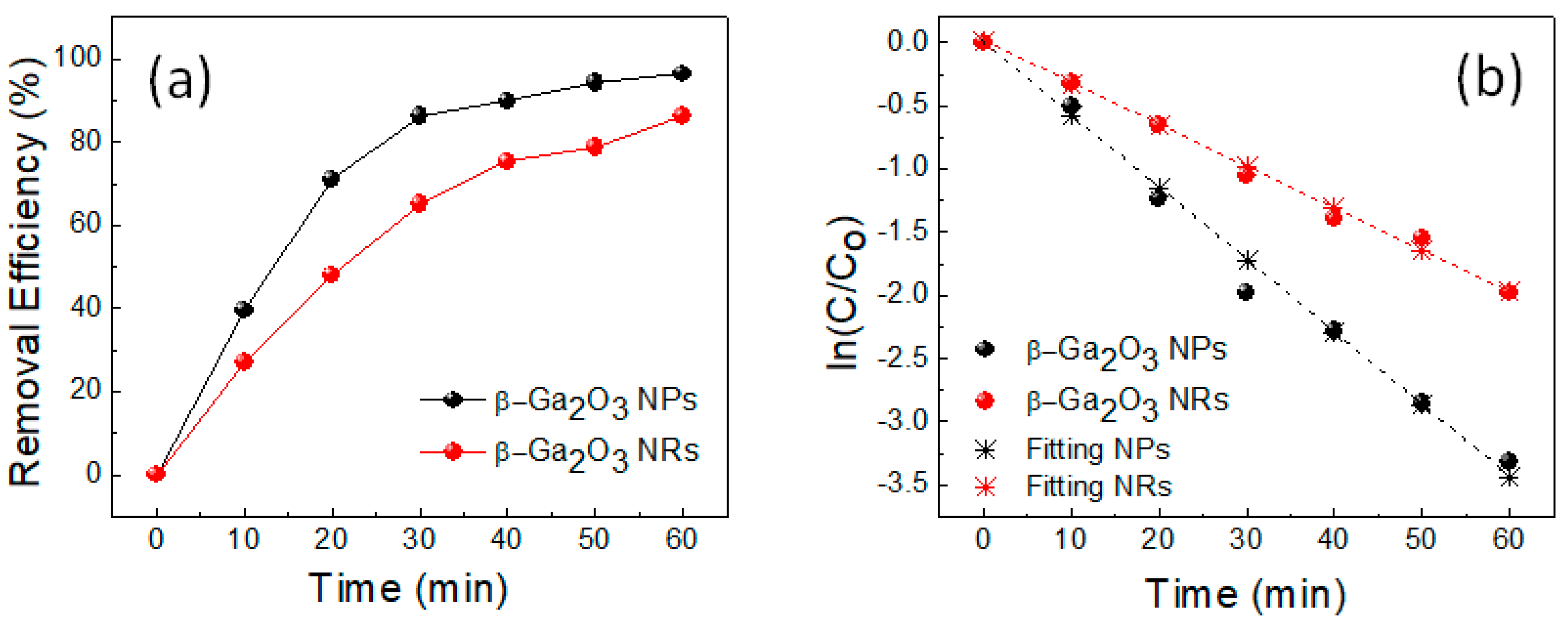
2. Results and Discussion

Figure 1a shows the SEM images of the as-synthesized β-Ga2O3 nanorods (NRs). It is seen that the uniform NRs were randomly distributed and covered on the whole surface of the Si substrate without any contamination. The average length of the NRs was found to be around 10 μm and 700 nm, respectively. The aspect ratio (length/width) was 14 for prepared β-Ga2O3 NRs. This value could be tailored by modification of the molar ratio of precursor:solvent:surfactant. The series of chemical reactions taking place in our system to produce β-Ga2O3 NRs is summarized as follows:
Ga + HCl   GaCl 3 +   H 2
GaCl 3 +   CO ( NH 2 ) 2   180   ° C   for   5   h   GaOOH
GaOOH   700   ° C   for   5   h   β Ga 2 O 3
The inset of Figure 1a indicates that hydrothermally synthesized GaOOH at 180 °C forms rod-like particles with a diameter of 700 nm. The rod-like morphology can be achieved using the neutral aqueous solution (i.e., in the absence of excess OH ions) [14]. The water medium favors a rod growth by unidirectional growth of one face while shutting down the growth on other faces [15]. GaOOH crystals are grown along the c-axis (001) direction only, thereby providing the rod-like morphology [16]. To verify this hypothesis, we used ethylene glycol instead of deionized water in the hydrothermal step, which resulted in the nanoparticle (NP) structure of β-Ga2O3 as shown in Figure 1b.
Figure 2a gives the XRD diffraction patterns of the hydrothermally synthesized GaOOH and calcinated β-Ga2O3. After the hydrothermal process, the obtained rod-like materials were identified as a single-phase GaOOH, referring to the Joint Committee on Powder Diffraction Standards (JCPDS no. 60180). The relatively broad diffraction peaks indicate that the rod-like materials had a polycrystalline nature. The peak of monoclinic β-Ga2O3 (JCPDS no. 760573) was obtained without any impurities through the calcination at 700 °C. The strong (110) peak disappeared and the intensity of (111) peak was significantly enhanced, suggesting that the β-Ga2O3 is somehow oriented along the (111) direction perpendicular to the substrate. It reveals that the original rod-like morphology of GaOOH was maintained after the calcinating process. These results further verify that GaOOH and β-Ga2O3 process along a preferential (111) direction. The Raman spectra of the white-colored β-Ga2O3 NRs, which were formed by calcinating GaOOH at 700 °C for 5 h, is exhibited in Figure 2b. A group of peaks at 168, 198, 345, 415, 474, 562, 653, 766 cm−1 were observed, which is consistent with those of Wang’s study [17]. These features correspond to Raman modes of monoclinic β-Ga2O3 [18]. The β-Ga2O3 crystal structure was comprised of a double chain of GaO3 octahedra connected by GaO4 tetrahedra. Therefore, GaI (tetrahedral coordination) and GaII (octahedral coordination) are two nonequivalent Ga3+ sites in the β-Ga2O3 lattice [19]. Three regions with different vibrational modes could be identified in the Raman spectra. In the low-frequency mode (below 200 cm−1), peaks at 168 and 198 cm−1 were ascribed to the vibrations involving the libration of Ga-O chains. In the mid-frequency range, the peak at 345 cm−1 was due to the vibration involving motions of octahedra (translations of GaII), while three peaks at 415, 475, 562 cm−1 were considered as bending modes of GaO4 tetrahedra groups. In the high-frequency range, two peaks at 653 and 766 cm−1 were related to stretching modes involving Ga-OI bonds, which are the shortest ones [20].
Figure 3a exhibits the PL spectra measured from β-Ga2O3 NRs and NPs. Both NRs and NPs have the same broad and strong green emission band at 510 nm. The origin of these emissions has not been clearly understood so far, and thus further systematic study is necessary to elucidate the mechanism of the observed emission. Binet and Gourier reported that the PL emission originates from the defect band emission of Ga2O3 crystals, which may be attributed to Ga vacancies, O vacancies, and Ga-O vacancy pairs [9]. It should be noted that the optical band gap of β-Ga2O3 nanostructures strongly depends on the dimension of nanomaterials’ morphology, as well as synthesis conditions [21]. In our experiment, β-Ga2O3 nanostructures were calcined at 700 °C for 5 h. At this high temperature under a reduction atmosphere, a considerable quantity of vacancies may have easily formed, which may have lowered the band-gap energy (red-shift) of our sample. The digital image of β-Ga2O3 NRs diluted in DI water also clearly shows a blue-green emission (Figure 3, inset), which is consistent with the PL result. As a comparison, the peak emission intensity of β-Ga2O3 NPs was found to be lower than that of β-Ga2O3 NRs. The HRXRD was performed to characterize the crystalline quality of β-Ga2O3 NRs and NPs, as shown in Figure 3b. Both samples possessed characteristic features of the monoclinic β-phase with (111) and (512) dominant peaks. In addition, it was clear to see that all diffraction peaks of NRs seemed to be clearer and sharper than that of NPs, especially for (111) and (113). It implies that the crystalline quality of synthesized NPs is lower than that of NRs, which may have caused the low emission peak intensity achieved in the PL spectrum of NPs as compared to NRs.
To examine the photocatalytic activity of β-Ga2O3 NRs and NPs, the photocatalyst oxidation of the HCHO in the gas phase was performed. The charge carriers are generated upon UVC irradiation and the electrons in the valence band get excited to the conduction band. The photogenerated charge carriers then travel to the surface of β-Ga2O3 where they take part in redox reactions [22,23]. The basic mechanism of photocatalytic decomposition is derived from the oxidation of organisms to H2O, CO2 along with –OH or O2 radicals produced on the surface of β-Ga2O3 under UCV irradiation [2,8]. In the heterogeneous reaction system, Ga2O3 is excited by the photon energy more so than the band gap energy. The photogenerated charge carriers (electron in conduction band and hole in valence band) can induce a redox reaction with the adsorbed reactant of the surface of Ga2O3, resulting in the formation of chemical intermediates. Figure 4a presents that the HCHO concentration in β-Ga2O3 NPs reduced from 0.382 ppm to 0.05 ppm after 30 min exposure under the UVC-LEDs irradiation. Herein, the β-Ga2O3 NPs agree with WHO guidelines which states that IAQ standards normally set the acceptable limit at 0.08 ppm for 30 min exposure [3,24]. However, the β-Ga2O3 NRs needed 60 min of irradiation time to reach a similar HCHO level. The longer irradiation time was possibly due to the low surface to volume ratio of NRs as compared to NPs. The HCHO degradation mechanism of β-Ga2O3 nanostructures can be explained as follows: the HCHO reacts prior with OH species generated on the excited β-Ga2O3 nanostructures surface under light exposure. It forms an intermediate of HCOOH which is eventually oxidized to CO2 and H2O vapor [2]. The HCHO photocatalytic decomposition involves the oxidation reaction between the hydroxyl radical (HO.) species and the HCHO. It runs according to the overall stoichiometry equation:
HCHO + 4 [ HO . ] CO 2 + 3 H 2 O
The above equation indicates that the CO2 concentration increases after the oxidation reaction is complete. In this case, the selectivity to CO2 (SCO2) and oxidized yield into CO2 (YCO2) are given by the following equations:
S CO 2 = C CO 2 C 0 C t × 100 ;   and   Y CO 2 = C CO 2 C 0 × 100
where C0 is the initial concentration, Ct is the real-time concentration of the HCHO during the photodegradation, and CCO2 is the concentration of the CO2 products. Figure 4b displays an increase of produced CO2 concentration measured as a function of UV irradiation time, indicating that the HCHO was completely oxidized to yield CO2. Under UVC irradiation for 30 min, the synthesized β-Ga2O3 NPs possessed CO2 selectivity of 78%, corresponding to an oxidized yield of 67%, whereas the synthesized β-Ga2O3 NRs had a CO2 selectivity of 62% and oxidized yield of 53%, respectively. The HCHO removal efficiency can be expressed as the percentage (PER) of initial concentration (Co) and the real-time concentration (Ct) of the HCHO as follows:
PER = C 0 C t C 0   ×   100
Figure 5a shows the calculated removal efficiency of the photodegradation of HCHO as a function of UV irradiation time for β-Ga2O3 NPs and NRs. The results confirmed that NPs have higher photocatalytic efficiency than NRs in terms of degradation of HCHO. The PER of β-Ga2O3 NPs and NRs were found to be 82 and 62%, respectively. Our results implied that photocatalytic activity depends on the surface to volume ratio rather than crystal quality, as usually observed elsewhere. Although the crystalline quality of the synthesized NRs was higher than that of NPs, the photocatalytic behavior of NRs was lower than that of NPs. The photocatalytic reactions typically took place on the surface of catalysts, where further investigations might be required to optimize the nanostructure dimension (surface to volume ratio), which significantly influences the surface reaction rate with beneficial consequences on the photocatalytic efficiency. The net decomposition of the HCHO under UVC irradiation followed the pseudo-first-order Langmuir-Hinshelwood kinetic model. The kinetic plots of the β-Ga2O3 nanostructures are expressed in terms of the linear regression analysis as shown in Figure 5b. As evaluated from the slope of the plot of ln(C/C0) = −kt, first-order degradation rate constant (min−1) for β-Ga2O3 NPs and β-Ga2O3 NRs was estimated to be 0.057 and 0.033 min−1, respectively.

3. Materials and Methods

3.1. Synthesis of β-Ga2O3 NRs

A total of 0.67 g commercial gallium metal (99.999% Sigma-Aldrich, St. Louis, MO, USA) was dissolved in 10 mL of diluted hydrochloric acid and vigorously stirred at 65 °C until a homogeneous solution with a light yellow, transparent color was obtained. After that, 0.8 mL of prepared solution was diluted with 8 mL deionized water and 1.8 g urea was slowly added into the solution while stirring at room temperature to obtain a well-dissolved solution. The as-prepared solution was transferred to a 20 mL Teflon-lined stainless steel autoclave. The autoclave was heated to the desired temperature of 180 °C with a heating rate of 1.5 °C/min and maintained for 5 h. After the autoclave was cooled down to room temperature, precipitates formed in the autoclave were redispersed in ethanol/DI water mixture (1:1 volume ratio), followed by centrifuging at 10,000 rpm for three times to wash. The supernatant was discarded and the precipitates were collected, followed by drying at 80 °C for 24 h. Finally, the obtained powder was calcinated at 700 °C of aging at 3.5 °C/min for 5 h in the air to obtain β-Ga2O3 NRs.

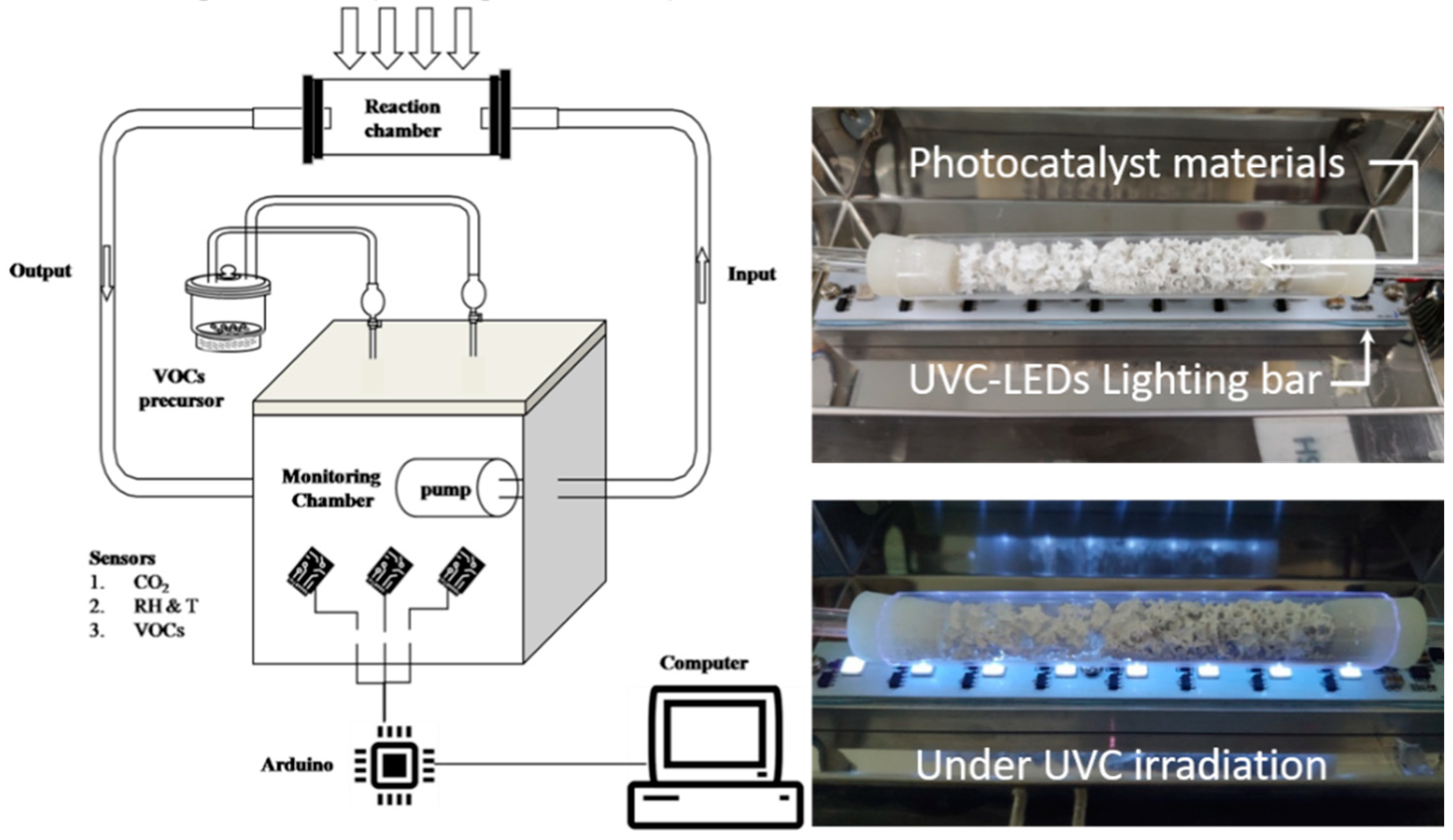
3.2. Photocatalytic Activity Test

The photocatalytic activity of β-Ga2O3 NRs for HCHO degradation was explored using a homemade system that consisted of three main components, including: (1) HCHO precursors, (2) reactor chamber, and (3) monitoring chamber, as illustrated in Figure 6. The experiments were performed at room temperature as follows: The 1.5 g synthesized β-Ga2O3 nanostructures (NRs and NPs) photocatalyst was coated on a ceramic porous platform before the sample was put into the reactor. An adhesive (Silicon Apollo A 500) was used as an HCHO precursor that was fed into the monitoring chamber. Here, the initial value (C0) for the concentration of HCHO was recorded through the sensor devices. Then, it was transferred to the reaction chamber (15 mm diameter quartz tube), where the UVC-PCO occurred, using mini air-pumping via input lines. The flow rate was controlled at 5 mL/min. When UVC-LEDs (278 nm, 10 mW) were irradiated to β-Ga2O3 nanostructures photocatalyst, HCHO was degraded by a photochemical reaction that takes place on the surface of the β-Ga2O3 NRs photocatalyst. The post-reaction product was circulated back to the monitoring chamber via output lines where the real-time concentration (Ct) of HCHO, and the generated concentration of CO2 (CCO2) was recorded. The change of the concentration due to photocatalytic activities was monitored, analyzed, and stored by a computer with DAQ (data acquisition board) and Lab-VIEW software (Austin, TX, USA).

4. Conclusions

In conclusion, β-Ga2O3 NPs and NRs were successfully synthesized using a solution-based process. Our results indicated that the solution used for the synthesis plays an important role in tailoring nanostructures, from rods to particle morphologies. The XRD result indicated that as-synthesized nanostructures show β-crystalline phase with a monoclinic structure. The PL spectrum exhibited a broad and strong green emission peak at 510 nm, which was due to a considerable quantity of oxygen and gallium vacancies formed during the high-temperature calcinating process. Furthermore, we have demonstrated the enhanced photocatalytic performance of synthesized β-Ga2O3 NPs for removing HCHO under UVC irradiation compared to that of β-Ga2O3 NRs. It could be attributed to higher surface to volume ratio for the β-Ga2O3 NPs. As-synthesized β-Ga2O3 NPs showed higher photocatalytic activity in eliminating HCHO than that of NRs with a photodegradation efficiency of 82% and a degradation rate of 0.057 min−1, compared to 62% and 0.033 min−1 of NRs after 30 min of UVC irradiation time. Our findings illustrated the potential application of exploiting β-Ga2O3 nanostructures as a photocatalyst for destroying VOCs under UVC irradiation at room temperature.

Author Contributions

Conceptualization, J.-H.L., J.-Y.Y., and T.V.C.; methodology, J.-H.L., T.A.D., and T.V.C.; formal analysis, J.-H.L., T.A.D., Y.J.P., and T.V.C.; measurements, H.T.M.H., P.H.P., D.T.L., N.H.H., Q.T.T., H.-S.L., and J.H.R.; data curation, J.-H.L. and T.A.D.; writing—original draft preparation, J.-H.L. and T.A.D.; writing—review and editing, Y.J.P. and T.V.C.; supervision, J.-Y.Y. and T.V.C. All authors have read and agreed to the published version of the manuscript.

Funding

This research received no external funding.

Acknowledgments

This research was supported by the Technology Development Program (S2800296) funded by the Ministry of SMEs and Startups (MSS, Korea), and the Vietnam National Foundation for Science and Technology Development (NAFOSTED) under grant number 05/2020/TN.

Conflicts of Interest

The authors declare no conflict of interest.

References

  1. Fukumura, T.; Sambandan, E.; Yamashita, H. Synthesis and VOC degradation ability of a CeO2/WO3 thin-layer visible-light photocatalyst. Mater. Res. Bull. 2017, 94, 493–499. [Google Scholar] [CrossRef]
  2. Huang, Y.; Ho, S.S.H.; Niu, R.; Xu, L.; Lu, Y.; Cao, J.; Lee, S. Removal of indoor volatile organic compounds via photocatalytic oxidation: A short review and prospect. Molecules 2016, 21, 56. [Google Scholar] [CrossRef] [PubMed] [Green Version]
  3. Tseng, C.; Hsieh, C.; Chen, S. The removal of indoor formaldehyde by various air cleaners. In Proceedings of the Air & Waste Management Association´s 98th Annual Conference, Minneapolis, MN, USA, 21–24 June 2005; p. 457. [Google Scholar]
  4. Keller, N.; Ducamp, M.N.; Robert, D.; Keller, V. Ethylene removal and fresh product storage: A challenge at the frontiers of chemistry. Toward an approach by photocatalytic oxidation. Chem. Rev. 2013, 113, 5029–5070. [Google Scholar] [CrossRef] [PubMed]
  5. Schneider, J.; Matsuoka, M.; Takeuchi, M.; Zhang, J.; Horiuchi, Y.; Anpo, M.; Bahnemann, D.W. Understanding TiO2 photocatalysis: Mechanisms and materials. Chem. Rev. 2014, 114, 9919–9986. [Google Scholar] [CrossRef]
  6. Tsukamoto, T.; Mitsutsuka, Y.; Takamura, T. Ga2O3-Photoassisted and Photolytic Degradation of the Flubendiamide Agrochemical in Aqueous Media under Irradiation. J. Water Environ. Technol. 2019, 17, 345–358. [Google Scholar] [CrossRef] [Green Version]
  7. Hidaka, H.; Tsukamoto, T.; Mitsutsuka, Y.; Oyama, T.; Serpone, N. Enhanced Ga2O3-photocatalyzed and photochemical degradation of the Fipronil insecticide by UVC irradiation in mixed aqueous/organic media under an inert atmosphere. Photochem. Photobiol. Sci. 2015, 14, 919–928. [Google Scholar] [CrossRef]
  8. Yoo, T.H.; Ryou, H.; Lee, I.G.; Cho, J.; Cho, B.J.; Hwang, W.S. Comparison of Ga2O3 and TiO2 Nanostructures for Photocatalytic Degradation of Volatile Organic Compounds. Catalysts 2020, 10, 545. [Google Scholar] [CrossRef]
  9. Binet, L.; Gourier, D. Origin of the blue luminescence of β−Ga2O3. J. Phys. Chem. Solids 1998, 59, 1241–1249. [Google Scholar] [CrossRef]
  10. Li, X.; Mo, X.; Li, K. Nanorod β−Ga2O3 semiconductor modified activated carbon as catalyst for improving power generation of microbial fuel cell. J. Solid State Electr. 2019, 23, 2843–2852. [Google Scholar] [CrossRef]
  11. Xiang, M.; Song, X.; Jia, M.; Fang, Y.; Lu, C.; Huang, Y. Hydro-thermal synthesis and photocatalytic reaction of Ga2O3 with different surfactants. Chin. J. Environ. Eng. 2016, 10, 2361–2366. [Google Scholar]
  12. Gai, L.; Jiang, H.; Tian, Y.; Cui, D.; Wang, Q. Low-temperature synthesis of β−Ga2O3 nanorods on SBA-15 microparticles by solvothermal method. Nanotechnology 2006, 17, 5858. [Google Scholar] [CrossRef]
  13. Han, W.Q.; Kohler-Redlich, P.; Ernst, F.; Ruhle, M. Growth and microstructure of Ga2O3 nanorods. Solid State Commun. 2005, 115, 527. [Google Scholar] [CrossRef]
  14. Tas, A.C.; Majewski, P.J.; Aldinger, F. Synthesis of Gallium Oxide Hydroxide Crystals in Aqueous Solutions with or without Urea and Their Calcination Behavior. J. Am. Ceram. Soc. 2002, 85, 1421–1429. [Google Scholar] [CrossRef] [Green Version]
  15. Manna, L.; Scher, E.C.; Alivisatos, A.P. Synthesis of soluble and processable rod-, arrow-, teardrop-, and tetrapod-shaped CdSe nanocrystals. J. Am. Chem. Soc. 2000, 122, 12700–12706. [Google Scholar] [CrossRef]
  16. Fujihara, S.; Shibata, Y.; Hosono, E. Chemical deposition of rodlike GaOOH and β-Ga2O3 films using simple aqueous solutions. J. Electrochem. Soc. 2005, 152, 764–768. [Google Scholar] [CrossRef]
  17. Wang, Y.; Li, N.; Duan, P.; Sun, X.; Chu, B.; He, Q. Properties and photocatalytic activity of β-Ga2O3 nanorods under simulated solar irradiation. J. Nanomater. 2015, 2015, 191793. [Google Scholar]
  18. Zhou, S.; Feng, G.; Wu, B.; Jiang, N.; Xu, S.; Qiu, J. Intense infrared luminescence in transparent glass-ceramics containing β-Ga2O3:Ni2+ Nanocrystals. J. Phys. Chem. C 2007, 111, 7335–7338. [Google Scholar] [CrossRef]
  19. López, I.; Utrilla, A.D.; Nogales, E.; Méndez, B.; Piqueras, J.; Peche, A.; Ramírez-Castellanos, J.; González-Calbet, J.M. In-doped gallium oxide micro- and nanostructures: Morphology, structure, and luminescence properties. J. Phys. Chem. C 2012, 116, 3935–3943. [Google Scholar] [CrossRef]
  20. Dohy, D.; Lucazeau, G.; Revcolevschi, A. Raman spectra and valence force field of single-crystalline β-Ga2O3. J. Solid State Chem. 1982, 45, 180–192. [Google Scholar] [CrossRef]
  21. Xu, Q.; Zhang, S. Fabrication and photoluminescence of β- Ga2O3 nanorods. Superlattices Microstruct. 2008, 44, 715–720. [Google Scholar] [CrossRef]
  22. Kumar, A.; Raizada, P.; Singh, P.; Saini, R.; Saini, A.; Hosseini-Bandegharaei, A. Perspective and status of polymeric graphitic carbon nitride based Z-scheme photocatalytic systems for sustainable photocatalytic water purification. Chem. Eng. J. 2019, 391, 123496. [Google Scholar] [CrossRef]
  23. Huang, C.-W.; Nguyen, V.-H.; Zhou, S.-R.; Hsu, S.-Y.; Tan, J.-X.; Wu, K.C.-W. Metal-organic frameworks: Preparation and applications in highly efficient heterogeneous photocatalysis. Sustain. Energy Fuels 2020, 4, 504–521. [Google Scholar] [CrossRef]
  24. Cano-Lerida, L.; Rose, M.; Walton, P. Polycyclic Aromatic Hydrocarbons Dalam Bioactive Compounds in Food; Blackwell Publishing: Oxford, UK, 2008; pp. 378–399. [Google Scholar]
Figure 1. SEM images of (a) β-Ga2O3 NRs. Inset: enlarged SEM image of the β-Ga2O3 NRs, and (b) β-Ga2O3 NPs.
Catalysts 10 01105 g001
Figure 2. (a) HRXRD diffraction spectra of as-synthesized β-Ga2O3 and GaOOH. (b) Raman spectra of β-Ga2O3 NRs.
Catalysts 10 01105 g002
Figure 3. (a) Photoluminescence (PL) spectra of the synthesized β-Ga2O3 NRs and NPs. Inset: digital image of green photoluminescence of β-Ga2O3 NRs dissolved in DI water under a 365 nm UV irradiation. (b) HRXRD diffraction spectra of synthesized β-Ga2O3 NRs and NPs.
Catalysts 10 01105 g003
Figure 4. Photocatalytic activity of β-Ga2O3 NRs and NPs for HCHO degradation under UVC irradiation: (a) concentration degradation of HCHO. (b) Amount of CO2 product as a function of time.
Catalysts 10 01105 g004
Figure 5. (a) HCHO removal efficiency of β-Ga2O3 NRs and NPs as a function of UVC irradiation time. (b) Langmuir-Hinshelwood pseudo-first order kinetic model for HCHO degradation of β-Ga2O3 NRs and NPs under UVC irradiation.
Catalysts 10 01105 g005
Figure 6. Schematic design of the experimental setup used for the study of photocatalytic activity of β-Ga2O3 related to degrade volatile organic compound (VOC) gas. Photographs of a reaction chamber consisting of a UVC lighting bar and β-Ga2O3 photocatalyst coated porous ceramic platform inside a quartz reactor.
Catalysts 10 01105 g006

