

Enhancing Brain Segmentation in MRI through Integration of Hidden Markov Random Field Model and Whale Optimization Algorithm



Abstract
1. Introduction
- -
- A new optimization method applied to MRI image segmentation.
- -
- A novel technique using the combination of HMRF and the WOA is proposed.
- -
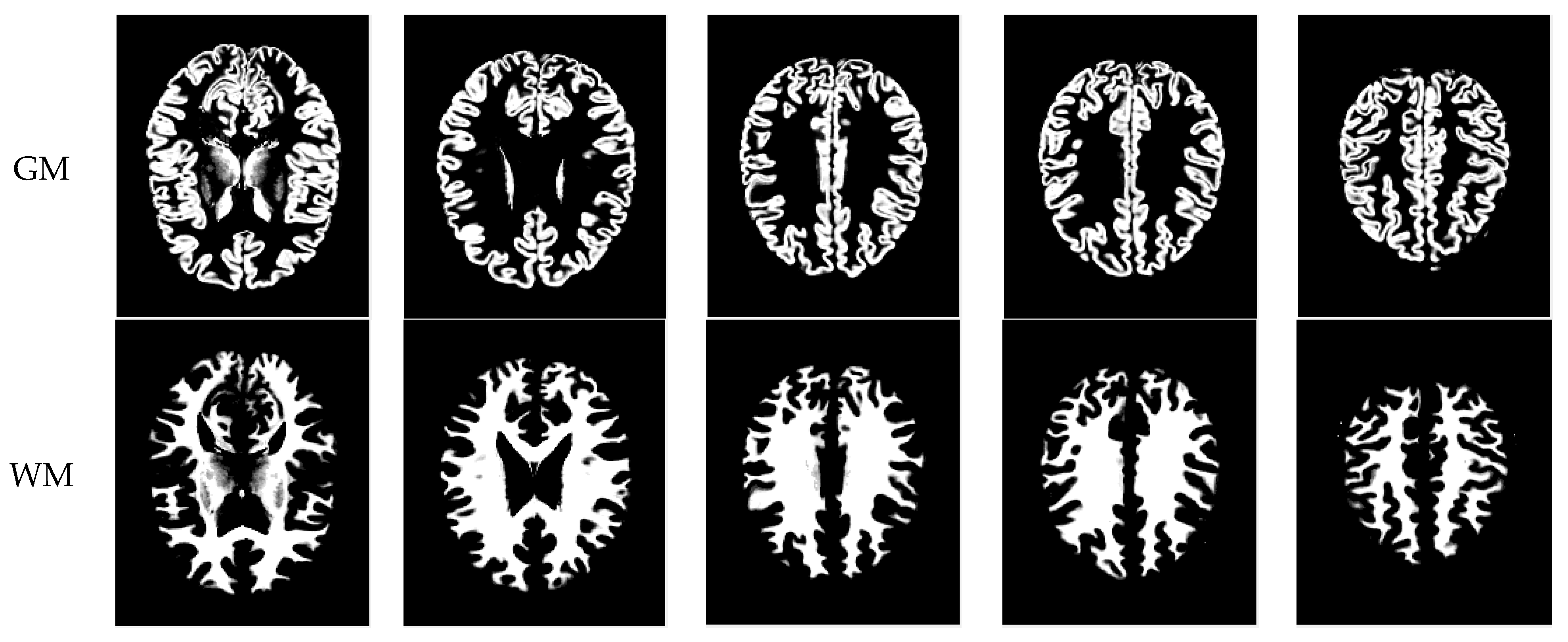
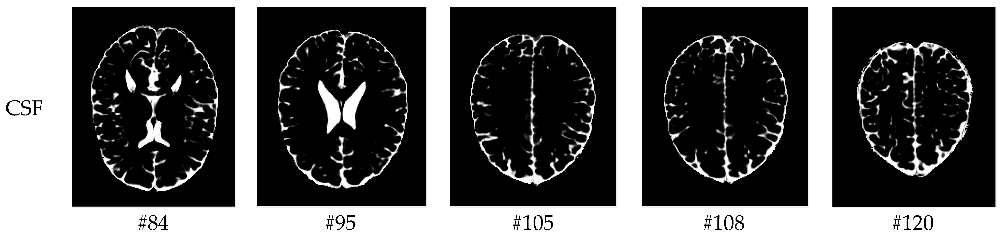
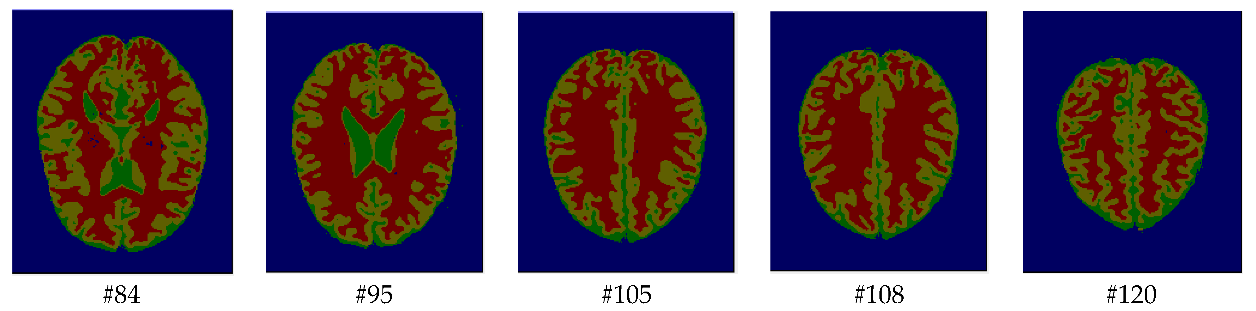
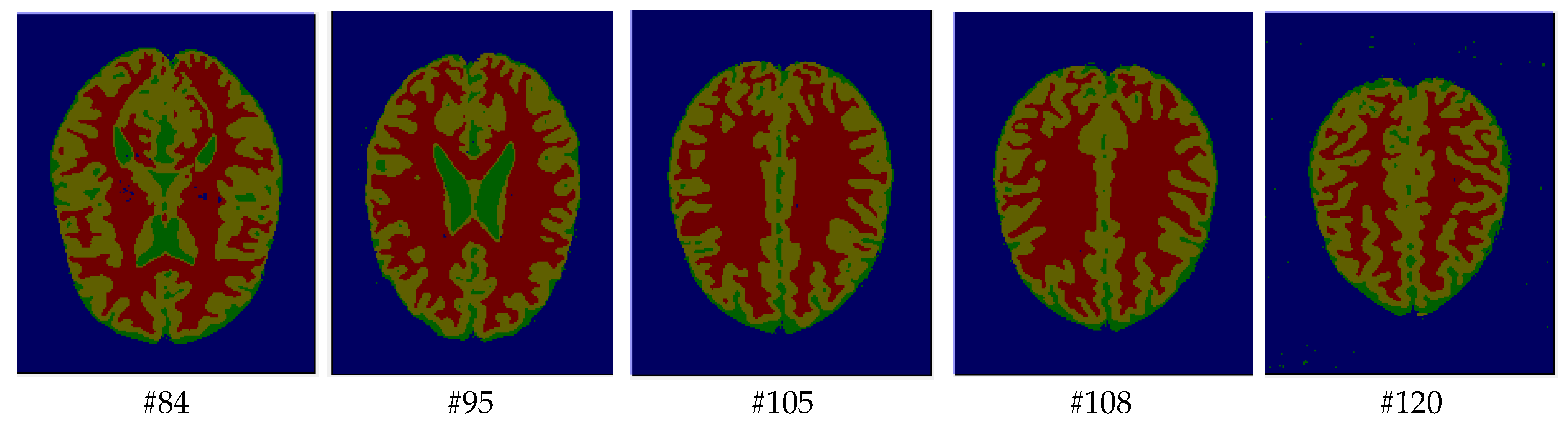
- The proposed method improves the accuracy of segmenting brain images into three tissues: GM, WM, and CSF.
- -
- This manuscript primarily focuses on brain tissue segmentation to aid experts and radiologists.
2. Hidden Markov Random Field Model
- Where s ∉ V(s) , a site is not a neighborhood to itself.
- Where s ∈ V(t) t ∈ V(s), the neighborhood relationship is symmetrical.
3. Whale Optimization Algorithm
- Shrinking encircling mechanism: According to Equation (15), the decrease in the value of A depends on the decrease in the value of the control parameter a. Note that the search agent approaches the current optimal solution when the value of the random variable A is set in the range [−1, 1].
- Spiral updating position: To capture food, humpback whales take a spiral-shaped path around their prey. Equations (18) and (19) are used to reproduce the helix-shaped movement of humpback whales and to calculate the distance between the whale and the prey, respectively:
4. HMRF and Whale Optimization Algorithm (HMRF-WOA)
| Algorithm 1: The main steps of our proposed segmentation method of brain MRIs by the HMRF-WOA. |
Input: Initialize randomly a population
|
5. Experimental Results
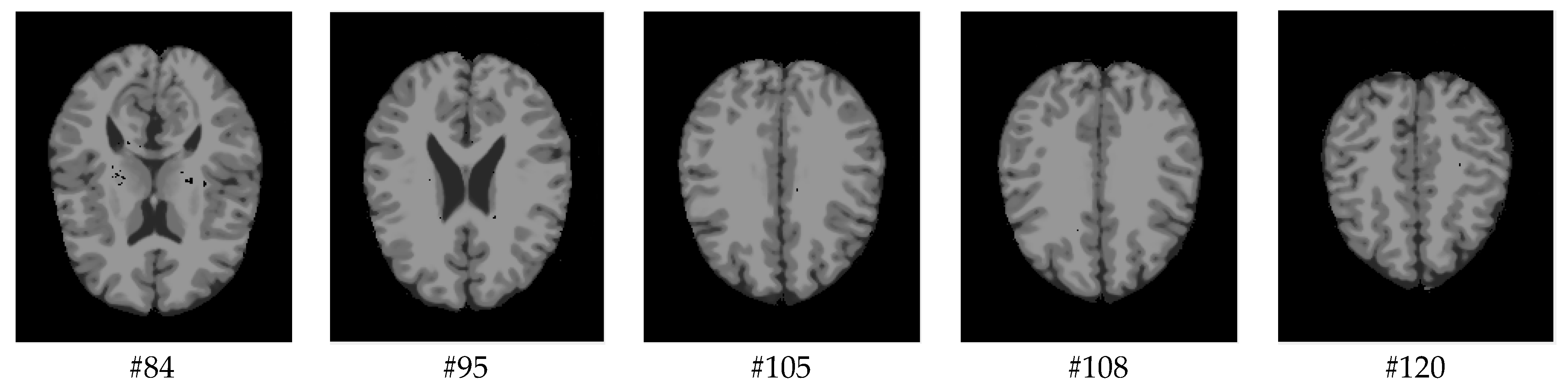
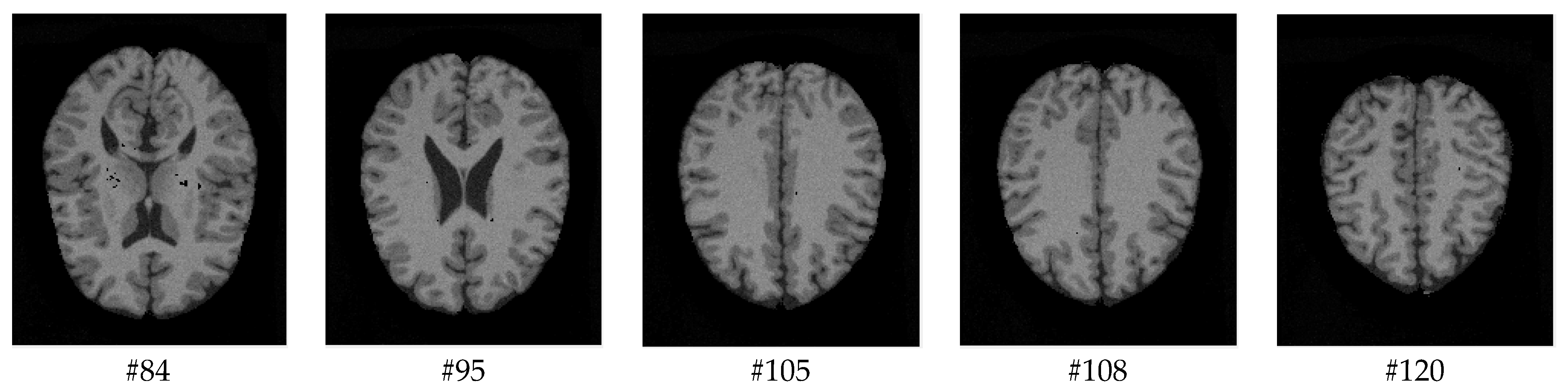
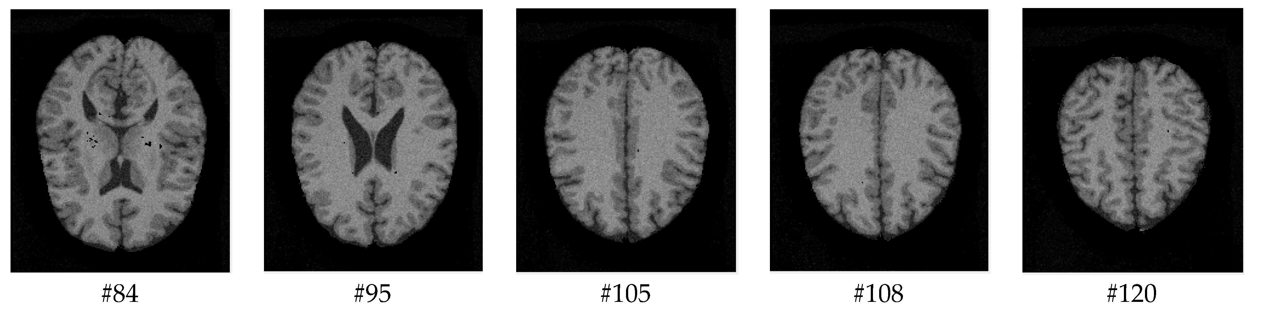
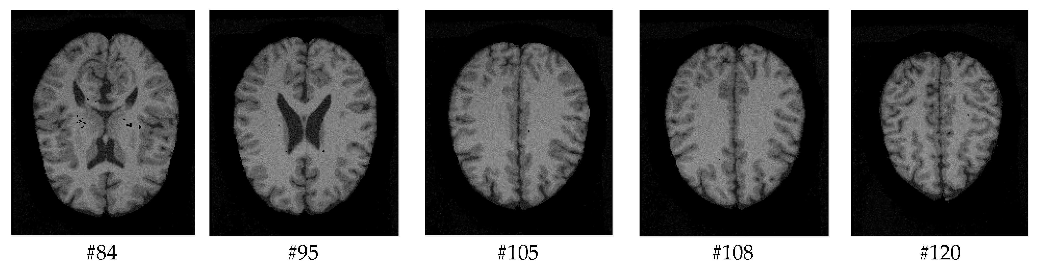
5.1. Datasets
5.2. Performance Measures
5.2.1. Dice Coefficient (Dice)
5.2.2. Jaccard Coefficient (JC)
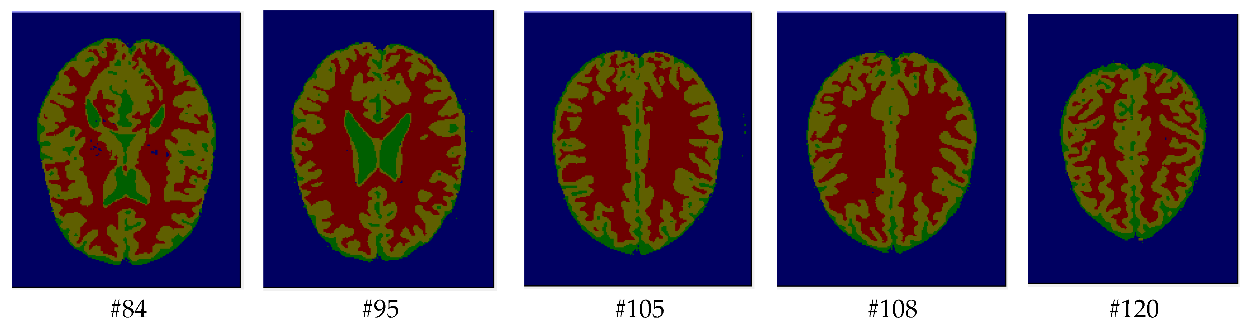
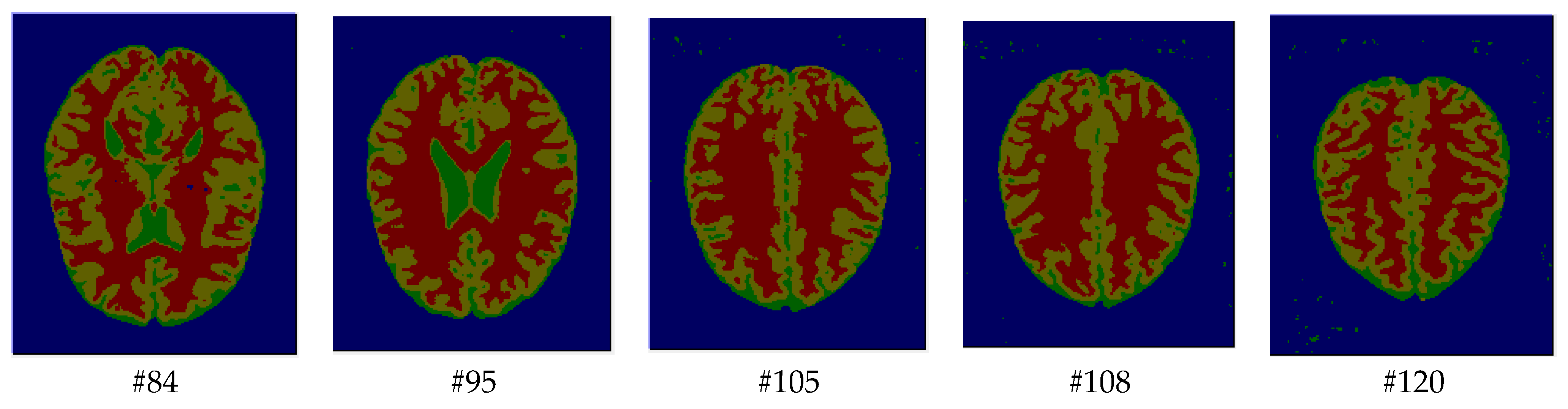
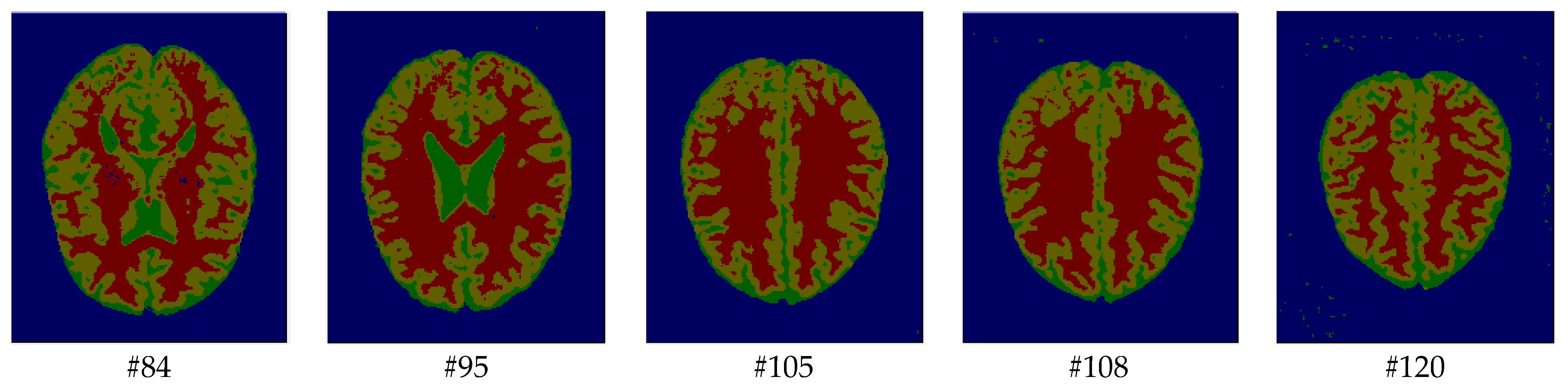
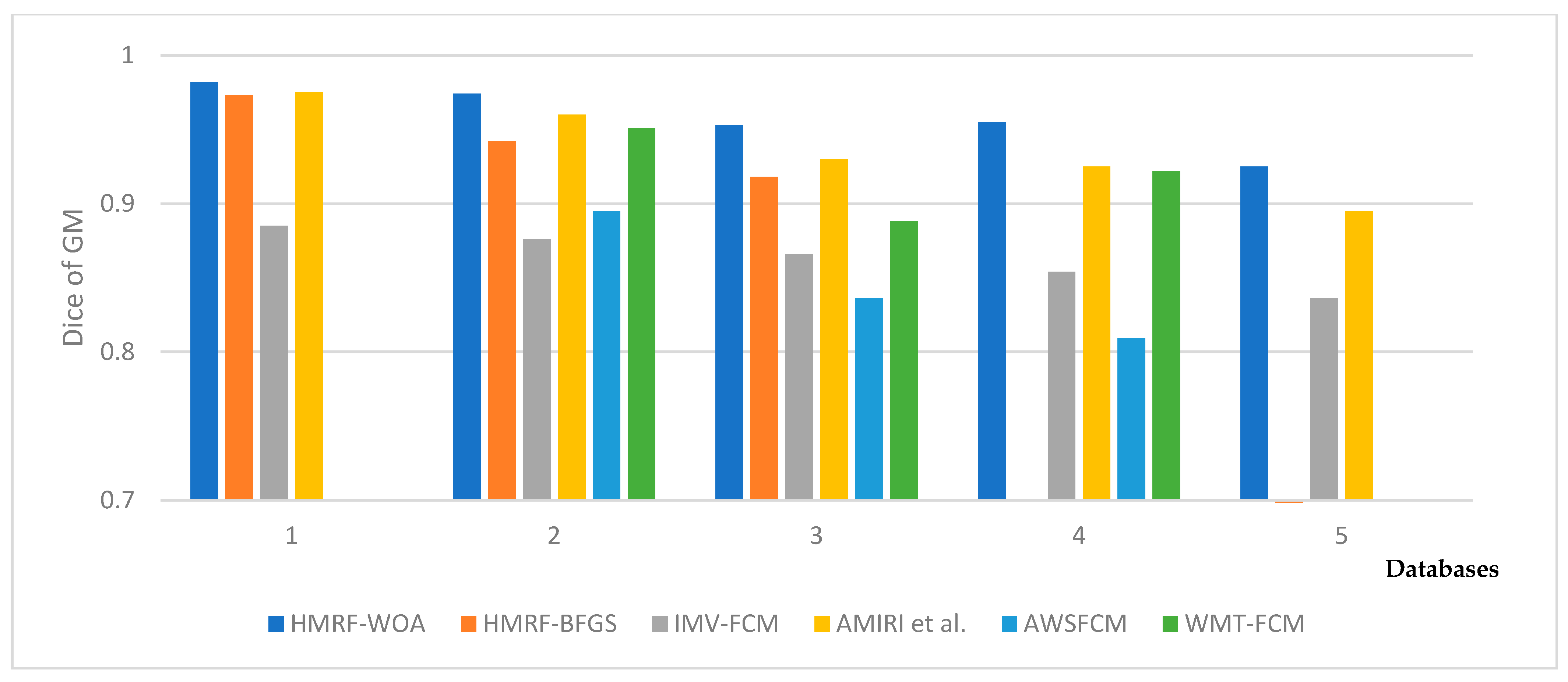
5.3. Discussion
- The weight β associated with the pair of neighboring pixels is a positive constant that controls the size of homogeneous regions. Also, increasing the value of the β parameter e can increase the contribution of the neighboring sites in the estimation of the class of a given pixel. So, we chose β = 1 as we obtained a good performance for the segmentation method using this value.
- The whale population size parameter represents the total number of whales used in the WOA to optimize the MRI image segmentation. We have tested three values of this parameter (10, 20, and 30). Using 20 search agents gave the best accuracy result and a good computational cost compared to other values.
- The number of iterations represents the number of loops used to evaluate the HMRF-WOA process. We tested values ranging between 5 and 40 iterations. The best result was obtained with 20 iterations.
Computational Complexity Analysis
- Initializing the whale population is O(N), where N is the size of the population.
- Computing the fitness value of the initial population is O(N).
- Obtaining the best solution is O(N2).
- Iteration, updating whale population, and evaluating fitness are O(2N).
- Iteration and obtaining the best solution are O(N2).
6. Conclusions
Author Contributions
Funding
Institutional Review Board Statement
Data Availability Statement
Conflicts of Interest
References
- Falaq, N.; Yasir, S. Human brain disorders: A Review. Open Biol. J. 2020, 8, 6–21. [Google Scholar] [CrossRef] [Scilit]
- Catalina, T.G.; Geers, A.J.; Peters, J.; Weese, J.; Pinto, K.; Karim, R.; Ammar, M.; Daoudi, A.; Margeta, J.; Sandoval, Z.; et al. Benchmark for Algorithms Segmenting the left atrium from 3D CT and MRI datasets. IEEE Trans. Med. Imaging 2015, 34, 1460–1473. [Google Scholar] [CrossRef] [Scilit] [PubMed]
- Abdelaziz, D.; Said, M.; Chikh, M. Automatic segmentation of the left atrium on CT images. In Statistical Atlases and Computational Models of the Heart. Imaging and Modelling Challenges. STACOM 2013; Springer: Berlin/Heidelberg, Germany, 2014; Volume 8330, pp. 14–23. [Google Scholar] [CrossRef] [Scilit]
- Abdelaziz, D.; Said, M.; Chikh, M. Automatic Segmentation of the Right Ventricle by Active Shape Model and a Distance Transform. JMIHI J. Med. Imaging Health Inform. 2015, 5, 27–35. [Google Scholar] [CrossRef] [Scilit]
- Qaiser, M.; Alipoor, M.; Chodorowski, A.; Mehnert, A.; Persson, M. Multimodal MR Brain segmentation using Bayesian based Adaptive Mean-Shift. MIDAS J. 2013. [Google Scholar] [CrossRef] [Scilit]
- Henri, A.V.; Fedde, V.; Wiro, J. Auto knn: Brain tissue segmentation using automatically trained k-nearest-neighbor classification. MIDAS J. 2011. [Google Scholar] [CrossRef] [Scilit]
- Fiaz, M.; Ali, K.; Rehman, A.; Gul, M.J.; Jung, S.K. Brain MRI Segmentation using rule-based hybrid approach. arXiv 2019, arXiv:1902.04207. [Google Scholar] [CrossRef] [Scilit]
- Jalab, H.A.; Hasan, A.M. Magnetic resonance imaging segmentation techniques of brain tumors: A Review. Arch. Neurosci. 2019, 6, e84920. [Google Scholar] [CrossRef] [Scilit]
- Hasan, A.M.; Meziane, F.; Aspin, R.; Jalab, H. Segmentation of Brain Tumors in MRI Images using three-dimensional active contour without edge. Symmetry 2016, 8, 132. [Google Scholar] [CrossRef] [Scilit]
- Ahmadvand, A.; Yousefi, S.; Manzuri, M. A novel markov random field model based on region adjacency graph for T1 magnetic resonance imaging brain segmentation. IJIST Int. J. Imaging Syst. Technol. 2017, 27, 78–88. [Google Scholar] [CrossRef] [Scilit]
- Jianhua, S.; Lei, Y. Brain tissue segmentation via non-local fuzzy c-means clustering combined with markov random field. MBE Math. Biosci. Eng. 2021, 19, 1891–1908. [Google Scholar] [CrossRef] [Scilit]
- Shah, S.A.; Chauhan, N.C. An automated approach for segmentation of Brain MR Images using gaussian mixture model based Hidden Markov Random Field with Expectation Maximization. BJHMR Br. J. Healthc. Med. Res. 2015, 2, 57. [Google Scholar] [CrossRef] [Scilit]
- Chen, M.; Yan, Q.; Qin, M. A segmentation of brain MRI images utilizing intensity and contextual information by Markov random field. Comput. Assist. Surg. 2017, 22, 200–211. [Google Scholar] [CrossRef] [Scilit] [PubMed]
- Alansary, A.; Soliman, A.; Khalifa, F.; Elnakib, A.; Mostapha, M.; Nitzken, M.; Casanova, M.; El-Baz, A. MAP Based framework for segmentation of MR Brain Images based on visual appearance and prior shape. MIDAS J. 2014. [Google Scholar] [CrossRef] [Scilit]
- Elhachemi, G.; Samy, A.; Dominique, M.; Ramdane, M. Hidden markov random field model and broyden fletcher goldfarb shanno algorithm for brain image segmentation. JETAI J. Exp. Theor. Artif. Intell. 2017, 30, 415–427. [Google Scholar] [CrossRef] [Scilit]
- Castillo, D.; Peis, I.; Martínez, F.; Segovia, F.; Górriz, J.; Ramírez, J.; Salas, D. A Heavy tailed expectation maximization hidden markov random field model with applications to segmentation of MRI. Front. Neurosci. 2017, 11, 66. [Google Scholar] [CrossRef] [Scilit]
- Hua, L.; Gu, Y.; Gu, X.; Xue, J.; Ni, T. A novel brain MR image segmentation method using an improved multi view fuzzy c-means clustering algorithm. Front. Neurosci. 2021, 15, 662674. [Google Scholar] [CrossRef] [Scilit]
- Zhao, L.; Asis-Cruz, J.D.; Feng, X.; Wu, Y.; Kapse, K.; Largent, A.; Quistorff, J.; Lopez, C.; Wu, D.; Qing, K.; et al. Automated 3D fetal brain segmentation using an optimized deep learning approach. AJNR Am. J. Neuroradiol. 2022, 43, 448–454. [Google Scholar] [CrossRef] [Scilit]
- Brudfors, M.; Balbastre, Y.; Ashburner, J.; Rees, G.; Nachev, P.; Ourselin, S.; Cardoso, M.J. An MRF Unet product of experts for image segmentation. In Proceedings of the Machine Learning Research, Proceedings of the Fourth Conference on Medical Imaging with Deep Learning, Lübeck, Germany, 7–9 July 2021; ML Research Press: Westminster, UK, 2021; Volume 143, pp. 48–59. [Google Scholar]
- Bento, M.; Fantini, I.; Park, J.; Rittner, L.; Frayne, R. Deep learning in large and multi site structural brain MR imaging datasets. Front. Neuroinform. 2022, 15, 805669. [Google Scholar] [CrossRef] [Scilit] [PubMed]
- Lee, B.; Yamanak, K.; Nagaraj, M.; Muhammad, A.; Choi, J. Automatic segmentation of brain MRI using a novel patch-wise U net deep architecture. PLoS ONE 2022, 17, e0264231. [Google Scholar] [CrossRef] [Scilit]
- Renukadevi, T.; Saraswathi, K.; Prabu, P.; Venkatachalam, K. Brain image classification using time frequency extraction with histogram intensity similarity. CSSE Comput. Syst. Sci. Eng. 2022, 41, 645–660. [Google Scholar] [CrossRef] [Scilit]
- Maqsood, S.; Damasevicius, R.; Shah, F.M. An Efficient Approach for the Detection of Brain Tumor Using Fuzzy Logic and U-NET CNN Classification. In Computational Science and Its Applications—ICCSA; Lecture Notes in Computer Science; Springer: Cham, Switzerland, 2021; pp. 105–118. [Google Scholar] [CrossRef] [Scilit]
- Khorram, B.; Yazdi, M. A New optimized thresholding method using ant colony algorithm for MR brain image segmentation. J. Digit. Imaging 2019, 32, 162–174. [Google Scholar] [CrossRef] [Scilit] [PubMed]
- Thuy, X.; Patrick, S.; Hamouche, O. Segmentation of MR Brain images through Hidden Markov Random Field and hybrid metaheuristic algorithm. IEEE Trans. Image Process. 2020, 29, 6507–6522. [Google Scholar] [CrossRef] [Scilit] [PubMed]
- Kadry, S.; Damaševičius, R.; Taniar, D.; Rajinikanth, V.; Lawal, I.A. U-Net Supported Segmentation of Ischemic-Stroke-Lesion from Brain MRI Slices. In Proceedings of the Seventh International Conference on Bio Signals, Images, and Instrumentation (ICBSII), Chennai, India, 25–27 March 2021; pp. 1–5. [Google Scholar] [CrossRef] [Scilit]
- Ramya, J.; Maheswari, B.U.; Rajakumar, M.P.; Sonia, R. Alzheimer’s Disease Segmentation and Classification on MRI Brain Images Using Enhanced Expectation Maximization Adaptive Histogram (EEM-AH) and Machine Learning. Inf. Technol. Control. 2022, 51, 786–800. [Google Scholar] [CrossRef] [Scilit]
- Rangaraju, B.; Chinnadurai, T.; Natarajan, S.; Raja, V. Dual Attention Aware Octave Convolution Network for Early-Stage Alzheimer’s Disease Detection. Inf. Technol. Control. 2024, 53, 302–316. [Google Scholar] [CrossRef] [Scilit]
- Clifford, P.; Hammersley, J. Markov Fields on Finite Graphs and Lattices; University of Oxford: Oxford, UK, 1971. [Google Scholar]
- Potts, R. Some generalized order-disorder transformations. Math. Proc. Camb. Philos. Soc. 1952, 48, 106–109. [Google Scholar] [CrossRef] [Scilit]
- Mirjalili, S.; Lewi, A. The whale optimization algorithm. AES Adv. Eng. Softw. 2016, 95, 51–67. [Google Scholar] [CrossRef] [Scilit]
- Goldbogen, J.; Friedlaender, A.; Calambokidis, J.; McKenna, M.; Malene, S.; Nowacek, D. Integrative approaches to the study of baleen whale diving behavior, feeding performance, and foraging ecology. BioScience 2013, 63, 90–100. [Google Scholar] [CrossRef] [Scilit]
- Hof, P.; Gucht, E. Structure of the cerebral cortex of the humpback whale, megaptera novaeangliae (Cetacea, Mysticeti, Balaenopteridae). Anat. Rec. 2007, 290, 1–31. [Google Scholar] [CrossRef] [PubMed]
- Watkins, W.; Schevill, W.E. Aerial Observation of feeding behavior in four baleen whales: Balaena glacialis, balaenoptera borealis, Megaptera novaeangliae, and Balaenoptera physalus. J. Mammal. 1979, 60, 155–163. [Google Scholar] [CrossRef] [Scilit]
- Nasiri, J.; Khiyabani, F.M.; Yoshise, A. A whale optimization algorithm (WOA) approach for clustering. CMS Cogent Math. Stat. 2018, 5, 1483565. [Google Scholar] [CrossRef] [Scilit]
- Yue, Y.; You, H.; Wang, S.; Cao, L. Improved whale optimization algorithm and its application in heterogeneous wireless sensor networks. IJDSN Int. J. Distrib. Sens. Netw. 2021, 17. [Google Scholar] [CrossRef] [Scilit]
- Mohammed, A.K.; Suhaib, A.A.; Zakariya, Y.A. Improving whale optimization algorithm for feature selection with a time-varying transfer function. NACO Numer. Algebra Control Optim. 2021, 11, 87–98. [Google Scholar] [CrossRef] [Scilit]
- Zhihong, Y.; Shuqian, W.; Bin, L.; Xinde, L. Application of whale Optimization Algorithm in Optimal Allocation of Water Resources. E3S Web Conf. 2018, 53, 04019. [Google Scholar] [CrossRef] [Scilit]
- Reddy, P.; Reddy, V.; Manohar, T. Whale optimization algorithm for optimal sizing of renewable resources for loss reduction in distribution systems. Renewables 2017, 4, 3. [Google Scholar] [CrossRef] [Scilit]
- Nadimi, S.; Zamani, H.; Asghari, V.Z.; Mirjalili, S. A Systematic Review of the Whale Optimization Algorithm: Theoretical Foundation, Improvements, and Hybridizations. Arch. Comput. Methods 2023, 30, 4113–4159. [Google Scholar] [CrossRef] [Scilit] [PubMed]
- Mostafa, A.; Hassanien, A.E.; Houseni, M.; Hefny, H. Liver segmentation in MRI images based on whale optimization algorithm. Multimed. Tools Appl. 2017, 76, 24931–24954. [Google Scholar] [CrossRef] [Scilit]
- Kaur, R.; Khehra, B.S. Modified Whale Optimisation Algorithm and minimum CROSS entropy-based segmentation of CT Liver image. J. Pharm. Negat. Results 2023, 14, 2908–2931. [Google Scholar]
- Chakraborty, S.; Saha, A.K.; Nama, S.; Debnath, S. COVID-19 X-ray image segmentation by modified whale optimization algorithm with population reduction. Comput. Biol. Med. 2021, 139, 104984. [Google Scholar] [CrossRef] [Scilit] [PubMed]
- Kwan, R.; Evans, A.; Pike, G. MRI simulation-based evaluation of image-processing and classification methods. IEEE Trans. Med. Imaging 1999, 18, 1085–1097. [Google Scholar] [CrossRef] [Scilit] [PubMed]
- Dice, L. Measures of the Amount of Ecologic Association between Species. Ecology 1945, 26, 297–302. [Google Scholar] [CrossRef] [Scilit]
- Jaccard, P. The Distribution of the Flora of the Alpine Zone. New Phytol. 1912, 11, 37–50. [Google Scholar] [CrossRef] [Scilit]
- Amiri, S.; Movahedi, M.; Kazemi, K.; Parsaei, H. 3D cerebral MR image segmentation using multiple-classifier system. Med. Biol. Eng. Comput. 2017, 55, 353–364. [Google Scholar] [CrossRef] [Scilit] [PubMed]
- Mishro, P.; Agrawal, S.; Panda, R.; Abraham, A. A Novel Type-2 Fuzzy C-Means Clustering for Brain MR Image Segmentation. IEEE Trans. Cybern. 2021, 51, 3901–3912. [Google Scholar] [CrossRef] [Scilit] [PubMed]
- Yunlan, Z.; Zhiyong, H.; Hangjun, C.; Fang, X.; Man, L.; Mengyao, W.; Daming, S. Segmentation of Brain Tissues from MRI Images Using Multitask Fuzzy Clustering Algorithm. JHE J. Health Eng. 2023, 4387134. [Google Scholar] [CrossRef] [Scilit] [PubMed]
- Jonas, G.B.; Pilar, G. Pseudo Label Assisted Self-Organizing Maps for Brain Tissue Segmentation in Magnetic Resonance Imaging. JDI J. Digit. Imaging 2022, 35, 180–192. [Google Scholar] [CrossRef] [Scilit] [PubMed]
- Jonas, G.B.; Pilar, G. Segmentation of MRI brain scans using spatial constraints and 3D features. Med. Biol. Eng. Comput. 2020, 58, 3101–3112. [Google Scholar] [CrossRef] [Scilit]
- Padmanaban, S.; Thiruvenkadam, K.; Karuppanagounder, S.; Rangasami, R. A Rapid Knowledge Based Partial Supervision Fuzzy C Means for Brain Tissue Segmentation with CUDA Enabled GPU Machine. IJIST Int. J. Imaging Syst. Technol. 2019, 29, 547–560. [Google Scholar] [CrossRef] [Scilit]

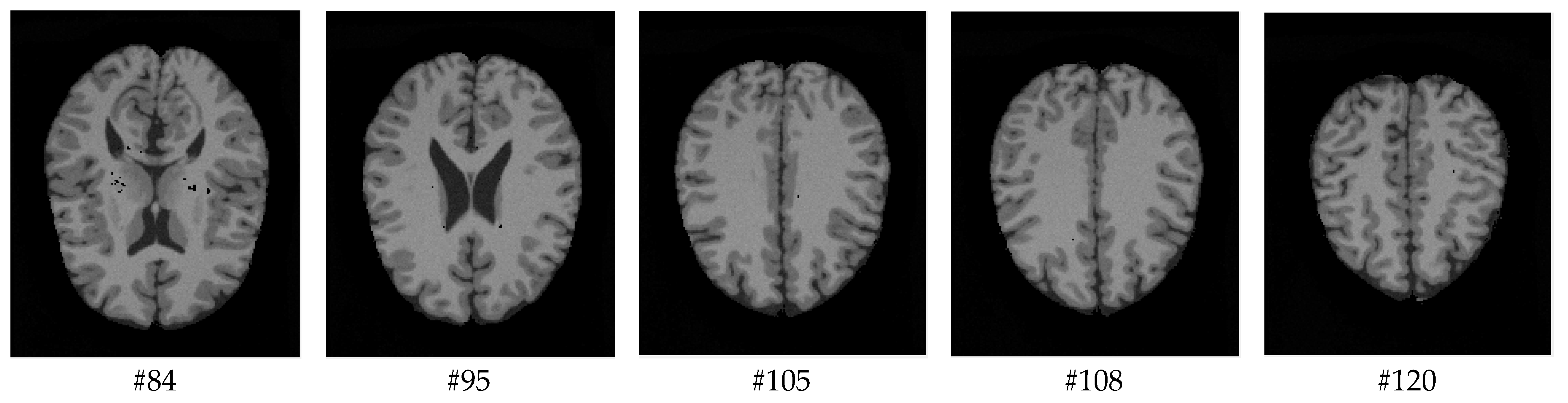




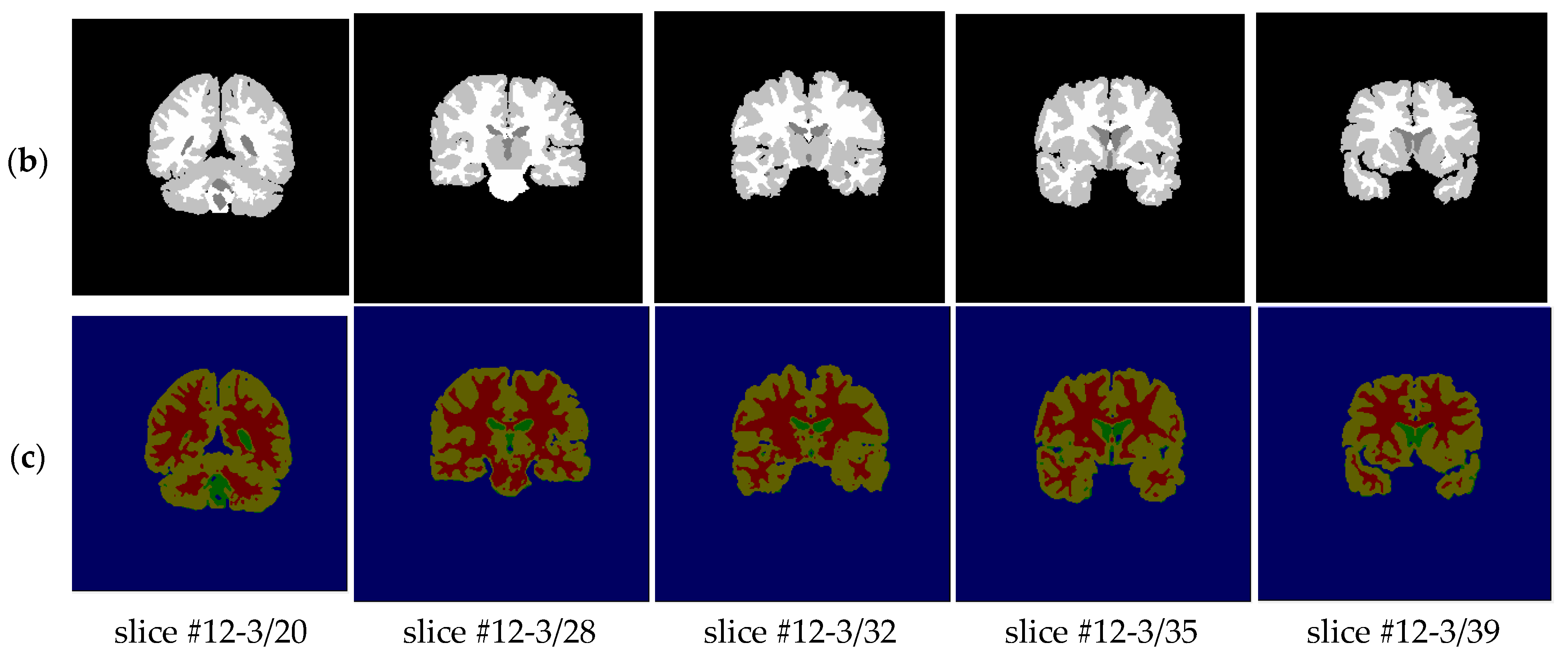

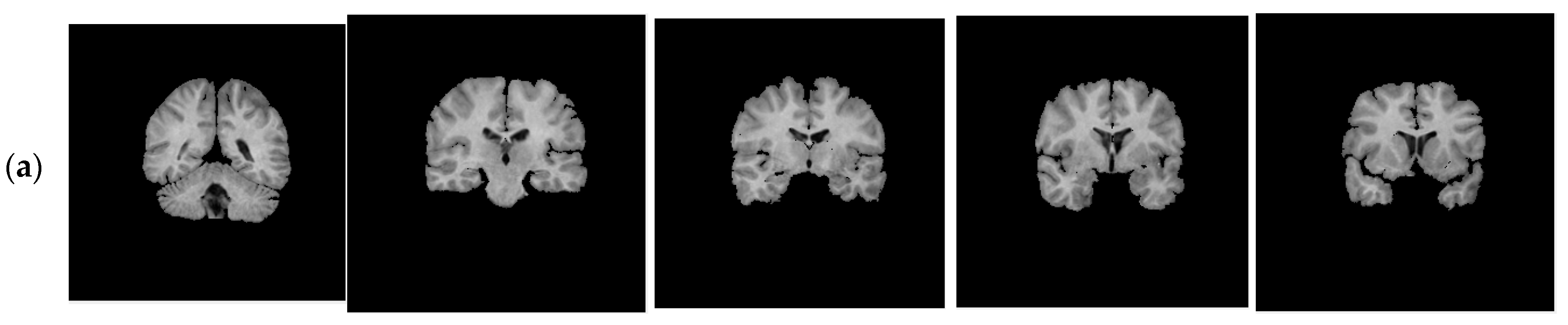
| Databases | Types | Dimensions | Noises (%) | INU * (%) | Voxels (mm) |
|---|---|---|---|---|---|
| 1 | Brainweb | 181 × 217 × 181 | 0 | 0 | 1 × 1 × 1 |
| 2 | Brainweb | 181 × 217 × 181 | 3 | 20 | 1 × 1 × 1 |
| 3 | Brainweb | 181 × 217 × 181 | 5 | 20 | 1 × 1 × 1 |
| 4 | Brainweb | 181 × 217 × 181 | 7 | 20 | 1 × 1 × 1 |
| 5 | Brainweb | 181 × 217 × 181 | 9 | 40 | 1 × 1 × 1 |
| 6 | IBSR | 256 × 256 × 63 | - | - | 1 × 3 × 1 |
| Tissue | Method | Database 1 | Database 2 | Database 3 | Database 4 | Database 5 |
|---|---|---|---|---|---|---|
| GM | HMRF-WOA | 0.982 | 0.974 | 0.953 | 0.955 | 0.935 |
| HMRF-BFGS | 0.973 | 0.942 | 0.918 | NA * | NA * | |
| MV-FCM | 0.885 | 0.876 | 0.866 | 0.854 | 0.836 | |
| Amiri et al. [47] | 0.975 | 0.960 | 0.930 | 0.925 | 0.895 | |
| AWSFCM | NA * | 0.895 | 0.836 | 0.809 | NA * | |
| WMT-FCM | NA * | 0.9508 | 0.9219 | 0.8818 | NA * | |
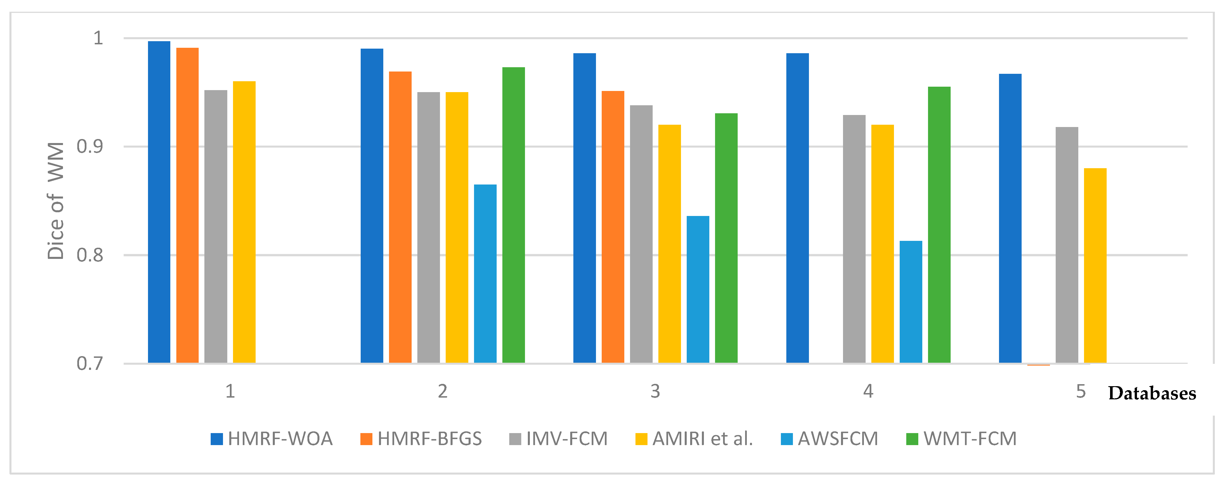
| WM | HMRF-WOA | 0.997 | 0.990 | 0.986 | 0.986 | 0.976 |
| HMRF-BFGS | 0.991 | 0.969 | 0.951 | NA * | NA * | |
| IMV-FCM | 0.952 | 0.950 | 0.938 | 0.929 | 0.918 | |
| Amiri et al. [47] | 0.960 | 0.950 | 0.920 | 0.920 | 0.880 | |
| AWSFCM | NA * | 0.865 | 0.836 | 0.813 | NA * | |
| WMT-FCM | NA * | 0.9729 | 0.9550 | 0.9306 | NA * | |
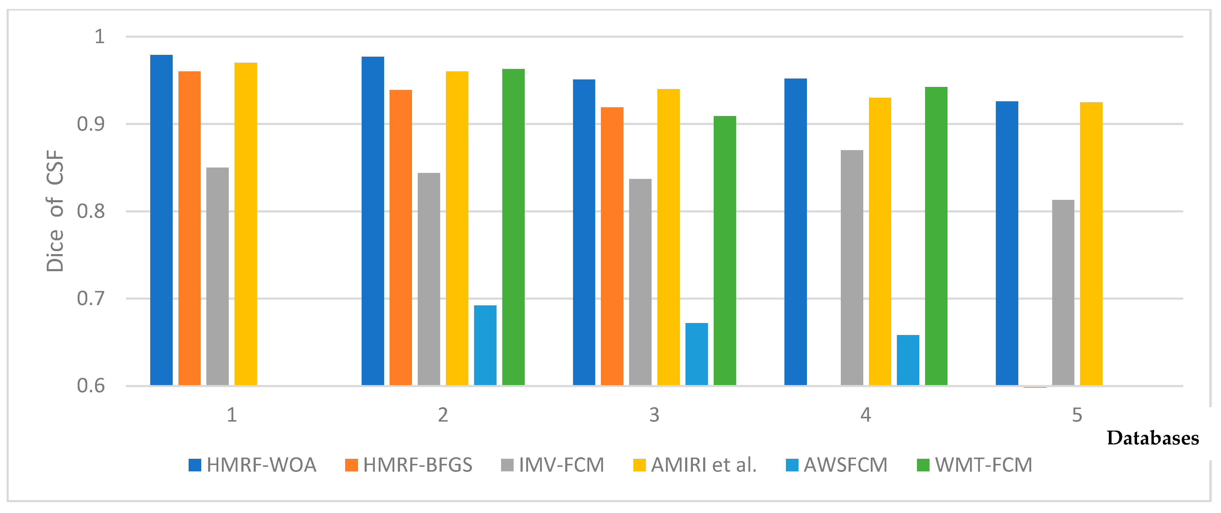
| CSF | HMRF-WOA | 0.979 | 0.977 | 0.951 | 0.952 | 0.968 |
| HMRF-BFGS | 0.960 | 0.939 | 0.919 | NA * | NA * | |
| IMV-FCM | 0.850 | 0.844 | 0.837 | 0.870 | 0.813 | |
| Amiri et al. [47] | 0.970 | 0.960 | 0.940 | 0.930 | 0.925 | |
| AWSFCM | NA * | 0.692 | 0.672 | 0.658 | NA * | |
| WMT-FCM | NA * | 0.9628 | 0.9424 | 0.9090 | NA * |
| Tissue | Method | Database 1 | Database 2 | Database 3 | Database 4 | Database 5 |
|---|---|---|---|---|---|---|
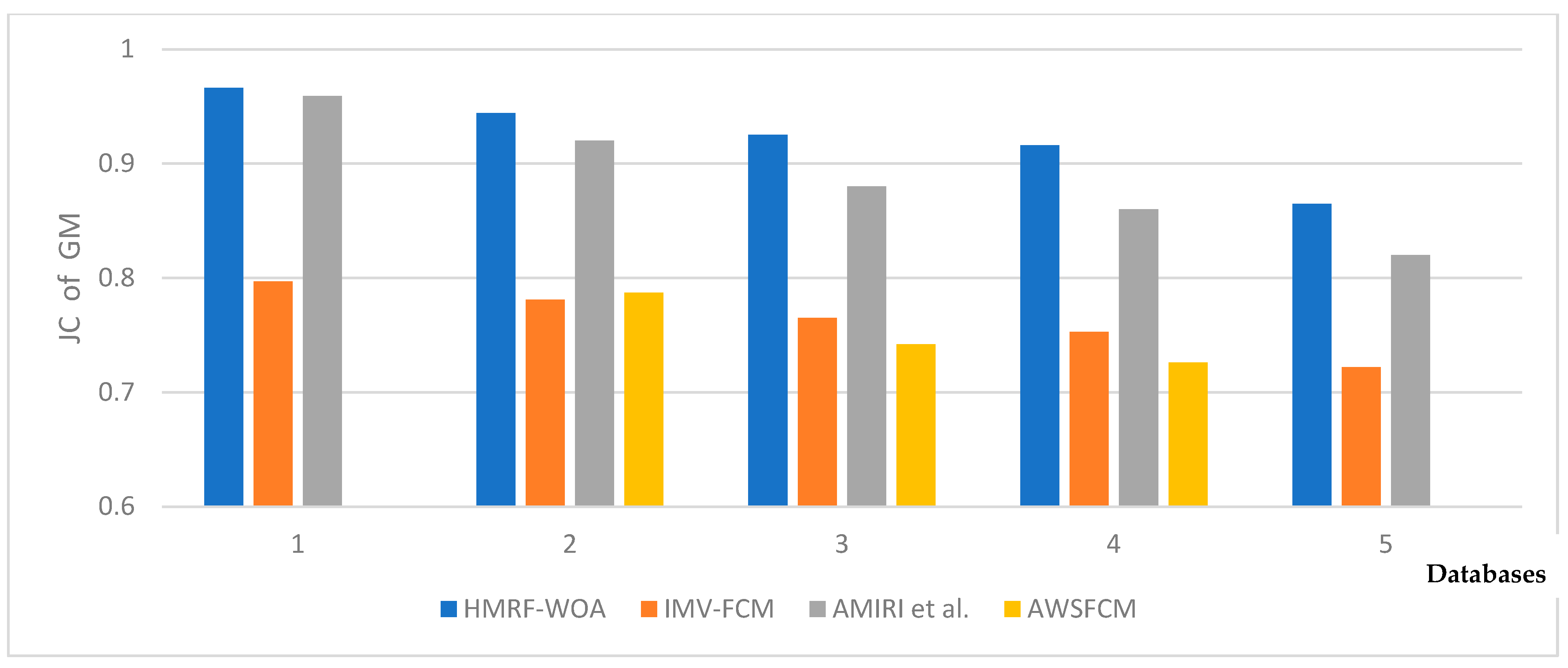
| GM | HMRF-WOA | 0.966 | 0.944 | 0.925 | 0.916 | 0.879 |
| HMRF-BFGS | NA * | NA * | NA * | NA * | NA * | |
| IMV-FCM | 0.797 | 0.781 | 0.765 | 0.753 | 0.722 | |
| Amiri et al. [47] | 0.959 | 0.920 | 0.880 | 0.860 | 0.820 | |
| AWSFCM | NA * | 0.787 | 0.742 | 0.726 | NA * | |
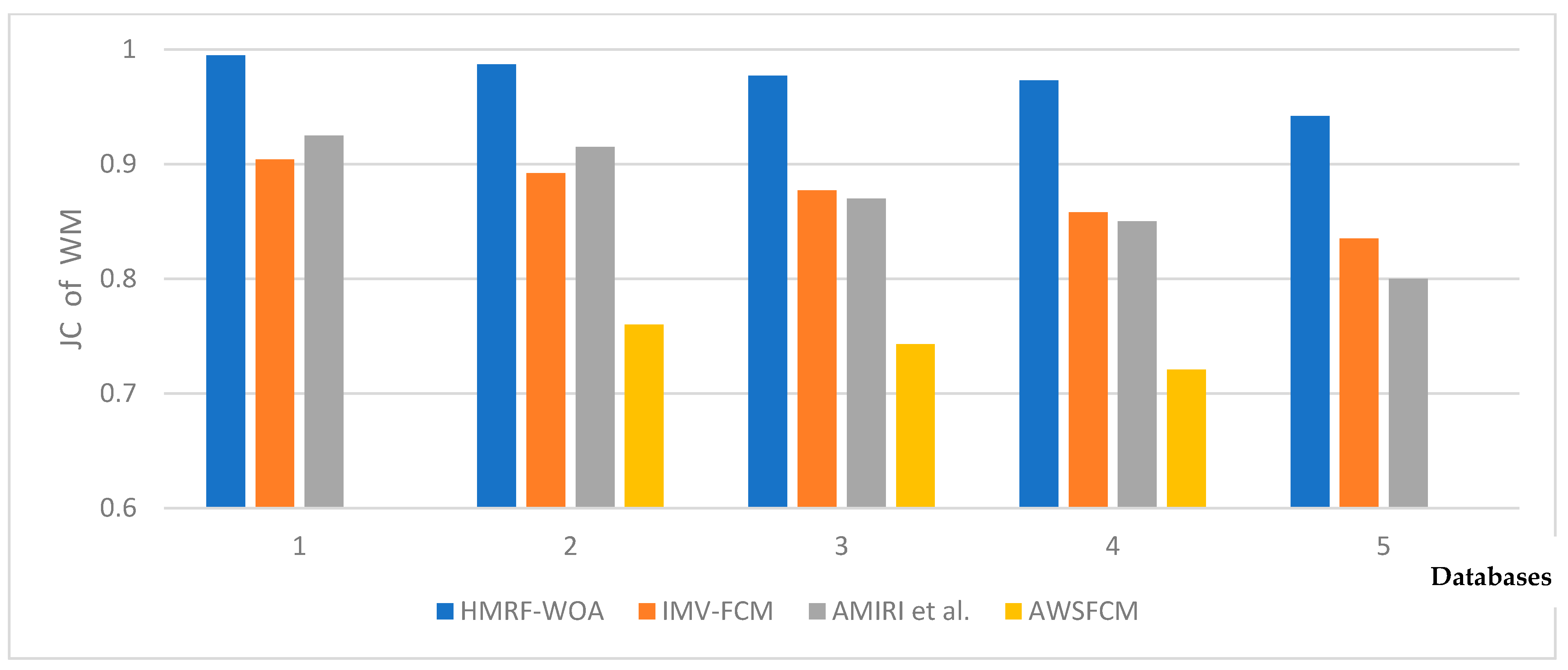
| WM | HMRF-WOA | 0.995 | 0.987 | 0.977 | 0.973 | 0.953 |
| HMRF-BFGS | NA * | NA * | NA * | NA * | NA * | |
| IMV-FCM | 0.904 | 0.892 | 0.877 | 0.858 | 0.835 | |
| Amiri et al. [47] | 0.925 | 0.915 | 0.870 | 0.850 | 0.800 | |
| AWSFCM | NA * | 0.760 | 0.743 | 0.721 | NA * | |
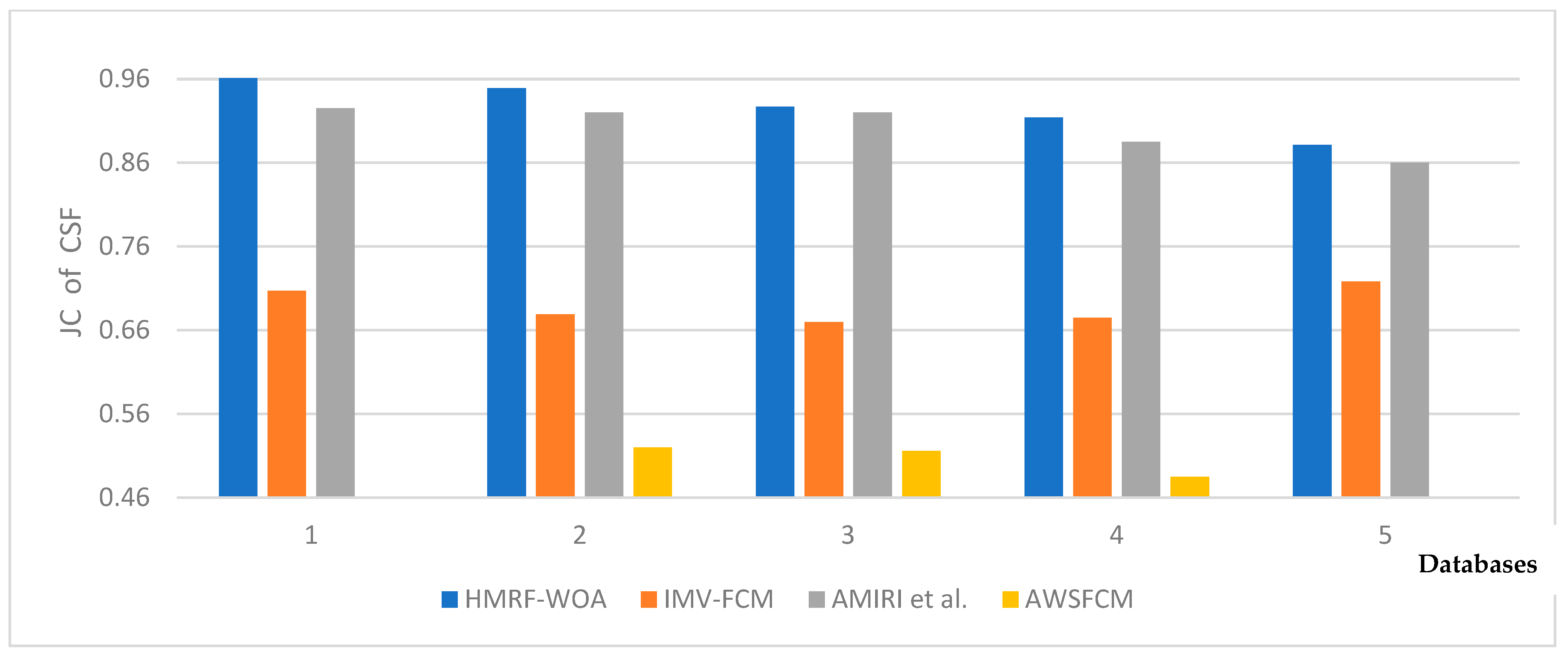
| CSF | HMRF-WOA | 0.961 | 0.949 | 0.927 | 0.914 | 0.939 |
| HMRF-BFGS | NA * | NA * | NA * | NA * | NA * | |
| IMV-FCM | 0.707 | 0.679 | 0.670 | 0.675 | 0.718 | |
| Amiri et al. [47] | 0.925 | 0.920 | 0.920 | 0.885 | 0.860 | |
| AWSFCM | NA * | 0.520 | 0.516 | 0.485 | NA * |
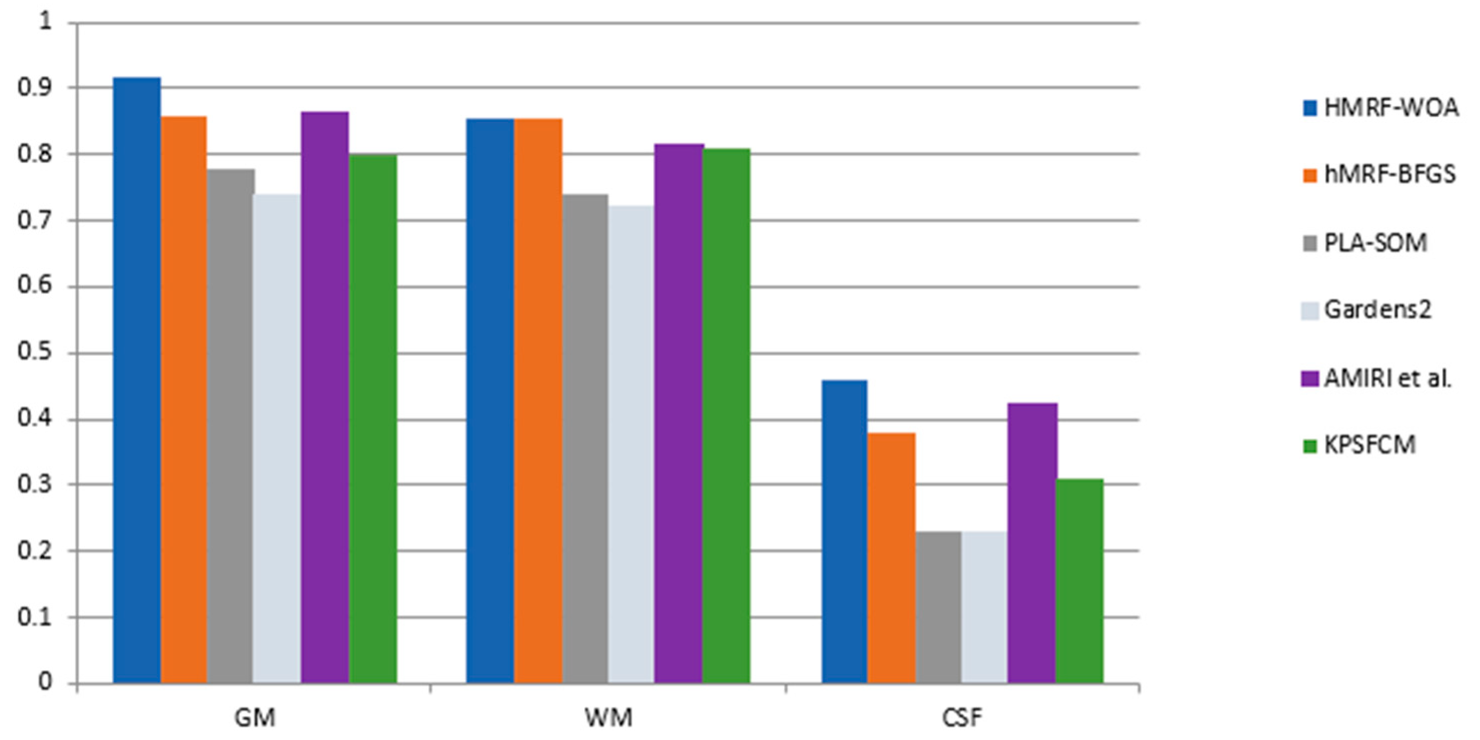
| Tissue | Method | Mean Dice |
|---|---|---|
| GM | HMRF-WOA | 0.916 |
| hMRF-BFGS | 0.859 | |
| PLA-SOM | 0.780 | |
| Amiri et al. [47] | 0.863 | |
| Gardens2 | 0.740 | |
| KPSFCM | 0.80 | |
| WM | HMRF-WOA | 0.856 |
| hMRF-BFGS | 0.855 | |
| PLA-SOM | 0.740 | |
| Amiri et al. [47] | 0.814 | |
| Gardens2 | 0.720 | |
| KPSFCM | 0.81 | |
| CSF | HMRF-WOA | 0.459 |
| hMRF-BFGS | 0.381 | |
| PLA-SOM | 0.230 | |
| Amiri et al. [47] | 0.423 | |
| Gardens2 | 0.230 | |
| KPSFCM | 0.31 |
Disclaimer/Publisher’s Note: The statements, opinions and data contained in all publications are solely those of the individual author(s) and contributor(s) and not of MDPI and/or the editor(s). MDPI and/or the editor(s) disclaim responsibility for any injury to people or property resulting from any ideas, methods, instructions or products referred to in the content. |
© 2024 by the authors. Licensee MDPI, Basel, Switzerland. This article is an open access article distributed under the terms and conditions of the Creative Commons Attribution (CC BY) license (https://creativecommons.org/licenses/by/4.0/).
Share and Cite
Daoudi, A.; Mahmoudi, S. Enhancing Brain Segmentation in MRI through Integration of Hidden Markov Random Field Model and Whale Optimization Algorithm. Computers 2024, 13, 124. https://doi.org/10.3390/computers13050124
Daoudi A, Mahmoudi S. Enhancing Brain Segmentation in MRI through Integration of Hidden Markov Random Field Model and Whale Optimization Algorithm. Computers. 2024; 13(5):124. https://doi.org/10.3390/computers13050124
Chicago/Turabian StyleDaoudi, Abdelaziz, and Saïd Mahmoudi. 2024. "Enhancing Brain Segmentation in MRI through Integration of Hidden Markov Random Field Model and Whale Optimization Algorithm" Computers 13, no. 5: 124. https://doi.org/10.3390/computers13050124
APA StyleDaoudi, A., & Mahmoudi, S. (2024). Enhancing Brain Segmentation in MRI through Integration of Hidden Markov Random Field Model and Whale Optimization Algorithm. Computers, 13(5), 124. https://doi.org/10.3390/computers13050124