


Management of PEComas: A Review of the Role of Radiotherapy

Simple Summary
Abstract
1. Introduction
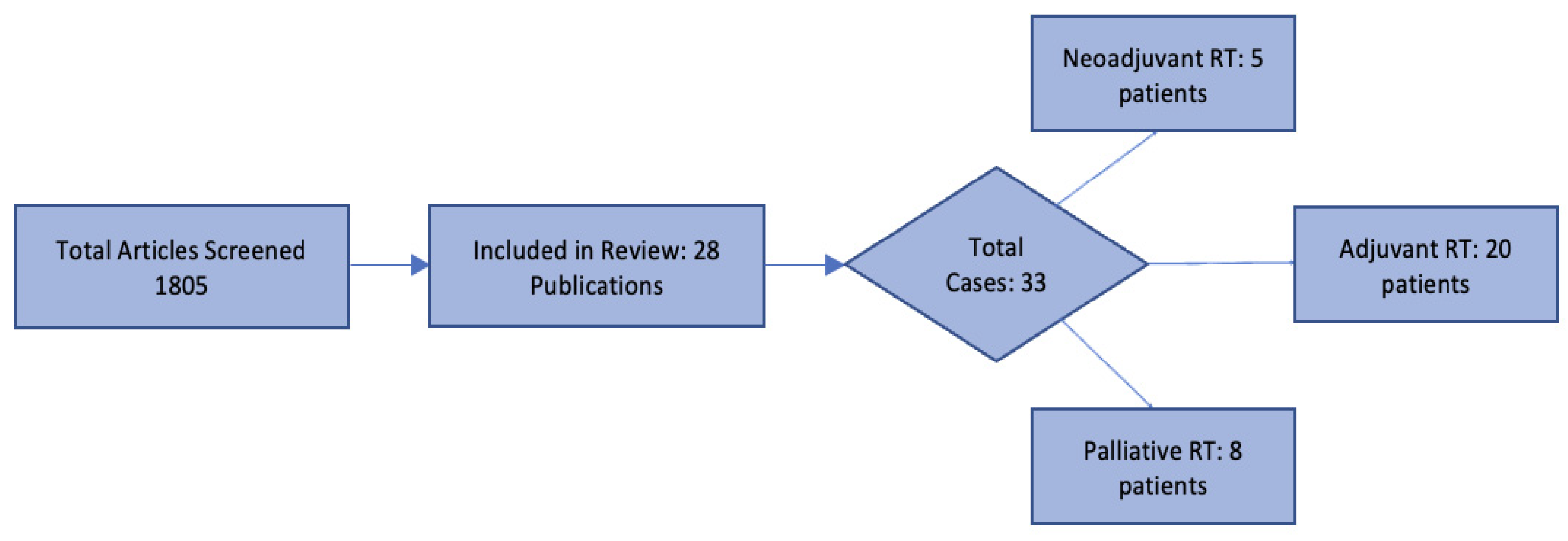
2. Materials and Methods
3. Statistical Analysis
4. Results
4.1. Neoadjuvant Radiotherapy
4.2. Adjuvant Radiotherapy
4.3. Metastatic Disease, Advanced/Unresectable PEComa and Palliative Radiotherapy
5. Conclusions and Future Directions
Author Contributions
Funding
Data Availability Statement
Acknowledgments
Conflicts of Interest
Abbreviations
| AC | adjuvant chemotherapy |
| ACR | adjuvant chemoradiotherapy |
| AR | adjuvant radiotherapy |
| CRT | chemoradiotherapy |
| CSF | colony-stimulating factor |
| Gy | Gray |
| HPF | high-power field |
| IA | Ifosfamide, Adriamycin |
| LN | lymph node |
| mTORi | mammalian target of rapamycin inhibitor |
| NC | neoadjuvant chemotherapy |
| NED | no evidence of disease |
| NR | neoadjuvant radiotherapy |
| PEComa | perivascular epithelioid cell tumor |
| RT | radiotherapy |
| SBRT | stereotactic body radiotherapy |
| UMP | uncertain malignant potential |
| VID | vincristine–ifosfamide–doxorubicin regimen |
Appendix A

| Total Cases | Main Sites | Median Age | Females % | Males % | Mean Follow-Up (Months) | PEComa Cases with Positive Composite Descriptive Outcome (NED for Neoadjuvant/Adjuvant RT; for Palliative RT: Local Response and/or Local Stability and/or Systemic Disease Stability and/or Symptom Relief) | |
|---|---|---|---|---|---|---|---|
| Neoadjuvant RT | 5 | Musculoskeletal 60% | 49 | 80% | 20% | 17 (6–34) | 4 (80%) |
| Adjuvant RT | 20 | Gynecological (including pelvis) 50% | 51 | 80% | 20% | 16 (3–50) | 15 (75%) |
| Palliative RT | 8 | Gynecological (including pelvis) 40% | 46 | 62.5% | 37.5% | 23 (4–48) | 4 (57%) * |
References
- Stacchiotti, S.; Frezza, A.M.; Blay, J.Y.; Baldini, E.H.; Bonvalot, S.; Bovée, J.; Callegaro, D.; Casali, P.G.; Chiang, R.C.; Demetri, G.D.; et al. Ultra-rare sarcomas: A consensus paper from the Connective Tissue Oncology Society community of experts on the incidence threshold and the list of entities. Cancer 2021, 127, 2934–2942. [Google Scholar] [CrossRef]
- Bonetti, F.; Pea, M.; Martignoni, G.; Zamboni, G. PEC and sugar. Am. J. Surg. Pathol. 1992, 16, 307–308. [Google Scholar] [CrossRef]
- Zamboni, G.; Pea, M.; Martignoni, G.; Zancanaro, C.; Faccioli, G.; Gilioli, E.; Pederzoli, P.; Bonetti, F. Clear Cell “Sugar” Tumor of the Pancreas: A Novel Member of the Family of Lesions Characterized by the Presence of Perivascular Epithelioid Cells. Am. J. Surg. Pathol. 1996, 20, 722–730. [Google Scholar] [CrossRef]
- Fletcher, C.D.M.; Unni, K.; Mertens, F. Pathology and Genetics of Tumours of Soft Tissue and Bone, 3rd ed.; World Health Organization Classification of Tumours; IARC Press: Lyon, France, 2002; Volume 5, pp. 221–222. [Google Scholar]
- Folpe, A.L.; Mentzel, T.; Lehr, H.A.; Fisher, C.; Balzer, B.L.; Weiss, S.W. Perivascular epithelioid cell neoplasms of soft tissue and gynecologic origin: A clinicopathologic study of 26 cases and review of the literature. Am. J. Surg. Pathol. 2005, 29, 1558–1575. [Google Scholar] [CrossRef] [PubMed]
- Stacchiotti, S.; Maria Frezza, A.; Demetri, G.D.; Blay, J.Y.; Bajpai, J.; Baldi, G.G.; Baldini, E.H.; Benjamin, R.S.; Bonvalot, S.; Bovée, J.; et al. Retrospective observational studies in ultra-rare sarcomas: A consensus paper from the Connective Tissue Oncology Society (CTOS) community of experts on the minimum requirements for the evaluation of activity of systemic treatments. Cancer Treat. Rev. 2022, 110, 102455. [Google Scholar] [CrossRef] [PubMed]
- Zekry, N.; Rettenmaier, M.A.; Abaid, L.N.; John, C.R.; Micha, J.P.; Brown, J.V., 3rd; Goldstein, B.H. Perivascular epithelioid cell neoplasms: A systematic review of prognostic factors. J. Minim. Invasive Gynecol. 2009, 16, 527–532. [Google Scholar] [CrossRef]
- Cuevas Herreros, Ó.; Escobar Lezcano, L.; Rodriguez Blaco, M.; Artigas Raventós, V. PEComa, a Rare Epithelioid Cell Tumor. Cir. Esp. (Engl. Ed.) 2015, 93, e65–e67. [Google Scholar] [CrossRef]
- Wagner, A.; Ravi, V.; Ganjoo, K.; Tine, B.; Riedel, R.; Chugh, R.; Cranmer, L.; Gordon, E.; Hornick, J.; Kwiatkowski, D.; et al. ABI-009 (nab-sirolimus) in advanced malignant perivascular epithelioid cell tumors (PEComa): Preliminary efficacy, safety, and mutational status from AMPECT, an open label phase II registration trial. J. Clin. Oncol. 2019, 37, 11005. [Google Scholar] [CrossRef]
- Wagner, A.J.; Ravi, V.; Riedel, R.F.; Ganjoo, K.; Van Tine, B.A.; Chugh, R.; Cranmer, L.; Gordon, E.M.; Hornick, J.L.; Du, H.; et al. nab-Sirolimus for Patients with Malignant Perivascular Epithelioid Cell Tumors. J. Clin. Oncol. 2021, 39, 3660–3670. [Google Scholar] [CrossRef]
- Yates, D.H. mTOR treatment in lymphangioleiomyomatosis: The role of everolimus. Expert Rev. Respir. Med. 2016, 10, 249–260. [Google Scholar] [CrossRef] [PubMed]
- Cao, B.; Huang, Y. Malignant perivascular epithelioid cell tumor (PEComa) of the uterus. BMC Women’s Health 2022, 22, 523. [Google Scholar] [CrossRef]
- Sanfilippo, R.; Jones, R.L.; Blay, J.Y.; Le Cesne, A.; Provenzano, S.; Antoniou, G.; Mir, O.; Fucà, G.; Fumagalli, E.; Bertulli, R.; et al. Role of Chemotherapy, VEGFR Inhibitors, and mTOR Inhibitors in Advanced Perivascular Epithelioid Cell Tumors (PEComas). Clin. Cancer Res. 2019, 25, 5295–5300. [Google Scholar] [CrossRef]
- Djerroudi, L.; Masliah-Planchon, J.; Brisse, H.J.; El Zein, S.; Helfre, S.; Tzanis, D.; Hamzaoui, N.; Bonnet, C.; Laurence, V.; Bonvalot, S.; et al. Metastatic Malignant Perivascular Epithelioid Cell Tumors with Microsatellite Instability Within Lynch Syndrome Successfully Treated with Anti-PD1 Pembrolizumab. JCO Precis. Oncol. 2023, 7, e2200627. [Google Scholar] [CrossRef] [PubMed]
- Liapi, A.; Mathevet, P.; Herrera, F.G.; Hastir, D.; Sarivalasis, A. VEGFR Inhibitors for Uterine Metastatic Perivascular Epithelioid Tumors (PEComa) Resistant to mTOR Inhibitors. A Case Report and Review of Literature. Front. Oncol. 2021, 11, 641376. [Google Scholar] [CrossRef]
- Utpatel, K.; Calvisi, D.F.; Köhler, G.; Kühnel, T.; Niesel, A.; Verloh, N.; Vogelhuber, M.; Neu, R.; Hosten, N.; Schildhaus, H.U.; et al. Complexity of PEComas: Diagnostic approach, molecular background, clinical management. Pathologe 2020, 41, 9–19. [Google Scholar] [CrossRef]
- Zagardo, V.; Ferini, G. Is there any role for radiotherapy in the management of uterine perivascular epithelioid cell tumors (PEComas)? Arch. Gynecol. Obstet. 2024, 310, 1789–1791. [Google Scholar] [CrossRef] [PubMed]
- Garzon, S.; Caliò, A.; Ferrari, F.A.; Iannicello, C.Q.; Zorzato, P.C.; Bosco, M.; Piazzola, E.; Martignoni, G.; Laganà, A.S.; Mariani, A.; et al. Uterine perivascular epithelioid cell tumors (PEComa) and the accuracy of proposed classification systems in predicting the malignant versus non-malignant behavior. Gynecol. Oncol. 2024, 188, 35–43. [Google Scholar] [CrossRef] [PubMed]
- Gantzer, J.; Toulmonde, M.; Severac, F.; Chamseddine, A.N.; Charon-Barra, C.; Vinson, C.; Hervieu, A.; Bourgmayer, A.; Bertucci, F.; Ryckewaert, T.; et al. PEC-PRO: A new prognostic score from a series of 87 patients with localized perivascular epithelioid cell neoplasms (PEComas) treated with curative intent. Cancer 2024, 130, 2304–2314. [Google Scholar] [CrossRef]
- Weinreb, I.; Howarth, D.; Latta, E.; Ghazarian, D.; Chetty, R. Perivascular epithelioid cell neoplasms (PEComas): Four malignant cases expanding the histopathological spectrum and a description of a unique finding. Virchows Arch. 2007, 450, 463–470. [Google Scholar] [CrossRef]
- Osei, D.A.; Alvandi, F.; Brooks, J.S.; Ogilvie, C.M. PEComa of the Upper Extremity: A Unique Case and Description of an Initial Response to Neoadjuvant Chemotherapy. Sarcoma 2007, 2007, 1–3. [Google Scholar] [CrossRef] [PubMed]
- Yamashita, K.; Fletcher, C.D. PEComa Presenting in Bone: Clinicopathologic Analysis of 6 Cases and Literature Review. Am. J. Surg. Pathol. 2010, 34, 1622–1629. [Google Scholar] [CrossRef]
- Kirste, S.; Kayser, G.; Zipfel, A.; Grosu, A.L.; Brunner, T. Unresectable hepatic PEComa: A rare malignancy treated with stereotactic body radiation therapy (SBRT) followed by complete resection. Radiat. Oncol. 2018, 13, 28. [Google Scholar] [CrossRef]
- Saluja, K.; Thomas, J.; Zhang, S.; Sturgis, E.M.; Jain, K.S.; Prieto, V.G.; El-Naggar, A.K.; Bell, D. Malignant perivascular epithelioid cell tumor of the oropharynx with strong TFE3 expression mimicking alveolar soft part sarcoma: A case report and review of the literature. Hum. Pathol. 2018, 76, 149–155. [Google Scholar] [CrossRef]
- Cole, D.W.; Menge, T.D.; Renati, S.; Bresler, S.C.; Patel, R.M.; Fullen, D.R.; Hamp, L.M. Primary cutaneous malignant perivascular epithelioid cell tumor: Case of a rare tumor with review of the literature. J. Cutan. Pathol. 2021, 48, 1088–1093. [Google Scholar] [CrossRef]
- Elousrouti, L.T.; Mouaddine, A.; Fadlallah, I.; Elhitmy, S.; Elloudi, S.; Mernissi, F.; Elidrissi, M.; Hammas, N.; Elfatemi, H.; Chbani, L. Primary Cutaneous Malignant Perivascular Epithelioïd Cell Tumor (PEComa): Case Report with Review of the Literature. Clin. Pathol. 2023, 16, 2632010x231178629. [Google Scholar] [CrossRef] [PubMed]
- Fink, D.; Marsden, D.E.; Edwards, L.; Camaris, C.; Hacker, N.F. Malignant perivascular epithelioid cell tumor (PEComa) arising in the broad ligament. Int. J. Gynecol. Cancer 2004, 14, 1036–1039. [Google Scholar] [CrossRef]
- Greveling, K.; Winnepenninckx, V.J.L.; Nagtzaam, I.F.; Lacko, M.; Tuinder, S.M.H.; de Jong, J.M.A.; Kelleners-Smeets, N.W.J. Malignant perivascular epithelioid cell tumor: A case report of a cutaneous tumor on the cheek of a male patient. J. Am. Acad. Dermatol. 2013, 69, e262–e264. [Google Scholar] [CrossRef] [PubMed]
- Jeon, I.S.; Lee, S.M. Multimodal treatment using surgery, radiotherapy, and chemotherapy in a patient with a perivascular epithelioid cell tumor of the uterus. J. Pediatr. Hematol. Oncol. 2005, 27, 681–684. [Google Scholar] [CrossRef] [PubMed]
- Akay, S.U.; Kesen, O.; Küçük, D.; Yener, E. Adrenal PEComa Treated by Surgical Resection and Postoperative Radiotherapy: A Case Report. Am. J. Case Rep. 2024, 25, e945177. [Google Scholar] [CrossRef]
- Komune, N.; Masuda, S.; Yasumatsu, R.; Hongo, T.; Jiromaru, R.; Matsuo, S.; Akiyama, O.; Tsuchihashi, N.; Matsumoto, N.; Yamamoto, H.; et al. Malignant perivascular epithelioid cell tumor mimicking jugular foramen schwannoma: A case report and literature review. Heliyon 2020, 6, e03200. [Google Scholar] [CrossRef]
- Liu, C.H.; Chao, W.T.; Lin, S.C.; Lau, H.Y.; Wu, H.H.; Wang, P.H. Malignant perivascular epithelioid cell tumor in the female genital tract: Preferred reporting items for systematic reviews and meta-analyses. Medicine 2019, 98, e14072. [Google Scholar] [CrossRef]
- Silva, E.G.; Deavers, M.T.; Bodurka, D.C.; Malpica, A. Uterine epithelioid leiomyosarcomas with clear cells: Reactivity with HMB-45 and the concept of PEComa. Am. J. Surg. Pathol. 2004, 28, 244–249. [Google Scholar] [CrossRef]
- Folpe, A.L.; Goodman, Z.D.; Ishak, K.G.; Paulino, A.F.; Taboada, E.M.; Meehan, S.A.; Weiss, S.W. Clear cell myomelanocytic tumor of the falciform ligament/ligamentum teres: A novel member of the perivascular epithelioid clear cell family of tumors with a predilection for children and young adults. Am. J. Surg. Pathol. 2000, 24, 1239–1246. [Google Scholar] [CrossRef]
- Fukunaga, M. Perivascular epithelioid cell tumor of the uterus: Report of four cases. Int. J. Gynecol. Pathol. 2005, 24, 341–346. [Google Scholar] [CrossRef]
- Hornick, J.L.; Fletcher, C.D. Sclerosing PEComa: Clinicopathologic analysis of a distinctive variant with a predilection for the retroperitoneum. Am. J. Surg. Pathol. 2008, 32, 493–501. [Google Scholar] [CrossRef]
- Lai, C.L.; Hsu, K.F.; Yu, J.C.; Chen, C.J.; Hsieh, C.B.; Chan, D.C.; Li, H.S.; Hsu, H.M. Malignant perivascular epithelioid cell tumor of the mesentery: A case report and literature review. Onkologie 2012, 35, 114–117. [Google Scholar] [CrossRef]
- Papoutsis, D.; Sahu, B.; Kelly, J.; Antonakou, A. Perivascular epithelioid cell tumour and mesonephric adenocarcinoma of the uterine cervix: An unknown co-existence. Oxf. Med. Case Rep. 2019, 2019, omy115. [Google Scholar] [CrossRef] [PubMed]
- Vang, R.; Kempson, R.L. Perivascular epithelioid cell tumor (‘PEComa’) of the uterus: A subset of HMB-45-positive epithelioid mesenchymal neoplasms with an uncertain relationship to pure smooth muscle tumors. Am. J. Surg. Pathol. 2002, 26, 1–13. [Google Scholar] [CrossRef]
- Bajaj, A.; Abazeed, M.E. Molecularly Targeted Radiation Therapy Using mTOR Inhibition for the Management of Malignant Perivascular Epithelioid Cell Tumor (PEComa): A Case Report and Review. Adv. Radiat. Oncol. 2021, 6, 100657. [Google Scholar] [CrossRef] [PubMed]
- Bonetti, F.; Martignoni, G.; Colato, C.; Manfrin, E.; Gambacorta, M.; Faleri, M.; Bacchi, C.; Sin, V.C.; Wong, N.L.; Coady, M.; et al. Abdominopelvic sarcoma of perivascular epithelioid cells. Report of four cases in young women, one with tuberous sclerosis. Mod. Pathol. 2001, 14, 563–568. [Google Scholar] [CrossRef] [PubMed]
- de León, D.C.; Pérez-Montiel, D.; Bandera, A.; Villegas, C.; Gonzalez-Conde, E.; Vilchis, J.C. Perivascular epithelioid cell tumor of abdominal origin. Ann. Diagn. Pathol. 2010, 14, 173–177. [Google Scholar] [CrossRef] [PubMed]
- Lao, I.W.; Yu, L.; Wang, J. Malignant perivascular epithelioid cell tumor (PEComa) of the femur: A case report and literature review. Diagn. Pathol. 2015, 10, 54. [Google Scholar] [CrossRef] [PubMed]
- Wang, Y.; Li, W.; Zuo, X.; Min, K.; Tang, Y.; Chen, H.; Wang, W.; Zhou, Y. Anti-PD-1 immunotherapy combined with stereotactic body radiation therapy and GM-CSF for the treatment of advanced malignant PEComa: A case report. Front. Oncol. 2023, 13, 1045119. [Google Scholar] [CrossRef] [PubMed]
- Alnajar, H.; Brickman, A.; Buckingham, L.; Arvanitis, L.D. Metastatic malignant PEComa of the leg with identification of ATRX mutation by next-generation sequencing. Virchows Arch. 2018, 472, 499–503. [Google Scholar] [CrossRef] [PubMed]
- Ross, C.; Sharma, S.; Louca, O.; Scurr, M.; Hayes, A.; Judson, I. A patient presenting with a perivascular epithelioid cell tumor in the broad ligament: A case report. J. Med. Case Rep. 2011, 5, 383. [Google Scholar] [CrossRef]
- Yin, Z.; Zhang, H.; Zhang, K.; Yue, J.; Tang, R.; Wang, Y.; Deng, Q.; Yu, Q. Impacts of combining PD-L1 inhibitor and radiotherapy on the tumour immune microenvironment in a mouse model of esophageal squamous cell carcinoma. BMC Cancer 2025, 25, 474. [Google Scholar] [CrossRef]
- Czarnecka, A.M.; Skoczylas, J.; Bartnik, E.; Świtaj, T.; Rutkowski, P. Management Strategies for Adults with Locally Advanced, Unresectable or Metastatic Malignant Perivascular Epithelioid Cell Tumor (PEComa): Challenges and Solutions. Cancer Manag. Res. 2023, 15, 615–623. [Google Scholar] [CrossRef]
- Bleeker, J.S.; Quevedo, J.F.; Folpe, A.L. “Malignant” perivascular epithelioid cell neoplasm: Risk stratification and treatment strategies. Sarcoma 2012, 2012, 541626. [Google Scholar] [CrossRef]
| Neoadjuvant Radiotherapy | |||||||
|---|---|---|---|---|---|---|---|
| Author | Patient Information | Diagnosis | Location | Characteristics | Treatment | Follow-Up (Months) | Outcome |
| Weinreb et al. (2007) [20] | 68 y/o male | Malignant PEComa | Thigh | 7.8 cm; necrosis, high mitotic activity (42/10 HPFs), dedifferentiated patternless sarcoma areas | NR + resection | 11 | NED |
| Osei et al. (2007) [21] | 49 y/o female | Malignant PEComa | Back/right shoulder | 5.3 × 4.7 cm; undifferentiated and pleomorphic cells, necrosis | NC + NR + resection, 80% of tumor reduction after NC (doxorubicin and ifosfamide), size increase after NR | 13 | Lung metastasis |
| Yamashita et al. (2010) [22] | 39 y/o female | Malignant PEComa | Right proximal tibia | 6.5 cm; prominent hypercellularity with high mitotic activity | NR + resection | 34 | NED |
| Kirste et al. (2018) [23] | 52 y/o female | Malignant PEComa | Liver | 1280 cm3 (around 10 cm in diameter); high mitotic activity, nuclear pleomorphism, multinuclear giant cells and lack of necrosis | NR with 49% of tumor reduction + resection | 21 | NED |
| Saluja et al. (2018) Neoadjuvant [24] | 28 y/o female | Malignant Pecoma | Oropharynx | 7.2 × 5.1 × 2.9 cm; high mitotic activity, nuclear atypia, necrosis | NC − 1 mo (mTORi, everolimus − 1 mo, then Adriamycin + Ifosfamide − 1 mo) + NR (palliative dose) + resection | 6 | NED |
| Adjuvant Radiotherapy | |||||||
|---|---|---|---|---|---|---|---|
| Author | Patient Information | Diagnosis | Location | Characteristics | Treatment | Follow-Up (Months) | Outcome |
| Folpe et al. (2000) [34] | 21 y/o male | Malignant PEComa | Falciform ligament/ligamentum teres | 20 cm; low mitotic activity, infiltrative growth, no necrosis, no vascular invasion | Resection + AR | 3 | Lung metastases |
| Folpe et al. (2005) [5] | 71 y/o male | Malignant PEComa | Forearm | 9 cm; high mitotic activity, high nuclear grade, mixed morphology with multinucleated giant cells (MNGCs) | Resection + AR | 10 | NED |
| Folpe et al. (2005) [5] | 48 y/o female | PEComa with UMP | Cervix | 2 cm; high nuclear grade, low mitotic activity, no necrosis, no vascular invasion, epithelioid and MNGC histology | Resection + AR | 21 | NED |
| Folpe et al. (2005) [5] | 77 y/o female | PEComa with UMP | Neck | 2.6 cm; low mitotic activity, mixed histology and MNGC, no necrosis, no vascular invasion | Resection + AR | 6 | NED |
| Folpe et al. (2005) [5] | 56 y/o female | Malignant PEComa | Uterus | 9 cm; high cellularity, high nuclear grade, high mitotic activity, atypia, no necrosis, no vascular invasion | Resection + AR + AC | 11 | Lung and bone metastases |
| Hornick et al. (2008) [36] | 50 y/o female | Malignant PEComa | Retroperitoneum | 13 cm; pleomorphic morphology, with marked nuclear atypia, high mitotic activity (22/10 HPF), necrosis | Resection + AR (for positive margins) | 39 | Lung, liver, abdominal wall metastases |
| Fukunaga et al. (2005) [35] | 40 y/o female | Malignant PEComa | Uterus | 30 × 27 × 18 cm and local metastases; high mitotic activity, vascular invasion, moderate atypia, necrosis | Resection + AC, AR | 16 | Died from multiple metastases |
| Silva et al. (2004) [33] | 76 y/o female | Malignant PEComa | Uterus | Size was not reported; necrosis, atypia, high mitotic rate | Resection + AR | 8 | NED |
| Vang et al. (2002) [39] | 75 y/o female | PEComa with UMP | Uterus | 5 cm; absence of necrosis, mitoses, pleomorphism, vascular invasion | Resection + AR | 30 | NED |
| Jeon et al. (2005) [29] | 9 y/o female | Malignant PEComa | Uterus | 6.5 × 5 × 3.5 cm; no necrosis, mitoses but polymorphism and metastasis to one of the regional LN | NC (VID) + resection +AC (VID) + AR (45 Gy) | 18 | NED |
| Fink et al. (2004) [27] | 51 y/o female | Malignant PEComa | Broad ligamentum | 17 cm; pleomorphism, extensive necrosis and hemorrhages | Resection + AR (50.4 Gy). | 15 | NED |
| Lai et al. (2012) [37] | 59-y/o male | Malignant PEComa | Mesenteric | 9 × 11 cm; high-grade nuclear atypia, necrosis, vascular invasion, and high mitotic activity | Resection + adjuvant concurrent CRT (IA + 60 Gy) | 6 | Recurrence |
| Cole et al. (2021) [25] | 42 y/o female | Malignant PEComa | Skin (cutaneous form) | 3.5 cm; high mitotic activity, pleomorphism, nuclear atypia and hypercellularity | Resection + AR (60 Gy) | 10 | NED |
| Greveling et al. (2013) [28] | 44 y/o male | Malignant PEComa | Cheek/cutaneous. | 1 cm; high mitotic activity, significant nuclear polymorphism, no necrosis | Resection + AR | 24 | NED |
| Elousrouti et al. (2023) [26] | 92 y/o female | Malignant PEComa | Skin/cutaneous form. | 7 × 5.5 × 5 cm; nuclear polymorphism, atypia, high mitotic activity, no necrosis, no vascular invasion | Resection + AR + AC (mTORi) | 6 | NED |
| Akay et al. (2024) [30] | 24 y/o female | Malignant PEComa | Adrenal gland | 10 cm; necrosis, nuclear pleomorphism, high mitotic activity | Resection + AR (for positive margins) (46.8 Gy/26 fractions) | 17 | NED |
| Komune et al. (2020) [31] | 24 y/o female | Malignant PEComa | Jugular foramen/neck | 5 cm; infiltrative growth, vascular invasion, nuclear atypia, and high mitotic activity | Resection + AR | 12 | NED |
| Liu et al. (2019) [32] | 80 y/o female | Malignant PEComa | Pelvis | 10 cm; no microscopy reported | Resection + re-resection for recurrence in 1 month + AR + AC (mTORi) | 50 | NED |
| Liu et al. (2019) [32] | 51 y/o female | Malignant PEComa | Pelvis | 8.3 cm; high mitotic rate, necrosis | Resection + AR + AC (mTORi) | 7 | NED |
| Papoutsis et al. (2019) [38] | 67 y/o female | PEcoma with UMP | Cervix | 4.5 cm × 2.5 cm; modest nuclear polymorphism, low mitotic activity | Surgery (positive margins) + AR + AC | 12 | NED |
| Palliative Radiotherapy for Metastatic Disease or for PEComa Not Amenable to Definitive Resection | |||||||
|---|---|---|---|---|---|---|---|
| Author | Patient Information | Diagnosis | Location | Characteristics | Treatment | Follow-Up (Months) | Outcome |
| Bonetti et al. (2001) [41] | 19 y/o female | Malignant PEComa | Uterine | 5.5 cm; necrosis, nuclear atypia, polymorphism and lymphovascular invasion, rare mitotic figures | Resection, recurrence in 1 month, another resection, inoperable residual disease, chemotherapy (IA) + consolidative RT to the pelvic mass for residual disease | 18 | Multiple metastases + local progression by 10th month |
| De León et al. (2010) [42] | 76 y/o female | Malignant PEComa | Retroperitoneum + vaginal canal + metastases to sacrum | 15 × 15 cm and 4 × 4 cm; necrosis, mild atypia, atypical mitoses | Primary resection, then RT (13 Gy) for sacrum metastasis | 48 | Multiple metastases to brain/spine |
| De León et al. (2010) [42] | 38 y/o female | Malignant PEComa | Spine metastases, unknown primary site | Extensive necrosis, mild atypia, atypical mitoses | Palliative RT (13 Gy) for lumbar/sacral metastases | 18 | Multiple metastases to liver, lungs, spine by 11 months post-RT for spinal metastases |
| Lao W et al. (2015) [43] | 47 y/o male | Malignant PEComa | Distal left femur + multiple lung metastases | 5.2 × 3.2 × 2.6 cm; hypercellularity, nuclear atypia, atypical mitoses, necrosis | Palliative RT and systemic therapy; no details were reported. No RT site was reported (but likely distal femur given clinical description) | 42 | Alive at 42 months (no progression reported) |
| Bajaj, A et al. (2021) [40] | 67 y/o male | Malignant PEComa | Lung (primary site) + metastases to ipsilateral lung, mediastinal, hilar nodes, ipsilateral malignant pleural effusion, chest wall, adrenal gland, etc. | 5.8 × 5 cm (primary lung mass); high-grade, abundant necrosis | RT to the left lung mass (24 Gy + 12 Gy) + mTORi | 4 | Resolution of left lung mass, 2 new metastases in extrapulmonary sites |
| Wang et al. (2023) [44] | 63 y/o female | Malignant PEComa | Primary uterine, recurrent pelvic mass + metastases to spine and lungs | 10 × 10 cm (primary lesion), 8 × 4 cm (first recurrence); high grade, necrosis, infiltrative growth, nuclear atypia | Primary resection, resection of the recurred pelvic mass, second recurrence, SBRT 30 Gy/6 fractions delivered during cycles 1 and 4 of a 4-cycle (q3w) anti-PD-1 (Tislelizumab on Day8) + GM-CSF regimen (Day 1–7). GM-CSF concurrent; immunotherapy sequential Subsequent SBRT 24 Gy/6 fractions delivered to spinal metastasis within the same anti-PD-1 + GM-CSF regimen | 4.3 (since progression in 2022—second recurrence) | Significant shrinkage of the recurred pelvic mass, stable spine metastasis post RT |
| Alnajar et al. (2018) [45] | 44 y/o male | Malignant PEComa | Popliteal Fossa + metastases to spine | 8.3 cm; nuclear atypia, necrosis, rare mitotic figures | Resection + systemic therapy (Pazopanib + Nivolumab) + palliative RT to spine metastases | 12 | Stable disease |
| Ross et al. (2011) [46] | 46 y/o Female | Malignant Pecoma | Pelvis (recurrent, non-operable) | 10 × 10 cm; pleomorphism, scattered mitoses, necrosis | Palliative RT + mTORi | 37 (estimated from post-palliative RT based on provided timelines) | Stable disease with a complex clinical course; partial local response to palliative RT leading to resolution of right-sided hydronephrosis, subsequently, tumor size reduction with sirolimus (53 mm from 92 mm) |
Disclaimer/Publisher’s Note: The statements, opinions and data contained in all publications are solely those of the individual author(s) and contributor(s) and not of MDPI and/or the editor(s). MDPI and/or the editor(s) disclaim responsibility for any injury to people or property resulting from any ideas, methods, instructions or products referred to in the content. |
© 2026 by the authors. Licensee MDPI, Basel, Switzerland. This article is an open access article distributed under the terms and conditions of the Creative Commons Attribution (CC BY) license.
Share and Cite
Nesterova, K.; Krcek, R.; Gupta, A.A.; Chung, P.W.M. Management of PEComas: A Review of the Role of Radiotherapy. Cancers 2026, 18, 1388. https://doi.org/10.3390/cancers18091388
Nesterova K, Krcek R, Gupta AA, Chung PWM. Management of PEComas: A Review of the Role of Radiotherapy. Cancers. 2026; 18(9):1388. https://doi.org/10.3390/cancers18091388
Chicago/Turabian StyleNesterova, Kristina, Reinhardt Krcek, Abha A. Gupta, and Peter W. M. Chung. 2026. "Management of PEComas: A Review of the Role of Radiotherapy" Cancers 18, no. 9: 1388. https://doi.org/10.3390/cancers18091388
APA StyleNesterova, K., Krcek, R., Gupta, A. A., & Chung, P. W. M. (2026). Management of PEComas: A Review of the Role of Radiotherapy. Cancers, 18(9), 1388. https://doi.org/10.3390/cancers18091388

