Article

Different SF3B1 Mutation Hotspots Show Hematopoietic Lineage-Specific VAF Patterns and Correlate with Distinct Genetic and Prognostic Profiles in Patients with Myeloid Neoplasms


by
Oriol Calvete
1,*,
Julia Mestre
1,2,
Lurdes Zamora
3,4,
Lorea Chaparro-González
1,2,
Lucía Ruiz Pérez-Hita
1,
Sara Torres-Esquius
5,
María Julia Montoro
5,
Blanca Xicoy
3,4 and
Francesc Solé
1,2,*
1
MDS Research Group, Josep Carreras Leukaemia Research Institute, ICO-Hospital Germans Trias i Pujol, Universitat Autònoma de Barcelona, 08916 Badalona, Spain
2
Facultat de Biociències, Universitat Autònoma de Barcelona, 08193 Barcelona, Spain
3
Haematology Department, Hospital Germans Trias i Pujol-Institut Català d’Oncologia, 08916 Badalona, Spain
4
Myeloid Neoplasm Group, Josep Carreras Leukaemia Research Institute, ICO-Hospital Germans Trias i Pujol, Universitat Autònoma de Barcelona, 08916 Badalona, Spain
5
Haematology Department, Vall d’Hebrón Institute of Oncology (VHIO), Hospital Universitari Vall d’Hebrón, 08035 Barcelona, Spain
*
Authors to whom correspondence should be addressed.
Cancers 2026, 18(8), 1308; https://doi.org/10.3390/cancers18081308
Submission received: 23 March 2026 / Revised: 15 April 2026 / Accepted: 19 April 2026 / Published: 20 April 2026

Simple Summary

Myeloid neoplasms (MNs) with SF3B1 mutations define an entity typically associated with a favorable prognosis. However, not all MN patients harboring these mutations meet the diagnostic criteria for this entity and exhibit distinct clinical outcomes. To better understand the prognostic heterogeneity among these patients, we evaluated the presence of myeloid SF3B1 mutations in CD3+ T lymphocyte cells (CD3+) and their potential clinical impact. Clinical and molecular data from 23 MN patients carrying SF3B1 mutations were analyzed according to mutation type and variant allele frequency (VAF) detected in myeloid and non-myeloid lineages. Notably, the SF3B1 VAF detected in CD3+ appeared to correlate with worse prognosis markers. In addition, the SF3B1 p.K700E mutation was restricted to the myeloid lineage, whereas non-p.K700E variants were frequently detected in both myeloid and lymphoid compartments, suggesting multilineage involvement. These findings indicate that, although CD3+ samples are not suitable for germline validation, mutation VAF in both affected lineages (myeloid and lymphoid compartments) may influence prognosis and clonal evolution in MN.

Abstract

Background/Objectives: Myeloid neoplasms (MNs) with SF3B1 mutations define a distinct entity associated with a favorable prognosis. However, not all MN patients harboring SF3B1 mutations meet the diagnostic criteria for this entity, and different mutation types may be associated with distinct clinical outcomes. We aimed to evaluate the impact of variant allele frequency (VAF) and SF3B1 mutation type across hematopoietic lineages to improve patient stratification. Methods: VAF and the distribution of the p.K700E hotspot compared with other SF3B1 variants were evaluated using paired sequencing data from bone marrow (myeloid) and CD3+ (non-myeloid) samples from 23 MN patients with SF3B1 mutations to assess their association with clinical outcomes. Results: Overall, 47.8% of SF3B1 mutations detected in myeloid samples (VAF 42.4%) were also identified in the lymphoid lineage (VAF 17.8%). SF3B1 VAF in CD3+ samples correlated with worse prognosis markers. No differences were observed in overall co-mutation burden; however, only myeloid-restricted SF3B1 mutations appeared to represent initiating events. p.K700E mutations (n = 12) were restricted to the myeloid lineage, whereas non-p.K700E mutations (n = 11) were predominantly detected in both myeloid and lymphoid lineages, suggesting multilineage involvement. Conclusions: Distinct mutational patterns and clonal progression mechanisms were observed for different SF3B1 mutation types and depending on the affected hematopoietic lineage. Our findings suggest that the SF3B1 VAF across different lineages may refine patient stratification beyond mutation type alone.

1. Introduction

SF3B1 mutations cause aberrant 3′ splice-site selection, leading to cryptic splicing [1,2] and defining a distinct myelodysplastic neoplasm (MDS) prognostic class consistent with the molecular Prognostic Scoring System [3]. This MDS subtype is characterized by ring sideroblasts (RSs) [4], low blast counts (<5%), and favorable prognosis [5,6]. According to the fifth edition of the WHO [7] and the International Consensus Classification (ICC) [8], this entity excludes del(5q), monosomy 7 or 7q deletion, or complex karyotypes and requires a variant allele frequency (VAF) greater than 10% with no TP53 or RUNX1 mutations, respectively [6]. However, only ~65% of SF3B1-mutated MDS cases meet these criteria, complicating classification and prognostic assessment [9]. In this regard, the MDS-SF3B1 entity is associated with longer survival, a lower AML transformation rate, and normal karyotypes and harbors fewer accompanying mutations compared to patients with SF3B1 mutations not falling into the proposed MDS-SF3B1 entity [10]. Patients with SF3B1 mutations outside this category often overlap with myelodysplastic/myeloproliferative neoplasms (MDS/MPN), show adverse cytogenetics, and have worse outcomes compared to the MDS-SF3B1 entity.
Moreover, different mutation hotspots confer distinct biological and clinical effects, as well as altered gene expression profiles, in MDS patients according to the International Working Group for the Prognosis of MDS [5,6]. The p.K700E hotspot is associated with impaired erythropoiesis, aberrant splicing, and spliceosome inhibitor sensitivity [11]. In contrast, p.K666N shows a unique splicing profile and poorer prognosis [12]. In this regard, a recent study also reported a distinct clinical and mutational profile in the SF3B1-p.K700E subgroup compared to the non-p.K700E subgroup [13].
However, the biological and clinical behavior of myeloid neoplasms, particularly in SF3B1-mutated cases, is not solely determined by the presence of specific mutations, but rather by their clonal hierarchy [14]. Myeloid neoplasms (MNs) arise through a step-wise acquisition of genetic lesions in hematopoietic stem and progenitor cells, leading to a complex clonal architecture composed of founder and subclonal populations [15]. In this context, the same mutation may exert distinct functional and clinical effects depending on its clonal position. Notably, acquired lesions exhibit high and heterogeneous variant allele frequencies (VAFs) in bone marrow (BM) and also in purified CD3+ T lymphocytes (CD3+) [12,16].
Thus, the clinical and molecular heterogeneity of SF3B1 mutations complicates the diagnosis and risk stratification of patients [17]. To address this issue, we propose a systematic characterization of SF3B1 mutations restricted to the myeloid lineage in comparison with multilineage SF3B1 variants (affecting both myeloid and lymphoid lineages) and their association with clinical and molecular features in a retrospective series of patients with myeloid neoplasms, including MDS, MDS/MPN, and acute myeloid leukemia (AML). In addition, we examine the clonal acquisition of the SF3B1-p.K700E and non-p.K700E mutation types in relation to the different hematopoietic lineages to delineate distinct clinical and mutational profiles associated with different prognostic outcomes.

2. Materials and Methods

A retrospective series of 208 patients with myeloid neoplasms, including myelodysplastic (MDS), myelodysplastic/myeloproliferative neoplasms (MDS/MPN), and acute myeloid leukemia (AML), with previously sequenced BM and isolated CD3+ T lymphocyte cells (CD3+), was considered.
CD3+ samples were obtained from peripheral blood samples as a source for non-myeloid lineage [18,19]. CD3+ were isolated via immunomagnetic selection using autoMACS technology (Miltenyi Biotec, Cologne, Germany), followed by DNA extraction using the Maxwell RSC Cultured Cells DNA Kit (Promega, Madison, WI, USA). Purity levels of the CD3+ samples were obtained by flow cytometry to assess the level of cross-contamination from the CD3 cell fraction. Regarding the selected cohort, 134 patients were studied by targeted NGS (tNGS), while 74 patients were previously studied by whole-exome sequencing (WES) according to the Spanish Guidelines for myelodysplastic syndromes and chronic myelomonocytic leukemia [20]. Bulk sequencing of total BM and isolated CD3+ samples was performed at a minimum coverage of 1000× to ensure reliable VAF estimation.
The tNGS was performed using an in-house panel covering selected exons of 50 myeloid neoplasms-related genes, including the SF3B1 gene [21]. The custom panel was designed with the technical specifications of the KAPA HyperCap Workflow 3.3 (Roche, Basel, Switzerland) protocol based on the use of KAPA HyperCap Target Enrichment Probes, which allow for the enrichment of targeted regions through the hybridization capture strategy. WES studies were performed using multiplexed samples and in-house pipelines [22]. Raw data was aligned against the reference genome (hg19/GRCh37). Variant calling was performed by Mutect (implemented in GATK 4.5.x version) and Strelka2 (Strelka 2.9.10) software. All variants were annotated with ANNOVAR software using ENSEMBL, ExAC, dbSNP, Exome Variant Server, GenomAD, ClinVar, and COSMIC. Only the 50 MN genes included in the tNGS custom panel were considered in WES studies.
Patients with variants in the SF3B1 gene in BM tissue were selected (n = 23). Variants were filtered according to variant type, population frequency, and damage predictor information. Synonymous and non-coding variants, as well as those annotated as polymorphisms in dbSNP [23] or with minor allele frequencies higher than 0.01, were excluded. Only variants with a VAF equal to or greater than 2% (>2%) were included, as this threshold was deemed indicative of true hematopoietic clonality rather than technical noise in tNGS studies. This cutoff is broadly accepted for recognizing clinically meaningful variants.
Clinical characteristics were initially assessed using Fisher’s exact test to verify comparable sample sizes across groups. Quantitative analyses of comparable clinical features and patient metrics were performed using Student’s t-test, whereas karyotype complexity was evaluated via ANOVA. The association between BMVAF and CD3+ VAF within each categorized group was examined using linear regression, with calculation of the coefficient of determination (R2) and the significance of the slope (p-value). To formally compare slopes across groups, an analysis of covariance (ANCOVA) incorporating the BM_VAF × Group interaction was conducted. For all statistical tests performed, a p-value of <0.05 was considered statistically significant.

3. Results

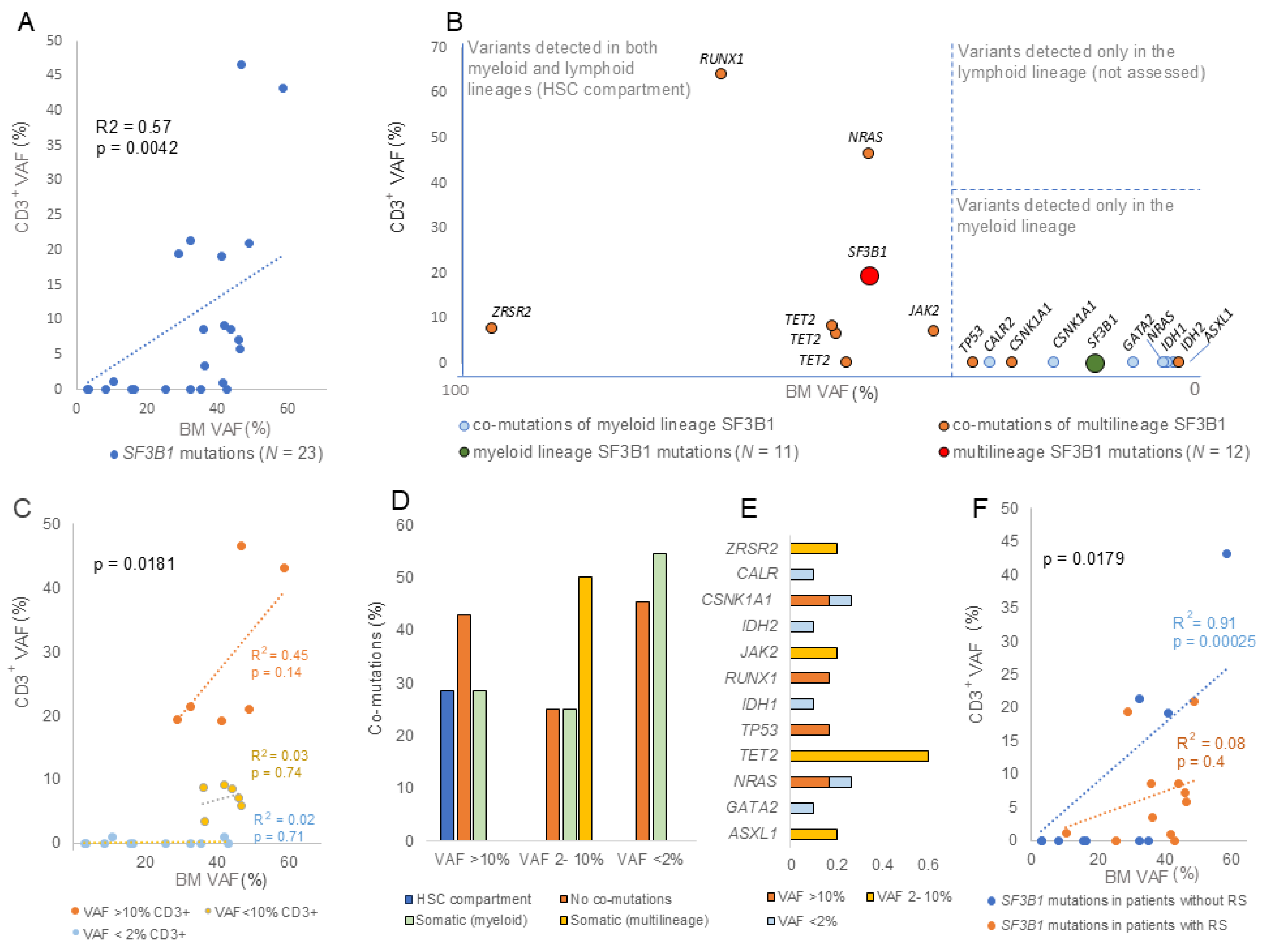
SF3B1 mutations were identified in 23 of 208 patients in the series (11.1%), which is concordant with rates described in prior WHO- and ICC-based studies. SF3B1 mutations exhibit mean VAFs of 32.3% in BM and 9.4% in CD3+ cell samples (Supplementary Table S1). Among them, 11 patients (47.8%) had an SF3B1 mutation detected in both BM and CD3+ samples, while 12 patients (52.2%) had an SF3B1 mutation restricted to BM samples (Table 1). A significant difference (p = 0.0003) in BM VAF was observed between mutations detected in CD3+ cells (42.4%) and those not detected in CD3+ cells (19.6%). Interestingly, the CD3+ VAF positively correlated with the BM VAF (p = 0.0042) across the cohort (Figure 1A). All identified variants in this cohort were documented as somatic in the COSMIC database (COSMIC GRCh38, v100+ series) [24]. In addition, to exclude the possibility of cross-contamination during cell sorting, CD3+ purity was also evaluated. No differences in CD3+ cell purity were observed between samples with variants restricted to BM and those also detected in CD3+ cells. Furthermore, an individualized VAF acceptance threshold was calculated for each case by multiplying the maximal non-pure fraction of the CD3+ sample by the corresponding BM VAF. The CD3+ VAFs of all patients exceeded the calculated threshold, and cross-contamination was ruled out as an explanation for the presence of acquired myeloid SF3B1 mutations in CD3+ samples (Supplementary Table S1).
Multilineage involvement was indirectly inferred through the detection of identical mutations across distinct cellular compartments. Although involvement at the progenitor level was not directly assessed, the presence of the same variant in both myeloid and lymphoid compartments supports the interpretation that these events originated in a common ancestral clone prior to hematopoietic stem cell (HSC) differentiation. Variants were classified as myeloid-restricted when absent in CD3+ samples (VAF < 2%), whereas they were considered multilineage when detected in both bone marrow (BM) and CD3+ compartments with VAF > 2%. In addition, a VAF between 2% and 10% was considered a low VAF, while a VAF > 10% was considered a high VAF. We acknowledge that the 10% VAF threshold is not universally standardized; however, it was used as a pragmatic cutoff to differentiate subclonal from dominant clonal populations. This stratification is supported by previous studies and consensus guidelines, which suggest that variants with higher VAF (≥10%) are more likely to reflect biologically and clinically relevant clonal expansions [25,26].

3.1. Comparison of Myeloid-Restricted and Multilineage SF3B1 Mutations

Patients with multilineage SF3B1 mutations were predominantly males (66.67%). There was a trend toward younger age at diagnosis among patients with multilineage SF3B1 mutations (mean age 65.8 years) compared to patients with BM-restricted mutations (mean age 70.4 years), but the difference did not reach statistical significance. No significant differences were observed between patients with multilineage (CD3+ VAF > 2%) and myeloid-restricted SF3B1 mutations (CD3+ VAF < 2%) across the clinical parameters analyzed (Table 1). However, patients with multilineage SF3B1 mutations showed lower karyotype alterations, blast percentage, and IPSS-R scores compared with patients harboring myeloid-restricted mutations, suggesting a more favorable prognosis in this subgroup. Accompanying pathogenic mutations are summarized in Supplementary Table S2, with no significant differences in co-mutation frequencies between groups (Table 1). However, a positive correlation was observed between the SF3B1 VAF in CD3+ cells and the clonal architecture of the co-mutations. Although VAF-based approaches are not the ideal method for chronologically ordering SF3B1 mutations and co-mutations, multilineage co-mutations (observed in both BM and CD3+ samples with a VAF > 2%) were found only in association with multilineage SF3B1 mutations (Figure 1B). In contrast, co-mutations associated with myeloid lineage SF3B1 mutations were not detected in CD3+ samples.
Multilineage patients were further stratified according to CD3+ VAF into high (VAF > 10%, n = 6) and low (VAF 2–10%, n = 6) groups.
BM VAFs were comparable between groups (42.9% vs. 41.9%) (Table 1). However, the correlation between BM and CD3+ VAFs differed significantly between groups (p = 0.0181) (Figure 1C). The stronger correlation observed in the high-VAF group suggests distinct proliferative dynamics, whereas additional molecular events may be required to drive clonal expansion in patients with low CD3+ VAF. Low-CD3+-VAF cases were associated with multilineage somatic co-mutations (Figure 1D) predominantly in TET2 (Figure 1E). In contrast, patients with high CD3+ T-cell VAF were enriched for co-mutations in the HSC compartment (Figure 1D). Notably, no germline or multilineage co-mutations were observed accompanying myeloid-restricted SF3B1 mutations, suggesting that only these SF3B1 mutations appeared to represent initiating events.
Finally, low-CD3+-VAF cases showed significantly higher ring sideroblast (RS) percentages (p = 0.0157) (Table 1). In contrast, cases of high CD3+ T-cell VAF exhibited more altered karyotypes and significantly correlated with low RS percentages (p = 0.0108), which suggests a negative association. Consistently, the correlation between BM and CD3+ VAFs differed significantly between RS groups (p = 0.0179). A stronger correlation was observed in RS− patients than in RS+ patients, suggesting distinct biological and prognostic implications associated with CD3+ VAF (Figure 1F).

3.2. Comparison of SF3B1 p.K700E with Other SF3B1 Mutations

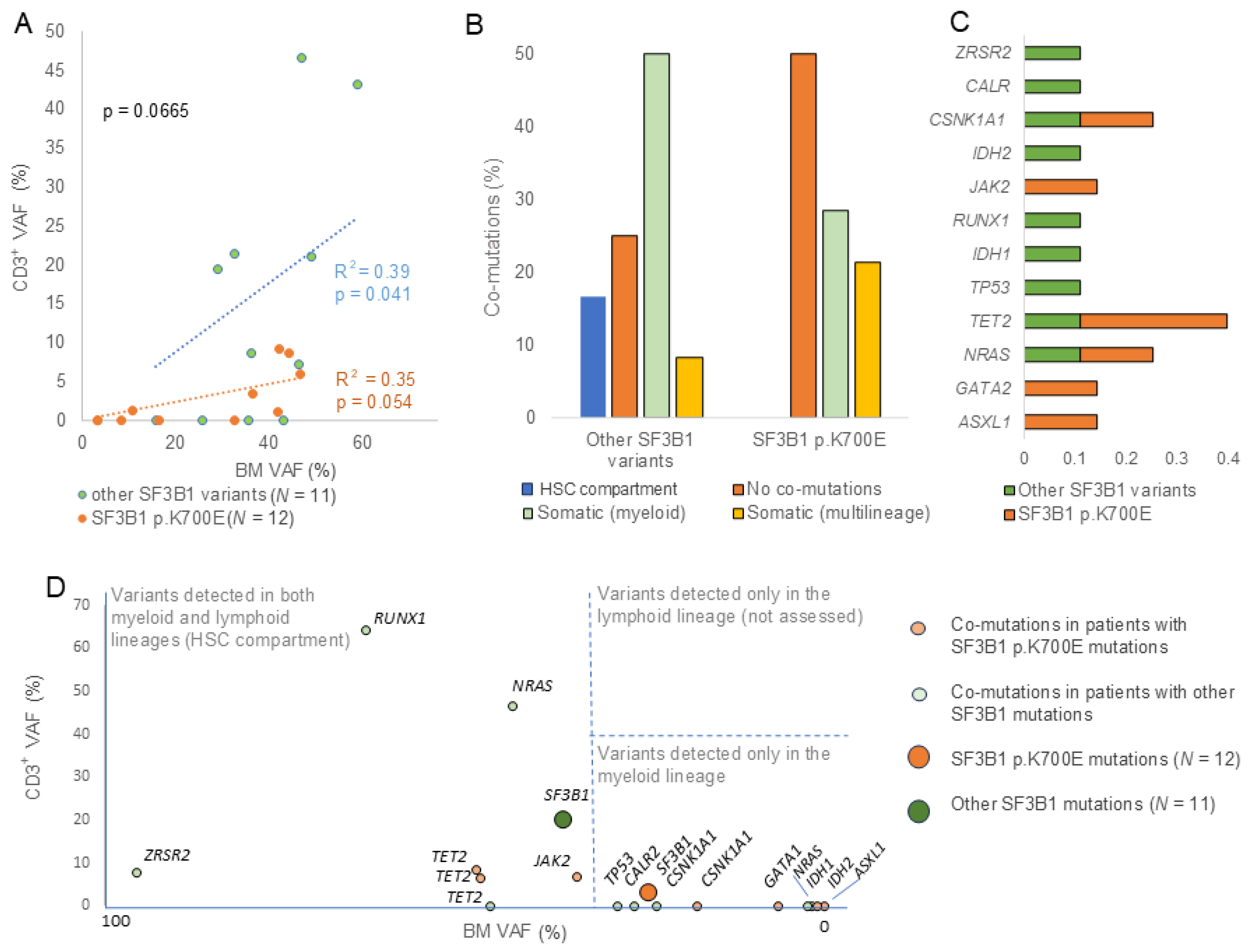
SF3B1 p.K700E and non-p.K700E mutation types were further evaluated across different hematopoietic lineages to interrogate differential patient characteristics associated with distinct mutational profiles. A total of 12 patients (52.2%) carried the recurrent SF3B1 p.K700E mutation, while 11 patients (47.8%) carried other mutations including p.Y141C (n = 1), p.E622D (n = 1), p.H662 (n = 2), p.K666 (n = 4), p.G742D (n = 1), and p.D781E (n = 2) (Supplementary Table S1). Non-p.K700E variants displayed on average higher BM (38.0%) and CD3+ (15.2%) VAFs compared to the BM VAF (27.2%) and CD3+ VAF (4%) of p.K700E mutations (Table 1). Additionally, the CD3+ VAF of the non-p.K700E mutations showed a positive correlation with its VAF in BM samples (Figure 2A). At the molecular level, p.K700E mutations were more frequently found accompanied by fewer co-mutations (Supplementary Table S2), while non-p.K700E variants co-occurred with mutations restricted to the myeloid lineage across a heterogeneous group of genes including TP53 and RUNX1 (Figure 2B,C). RUNX1 mutations have been recurrently described in SF3B1 mutated MDS (~5% frequency) and associated with worse overall survival and disease progression [10]. However, no correlation was observed between the VAFs in CD3+ cells of the co-mutations and the SF3B1 mutation type, as multilineage and myeloid-restricted co-mutations were found accompanying different mutation types (Figure 2D).
Finally, lower BM and CD3+ VAFs were observed in patients harboring del(5q) alterations (either isolated or accompanied by additional cytogenetic abnormalities) compared with patients lacking del(5q) alteration (Table 2). Interestingly, RSs were significantly more prevalent (p = 0.0135) in non-del(5q) patients (33.1%) than in those with del(5q) (2.8%). Notably, the SF3B1 p.K700E hotspot (associated with myeloid lineage) was more frequently observed in del(5q) karyotypes (66.7%) than other SF3B1 mutations, which were detected in both BM and CD3+ samples (33.3%) (Table 2).

4. Discussion

Our study underscores the clinical complexity and heterogeneity of somatic variants in SF3B1 driving myeloid disease. High BM VAF (52% of patients exhibited VAF > 35%) and detection of SF3B1 mutations in purified CD3+ cells support a broader, multilineage distribution of the mutated clones (founder events). Indeed, SF3B1 mutations in MDS-RS have been previously shown to be recurrent driver events originating in progenitor HSCs and retained throughout differentiation [27]. Our results support the current germline-testing recommendations of not using CD3+ samples as non-tumoral tissue, as they may be insufficient to reliably distinguish germline variants from de novo mutations arising in HSCs [18,28,29]. Alternative tissues such as fibroblasts or hair follicles should be employed as suggested [30].
Nevertheless, CD3+ analysis provides valuable prognostic insight. High CD3+ VAFs correlated with BM VAFs, indicating parallel clonal advantage across lineages, as previously described [31]. In contrast, low CD3+ VAFs lacked this correlation (Figure 1C), implying that secondary mutations are required for myeloid expansion to drive disease progression. Additionally, low-CD3+-VAF cases frequently harbored TET2 mutations and exhibited higher RS, consistent with previous observations in MDS with mutations in SF3B1 [10,17]. Conversely, multilineage SF3B1 mutations with high-CD3+-VAF cases were enriched for altered karyotypes. Altogether, these findings could suggest a worse prognosis in patients with multilineage SF3B1 mutations and high CD3+ VAF (altered karyotypes and RS−) compared to those with multilineage mutations and low CD3+ cell VAF (RS+). Thus, assessing VAF in CD3+ cells and not only in BM samples may provide improved biological and prognostic insight in SF3B1-mutated MNs.
Furthermore, distinct co-mutation profiles were also associated with different SF3B1 mutations. Previous studies provided evidence that SF3B1 mutations may represent an initiating event driving the expansion of clonal hematopoiesis and usually precede diverse recurrent mutations [32]. However, only myeloid-restricted SF3B1 mutations appeared to represent initiating events without co-mutations in the HSC compartment (Figure 1B). Additionally, no concomitant mutations were detected in 34.7% of patients. Notably, in up to 10–20% of patients diagnosed with MDS, SF3B1 mutation represents the only detectable genetic lesion, supporting the notion that this mutation alone may be sufficient to drive clonal expansion [6,33]. No differences regarding the proportion of SF3B1-mutated patients lacking co-mutations were observed across different CD3+ VAF groups (Figure 1D). However, patients without co-mutations were more frequently associated with the SF3B1 p.K700E hotspot compared with other SF3B1 mutation cases (Figure 2B), suggesting that this hotspot, which is predominantly associated with myeloid lineage, may represent a distinct unique event. Thus, the clonal architecture of SF3B1 mutations may play a key role in shaping the step-wise model of initiation events and mutational acquisition in MN, as previously suggested when evaluating the clonal hierarchy of founder and subclonal SF3B1 mutations [14,34]. However, our data suggests that mutation type may also correlate with clonal succession, thereby contributing to the definition of specific clinical features of myeloid neoplasms.
Furthermore, we found that mutation type also correlated with VAF in CD3+. SF3B1-p.K700E mutations were associated with the myeloid lineage, whereas other SF3B1 mutations were more often observed in both myeloid and lymphoid lineages. Finally, del(5q) and RSs were differentially associated with mutation type and VAF in affected lineages, which suggests two distinct biological subgroups. Notably, an increased percentage of RS, which is a hallmark of SF3B1-mutated MDS [35], was observed to be associated with patients with multilineage SF3B1 mutations and low CD3+ VAF. Importantly, increased RSs were found in non-del(5q) patients, suggesting a negative association between del(5q) alteration and RS. Remarkably, the co-occurrence of del(5q) and SF3B1 mutations, both typically associated with favorable outcomes when occurring independently, defined a more aggressive course [3]. Importantly, as only one AML case (harboring the p.K700E mutation) was included (Supplementary Table S1), the observed effect can be attributed to the variants per se rather than to enrichment for MDS-EB cases.

5. Conclusions

In summary, distinct patterns were identified not only according to SF3B1 variant type but also based on the affected hematopoietic lineage and CD3+ VAF. We observed statistically significant differences in CD3+ VAF distribution across compartments and in the percentage of ring sideroblasts, a clinically relevant feature in SF3B1-mutated patients, whereas other prognostic variables (including karyotype complexity and IPSS-R) showed only non-significant trends. Furthermore, SF3B1 p.K700E and non-K700E variants were associated with different lineage involvement. Specifically, the p.K700E mutation was largely restricted to the myeloid lineage, whereas non-p.K700E variants more frequently involved HSC and were associated with altered karyotypes.
Thus, although CD3+ samples are not recommended to establish the germline status of mutations, our findings highlight the importance of considering not only the mutation subtype but also lineage involvement and CD3+ T-cell VAF to improve risk stratification in SF3B1-mutated MNs. Accordingly, integrating clonal architecture into molecular assessment may provide a more refined framework for understanding disease biology and improving the clinical management of patients.
We acknowledge that, given the limited number of patients included in our cohort, potential differences in clinical profiles should be considered exploratory, and the absence of an independent validation cohort limits the generalizability of these findings. Validation in larger cohorts is required to enable robust multivariate analyses. In addition, although samples were obtained from a pre-existing biobank collection, which reduces bias associated with prospective patient recruitment, selection bias related to clinical indication and sample availability cannot be excluded.
Nonetheless, this remains a preliminary study and despite these limitations, the observed associations should be considered hypothesis-generating, and lineage-based analyses of other recurrently mutated genes in MNs are warranted to further extend the relevance of CD3+ VAF in prognosis.

Supplementary Materials

The following supporting information can be downloaded at https://www.mdpi.com/article/10.3390/cancers18081308/s1, Table S1: SF3B1 patients; Table S2: Accompanying mutations in SF3B1-mutated patients.

Author Contributions

Conceptualization, O.C.; Methodology, O.C.; Formal analysis, O.C.; Investigation, O.C. and J.M.; Resources, L.C.-G., L.R.P.-H., S.T.-E., M.J.M. and B.X.; Data curation, J.M.; Writing—original draft, O.C.; Writing—review & editing, J.M., L.Z. and M.J.M.; Supervision, F.S.; Funding acquisition, F.S. All authors have read and agreed to the published version of the manuscript.

Funding

This research was funded by Instituto de Salud Carlos III (co-funded by European Regional Development Fund, ERDF, a way to build Europe), grant number PI23/00007; Generalitat de Catalunya, grant number 2021 SGR0056 and Proyectos de Medicina Personalizada del Instituto de Salud Carlos III, grant number PMP24/00025. OC was partially funded by la Fundación Española de Hematología y Hemoterapia (FEHH). JM was supported by the Joan Oró predoctoral program from Secretaria d’Universitats i Recerca del Departament de Recerca i Universitats de la Generalitat de Catalunya, Agència de Gestió d’Ajuts Universitaris i de Recerca (AGAUR) co-funded by European Social Funding Plus (FSE+), grant number 2023 FI-1 00200.

Institutional Review Board Statement

The study was conducted in accordance with the Declaration of Helsinki and approved by the Ethics Committee of Hospital Universitario GermansTrias i Pujol (PI-19-008), approved on 11 January 2019.

Informed Consent Statement

Informed consent was obtained from all subjects involved in the study.

Data Availability Statement

The original contributions presented in this study are included in the article/Supplementary Materials. Further inquiries can be directed to the corresponding authors.

Acknowledgments

Samples included in this study were provided by the collection of samples of IJC (C002922 and C0006786).

Conflicts of Interest

The authors declare no conflicts of interest.

Abbreviations

The following abbreviations are used in this manuscript:
AMLAcute myeloid leukemia
BMBone marrow
CD3+Three-letter acronym for CD3+ T lymphocyte cells
HSCHematopoietic stem cell
ICCInternational Consensus Classification
MDSMyelodysplastic neoplasm
MDS/MPNMyelodysplastic/myeloproliferative neoplasms
MNMyeloid neoplasm
RSRing sideroblast
tNGSTargeted next-generation sequencing
VAFVariant allele frequency
WESWhole-exome sequencing
WHOWorld Health Organization

References

  1. Alsafadi, S.; Houy, A.; Battistella, A.; Popova, T.; Wassef, M.; Henry, E.; Tirode, F.; Constantinou, A.; Piperno-Neumann, S.; Roman-Roman, S.; et al. Cancer-Associated SF3B1 Mutations Affect Alternative Splicing by Promoting Alternative Branchpoint Usage. Nat. Commun. 2016, 7, 10615. [Google Scholar] [CrossRef] [Scilit]
  2. Darman, R.B.; Seiler, M.; Agrawal, A.A.; Lim, K.H.; Peng, S.; Aird, D.; Bailey, S.L.; Bhavsar, E.B.; Chan, B.; Colla, S.; et al. Cancer-Associated SF3B1 Hotspot Mutations Induce Cryptic 3′ Splice Site Selection through Use of a Different Branch Point. Cell Rep. 2015, 13, 1033–1045. [Google Scholar] [CrossRef] [Scilit] [PubMed]
  3. Bernard, E.; Tuechler, H.; Greenberg, P.L.; Hasserjian, R.P.; Arango Ossa, J.E.; Nannya, Y.; Devlin, S.M.; Creignou, M.; Pinel, P.; Monnier, L.; et al. Molecular International Prognostic Scoring System for Myelodysplastic Syndromes. NEJM Evid. 2022, 1, EVIDoa2200008. [Google Scholar] [CrossRef] [Scilit] [PubMed]
  4. Visconte, V.; Makishima, H.; Jankowska, A.; Szpurka, H.; Traina, F.; Jerez, A.; O’Keefe, C.; Rogers, H.J.; Sekeres, M.A.; Maciejewski, J.P.; et al. SF3B1, a Splicing Factor Is Frequently Mutated in Refractory Anemia with Ring Sideroblasts. Leukemia 2012, 26, 542–545. [Google Scholar] [CrossRef] [Scilit] [PubMed]
  5. Malcovati, L.; Karimi, M.; Papaemmanuil, E.; Ambaglio, I.; Jädersten, M.; Jansson, M.; Elena, C.; Gallì, A.; Walldin, G.; Della Porta, M.G.; et al. SF3B1 Mutation Identifies a Distinct Subset of Myelodysplastic Syndrome with Ring Sideroblasts. Blood 2015, 126, 233–241. [Google Scholar] [CrossRef] [Scilit]
  6. Malcovati, L.; Stevenson, K.; Papaemmanuil, E.; Neuberg, D.; Bejar, R.; Boultwood, J.; Bowen, D.T.; Campbell, P.J.; Ebert, B.L.; Fenaux, P.; et al. SF3B1-Mutant MDS as a Distinct Disease Subtype: A Proposal from the International Working Group for the Prognosis of MDS. Blood 2020, 136, 157–170. [Google Scholar] [CrossRef] [Scilit]
  7. Khoury, J.D.; Solary, E.; Abla, O.; Akkari, Y.; Alaggio, R.; Apperley, J.F.; Bejar, R.; Berti, E.; Busque, L.; Chan, J.K.C.; et al. The 5th Edition of the World Health Organization Classification of Haematolymphoid Tumours: Myeloid and Histiocytic/Dendritic Neoplasms. Leukemia 2022, 36, 1703–1719. [Google Scholar] [CrossRef] [Scilit]
  8. Arber, D.A.; Orazi, A.; Hasserjian, R.P.; Borowitz, M.J.; Calvo, K.R.; Kvasnicka, H.-M.; Wang, S.A.; Bagg, A.; Barbui, T.; Branford, S.; et al. International Consensus Classification of Myeloid Neoplasms and Acute Leukemias: Integrating Morphologic, Clinical, and Genomic Data. Blood 2022, 140, 1200–1228. [Google Scholar] [CrossRef] [Scilit]
  9. Palomo, L.; Solé, F. SF3B1: The Lord of the Rings in MDS. Blood 2020, 136, 149–151. [Google Scholar] [CrossRef] [Scilit]
  10. Huber, S.; Haferlach, T.; Meggendorfer, M.; Hutter, S.; Hoermann, G.; Baer, C.; Kern, W.; Haferlach, C. SF3B1 Mutated MDS: Blast Count, Genetic Co-Abnormalities and Their Impact on Classification and Prognosis. Leukemia 2022, 36, 2894–2902. [Google Scholar] [CrossRef] [Scilit]
  11. Obeng, E.A.; Chappell, R.J.; Seiler, M.; Chen, M.C.; Campagna, D.R.; Schmidt, P.J.; Schneider, R.K.; Lord, A.M.; Wang, L.; Gambe, R.G.; et al. Physiologic Expression of Sf3b1 K700E Causes Impaired Erythropoiesis, Aberrant Splicing, and Sensitivity to Therapeutic Spliceosome Modulation. Cancer Cell 2016, 30, 404–417. [Google Scholar] [CrossRef] [Scilit] [PubMed]
  12. Brian Dalton, W.; Helmenstine, E.; Pieterse, L.; Li, B.; Gocke, C.D.; Donaldson, J.; Xiao, Z.; Gondek, L.P.; Ghiaur, G.; Gojo, I.; et al. The K666N Mutation in SF3B1 Is Associated with Increased Progression of MDS and Distinct RNA Splicing. Blood Adv. 2020, 4, 1192–1196. [Google Scholar] [CrossRef] [Scilit] [PubMed]
  13. Kanagal-Shamanna, R.; Montalban-Bravo, G.; Sasaki, K.; Darbaniyan, F.; Jabbour, E.; Bueso-Ramos, C.; Wei, Y.; Chien, K.; Kadia, T.; Ravandi, F.; et al. Only SF3B1 Mutation Involving K700E Independently Predicts Overall Survival in Myelodysplastic Syndromes. Cancer 2021, 127, 3552–3565. [Google Scholar] [CrossRef] [Scilit] [PubMed]
  14. Awada, H.; Kerr, C.M.; Durmaz, A.; Adema, V.; Gurnari, C.; Pagliuca, S.; Zawit, M.; Kongkiatkamon, S.; Rogers, H.J.; Saunthararajah, Y.; et al. Clonal Trajectories and Cellular Dynamics of Myeloid Neoplasms with SF3B1 Mutations. Leukemia 2021, 35, 3324–3328. [Google Scholar] [CrossRef] [Scilit]
  15. Takahashi, K.; Tanaka, T. Clonal Evolution and Hierarchy in Myeloid Malignancies. Trends Cancer 2023, 9, 707–715. [Google Scholar] [CrossRef] [Scilit]
  16. Mestre, J.; Chaparro, L.; Manzanares, A.; Xicoy, B.; Zamora, L.; Sole, F.; Calvete, O. Beyond Myeloid Neoplasms Germline Guidelines: Validation of the Thresholds Criteria in the Search of Germline Predisposition Variants. eJHaem 2024, 5, 1021–1027. [Google Scholar] [CrossRef] [Scilit]
  17. Kumaar, J.; Lewis, N.E.; Sherpa, S.; Londono, D.; Sun, X.; Gao, Q.; Arcila, M.E.; Roshal, M.; Zhang, Y.; Xiao, W.; et al. Diagnostic Challenges and Proposed Classification of Myeloid Neoplasms with Overlapping Features of Thrombocytosis, Ring Sideroblasts and Concurrent Del(5q) and SF3B1 Mutations. Haematologica 2024, 109, 2676. [Google Scholar] [CrossRef] [Scilit]
  18. Baliakas, P.; Tesi, B.; Wartiovaara-Kautto, U.; Stray-Pedersen, A.; Friis, L.S.; Dybedal, I.; Hovland, R.; Jahnukainen, K.; Raaschou-Jensen, K.; Ljungman, P.; et al. Nordic Guidelines for Germline Predisposition to Myeloid Neoplasms in Adults: Recommendations for Genetic Diagnosis, Clinical Management and Follow-Up. Hemasphere 2019, 3, e321. [Google Scholar] [CrossRef] [Scilit]
  19. Speight, B.; Hanson, H.; Turnbull, C.; Hardy, S.; Drummond, J.; Khorashad, J.; Wragg, C.; Page, P.; Parkin, N.W.; Rio-Machin, A.; et al. Germline Predisposition to Haematological Malignancies: Best Practice Consensus Guidelines from the UK Cancer Genetics Group (UKCGG), CanGene-CanVar and the NHS England Haematological Oncology Working Group. Br. J. Haematol. 2023, 201, 25–34. [Google Scholar] [CrossRef] [Scilit]
  20. Palomo, L.; Ibáñez, M.; Abáigar, M.; Vázquez, I.; Álvarez, S.; Cabezón, M.; Tazón-Vega, B.; Rapado, I.; Fuster-Tormo, F.; Cervera, J.; et al. Spanish Guidelines for the Use of Targeted Deep Sequencing in Myelodysplastic Syndromes and Chronic Myelomonocytic Leukaemia. Br. J. Haematol. 2020, 188, 605–622. [Google Scholar] [CrossRef] [Scilit]
  21. Bersanelli, M.; Travaglino, E.; Meggendorfer, M.; Matteuzzi, T.; Sala, C.; Mosca, E.; Chiereghin, C.; di Nanni, N.; Gnocchi, M.; Zampini, M.; et al. Classification and Personalized Prognostic Assessment on the Basis of Clinical and Genomic Features in Myelodysplastic Syndromes. J. Clin. Oncol. 2021, 39, 1223–1233. [Google Scholar] [CrossRef] [Scilit] [PubMed]
  22. Calvete, O.; Mestre, J.; Risueño, R.M.; Manzanares, A.; Acha, P.; Xicoy, B.; Solé, F. Two-Time Multiplexed Targeted Next-Generation Sequencing Might Help the Implementation of Germline Screening Tools for Myelodysplastic Syndromes/Hematologic Neoplasms. Biomedicines 2023, 11, 3222. [Google Scholar] [CrossRef] [Scilit] [PubMed]
  23. Sherry, S.T.; Ward, M.H.; Kholodov, M.; Baker, J.; Phan, L.; Smigielski, E.M.; Sirotkin, K. DbSNP: The NCBI Database of Genetic Variation. Nucleic Acids Res. 2001, 29, 308–311. [Google Scholar] [CrossRef] [Scilit] [PubMed]
  24. Singh, J.; Li, N.; Ashrafi, E.; Thao, L.T.P.; Curtis, D.J.; Wood, E.M.; McQuilten, Z.K. Clonal Hematopoiesis of Indeterminate Potential as a Prognostic Factor: A Systematic Review and Meta-Analysis. Blood Adv. 2024, 8, 3771–3784. [Google Scholar] [CrossRef] [Scilit]
  25. Pandzic, T.; Ladenvall, C.; Engvall, M.; Mattsson, M.; Hermanson, M.; Cavelier, L.; Ljungström, V.; Baliakas, P. Five Percent Variant Allele Frequency Is a Reliable Reporting Threshold for TP53 Variants Detected by Next Generation Sequencing in Chronic Lymphocytic Leukemia in the Clinical Setting. Hemasphere 2022, 6, e761. [Google Scholar] [CrossRef] [Scilit]
  26. Tate, J.G.; Bamford, S.; Jubb, H.C.; Sondka, Z.; Beare, D.M.; Bindal, N.; Boutselakis, H.; Cole, C.G.; Creatore, C.; Dawson, E.; et al. COSMIC: The Catalogue Of Somatic Mutations In Cancer. Nucleic Acids Res. 2019, 47, D941–D947. [Google Scholar] [CrossRef] [Scilit]
  27. Mortera-Blanco, T.; Dimitriou, M.; Woll, P.S.; Karimi, M.; Elvarsdottir, E.; Conte, S.; Tobiasson, M.; Jansson, M.; Douagi, I.; Moarii, M.; et al. SF3B1-Initiating Mutations in MDS-RSs Target Lymphomyeloid Hematopoietic Stem Cells. Blood 2017, 130, 881–890. [Google Scholar] [CrossRef] [Scilit]
  28. Schlegelberger, B.; Mecucci, C.; Wlodarski, M. Review of Guidelines for the Identification and Clinical Care of Patients with Genetic Predisposition for Hematological Malignancies. Fam. Cancer 2021, 20, 295–303. [Google Scholar] [CrossRef] [Scilit]
  29. Servei Català de la Salut. Determinacions Del Perfil Genètic de Les Síndromes Hereditàries de Càncer En l’adult i Pediatria; Versió 2; Servei Català de La Salut: Barcelona, Spain, 2022. [Google Scholar]
  30. Duncavage, E.J.; Bagg, A.; Hasserjian, R.P.; DiNardo, C.D.; Godley, L.A.; Iacobucci, I.; Jaiswal, S.; Malcovati, L.; Vannucchi, A.M.; Patel, K.P.; et al. Genomic Profiling for Clinical Decision Making in Myeloid Neoplasms and Acute Leukemia. Blood 2022, 140, 2228–2247. [Google Scholar] [CrossRef] [Scilit]
  31. Montalban-Bravo, G.; Takahashi, K.; Patel, K.; Wang, F.; Xingzhi, S.; Nogueras, G.M.; Huang, X.; Pierola, A.A.; Jabbour, E.; Colla, S.; et al. Impact of the Number of Mutations in Survival and Response Outcomes to Hypomethylating Agents in Patients with Myelodysplastic Syndromes or Myelodysplastic/Myeloproliferative Neoplasms. Oncotarget 2018, 9, 9714–9727. [Google Scholar] [CrossRef] [Scilit]
  32. Creignou, M.; Sarchi, M.; Bernard, E.; Malcovati, L. Evolutionary Trajectories of Myelodysplastic Syndromes/Neoplasms. Semin. Cancer Biol. 2026, 120, 16–30. [Google Scholar] [CrossRef] [Scilit]
  33. Bernard, E.; Hasserjian, R.P.; Greenberg, P.L.; Arango Ossa, J.E.; Creignou, M.; Tuechler, H.; Gutierrez-Abril, J.; Domenico, D.; Medina-Martinez, J.S.; Levine, M.; et al. Molecular Taxonomy of Myelodysplastic Syndromes and Its Clinical Implications. Blood 2024, 144, 1617–1632. [Google Scholar] [CrossRef] [Scilit]
  34. Nagata, Y.; Makishima, H.; Kerr, C.M.; Przychodzen, B.P.; Aly, M.; Goyal, A.; Awada, H.; Asad, M.F.; Kuzmanovic, T.; Suzuki, H.; et al. Invariant Patterns of Clonal Succession Determine Specific Clinical Features of Myelodysplastic Syndromes. Nat. Commun. 2019, 10, 5386. [Google Scholar] [CrossRef] [Scilit]
  35. Papaemmanuil, E.; Cazzola, M.; Boultwood, J.; Malcovati, L.; Vyas, P.; Bowen, D.; Pellagatti, A.; Wainscoat, J.S.; Hellstrom-Lindberg, E.; Gambacorti-Passerini, C.; et al. Somatic SF3B1 Mutation in Myelodysplasia with Ring Sideroblasts. N. Engl. J. Med. 2011, 365, 1384–1395. [Google Scholar] [CrossRef] [Scilit]
Figure 1. Comparison of myeloid-restricted and multilineage SF3B1 mutations. (A) Scatter plot showing CD3+ and corresponding BM VAFs in the SF3B1-mutated studied patients (N = 23). The trendline is shown. (B) Scatter plot showing CD3+ and corresponding BM VAFs for co-mutations associated with myeloid-restricted (blue dots) and multilineage (orange dots) SF3B1 mutations. The average and BM VAFs for myeloid-restricted (green dot) and multilineage (red dot) SF3B1 mutations are also shown. Threshold lines indicating mutations arising in the HSC compartment (multilineage mutations) and myeloid-restricted mutations (not found in CD3+ samples) are shown. Mutations detected in CD3+ samples but not in BM samples (lymphoid lineage) were not considered. (C) Scatter plot showing CD3+ and corresponding BM VAFs for the SF3B1-mutated cohort categorized by high (>10%), low (<10%), or absent (<2%) CD3+ VAFs. Trendlines per group are shown. (D) Percentage of patients according to co-mutation type, grouped by high (>10%), low (<10%), or absent (<2%) CD3+ VAFs of the SF3B1 mutation. Multilineage co-mutations are shown based on their VAF in CD3+ cells, distinguishing between high (annotated as HSC compartment mutations) and low CD3+ VAF (annotated as somatic mutations). (E) Percentage of co-mutated genes according to patient groups defined by high VAF (>10%), low VAF (<10%) or absent (VAF < 2%) CD3+ VAFs of the SF3B1 mutation. (F) Scatter plot showing CD3+ and corresponding BM VAFs in SF3B1-mutated patients with and without RS. Trendlines for each group are shown. HSC, hematopoietic stem cell; VAF, variant allele frequency; CD3+, CD3+ T lymphocytes; BM, bone marrow; RS, ring sideroblast.
Cancers 18 01308 g001
Figure 2. Comparison of the SF3B1 p.K700E hotspot with other SF3B1 mutations. (A) Scatter plot showing CD3+ and corresponding BM VAFs for SF3B1 mutations categorized according to mutation position. Trendlines for each group are shown. (B) Percentage of patients according to co-mutation type, grouped by the position of the SF3B1 mutation. Multilineage co-mutations are shown based on their VAF in CD3+ cells, distinguishing between high (annotated as HSC compartment mutations) and low CD3+ VAF (annotated as somatic mutations). (C) Percentage of co-mutated genes according to patient groups defined by the position of the SF3B1 mutation. (D) Scatter plot showing CD3+ and corresponding BM VAFs for co-mutations associated with the SF3B1 p.K700E hotspot (orange dots) and other SF3B1 mutations (green dots). The average CD3+ and BM VAFs for SF3B1 p.K700E (dark green dot) and other SF3B1 mutations (dark orange dot) are also shown. Threshold lines indicating mutations arising in the HSC compartment (multilineage mutations) and myeloid-restricted mutations (not found in CD3+ samples) are indicated. Mutations detected in CD3+ samples but not in BM samples (lymphoid lineage) were not considered. HSC, hematopoietic stem cell; VAF, variant allele frequency; CD3+, CD3+ T lymphocytes; BM, bone marrow.
Cancers 18 01308 g002
Table 1. Comparison of SF3B1 mutations. (A) Multilineage SF3B1 mutations (VAF > 2% CD3+) compared to myeloid SF3B1 mutations (VAF < 2% CD3+). (B) Multilineage SF3B1 mutations with high VAF (VAF > 10% CD3+) compared to low VAF (VAF 2–10% CD3+). (C) SF3B1 p.K700E mutation compared to other SF3B1 mutations. The p-value for the comparison of clinical features between groups was calculated using Student’s t-test. The p-value for the comparison of karyotype complexity between groups was calculated using the ANOVA test.
A. Multilineage vs. Myeloid
Restricted Variants
B. Multilineage Variants
(High VAF vs. Low VAF)
C. SF3B1 Hotspot
Comparison
TotalVAF > 2% CD3+VAF < 2% CD3+p-ValueVAF > 10% CD3+VAF 2–10%
CD3+
Valuep.K700ENon-p.K700Ep-Value
N231211 66 1211
Females (%)47.833.363.6 66.70.0 50.045.5
Males (%)52.266.736.4 33.3100.0 50.054.5
VAF in BM (%)32.342.419.60.0003 *42.941.90.840427.238.00.1018
VAF in CD3+ (%)9.417.80.20.0004 *28.57.20.0090 *4.015.20.0598
CD3+ Purity (%)93.191.295.20.110592.590.40.631191.894.30.3139
Age at onset68.165.870.40.294665.765.80.985764.572.40.0701
Altered karyotype (%)66.754.580.00.216583.333.30.121781.850.00.1224
Normal karyotype (%)33.345.4520.0 16.766.7 18.250.0
Blasts (%)3.32.54.10.38822.03.20.44263.72.90.6616
Platelets (×109/L)223.3224.5222.00.9703187.2261.80.3807251.8192.20.3667
H (×109/L)9.810.39.20.173110.810.10.618410.19.50.4950
N (×109/L)3.43.73.00.43603.63.90.72383.03.90.2719
L (×109/L)5.65.72.70.97625.06.40.25755.85.50.8072
RS (%)23.528.917.60.356911.446.40.0157 *25.521.30.7379
IPSS-R2.31.92.90.22542.2NDND2.62.20.6695
Patients with co-mutations (%)52.463.650.00.528350.080.00.303150.066.670.4450
* p < 0.05. VAF, variant allele frequency; CD3+, CD3+ T lymphocytes; BM, bone marrow; ND, not determined; RS, ring sideroblast; H, hemoglobin; N, neutrophil; L, leucocyte.
Table 2. SF3B1-mutated patients per del(5q) alteration.
(%)Non-del(5q)del(5q) °p-Value
N156
VAF in BM39.017.00.0007 *
VAF in CD3+12.13.20.1507
Normal karyotype46.666.6 °°NA
Altered karyotype53.333.30.0219 *
Complex karyotype0.00.0NA
Ring sideroblasts33.12.80.0135 *
p.K700E47.066.70.2214
Non-p.K700E53.033.30.3765
° Including 4 patients with isolated del(5q) and 2 patients with an additional cytogenetic alteration. °° del(5q) as unique alteration. * p < 0.05. NA, not applicable; VAF, variant allele frequency; CD3+, CD3+ T lymphocytes; BM, bone marrow.
Disclaimer/Publisher’s Note: The statements, opinions and data contained in all publications are solely those of the individual author(s) and contributor(s) and not of MDPI and/or the editor(s). MDPI and/or the editor(s) disclaim responsibility for any injury to people or property resulting from any ideas, methods, instructions or products referred to in the content.