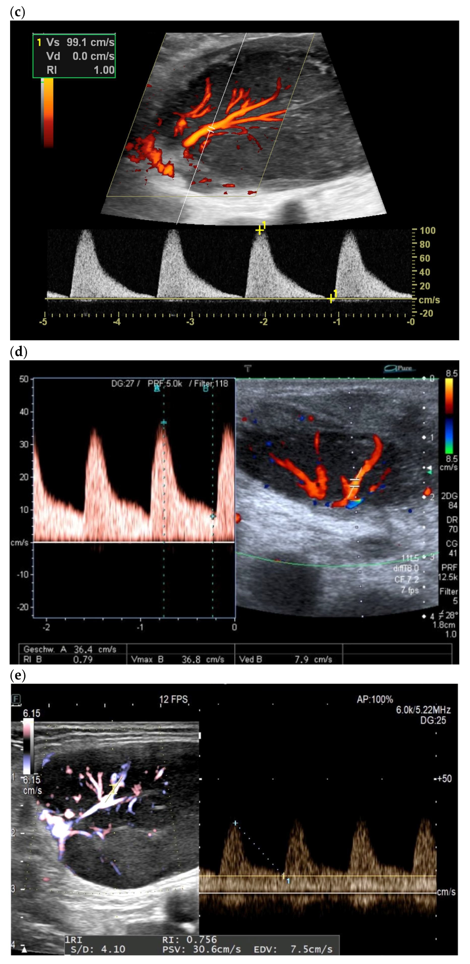
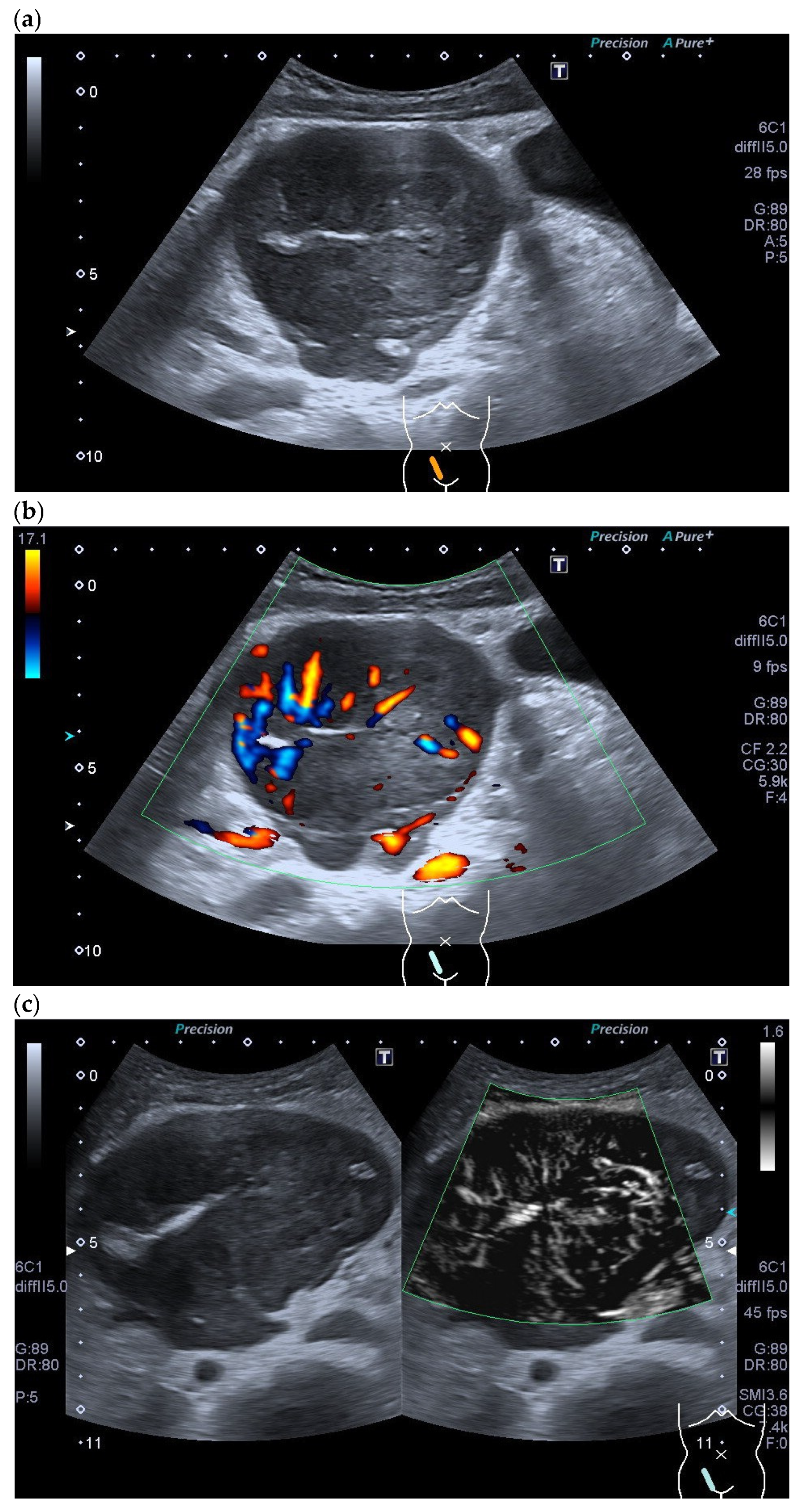
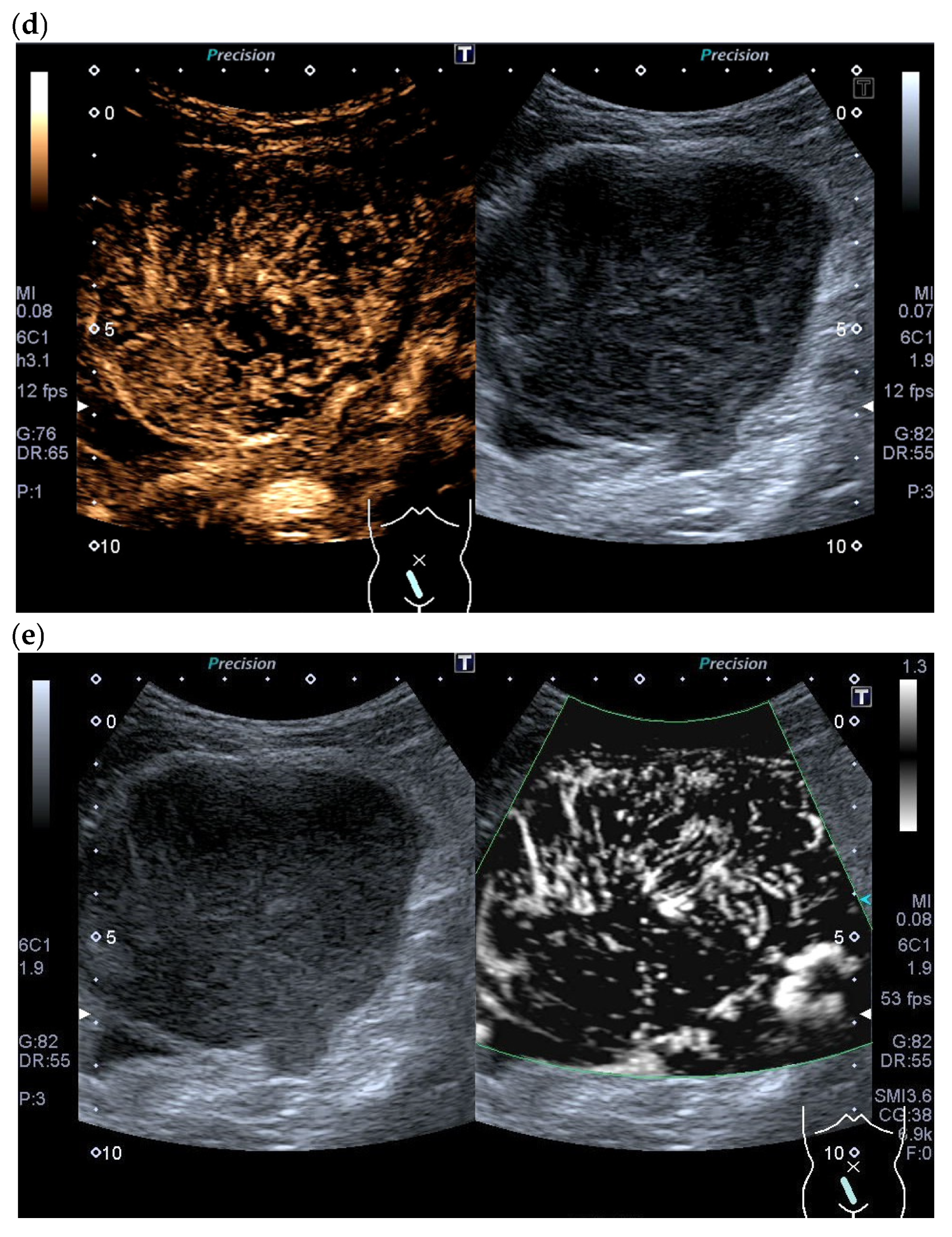
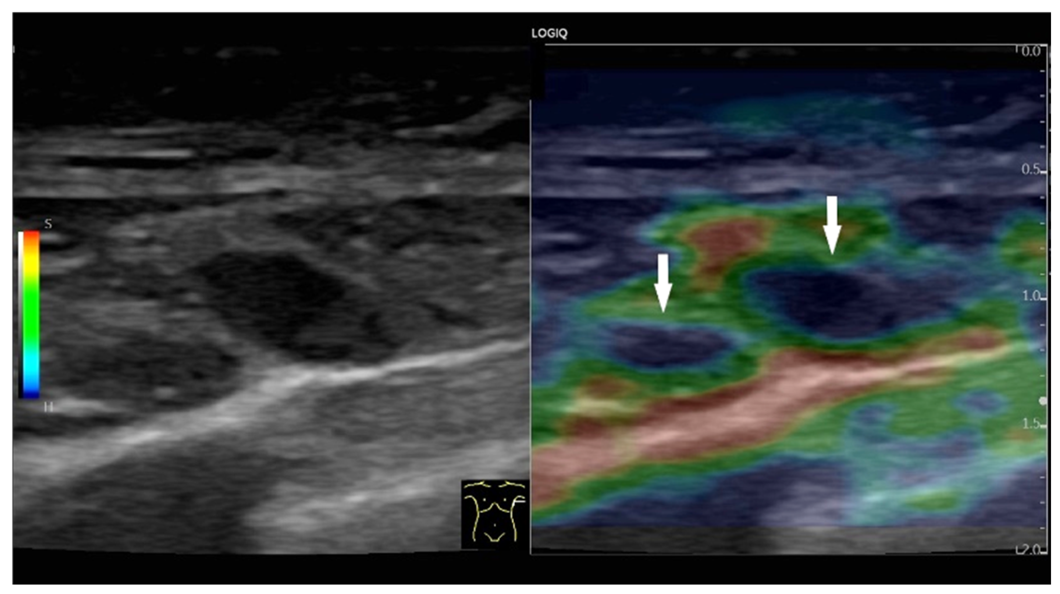
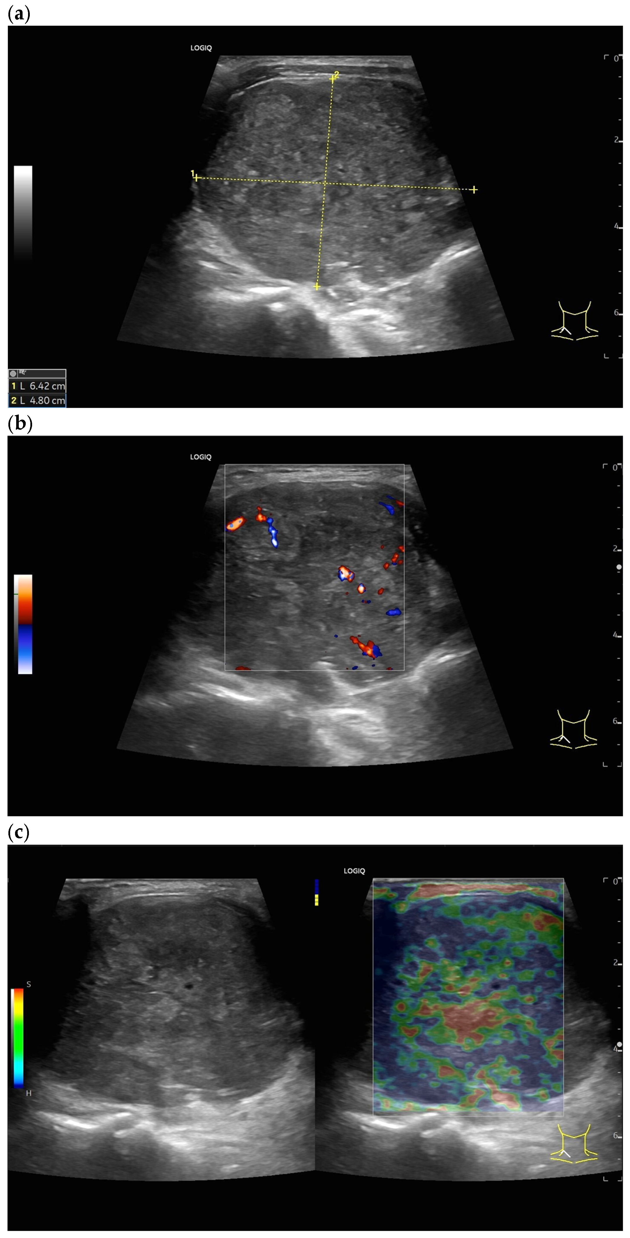
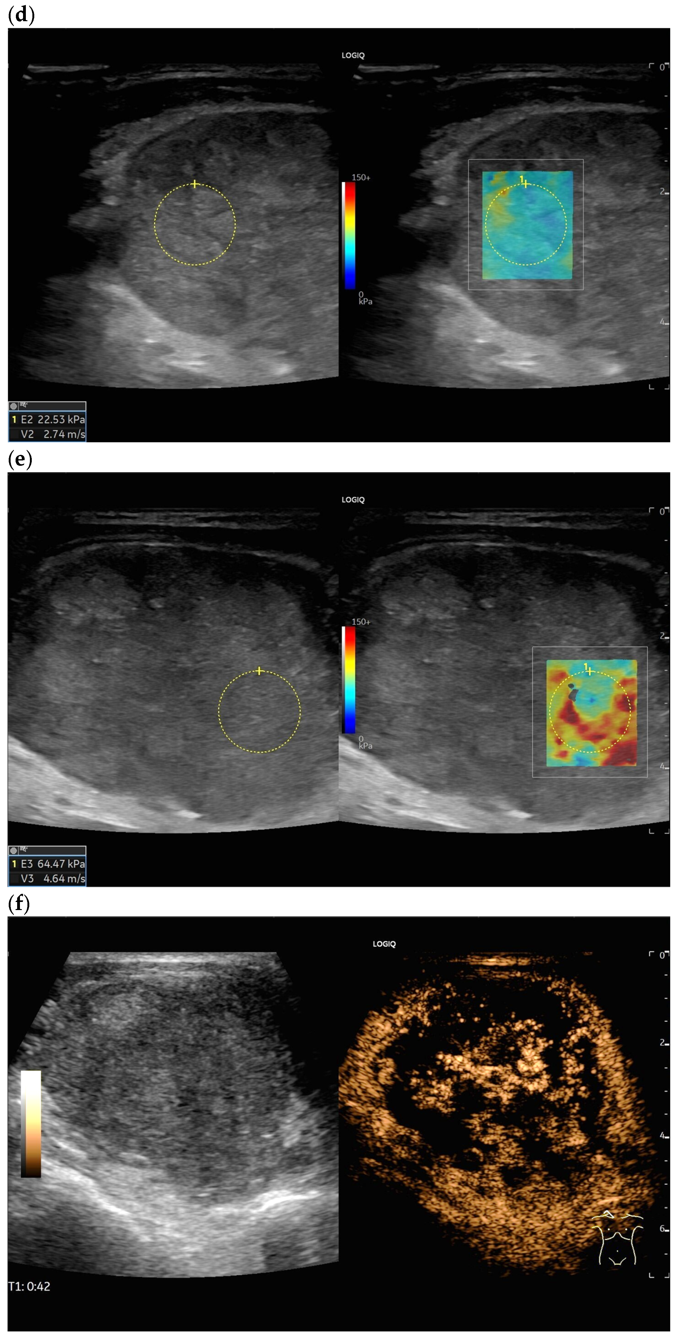
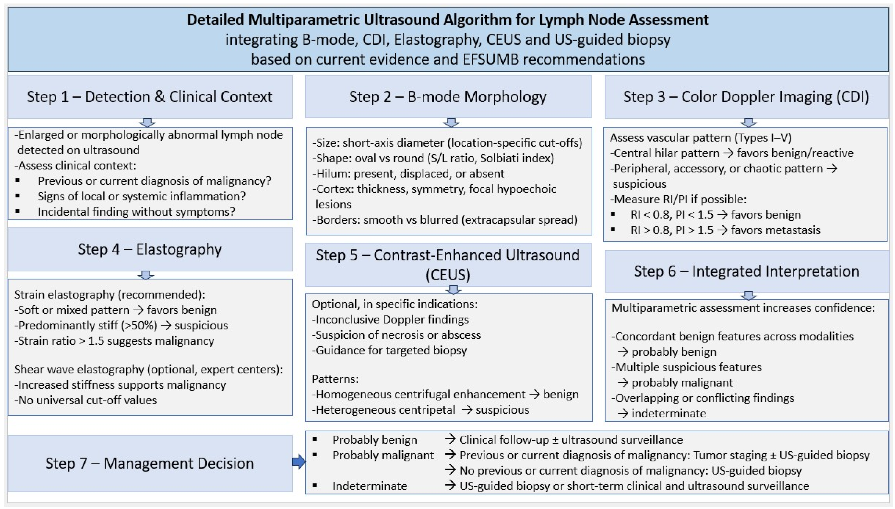
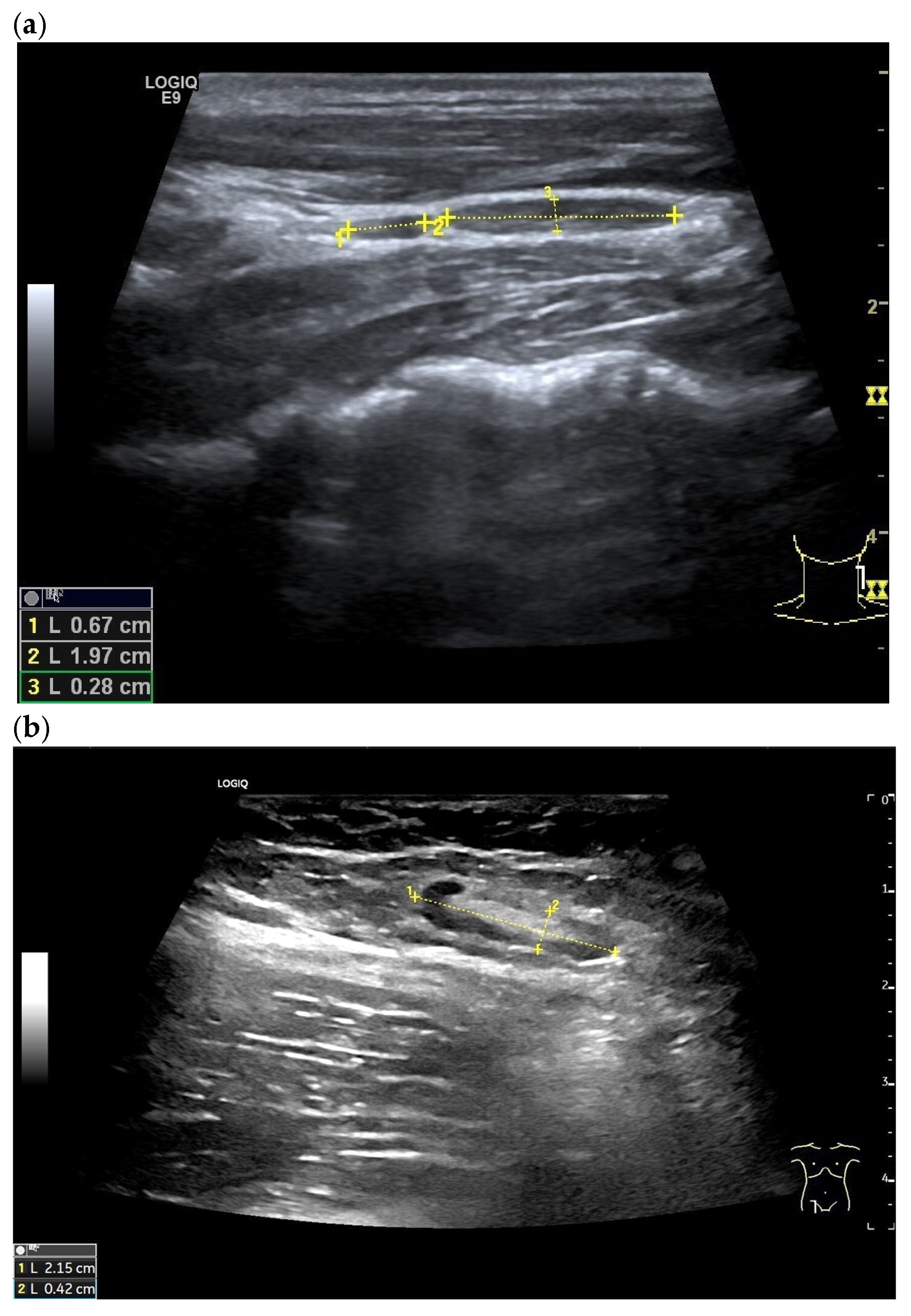
Lymph Node Assessment with Multiparametric Ultrasound: Normal Values, Morphologic Patterns, and Diagnostic Algorithms

 ,
,  ,
,  , ,
, ,  ,
,
Simple Summary
Abstract
1. Introduction
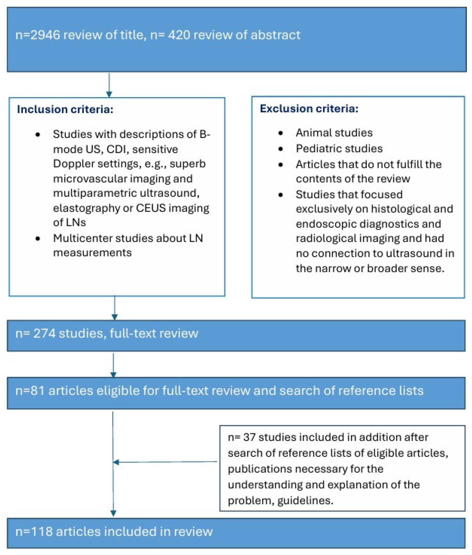
2. Method
2.1. Investigation Requirements
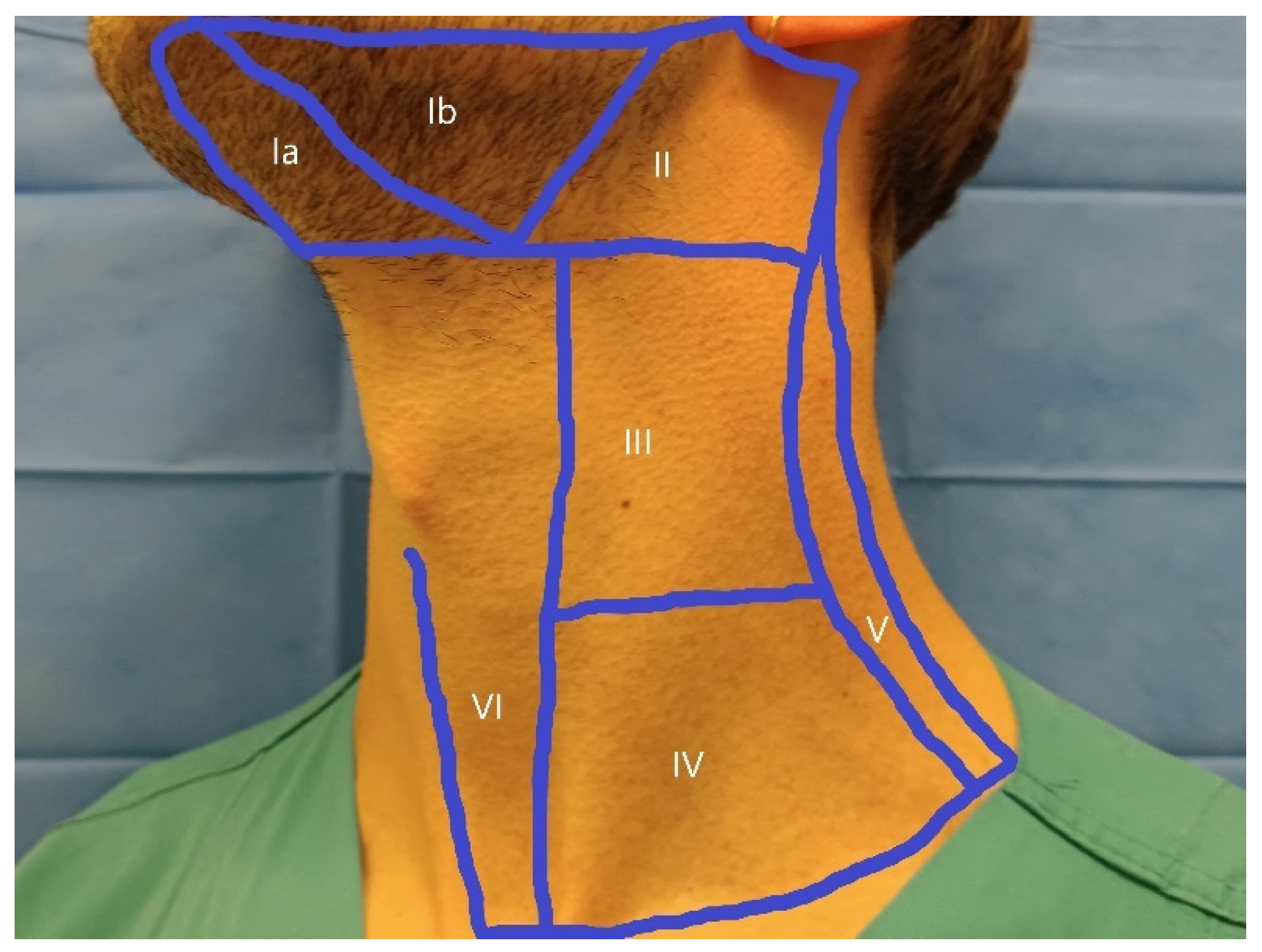
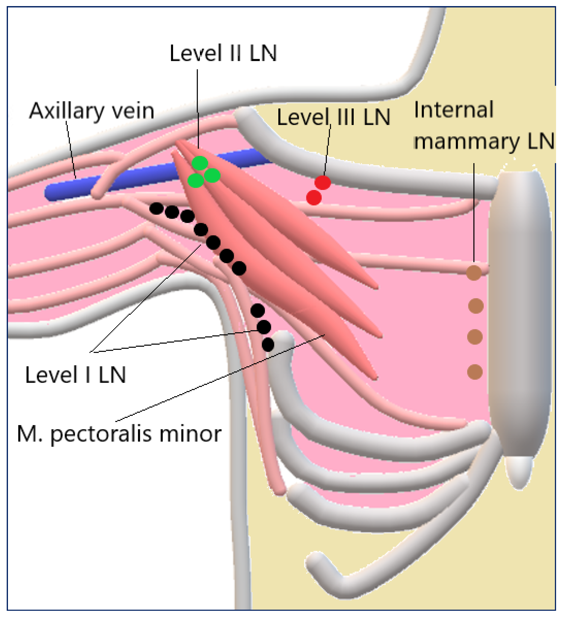
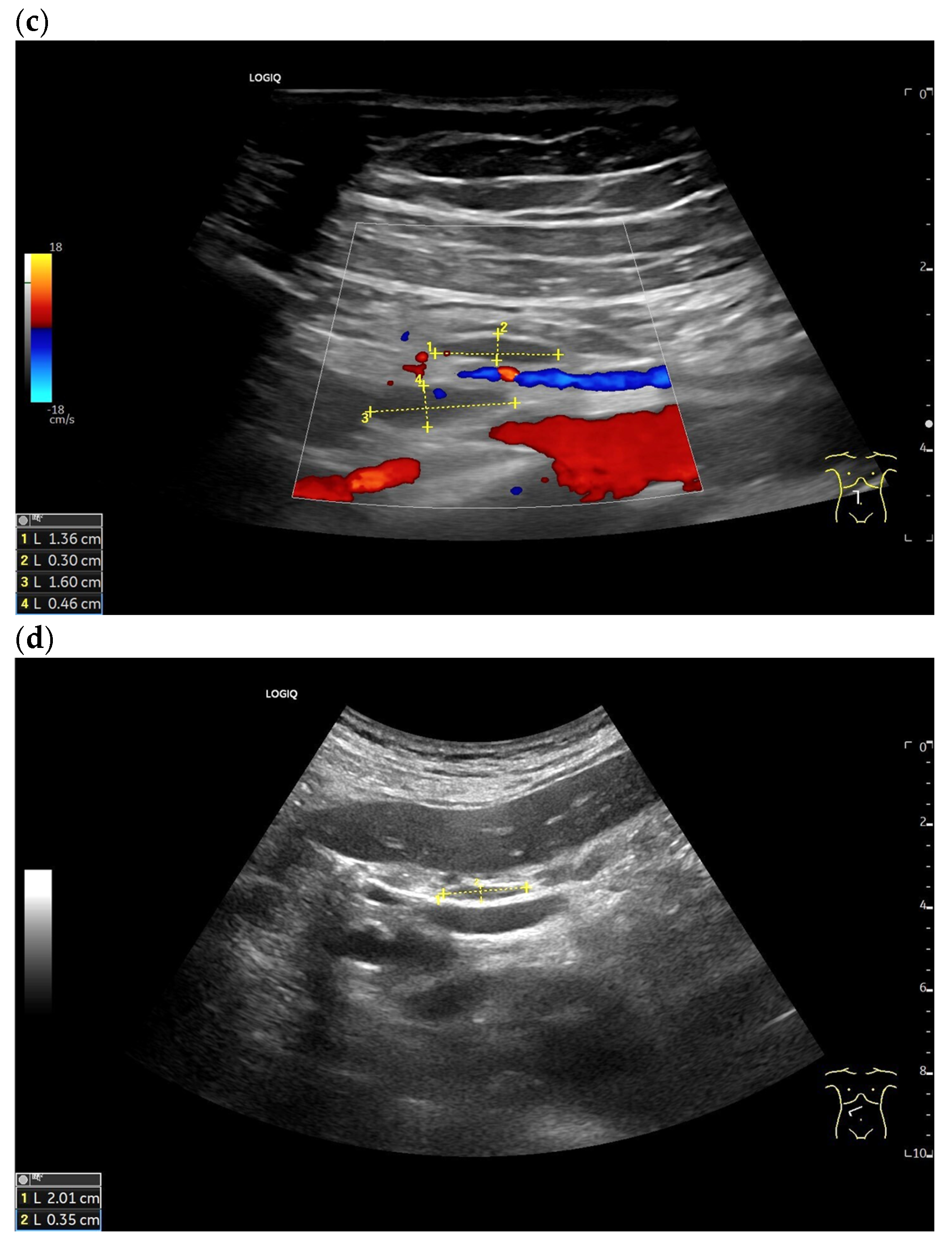
2.2. Lymph Node Stations
2.3. Lymph Node Sonomorphology
2.3.1. What Is Normal (Reference Values)
2.3.2. What Suggests Malignancy
2.3.3. Overlap and Pitfalls
2.4. Size
2.4.1. What Is Normal (Reference Values)
2.4.2. What Suggests Malignancy
2.4.3. Overlap and Pitfalls
2.5. Solbiati Index and Short/Long-Axis Ratio
2.6. Solbiati Index
2.6.1. What Is Normal (Reference Values)
2.6.2. What Suggests Malignancy
2.6.3. Overlap and Pitfalls
2.7. Short/Long-Axis Ratio
2.7.1. What Is Normal (Reference Values)
2.7.2. What Suggests Malignancy
2.8. Influence of Age and Gender on Lymph Nodes Size and S/L-Axis Ratio
2.9. Shape
2.9.1. What Is Normal (Reference Values)
2.9.2. What Suggests Malignancy
2.9.3. Overlap and Pitfalls
2.10. Uniformity of Cortical Thickness
2.10.1. What Is Normal (Reference Values)
2.10.2. What Suggests Malignancy
2.10.3. Overlap and Pitfalls
2.11. Heterogeneity and Echogenicity
2.11.1. What Is Normal (Reference Values)
2.11.2. What Suggests Malignancy
2.11.3. Overlap and Pitfalls
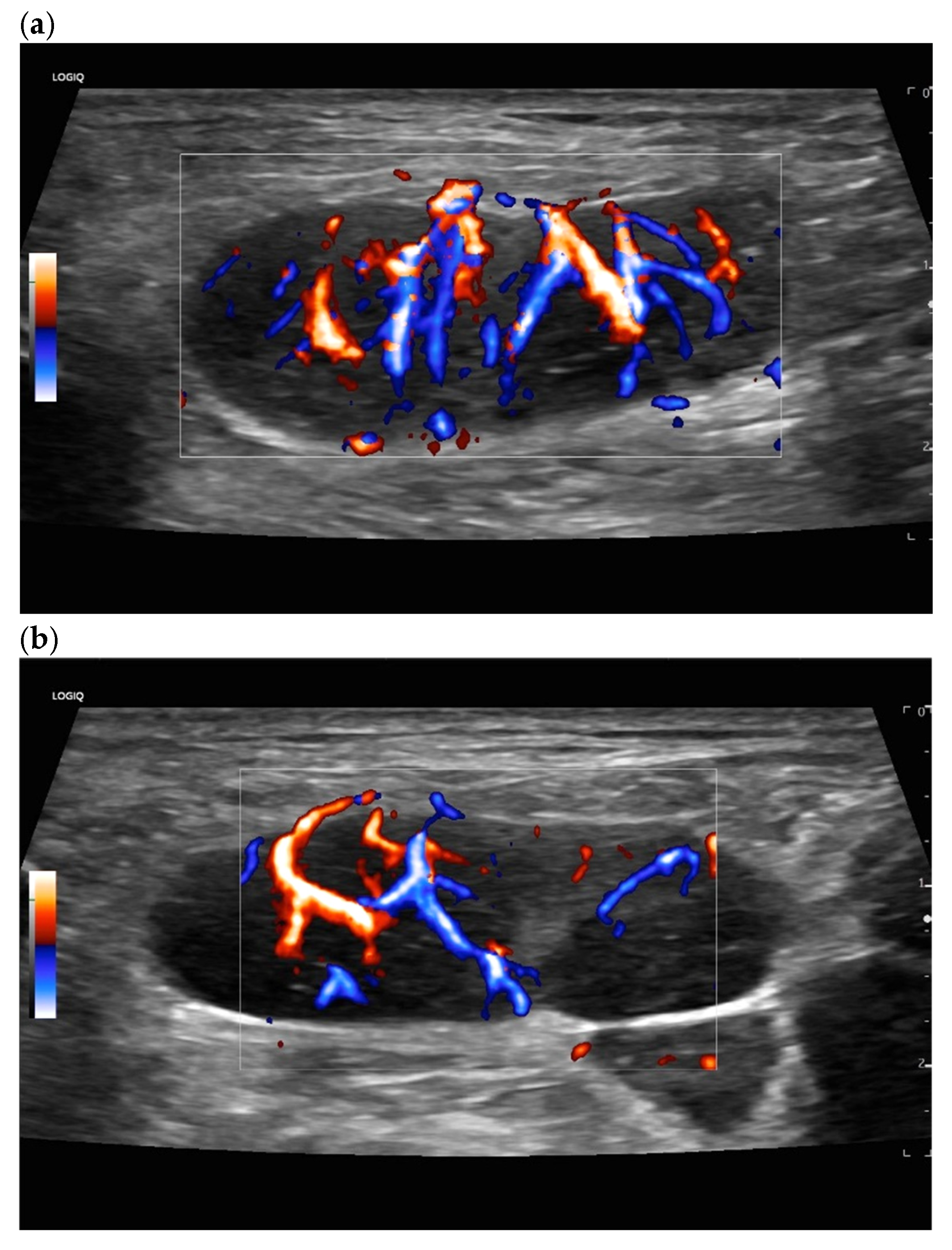
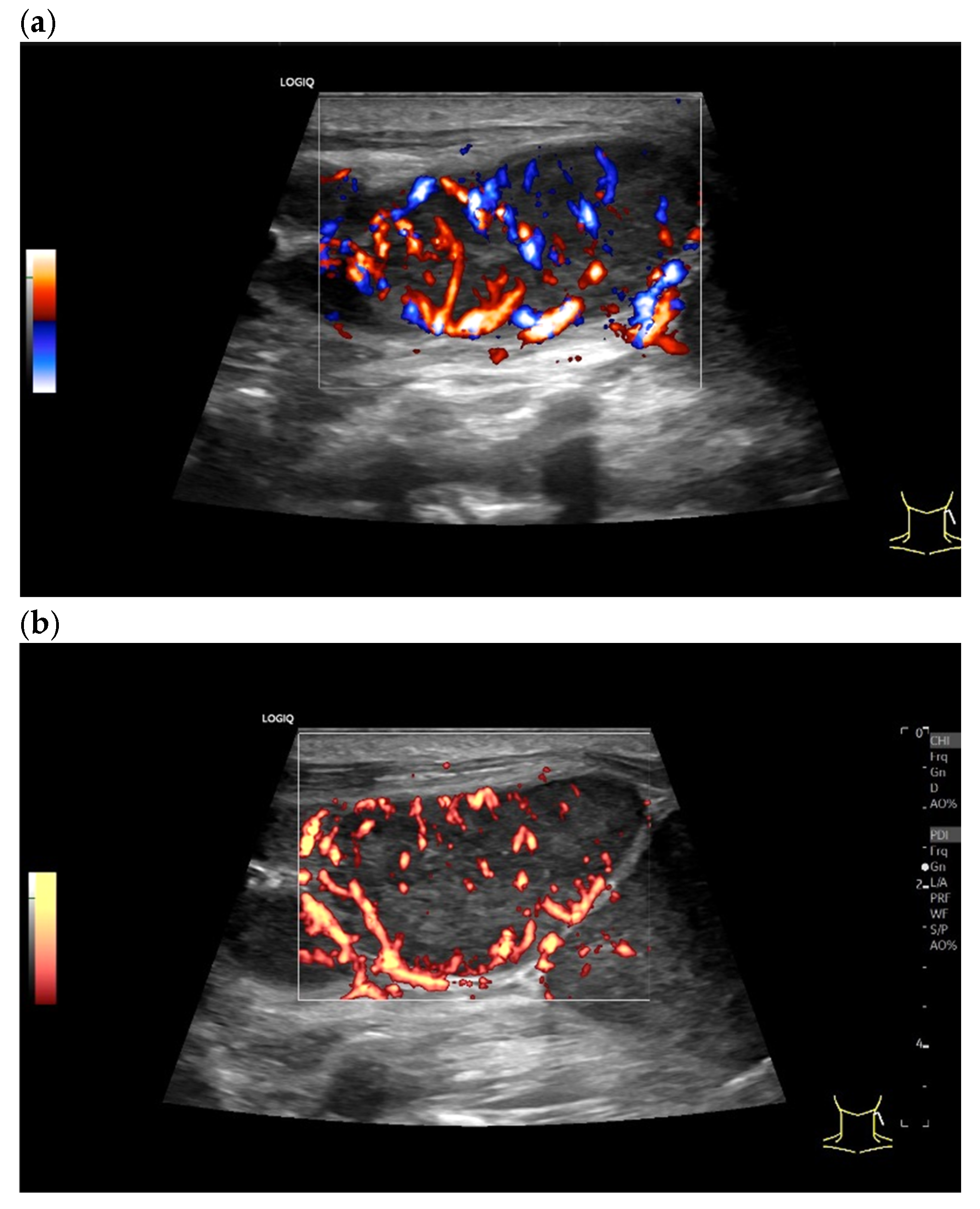
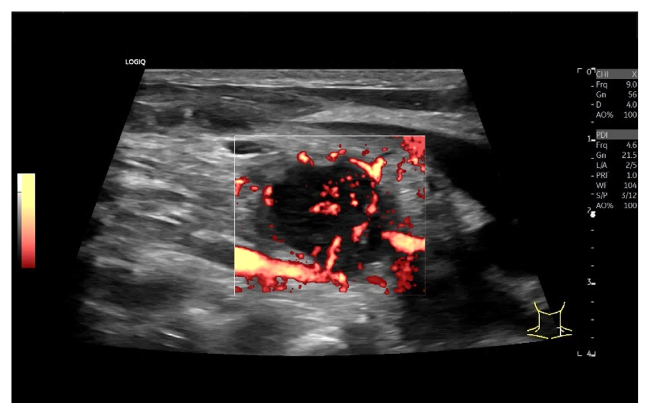
2.12. Color Doppler Imaging (CDI)
2.12.1. What Is Normal (Reference Values)
2.12.2. What Suggests Malignancy
2.12.3. Overlap and Pitfalls
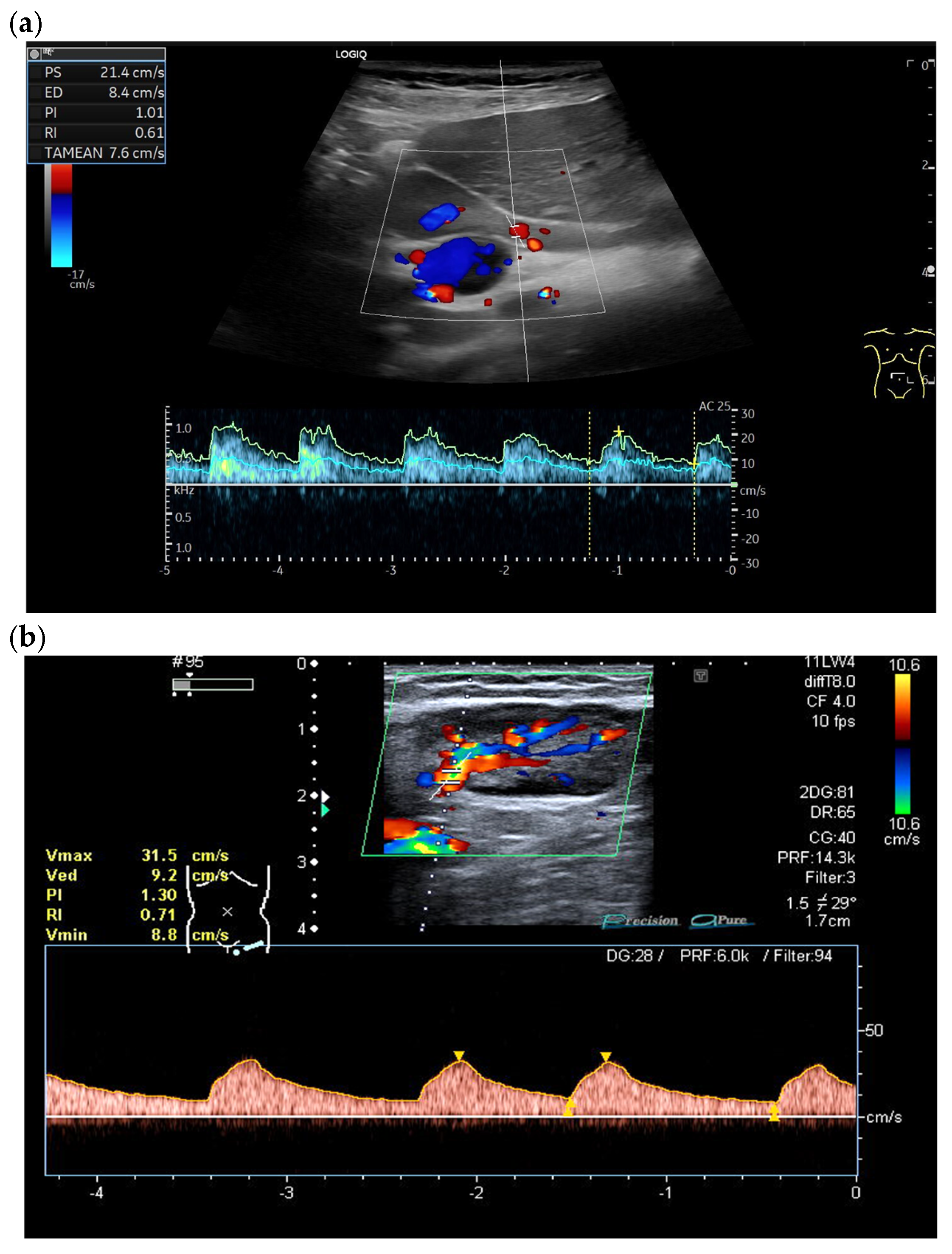
2.13. Resistive Index (RI) and Pulsatility Index (PI)
2.13.1. What Is Normal (Reference Values)
2.13.2. What Suggests Malignancy
2.13.3. Overlap and Pitfalls
2.14. CEUS
2.14.1. What Is Normal (Reference Values)
2.14.2. What Suggests Malignancy
2.14.3. Overlap and Pitfalls
2.15. Superb Microvascular Imaging (SMI)
2.16. Elastography
2.17. Strain Elastography
2.17.1. What Is Normal (Reference Values)
2.17.2. What Suggests Malignancy
2.17.3. Overlap and Pitfalls
2.18. Shear Wave Elastography
3. Multiparametric US
4. Lymph Node Biopsy, US-Guided Sampling
5. Future Development, Structured Diagnosis
6. Conclusions
Author Contributions
Funding
Institutional Review Board Statement
Informed Consent Statement
Data Availability Statement
Conflicts of Interest
References
- Dietrich, C.F.; Zeuzem, S.; Caspary, W.F.; Wehrmann, T. Ultrasound lymph node imaging in the abdomen and retroperitoneum of healthy probands. Ultraschall Med. 1998, 19, 265–269. [Google Scholar] [CrossRef] [PubMed]
- Sienz, M.; Ignee, A.; Dietrich, C.F. Reference values in abdominal ultrasound—Liver and liver vessels. Z. Gastroenterol. 2010, 48, 1141–1152. [Google Scholar] [CrossRef] [PubMed]
- Sienz, M.; Ignee, A.; Dietrich, C.F. Reference values in abdominal ultrasound—Biliopancreatic system and spleen. Z. Gastroenterol. 2011, 49, 845–870. [Google Scholar] [CrossRef] [PubMed]
- Sienz, M.; Ignee, A.; Dietrich, C.F. Sonography today: Reference values in abdominal ultrasound: Aorta, inferior vena cava, kidneys. Z. Gastroenterol. 2012, 50, 293–315. [Google Scholar] [CrossRef]
- Meier, J.; Lucius, C.; Möller, K.; Jenssen, C.; Zervides, C.; Gschmack, A.M.; Dong, Y.; Srivastava, D.; Dietrich, C.F. Pancreatic ultrasound: An update of measurements, reference values, and variations of the pancreas. Ultrasound Int. Open 2024, 10, a23899085. [Google Scholar] [CrossRef]
- Srivastava, S.; Dighe, M.; Möller, K.; Chammas, M.C.; Dong, Y.; Cui, X.C.; Dietrich, C.F. Ultrasound measurements and normal findings in the thyroid gland. Med. Ultrason. 2025, 27, 341–351. [Google Scholar] [CrossRef]
- Möller, K.; Saborio, M.; Gottschall, H.; Blaivas, M.; Borges, A.C.; Morf, S.; Möller, B.; Dietrich, C.F. The Perception of the Diaphragm with Ultrasound: Always There Yet Overlooked? Life 2025, 15, 239. [Google Scholar] [CrossRef]
- Möller, K.; Fischer, P.; Gilja, O.H.; Gottschall, H.; Jenssen, C.; Hollerweger, A.; Lucius, C.; Meier, J.; Rogler, G.; Misselwitz, B.; et al. Gastrointestinal Ultrasound: Measurements and Normal Findings—What Do You Need to Know? Dig. Dis. 2025, 43, 300–335. [Google Scholar] [CrossRef]
- Lucius, C.; Flückiger, A.; Meier, J.; Möller, K.; Jenssen, C.; Braden, B.; Kallenbach, M.; Misselwitz, B.; Nolsøe, C.; Sienz, M.; et al. Ultrasound of Bile Ducts-An Update on Measurements, Reference Values, and Their Influencing Factors. Diagnostics 2025, 15, 919. [Google Scholar] [CrossRef]
- Lucius, C.; Braden, B.; Jenssen, C.; Möller, K.; Sienz, M.; Zervides, C.; Essig, M.W.; Dietrich, C.F. Ultrasound of the Gallbladder-An Update on Measurements, Reference Values, Variants and Frequent Pathologies: A Scoping Review. Life 2025, 15, 941. [Google Scholar] [CrossRef]
- Lucius, C.; Meier, J.; Gschmack, A.; Zervides, C.; Jenssen, C.; Dong, Y.; Graumann, O.; Petry, M.; Dietrich, C.F. Ultrasound of the spleen—An update on measurements, reference values, and influencing factors. A systematic review. Med. Ultrason. 2025, 27, 185–194. [Google Scholar] [CrossRef] [PubMed]
- Gschmack, A.M.; Karlas, T.; Lucius, C.; Barth, G.; Blaivas, M.; Daum, N.; Dong, Y.; Goudie, A.; Hoffmann, B.; Jenssen, C.; et al. Measurement and Normal Values, Pathologies, Interpretation of findings, and Interventional Ultrasound as part of student ultrasound education. Z. Gastroenterol. 2025, 63, 513–520. [Google Scholar] [CrossRef] [PubMed]
- Yadav, M.; Srivastava, S.; Dighe, M.; Möller, K.; Jenssen, C.; Dietrich, C.F. Sonographic Anatomy and Normal Measurements of the Human Kidneys: A Comprehensive Review. Diagnostics 2025, 15, 3208. [Google Scholar] [CrossRef] [PubMed]
- Möller, K.; Ludwig, M.; Jenssen, C.; Dong, Y.; Daum, N.; Wu Cui, X.; Dighe, M.; Graumann, O.; Kallenbach, M.; Lucius, C.; et al. Ultrasound measurements and normal values of the liver: A comprehensive review and practical guide. Med. Ultrason 2026. Online ahead of print. [Google Scholar] [CrossRef]
- Nylund, K.; Hausken, T.; Odegaard, S.; Eide, G.E.; Gilja, O.H. Gastrointestinal wall thickness measured with transabdominal ultrasonography and its relationship to demographic factors in healthy subjects. Ultraschall Med. 2012, 33, E225–E232. [Google Scholar] [CrossRef]
- Som, P.M.; Curtin, H.D.; Mancuso, A.A. Imaging-based nodal classification for evaluation of neck metastatic adenopathy. AJR Am. J. Roentgenol. 2000, 174, 837–844. [Google Scholar] [CrossRef]
- Dietrich, C.; Gottschalk, R.; Herrmann, G.; Caspary, W.; Zeuzem, S. Sonographischer Nachweis von Lymphknoten im Ligamentum hepatoduodenale. DMW—Dtsch. Med. Wochenschr. 1997, 122, 1269–1274. [Google Scholar] [CrossRef]
- Dietrich, C.F.; Liesen, M.; Buhl, R.; Herrmann, G.; Kirchner, J.; Caspary, W.F.; Wehrmann, T. Detection of normal mediastinal lymph nodes by ultrasonography. Acta Radiol. 1997, 38, 965–969. [Google Scholar] [CrossRef]
- Aoki, T.; Moriyasu, F.; Yamamoto, K.; Shimizu, M.; Yamada, M.; Imai, Y. Image of tumor metastasis and inflammatory lymph node enlargement by contrast-enhanced ultrasonography. World J. Radiol. 2011, 3, 298–305. [Google Scholar] [CrossRef]
- Pan, W.R.; Zeng, F.Q.; Wang, D.G.; Qiu, Z.Q. Perforating and deep lymphatic vessels in the knee region: An anatomical study and clinical implications. ANZ J. Surg. 2017, 87, 404–410. [Google Scholar] [CrossRef]
- Shinaoka, A.; Koshimune, S.; Suami, H.; Yamada, K.; Kumagishi, K.; Boyages, J.; Kimata, Y.; Ohtsuka, A. Lower-Limb Lymphatic Drainage Pathways and Lymph Nodes: A CT Lymphangiography Cadaver Study. Radiology 2020, 294, 223–229. [Google Scholar] [CrossRef]
- Tokur, O.; Aydin, S.; Kilinc, F.; Degirmenci, T.; Oztekin, P.S. Diagnostic value of super microvascular imaging in differentiating axillary lymph nodes: Semi-quantitative and qualitative approach in primary breast cancer and suspicious axillary nodes. BMC Cancer 2025, 25, 1705. [Google Scholar] [CrossRef] [PubMed]
- Rettenbacher, T. Sonography of peripheral lymph nodes part 1: Normal findings and B-image criteria. Ultraschall Med. 2010, 31, 344–362. [Google Scholar] [CrossRef] [PubMed]
- Ying, M.; Ahuja, A.; Brook, F.; Brown, B.; Metreweli, C. Sonographic appearance and distribution of normal cervical lymph nodes in a Chinese population. J. Ultrasound Med. 1996, 15, 431–436. [Google Scholar] [CrossRef] [PubMed]
- Ying, M.; Ahuja, A.; Brook, F. Sonographic appearances of cervical lymph nodes: Variations by age and sex. J. Clin. Ultrasound 2002, 30, 1–11. [Google Scholar] [CrossRef]
- Vassallo, P.; Edel, G.; Roos, N.; Naguib, A.; Peters, P.E. In-vitro high-resolution ultrasonography of benign and malignant lymph nodes. A sonographic-pathologic correlation. Investig. Radiol. 1993, 28, 698–705. [Google Scholar] [CrossRef]
- Gritzmann, N.; Hollerweger, A.; Macheiner, P.; Rettenbacher, T. Sonography of soft tissue masses of the neck. J. Clin. Ultrasound 2002, 30, 356–373. [Google Scholar] [CrossRef]
- Esen, G. Ultrasound of superficial lymph nodes. Eur. J. Radiol. 2006, 58, 345–359. [Google Scholar] [CrossRef]
- Moore, A.; Hester, M.; Nam, M.W.; Brill, Y.M.; McGrath, P.; Wright, H.; Weisinger, K.; Romond, E.; Samayoa, L.M. Distinct lymph nodal sonographic characteristics in breast cancer patients at high risk for axillary metastases correlate with the final axillary stage. Br. J. Radiol. 2008, 81, 630–636. [Google Scholar] [CrossRef]
- Dietrich, C.F.; Hocke, M.; Jenssen, C. Ultrasound for abdominal lymphadenopathy. Dtsch. Med. Wochenschr. 2013, 138, 1001–1018. [Google Scholar] [CrossRef]
- Ying, M.; Ahuja, A.; Brook, F.; Brown, B.; Metreweli, C. Nodal shape (S/L) and its combination with size for assessment of cervical lymphadenopathy: Which cut-off should be used? Ultrasound Med. Biol. 1999, 25, 1169–1175. [Google Scholar] [CrossRef]
- van den Brekel, M.W.; Stel, H.V.; Castelijns, J.A.; Nauta, J.J.; van der Waal, I.; Valk, J.; Meyer, C.J.; Snow, G.B. Cervical lymph node metastasis: Assessment of radiologic criteria. Radiology 1990, 177, 379–384. [Google Scholar] [CrossRef] [PubMed]
- Ghafoori, M.; Azizian, A.; Pourrajabi, Z.; Vaseghi, H. Sonographic Evaluation of Cervical Lymphadenopathy; Comparison of Metastatic and Reactive Lymph Nodes in Patients With Head and Neck Squamous Cell Carcinoma Using Gray Scale and Doppler Techniques. Iran. J. Radiol. 2015, 12, e11044. [Google Scholar] [CrossRef] [PubMed]
- Sumi, M.; Ohki, M.; Nakamura, T. Comparison of sonography and CT for differentiating benign from malignant cervical lymph nodes in patients with squamous cell carcinoma of the head and neck. AJR Am. J. Roentgenol. 2001, 176, 1019–1024. [Google Scholar] [CrossRef] [PubMed]
- Lyshchik, A.; Higashi, T.; Asato, R.; Tanaka, S.; Ito, J.; Hiraoka, M.; Insana, M.F.; Brill, A.B.; Saga, T.; Togashi, K. Cervical lymph node metastases: Diagnosis at sonoelastography--initial experience. Radiology 2007, 243, 258–267. [Google Scholar] [CrossRef]
- Steinkamp1, H.; Knöbber2, D.; Schedel1, H.; Mäurer1, J.; Felix1, R. Palpation und Sonographie in der Nachsorge von Kopf-Hals-Tumorpatienten: Vergleich sonographischer Dignitätsparameter. Laryngo-Rhino-Otologie 1993, 72, 431–438. [Google Scholar] [CrossRef]
- Öztoprak, B.; Öztoprak, İ.; Sönmez, M.; Haydar Atalar, M.; Arslan, M. Sonographic evaluation of jugulodigastric and juguloomohyoid lymph nodes. Clin. Anat. 2016, 29, 943–948. [Google Scholar] [CrossRef]
- Lucey, B.C.; Stuhlfaut, J.W.; Soto, J.A. Mesenteric lymph nodes: Detection and significance on MDCT. AJR Am. J. Roentgenol. 2005, 184, 41–44. [Google Scholar] [CrossRef]
- Dayanand, S.M.; Desai, R.; Reddy, P.B. Efficiency of ultrasonography in assessing cervical lymph node metastasis in oral carcinoma. Natl. J. Maxillofac. Surg. 2010, 1, 117–122. [Google Scholar] [CrossRef]
- Schwartz, L.H.; Litière, S.; de Vries, E.; Ford, R.; Gwyther, S.; Mandrekar, S.; Shankar, L.; Bogaerts, J.; Chen, A.; Dancey, J.; et al. RECIST 1.1-Update and clarification: From the RECIST committee. Eur. J. Cancer 2016, 62, 132–137. [Google Scholar] [CrossRef]
- Schwartz, L.H.; Bogaerts, J.; Ford, R.; Shankar, L.; Therasse, P.; Gwyther, S.; Eisenhauer, E.A. Evaluation of lymph nodes with RECIST 1.1. Eur. J. Cancer 2009, 45, 261–267. [Google Scholar] [CrossRef]
- Chen, J.; Deng, Y.; Xiong, J.; Li, W.; Shang, G.; Li, H.; Wu, Y.; Qin, X. The diagnostic value of two-dimensional shear-wave elastography in identifying malignant lesions in lymph nodes: A prospective study. Sci. Rep. 2025, 15, 17395. [Google Scholar] [CrossRef] [PubMed]
- Cui, Q.L.; Yin, S.S.; Fan, Z.H.; Yang, W.; Wang, S.; Yan, K. Diagnostic Value of Contrast-Enhanced Ultrasonography and Time-Intensity Curve in Differential Diagnosis of Cervical Metastatic and Tuberculous Lymph Nodes. J. Ultrasound Med. 2018, 37, 83–92. [Google Scholar] [CrossRef] [PubMed]
- Tschammler, A.; Ott, G.; Schang, T.; Seelbach-Goebel, B.; Schwager, K.; Hahn, D. Lymphadenopathy: Differentiation of benign from malignant disease--color Doppler US assessment of intranodal angioarchitecture. Radiology 1998, 208, 117–123. [Google Scholar] [CrossRef] [PubMed]
- Bhatia, K.S.; Cho, C.C.; Tong, C.S.; Yuen, E.H.; Ahuja, A.T. Shear wave elasticity imaging of cervical lymph nodes. Ultrasound Med. Biol. 2012, 38, 195–201. [Google Scholar] [CrossRef]
- Ishibashi, N.; Yamagata, K.; Sasaki, H.; Seto, K.; Shinya, Y.; Ito, H.; Shinozuka, K.; Yanagawa, T.; Onizawa, K.; Bukawa, H. Real-Time Tissue Elastography for the Diagnosis of Lymph Node Metastasis in Oral Squamous Cell Carcinoma. Ultrasound Med. Biol. 2012, 38, 389–395. [Google Scholar] [CrossRef]
- Hinz, T.; Hoeller, T.; Wenzel, J.; Bieber, T.; Schmid-Wendtner, M.H. Real-time tissue elastography as promising diagnostic tool for diagnosis of lymph node metastases in patients with malignant melanoma: A prospective single-center experience. Dermatology 2013, 226, 81–90. [Google Scholar] [CrossRef]
- Okumuş, Ö.; Dönmez, M.; Pekiner, F.N. Ultrasonographic Appearances of Cervical Lymph Nodes in Healthy Turkish Adults Subpopulation: Preliminary Study. Open Dent. J. 2017, 11, 404–412. [Google Scholar] [CrossRef]
- Lerchbaumer, M.H.; Wakonig, K.M.; Arens, P.; Dommerich, S.; Fischer, T. Quantitative Multiparametric Ultrasound (mpUS) in the Assessment of Inconclusive Cervical Lymph Nodes. Cancers 2022, 14, 1597. [Google Scholar] [CrossRef]
- Çetin Tunçez, H.; Murat Koç, A.; Hilal Adıbelli, Z.; Zeynep Arslan, F.; Argon, A.; Yücel Oğuzdoğan, G. Diagnostic Efficacy of Ultrasonography, Doppler Ultrasonography and Elastography in the Evaluation of Suspected Malignant Lymph Nodes. J. Ultrason. 2023, 23, e1–e9. [Google Scholar] [CrossRef]
- He, J.; Yi, H.; Tang, D.; Zhang, X.; Cui, X.; Zhang, W. Differentiating benign and malignant superficial lymph nodes using super-resolution contrast enhanced ultrasound—A pilot study. BMC Med. Imaging 2025, 25, 170. [Google Scholar] [CrossRef]
- Steinkamp, H.J.; Mäurer, J.; Cornehl, M.; Knöbber, D.; Hettwer, H.; Felix, R. Recurrent cervical lymphadenopathy: Differential diagnosis with color-duplex sonography. Eur. Arch. Otorhinolaryngol. 1994, 251, 404–409. [Google Scholar] [CrossRef]
- Cui, X.W.; Hocke, M.; Jenssen, C.; Ignee, A.; Klein, S.; Schreiber-Dietrich, D.; Dietrich, C.F. Conventional ultrasound for lymph node evaluation, update 2013. Z. Gastroenterol. 2014, 52, 212–221. [Google Scholar] [CrossRef] [PubMed]
- Solbiati, L.; Rizzato, G.; Belotti, E. High resolution sonography of cervical lymph nodes in head and neck cancer. Criterias for differentiation of reactive versus malignant nodes. Radiology 1988, 169, 113–117. [Google Scholar]
- Osanai, H.; Kuroiwa, H.; Uchida, K.; Kagami, H.; Yamada, K.; Taguchi, A. Sonographic appearances of cervical lymph nodes in healthy young Japanese adults: Association with age, sex, and body mass index. J. Clin. Ultrasound 2015, 43, 295–301. [Google Scholar] [CrossRef] [PubMed]
- Ying, M.; Ahuja, A.; Brook, F. Gray scale and power Doppler sonography of normal cervical lymph nodes: Comparison between Chinese and white subjects. J. Ultrasound Med. 2002, 21, 59–65. [Google Scholar] [CrossRef]
- Aladag Kurt, S.; Kayadibi, Y.; Onur, I.; Uslu Besli, L.; Necati Sanli, A.; Velidedeoglu, M. Predicting axillary nodal metastasis based on the side of asymmetrical cortical thickening in breast cancer: Evaluation with grayscale and microvascular imaging findings. Eur. J. Radiol. 2023, 158, 110643. [Google Scholar] [CrossRef]
- Jenssen, C.; Möller, K.; Sarbia, M.; Wagner, S. Endosonographische Biopsie (EUS-FNA, EUS-TCB)—Fallstricke, Probleme und Problemlösungen. In Endosonographie. Lehrbuch und Atlas des Endoskopischen Ultraschalls; Dietrich, C.F., Ed.; Thieme: Stuttgart, Germany, 2008; pp. 87–140. [Google Scholar]
- Ahuja, A.; Ying, M. Sonography of neck lymph nodes. Part II: Abnormal lymph nodes. Clin. Radiol. 2003, 58, 359–366. [Google Scholar] [CrossRef]
- Xiang, D.; Hong, Y.; Zhang, B.; Huang, P.; Li, G.; Wang, P.; Li, Z. Contrast-enhanced ultrasound (CEUS) facilitated US in detecting lateral neck lymph node metastasis of thyroid cancer patients: Diagnosis value and enhancement patterns of malignant lymph nodes. Eur. Radiol. 2014, 24, 2513–2519. [Google Scholar] [CrossRef]
- Moon, I.S.; Kim, D.W.; Baek, H.J. Ultrasound-based diagnosis for the cervical lymph nodes in a tuberculosis-endemic area. Laryngoscope 2015, 125, 1113–1117. [Google Scholar] [CrossRef]
- Zhang, J.; Hu, Y.; Huang, X.; Huang, X.; Deng, Q.; Zhou, Q. Ultrafast ultrasound localization microscopy for differential diagnosis of reactive hyperplasia and metastatic cervical lymph nodes: A pilot study. Quant. Imaging Med. Surg. 2025, 15, 10204–10214. [Google Scholar] [CrossRef]
- Xu, L.L.; Zhou, L.; Yu, X.L.; Li, S.Y. A Novel Noninvasive Approach for Evaluating Suspicious Cervical Lymph Nodes in Papillary Thyroid Carcinoma-Superb Microvascular Imaging. J. Clin. Ultrasound 2025, 53, 1801–1809. [Google Scholar] [CrossRef]
- Seher, N.; Nayman, A.; Dilek, I.; Erol, S.; Ari, A.; Baytok, A.; Ozer, H.; Ugras, N.S.; Koplay, M. Evaluation of Lymph Nodes Using a Combination of Advanced Sonographic Techniques: Superb Microvascular Imaging and Shear Wave Elastography. J. Clin. Ultrasound 2025, 53, 1213–1220. [Google Scholar] [CrossRef]
- Yang, Z.; Cai, J.; Wang, Y.; Shu, L.; Liu, W.; Wang, Z. Comparison of Doppler Imaging and Microvascular Imaging in Cervical Lymph Node Blood Flow Analysis. Curr. Med. Imaging 2024, 20, e15734056306197. [Google Scholar] [CrossRef] [PubMed]
- Huang, T.; Huang, P.T.; Luo, Z.Y.; Lv, J.F.; Jin, P.L.; Zhang, T.; Zhao, Y.L.; Wang, Y.; Hong, Y.R. Use superb microvascular imaging to diagnose and predict metastatic cervical lymph nodes in patients with papillary thyroid carcinoma. J. Cancer Res. Clin. Oncol. 2024, 150, 268. [Google Scholar] [CrossRef] [PubMed]
- Bulut, I.N.; Kayadibi, Y.; Deger, E.; Kurt, S.A.; Velidedeoglu, M.; Onur, I.; Ozturk, T.; Adaletli, I. Preoperative Role of Superb Microvascular Imaging and Shear-Wave Elastography for Prediction of Axillary Lymph Node Metastasis in Patients With Breast Cancer. Ultrasound Q. 2024, 40, 111–118. [Google Scholar] [CrossRef] [PubMed]
- Kurt, S.A.; Eryurekli, A.E.; Kayadibi, Y.; Samanci, C.; Velidedeoglu, M.; Onur, I.; Arslan, S.; Taskin, F. Diagnostic Performance of Superb Microvascular Imaging in Differentiating Benign and Malignant Axillary Lymph Nodes. Ultrasound Q. 2023, 39, 74–80. [Google Scholar] [CrossRef]
- Cui, X.W.; Jenssen, C.; Saftoiu, A.; Ignee, A.; Dietrich, C.F. New ultrasound techniques for lymph node evaluation. World J. Gastroenterol. 2013, 19, 4850–4860. [Google Scholar] [CrossRef]
- Bialek, E.J.; Jakubowski, W.; Szczepanik, A.B.; Maryniak, R.K.; Prochorec-Sobieszek, M.; Bilski, R.; Szopinski, K.T. Vascular patterns in superficial lymphomatous lymph nodes: A detailed sonographic analysis. J. Ultrasound 2007, 10, 128–134. [Google Scholar] [CrossRef]
- Ying, M.; Ahuja, A.; Brook, F. Accuracy of sonographic vascular features in differentiating different causes of cervical lymphadenopathy. Ultrasound Med. Biol. 2004, 30, 441–447. [Google Scholar] [CrossRef]
- Rettenbacher, T. Sonography of peripheral lymph nodes part 2: Doppler criteria and typical findings of distinct entities. Ultraschall Med. 2014, 35, 10–27; quiz 28-32. [Google Scholar] [CrossRef]
- Chiorean, L.; Barr, R.G.; Braden, B.; Jenssen, C.; Cui, X.W.; Hocke, M.; Schuler, A.; Dietrich, C.F. Transcutaneous Ultrasound: Elastographic Lymph Node Evaluation. Current Clinical Applications and Literature Review. Ultrasound Med. Biol. 2016, 42, 16–30. [Google Scholar] [CrossRef] [PubMed]
- Choi, M.Y.; Lee, J.W.; Jang, K.J. Distinction between benign and malignant causes of cervical, axillary, and inguinal lymphadenopathy: Value of Doppler spectral waveform analysis. Am. J. Roentgenol. 1995, 165, 981–984. [Google Scholar] [CrossRef] [PubMed]
- Sidhu, P.S.; Cantisani, V.; Dietrich, C.F.; Gilja, O.H.; Saftoiu, A.; Bartels, E.; Bertolotto, M.; Calliada, F.; Clevert, D.A.; Cosgrove, D.; et al. The EFSUMB Guidelines and Recommendations for the Clinical Practice of Contrast-Enhanced Ultrasound (CEUS) in Non-Hepatic Applications: Update 2017 (Short Version). Ultraschall Med. 2018, 39, 154–180. [Google Scholar] [CrossRef] [PubMed]
- Nielsen Moody, A.; Bull, J.; Culpan, A.M.; Munyombwe, T.; Sharma, N.; Whitaker, M.; Wolstenhulme, S. Preoperative sentinel lymph node identification, biopsy and localisation using contrast enhanced ultrasound (CEUS) in patients with breast cancer: A systematic review and meta-analysis. Clin. Radiol. 2017, 72, 959–971. [Google Scholar] [CrossRef]
- Cui, Q.; Dai, L.; Li, J.; Xue, J. Accuracy of CEUS-guided sentinel lymph node biopsy in early-stage breast cancer: A study review and meta-analysis. World J. Surg. Oncol. 2020, 18, 112. [Google Scholar] [CrossRef]
- Spiesecke, P.; Neumann, K.; Wakonig, K.; Lerchbaumer, M.H. Contrast-enhanced ultrasound (CEUS) in characterization of inconclusive cervical lymph nodes: A meta-analysis and systematic review. Sci. Rep. 2022, 12, 7804. [Google Scholar] [CrossRef]
- Wakonig, K.M.; Dommerich, S.; Fischer, T.; Arens, P.; Hamm, B.; Olze, H.; Lerchbaumer, M.H. The Diagnostic Performance of Multiparametric Ultrasound in the Qualitative Assessment of Inconclusive Cervical Lymph Nodes. Cancers 2023, 15, 5035. [Google Scholar] [CrossRef]
- Ling, W.; Nie, J.; Zhang, D.; Yang, Q.; Jin, H.; Ou, X.; Ma, X.; Luo, Y. Role of Contrast-Enhanced Ultrasound (CEUS) in the Diagnosis of Cervical Lymph Node Metastasis in Nasopharyngeal Carcinoma (NPC) Patients. Front. Oncol. 2020, 10, 972. [Google Scholar] [CrossRef]
- Yin, S.S.; Cui, Q.L.; Fan, Z.H.; Yang, W.; Yan, K. Diagnostic Value of Arrival Time Parametric Imaging Using Contrast-Enhanced Ultrasonography in Superficial Enlarged Lymph Nodes. J. Ultrasound Med. 2019, 38, 1287–1298. [Google Scholar] [CrossRef]
- Nie, J.; Ling, W.; Yang, Q.; Jin, H.; Ou, X.; Ma, X. The Value of CEUS in Distinguishing Cancerous Lymph Nodes From the Primary Lymphoma of the Head and Neck. Front. Oncol. 2020, 10, 473. [Google Scholar] [CrossRef]
- Dietrich, C.F.; Correas, J.M.; Cui, X.W.; Dong, Y.; Havre, R.F.; Jenssen, C.; Jung, E.M.; Krix, M.; Lim, A.; Lassau, N.; et al. EFSUMB Technical Review—Update 2023: Dynamic Contrast-Enhanced Ultrasound (DCE-CEUS) for the Quantification of Tumor Perfusion. Ultraschall Med. 2024, 45, 36–46. [Google Scholar] [CrossRef]
- Sim, J.K.; Lee, J.Y.; Hong, H.S. Differentiation Between Malignant and Benign Lymph Nodes: Role of Superb Microvascular Imaging in the Evaluation of Cervical Lymph Nodes. J. Ultrasound Med. 2019, 38, 3025–3036. [Google Scholar] [CrossRef] [PubMed]
- Ryoo, I.; Suh, S.; You, S.H.; Seol, H.Y. Usefulness of Microvascular Ultrasonography in Differentiating Metastatic Lymphadenopathy from Tuberculous Lymphadenitis. Ultrasound Med. Biol. 2016, 42, 2189–2195. [Google Scholar] [CrossRef] [PubMed]
- Lee, S.; Lee, J.Y.; Yoon, R.G.; Kim, J.H.; Hong, H.S. The Value of Microvascular Imaging for Triaging Indeterminate Cervical Lymph Nodes in Patients with Papillary Thyroid Carcinoma. Cancers 2020, 12, 2839. [Google Scholar] [CrossRef] [PubMed]
- Săftoiu, A.; Gilja, O.H.; Sidhu, P.S.; Dietrich, C.F.; Cantisani, V.; Amy, D.; Bachmann-Nielsen, M.; Bob, F.; Bojunga, J.; Brock, M.; et al. The EFSUMB Guidelines and Recommendations for the Clinical Practice of Elastography in Non-Hepatic Applications: Update 2018. Ultraschall Med. 2019, 40, 425–453. [Google Scholar] [CrossRef]
- Cui, X.W.; Chang, J.M.; Kan, Q.C.; Chiorean, L.; Ignee, A.; Dietrich, C.F. Endoscopic ultrasound elastography: Current status and future perspectives. World J. Gastroenterol. 2015, 21, 13212–13224. [Google Scholar] [CrossRef]
- Cui, X.W.; Li, K.N.; Yi, A.J.; Wang, B.; Wei, Q.; Wu, G.G.; Dietrich, C.F. Ultrasound elastography. Endosc. Ultrasound 2022, 11, 252–274. [Google Scholar] [CrossRef]
- Dietrich, C.F.; Barr, R.G.; Farrokh, A.; Dighe, M.; Hocke, M.; Jenssen, C.; Dong, Y.; Saftoiu, A.; Havre, R.F. Strain Elastography—How To Do It? Ultrasound Int. Open 2017, 3, E137–E149. [Google Scholar] [CrossRef]
- Wang, B.; Guo, Q.; Wang, J.Y.; Yu, Y.; Yi, A.J.; Cui, X.W.; Dietrich, C.F. Ultrasound Elastography for the Evaluation of Lymph Nodes. Front. Oncol. 2021, 11, 714660. [Google Scholar] [CrossRef]
- Ferraioli, G.; Barr, R.G.; Farrokh, A.; Radzina, M.; Cui, X.W.; Dong, Y.; Rocher, L.; Cantisani, V.; Polito, E.; D’Onofrio, M.; et al. How to perform shear wave elastography. Part II. Med. Ultrason. 2022, 24, 196–210. [Google Scholar] [CrossRef]
- Zhang, X.Y.; Wei, Q.; Wu, G.G.; Tang, Q.; Pan, X.F.; Chen, G.Q.; Zhang, D.; Dietrich, C.F.; Cui, X.W. Artificial intelligence—Based ultrasound elastography for disease evaluation—A narrative review. Front. Oncol. 2023, 13, 1197447. [Google Scholar] [CrossRef] [PubMed]
- Shiina, T.; Nightingale, K.R.; Palmeri, M.L.; Hall, T.J.; Bamber, J.C.; Barr, R.G.; Castera, L.; Choi, B.I.; Chou, Y.H.; Cosgrove, D.; et al. WFUMB guidelines and recommendations for clinical use of ultrasound elastography: Part 1: Basic principles and terminology. Ultrasound Med. Biol. 2015, 41, 1126–1147. [Google Scholar] [CrossRef] [PubMed]
- Dietrich, C.F.; Bamber, J.; Berzigotti, A.; Bota, S.; Cantisani, V.; Castera, L.; Cosgrove, D.; Ferraioli, G.; Friedrich-Rust, M.; Gilja, O.H.; et al. EFSUMB Guidelines and Recommendations on the Clinical Use of Liver Ultrasound Elastography, Update 2017 (Short Version). Ultraschall Med. 2017, 38, 377–394. [Google Scholar] [CrossRef] [PubMed]
- Bamber, J.; Cosgrove, D.; Dietrich, C.F.; Fromageau, J.; Bojunga, J.; Calliada, F.; Cantisani, V.; Correas, J.M.; D’Onofrio, M.; Drakonaki, E.E.; et al. EFSUMB guidelines and recommendations on the clinical use of ultrasound elastography. Part 1: Basic principles and technology. Ultraschall Med. 2013, 34, 169–184. [Google Scholar] [CrossRef]
- Cosgrove, D.; Piscaglia, F.; Bamber, J.; Bojunga, J.; Correas, J.M.; Gilja, O.H.; Klauser, A.S.; Sporea, I.; Calliada, F.; Cantisani, V.; et al. EFSUMB guidelines and recommendations on the clinical use of ultrasound elastography. Part 2: Clinical applications. Ultraschall Med. 2013, 34, 238–253. [Google Scholar] [CrossRef]
- Giovannini, M.; Thomas, B.; Erwan, B.; Christian, P.; Fabrice, C.; Benjamin, E.; Geneviève, M.; Paolo, A.; Pierre, D.; Robert, Y.; et al. Endoscopic ultrasound elastography for evaluation of lymph nodes and pancreatic masses: A multicenter study. World J. Gastroenterol. 2009, 15, 1587–1593. [Google Scholar] [CrossRef]
- Jiang, M.; Li, C.L.; Luo, X.M.; Chuan, Z.R.; Chen, R.X.; Tang, S.C.; Lv, W.Z.; Cui, X.W.; Dietrich, C.F. Radiomics model based on shear-wave elastography in the assessment of axillary lymph node status in early-stage breast cancer. Eur. Radiol. 2022, 32, 2313–2325. [Google Scholar] [CrossRef]
- Suh, C.H.; Choi, Y.J.; Baek, J.H.; Lee, J.H. The diagnostic performance of shear wave elastography for malignant cervical lymph nodes: A systematic review and meta-analysis. Eur. Radiol. 2017, 27, 222–230. [Google Scholar] [CrossRef]
- Kritcharoen, W.; Pradaranon, V.; Rohitopakarn, P.; Kaewpiboon, W. Accuracy of shear wave elastography in diagnosing axillary lymph node metastasis in patients with suspicious axillary lymph nodes: Development of a predictive nomogram. J. Ultrasound 2025, 28, 627–633. [Google Scholar] [CrossRef]
- Ying, L.; Hou, Y.; Zheng, H.M.; Lin, X.; Xie, Z.L.; Hu, Y.P. Real-time elastography for the differentiation of benign and malignant superficial lymph nodes: A meta-analysis. Eur. J. Radiol. 2012, 81, 2576–2584. [Google Scholar] [CrossRef]
- Ghajarzadeh, M.; Mohammadifar, M.; Azarkhish, K.; Emami-Razavi, S.H. Sono-elastography for Differentiating Benign and Malignant Cervical Lymph Nodes: A Systematic Review and Meta-Analysis. Int. J. Prev. Med. 2014, 5, 1521–1528. [Google Scholar] [PubMed]
- Dietrich, C.F.; Dong, Y.; Cui, X.W.; Fink, M.; Jenssen, C.; Moeller, K.; Sandrin, L.; Tsuneyoshi, S.; Tanter, M. Ultrasound elastography: A brief clinical history of an evolving technique. Ultrasound Int. Open 2024, 10, a23786926. [Google Scholar] [CrossRef] [PubMed]
- Kanagaraju, V.; Rakshith, A.V.B.; Devanand, B.; Rajakumar, R. Utility of Ultrasound Elastography to Differentiate Benign from Malignant Cervical Lymph Nodes. J. Med. Ultrasound 2020, 28, 92–98. [Google Scholar] [CrossRef] [PubMed]
- Lahtinen, O.; Pulkkinen, M.; Sironen, R.; Vanninen, R.; Rautiainen, S. 2D-shear wave elastography in the evaluation of suspicious superficial inguinal lymph nodes: Reproducibility and region of interest selection. PLoS ONE 2022, 17, e0265802. [Google Scholar] [CrossRef]
- Sidhu, P.S.; Brabrand, K.; Cantisani, V.; Correas, J.M.; Cui, X.W.; D’Onofrio, M.; Essig, M.; Freeman, S.; Gilja, O.H.; Gritzmann, N.; et al. EFSUMB Guidelines on Interventional Ultrasound (INVUS), Part II. Diagnostic Ultrasound-Guided Interventional Procedures (Long Version). Ultraschall Med. 2015, 36, E15–E35. [Google Scholar] [CrossRef]
- Chiorean, L.; Cantisani, V.; Jenssen, C.; Sidhu, P.S.; Baum, U.; Dietrich, C.F. Focal masses in a non-cirrhotic liver: The additional benefit of CEUS over baseline imaging. Eur. J. Radiol. 2015, 84, 1636–1643. [Google Scholar] [CrossRef]
- He, Y.; Ji, X.; Xie, Y.; He, B.; Xu, X.; Chen, X.; Zhang, Q. Clinical application of ultrasound-guided core needle biopsy with multiple punches in the diagnosis of lymphoma. World J. Surg. Oncol. 2015, 13, 126. [Google Scholar] [CrossRef]
- Lachar, W.A.; Shahab, I.; Saad, A.J. Accuracy and cost-effectiveness of core needle biopsy in the evaluation of suspected lymphoma: A study of 101 cases. Arch. Pathol. Lab. Med. 2007, 131, 1033–1039. [Google Scholar] [CrossRef]
- Jenssen, C.; Hocke, M.; Fusaroli, P.; Gilja, O.H.; Buscarini, E.; Havre, R.F.; Ignee, A.; Saftoiu, A.; Vilmann, P.; Burmester, E.; et al. EFSUMB Guidelines on Interventional Ultrasound (INVUS), Part IV—EUS-guided interventions: General Aspects and EUS-guided Sampling (Short Version). Ultraschall Med. 2016, 37, 157–169. [Google Scholar] [CrossRef]
- Jenssen, C.; Annema, J.T.; Clementsen, P.; Cui, X.W.; Borst, M.M.; Dietrich, C.F. Ultrasound techniques in the evaluation of the mediastinum, part 2: Mediastinal lymph node anatomy and diagnostic reach of ultrasound techniques, clinical work up of neoplastic and inflammatory mediastinal lymphadenopathy using ultrasound techniques and how to learn mediastinal endosonography. J. Thorac. Dis. 2015, 7, E439–E458. [Google Scholar] [CrossRef]
- Han, F.; Xu, M.; Xie, T.; Wang, J.W.; Lin, Q.G.; Guo, Z.X.; Zheng, W.; Han, J.; Lin, X.; Zou, R.H.; et al. Efficacy of ultrasound-guided core needle biopsy in cervical lymphadenopathy: A retrospective study of 6,695 cases. Eur. Radiol. 2018, 28, 1809–1817. [Google Scholar] [CrossRef]
- Elhamdoust, E.; Motamedfar, A.; Gharibvand, M.M.; Jazayeri, S.N. Investigation of the value of ultrasound-guided core needle biopsy from pathologic lymph nodes to the diagnosis of lymphoma. J. Fam. Med. Prim. Care 2020, 9, 2801–2805. [Google Scholar] [CrossRef]
- Strobel, D.; Bernatik, T.; Blank, W.; Will, U.; Reichel, A.; Wustner, M.; Keim, V.; Schacherer, D.; Barreiros, A.P.; Kunze, G.; et al. Incidence of bleeding in 8172 percutaneous ultrasound-guided intraabdominal diagnostic and therapeutic interventions—Results of the prospective multicenter DEGUM interventional ultrasound study (PIUS study). Ultraschall Med. 2015, 36, 122–131. [Google Scholar] [CrossRef]
- Elsholtz, F.H.J.; Asbach, P.; Haas, M.; Becker, M.; Beets-Tan, R.G.H.; Thoeny, H.C.; Padhani, A.R.; Hamm, B. Introducing the Node Reporting and Data System 1.0 (Node-RADS): A concept for standardized assessment of lymph nodes in cancer. Eur. Radiol. 2021, 31, 6116–6124. [Google Scholar] [CrossRef]
- Zhong, J.; Mao, S.; Chen, H.; Wang, Y.; Yin, Q.; Cen, Q.; Lu, J.; Yang, J.; Hu, Y.; Xing, Y.; et al. Node-RADS: A systematic review and meta-analysis of diagnostic performance, category-wise malignancy rates, and inter-observer reliability. Eur. Radiol. 2025, 35, 2723–2735. [Google Scholar] [CrossRef]
- Parillo, M.; Quattrocchi, C.C. Node Reporting and Data System 1.0 (Node-RADS) for the Assessment of Oncological Patients’ Lymph Nodes in Clinical Imaging. J. Clin. Med. 2025, 14, 263. [Google Scholar] [CrossRef]




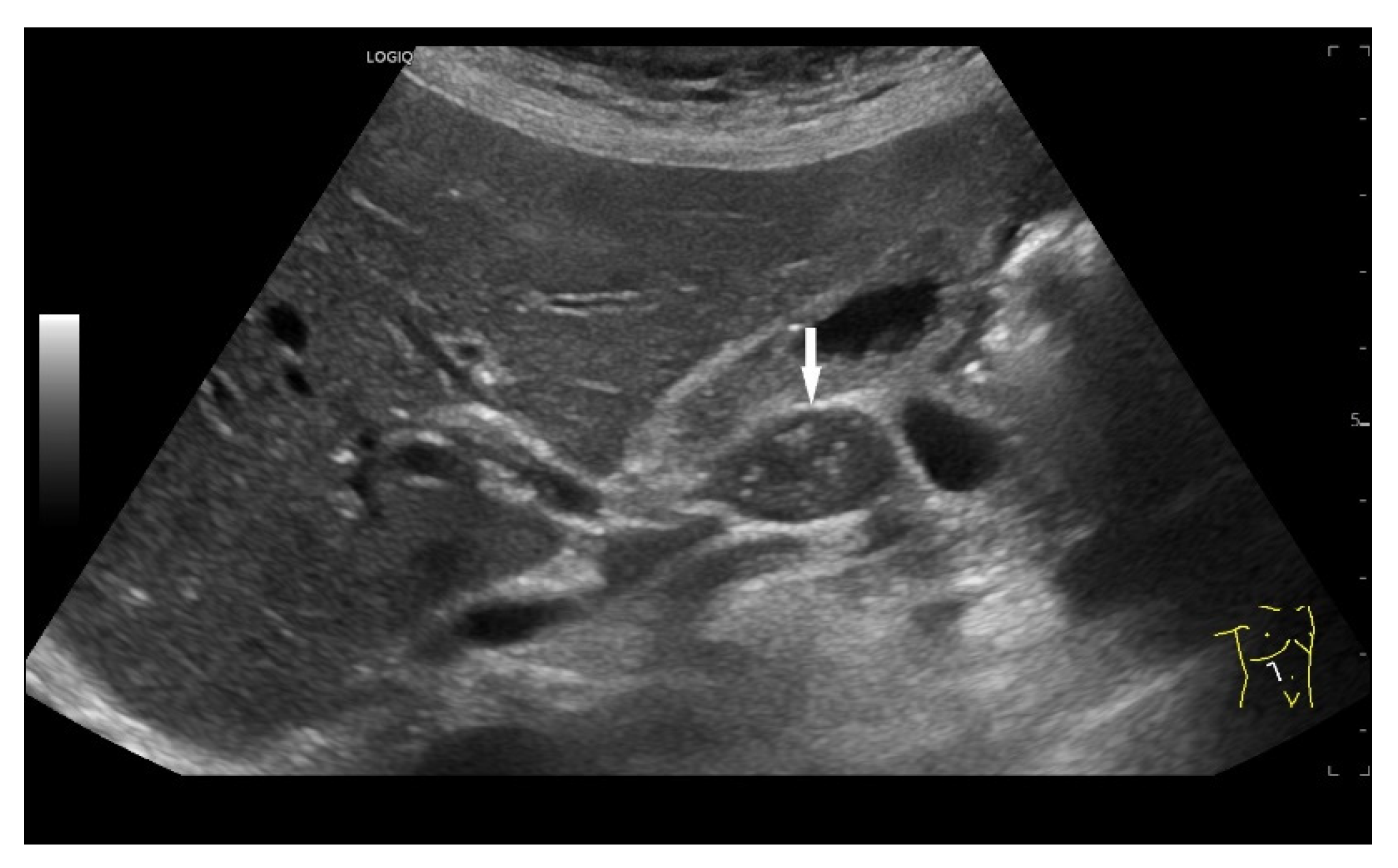

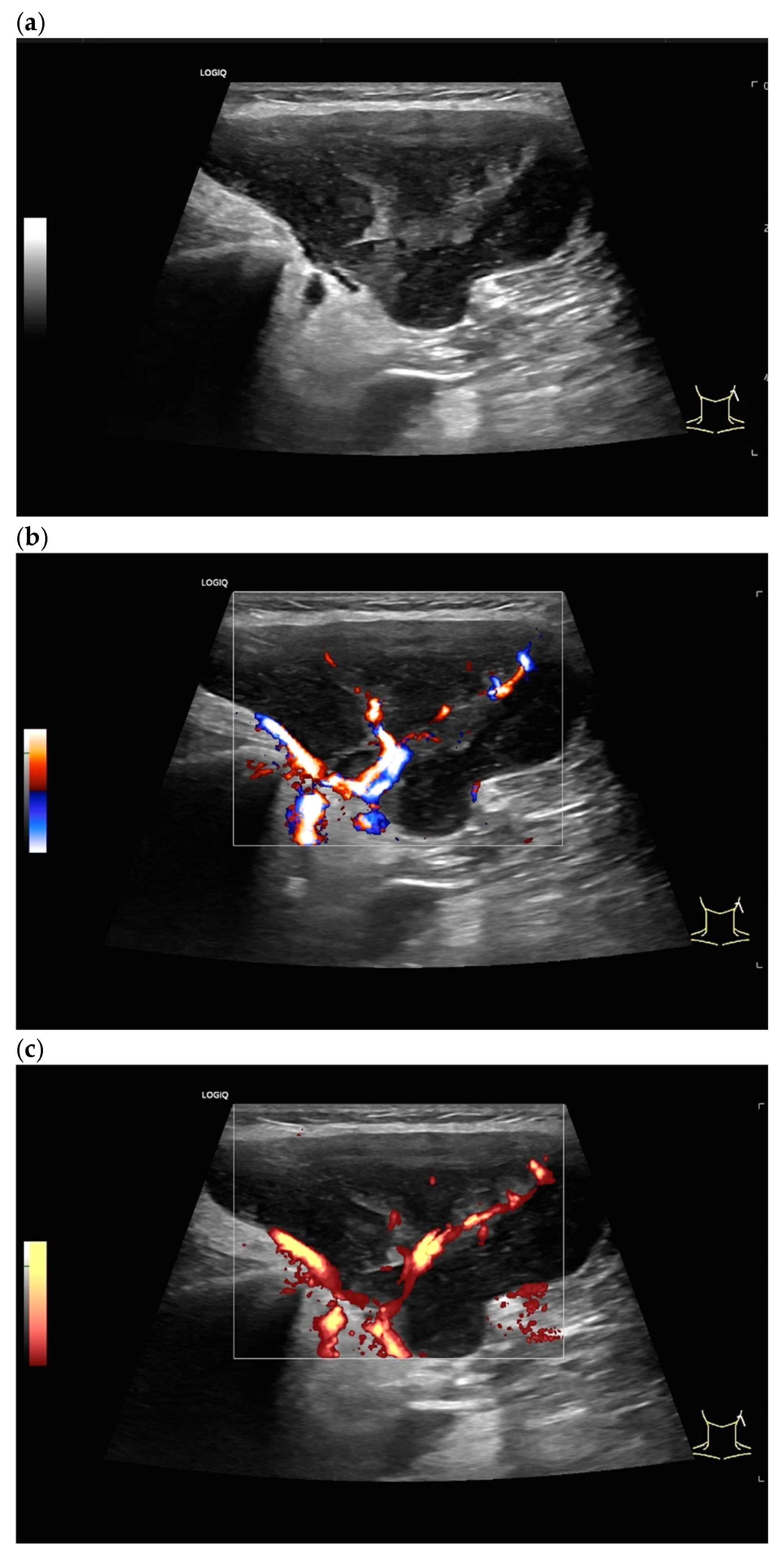


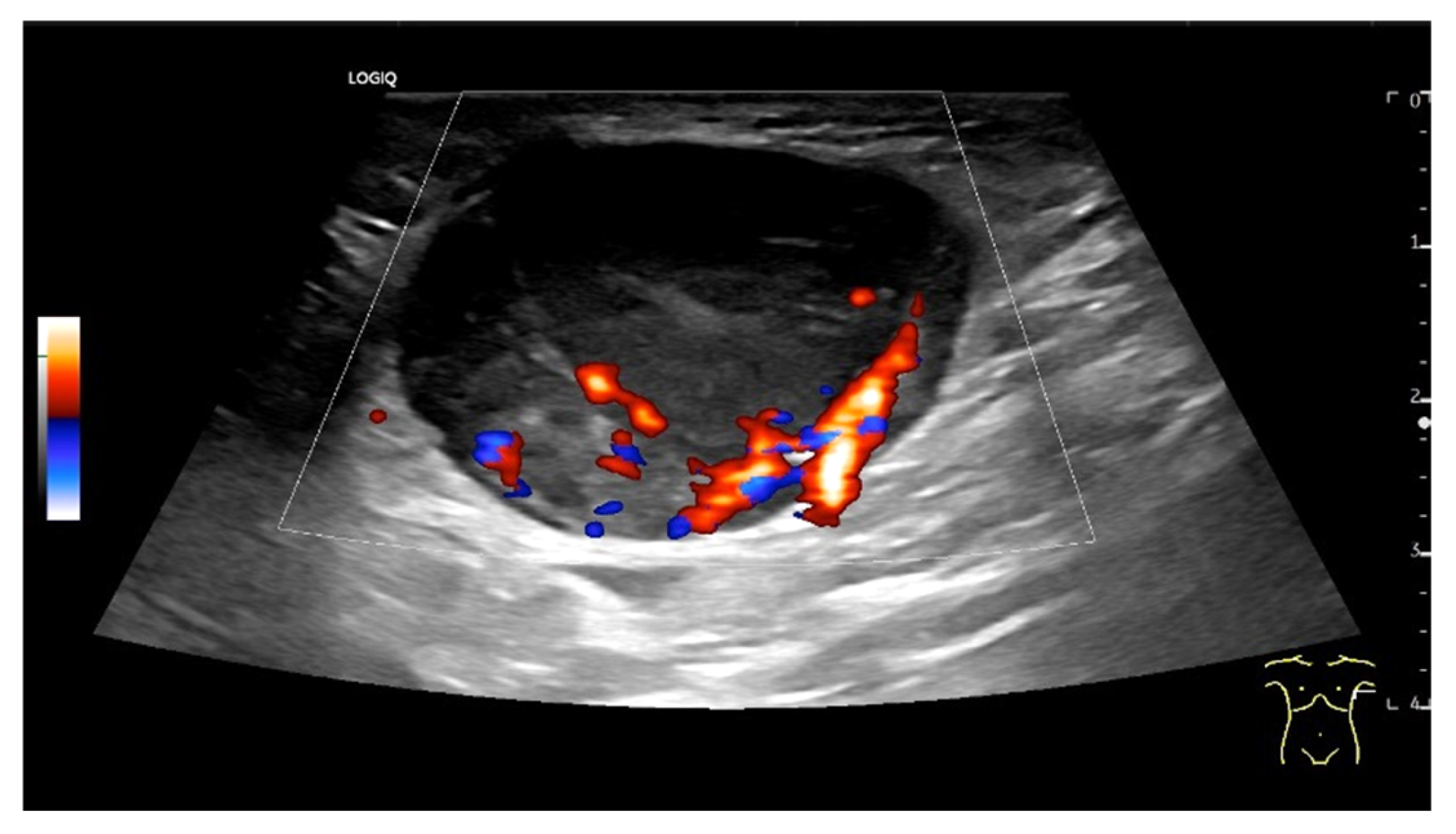



| Localization | Body Position and Transducer Selection | Transducer Position |
|---|---|---|
| Cervical | In supine position with head slightly extended backward and tilted to the opposite side. Linear transducer 9–18 MHz | The LN stations according to Som are examined using transverse and longitudinal transducer position. |
| Axillary | In the supine position, the arm should be slightly bent above the head. Linear transducer 2–9/9–18 MHz. | Transverse and longitudinal transducer guidance along the axillary vessels, lateral, medial, caudal, and cranial to the pectoralis minor muscle. The internal mammary LNs are examined parasternal intercostally. |
| Inguinal | Supine position. Linear transducer 2–9/9–18 MHz. | Examination of the LNs along the large vessels in transverse and longitudinal transducer positions. |
| Hepatoduodenal ligament | In supine or left-lateral position. Convex transducer 1–6 MHz | Position the hepatic hilum in the subcostal transducer position with transverse and longitudinal transducer guidance and shoulder–navel transducer position. Use deep inspiration or protrusion of the abdomen. |
| Paraaortal, paracaval, interaortocaval, parailiac | Supine position. Convex transducer 1–6 MHz, if sonographic imaging allows, then preferably with a linear transducer 2–9 MHz. | Examination of the LNs along the large vessels in transverse and longitudinal transducer positions. |
| Peripancreatic | Supine position. Convex transducer 1–6 MHz, if sonographic imaging allows, then preferably with a linear transducer 2–9 MHz. | Use protrusion of the abdomen for better visuality of the pancreas. Transversal and longitudinal transducer position. |
| Along the renal vessels | Supine position, convex transducer 1–6 MHz | Arms should be slightly bent above the head. Adjusting the renal hilum in the transverse section in the right and left flank |
| Mesenteric | Supine position. Convex transducer 1–6 MHz, preferably with a linear transducer 2–9 MHz. | Adjustment of the small and large intestine with the linear transducer. Special attention should be paid to the ileocecal region. This should be examined in transverse and longitudinal sections. Interenteric LNs should be assessed in all four quadrants. LNs should be sought in particular in areas of inflammatory or neoplastic bowel segments. |
| Iliac | Supine position. Convex transducer 1–6 MHz, optionally linear transducer 2–9 MHz. | Examination starting at aortic branch following along the common, external, and internal iliac vessels in transverse and longitudinal planes. |
| Study | Localization | Lymph Node Long-Axis Diameter (Range) | Lymph Node Short-Axis Diameters (Range) | Comments |
|---|---|---|---|---|
| Tschammler 1998 [44] n = 130 LNs (n = 48 benign, n = 82 malignant: metastases and lymphoma) | Cervical, submandibular, supraclavicular, inguinal LN n = 48 reactive, benign LN | Benign: 13.5 ± 6.0 mm (6–34 mm) Metastases: 19.2 ± 8.8 mm (6–43 mm) Lymphoma: 23.2 ± 10.5 mm (12–50 mm) | Benign: Thickness: 6.0 ± 2.9 mm (3–17 mm) Width: 9.1 ± 3.8 mm (4–24 mm) Metastases: Thickness: 11.6 ± 5.4 mm (4–29 mm) Width: 15.6 ± 7.4 mm (4–42 mm) Lymphoma: Thickness: 16.2 ± 9.9 mm (9–45 mm) Width: 19.4 ± 8.9 mm (10–45 mm) | Significant difference to malignant LNs (Metastases and lymphoma) |
| Bhatia 2012 [45] n = 55 LNs (n = 31 malignant, n = 24 benign) | Cervical LNs n = 23 reactive LNs n = 1 tuberculous LNs | N/A | Benign: 8.8 ± 3.4 mm (range 3–30 mm) Malignant: 11.4 ± 6.8 mm (range 4–30 mm) | No significant difference to malignant LNs |
| Ishibashi 2012 [46] N = 71 cervical LNs in oral squamous cell carcinoma (n = 31 metastatic LNs, n = 40 reactive benign LN) | n = 40 reactive LNs | N/A | Benign: 4.0 mm (2.4–8.3 mm) Malignant: 9.1 mm (3.2–19.2 mm) | |
| Hinz 2013 [47] N = 42 LNs (n = 21 melanoma LN metastasis, n = 21 benign) | n = 21 benign LNs cervical n = 3 (14.3%) axillary n = 3 (14.3%) inguinal n = 15 (71.4%) | Benign: 12.5 ± 3.66 mm Malignant: 19.37 ± 10.77 mm | Benign: 5.94 ± 1.90 mm Malignant: 12.30 ± 6.41 mm) | Solbiati index > 2: n = 76.21% in the benign group Solbiati index < 2: 80.9% in the malignant group. |
| Ghafoori 2015 [33] N = 43 patients with squamous cell carcinoma, malignant and reactive LNs | Cervical LNs n = 17 reactive LNs | Benign: 31.25 ± 4.82 mm Malignant: 34.98 ± 6.39 mm | Benign: 16.63 ± 5.11 mm Malignant: 22.62 ± 7.43 mm | Exact regional distributions of detected LNs were not specified. |
| Okumuş 2017 [48] Healthy probands n = 25 | Cervical LNs | Right: 11.11 ± 3.36 mm (5.9–18 mm) Left: 11.8 ± 2.67 mm (7.1–16.9 mm) | Right: 3.56 ± 0.73 mm (2–5 mm) Left: 3.84 ± 0.75 mm (2.7–5.5 mm) | Short/long-axis ratio: Right: 0.34 ± 0.11 (0.21–0.64) Left: 0.34 ± 0.07 (0.22–0.46) |
| Submandibular region | Right: 12.84 ± 4.4 mm (5.7–23.3 mm) Left: 11.94 ± 1.95 mm (7.6–16 mm) | Right: 3.87 ± 1.01 mm (1.8–5,8 mm) Left: 4.24 ± 1.07 mm (2.6–7.7 mm) | Short/long-axis ratio: Right: 0.33 ± 0.11 (0.19–0.54) Left: 0.36 ± 0.11 (0.2–0.66) | |
| Submental region | Right: 15.19 ± 4.62 mm (7.4–25.2 mm) Left: 16.18 ± 4.72 mm (9.6–29 mm) | Right: 4.65 ± 1.29 mm (2.4–8.8 mm) Left: 4.54 ± 1.28 mm (2.3–8 mm) | Short/long-axis ratio: Right: 0.32 ± 0.1 (0.2–0.66) Left: 0.29 ± 0.07 (0.18–0.48) | |
| Lerchbaumer 2022 [49] Healthy Probands n = 34 | Cervical LNs | Benign: 14.7 ± 6.9 mm | Benign: 7.4 ± 3.4 mm | Solbiati index: 2.11 ± 0.81 |
| Tunçez 2023 [50] n = 100 cervical or axillary LNs, 80% were axillary | Cervical or axillary LNs in suspected malignancy, n = 36 benign LNs | N/A | Benign: 9.36 ± 2.93 mm | Solbiati index: 2.17 ± 0.59 in the benign group |
| Chen 2025 [42] n = 159 LNs (benign, metastatic, Lymphomas) | Cervical, axillary and inguinal. n = 37 benign LNs. The malignant LNs included LN metastases and lymphomas | 20.52 ± 8.63 mm | Benign: 0.64 ± 3.23 mm | The benign LNs included reactive, unspecific and granulomatous lesions or tuberculosis. |
| He 2025 [51] n = 54 enlarged superficial LNs (benign, metastatic, lymphomas) Cervical (90%), axillary and inguinal localizations. | n = 20 benign (n = 16 reactive n = 4 tuberculous) | 27 ± 7.2 mm | 12 ± 6.1 mm | These were primarily enlarged LNs, including reactive, tuberculous, and malignant LNs (metastatic and lymphoma). |
| Study | Localization | Lymph Node Long-Axis Diameter (Range) | Lymph Node Short-Axis Diameter (Range) |
|---|---|---|---|
| Dietrich 1997 [17] n = 83 healthy probands and n = 20 autopsies from patients without hepatobiliary diseases. | Ventral to the portal vein Healthy probands n = 60/83 (72.3%) | 13 ± 2 mm (8–17 mm) | 5 ± 1 mm (0–7 mm) |
| Ventral to the portal vein/autopsy study/n = 20/ultrasound | 12 ± 3 mm (8–20 mm) | 5 ± 1 mm (3–10 mm) | |
| Ventral to the portal vein/autopsy study/n = 20/macroscopically | 11 ± 3 mm (8–18 mm) | 5 ± 1 mm (3–8 mm) | |
| between portal vein and inferior vena cava Healthy probands n = 60/83 (72.3%) | 14 ± 3 mm (8–20 mm) | 5 ± 1 mm (3–9 mm) | |
| between portal vein and inferior vena cava/autopsy study/n = 20/ultrasound | 13 ± 5 mm (8–20 mm) | 5 ± 2 mm (3–7 mm) | |
| between portal vein and inferior vena cava/autopsy study/n = 20/macroscopically | 12 ± 4 mm (8–19 mm) | 5 ±2 mm (3–8 mm) | |
| Dietrich 1998 [1] n = 80 healthy probands | Interaortocaval 44 /80 (55%) | 11 ± 3 mm (8–18 mm) | 5 ± 1 mm (3–10 mm) |
| Peripancreatic 14/80 (18%) | 9 ± 2 mm (8–13 mm) | 5 ± 1 mm (3–7 mm). | |
| left aortic 32/80 (40%) | 10 ± 2 mm (8–16 mm) | 5 ± 1 mm (3–8 mm) | |
| right mesenteric 31/80 (39%) | 11 ± 3 mm (8–19 mm) | 5 ± 1 mm (3–9 mm) | |
| along the renal arteries 2/80 | 12 × 5 mm and 11 × 4 mm | ||
| Splenic hilum 0/80 | No evidence of LNs in the splenic hilum. | ||
| Localization | Reference Value Long-Axis | Reference Value Short-Axis |
|---|---|---|
| Submandibular and in the upper cervical region (regions Ib and II) [24,27,31,32,46,48,52] | ≤8 mm | |
| Cervical regions (Ia, III, IV, V) [24,31,48] | ≤5 mm | |
| Jugulodigastric LN [23] | <30 mm | ≤8 mm |
| Infraclavicular, internal mammary region [28] | ≤4 mm | |
| Axillary [23] | Cortical thickness < 4 mm | |
| Hepatoduodenal ligament [17,53] | <20 mm | <8 mm |
| Retroperitoneal [1,53] | <30 | <9 mm |
| Mesenterial [38] | ≤5 mm | |
| Inguinal [23] | <40 mm | Cortical thickness <2.5 mm |
| Study | Localization | Resistive Index (RI) | Pulsatility Index (PI) | Comments |
|---|---|---|---|---|
| Steinkamp 1994 [52] n = 245 cervical LNs (reactive, metastatic, lymphoma, other) | Reactive LNs n = 104 | 0.65 ± 0.08 Cut-off 0.8 | 0.92 ± 0.26 Cut-off 1.6 | |
| Choi 1995 [74] n = 41 cervical LNs, n = 1 axillary LN, n = 1 inguinal LN | n = 19 benign LNs | 0.59 ± 0.11 Cut-off < 0.8 | 0.90 ± 0.23 Cut-off < 1.5 | Malignant LNs: RI 0.92 ± 0.23 PI 2.66 ± 1.59 |
| Ghafoori 2015 [33] n = 43 patients with squamous cell carcinoma, metastatic and reactive LNs | Cervical LNs n = 17 reactive LNs | 0.64 ± 0.08 Cut-off 0.69 | 1.18 ± 0.38 Cut-off 1.35 | Malignant LNs: RI 0.79 ± 0.08 (p < 0.001) PI 1.83 ± 0.52 (p < 0.001) |
| Ying 2004 [71] n = 270 patients (metastases n = 101, lymphoma n = 21, tuberculosis n = 76, reactive n = 72? | Cervical LNs n = 72 reactive LNs | 0.64 ± 0.08 Cut-off 0.7 | 1.05 ± 0.24 Cut-off 1.4 | Tuberculosis: RI 0.71 ± 0.11 |
| Benign LN | Malignant LN | |||
|---|---|---|---|---|
| Reactive | Tuberculous | Metastatic | Lymphoma | |
| B-mode US | Elongated oval shape, central hyperechoic hilum depending on location, uniform cortex width | Enlarged, rounded, hypoechoic LNs with a heterogeneous pattern, hypoechoic changes with hyperechoic margin in caseous abscesses. | Increase in short-axis and round shape, irregular widening of the cortex, hypoechoic inhomogeneities with focal metastatic infiltration. Compression/destruction of the central hilar region. | Round shape, often very large hypoechoic LNs with asymmetrical cortical widening or loss of echogenic hilum. Pronounced hypoechogenicity. LN conglomerates (bulky lesions). |
| Solbiati index | >2 | variable | <2 | <2 |
| Short/long-axis ratio | <0.5 | variable | >0.5 | >0.5 |
| Vascularization (color Doppler imaging) | Central vessel with regular branching; inflammation causes increased vascularization | Destroyed vascularization. | Destroyed vascularization; peripheral accessory vessels, irregular vessels. | Central and/or peripheral vessels with (irregular and pronounced) branching, high vessel density, compression of the central artery may cause necrosis. |
| Doppler parameters | RI < 0.8 PI < 1.5 | RI < 0.8 PI < 1.5 | RI > 0.8 PI > 1.5 | No definite recommendation, often “in between” |
| CEUS | Fast and strong centrifugal enhancement, central vessel, homogeneous | Centrifugal enhancement. Melting abscesses present with hyperenhancing margins and central non-enhancing areas. | (Delayed) centripetal peripheral enhancement, non-enhanced areas in case of necroses. | Central and/or peripheral vessels, strong enhancement, in case of necrosis non-enhancing areas. |
| Strain elastography | Soft, cortex is stiffer than hilum | Stiffer compared to surrounding due to inflammation, melting caseating abscesses may be softer. | Stiffer compared to surrounding. | Stiffer compared to surrounding. |
Disclaimer/Publisher’s Note: The statements, opinions and data contained in all publications are solely those of the individual author(s) and contributor(s) and not of MDPI and/or the editor(s). MDPI and/or the editor(s) disclaim responsibility for any injury to people or property resulting from any ideas, methods, instructions or products referred to in the content. |
© 2026 by the authors. Licensee MDPI, Basel, Switzerland. This article is an open access article distributed under the terms and conditions of the Creative Commons Attribution (CC BY) license.
Share and Cite
Möller, K.; Jenssen, C.; Lerchbaumer, M.H.; Hollerweger, A.; Yadav, M.; Dighe, M.; Serra, C.; Boccatonda, A.; Faiss, S.; Dietrich, C.F. Lymph Node Assessment with Multiparametric Ultrasound: Normal Values, Morphologic Patterns, and Diagnostic Algorithms. Cancers 2026, 18, 1045. https://doi.org/10.3390/cancers18061045
Möller K, Jenssen C, Lerchbaumer MH, Hollerweger A, Yadav M, Dighe M, Serra C, Boccatonda A, Faiss S, Dietrich CF. Lymph Node Assessment with Multiparametric Ultrasound: Normal Values, Morphologic Patterns, and Diagnostic Algorithms. Cancers. 2026; 18(6):1045. https://doi.org/10.3390/cancers18061045
Chicago/Turabian StyleMöller, Kathleen, Christian Jenssen, Markus Herbert Lerchbaumer, Alois Hollerweger, Madhvi Yadav, Manjiri Dighe, Carla Serra, Andrea Boccatonda, Siegbert Faiss, and Christoph Frank Dietrich. 2026. "Lymph Node Assessment with Multiparametric Ultrasound: Normal Values, Morphologic Patterns, and Diagnostic Algorithms" Cancers 18, no. 6: 1045. https://doi.org/10.3390/cancers18061045
APA StyleMöller, K., Jenssen, C., Lerchbaumer, M. H., Hollerweger, A., Yadav, M., Dighe, M., Serra, C., Boccatonda, A., Faiss, S., & Dietrich, C. F. (2026). Lymph Node Assessment with Multiparametric Ultrasound: Normal Values, Morphologic Patterns, and Diagnostic Algorithms. Cancers, 18(6), 1045. https://doi.org/10.3390/cancers18061045