Article

Circulating ERVFRD-1 and MFSD2A Are Associated with Immunotherapy Response in Metastatic Clear Cell Renal Cell Carcinoma


by
Hector Katifelis
1,
Styliani-Evangelia Zerva
1,
Aristotelis Bamias
2,
Michalis V. Karamouzis
3,
Konstantinos Stravodimos
4,
Leonardo A. Sechi
5,
Dimitra-Ioanna Lampropoulou
6,
Evangelia Pliakou
7 and
Maria Gazouli
1,*
1
Laboratory of Biology, Department of Basic Medical Sciences, Medical School, National and Kapodistrian University of Athens, Michalakopoulou 176, 11527 Athens, Greece
2
2nd Propaedeutic Department of Internal Medicine, ATTIKON University Hospital, School of Medicine, National and Kapodistrian University of Athens, 11527 Athens, Greece
3
Molecular Oncology Unit, Department of Biological Chemistry, National and Kapodistrian University of Athens, 11527 Athens, Greece
4
Department of Urology, National and Kapodistrian University of Athens, “Laiko” Hospital, 11527 Athens, Greece
5
Department of Biomedical Sciences, University of Sassari, 07100 Sassari, Italy
6
Department of Nutritional Science and Dietetics, Faculty of Health Sciences, University of Peloponnese, 24100 Kalamata, Greece
7
Department of Medical Oncology, Athens Naval and Veterans Hospital, 11521 Athens, Greece
*
Author to whom correspondence should be addressed.
Cancers 2026, 18(4), 716; https://doi.org/10.3390/cancers18040716
Submission received: 15 January 2026 / Revised: 15 February 2026 / Accepted: 20 February 2026 / Published: 23 February 2026
(This article belongs to the Section Cancer Biomarkers)

Simple Summary

Immune checkpoint inhibitor (ICI) therapies have improved outcomes for patients with metastatic clear cell renal cell carcinoma (mccRCC). However, many patients do not respond to treatment. Therefore, reliable biomarkers associated with therapeutic response are urgently needed. Blood-based biomarkers offer a non-invasive alternative to tissue analysis. In this study, we investigated the expression of two immunity-related genes, ERVFRD-1 and MFSD2A, in peripheral blood samples from mccRCC patients receiving PD-1-based treatment. Both genes were dysregulated compared with healthy controls and demonstrated differential baseline expression between patients who achieved clinical benefit and those with progressive disease. Patients with progressive disease exhibited decreased expression of ERVFRD-1 and increased expression of MFSD2A. These findings suggest that ERVFRD-1 and MFSD2A may serve as candidate blood-based biomarkers associated with response to ICI, although confirmation in larger prospective studies is required.

Abstract

Background/Objectives: Immune checkpoint inhibitor (ICI)-based combinations have significantly improved outcomes in metastatic clear cell renal cell carcinoma (mccRCC). However, a substantial number of patients fail to derive clinical benefit, highlighting the need for reliable predictive biomarkers. Circulating biomarkers represent an attractive, non-invasive alternative to tissue-based assays. This study aimed to evaluate the immunity-related genes ERVFRD-1 and MFSD2A as potential blood-based candidate biomarkers associated with response to ICI-based therapy in mccRCC. Methods: Peripheral blood samples were collected prior to treatment initiation from 34 patients with mccRCC receiving PD-1-based therapy. Gene expression levels of ERVFRD-1 and MFSD2A were quantified using real-time PCR. Treatment response was assessed according to the Response Evaluation Criteria in Solid Tumors (RECIST) version 1.1. Peripheral blood samples from healthy individuals were included as controls. Results: Both ERVFRD-1 and MFSD2A were significantly dysregulated in mccRCC patients compared with healthy controls. Their expression differed between patients with clinical benefit and those with progressive disease. Specifically, patients with progressive disease exhibited reduced ERVFRD-1 expression and increased MFSD2A expression compared with patients showing clinical benefit. Conclusions: ERVFRD-1 and MFSD2A were associated with treatment response in this pilot cohort and may represent promising blood-based biomarker candidates, requiring validation in larger prospective multicenter studies.

1. Introduction

Renal cell carcinoma (RCC) represents a heterogeneous malignancy with a rising global incidence, accounting for more than 80% of all kidney cancers. Recent data further indicate that RCC represents approximately 4% of all new cancer cases [1,2]. The most common histological subtype of RCC is clear cell RCC (ccRCC), which accounts for approximately 75% of renal malignancies, followed by papillary and chromophobe tumors [3]. Unfortunately, ccRCC tends to be highly metastatic (metastatic ccRCC, mccRCC). Nearly one out of three patients present with distant metastases (including bone, brain, and pancreas) at the time of diagnosis [4,5].
Immune-checkpoint inhibitors (ICIs) have drastically improved the outcomes of mccRCC patients over the past years [6]. These agents are used in combinations that typically include programmed cell death-1 (PD-1) inhibitors combined with another type of ICI, namely cytotoxic T-lymphocyte-associated protein 4 (CTLA-4) inhibitors. Alternatively, PD-1 inhibitors can be used in combination with a tyrosine kinase inhibitor (TKI). Both combinations are recommended by the European Society of Medical Oncology (ESMO) [7] and the American Society of Clinical Oncology (ASCO) [8].
However, despite these advances, approximately 40–50% of mccRCC patients fail to derive durable benefit from ICI/ICI or ICI/TKI combinations, highlighting a critical unmet clinical need for robust predictive biomarkers [9,10]. It remains a major challenge for personalized oncology approaches to identify these patients, and the optimal predictive biomarker remains to be established. The first biomarker to be used for this purpose was PD-L1. Its expression has been associated with immunotherapy response in several cancers, but its use has been problematic in several malignancies, including mccRCC [11,12]. From a clinical perspective, repeated tumor biopsies in patients with advanced disease are often impractical or unsafe. Therefore, circulating biomarkers represent a particularly attractive, minimally invasive alternative with increasing clinical relevance [13,14,15].
Among circulating candidates, genes involved in immune regulation and interactions between tumor and immune components have attracted increasing attention, particularly those with established roles in immune barrier integrity and immune tolerance [14,15]. In this context, two genes of particular importance are ERVFRD-1 (Endogenous Retrovirus Group FRD member 1) [16] and MFSD2A (Major Facilitator Superfamily Domain Containing 2A) [17]. ERVFRD-1 encodes Syncytin-2, which engages MFSD2A as its cognate receptor, forming a defined ligand–receptor pair in human placentation, and has been shown to possess immunosuppressive properties that have been proposed to contribute to immune tolerance mechanisms [18,19].
In addition to its physiological role in placentation and maternal immune tolerance [16], ERVFRD-1 has also been implicated in tumor–immune interactions. A recent comprehensive bioinformatic analysis by Wen et al. [16] demonstrated that ERVFRD-1 expression in ccRCC tumor tissue is significantly associated with immune-related pathways, tumor mutation burden, and patient survival, proposing ERVFRD-1 as a promising biomarker for prognosis and immunotherapy response. Importantly, however, evidence regarding its potential utility as a circulating biomarker remains extremely limited. ERVFRD-1 promotes the inhibition of anti-tumor immune responses in experimental models and has been shown to influence tumor growth in human cancer cells. Conversely, in the context of RCC, reduced expression has been reported, while higher expression appears to correlate with improved survival [16,20]. Furthermore, the current literature suggests that it acts as a regulator of the tumor microenvironment in patients with melanoma [16].
Beyond its role as the Syncytin-2 receptor, MFSD2A has been implicated in several processes relevant to immune regulation and tumor biology [21]. This gene encodes a protein involved in lipid metabolism with a major role in the maintenance of the blood–brain barrier (BBB) and its deficiency is linked to BBB leakage and developmental disorders [22]. At the same time, it represents an important component of CD8 memory T cell function participating in both T cell maintenance and immune response [23]. In cancer, it has been described as a tumor suppressor in hepatocellular carcinoma and lung cancer [24,25]. Its overexpression has been found to repress cellular proliferation and migration in vitro and tumor growth in vivo [26]. Interestingly, it has been studied as a predictive biomarker for anti-PD-1 immunotherapy in gastric cancer [17]. In addition, Shi et al. [27] reported that the expression of MFSD2A is reduced in gastric tumors while increased expression was associated with improved survival and clinicopathologic features consistent with a less aggressive phenotype. These findings support the tumor-suppressive role of MFSD2A in the context of this malignancy.
In our previous work [28], we investigated the potential of the expression of circulating inflammation- and immunity-related genes as biomarkers of immunotherapy effectiveness in mccRCC individuals. In the present study, we aimed to further explore the potential of the expression of the immunity-related genes MFSD2A and ERVFRD-1 as candidate biomarkers associated with response to ICI-based therapies in mccRCC. To our knowledge, this is the first study investigating the association between circulating ERVFRD-1 and MFSD2A expression levels and response to ICI-based therapies in metastatic clear cell renal cell carcinoma.

2. Materials and Methods

2.1. Patients

Peripheral blood was obtained from 34 patients diagnosed with mccRCC before the start of therapy. All patients received first-line anti-PD-1 blockade (Nivolumab or Pembrolizumab) combined with either CTLA-4 inhibition (Ipilimumab) or a TKI (Cabozantinib or Axitinib) for metastatic disease.
Treatment outcomes were grouped by clinical benefit (CB) [comprising complete response (CR), partial response (PR), and stable disease, (SD)] or progressive disease (PD). Response categorization was evaluated according to RECIST version 1.1 criteria [29].
Patients were managed at Attikon University Hospital and Laikon General Hospital during the period between 2021 and 2023. Blood samples from 30 healthy donors served as the control group. Written informed consent was obtained from all individuals prior to inclusion, and the protocol received approval from the ethics committees of both institutions. All samples were collected and processed according to a standardized workflow across participating centers. The time from collection to processing was minimized and samples were handled under uniform temperature conditions. Repeated freeze–thaw cycles were avoided. Duplicate measurements differing by more than one Ct cycle were considered unreliable and were excluded from further analysis.

2.2. RNA Extraction and cDNA Synthesis

Total RNA was isolated from whole blood using the Qiagen AllPrep RNA/DNA Mini Kit (Qiagen, Hilden, Germany) according to the manufacturer’s instructions. Complementary DNA (cDNA) was generated from the extracted RNA with a TAKARA kit (Takara Bio Europe SAS, Saint-Germain-en-Laye, France). Reverse transcription was carried out in a SuperCycler Thermal Cycler (Kyratec, Mansfield, Australia) at 37 °C for 30 min, followed by 5 min at 85 °C to deactivate reverse transcriptase.

2.3. Real-Time PCR and Gene Expression Analysis

Real-time PCR was performed using KAPA SYBR FAST qPCR mix (KAPA BIOSYSTEMS, Cape Town, South Africa). GAPDH served as a reference gene, and primer sequences for ERVFRD-1, MFSD2A and GAPDH are listed in Table 1.
Each sample was analyzed in duplicate, and relative expression levels were calculated after normalization to GAPDH. Amplification reactions were run on a SaCycler-96 (Sacace Biotechnologies, Como, Italy). Changes in transcript levels were determined using the 2−ΔΔCt is presented as fold regulation. Gene downregulation is shown as the negative inverse of fold change and overexpressed genes are shown as fold change [30]. All RT-qPCR reactions were performed in technical duplicates. Intra-assay variability was assessed using Ct values, with coefficients of variation consistently below 5%. Duplicate measurements differing by more than one Ct cycle were excluded from analysis.

2.4. Statistical Analysis

Data analysis was conducted with GraphPad Prism (version 10.6.1; GraphPad Software, San Diego, CA, USA). Statistical significance was defined as a p-value < 0.05.

3. Results

3.1. Patient Characteristics, Treatment and Clinical Response

The study cohort consisted of 34 patients with males representing the majority (n = 26). The mean age at treatment initiation was 67.1 ± 10.6 years. Regarding treatment regimens, 16 patients (47%) received nivolumab plus Ipilimumab as first-line treatment. Nine patients (26.5%) received nivolumab plus Cabozantinib whereas another nine patients (26.5%) received Pembrolizumab plus Axitinib. Clinical benefit was observed for 53% of patients, while 47% showed PD. A summary of the study cohort is presented in Table 2.

3.2. ERVFRD-1 and MFSD2A Are Downregulated in mccRCC Patients

The expression of MFSD2A and ERVFRD-1 was evaluated among mccRCC patients and healthy individuals. Both genes exhibited significantly decreased expression in mccRCC patients compared with healthy individuals, suggesting systemic dysregulation of immune-related pathways in this disease. ERVFRD-1 was significantly downregulated in mccRCC patients (0.55-fold change, p = 0.002), while MFSD2A was similarly reduced (0.65-fold change, p = 0.0021), supporting the biological relevance of these circulating transcripts in the context of disease presence. The results are shown in Figure 1 (panel A for MFSD2A and panel B for ERVFRD-1).

3.3. Transcriptomic Differences Between CB and PD Groups at Baseline

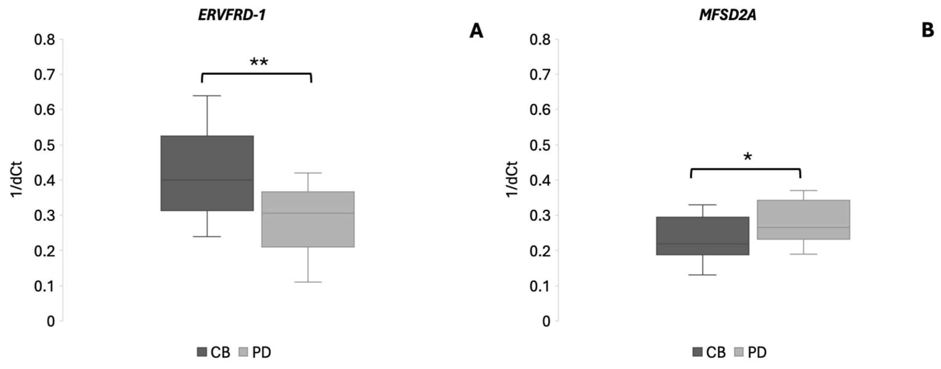
To evaluate potential differences in ERVFRD-1 and MFSD2A expression between PD and CB groups we investigated gene expression across these patient groups. Regarding ERVFRD-1, compared to PD patients, the CB group showed increased expression by approximately 1.4-fold (p = 0.0014). Conversely, MFSD2A showed a modest but statistically significant increase in PD patients compared with the CB group (1.2-fold, p = 0.044). Further subgroup analyses based on specific treatment regimens were not feasible due to small sample sizes. The results are shown in Figure 2.

4. Discussion

The use of ICI-based approaches has been revolutionary in cancer therapeutics; nevertheless, only a subset of mccRCC patients benefits from these treatments [31]. Thus, the need for predictive biomarkers to achieve personalized oncology approaches is urgent. Extending our previous work [28], we investigated potential differences in the expression of the immunity-related genes MFSD2A and ERVFRD-1 between CB and PD patients.
Regarding ERVFRD-1 and other members of the HERV family, there is a growing body of evidence that they are dysregulated in several malignancies, including ccRCC, prostate and bladder cancer [16,32,33]. Indeed, our results show that ERVFRD-1 is downregulated in the blood of mccRCC patients. To the best of our knowledge, this is the first study to evaluate ERVFRD-1 expression in the peripheral blood of mccRCC patients. A recent large-scale bioinformatic analysis demonstrated that ERVFRD-1 expression correlates with immune activation signatures, tumor mutation burden, and overall survival in ccRCC, supporting a biologically plausible link between ERVFRD-1 and immunotherapy responsiveness [16]. Although the results shown by Wen et al. did not show a statistically significant association with response to PD-L1/PD-1 blockade, this analysis was performed in a retrospective subset comprising a limited number of treated patients and was based on tumor tissue. Nevertheless, the authors suggested an important role of ERVFRD-1 in immune activation mediating an anti-tumor effect in ccRCC and found that higher ERVFRD-1 expression is associated with progression-free survival.
In line with these observations, ERVFRD-1 (Syncytin-2) expression in our cohort was downregulated in PD patients, whereas higher expression levels were observed in CB patients. Taken together, these findings, combined with our observation that higher circulating ERVFRD-1 expression is associated with clinical benefit, support the investigation of ERVFRD-1 as a potential blood-based biomarker for patient stratification in mccRCC.
At first glance, this observation seems to contrast with the concept of viral mimicry, according to which derepression of epigenetically silenced ERVs generates double-stranded RNA that activates innate sensors and induces interferon-driven antitumor immunity [34,35]. However, circulating RNA levels do not necessarily mirror intratumoral ERV activity and are more likely to represent a systemic immune context shaped by host inflammatory status and immune cell composition [15]. We therefore hypothesize that reduced peripheral ERVFRD-1 expression may indicate weakened interferon pathway activation and impaired cytotoxic competence. Such defects have been repeatedly associated with reduced responsiveness to ICIs [36,37], offering a biologically plausible explanation for the inverse relationship observed in our cohort. Additionally, Syncytin-2 possesses immunomodulatory properties and is subject to epigenetic regulation [16].
From an evolutionary perspective, Syncytin-2 and its receptor (MFSD2A) represent typical examples of endogenous retroviral exaptation, where ancestral viral envelope genes were co-opted to enable placentation and maternal-fetal immune tolerance [18,38]. During pregnancy, trophoblast cells deploy coordinated immune-evasion strategies (including local suppression of cytotoxic responses) to prevent immune rejection of the semi-allogenic fetus. Several of these mechanisms are similar to strategies exploited by tumors to achieve evasion of host immunity [39]. Consequently, altered ERVFRD-1 and MFSD2A expression in cancer may indicate activation of tolerance-like mechanisms within the tumor-host interaction and not intrinsic tumor immunogenicity per se. This evolutionary parallel provides a conceptual framework for interpreting why expression patterns associated with immunity regulation in pregnancy may influence ICI responsiveness. It further suggests that characterization of placenta-derived tolerance pathways in tumors could refine patient stratification and could identify therapeutic opportunities aiming at reversing immune permissiveness, for example through combinations of ICIs with agents that modulate epigenetic or interferon-related mechanisms. Nevertheless, future studies that integrate tumor tissue analyses with peripheral measurements are necessary to clarify compartment-specific effects.
In other malignancies, such as acute myeloid leukemia, increased Syncytin-2 levels indicate a favorable prognosis in particular subgroups, including patients with low white blood count and those of white race [40]. However, its exact role in immunotherapy response remains veiled. Interestingly, ERVFRD-1 has been proposed as a therapeutic target to enhance immunotherapy efficacy [34], with hypomethylating agents such as decitabine potentially sensitizing tumors to immunotherapy. Indeed, methylation has been shown [16] to play a key role in ERVFRD-1 regulation with increased methylation associated with an unfavorable prognosis.
MFSD2A has also been implicated in multiple cancers. Loss of PRMT6 (a gene encoding a methyltransferase) induces MFSD2A upregulation, promoting leukemia stem cells maintenance [41]. While MFSD2A has been investigated in tumor tissue across several malignancies, data regarding its circulating expression in mccRCC are currently lacking. In our cohort, MFSD2A was significantly downregulated in peripheral blood of mccRCC patients compared with healthy individuals. Interestingly, Wang et al. [42] showed that ccRCC, MFSD2A and CYP24A1 were positively correlated with risk scores in bioinformatic analyses, and patients in the high-risk group treated for advanced RCC showed lower half-maximum inhibition concentration of targeted therapeutic agents (Sunitinib, Temsirolimus and Pazopanib).
In our study, MFSD2A was found to be upregulated in PD patients. Although the therapeutic contexts differ, these findings suggest that MFSD2A upregulation may be associated with unfavorable outcomes, such as a high-risk score or PD. Interestingly, MFSD2A has been shown to enhance response to PD-1 blockade in gastric cancer by reprogramming the tumor microenvironment and promoting cytotoxic T-cell activation [17]. The contrasting directionality observed in our cohort underscores the likelihood that MFSD2A exerts tumor-specific immunobiological functions, warranting further mechanistic investigation [17]. The favorable prognostic association of higher MFSD2A expression observed in gastric cancer [27] further supports that the biological role of this molecule could be highly context dependent. One possible explanation for this discrepancy may lie in the fundamental differences in tumor microenvironment, immune contexture and therapeutic exposure between gastric cancer and mccRCC. Clear cell renal cell carcinoma is widely recognized as an immunogenic malignancy characterized by dense immune infiltration and strong tumor-host immune interplay, features that influence responsiveness to ICIs [43]. In contrast, gastric cancer has traditionally been managed with chemotherapy-based treatments and the immune landscape driving treatment sensitivity may substantially differ [44]. Thus, the biological consequences of MFSD2A expression may substantially vary across tumor types, reflecting distinct immune escape mechanisms and microenvironmental pressures. While in gastric malignancies its increased expression has been linked to reduced angiogenesis and a less aggressive phenotype, our findings in mccRCC derive from circulating RNA levels in patients who received ICIs. These important differences in tumor type, sampling compartment and therapeutic context are possibly major contributors to these divergent observations. Overall, this variability highlights the presence of tumor-specific biological mechanisms that mediate response to immunotherapy.
Taken together, both genes show statistically significant differences between CB and PD groups supporting their potential as candidate biomarkers in this pilot cohort. Nevertheless, the interaction between ERVFRD-1 (Syncytin-2) and MFSD2A in immunotherapy response or resistance remains largely unexplored.
This study has some limitations. The cohort size was limited, reflecting the difficulty of assembling uniformly treated mccRCC populations in real-world settings. This challenge is further complicated by the different therapeutic combinations (ICI/ICI or ICI/TKI) which limit the numbers of patients in each group. Given the exploratory nature of the study and the modest cohort size, no predictive model was constructed and internal validation approaches such as cross-validation or bootstrap resampling were not applied; these analyses will be essential in future larger cohorts aimed at model development and clinical prediction. While standardized sample handling and duplicate RT-qPCR measurements were implemented to enhance analytical robustness, only a single baseline blood sample per patient was available. As a result, intra-patient temporal variability and short-term biological fluctuations of ERVFRD-1 and MFSD2A expression prior to treatment initiation could not be formally assessed. Consequently, it remains unclear whether the observed low or high circulating expression levels represent stable systemic phenotypes or dynamic transcriptional states. In addition, relevant clinical variables that may influence circulating RNA expression, such as systemic inflammatory status (e.g., neutrophil-to-lymphocyte ratio), tumor burden, performance status, and prior anti-angiogenic therapy, were not uniformly available and could not be included in correlation analyses or multivariable adjustment. Therefore, the independence of the observed associations from these potential confounders cannot be established in the present cohort. Intra-assay reproducibility was high, with coefficients of variation for Ct values consistently below 5%; however, longitudinal sampling will be required to establish biological coefficients of variation and transcript stability over time.
From a biological perspective, circulating gene expression may not fully reflect the tumor microenvironment. Importantly, the present study was not designed to establish causal or mechanistic relationships between circulating ERVFRD-1 and MFSD2A expression and immunotherapy response. Therefore, the observed associations should be interpreted as hypothesis-generating rather than mechanistically explanatory. Functional studies, as well as paired tumor–blood and longitudinal analyses, will be necessary to clarify the biological mechanisms underlying these observations. Technical and pre-analytical considerations should also be acknowledged, as circulating RNA measurements are inherently sensitive to pre-analytical and biological variability. The dichotomization of patients using cohort-derived cut-offs was performed for exploratory purposes and does not imply the existence of clinically validated thresholds. Establishment of fixed reference ranges for circulating ERVFRD-1 and MFSD2A will require assay harmonization and evaluation across multiple independent populations, including healthy controls. While standardized handling and duplicate real-time PCR measurements were implemented to enhance robustness, only a single baseline sample per patient was available and intra-patient temporal variability could not be assessed. Confirmation of transcript stability via serial sampling will be required before routine clinical application can be considered. From an analytical perspective, establishment of clinically applicable thresholds will require assay harmonization and the definition of validated reference ranges across independent populations. In addition, detailed time-to-event follow-up data were not uniformly available in this cohort. Therefore, survival analyses such as Kaplan–Meier estimation were not performed. Important potential confounders, including systemic inflammatory indices and tumor burden measurements were not uniformly available, precluding formal multivariable adjustment.
Finally, this was a two-center cohort from a single country. Thus, larger prospective studies are needed to validate the predictive value of MFSD2A and ERVFRD-1. The cut-off values applied for group comparisons were derived from the distribution of the present cohort and should be considered exploratory. Taken together, these considerations emphasize the need for external validation while supporting the role of the present findings as hypothesis-generating.

5. Conclusions

Circulating ERVFRD-1 and MFSD2A expression showed an association with response to ICI-based immunotherapy in mccRCC patients. Downregulated ERVFRD-1 expression and upregulated MFSD2A expression were associated with progressive disease. Collectively, our findings suggest that circulating ERVFRD-1 and MFSD2A expression profiles represent non-invasive candidate biomarkers associated with response to ICI-based therapy in mccRCC. If validated in larger prospective cohorts, these biomarkers could contribute to improved patient stratification and support the implementation of precision immuno-oncology in routine clinical practice. Given the inherent sensitivity of circulating RNA measurements to pre-analytical and biological variability, confirmation of these observations with longitudinal sampling and standardized workflow will be essential prior to routine clinical implementation. Therefore, these results should be considered hypothesis-generating and interpreted in the context of the modest sample size, requiring independent validation.

Author Contributions

Conceptualization, H.K., L.A.S. and M.G.; methodology, S.-E.Z.; formal analysis, A.B.; investigation, H.K.; resources, K.S., D.-I.L., E.P.; data curation, S.-E.Z.; writing—original draft preparation, H.K. and S.-E.Z.; writing—review and editing, H.K., K.S. and M.G.; visualization, M.V.K.; supervision, A.B.; project administration, M.G.; funding acquisition, M.G. All authors have read and agreed to the published version of the manuscript.

Funding

This work was supported by the KATY project funded from the European Union’s Horizon 2020 research and innovation program under grant agreement No. 10101745 and from the HeSMO grant to Evangelia Pliakou No. 9867/01.04.2025.

Institutional Review Board Statement

The study was conducted in accordance with the Declaration of Helsinki and approved by the Ethics Committee of Medical School, National and Kapodistrian University of Athens (566/19/10/2021, approved on 14 June 2021) for studies involving humans.

Informed Consent Statement

Informed consent was obtained from all subjects involved in the study.

Data Availability Statement

The data generated and/or analyzed during the current study are available from the corresponding author on reasonable request.

Conflicts of Interest

The authors declare no conflicts of interest.

References

  1. Peshin, S.; Dharia, A.; Moka, N.; Skelton, W.P., IV. A Review of Immunotherapy in Renal Cell Carcinoma: Current Landscape and Future Directions. Cancers 2025, 17, 3139. [Google Scholar] [CrossRef] [PubMed]
  2. Bhargava, V.K.; Valame, S.; Sahu, H.; Gupta, V.; Jain, R.; Azad, S.; Manek, D.; Bharati, V. Oncological Outcomes of Non-clear Cell Renal Cell Carcinomas: A Retrospective Study From a Tertiary Care Center. Cureus 2025, 17, e89022. [Google Scholar] [CrossRef] [PubMed]
  3. Baytok, A.; Ecer, G.; Balasar, M.; Koplay, M. Computed tomography and magnetic resonance imaging characteristics of renal cell carcinoma: Differences between subtypes and clinical evaluation. J. Clin. Imaging Sci. 2025, 15, 10. [Google Scholar] [CrossRef] [PubMed]
  4. Apanovich, N.; Matveev, A.; Ivanova, N.; Burdennyy, A.; Apanovich, P.; Pronina, I.; Filippova, E.; Kazubskaya, T.; Loginov, V.; Braga, E.; et al. Prediction of Distant Metastases in Patients with Kidney Cancer Based on Gene Expression and Methylation Analysis. Diagnostics 2023, 13, 2289. [Google Scholar] [CrossRef]
  5. Kanwal, R. Metastasis in renal cell carcinoma: Biology and treatment. Adv. Cancer Biol.-Met. 2023, 7, 100094. [Google Scholar] [CrossRef]
  6. Thouvenin, J.; Masson, C.; Boudier, P.; Maillet, D.; Kuchler-Bopp, S.; Barthélémy, P.; Massfelder, T. Complete Response in Metastatic Clear Cell Renal Cell Carcinoma Patients Treated with Immune-Checkpoint Inhibitors: Remission or Healing? How to Improve Patients’ Outcomes? Cancers 2023, 15, 793. [Google Scholar] [CrossRef]
  7. European Society for Medical Oncology (ESMO). ESMO Living Guidelines: Renal Cell Carcinoma. Management of Advanced and Metastatic Disease: First-Line Treatment for Clear-Cell RCC. Version 1.1, June 2025. Available online: https://www.esmo.org/guidelines/living-guidelines/esmo-living-guideline-renal-cell-carcinoma/advanced-and-metastatic-disease/systemic-treatment-for-advanced-and-metastatic-ccrcc/management-of-advanced-and-metastatic-disease-first-line-treatment-for-clear-cell-rcc (accessed on 26 December 2025).
  8. Rathmell, W.K.; Rumble, R.B.; Van Veldhuizen, P.J.; Al-Ahmadie, H.; Emamekhoo, H.; Hauke, R.J.; Louie, A.V.; Milowsky, M.I.; Molina, A.M.; Rose, T.L.; et al. Management of Metastatic Clear Cell Renal Cell Carcinoma: ASCO Guideline. J. Clin. Oncol. 2022, 40, 2957–2995. [Google Scholar] [CrossRef]
  9. Schoenfeld, A.J.; Hellmann, M.D. Acquired Resistance to Immune Checkpoint Inhibitors. Cancer Cell 2020, 37, 443–455. [Google Scholar] [CrossRef]
  10. Ou, X.; Gao, G.; Habaz, I.A.; Wang, Y. Mechanisms of resistance to tyrosine kinase inhibitor-targeted therapy and overcoming strategies. MedComm 2024, 5, e694. [Google Scholar] [CrossRef]
  11. Tripathi, A.; Lin, E.; Agarwal, N. Biomarkers in metastatic renal cell carcinoma in the era of immune checkpoint inhibitors. Ann. Transl. Med. 2019, 7, S203. [Google Scholar] [CrossRef]
  12. Derraze, Y.; Hashemi, S.M.S.; Ulas, E.B.; Ziesemer, K.A.; Lissenberg-Witte, B.; Radonic, T.; Bahce, I. Evaluating the significance of combining PD-L1 and TILs as biomarkers in non-small cell lung cancer patients treated with immunotherapy: A systematic review. BJC Rep. 2025, 3, 65. [Google Scholar] [CrossRef] [PubMed]
  13. Kaczmarek, F.; Marcinkowska-Gapińska, A.; Bartkowiak-Wieczorek, J.; Nowak, M.; Kmiecik, M.; Brzezińska, K.; Dotka, M.; Brosz, P.; Firlej, W.; Wojtyła-Buciora, P. Blood-Based Biomarkers as Predictive and Prognostic Factors in Immunotherapy-Treated Patients with Solid Tumors-Currents and Perspectives. Cancers 2025, 17, 2001. [Google Scholar] [CrossRef] [PubMed]
  14. Goh, K.Y.; Cheng, T.Y.; Tham, S.C.; Lim, D.W. Circulating Biomarkers for Prediction of Immunotherapy Response in NSCLC. Biomedicines 2023, 11, 508. [Google Scholar] [CrossRef] [PubMed]
  15. Song, Y.; Li, X.; Wei, X.; Cui, J. Human Endogenous Retroviruses as Biomedicine Markers. Virol. Sin. 2021, 36, 852–858. [Google Scholar] [CrossRef]
  16. Wen, X.; Shen, J.; De Miglio, M.R.; Zeng, D.; Sechi, L.A. Endogenous retrovirus group FRD member 1 is a potential biomarker for prognosis and immunotherapy for kidney renal clear cell carcinoma. Front. Cell. Infect. Microbiol. 2023, 13, 1252905. [Google Scholar] [CrossRef]
  17. Zhang, B.; Wang, C.M.; Wu, H.X.; Wang, F.; Chai, Y.Y.; Hu, Y. MFSD2A potentiates gastric cancer response to anti-PD-1 immunotherapy by reprogramming the tumor microenvironment to activate T cell response. Cancer Commun. 2023, 43, 1097–1116. [Google Scholar] [CrossRef]
  18. Esnault, C.; Priet, S.; Ribet, D.; Vernochet, C.; Bruls, T.; Lavialle, C. A placenta-specific receptor for the fusogenic, endogenous retrovirus-derived, human syncytin-2. Proc. Natl. Acad. Sci. USA 2008, 105, 17532–17537. [Google Scholar] [CrossRef]
  19. Liang, C.-Y.; Wang, L.-J.; Chen, C.-P.; Chen, L.-F.; Chen, Y.-H.; Chen, H. GCM1 regulation of the expression of syncytin 2 and its cognate receptor MFSD2A in human placenta. Biol. Reprod. 2010, 83, 387–395. [Google Scholar] [CrossRef]
  20. Namba, S.; Ueno, T.; Kojima, S.; Kobayashi, K.; Kawase, K.; Tanaka, Y.; Inoue, S.; Kishigami, F.; Kawashima, S.; Maeda, N.; et al. Transcript-targeted analysis reveals isoform alterations and double-hop fusions in breast cancer. Commun. Biol. 2021, 4, 1320. [Google Scholar] [CrossRef]
  21. Sun, L.; Li, X.; Xiao, Y.; Yu, W.; Chen, X.; Wang, Z.; Xia, N.; Chen, X.; Chen, M.; Zhu, H.; et al. Mfsd2a suppresses colorectal cancer progression and liver metastasis via the S100A14/STAT3 axis. J. Transl. Med. 2025, 23, 59. [Google Scholar] [CrossRef]
  22. Ben-Zvi, A.; Lacoste, B.; Kur, E.; Andreone, B.J.; Mayshar, Y.; Yan, H.; Gu, C. Mfsd2a is critical for the formation and function of the blood-brain barrier. Nature 2014, 509, 507–511. [Google Scholar] [CrossRef]
  23. Piccirillo, A.R.; Hyzny, E.J.; Beppu, L.Y.; Menk, A.V.; Wallace, C.T.; Hawse, W.F. The Lysophosphatidylcholine Transporter MFSD2A Is Essential for CD8+ Memory T Cell Maintenance and Secondary Response to Infection. J. Immunol. 2019, 203, 117–126. [Google Scholar] [CrossRef] [PubMed]
  24. Chaudhary, R.; Weiskirchen, R.; Ehrlich, M.; Henis, Y.I. Dual signaling pathways of TGF-β superfamily cytokines in hepatocytes: Balancing liver homeostasis and disease progression. Front. Pharmacol. 2025, 16, 1580500. [Google Scholar] [CrossRef] [PubMed]
  25. Spinola, M.; Falvella, F.S.; Colombo, F.; Sullivan, J.P.; Shames, D.S.; Girard, L.; Spessotto, P.; Minna, J.D.; Dragani, T.A. MFSD2A is a novel lung tumor suppressor gene modulating cell cycle and matrix attachment. Mol. Cancer 2010, 9, 62. [Google Scholar] [CrossRef] [PubMed]
  26. Xiao, C.; Zhao, X.; Hu, Z.; Long, G. MFSD2A Overexpression Inhibits Hepatocellular Carcinoma Through TGF-β/Smad Signaling. Mol. Carcinog. 2025, 64, 597–611. [Google Scholar] [CrossRef]
  27. Shi, X.; Huang, Y.; Wang, H.; Zheng, W.; Chen, S. MFSD2A expression predicts better prognosis in gastric cancer. Biochem. Biophys. Res. Commun. 2018, 505, 699–704. [Google Scholar] [CrossRef]
  28. Dovrolis, N.; Katifelis, H.; Grammatikaki, S.; Zakopoulou, R.; Bamias, A.; Karamouzis, M.V.; Souliotis, K.; Gazouli, M. Inflammation and Immunity Gene Expression Patterns and Machine Learning Approaches in Association with Response to Immune-Checkpoint Inhibitors-Based Treatments in Clear-Cell Renal Carcinoma. Cancers 2023, 15, 5637. [Google Scholar] [CrossRef]
  29. Eisenhauer, E.A.; Therasse, P.; Bogaerts, J.; Schwartz, L.H.; Sargent, D.; Ford, R.; Dancey, J.; Arbuck, S.; Gwyther, S.; Mooney, M.; et al. New Response Evaluation Criteria in Solid Tumours: Revised RECIST Guideline (Version 1.1). Eur. J. Cancer 2009, 45, 228–247. [Google Scholar] [CrossRef]
  30. Gazouli, M.; Dovrolis, N.; Bourdakou, M.M.; Gizis, M.; Kokkotis, G.; Kolios, G.; Michalopoulos, G.; Michopoulos, S.; Papaconstantinou, I.; Tzouvala, M.; et al. Response to Anti-α4β7 Blockade in Patients With Ulcerative Colitis Is Associated With Distinct Mucosal Gene Expression Profiles at Baseline. Inflamm. Bowel Dis. 2022, 28, 87–95. [Google Scholar] [CrossRef]
  31. Lin, J.; Cai, Y.; Ma, Y.; Pan, J.; Wang, Z.; Zhang, J.; Liu, Y.; Zhao, Z. A New Signature That Predicts Progression-Free Survival of Clear Cell Renal Cell Carcinoma with Anti-PD-1 Therapy. Int. J. Mol. Sci. 2023, 24, 5332. [Google Scholar] [CrossRef]
  32. Ma, W.; Ji, C.; Abudushataer, A.; Liu, N.; Xu, T.; Zhao, K.; Qian, Y.; Tuerxun, P.; Jiang, X.; Xiong, Z.; et al. The expression, regulation, and function of human endogenous retroviruses in genitourinary cancers. Cell Death Discov. 2025, 11, 553. [Google Scholar] [CrossRef] [PubMed]
  33. Rezaei, S.D.; Hayward, J.A.; Norden, S.; Pedersen, J.; Mills, J.; Hearps, A.C. HERV-K Gag RNA and Protein Levels Are Elevated in Malignant Regions of the Prostate in Males with Prostate Cancer. Viruses 2021, 13, 449. [Google Scholar] [CrossRef] [PubMed]
  34. de Cubas, A.A.; Dunker, W.; Zaninovich, A.; Hongo, R.A.; Bhatia, A.; Panda, A.; Beckermann, K.E.; Bhanot, G.; Ganesan, S.; Karijolich, J.; et al. DNA hypomethylation promotes transposable element expression and activation of immune signaling in renal cell cancer. JCI Insight 2020, 5, e137569. [Google Scholar] [CrossRef] [PubMed]
  35. Wang, R.; Dong, X.; Zhang, X.; Liao, J.; Cui, W.; Li, W. Exploring viral mimicry combined with epigenetics and tumor immunity: New perspectives in cancer therapy. Int. J. Biol. Sci. 2025, 21, 958–973. [Google Scholar] [CrossRef]
  36. Rooney, M.S.; Shukla, S.A.; Wu, C.J.; Getz, G.; Hacohen, N. Molecular and genetic properties of tumors associated with local immune cytolytic activity. Cell 2015, 160, 48–61. [Google Scholar] [CrossRef]
  37. Benci, J.L.; Johnson, L.R.; Choa, R.; Xu, Y.; Qiu, J.; Zhou, Z.; Xu, B.; Ye, D.; Nathanson, K.L.; June, C.H.; et al. Opposing functions of interferon coordinate adaptive and innate immune responses to cancer immune checkpoint blockade. Cell 2019, 178, 933–948.e14. [Google Scholar] [CrossRef]
  38. Shimode, S. Acquisition and exaptation of endogenous retroviruses in mammalian placenta. Biomolecules 2023, 13, 1482. [Google Scholar] [CrossRef]
  39. Muralidhara, P.; Sood, V.; Vinayak Ashok, V.; Bansal, K. Pregnancy and tumour: The parallels and differences in regulatory T cells. Front. Immunol. 2022, 13, 866937. [Google Scholar] [CrossRef]
  40. Shen, J.; Wen, X.; Xing, X.; Fozza, C.; Sechi, L.A. Endogenous retroviruses Suppressyn and Syncytin-2 as innovative prognostic biomarkers in Acute Myeloid Leukemia. Front. Cell. Infect. Microbiol. 2024, 13, 1339673. [Google Scholar] [CrossRef]
  41. Li, D.; Hu, S.; Ye, J.; Zhai, C.; Liu, J.; Wang, Z.; Zhou, X.; Chen, L.; Zhou, F. The Emerging Role of IGF2BP2 in Cancer Therapy Resistance: From Molecular Mechanism to Future Potential. Int. J. Mol. Sci. 2024, 25, 12150. [Google Scholar] [CrossRef]
  42. Wang, Y.; Zhou, J.; Zhang, N.; Zhu, Y.; Zhong, Y.; Wang, Z.; Jin, H.; Wang, X. A Novel Defined PANoptosis-Related miRNA Signature for Predicting the Prognosis and Immune Characteristics in Clear Cell Renal Cell Carcinoma: A miRNA Signature for the Prognosis of ccRCC. Int. J. Mol. Sci. 2024, 24, 9392. [Google Scholar] [CrossRef]
  43. Shapiro, D.D.; Dolan, B.; Laklouk, I.A.; Rassi, S.; Lozar, T.; Emamekhoo, H.; Wentland, A.L.; Lubner, M.G.; Abel, E.J. Understanding the tumor immune microenvironment in renal cell carcinoma. Cancers 2023, 15, 2500. [Google Scholar] [CrossRef]
  44. Sato, Y.; Okamoto, K.; Kida, Y.; Mitsui, Y.; Kawano, Y.; Sogabe, M.; Miyamoto, H.; Takayama, T. Overview of chemotherapy for gastric cancer. J. Clin. Med. 2023, 12, 1336. [Google Scholar] [CrossRef]
Figure 1. Differentially expressed genes ERVFRD-1 (panel (A)) and MFSD2A (panel (B)) between mccRCC patients and healthy individuals. Data are shown as boxplots with the y axis representing 1/dCt values. Median 1/dCt values of each group are shown with a horizontal line. ** p < 0.01.
Cancers 18 00716 g001
Figure 2. Differentially expressed genes ERVFRD-1 (panel (A)) and MFSD2A (panel (B)) among mccRCC patients in CB and PD groups. Data are shown as boxplots with the y axis representing 1/dCt values. Median 1/dCt values of each group are shown with a horizontal line. * p < 0.05; ** p < 0.01.
Cancers 18 00716 g002
Table 1. Primer sequences.
GeneForward PrimerReverse Primer
ERVFRD-15′-CCCTCACCCC
CTTATTTCAT-3′
5′-TTTGAAGGACTA
CGGCTGCT-3′
MFSD2A5′-ATCAGCACCGAGCAGACTG-3′5′-GCTATTGAGGTCCTGGAAACAAG-3′
GAPDH5′-AGGTGGTCT
CCTCTGACT
TC-3′
5′-CTGTTGCTGTAGCCAAAT
TCG-3′

Table 2. Patient clinicopathological data.
Characteristic Number of Patients (%)
GenderMale
Female
26 (76%)
8 (24%)
Average age (Range) 67.1 ± 10.6 years
TreatmentNivolumab + Ipilimumab
Nivolumab + Cabozantinib
Pembrolizumab + Axitinib
16 (47%)
9 (26.5%)
9 (26.5%)
Response StatusClinical Benefit
Progressive Disease
18 (53%)
16 (47%)
Disclaimer/Publisher’s Note: The statements, opinions and data contained in all publications are solely those of the individual author(s) and contributor(s) and not of MDPI and/or the editor(s). MDPI and/or the editor(s) disclaim responsibility for any injury to people or property resulting from any ideas, methods, instructions or products referred to in the content.