1. Introduction
Wilms tumor (WT) is the most common pediatric renal tumor across all global regions [
1]. In recent decades, significant improvements in survival outcomes for patients with WT have been achieved, leading to overall survival (OS) rates exceeding 90% [
2]. A fundamental factor driving these improvements is the refined risk stratification facilitated by collaborative working groups like the International Society of Pediatric Oncology (SIOP) and the Children’s Oncology Group (COG). These distinct protocols utilize a risk-based approach where multiple factors, including stage, pathology, and molecular markers (such as loss of heterozygosity at 1p and 16q), play a critical role in determining the necessity for treatment intensification [
3,
4,
5,
6,
7,
8,
9]. However, while post-operative histopathological and molecular profiling represent cornerstone advances in WT risk stratification, their dependence on surgical tissue limits their applicability at the point of initial clinical presentation, particularly in low- and middle-income countries where access to molecular diagnostics remains constrained. Consequently, approximately 10–15% of patients still experience relapse, and the prognosis for recurrent WT remains dismal [
10]. There is an unmet clinical need for accessible, pre-operative, and non-invasive markers that can identify high-risk patients at the time of initial diagnosis. Identifying such patients early could justify the integration of novel therapeutics or altered surgical approaches prior to definitive resection.
The systemic inflammatory response is well-documented to contribute to cancer initiation, progression, and metastasis. This physiological response affects tumor cell proliferation, alters the tumor microenvironment, and promotes metastasis [
11,
12]. For example, tumor-associated neutrophils are recognized as powerful promoters of angiogenesis, secreting pro-tumor cytokines that facilitate tumor growth [
13]. Accordingly, inflammatory markers and composite scores have emerged as prognostic indicators across numerous solid and hematological malignancies. Scores such as the Glasgow Prognostic Score and the Pan-Immune-Inflammation Value (PIV) have been investigated and shown to serve as independent predictors of survival in adult patients with a range of cancers [
14,
15,
16]. Furthermore, an elevated neutrophil-to-lymphocyte ratio (NLR) and low lymphocyte-to-monocyte ratio (LMR) have been associated with worse outcomes in many adult cancers, including gastric, colorectal, esophageal, breast, and oral cancers [
14,
17,
18,
19,
20,
21,
22].
Despite the established role of systemic inflammation in driving tumor progression, the prognostic utility of these inflammatory markers in pediatric tumors overall, and WT specifically, has been largely underexplored. While embryonal pediatric tumors like WT differ biologically from adult carcinomas, the tumor microenvironment’s reliance on immune evasion and angiogenesis remains a universal hallmark of cancer progression [
11,
12]. Unlike single-lineage markers, novel composite indices like the PIV incorporate the entirety of the peripheral immune response—including neutrophils, platelets, monocytes, and lymphocytes—potentially offering a more comprehensive reflection of tumor-host immune dynamics [
17,
23,
24]. Therefore, this study aims to investigate the prognostic value of pretreatment inflammatory markers, specifically PIV, NLR, and LMR, on survival outcomes in patients with unilateral WT and to establish optimal cut-off values for potential clinical application. A recent study by Cui et al. reported concordant findings in a Chinese pediatric cohort, with PIV, NLR, and stage predicting EFS, and PIV and stage predicting OS [
25]. The present study extends this work by additionally evaluating LMR, applying a unified cutpoint strategy across both survival endpoints, and characterizing outcomes in a Middle Eastern Arab pediatric cohort—a population in whom baseline hematological profiles may differ systematically from those previously studied, underscoring the need for region-specific validation of inflammatory biomarker thresholds.
2. Materials and Methods
2.1. Patient Cohort and Study Design
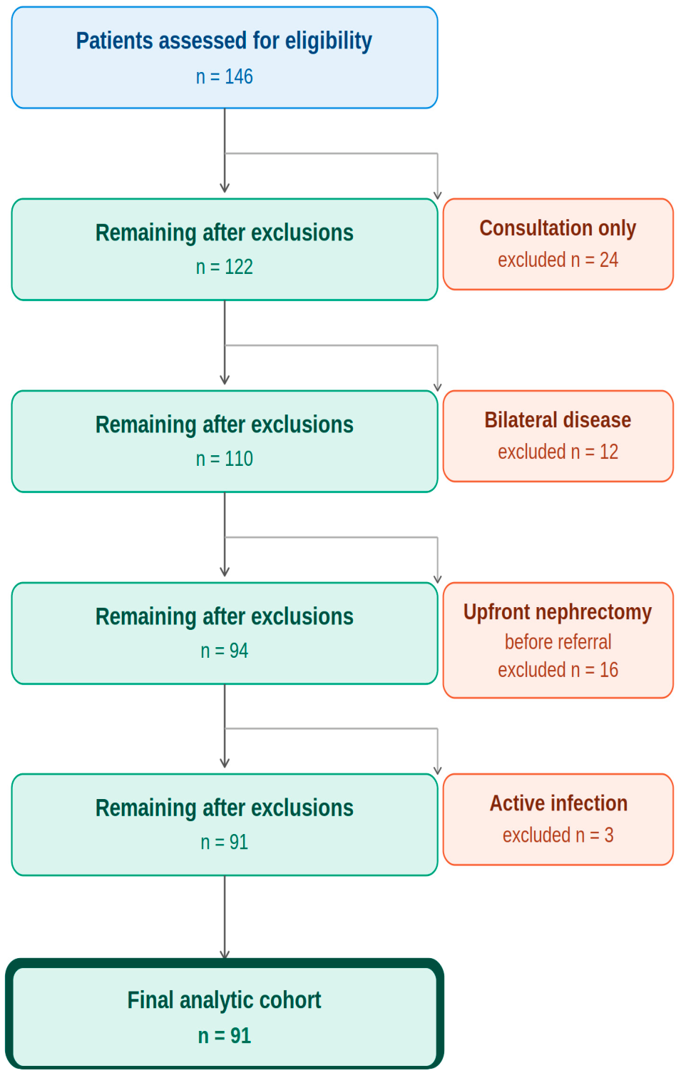
Following King Hussein Cancer Center (KHCC) institutional review board approval (24KHCC196), we performed a comprehensive review of medical records for patients aged ≤ 18 years diagnosed with WT at KHCC, Jordan. Children treated between November 2014 and December 2023 were included in this study. The analysis focused on cases of unilateral disease. Patients with bilateral disease were excluded, as they represent a distinct clinical entity. Furthermore, we excluded individuals referred solely for consultation, radiation therapy, or surgery, as well as those who had undergone nephrectomy or chemotherapy prior to their referral to our institution. To prevent confounding of the baseline inflammatory markers, patients presenting with fever or an active infection at the time of diagnosis were also excluded. The patient selection process, including the number of patients excluded at each stage, is summarized in
Figure 1.
All patients were treated according to the International Society of Pediatric Oncology (SIOP) protocol. Laboratory data, including differential white blood cell (WBC) count and platelet count, were obtained at initial presentation as part of the routine baseline workup, prior to the initiation of any neoadjuvant chemotherapy.
2.2. Data Collection and Definitions
Demographic and clinical characteristics collected included age at diagnosis, sex, presence of metastasis, tumor stage, histology, and clinical outcomes. Laboratory data obtained at initial presentation, as part of the routine baseline workup prior to any therapeutic intervention, included differential WBC and platelet counts.
Systemic inflammatory markers were calculated using the following established formulas:
Absolute neutrophil count (ANC) = WBC × neutrophil percentage
Absolute lymphocyte count (ALC) = WBC × lymphocyte percentage
Absolute monocyte count (AMC) = WBC × monocyte percentage
Neutrophil-to-lymphocyte ratio (NLR) = ANC/ALC
Lymphocyte-to-monocyte ratio (LMR) = ALC/AMC
Pan-Immune-Inflammation Value (PIV) = (ANC × Platelet count × AMC)/ALC
2.3. Statistical Analysis
Demographic, tumor, and treatment characteristics were summarized by descriptive statistics. OS was defined as the time from diagnosis to death from any cause, or to the last follow-up for patients remaining alive. Event-free survival (EFS) was defined as the time from diagnosis to the occurrence of disease recurrence, progression, death, or last follow-up for patients who did not experience an event.
Optimal cut-off values were determined using the surv_cutpoint function (R package survminer, maxstat method), which evaluates the log-rank test statistic at every possible split point of the continuous marker and selects the threshold producing the greatest survival curve separation. This approach is data-driven; however, because cutpoints are derived and tested within the same dataset, performance metrics are subject to optimism bias. A unified set of EFS-optimized cutpoints was applied consistently to both EFS and OS analyses, across all survival analyses, and for reporting sensitivity and specificity in ROC curve evaluations, allowing a single threshold to be used across endpoints. Patients were subsequently stratified into “low” and “high” groups based on these determined thresholds. EFS and OS rates were then compared across these groups and other patient characteristics.
Cox proportional hazards regression was used for univariable and multivariable comparisons of different covariates. The clinical variables age at diagnosis and stage were included as covariates along with inflammatory markers. Variables with p ≤ 0.05 in univariable analysis were considered for multivariable models.
Given the mathematical interdependence of PIV, NLR, and LMR—which share ALC in their denominators—pairwise Spearman rank correlations were calculated between all three markers to quantify collinearity prior to survival analysis. In view of the strong inter-correlations identified and the limited number of outcome events (25 EFS events, 16 OS events), the three markers were not entered simultaneously into any single multivariable Cox model. Instead, each inflammatory marker was evaluated independently in univariable analysis. In the multivariable models, inflammatory markers were included individually alongside the pre-specified clinical covariates (age and stage), rather than as a combined set, to avoid variance inflation from collinear predictors.
4. Discussion
Several prognostic factors have historically been identified in WT, prominently including tumor histopathology, the presence of distant metastases, per-operative tumor volume, and specific molecular alterations [
2,
26,
27,
28,
29]. While the prognostic utility of inflammatory markers has been extensively validated in adult oncology, there remains a distinct paucity of data regarding their application in pediatric cohorts [
14,
18,
23,
24]. This study presents evidence that systemic inflammatory markers, readily assessed via peripheral blood prior to the initiation of any therapy, hold significant prognostic value in children with unilateral WT. These markers not only reflect the baseline inflammatory and immunologic status of the host but also correlate strongly with survival outcomes, functioning as surrogate markers for high-risk disease that can be assessed prior to surgical staging.
Our findings largely corroborate those of Cui et al., who reported that elevated PIV and NLR predicted worse EFS and OS in WT patients treated at a Chinese center [
25]. The biological convergence across two geographically and ethnically distinct populations strengthens confidence in the prognostic relevance of these markers. Notable differences exist, however: Cui et al. did not report LMR as a significant predictor, while in our cohort, LMR demonstrated a significant association with EFS on univariable analysis [
25]. Differences in institutional treatment protocols, histological distribution, and baseline hematological norms across ethnic groups may account for this discrepancy and deserve further study.
A key methodological consideration in studies evaluating composite inflammatory indices simultaneously is the collinearity inherent to their mathematical structure. In the present cohort, pairwise Spearman correlations between PIV, NLR, and LMR were uniformly strong (rho range: 0.83–0.85), reflecting the fact that ALC appears in the denominator of all three indices. This degree of inter-correlation means that simultaneous entry of all three markers into a single multivariable model would produce unstable and uninterpretable coefficient estimates, particularly given the limited number of outcome events in this cohort. Accordingly, markers were evaluated independently in univariable analysis and not entered as a combined set in multivariable models. The loss of statistical significance for PIV and NLR in the multivariable EFS model—where high stage remained the dominant predictor—most likely reflects two overlapping mechanisms: collinearity between the inflammatory markers themselves, and the established biological relationship between advanced stage and systemic immune perturbation, whereby higher tumor burden drives the neutrophilia, thrombocytosis, and lymphopenia that composite indices capture. This collinearity between stage and inflammatory markers does not diminish the pre-operative clinical utility of PIV and NLR; on the contrary, it confirms that these markers function as accessible surrogates for tumor aggressiveness at a point in time when surgical staging is not yet available.
In the broader oncologic literature, patients presenting with high NLR and PIV and low LMR generally exhibit inferior clinical trajectories. Biologically, these parameters likely represent an underlying immunologic imbalance. Elevated neutrophil or platelet counts, coupled with reduced lymphocyte levels, point toward a tumor-growth-promoting systemic environment. Conversely, relative lymphopenia reflects a profound suppression of the adaptive immune system, specifically a depletion of the cytotoxic CD8+ T-cells necessary for anti-tumor surveillance [
30,
31].
The identification of the PIV as a powerful risk stratifier is particularly important and represents a highly novel finding in pediatric renal tumors. Combining four parameters—neutrophils, platelets, monocytes, and lymphocytes—PIV offers a comprehensive snapshot of the immune system. Specifically, the incorporation of platelets is crucial; tumor-educated platelets are known to secrete pro-angiogenic factors, shield circulating tumor cells from natural killer (NK) cell-mediated immune destruction, and facilitate extravasation during metastasis. This multi-faceted role in immune evasion and tumor dissemination provides a strong biological rationale for PIV’s superior prognostic depth [
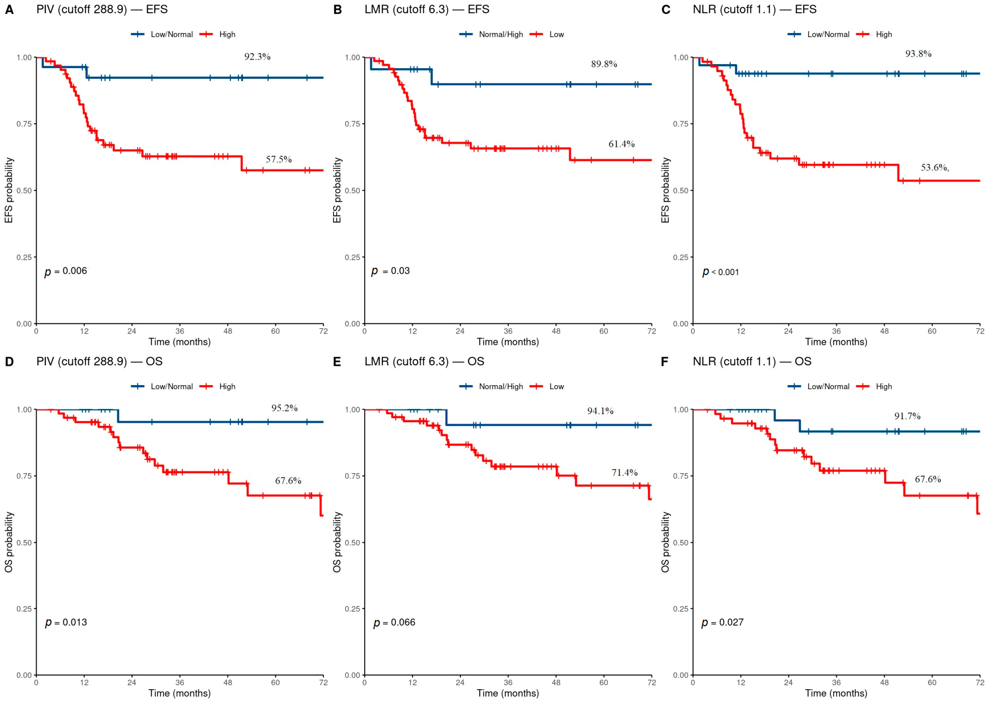
32]. Patients in our cohort with an elevated PIV had a markedly reduced 5-year EFS of 57.5%, compared to 92.3% in the low-PIV group. As discussed above, PIV and NLR did not retain independent significance in the multivariable EFS model—a finding best understood through the collinearity framework described earlier—yet their high univariate hazard ratios and exceptional sensitivity (93.3% for 5-year mortality) underscore their value precisely at the moment of initial presentation, before surgical staging is available. The identification of elevated NLR and PIV, alongside low LMR, as specific markers of poor prognosis in our WT cohort aligns with prior observations in other adult solid tumors, suggesting that the fundamental mechanisms of cancer-associated inflammation exceed age and histology [
14,
17,
23].
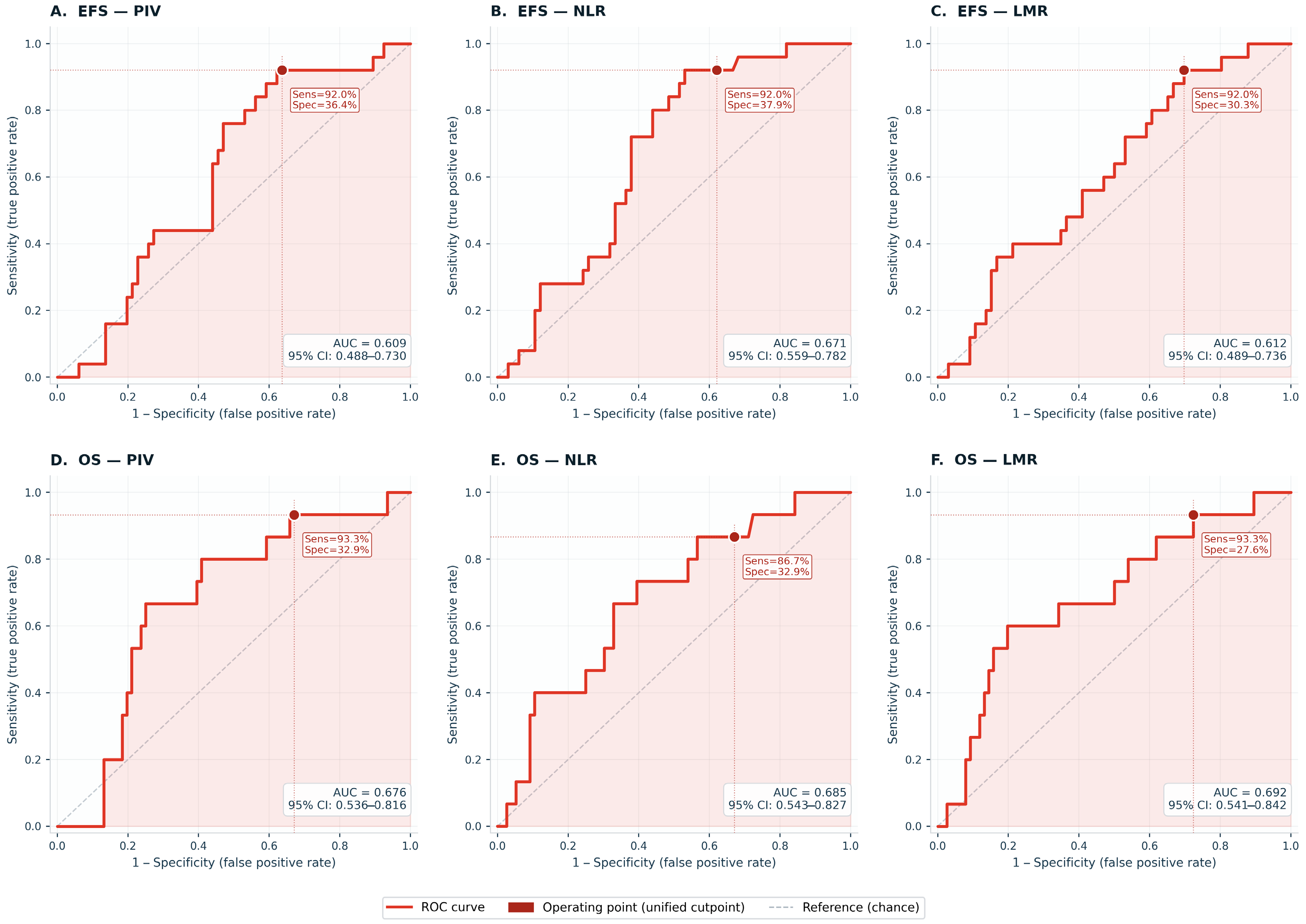
The clinical utility of our findings is supported by the use of ROC curve analysis and data-derived cutpoints to establish specific, quantifiable cut-off values for both mortality and relapse. It is important to contextualize the predictive performance of these markers. While the ROC analysis demonstrated statistically significant AUC values (ranging from 0.609 to 0.692), these represent moderate discriminative ability. Therefore, pre-treatment inflammatory markers should not be viewed as standalone definitive predictors of relapse or mortality. Rather, they serve as valuable, highly accessible clinical adjuncts intended to complement—not replace—established risk stratification models based on tumor stage, histology, and molecular profiling. However, when comparing our derived thresholds to published data, notable variations emerge. Previous studies exploring inflammatory markers in pediatric solid tumors have frequently reported higher cut-points for NLR, often exceeding 2.0 or 2.5, to predict adverse outcomes. In contrast, our cohort’s data-derived, EFS-optimized cut-point for NLR was relatively lower (1.1). Similarly, optimal LMR thresholds in our analysis differed from established adult and pediatric literature benchmarks. These biomarkers are derived from routine complete blood counts and require no additional cost or specialized infrastructure, making them particularly valuable in low- and middle-income countries where access to molecular diagnostics remains limited.
These discrepancies highlight a critical pathophysiological and epidemiological consideration: while methodological approaches (such as maximum survival curve separation) naturally influence cut-point generation, the baseline hematological and inflammatory profiles of Arab children in the Middle East may inherently differ from those of Western cohorts. Regional environmental factors, endemic subclinical infections, nutritional variations, and distinct immunogenetic backgrounds (such as polymorphisms in cytokine promoter regions) can shift the “normal” inflammatory baseline. Consequently, applying universal, Western-derived cut-points may lead to inaccurate risk stratification in our demographic. This underscores the necessity for population-specific reference intervals rather than assuming a one-size-fits-all threshold for inflammatory biomarkers in global pediatric oncology.
It is important to situate these pre-treatment markers within the full clinical complexity of WT management. Several factors with major prognostic impact are, by definition, unavailable at initial presentation: histological subtype and chemotherapy response are only assessable from the post-nephrectomy specimen; lymph node status depends on intraoperative sampling technique and surgeon decision-making; anaplasia (focal or diffuse) may not be clinically suspected before resection; and chemotherapy can induce downstaging that shortens or alters treatment duration. Pre-treatment inflammatory markers do not replace any of these assessments, nor do they inform them. Their utility lies specifically in the narrow clinical window at initial diagnosis—before imaging results in surgical staging, before histology is available, and before treatment has commenced. In this window, a simple complete blood count may identify patients who are likely to have a turbulent clinical course, warranting heightened clinical vigilance even if formal risk re-stratification must await post-operative information.
To translate these findings into a practical clinical framework, we propose a risk-stratification approach as a hypothesis for prospective evaluation. These markers are particularly relevant in low- and middle-income countries, where access to molecular diagnostics is limited yet a routine complete blood count is universally available at diagnosis, making inflammatory indices a genuinely zero-cost addition to standard workup. At initial presentation, PIV, NLR, and LMR are calculated from the routine pre-treatment complete blood count and integrated with established clinical parameters—tumor stage on imaging, presence of metastasis, and age at diagnosis. Patients with advanced stage (III–IV) and concurrent elevated PIV or NLR may represent a particularly high-risk subgroup in whom closer clinical monitoring during neoadjuvant chemotherapy is warranted, and in whom treating physicians may exercise a lower threshold for treatment modification based on early response assessment. For patients with early-stage disease (I–II), elevated PIV or NLR at diagnosis may identify the minority at unexpected risk for relapse—a group in whom standard staging alone would not prompt heightened vigilance—supporting more frequent post-treatment clinical follow-up and ultrasound-based monitoring beyond the standard schedule. In low- and middle-income settings where resources for uniform intensive follow-up are limited, inflammatory markers at diagnosis could help triage which patients warrant closer surveillance, allowing more efficient allocation of available monitoring capacity. Patients with low PIV, low NLR, and high LMR may conversely represent candidates for treatment de-escalation strategies in future prospective studies, potentially reducing chemotherapy-related morbidity—a consideration of particular importance in resource-constrained settings where long-term toxicity monitoring is less accessible. We emphasize that this framework is hypothesis-generating and requires prospective validation before clinical adoption.
5. Study Limitations
Despite the strengths of our analysis, we acknowledge several limitations. The retrospective, single-center design and the relatively modest sample size inherently limit statistical power for detecting highly subtle associations and may affect broad generalizability. The excellent prognosis of early-stage disease at our center resulted in zero mortality events for Stage I and II patients; while this limited the calculation of hazard ratios for stage in the OS model, it underscores the critical need for biomarkers like PIV and NLR to identify the minority of patients at risk for lethal recurrence. Furthermore, a fundamental methodological limitation of this work is the use of data-derived cut-points evaluated within the same dataset from which they were generated. The maxstat method (surv_cutpoint) selects the threshold that maximizes separation of the log-rank statistic across all possible values—an exhaustive search that, by definition, identifies the split most favorable to demonstrating a survival difference in this particular sample. When these same thresholds are then tested in Kaplan–Meier analyses and ROC curves on the same data, the resulting performance estimates (AUC 0.609–0.692; sensitivity 86.7–93.3%) are subject to optimistic bias of unknown magnitude. External validation in an independent prospective cohort—ideally from a different geographic or institutional setting—is essential before these specific thresholds can be recommended for clinical use. We explicitly caution against applying the derived NLR cut-off of 1.1 or PIV cut-off of 288.9 in routine practice without such validation. Our findings are best understood as generating specific, testable hypotheses for future prospective study.
A further limitation is the non-specific nature of these indices. Subclinical infections—including otitis media, urinary tract infections, and low-grade enteritis—can independently produce neutrophilia, monocytosis, and relative lymphopenia, potentially misclassifying non-oncological immune activation as tumor-related risk. Although patients with documented fever or active infection were excluded, subclinical infections without fever were not systematically screened for. Future prospective studies should incorporate baseline CRP or procalcitonin to allow identification and adjustment for occult infection. It should further be noted that these indices are elevated across a broad range of non-malignant inflammatory conditions, including appendicitis and inflammatory bowel disease, confirming that their interpretation as tumor-specific prognostic signals requires careful clinical contextualization and cannot be made in isolation from the full clinical picture.




 ,
,