Review

Myeloid-Derived Suppressor Cells in Cancer: Metabolic Reprogramming, Immune Crosstalk, and Therapeutic Targeting


Innate Lymphoid Cells Unit, Bambino Gesù Children’s Hospital, IRCCS, 00146 Rome, Italy
*
Authors to whom correspondence should be addressed.
These authors contributed equally to this work.
Cancers 2026, 18(13), 2150; https://doi.org/10.3390/cancers18132150
Submission received: 29 May 2026 / Revised: 30 June 2026 / Accepted: 1 July 2026 / Published: 3 July 2026

Simple Summary

Myeloid-derived suppressor cells are a group of immature immune cells that accumulate in various types of cancer. They hinder the immune system’s ability to recognize and eliminate tumor cells. By compromising the body’s natural defenses, these cells facilitate tumor growth and spread and reduce responsiveness to treatments, including immunotherapy. In this review, we summarize current knowledge about the development of myeloid-derived suppressor cells, their interactions with other immune cells, and their adaptation of energy use to survive within the tumor microenvironment. These adaptations enable them to maintain their potent immune-suppressing activity and further promote cancer progression. We also discuss new therapeutic strategies aimed at preventing their recruitment to tumors, reducing their numbers, and interfering with the biological processes that support their function. Although many of these approaches are still being researched, targeting myeloid-derived suppressor cells is a promising strategy to restore effective anti-tumor immunity and enhance the success of existing cancer treatments. A better understanding of these cells could lead to the development of more effective and personalized therapies for cancer patients.

Abstract

Myeloid-derived suppressor cells (MDSCs) are a heterogeneous population of immature myeloid cells that accumulate in cancer and represent one of the major drivers of tumor-associated immunosuppression. MDSCs actively contribute to tumor progression by inhibiting both innate and adaptive immune responses, promoting angiogenesis, metastatic dissemination, and resistance to immunotherapy. Two major subsets have been identified, polymorphonuclear (PMN-) and monocytic (Mo-) MDSCs, each characterized by distinct phenotypic, metabolic, and suppressive properties. Within the tumor microenvironment (TME), MDSCs establish a complex network of interactions with T-, B-, NK-cells, dendritic cells, and macrophages, thereby orchestrating immune escape and tumor persistence. Recent evidence highlights the pivotal role of metabolic rewiring in regulating MDSC survival and suppressive activity. Enhanced aerobic glycolysis, fatty acid oxidation, amino acid depletion, reactive oxygen species (ROS) production, and adenosine metabolism collectively sustain MDSC-mediated immune dysfunction and shape the immunosuppressive TME. In particular, the crosstalk between PMN-MDSCs and NK cells has emerged as a critical mechanism of tumor immune evasion, leading to impaired NK cell cytotoxicity, altered activating receptor expression, and defective cytokine production. In this review, we summarize the current knowledge on the phenotypic and functional heterogeneity of MDSCs, their metabolic adaptations, and their interactions with immune effector populations in cancer. Furthermore, we discuss emerging therapeutic strategies aimed at targeting MDSC recruitment, differentiation, metabolic pathways, and suppressive functions. Understanding the molecular and metabolic mechanisms governing MDSC biology may provide novel opportunities to overcome tumor-induced immunosuppression and improve the efficacy of current cancer immunotherapies.

1. Introduction

Myeloid-derived suppressor cells (MDSCs) are a heterogeneous subpopulation with a potent regulatory role that modulates immune responses in the inflammatory milieu. The immunosuppressive role of MDSCs may be dual: beneficial in contexts such as pregnancy, sepsis, trauma, and inflammatory diseases [1,2,3], or detrimental in contexts such as infections or cancer [1].
MDSCs are virtually absent under physiological conditions but are abundant in both peripheral blood (PB) and tissues during pathological states, including hematological [2,3] and solid tumors [4,5]. The first studies on the accumulation of myeloid cells in cancer were published in the late ’70s [6], but subsequent investigations demonstrated the existence of distinct myeloid subsets with immunosuppressive functions [5,6], although many aspects of their complex biology remain to be fully elucidated. MDSCs contribute to tumor progression through tumor angiogenesis [7], drug resistance [8], and promotion of tumor metastases [9], limiting the efficacy of immunotherapy in cancer [10,11]. In the tumor microenvironment (TME), MDSCs can affect the capability of the immune response to eliminate tumor cells and help them to evade the immune response, spread, and form metastasis through different mechanisms [12,13]. Understanding the immunosuppressive mechanisms, the interaction with other immune cells, and therefore strategies to target these cells may represent an important milestone for cancer treatments [8].
In this review, we discuss the phenotypic and functional heterogeneity of MDSCs as well as the suppressive mechanisms, the interactions with immune cells (e.g., T-cells, B-cells, and natural killer cells), the metabolic reprogramming, and therapeutic strategies, aiding in overcoming resistance to cancer immunotherapies. Unlike previous reviews, we offer an integrated and translational perspective that connects the metabolic underpinnings of MDSC suppressive activity to immune cell crosstalk and to actionable therapeutic targeting strategies, providing a more cohesive framework for understanding MDSC-driven immune evasion in cancer.

2. Phenotypic and Functional Characterization of MDSCs

MDSCs are a heterogeneous population with diverse morphological, phenotypic, and functional features. Consistent with this, they express a range of myeloid-associated surface markers while lacking lineage-specific markers of B- and T-lymphocytes, natural killer (NK) cells, macrophages, and dendritic cells (DCs). MDSCs consist of two major subsets of described cells, polymorphonuclear (PMN-) and monocytic (Mo-) MDSCs. Morphologically, PMN-MDSCs are similar to neutrophils, while Mo-MDSCs are similar to monocytes. These cells have been phenotypically characterized by flow cytometry, and the criteria for the nomenclature and characterization standards have been defined [14]. In mice, MDSCs co-express CD11b and Gr-1 and are classified into PMN-MDSCs (Ly6G+Ly6Clo) and Mo-MDSCs (Ly6GLy6Chi) [15,16]. In humans, MDSCs are identified as Lineage (CD3-14-19-56-123), CD11b+, CD33+, HLA-DRlow/−, and subdivided into two subsets: Mo-MDSCs are CD14+CD15 cells, and PMN-MDSCs are CD14CD15+CD66b+CD124+ [17].
Until recently, separation of neutrophils from PMN-MDSCs relied on density gradient centrifugation, whereby PMN-MDSCs are enriched in the low-density peripheral blood mononuclear cells (PBMC) fraction, whereas neutrophils segregate with high-density cells [1]. Additional markers, such as lectin-type oxidized LDL receptor 1 (LOX-1) and S100A9, have emerged as candidate markers of human PMN-MDSCs [18].
MDSCs share a common myeloid progenitor (CMP) with other myeloid cells (e.g., macrophages, granulocytes, and DCs). MDSC development is governed by the same growth factors that control normal myelopoiesis, e.g., macrophage colony-stimulating factor (M-CSF), granulocyte colony-stimulating factor (G-CSF), and granulocyte-monocyte colony-stimulating factor (GM-CSF). These mediators, together with IL-6, are abundant in pathological conditions and TME [19]. In particular, tumor-derived GM-CSF plays a key role in the generation, maintenance, and survival of MDSCs, and its sustained production may underlie the marked accumulation of these cells observed in cancer patients [11,14]. GM-CSF, together with IL-6, can activate the JAK/STAT pathway, involving STAT3 and STAT5, which induces the expression of key genes that drive the proliferation, survival, and suppressive activity of MDSCs [20]. Elevated serum levels of these cytokines have been associated with increased frequencies of circulating MDSCs.

3. Immunosuppressive Mechanisms of MDSCs

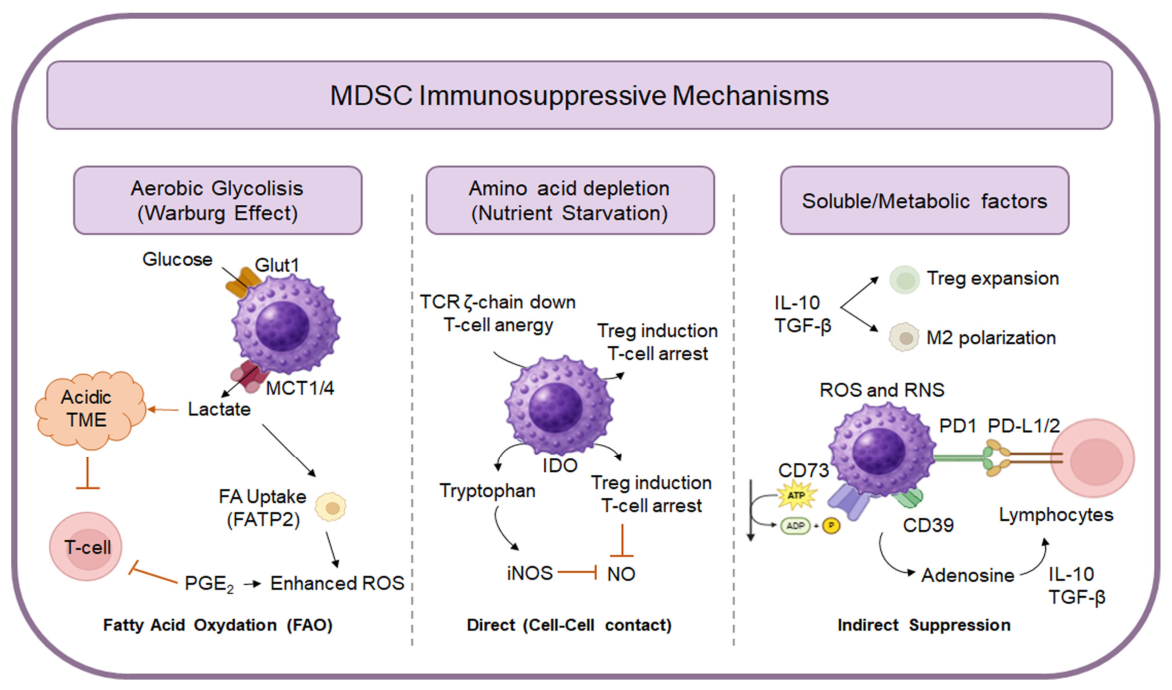
The main characteristic of MDSCs is the ability to suppress immune effector cells, including those from innate (monocytes, DCs, NK cells) and adaptive (T- and B-cells) compartments (Figure 1).
They can produce different cytokines and soluble factors that have direct immunoregulatory effects, such as IL-10, TGF-β, reactive oxygen (ROS) and nitrogen species (RNS), nitric oxide (NO), cyclooxygenase 2 (COX-2), and indoleamine 2,3-dioxygenase (IDO), or arginase (Arg) 1 and 2 [8]. Moreover, immune regulation is enhanced by the contextual recruitment of other regulatory cells, including regulatory T (Treg) and B cells (Breg), M2 macrophages, and tolerogenic DCs (TolDCs) [21]. MDSCs rewire their metabolic pathways, specifically involving glucose, lipids, amino acids, and adenosine, to survive in hostile microenvironments and effectively suppress immune responses.
One of the key features of MDSCs is the nutrient depletion induced by the production of enzymes like Arg-1 and -2, IDO, and iNOS. These molecules deplete arginine (ʟ-Arg), cysteine, and tryptophan, three important amino acids involved in lymphocyte proliferation and function, leading to a progressive immune cell anergy [22]. Arg-1 degrades ʟ-Arg, which is vital for T-cell receptor signaling, while the enzyme IDO breaks down tryptophan into kynurenine, a metabolite that inhibits T-cell proliferation and promotes the development of Tregs [23,24,25,26,27,28].
As already mentioned, another MDSC feature is reactive species production. Increased ROS and RNS secretion disturb the equilibrium status of pro-oxidant/antioxidant reactions and is also important for the impairment of immune cells, especially T-cells. Indeed, ROS catalyzes the nitration of TCR/CD8 molecules to prevent the TCR/MHC–peptide interactions [29,30].
Secretion of IL-10 and TGF-β is pivotal in shifting responses toward Tregs and suppressive macrophages, ensuring the formation of an immunosuppressive environment [31].
MDSCs are also involved in cell–cell communication, upregulating immune checkpoints, and driving T-cell exhaustion. In this context, an important role is played by Programmed Death-Ligand 1 (PD-L1), which is expressed by MDSCs and inhibits cell proliferation, survival, cytokine production, and other effector functions upon interaction with PD-1. Besides adaptive immune cells, MDSCs can incite NK and DC impairment through a decrease in NK cytotoxicity, DC maturation, and antigen presentation [32].
MDSCs heavily utilize aerobic glycolysis (the Warburg effect), converting glucose into lactate even when oxygen is present. This high glucose consumption depletes resources for immune cells, while the resulting lactate accumulation acidifies the environment and impairs cell function by disrupting their redox balance [33,34,35]. MDSCs also rely on fatty acid oxidation (FAO) to generate energy and ROS, which enhances their suppressive activity. Tumor-derived factors like GM-CSF induce the transporter fatty acid transport protein 2 (FATP2), allowing MDSCs to take up arachidonic acid and produce PGE2, a major mediator of immune suppression [36,37,38].
Finally, in the TME, MDSCs use the enzymes CD39 and CD73 to convert extracellular ATP into adenosine that binds their receptors (e.g., A2AR/A2BR) on MDSCs, triggering signaling pathways that increase the production of immunosuppressive molecules like TGF-β and IL-10 [39,40,41,42,43].

4. MDSCs and Immune Cell Interactions in Cancer

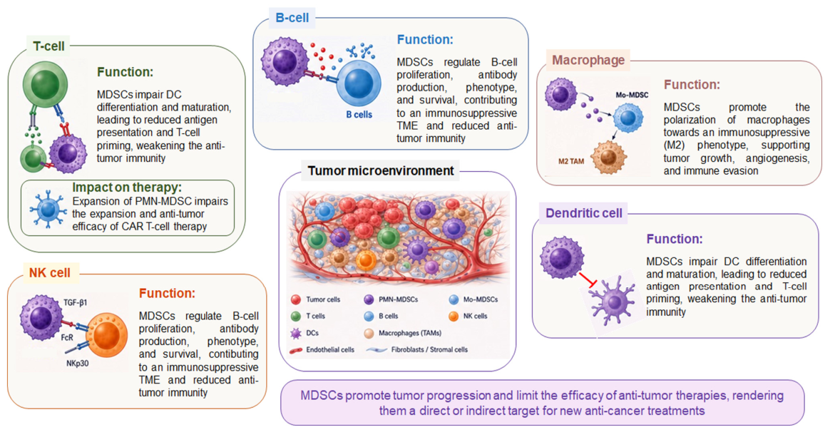
The TME is a heterogeneous ecosystem in which malignant cells coexist with non-tumoral elements, including mesenchymal stromal cells, endothelial cells, fibroblasts, and a spectrum of immune cell populations. Within the TME, PMN-MDSCs constitute the predominant subset of MDSCs, accounting for about 80% of the total MDSC population. MDSCs play a central role in orchestrating immunosuppression within the TME, employing a broad array of mediators and molecular mechanisms to inhibit several critical immune effectors, including T-, B-, NK-ells, DC, and macrophages [44,45] (Figure 2).

4.1. T-Cells

MDSCs are potent inhibitors of T-cell immune responses [46] and employ multiple mechanisms to suppress T-cell function. As mentioned before, one major pathway involves the modulation of amino acid metabolism. In in vivo models, MDSCs impair T-cell activation by sequestering cystine and limiting its availability. Because T-cells are unable to efficiently synthesize or import cysteine, they rely on antigen-presenting cells (APCs), such as macrophages and DCs, for their supply. In contrast, MDSCs take up cystine but do not release cysteine, thereby competing with APCs and reducing extracellular cysteine levels, ultimately impairing T-cell activation [47].
Arg-1 and Arg-2 enzymes reduce ʟ-Arg availability, impairing T-cell activation and proliferation by downregulating the CD3ζ chain of the TCR complex [48]. The accumulation of Arg-1+ MDSCs results in systemic ʟ-Arg depletion, which is associated with tumor progression [49]. Notably, T-cell response suppression can be reversed by arginase inhibition or ʟ-Arg supplementation [50]. In PBMC and tissue samples from lung cancer patients, increased frequencies of PMN-MDSCs correlate with reduced CD4+ and CD8+ T-cell populations, and elevated Arg-1 mRNA expression in PBMC is associated with MDSC abundance and suppressive activity [51,52]. Similar Arg-1 upregulation has been reported across multiple tumor types, including gastric [53,54], colorectal [55], renal [56], and hepatocellular carcinomas [57].
In melanoma and colorectal cancer patients, interactions between MDSCs and activated T-cells induce IL-10 production, which promotes STAT3 activation in MDSCs, leading to an upregulation of PD-L1 expression. These PD-L1+ MDSCs mediate immunosuppression through mechanisms involving Arg-1 and IDO [58]. IDO overexpression by MDSCs leads to tryptophan depletion, resulting in T-cell cycle arrest and impaired immune function [59]. PMN-MDSCs also release ROS into the extracellular space [60], thereby directly and indirectly promoting tumor progression. In tumor mouse models, ROS production by PMN-MDSCs suppresses T-cell responses through pathways involving FATP2 and STAT3 signaling [61].
New treatment approaches based on adoptive cell therapy (CAR T-cells) could be influenced by the presence and expansion of PMN-MDSCs in the PB of hematological and solid tumor-treated patients. The expansion of PMN-MDSCs affects the expansion and the potent anti-tumor activity of CAR T-cells [11].

4.2. B-Cells

B-cells contribute to tumor progression through multiple mechanisms, including the production of lymphotoxin, which induces angiogenesis and tumor growth. Tumor-derived extracellular vesicles can activate B-cells to produce antibodies that form immune complexes, activating Fcγ receptors on myeloid cells, and driving their differentiation into MDSCs [62]. Despite extensive research, evidence supporting crosstalk between MDSCs and B-cells remains limited. In a cancer mouse model, MDSCs were found to accumulate around splenic germinal centers, co-localizing with B-cells and enhancing their proliferation and IgA-production, mediated by IL-10 and TGF-β1 [63]. A similar mechanism involving direct cell-to-cell contact has also been described in a lung cancer model, where MDSCs suppress B-cell proliferation in an Arg-dependent manner [64]. In tumor-bearing mice, a reduction in B-cells in the bone marrow was linked to the infiltration of MDSCs. Tumor progression disrupted key pathways for B-cell development, including IL-7 and STAT5 signaling, leading to decreased serum IgG levels. The extent of B-cell impairment correlated positively with MDSC infiltration and cancer prognosis [64]. In breast cancer mouse models, MDSCs and splenic B-cells increased significantly. MDSCs altered B-cell phenotypes, affecting immune checkpoints like PD-1 and PD-L1, and regulating functions such as proliferation, apoptosis, antibody production, and cytokine secretion [65].
MDSCs, Tregs, and Bregs play a significant role in the immunosuppressive microenvironment of glioblastoma (GBM) in both preclinical models and patient samples. Bregs exhibited immunosuppressive activity against activated CD8+ T-cells and overexpressed inhibitory molecules like PD-L1 and CD155, along with producing TGF-β and IL-10. Additionally, MDSCs enhance the immunosuppressive effects of Bregs by transferring PD-L1 via microvesicles, creating a critical MDSC–B-cell axis that contributes to immune suppression in GBM [21].
Studies in human patients remain relatively limited. Bodogai et al., showed that MDSCs from B-cell chronic lymphocytic leukemia patients displayed only partial regulatory function unless educated by Bregs via TGF-β receptor signaling. This interaction enhanced MDSC production of ROS and NO, strengthening their ability to suppress CD4+ and CD8+ T-cells, and promoting tumor growth and metastasis. Disruption of Bregs or TGF-β signaling impaired MDSC function and blocked metastasis [66]. PMN-MDSCs can also differentially regulate B-cell function by suppressing their proliferation and antibody production in a cell contact-dependent manner. This effect involves established mediators (i.e., Arg-1, NO, ROS) and is associated with the induction of B-cell death [67].
A study found that human Mo-MDSCs inhibit B-cell proliferation and function in vitro through soluble mediators like IDO, Arg-1, and NO, without direct contact. They also alter B-cell phenotype by downregulating activation markers and affecting gene expression related to apoptosis, class-switch regulation, and B-cell differentiation [68].

4.3. NK Cells

NK cells are key effectors of antitumor immunity, primarily acting through their cytotoxic capacity and the secretion of soluble factors and cytokines, which can directly eliminate tumor cells or indirectly shape the immune response by recruiting and activating other immune populations [69].
Several experimental models have demonstrated that MDSCs exert a potent inhibitory effect on NK cell function. In multiple murine tumor models, NK cell activity is significantly impaired, a phenomenon closely associated with MDSC accumulation. This dysfunction is characterized by reduced cytotoxicity, decreased expression of activating receptors, and impaired IFN-γ production. Mechanistically, MDSCs inhibit NK cell activation predominantly through membrane-bound TGF-β1 [70,71]. Consequentially, NK cell activation can counteract MDSC-mediated immunosuppression by reducing their accumulation via the NKG2D-NKG2DL axis and promoting their maturation through IFN-γ-dependent signaling, an effect associated with improved clinical outcomes [72]. Additional suppressive mechanisms involve NO production and activation of the IL-6/STAT3 axis, both of which contribute to NK cell dysfunction and tumor immune escape [73,74]. Among the inhibitory mechanisms, as mentioned above, one involves direct cell-to-cell contact. In vitro co-culture systems have shown that MDSCs suppress NK cell functions, including antibody-dependent cellular cytotoxicity, cytokine production, and downstream signaling, through Fc receptors on NK cells [73]. Additional studies in hepatocarcinoma and lung cancer further demonstrate that MDSCs inhibit NK cell activity via receptors such as NKp30 [4,75].
PMN-MDSCs are highly enriched in the PB and TME of lung cancer patients, where their accumulation correlates with poor clinical outcome and reduced NK cell frequency and functionality. PMN-MDSCs profoundly impair NK cell activity by downregulating activating receptors, reducing cytotoxicity, degranulation, and cytokine production through both cell-to-cell contact and soluble mediators, including PMN-MDSC-derived exosomes. Importantly, these findings identify PMN-MDSCs as key regulators of NK cell dysfunction and suggest that targeting PMN-MDSC-mediated immunosuppressive mechanisms may represent a promising strategy to restore NK cell-mediated anti-tumor immunity [4].
In clinical contexts, MDSC-mediated suppression is also evident in hematological malignancies. In myelodysplastic syndromes, co-culture with MDSCs results in reduced expression of NK activating receptors (NKG2D, NKp30, NKp46), decreased effector molecule production (CD107a, IFN-γ, perforin, and granzyme B), and increased apoptosis of NK cells. Importantly, blockade of the TIGIT/CD155 axis partially restores NK cell function, highlighting an additional checkpoint involved in MDSC-mediated immune suppression [76].
Finally, clinical studies support the immunoregulatory role of MDSCs. In patients with head and neck squamous cell carcinoma (HNSCC), elevated levels of circulating and tumor-infiltrating PMN-MDSCs and Mo-MDSCs correlate with strong immunosuppressive activity, driven by TGF-β and NO pathways [77]. In the context of immunotherapy, a higher NK cell-to-Lox-1+ PMN-MDSC ratio has been associated with better responses to anti-PD-1 therapy, longer progression-free survival, and improved overall survival [78]. A small-molecule screen identified PI3K-γ inhibitors as effective modulators of MDSCs’ function [79]. Finally, in sarcoma and breast cancer patients, IL-6 produced by tumor-infiltrating NK cells correlates with MDSC-associated markers, such as S100A8/9 and Arg-1, further linking NK-MDSC crosstalk to tumor progression and immune remodeling [74].

4.4. Dendritic Cells

DCs are specialized APCs that connect innate and adaptive immunity by providing signals essential for T-cell activation and differentiation. Tumor-related conditions lead to the accumulation of immature DCs and hinder their maturation, causing functional impairments [80,81,82].
The accumulation of MDSCs represents a key mechanism underlying defective DC differentiation in cancer, thereby contributing to defective antitumor immunity. In murine models, this process is mediated in part by the STAT3/S100A9 axis, which promotes MDSC expansion and inhibits DC development [83]. Mechanistically, PMN-MDSCs selectively impair DC cross-presentation, without affecting direct antigen presentation, via a contact-independent mechanism involving the transfer of oxidized lipids. Although both neutrophils and PMN-MDSCs transfer lipids to DCs, only PMN-MDSCs suppress cross-presentation due to their capacity to generate oxidatively truncated lipids, driven by myeloperoxidase (MPO) [84].
Consistent with these findings, in vitro studies have shown that mouse MDSCs differentiated from c-kit+ bone marrow progenitor cells in the presence of IL-4, GM-CSF, and PGE2 are associated with a proportional decline in the number of mature DCs [85]. Similarly, stimulation with LPS and IFN-γ inhibits DC development while enhancing MDSC suppressive functions [86]. In several cancer types, patients exhibit decreased DC levels alongside increased MDSC frequencies, correlating with poorer overall and progression-free survival [87]. Finally, patients with HNSCC display elevated levels of Mo-MDSCs and tolerogenic DCs, both of which suppress activated T-cells through distinct signaling pathways. PI3K-AKT signaling is critical for the induction and activity of Mo-MDSCs, whereas β-catenin-dependent Wnt signaling drives the tolerogenic properties of DCs [88].

4.5. Macrophages

Macrophages support host defense by regulating adaptive immunity, promoting tissue repair, and eliminating pathogens. In cancer, however, they represent a highly heterogeneous population: M1-macrophages promote anti-tumor immunity, while M2-macrophages exhibit immunosuppressive functions and support tumor growth [89]. Within the TME, tumor-associated macrophages (TAMs) are predominantly polarized towards an M2 phenotype, which contributes to tumor progression by promoting angiogenesis, tumor cell invasion, and metastasis [81]. The immunosuppressive activity of TAMs is further reinforced by MDSCs. These interactions modulate key mediators, such as IL-6, IL-10, TNF-α, and NO. For instance, MDSC-derived IL-10 suppresses macrophage pro-inflammatory cytokines while enhancing NO production [81]. In parallel, PMN-MDSCs can promote the differentiation of Mo-MDSCs into M2-macrophages via exosome-mediated mechanisms regulated by the IL-6R/JAK/STAT3 pathway, thereby sustaining tumor-promoting inflammation [90].
Metabolic and inflammatory signaling pathways also critically shape myeloid cell function. The nutrient-sensing kinase regulates macrophage and MDSC polarization in the TME [91]. Conversely, pro-inflammatory stimuli such as Toll-like receptors 7/8 agonists, IFN-γ, or TNF-α plus IL-6, can reprogram human Mo-MDSCs into M1-macrophages with antitumor activity via NF-κB and STAT4 signaling [92]. In esophageal cancer, elevated circulating MDSCs are associated with increased accumulation of M2-macrophages and a Th2-polarized immune profile, characterized by high IL-4, IL-13, IL-6, and GATA3, alongside reduced IFN-γ and IL-12 [93,94]. A similar pattern is observed in Hodgkin lymphoma, where the MDSC expansion correlates with the accumulation of pro-tumoral M2-macrophages [95].
Functional differences between myeloid subsets also emerge during tumor progression. In breast cancer models, MDSCs exhibit a more pro-inflammatory and pro-angiogenic profile and preferentially localize to hypoxic tumor regions, suggesting a role in early tumor development. As tumors progress, a shift toward TAMs is observed, with TAMs exerting stronger immunosuppressive effects and driving immune evasion [96]. At the molecular level, several regulators of myeloid cell function have been identified. The scavenger receptor MARCO is highly expressed in M2-TAMs and plays a key role in MDSC differentiation and immunosuppression. Its targeting reduces MDSC and TAM accumulation while enhancing CD8+ T-cells and NK cell responses [97]. Additionally, macrophages derived from Mo-MDSCs display a distinct immunosuppressive phenotype driven by persistent S100A9 expression and regulated by the transcription factor C/EBPβ. The presence of S100A9+ macrophages correlates with poor prognosis in patients with head and neck cancer [98].
Finally, in desmoplastic tumors such as cholangiocarcinoma (CCA), targeting TAMs alone induces a compensatory expansion of PMN-MDSCs, which sustains immune evasion by suppressing T-cell responses. Notably, a distinct ApoE+ PMN-MDSC subset emerges following TAM depletion and is also detectable in human CCA, underscoring the need for combinatorial strategies targeting multiple immunosuppressive myeloid populations [99].

5. Targeting PMN-MDSCs for Cancer Treatment

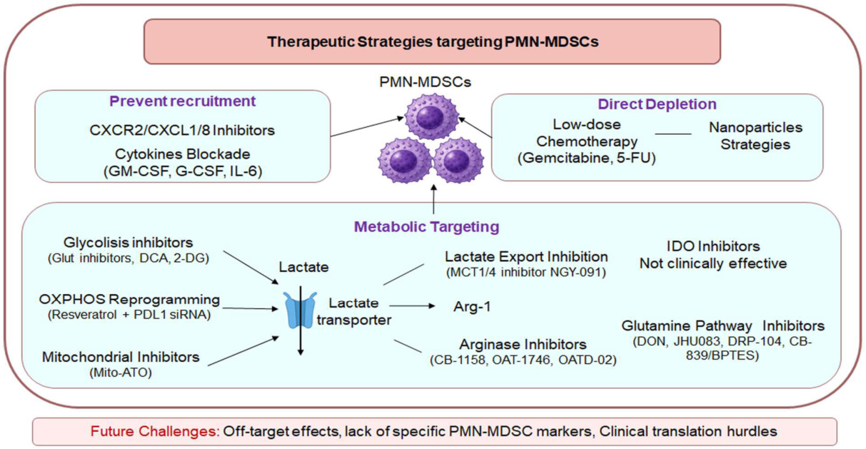
A deeper understanding of the mechanisms through which MDSCs mediate immunosuppression is essential for the development of strategies aimed at restoring effective anti-tumor immunity (Figure 3).
Based on these acknowledgements, the major therapeutic strategies that can be applied to target PMN-MDSCs consist of preventing the recruitment or directly depleting them at the tumor site, limiting in this manner the interaction with other immune cells, and improving the local anti-tumor response. In addition, targeting the immunosuppressive mechanisms described above is a possible strategy. New insights from the literature focus on the importance of MDSC metabolism and the influence of TME-derived metabolites, although more studies in this field are needed. Several ongoing studies are investigating MDSC-targeting strategies in combination with other anti-cancer therapies to improve therapeutic efficacy.

5.1. Preventing PMN-MDSC Recruitment to Tumors

Tumors recruit PMN-MDSCs through chemokines and inflammatory cytokines. Blocking these pathways can reduce their accumulation in the TME. Targeting the CXCR2/CXCL1-CXCL8 axis is one of the most important pathways controlling PMN-MDSC migration [77]. Indeed, CXCR2 inhibitors have shown promise in preclinical models by improving T-cell infiltration and enhancing checkpoint blockade therapy, as well as targeting inflammatory tumor-derived cytokines (such as GM-CSF, G-CSF, IL-6) [100].

5.2. Depleting PMN-MDSCs Directly

Another therapeutic strategy involves the selective depletion of PMN-MDSCs, although the implementation of this approach remains challenging due to several biological and technical limitations. Indeed, some of the possible strategies, such as low doses of chemotherapeutic agents (e.g., gemcitabine, 5-fluorouracil), could affect the other immune cells. All-trans retinoic acid (ATRA), the active metabolite of vitamin A, has emerged as a key regulator of PMN-MDSC differentiation and immunosuppressive activity within the tumor microenvironment [101]. Several studies demonstrated that ATRA promotes the maturation of immature myeloid cells into differentiated non-suppressive populations, thereby reducing the accumulation and suppressive function of PMN-MDSCs through activation of the ERK1/2 pathway and increased glutathione synthesis, which neutralizes ROS-mediated immunosuppressive mechanisms [102,103,104]. Importantly, ATRA-mediated targeting of MDSCs has been associated with restoration of anti-tumor immune responses, including improved T-cell and NK cell activity. In preclinical and clinical settings, ATRA reduced circulating MDSC frequencies and enhanced responsiveness to immune checkpoint blockade, supporting its use as an adjuvant immunomodulatory strategy in cancer therapy [105].
Moreover, the use of antibodies targeting myeloid markers (e.g., nanoparticle-mediated drug delivery systems, etc.) is not yet feasible, since the common phenotype that PMN-MDSC shares with neutrophils, in fact, a unique antigen has not yet been characterized [8].

5.3. Targeting PMN-MDSC Metabolism

In the TME, the capability of MDSCs to rewire their metabolism and to promote the suppression of immune cells made it possible to investigate the potential targets of different pathways involved in their bioenergetic activity. Several strategies aim to disrupt glycolysis, since the PMN-MDSCs depend on it for their energetic capacity. For this reason, glucose depletion using agents such as 2-deoxyglucose (2-DG), inhibition of glucose transporters like GLUT3, and targeting metabolic enzymes such as pyruvate dehydrogenase with dichloroacetate (DCA) [106]. Additional approaches include metabolic reprogramming therapies, such as resveratrol, combined with PD-L1 siRNA, which shifts MDSCs from glycolysis back toward OXPHOS, reducing their suppressive activity and enhancing anti-tumor immunity [107]. Mitochondria-targeted atovaquone (Mito-ATO), a modified form of the antimalarial drug atovaquone, selectively impairs PMN-MDSC bioenergetics by inhibiting mitochondrial complex I and glycolytic pathways. This decreases PMN-MDSC survival, lowers Arg-1 and ROS production, and restores T-cell anti-tumor responses [108].
Enhanced glycolysis in MDSCs also leads to increased lactate production. Lactate is exported through monocarboxylate transporters, MCT1 and MCT4, to prevent intracellular acidification and sustain glycolytic activity. Extracellular lactate further activates MDSCs and strengthens their tumor-promoting and immunosuppressive functions [109]. Preclinical evidence suggests that dual inhibition of MCT1/4, including NGY-091, may attenuate lactate-driven immunosuppressive functions [110].
Although these metabolic-targeting strategies have shown promise in preclinical studies, their clinical application remains challenging because many normal immune cells in the tumor microenvironment also rely on glycolysis, raising concerns about off-target immune suppression. Also targeting the lipid metabolic pathways weakens PMN-MDSC suppressive activity, promotes anti-tumor immunity, and enhances the efficacy of immunotherapies, particularly immune checkpoint blockade [37].
Pharmacological inhibition of Arg-1 and, in some cases, Arg-2, using small molecules such as CB-1158, OAT-1746, and OATD-02, demonstrated restoration of T-cell anti-tumor response [24] and increased the arginine levels in TME, reduced the MDSC frequency [111]. Similarly, phosphodiesterase-5 (PDE5) inhibitors, such as sildenafil and tadalafil, downregulate Arg-1, iNOS, and inflammatory cytokine production, thereby attenuating MDSC-mediated immunosuppression and promoting T-cell infiltration and activation within the TME [112,113]. These immunomodulatory effects have also been translated into early clinical studies. In patients undergoing surgery for abdominal malignancies, perioperative sildenafil reduces the immunosuppressive activity of surgery-induced PMN-MDSCs and is associated with reduced postoperative recurrence, metastasis, and cancer-specific mortality [114,115]. Likewise, tadalafil treatment in patients with HNSCC and GBM significantly decreased both circulating and intratumoral MDSCs and Tregs, while increasing tumor antigen-specific CD8+ T-cell responses [116,117], supporting the clinical potential of PDE5 inhibition as an adjunct to cancer immunotherapy.
In contrast, molecules involved in tryptophan catabolism, such as IDO-1 inhibitors (e.g., INCB023843 and RY103), were developed to counteract tumor-induced immunosuppression, but they showed limited effects on MDSC functions and failed to improve clinical outcomes when combined with a PD-1 inhibitor in phase III clinical trials [118].
Targeting glutamine metabolism showed promising results; for example, 6-Diazo-5-oxo-L-norleucine (DON), a glutamine antagonist, showed anticancer properties. Due to the failure of DON in depleting MDSCs, some prodrugs of DON (e.g., JHU083, DRP-104, JHU395) that target the inhibition of glutamine pathways have emerged [119]. Pharmacological inhibition of glutaminase with CB-839 (telaglenastat), BPTES (bis-2-(5-phenylacetamido-1,2,4-thiadiazol-2-yl) ethyl sulfide), has been studied in different tumor types and in combination with other drugs; however, their direct impact on MDSCs depends on the tumor type, drug dosing, and compensatory pathways, which remain to be examined [95].

6. Concluding Remarks and Future Perspectives

MDSCs have evolved in scientific understanding from being viewed as simple immature myeloid populations to being recognized as sophisticated metabolic and regulatory hubs within the TME. Their extraordinary plasticity allows them to adapt to hostile conditions, effectively orchestrating a multi-layered immunosuppressive network through complex crosstalk with T-, B-, and NK-cells, as well as DCs and macrophages. In recent years, MDSC research has been reshaped by emerging technologies that enable more precise cell-state definition, tissue localization, and assessment of therapeutic relevance. Single-cell transcriptomics, multi-omics approaches, and spatial or multiplex imaging are beginning to resolve MDSC heterogeneity and assign specific functional states, linking them to suppressive niches and prognostic signatures [120,121,122]. These technologies are particularly valuable in the MDSC field, where universally validated unique markers are still lacking and where spatial context is essential to distinguish suppressive myeloid states from phenotypically similar populations, improving the possibility of identifying specific, solid, and valid markers for MDSC identification. Moreover, the future of MDSC-targeted therapy lies in moving beyond simple cellular depletion, which remains hindered by the lack of unique markers distinguishing PMN-MDSCs from mature neutrophils. Instead, the research focus is shifting toward functional reprogramming. By targeting the specific metabolic “engines” that drive their suppressive capacity, such as aerobic glycolysis, fatty acid oxidation, or the arginase and adenosine pathways, it may be possible to neutralize their inhibitory effects while preserving essential myeloid functions. Furthermore, overcoming resistance to current immunotherapies, particularly immune checkpoint inhibitors, will likely require combinatorial strategies. Ultimately, the identification of precise biomarkers and specific metabolic signatures will be crucial for personalizing these interventions and improving clinical outcomes in oncological patients.

Author Contributions

Writing—original draft preparation, A.S., M.R.A., M.T.B., P.V., N.T.; writing—review and editing, all authors; supervision, P.V.; funding acquisition, P.V. All authors have read and agreed to the published version of the manuscript.

Funding

This work was supported by “AIRC—Associazione Italiana per la Ricerca sul Cancro” (project no IG 2022 ID 27065 to P.V., fellowship for Italy to M.T.B.) and the Italian Ministry of Health with “Current Research funds”, and European Union-Next Generation EU-NRRP M6C2-Investment 2.1 Enhancement and strengthening of biomedical research in the NHS (PNRR-POC-2023-12377131, CUP: E83C24000760006 to P.V.).

Data Availability Statement

No new datasets were generated or analyzed in this narrative review. All cited data derive from previously published sources.

Acknowledgments

The authors acknowledge the use of ChatGPT (OpenAI, GPT-5.5 Thinking version) solely for cell cartoon generation. No generative artificial intelligence tool was used to generate scientific content, perform literature searches, select references, analyze data, draw conclusions.

Conflicts of Interest

The authors declare no conflicts of interest.

Abbreviations

The following abbreviations are used in this manuscript:
MDSCMyeloid-Derived Suppressor Cells
PMN-MDSCPolymorphonuclear MDSC
Mo-MDSCMonocytic MDSC
TMETumor Microenvironment
NKNatural Killer
ROSReactive Oxygen Species
RNSReactive Nitrogen Species
NONitric Oxide
DCDendritic Cells
TolDCTolerogenic Dendritic Cell
TregRegulatory T-Cells
B-regRegulatory B Cells
Arg-1Arginase 1
IDOIndoleamine 2,3-Dioxygenase
iNOSInducible Nitric Oxide Synthase
COX-2Cyclooxygenase-2
PD-1Programmed Cell Death Protein 1
PD-L1Programmed Death-Ligand 1
FAOFatty Acid Oxidation
FATP2Fatty Acid Transport Protein 2
PGE2Prostaglandin E2
ATPAdenosine Triphosphate
A2ARAdenosine A2A Receptor
A2BRAdenosine A2B Receptor
PBMCPeripheral Blood Mononuclear Cells
PBPeripheral Blood
CMPCommon Myeloid Progenitor
M-CSFMacrophage Colony-Stimulating Factor
G-CSFGranulocyte Colony-Stimulating Factor
GM-CSFGranulocyte-Macrophage Colony-Stimulating Factor
ILInterleukin
IFN-γInterferon Gamma
TNF-αTumor Necrosis Factor Alpha
JAKJanus Kinase
STATSignal Transducer and Activator of Transcription
TCRT-Cell Receptor
MHCMajor Histocompatibility Complex
APCAntigen-Presenting Cells
LOX-1Lectin-Type Oxidized LDL Receptor 1
LDLLow-Density Lipoprotein
MPOMyeloperoxidase
TAMTumor-Associated Macrophages
MARCOMacrophage Receptor with Collagenous Structure
NF-κBNuclear Factor Kappa B
C/EBPβCCAAT/Enhancer-Binding Protein Beta
HNSCCHead and Neck Squamous Cell Carcinoma
GBMGlioblastoma
CCACholangiocarcinoma
ATRAAll-Trans Retinoic Acid
ERK1/2Extracellular Signal-Regulated Kinases 1/2
GLUT3Glucose Transporter 3
2-DG2-Deoxyglucose
DCADichloroacetate
OXPHOSOxidative Phosphorylation
Mito-ATOMitochondria-Targeted Atovaquone
MCT1/4Monocarboxylate Transporters 1/4
CAR T-cellsChimeric Antigen Receptor T-Cells
TIGITT-cell immunoreceptor with Ig and ITIM domains
IgImmunoglobulin

References

  1. Vacca, P.; Bilotta, M.T.; Moretta, L.; Tumino, N. Myeloid-derived suppressor cells: Identification and function. Methods Cell Biol. 2024, 190, 151–169. [Google Scholar] [CrossRef] [PubMed]
  2. Yu, S.; Ren, X.; Li, L. Myeloid-derived suppressor cells in hematologic malignancies: Two sides of the same coin. Exp. Hematol. Oncol. 2022, 11, 43. [Google Scholar] [CrossRef] [PubMed]
  3. Tumino, N.; Di Pace, A.L.; Besi, F.; Quatrini, L.; Vacca, P.; Moretta, L. Interaction Between MDSC and NK Cells in Solid and Hematological Malignancies: Impact on HSCT. Front. Immunol. 2021, 12, 638841. [Google Scholar] [CrossRef] [PubMed]
  4. Tumino, N.; Besi, F.; Martini, S.; Di Pace, A.L.; Munari, E.; Quatrini, L.; Pelosi, A.; Fiore, P.F.; Fiscon, G.; Paci, P.; et al. Polymorphonuclear Myeloid-Derived Suppressor Cells Are Abundant in Peripheral Blood of Cancer Patients and Suppress Natural Killer Cell Anti-Tumor Activity. Front. Immunol. 2021, 12, 803014. [Google Scholar] [CrossRef] [PubMed]
  5. Wang, P.F.; Song, S.Y.; Wang, T.J.; Ji, W.J.; Li, S.W.; Liu, N.; Yan, C.X. Prognostic role of pretreatment circulating MDSCs in patients with solid malignancies: A meta-analysis of 40 studies. Oncoimmunology 2018, 7, e1494113. [Google Scholar] [CrossRef] [PubMed]
  6. Duwe, A.K.; Singhal, S.K. The immunoregulatory role of bone marrow. I. Suppression of the induction of antibody responses to T-dependent and T-independent antigens by cells in the bone marrow. Cell. Immunol. 1979, 43, 362–371. [Google Scholar] [CrossRef] [PubMed]
  7. Wang, Y.; Ding, Y.; Guo, N.; Wang, S. MDSCs: Key Criminals of Tumor Pre-metastatic Niche Formation. Front. Immunol. 2019, 10, 172. [Google Scholar] [CrossRef] [PubMed]
  8. Raskov, H.; Orhan, A.; Gaggar, S.; Gogenur, I. Neutrophils and polymorphonuclear myeloid-derived suppressor cells: An emerging battleground in cancer therapy. Oncogenesis 2022, 11, 22. [Google Scholar] [CrossRef] [PubMed]
  9. Sprouse, M.L.; Welte, T.; Boral, D.; Liu, H.N.; Yin, W.; Vishnoi, M.; Goswami-Sewell, D.; Li, L.; Pei, G.; Jia, P.; et al. PMN-MDSCs Enhance CTC Metastatic Properties through Reciprocal Interactions via ROS/Notch/Nodal Signaling. Int. J. Mol. Sci. 2019, 20, 1916. [Google Scholar] [CrossRef] [PubMed]
  10. He, G.; Zhang, H.; Zhou, J.; Wang, B.; Chen, Y.; Kong, Y.; Xie, X.; Wang, X.; Fei, R.; Wei, L.; et al. Peritumoural neutrophils negatively regulate adaptive immunity via the PD-L1/PD-1 signalling pathway in hepatocellular carcinoma. J. Exp. Clin. Cancer Res. 2015, 34, 141. [Google Scholar] [PubMed]
  11. Tumino, N.; Weber, G.; Besi, F.; Del Bufalo, F.; Bertaina, V.; Paci, P.; Quatrini, L.; Antonucci, L.; Sinibaldi, M.; Quintarelli, C.; et al. Polymorphonuclear myeloid-derived suppressor cells impair the anti-tumor efficacy of GD2.CAR T-cells in patients with neuroblastoma. J. Hematol. Oncol. 2021, 14, 191. [Google Scholar] [PubMed]
  12. De Cicco, P.; Ercolano, G.; Ianaro, A. The New Era of Cancer Immunotherapy: Targeting Myeloid-Derived Suppressor Cells to Overcome Immune Evasion. Front. Immunol. 2020, 11, 1680. [Google Scholar] [CrossRef] [PubMed]
  13. Bruno, A.; Mortara, L.; Baci, D.; Noonan, D.M.; Albini, A. Myeloid Derived Suppressor Cells Interactions With Natural Killer Cells and Pro-angiogenic Activities: Roles in Tumor Progression. Front. Immunol. 2019, 10, 771. [Google Scholar] [CrossRef] [PubMed]
  14. Bronte, V.; Brandau, S.; Chen, S.H.; Colombo, M.P.; Frey, A.B.; Greten, T.F.; Mandruzzato, S.; Murray, P.J.; Ochoa, A.; Ostrand-Rosenberg, S.; et al. Recommendations for myeloid-derived suppressor cell nomenclature and characterization standards. Nat. Commun. 2016, 7, 12150. [Google Scholar] [CrossRef] [PubMed]
  15. Millrud, C.R.; Bergenfelz, C.; Leandersson, K. On the origin of myeloid-derived suppressor cells. Oncotarget 2017, 8, 3649–3665. [Google Scholar] [PubMed]
  16. Lu, J.; Luo, Y.; Rao, D.; Wang, T.; Lei, Z.; Chen, X.; Zhang, B.; Li, Y.; Liu, B.; Xia, L.; et al. Myeloid-derived suppressor cells in cancer: Therapeutic targets to overcome tumor immune evasion. Exp. Hematol. Oncol. 2024, 13, 39. [Google Scholar] [CrossRef] [PubMed]
  17. Veglia, F.; Sanseviero, E.; Gabrilovich, D.I. Myeloid-derived suppressor cells in the era of increasing myeloid cell diversity. Nat. Rev. Immunol. 2021, 21, 485–498. [Google Scholar] [CrossRef] [PubMed]
  18. Condamine, T.; Dominguez, G.A.; Youn, J.I.; Kossenkov, A.V.; Mony, S.; Alicea-Torres, K.; Tcyganov, E.; Hashimoto, A.; Nefedova, Y.; Lin, C.; et al. Lectin-type oxidized LDL receptor-1 distinguishes population of human polymorphonuclear myeloid-derived suppressor cells in cancer patients. Sci. Immunol. 2016, 1, aaf8943. [Google Scholar] [PubMed]
  19. Wu, S.; Cao, Z.; Lu, R.; Zhang, Z.; Sethi, G.; You, Y. Interleukin-6 (IL-6)-associated tumor microenvironment remodelling and cancer immunotherapy. Cytokine Growth Factor. Rev. 2025, 85, 93–102. [Google Scholar] [CrossRef] [PubMed]
  20. Lv, J.; Zhong, X.; Wang, L.; Fan, W. Plasticity of myeloid-derived suppressor cells in cancer and cancer therapy. Oncol. Res. 2025, 33, 1581–1592. [Google Scholar] [CrossRef] [PubMed]
  21. Lee-Chang, C.; Rashidi, A.; Miska, J.; Zhang, P.; Pituch, K.C.; Hou, D.; Xiao, T.; Fischietti, M.; Kang, S.J.; Appin, C.L.; et al. Myeloid-Derived Suppressive Cells Promote B cell-Mediated Immunosuppression via Transfer of PD-L1 in Glioblastoma. Cancer Immunol. Res. 2019, 7, 1928–1943. [Google Scholar] [CrossRef] [PubMed]
  22. Li, Q.; Xiang, M. Metabolic reprograming of MDSCs within tumor microenvironment and targeting for cancer immunotherapy. Acta Pharmacol. Sin. 2022, 43, 1337–1348. [Google Scholar] [PubMed]
  23. Li, Y.N.; Wang, Z.W.; Li, F.; Zhou, L.H.; Jiang, Y.S.; Yu, Y.; Ma, H.H.; Zhu, L.P.; Qu, J.M.; Jia, X.M. Inhibition of myeloid-derived suppressor cell arginase-1 production enhances T-cell-based immunotherapy against Cryptococcus neoformans infection. Nat. Commun. 2022, 13, 4074. [Google Scholar] [CrossRef] [PubMed]
  24. Grzybowski, M.M.; Ucal, Y.; Muchowicz, A.; Rejczak, T.; Kikulska, A.; Gluchowska, K.M.; Szostakowska-Rodzos, M.; Zagozdzon, A.; Bausbacher, T.; Tkaczyk, A.; et al. Metabolomic reprogramming of the tumor microenvironment by dual arginase inhibitor OATD-02 boosts anticancer immunity. Sci. Rep. 2025, 15, 18741. [Google Scholar] [CrossRef] [PubMed]
  25. Wang, X.; Chen, Z.; Chen, L.; Qiu, C. IDO family: The metabolic crossroads connecting immunity, nerves and tumors. J. Transl. Med. 2026, 24, 289. [Google Scholar] [CrossRef] [PubMed]
  26. Belladonna, M.L.; Puccetti, P.; Orabona, C.; Fallarino, F.; Vacca, C.; Volpi, C.; Gizzi, S.; Pallotta, M.T.; Fioretti, M.C.; Grohmann, U. Immunosuppression via tryptophan catabolism: The role of kynurenine pathway enzymes. Transplantation 2007, 84, S17–S20. [Google Scholar] [CrossRef] [PubMed]
  27. Stone, T.W.; Williams, R.O. Modulation of T cells by tryptophan metabolites in the kynurenine pathway. Trends Pharmacol. Sci. 2023, 44, 442–456. [Google Scholar] [CrossRef] [PubMed]
  28. Chen, J.; Cui, L.; Lu, S.; Xu, S. Amino acid metabolism in tumor biology and therapy. Cell Death Dis. 2024, 15, 42. [Google Scholar] [CrossRef] [PubMed]
  29. Ohl, K.; Tenbrock, K. Reactive Oxygen Species as Regulators of MDSC-Mediated Immune Suppression. Front. Immunol. 2018, 9, 2499. [Google Scholar] [CrossRef] [PubMed]
  30. Gabrilovich, D.I.; Nagaraj, S. Myeloid-derived suppressor cells as regulators of the immune system. Nat. Rev. Immunol. 2009, 9, 162–174. [Google Scholar] [CrossRef] [PubMed]
  31. Binnewies, M.; Roberts, E.W.; Kersten, K.; Chan, V.; Fearon, D.F.; Merad, M.; Coussens, L.M.; Gabrilovich, D.I.; Ostrand-Rosenberg, S.; Hedrick, C.C.; et al. Understanding the tumor immune microenvironment (TIME) for effective therapy. Nat. Med. 2018, 24, 541–550. [Google Scholar] [CrossRef] [PubMed]
  32. Kim, J.T.; Park, E.S.; Hwang, Y.S.; Yoon, H.R.; Yoon, S.R.; Cho, H.J.; Lee, H.G. Attenuation of natural killer cell cytotoxicity by interaction between NKp30 of NK cells and dipeptidase 1 of colon cancer cells. Sci. Rep. 2025, 15, 34777. [Google Scholar] [CrossRef] [PubMed]
  33. Sanderson, S.M.; Locasale, J.W. Revisiting the Warburg Effect: Some Tumors Hold Their Breath. Cell Metab. 2018, 28, 669–670. [Google Scholar] [CrossRef] [PubMed]
  34. Gao, Y.; Zhou, H.; Liu, G.; Wu, J.; Yuan, Y.; Shang, A. Tumor Microenvironment: Lactic Acid Promotes Tumor Development. J. Immunol. Res. 2022, 2022, 3119375. [Google Scholar] [CrossRef] [PubMed]
  35. Goldmann, O.; Medina, E. Myeloid-derived suppressor cells impair CD4+ T cell responses during chronic Staphylococcus aureus infection via lactate metabolism. Cell. Mol. Life Sci. 2023, 80, 221. [Google Scholar] [CrossRef] [PubMed]
  36. Li, W.; Tanikawa, T.; Kryczek, I.; Xia, H.; Li, G.; Wu, K.; Wei, S.; Zhao, L.; Vatan, L.; Wen, B.; et al. Aerobic Glycolysis Controls Myeloid-Derived Suppressor Cells and Tumor Immunity via a Specific CEBPB Isoform in Triple-Negative Breast Cancer. Cell Metab. 2018, 28, 87–103.e6. [Google Scholar] [PubMed]
  37. Al-Khami, A.A.; Zheng, L.; Del Valle, L.; Hossain, F.; Wyczechowska, D.; Zabaleta, J.; Sanchez, M.D.; Dean, M.J.; Rodriguez, P.C.; Ochoa, A.C. Exogenous lipid uptake induces metabolic and functional reprogramming of tumor-associated myeloid-derived suppressor cells. Oncoimmunology 2017, 6, e1344804. [Google Scholar] [PubMed]
  38. Veglia, F.; Tyurin, V.A.; Blasi, M.; De Leo, A.; Kossenkov, A.V.; Donthireddy, L.; To, T.K.J.; Schug, Z.; Basu, S.; Wang, F.; et al. Fatty acid transport protein 2 reprograms neutrophils in cancer. Nature 2019, 569, 73–78. [Google Scholar] [CrossRef] [PubMed]
  39. Leone, R.D.; Emens, L.A. Targeting adenosine for cancer immunotherapy. J. Immunother. Cancer 2018, 6, 57. [Google Scholar] [CrossRef] [PubMed]
  40. Ahmed, A.; Tait, S.W.G. Targeting immunogenic cell death in cancer. Mol. Oncol. 2020, 14, 2994–3006. [Google Scholar] [CrossRef] [PubMed]
  41. Xia, C.; Yin, S.; To, K.K.W.; Fu, L. CD39/CD73/A2AR pathway and cancer immunotherapy. Mol. Cancer 2023, 22, 44. [Google Scholar] [PubMed]
  42. Vijayan, D.; Young, A.; Teng, M.W.L.; Smyth, M.J. Targeting immunosuppressive adenosine in cancer. Nat. Rev. Cancer 2017, 17, 709–724. [Google Scholar] [CrossRef] [PubMed]
  43. Zhou, D.; Yao, M.; Zhang, L.; Chen, Y.; He, J.; Zhang, Y.; Xu, H.; Zhou, P.; Zhong, W.; Yao, Z.; et al. Adenosine Alleviates Necrotizing Enterocolitis by Enhancing the Immunosuppressive Function of Myeloid-Derived Suppressor Cells in Newborns. J. Immunol. 2022, 209, 401–411. [Google Scholar] [CrossRef] [PubMed]
  44. Tumino, N.; Fiore, P.F.; Pelosi, A.; Moretta, L.; Vacca, P. Myeloid derived suppressor cells in tumor microenvironment: Interaction with innate lymphoid cells. Semin. Immunol. 2022, 61–64, 101668. [Google Scholar] [CrossRef] [PubMed]
  45. Kalathil, S.G.; Thanavala, Y. Importance of myeloid derived suppressor cells in cancer from a biomarker perspective. Cell. Immunol. 2021, 361, 104280. [Google Scholar] [PubMed]
  46. Chen, C.; Liu, X.; Chang, C.Y.; Wang, H.Y.; Wang, R.F. The Interplay between T Cells and Cancer: The Basis of Immunotherapy. Genes 2023, 14, 1008. [Google Scholar] [CrossRef] [PubMed]
  47. Srivastava, M.K.; Sinha, P.; Clements, V.K.; Rodriguez, P.; Ostrand-Rosenberg, S. Myeloid-derived suppressor cells inhibit T-cell activation by depleting cystine and cysteine. Cancer Res. 2010, 70, 68–77. [Google Scholar] [PubMed]
  48. Geiger, R.; Rieckmann, J.C.; Wolf, T.; Basso, C.; Feng, Y.; Fuhrer, T.; Kogadeeva, M.; Picotti, P.; Meissner, F.; Mann, M.; et al. L-Arginine Modulates T Cell Metabolism and Enhances Survival and Anti-tumor Activity. Cell 2016, 167, 829–842.e13. [Google Scholar] [PubMed]
  49. Sosnowska, A.; Chlebowska-Tuz, J.; Matryba, P.; Pilch, Z.; Greig, A.; Wolny, A.; Grzywa, T.M.; Rydzynska, Z.; Sokolowska, O.; Rygiel, T.P.; et al. Inhibition of arginase modulates T-cell response in the tumor microenvironment of lung carcinoma. Oncoimmunology 2021, 10, 1956143. [Google Scholar] [CrossRef] [PubMed]
  50. Highfill, S.L.; Rodriguez, P.C.; Zhou, Q.; Goetz, C.A.; Koehn, B.H.; Veenstra, R.; Taylor, P.A.; Panoskaltsis-Mortari, A.; Serody, J.S.; Munn, D.H.; et al. Bone marrow myeloid-derived suppressor cells (MDSCs) inhibit graft-versus-host disease (GVHD) via an arginase-1-dependent mechanism that is up-regulated by interleukin-13. Blood 2010, 116, 5738–5747. [Google Scholar] [CrossRef] [PubMed]
  51. Heuvers, M.E.; Muskens, F.; Bezemer, K.; Lambers, M.; Dingemans, A.C.; Groen, H.J.M.; Smit, E.F.; Hoogsteden, H.C.; Hegmans, J.; Aerts, J. Arginase-1 mRNA expression correlates with myeloid-derived suppressor cell levels in peripheral blood of NSCLC patients. Lung Cancer 2013, 81, 468–474. [Google Scholar] [CrossRef] [PubMed]
  52. Miret, J.J.; Kirschmeier, P.; Koyama, S.; Zhu, M.; Li, Y.Y.; Naito, Y.; Wu, M.; Malladi, V.S.; Huang, W.; Walker, W.; et al. Suppression of Myeloid Cell Arginase Activity leads to Therapeutic Response in a NSCLC Mouse Model by Activating Anti-Tumor Immunity. J. Immunother. Cancer 2019, 7, 32. [Google Scholar] [CrossRef] [PubMed]
  53. Ren, W.; Zhang, X.; Li, W.; Feng, Q.; Feng, H.; Tong, Y.; Rong, H.; Wang, W.; Zhang, D.; Zhang, Z.; et al. Circulating and tumor-infiltrating arginase 1-expressing cells in gastric adenocarcinoma patients were mainly immature and monocytic Myeloid-derived suppressor cells. Sci. Rep. 2020, 10, 8056. [Google Scholar] [CrossRef] [PubMed]
  54. Mao, F.Y.; Zhao, Y.L.; Lv, Y.P.; Teng, Y.S.; Kong, H.; Liu, Y.G.; Wu, X.L.; Hao, C.J.; Chen, W.; Duan, M.B.; et al. CD45(+)CD33(low)CD11b(dim) myeloid-derived suppressor cells suppress CD8(+) T cell activity via the IL-6/IL-8-arginase I axis in human gastric cancer. Cell Death Dis. 2018, 9, 763. [Google Scholar] [CrossRef] [PubMed]
  55. Leonard, W.; Dufait, I.; Schwarze, J.K.; Law, K.; Engels, B.; Jiang, H.; Van den Berge, D.; Gevaert, T.; Storme, G.; Verovski, V.; et al. Myeloid-derived suppressor cells reveal radioprotective properties through arginase-induced l-arginine depletion. Radiother. Oncol. 2016, 119, 291–299. [Google Scholar] [CrossRef] [PubMed]
  56. Rodriguez, P.C.; Ernstoff, M.S.; Hernandez, C.; Atkins, M.; Zabaleta, J.; Sierra, R.; Ochoa, A.C. Arginase I-producing myeloid-derived suppressor cells in renal cell carcinoma are a subpopulation of activated granulocytes. Cancer Res. 2009, 69, 1553–1560. [Google Scholar] [CrossRef] [PubMed]
  57. Hoechst, B.; Ormandy, L.A.; Ballmaier, M.; Lehner, F.; Kruger, C.; Manns, M.P.; Greten, T.F.; Korangy, F. A new population of myeloid-derived suppressor cells in hepatocellular carcinoma patients induces CD4(+)CD25(+)Foxp3(+) T cells. Gastroenterology 2008, 135, 234–243. [Google Scholar] [CrossRef] [PubMed]
  58. Pinton, L.; Solito, S.; Damuzzo, V.; Francescato, S.; Pozzuoli, A.; Berizzi, A.; Mocellin, S.; Rossi, C.R.; Bronte, V.; Mandruzzato, S. Activated T cells sustain myeloid-derived suppressor cell-mediated immune suppression. Oncotarget 2016, 7, 1168–1184. [Google Scholar] [CrossRef] [PubMed]
  59. Holmgaard, R.B.; Zamarin, D.; Li, Y.; Gasmi, B.; Munn, D.H.; Allison, J.P.; Merghoub, T.; Wolchok, J.D. Tumor-Expressed IDO Recruits and Activates MDSCs in a Treg-Dependent Manner. Cell Rep. 2015, 13, 412–424. [Google Scholar] [CrossRef] [PubMed]
  60. Chen, X.; Song, M.; Zhang, B.; Zhang, Y. Reactive Oxygen Species Regulate T Cell Immune Response in the Tumor Microenvironment. Oxid. Med. Cell. Longev. 2016, 2016, 1580967. [Google Scholar] [CrossRef] [PubMed]
  61. Adeshakin, A.O.; Liu, W.; Adeshakin, F.O.; Afolabi, L.O.; Zhang, M.; Zhang, G.; Wang, L.; Li, Z.; Lin, L.; Cao, Q.; et al. Regulation of ROS in myeloid-derived suppressor cells through targeting fatty acid transport protein 2 enhanced anti-PD-L1 tumor immunotherapy. Cell. Immunol. 2021, 362, 104286. [Google Scholar] [CrossRef] [PubMed]
  62. Yuen, G.J.; Demissie, E.; Pillai, S. B lymphocytes and cancer: A love-hate relationship. Trends Cancer 2016, 2, 747–757. [Google Scholar] [CrossRef] [PubMed]
  63. Xu, X.; Meng, Q.; Erben, U.; Wang, P.; Glauben, R.; Kuhl, A.A.; Wu, H.; Ma, C.W.; Hu, M.; Wang, Y.; et al. Myeloid-derived suppressor cells promote B-cell production of IgA in a TNFR2-dependent manner. Cell. Mol. Immunol. 2017, 14, 597–606. [Google Scholar] [PubMed]
  64. Wang, Y.; Schafer, C.C.; Hough, K.P.; Tousif, S.; Duncan, S.R.; Kearney, J.F.; Ponnazhagan, S.; Hsu, H.C.; Deshane, J.S. Myeloid-Derived Suppressor Cells Impair B Cell Responses in Lung Cancer through IL-7 and STAT5. J. Immunol. 2018, 201, 278–295. [Google Scholar] [CrossRef] [PubMed]
  65. Shen, M.; Wang, J.; Yu, W.; Zhang, C.; Liu, M.; Wang, K.; Yang, L.; Wei, F.; Wang, S.E.; Sun, Q.; et al. A novel MDSC-induced PD-1(-)PD-L1(+) B-cell subset in breast tumor microenvironment possesses immuno-suppressive properties. Oncoimmunology 2018, 7, e1413520. [Google Scholar] [PubMed]
  66. Bodogai, M.; Moritoh, K.; Lee-Chang, C.; Hollander, C.M.; Sherman-Baust, C.A.; Wersto, R.P.; Araki, Y.; Miyoshi, I.; Yang, L.; Trinchieri, G.; et al. Immunosuppressive and Prometastatic Functions of Myeloid-Derived Suppressive Cells Rely upon Education from Tumor-Associated B Cells. Cancer Res. 2015, 75, 3456–3465. [Google Scholar] [CrossRef] [PubMed]
  67. Lelis, F.J.N.; Jaufmann, J.; Singh, A.; Fromm, K.; Teschner, A.C.; Poschel, S.; Schafer, I.; Beer-Hammer, S.; Rieber, N.; Hartl, D. Myeloid-derived suppressor cells modulate B-cell responses. Immunol. Lett. 2017, 188, 108–115. [Google Scholar] [CrossRef] [PubMed]
  68. Jaufmann, J.; Lelis, F.J.N.; Teschner, A.C.; Fromm, K.; Rieber, N.; Hartl, D.; Beer-Hammer, S. Human monocytic myeloid-derived suppressor cells impair B-cell phenotype and function in vitro. Eur. J. Immunol. 2020, 50, 33–47. [Google Scholar] [PubMed]
  69. Moretta, L.; Pietra, G.; Vacca, P.; Pende, D.; Moretta, F.; Bertaina, A.; Mingari, M.C.; Locatelli, F.; Moretta, A. Human NK cells: From surface receptors to clinical applications. Immunol. Lett. 2016, 178, 15–19. [Google Scholar] [CrossRef] [PubMed]
  70. Li, H.; Han, Y.; Guo, Q.; Zhang, M.; Cao, X. Cancer-expanded myeloid-derived suppressor cells induce anergy of NK cells through membrane-bound TGF-beta 1. J. Immunol. 2009, 182, 240–249. [Google Scholar] [CrossRef] [PubMed]
  71. Li, Z.; Pang, Y.; Gara, S.K.; Achyut, B.R.; Heger, C.; Goldsmith, P.K.; Lonning, S.; Yang, L. Gr-1+CD11b+ cells are responsible for tumor promoting effect of TGF-beta in breast cancer progression. Int. J. Cancer 2012, 131, 2584–2595. [Google Scholar] [PubMed]
  72. Peng, P.; Lou, Y.; Wang, S.; Wang, J.; Zhang, Z.; Du, P.; Zheng, J.; Liu, P.; Xu, L.X. Activated NK cells reprogram MDSCs via NKG2D-NKG2DL and IFN-gamma to modulate antitumor T-cell response after cryo-thermal therapy. J. Immunother. Cancer 2022, 10, e005769. [Google Scholar] [PubMed]
  73. Stiff, A.; Trikha, P.; Mundy-Bosse, B.; McMichael, E.; Mace, T.A.; Benner, B.; Kendra, K.; Campbell, A.; Gautam, S.; Abood, D.; et al. Nitric Oxide Production by Myeloid-Derived Suppressor Cells Plays a Role in Impairing Fc Receptor-Mediated Natural Killer Cell Function. Clin. Cancer Res. 2018, 24, 1891–1904. [Google Scholar] [CrossRef] [PubMed]
  74. Neo, S.Y.; Tong, L.; Chong, J.; Liu, Y.; Jing, X.; Oliveira, M.M.S.; Chen, Y.; Chen, Z.; Lee, K.; Burduli, N.; et al. Tumor-associated NK cells drive MDSC-mediated tumor immune tolerance through the IL-6/STAT3 axis. Sci. Transl. Med. 2024, 16, eadi2952. [Google Scholar] [CrossRef] [PubMed]
  75. Hoechst, B.; Voigtlaender, T.; Ormandy, L.; Gamrekelashvili, J.; Zhao, F.; Wedemeyer, H.; Lehner, F.; Manns, M.P.; Greten, T.F.; Korangy, F. Myeloid derived suppressor cells inhibit natural killer cells in patients with hepatocellular carcinoma via the NKp30 receptor. Hepatology 2009, 50, 799–807. [Google Scholar] [CrossRef] [PubMed]
  76. Yue, J.; Li, J.; Ma, J.; Zhai, Y.; Shen, L.; Zhang, W.; Li, L.; Fu, R. Myeloid-derived suppressor cells inhibit natural killer cells in myelodysplastic syndromes through the TIGIT/CD155 pathway. Hematology 2023, 28, 2166333. [Google Scholar] [CrossRef] [PubMed]
  77. Greene, S.; Robbins, Y.; Mydlarz, W.K.; Huynh, A.P.; Schmitt, N.C.; Friedman, J.; Horn, L.A.; Palena, C.; Schlom, J.; Maeda, D.Y.; et al. Inhibition of MDSC Trafficking with SX-682, a CXCR1/2 Inhibitor, Enhances NK-Cell Immunotherapy in Head and Neck Cancer Models. Clin. Cancer Res. 2020, 26, 1420–1431. [Google Scholar] [CrossRef] [PubMed]
  78. Youn, J.I.; Park, S.M.; Park, S.; Kim, G.; Lee, H.J.; Son, J.; Hong, M.H.; Ghaderpour, A.; Baik, B.; Islam, J.; et al. Peripheral natural killer cells and myeloid-derived suppressor cells correlate with anti-PD-1 responses in non-small cell lung cancer. Sci. Rep. 2020, 10, 9050. [Google Scholar] [PubMed]
  79. Angka, L.; Tennakoon, G.; Cook, D.P.; Martel, A.B.; Market, M.; Tanese de Souza, C.; Cummins, E.; Samudio, I.; Kekre, N.; Ardolino, M.; et al. Preventing surgery-induced natural killer cell suppression and metastases by inhibiting PI3K-gamma signaling in myeloid-derived suppressor cells. J. Immunother. Cancer 2026, 14, e013304. [Google Scholar] [CrossRef] [PubMed]
  80. Abbaszadeh, M.; Naseri, B.; Taghizadeh-Teymorloei, M.; Mardi, A.; Javan, M.R.; Masoumi, J.; Ghorbaninezhad, F.; Hatami-Sadr, A.; Tural, S.; Baradaran, B.; et al. Overview of dendritic cells subsets and their involvement in immune-related pathological disease. Bioimpacts 2025, 15, 30671. [Google Scholar] [CrossRef] [PubMed]
  81. Ostrand-Rosenberg, S.; Sinha, P.; Beury, D.W.; Clements, V.K. Cross-talk between myeloid-derived suppressor cells (MDSC), macrophages, and dendritic cells enhances tumor-induced immune suppression. Semin. Cancer Biol. 2012, 22, 275–281. [Google Scholar] [CrossRef] [PubMed]
  82. Kong, Y.Y.; Wilson, K.; Apostolopoulos, V.; Plebanski, M. Dendritic Cells and Myeloid Derived Suppressor Cells Fully Responsive to Stimulation via Toll-Like Receptor 4 Are Rapidly Induced from Bone-Marrow Cells by Granulocyte-Macrophage Colony-Stimulating Factor. Vaccines 2020, 8, 522. [Google Scholar] [CrossRef] [PubMed]
  83. Cheng, P.; Corzo, C.A.; Luetteke, N.; Yu, B.; Nagaraj, S.; Bui, M.M.; Ortiz, M.; Nacken, W.; Sorg, C.; Vogl, T.; et al. Inhibition of dendritic cell differentiation and accumulation of myeloid-derived suppressor cells in cancer is regulated by S100A9 protein. J. Exp. Med. 2008, 205, 2235–2249. [Google Scholar] [PubMed]
  84. Ugolini, A.; Tyurin, V.A.; Tyurina, Y.Y.; Tcyganov, E.N.; Donthireddy, L.; Kagan, V.E.; Gabrilovich, D.I.; Veglia, F. Polymorphonuclear myeloid-derived suppressor cells limit antigen cross-presentation by dendritic cells in cancer. JCI Insight 2020, 5, e138581. [Google Scholar] [CrossRef] [PubMed]
  85. Sinha, P.; Clements, V.K.; Fulton, A.M.; Ostrand-Rosenberg, S. Prostaglandin E2 promotes tumor progression by inducing myeloid-derived suppressor cells. Cancer Res. 2007, 67, 4507–4513. [Google Scholar] [CrossRef] [PubMed]
  86. Greifenberg, V.; Ribechini, E.; Rossner, S.; Lutz, M.B. Myeloid-derived suppressor cell activation by combined LPS and IFN-gamma treatment impairs DC development. Eur. J. Immunol. 2009, 39, 2865–2876. [Google Scholar] [CrossRef] [PubMed]
  87. Chevolet, I.; Speeckaert, R.; Schreuer, M.; Neyns, B.; Krysko, O.; Bachert, C.; Van Gele, M.; van Geel, N.; Brochez, L. Clinical significance of plasmacytoid dendritic cells and myeloid-derived suppressor cells in melanoma. J. Transl. Med. 2015, 13, 9. [Google Scholar] [CrossRef] [PubMed]
  88. van Wigcheren, G.F.; Cuenca-Escalona, J.; Stelloo, S.; Brake, J.; Peeters, E.; Horrevorts, S.K.; Frolich, S.; Ramos-Tomillero, I.; Wesseling-Rozendaal, Y.; van Herpen, C.M.L.; et al. Myeloid-derived suppressor cells and tolerogenic dendritic cells are distinctively induced by PI3K and Wnt signaling pathways. J. Biol. Chem. 2023, 299, 105276. [Google Scholar] [CrossRef] [PubMed]
  89. Mantovani, A.; Marchesi, F.; Malesci, A.; Laghi, L.; Allavena, P. Tumour-associated macrophages as treatment targets in oncology. Nat. Rev. Clin. Oncol. 2017, 14, 399–416. [Google Scholar] [CrossRef] [PubMed]
  90. Wang, Y.; Liu, H.; Zhang, Z.; Bian, D.; Shao, K.; Wang, S.; Ding, Y. G-MDSC-derived exosomes mediate the differentiation of M-MDSC into M2 macrophages promoting colitis-to-cancer transition. J. Immunother. Cancer 2023, 11, e006166. [Google Scholar] [PubMed]
  91. Halaby, M.J.; Hezaveh, K.; Lamorte, S.; Ciudad, M.T.; Kloetgen, A.; MacLeod, B.L.; Guo, M.; Chakravarthy, A.; Medina, T.D.S.; Ugel, S.; et al. GCN2 drives macrophage and MDSC function and immunosuppression in the tumor microenvironment. Sci. Immunol. 2019, 4, eaax8189. [Google Scholar] [CrossRef] [PubMed]
  92. Bayik, D.; Tross, D.; Klinman, D.M. Factors Influencing the Differentiation of Human Monocytic Myeloid-Derived Suppressor Cells Into Inflammatory Macrophages. Front. Immunol. 2018, 9, 608. [Google Scholar] [CrossRef] [PubMed]
  93. Gao, J.; Wu, Y.; Su, Z.; Amoah Barnie, P.; Jiao, Z.; Bie, Q.; Lu, L.; Wang, S.; Xu, H. Infiltration of alternatively activated macrophages in cancer tissue is associated with MDSC and Th2 polarization in patients with esophageal cancer. PLoS ONE 2014, 9, e104453. [Google Scholar] [CrossRef] [PubMed]
  94. Wang, Y.; Sun, H.; Zhu, N.; Wu, X.; Sui, Z.; Gong, L.; Yu, Z. Myeloid-Derived Suppressor Cells in Immune Microenvironment Promote Progression of Esophagogastric Junction Adenocarcinoma. Front. Oncol. 2021, 11, 640080. [Google Scholar] [CrossRef] [PubMed]
  95. Dash, S.; Firmanty, P.; Chomczyk, M.; Mohanty, V.; Ma, W.; Baran, N. Targeting MDSCs in cancer: Emerging immunotherapeutic and metabolic strategies. Front. Immunol. 2026, 17, 1749965. [Google Scholar] [CrossRef] [PubMed]
  96. Fang, Z.; Wen, C.; Chen, X.; Yin, R.; Zhang, C.; Wang, X.; Huang, Y. Myeloid-derived suppressor cell and macrophage exert distinct angiogenic and immunosuppressive effects in breast cancer. Oncotarget 2017, 8, 54173–54186. [Google Scholar] [CrossRef] [PubMed]
  97. Liu, S.; Tian, B.; Wang, N.; Wang, Z.; Zhang, W.; Li, Q.; Wang, J.; Fan, G.H.; Zhou, C. MARCO expression on myeloid-derived suppressor cells is essential for their differentiation and immunosuppression. Cell Death Discov. 2025, 11, 337. [Google Scholar] [CrossRef] [PubMed]
  98. Kwak, T.; Wang, F.; Deng, H.; Condamine, T.; Kumar, V.; Perego, M.; Kossenkov, A.; Montaner, L.J.; Xu, X.; Xu, W.; et al. Distinct Populations of Immune-Suppressive Macrophages Differentiate from Monocytic Myeloid-Derived Suppressor Cells in Cancer. Cell Rep. 2020, 33, 108571. [Google Scholar] [PubMed]
  99. Loeuillard, E.; Yang, J.; Buckarma, E.; Wang, J.; Liu, Y.; Conboy, C.; Pavelko, K.D.; Li, Y.; O’Brien, D.; Wang, C.; et al. Targeting tumor-associated macrophages and granulocytic myeloid-derived suppressor cells augments PD-1 blockade in cholangiocarcinoma. J. Clin. Investig. 2020, 130, 5380–5396. [Google Scholar] [PubMed]
  100. Highfill, S.L.; Cui, Y.; Giles, A.J.; Smith, J.P.; Zhang, H.; Morse, E.; Kaplan, R.N.; Mackall, C.L. Disruption of CXCR2-mediated MDSC tumor trafficking enhances anti-PD1 efficacy. Sci. Transl. Med. 2014, 6, 237ra67. [Google Scholar] [PubMed]
  101. Nefedova, Y.; Fishman, M.; Sherman, S.; Wang, X.; Beg, A.A.; Gabrilovich, D.I. Mechanism of all-trans retinoic acid effect on tumor-associated myeloid-derived suppressor cells. Cancer Res. 2007, 67, 11021–11028. [Google Scholar] [CrossRef] [PubMed]
  102. Iclozan, C.; Antonia, S.; Chiappori, A.; Chen, D.T.; Gabrilovich, D. Therapeutic regulation of myeloid-derived suppressor cells and immune response to cancer vaccine in patients with extensive stage small cell lung cancer. Cancer Immunol. Immunother. 2013, 62, 909–918. [Google Scholar] [CrossRef] [PubMed]
  103. Tobin, R.P.; Jordan, K.R.; Robinson, W.A.; Davis, D.; Borges, V.F.; Gonzalez, R.; Lewis, K.D.; McCarter, M.D. Targeting myeloid-derived suppressor cells using all-trans retinoic acid in melanoma patients treated with Ipilimumab. Int. Immunopharmacol. 2018, 63, 282–291. [Google Scholar] [CrossRef] [PubMed]
  104. Liang, Y.; Wang, W.; Zhu, X.; Yu, M.; Zhou, C. Inhibition of myeloid-derived suppressive cell function with all-trans retinoic acid enhanced anti-PD-L1 efficacy in cervical cancer. Sci. Rep. 2022, 12, 9619. [Google Scholar] [PubMed]
  105. Tobin, R.P.; Cogswell, D.T.; Cates, V.M.; Davis, D.M.; Borgers, J.S.W.; Van Gulick, R.J.; Katsnelson, E.; Couts, K.L.; Jordan, K.R.; Gao, D.; et al. Targeting MDSC Differentiation Using ATRA: A Phase I/II Clinical Trial Combining Pembrolizumab and All-Trans Retinoic Acid for Metastatic Melanoma. Clin. Cancer Res. 2023, 29, 1209–1219. [Google Scholar] [PubMed]
  106. Fu, C.; Fu, Z.; Jiang, C.; Xia, C.; Zhang, Y.; Gu, X.; Zheng, K.; Zhou, D.; Tang, S.; Lyu, S.; et al. CD205(+) polymorphonuclear myeloid-derived suppressor cells suppress antitumor immunity by overexpressing GLUT3. Cancer Sci. 2021, 112, 1011–1025. [Google Scholar] [CrossRef] [PubMed]
  107. Jia, L.; Gao, Y.; Zhou, T.; Zhao, X.L.; Hu, H.Y.; Chen, D.W.; Qiao, M.X. Enhanced response to PD-L1 silencing by modulation of TME via balancing glucose metabolism and robust co-delivery of siRNA/Resveratrol with dual-responsive polyplexes. Biomaterials 2021, 271, 120711. [Google Scholar] [PubMed]
  108. Xiong, D.; Yin, Z.; Huang, M.; Wang, Y.; Hardy, M.; Kalyanaraman, B.; Wong, S.T.; You, M. Mitochondria-targeted atovaquone promotes anti-lung cancer immunity by reshaping tumor microenvironment and enhancing energy metabolism of anti-tumor immune cells. Cancer Commun. 2024, 44, 448–452. [Google Scholar]
  109. Wei, X.; Li, X.; Pan, Y.; Zhang, Y.; Zhong, D.; Kong, W.; Wang, Y.; Yu, Z.; Shao, W.; Dong, Y.; et al. LDHA-driven lactate metabolism promotes MDSC activation and immunosuppressive microenvironment in prostate cancer. Oncogene 2026, 45, 1544–1556. [Google Scholar] [CrossRef] [PubMed]
  110. Brand, A.; Singer, K.; Koehl, G.E.; Kolitzus, M.; Schoenhammer, G.; Thiel, A.; Matos, C.; Bruss, C.; Klobuch, S.; Peter, K.; et al. LDHA-Associated Lactic Acid Production Blunts Tumor Immunosurveillance by T and NK Cells. Cell Metab. 2016, 24, 657–671. [Google Scholar] [CrossRef] [PubMed]
  111. Steggerda, S.M.; Bennett, M.K.; Chen, J.; Emberley, E.; Huang, T.; Janes, J.R.; Li, W.; MacKinnon, A.L.; Makkouk, A.; Marguier, G.; et al. Inhibition of arginase by CB-1158 blocks myeloid cell-mediated immune suppression in the tumor microenvironment. J. Immunother. Cancer 2017, 5, 101. [Google Scholar] [CrossRef] [PubMed]
  112. Serafini, P.; Meckel, K.; Kelso, M.; Noonan, K.; Califano, J.; Koch, W.; Dolcetti, L.; Bronte, V.; Borrello, I. Phosphodiesterase-5 inhibition augments endogenous antitumor immunity by reducing myeloid-derived suppressor cell function. J. Exp. Med. 2006, 203, 2691–2702. [Google Scholar] [CrossRef] [PubMed]
  113. Meyer, C.; Sevko, A.; Ramacher, M.; Bazhin, A.V.; Falk, C.S.; Osen, W.; Borrello, I.; Kato, M.; Schadendorf, D.; Baniyash, M.; et al. Chronic inflammation promotes myeloid-derived suppressor cell activation blocking antitumor immunity in transgenic mouse melanoma model. Proc. Natl. Acad. Sci. USA 2011, 108, 17111–17116. [Google Scholar] [CrossRef] [PubMed]
  114. Tai, L.H.; Alkayyal, A.A.; Leslie, A.L.; Sahi, S.; Bennett, S.; Tanese de Souza, C.; Baxter, K.; Angka, L.; Xu, R.; Kennedy, M.A.; et al. Phosphodiesterase-5 inhibition reduces postoperative metastatic disease by targeting surgery-induced myeloid derived suppressor cell-dependent inhibition of Natural Killer cell cytotoxicity. Oncoimmunology 2018, 7, e1431082. [Google Scholar] [PubMed]
  115. Zhang, Z.; Huang, W.; Huang, D.; Xu, Z.; Xie, Q.; Tan, X.; He, W.; Yang, W.; Li, G.; Ji, J.; et al. Repurposing of phosphodiesterase-5 inhibitor sildenafil as a therapeutic agent to prevent gastric cancer growth through suppressing c-MYC stability for IL-6 transcription. Commun. Biol. 2025, 8, 85. [Google Scholar] [CrossRef] [PubMed]
  116. Weed, D.T.; Vella, J.L.; Reis, I.M.; De la Fuente, A.C.; Gomez, C.; Sargi, Z.; Nazarian, R.; Califano, J.; Borrello, I.; Serafini, P. Tadalafil reduces myeloid-derived suppressor cells and regulatory T cells and promotes tumor immunity in patients with head and neck squamous cell carcinoma. Clin. Cancer Res. 2015, 21, 39–48. [Google Scholar] [CrossRef] [PubMed]
  117. Ghosh, S.; Johanns, T.M.; Chheda, M.G.; Liu, E.; Butt, O.; Abraham, C.; Badiyan, S.; Huang, Y.; DeNardo, D.; Kim, A.H.; et al. A pilot phase Ib study to evaluate tadalafil to overcome immunosuppression during chemoradiotherapy for IDH-wild-type glioblastoma. Neurooncol. Adv. 2023, 5, vdad088. [Google Scholar] [PubMed]
  118. Seo, S.K.; Kwon, B. Immune regulation through tryptophan metabolism. Exp. Mol. Med. 2023, 55, 1371–1379. [Google Scholar] [CrossRef] [PubMed]
  119. Lemberg, K.M.; Gori, S.S.; Tsukamoto, T.; Rais, R.; Slusher, B.S. Clinical development of metabolic inhibitors for oncology. J. Clin. Investig. 2022, 132, e148550. [Google Scholar] [CrossRef] [PubMed]
  120. Darden, D.B.; Bacher, R.; Brusko, M.A.; Knight, P.; Hawkins, R.B.; Cox, M.C.; Dirain, M.L.; Ungaro, R.; Nacionales, D.C.; Rincon, J.C.; et al. Single-Cell RNA-seq of Human Myeloid-Derived Suppressor Cells in Late Sepsis Reveals Multiple Subsets with Unique Transcriptional Responses: A Pilot Study. Shock 2021, 55, 587–595. [Google Scholar] [PubMed]
  121. Cai, Y.; Li, S.; Li, H.; Zou, Z.; Zheng, X.; Tang, H.; Yang, M.; Wang, P.; Wu, W.; Teng, H.; et al. Pan-cancer analysis reveals TREM1(+) PMN-MDSCs as critical regulators of immune suppression and tumor microenvironment remodeling. Commun. Biol. 2025, 9, 75. [Google Scholar] [CrossRef] [PubMed]
  122. Erreni, M.; Fumagalli, M.R.; Marozzi, M.; Leone, R.; Parente, R.; D’Anna, R.; Doni, A. From surfing to diving into the tumor microenvironment through multiparametric imaging mass cytometry. Front. Immunol. 2025, 16, 1544844. [Google Scholar] [CrossRef] [PubMed]
Figure 1. Schematic representation of the major immunosuppressive mechanisms exerted by MDSCs. MDSCs suppress anti-tumor immunity through multiple interconnected mechanisms encompassing metabolic reprogramming, amino acid depletion, and intercellular signaling. Aerobic glycolysis and fatty acid oxidation alter the TME and generate immunosuppressive mediators, including lactate, ROS, and PGE2. Depletion of key amino acids such as ʟ-arginine and tryptophan, mediated by Arg-1, IDO, and iNOS, impairs T-cell activation and promotes Treg induction. Indirect suppression is further sustained by secretion of IL-10 and TGF-β and by purinergic adenosine signaling. Direct cell-contact suppression occurs through upregulation of immune checkpoints, including PD-L1, driving T-cell exhaustion.
Cancers 18 02150 g001
Figure 2. Interactions between MDSCs and immune cells within the TME. The TME is a complex and heterogeneous ecosystem composed of malignant cells and multiple stromal and immune cell populations. Within the TME, MDSCs play a central role in tumor-induced immunosuppression by releasing soluble mediators and engaging multiple molecular pathways that impair the function of T-cells, B-cells, NK-cells, DCs, and macrophages, thereby promoting immune evasion and tumor progression.
Cancers 18 02150 g002
Figure 3. Targeting PMN-MDSC for cancer treatment. The main therapeutic approaches and challenges that are currently under investigation to counteract the immunosuppressive activity of PMN-MDSC. Strategies are grouped into three major categories: (i) prevention of PMN-MDSC recruitment to the TME through inhibition of chemokine signaling and cytokine blockade; (ii) direct depletion of PMN-MDSC using low-dose chemotherapy or nanoparticle-based approaches; and (iii) metabolic targeting, aimed at disrupting the metabolic pathways that sustain their survival and suppressive function, including glycolysis, oxidative phosphorylation, mitochondrial metabolism, lactate transport, arginase activity, glutamine metabolism, and IDO signaling.
Cancers 18 02150 g003
Disclaimer/Publisher’s Note: The statements, opinions and data contained in all publications are solely those of the individual author(s) and contributor(s) and not of MDPI and/or the editor(s). MDPI and/or the editor(s) disclaim responsibility for any injury to people or property resulting from any ideas, methods, instructions or products referred to in the content.