Article

Ultra-High Dose-Rate Oxygen Depletion and Skin Response to Irradiation

1
Physics Division, Department of Radiation Oncology, Massachusetts General Hospital and Harvard Medical School, Boston, MA 02114, USA
2
Edwin L. Steele Laboratory, Department of Radiation Oncology, Massachusetts General Hospital and Harvard Medical School, Boston, MA 02114, USA
3
Thayer School of Engineering, Dartmouth College, Hanover, NH 03755, USA
*
Authors to whom correspondence should be addressed.
Cancers 2026, 18(12), 2011; https://doi.org/10.3390/cancers18122011
Submission received: 8 April 2026 / Revised: 5 June 2026 / Accepted: 8 June 2026 / Published: 22 June 2026
(This article belongs to the Section Cancer Therapy)

Simple Summary

Radiation therapy is a principal form of cancer treatment. However, the dose that can be delivered to tumors is limited by the tolerance of surrounding healthy tissues, such as the heart, lung, and liver. In 2014, it was reported that ultra-high dose-rate irradiation reduced fibrosis in normal lung tissue. Similar findings have since been reported; however, a consensus mechanism underlying normal tissue sparing has not yet been established. In this study, we report that, in contrast to conventional dose-rate irradiation, ultra-high dose-rate irradiation depletes the potent radiation sensitizer, tissue oxygen, more rapidly than it is replenished from adjacent capillaries and reduces mouse skin contraction and fibrosis.

Abstract

Background/Objectives: This study investigates the hypothesis that transient oxygen depletion is the mechanism of the skin sparing effect of ultra-high dose-rate irradiation, commonly referred to as FLASH irradiation. Methods: Two skin tattoo dots were placed approximately 1.0 cm apart on the thigh of FVB/N mice. The area overlapping the dots was irradiated with a single dose of 27 Gy protons delivered with either FLASH (~120 Gy/s) or 0.5 Gy/s conventional dose-rate (CDR) irradiation. Skin contraction was assessed by measuring the distance between the tattoo dots and complemented by histopathological skin analyses. Mice were placed in a 1.4 L chamber flushed with 5%, 7%, 20.9% or 100% oxygen (balance nitrogen, where applicable) prior to and during irradiation. Skin oxygenation was measured non-invasively using the phosphorescence quenching method. Results: Compared to air-breathing mice, skin contraction increases in mice breathing 100% oxygen and decreases when breathing 7% and 5% oxygen following CDR irradiation, showing that skin is neither fully oxygenated nor hypoxic. FLASH irradiation reduced skin contraction, epidermal thickening, and fibrosis in air-breathing mice compared to CDR irradiation. The difference between FLASH and CDR skin contraction decreases as the inspired gas oxygen content is reduced from 20.9% to 7%. Under 5% oxygen breathing conditions, the FLASH sparing effect is eliminated. Conclusions: Mean normal tissue pO2 does not reveal the presence of cells at low pO2 that could become susceptible to FLASH-induced radiobiological hypoxia at doses lower than would be predicted from the mean tissue pO2 value. In the absence of oxygen, FLASH skin sparing for the late normal tissue effect, skin contraction, is eliminated.

1. Introduction

Most cancer patients in the United States are treated by radiation therapy alone or in combination with other treatment modalities [1]. Notable improvements in target dose conformality have been achieved over the past 75 years; however, normal tissue toxicities continue to limit the therapeutic tumor dose. Late toxicities (occurring months to years post-treatment in patients) commonly limit the total dose of radiotherapy, as these effects are often permanent, progressive, and severe (e.g., fibrosis, organ dysfunction [2,3]). Early (acute) reactions (e.g., skin/mucosa) occurring during treatment are usually reversible and manageable [3,4]. In this study, we focus on the relationship between tissue oxygenation and the FLASH sparing of skin contraction, a late effect in skin.
In 2014, Favaudon et al. reported that irradiation at dose rates greater than 40 Gy/s spared normal lung tissue compared to conventional dose-rate irradiation (CDR), typically in the range of 0.05–0.5 Gy/s, but was as effective against tumors as CDR irradiation [5]. This paradigm-shifting report has given rise to an expanding number of papers reporting FLASH tissue sparing and proposing potential mechanisms of normal tissue sparing, reviewed in [6,7,8,9]. In spite of the number of papers reporting a FLASH sparing effect, the mechanism of the FLASH normal tissue sparing effect remains the subject of debate [6,7,8,9]. The absence of a settled mechanism or explanation for the FLASH effect hinders its introduction into the clinic. Nevertheless, at least three clinical trials are currently in progress [9]: a randomized phase II selection trial of FLASH and conventional radiotherapy for patients with localized cutaneous squamous cell carcinoma or basal cell carcinoma, a single-arm FLASH treatment of symptomatic bone metastases to thorax with the assessment of pain relief and safety, and a cutaneous melanoma metastases efficacy and toxicity trial.
A frequently proposed explanation of the damage sparing effect of FLASH irradiation is the radiation-induced depletion of oxygen, a powerful enhancer of radiation damage. When exposed to radiation, the water radiolysis radical •OH extracts one hydrogen from intracellular biomolecules, including DNA. Oxygen oxidizes the resulting DNA• radical, yielding the resultant peroxyl DNA-OO•, which is difficult to repair and leads to DNA strand breaks. At low oxygen concentration and in the absence of oxygen, a competing reaction, the sulfhydryl donation of hydrogen to DNAOH•, precludes the oxygen fixation of DNA damage, allowing the repair of the DNA strand break and increased radiation resistance [10,11]. In the absence of oxygen, cells and tissues are ~3 times more resistant to electron, photon and proton irradiation than in its presence. The oxygen concentration dependent 1 to 3-fold change in radiation sensitivity is called the Oxygen Enhancement Ratio (OER). In oxygenated cells and tissues, a loss of oxygen sensitization may occur if oxygen is depleted more rapidly than it is supplied by the nearest capillary, which may happen during FLASH irradiation. Interestingly, in acute skin studies with high-LET low-OER carbon, increased collagen deposition was observed following CDR but not FLASH irradiation, but acute skin reaction did not differ between FLASH and CDR irradiation [12]. In contrast, in studies with low LET entry carbon irradiation, a significant FLASH sparing effect was observed at all evaluated doses, with a dose modifying factor of ~1.5 [12,13].
A dose of radiation sufficiently large to deplete mean human normal tissue pO2 [14,15] substantially exceeds clinically relevant radiation doses and the dose (e.g., 10 Gy) at which the FLASH effect has been reported [16,17,18]. For this reason, oxygen depletion is commonly dismissed as a mechanism of the FLASH normal tissue sparing effect [19,20,21,22]. However, median or average tissue pO2 values do not reveal the range of pO2 values above and below the mean value [14,15] or the change in pO2 with increasing distance from oxygen-supplying capillaries. In an analysis considering the impact of the full spectrum of pO2 values measured in normal human tissue, including those below the mean value, Zhu et al. reported that oxygen depletion could lead to a FLASH sparing effect in the context of stereotactic body radiotherapy [23]. Prior studies have demonstrated the FLASH sparing of radiation-induced acute skin damage [24,25,26]. Two recent publications evaluated the dose modifying factor of FLASH vs. CDR irradiation for acute skin damage under air- and oxygen-breathing conditions in skin and gut [27], and under air-breathing and tourniquet hypoxic conditions in skin [28].
The present study was designed to test the hypothesis about the relationship between FLASH oxygen depletion and FLASH tissue sparing. Furthermore, we hypothesize that the distribution of cellular oxygen tensions, rather than the mean tissue pO2, is the biologically relevant parameter governing the FLASH tissue sparing effect. The response metric is late normal tissue damage, i.e., persisting skin contraction after 25 days. The underlying basis for the design of the study and interpretation of the results of the study reported here are based on the established relationship between oxygen concentration and radiation sensitivity. Oxygen is a strong modifier of radiation sensitivity in the range of 0–15 mmHg (pO2) and especially over the range of 0–5 mmHg. Half-maximal radiosensitization occurs at approximately 3.5 mmHg, whereas near-maximal and relatively invariant radiosensitization is observed over the oxygen concentration range of approximately 15–20 mmHg, up to 760 mmHg (100% oxygen at atmospheric pressure). This relationship has been demonstrated and validated in viruses, bacteria, yeast, and mammalian cells, including human tissue-derived cells [29].

2. Materials and Methods

2.1. Animals

A total of 284 ten- to twelve-week-old male and female FVB/N mice, bred and raised in our defined flora colony, were used in the experiments. Upon entry into experiments, all animals were housed and maintained in micro-isolator cages. Mice were provided standard laboratory chow and acidified water ad libitum. All animal care and procedures were performed in accordance with the Public Health Service Policy on Humane Care of Laboratory Animals and approved by the IACUC (Institutional Animal Care and Use Committee) at MGH.

2.2. Beamline and Dosimetry

Proton irradiation was performed at the experimental beamline in the Burr Proton Therapy Center, which delivers a 230 MeV proton beam. The beam was optimized to deliver ultra-high dose-rate protons to a 1.6 × 1.2 cm field [30]. The range of the average dose rates was between 100 and 130 Gy/s for FLASH irradiations and around 0.5 Gy/s for CDR irradiations. Protons were delivered by a C230 isochronous cyclotron (Ion Beam Applications SA, Louvain-la-Neuve, Belgium) with an energy of 228.9 MeV and a radiofrequency of 106 MHz, resulting in bunches of 3 ns with a spacing between the start of each bunch of 9.4 ns. The dose rate was adjusted by varying the beam current at the cyclotron exit by modulating the arc current in the ion source and thus controlling the number of protons available for acceleration. For FLASH irradiations, the cyclotron output current was set to the maximum allowed of 300 nA. For CDR irradiations, a cyclotron exit current of 1.2 nA produced a dose rate of 0.5 Gy/s using the same scattering configuration [31]. Additional details of the beam characteristics and dosimetry can be found in Zhang et al. [32]. Before each irradiation, the beam flatness was optimized using a custom-made 2D scanner [31]. The mice were then placed in the entry plateau region of the beam. Additional details are provided in Supplementary Figure S1.

2.3. Response Metric and Irradiation

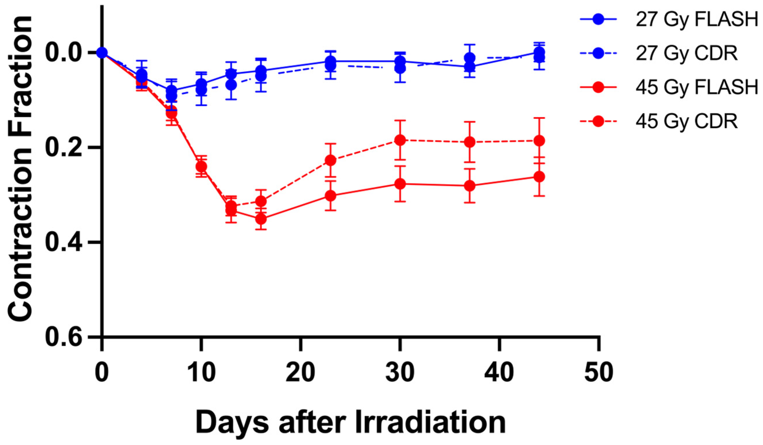
For the assessment of skin response to irradiation, two tattoo dots (ATS-3 General Rodent Tattoo System, AIMS, Hornell, NY, USA) were placed approximately 1 cm apart on the shaved right rear leg of the mice 1 day prior to irradiation. Following anesthetization with a mix of ketamine (80–100 mg/kg, Zoetis, Parsippany, NJ, USA) and xylazine (10 mg/kg, AnaSed®, Dechra Veterinary Products, Elwood, KS, USA), the mice were placed in a custom plastic mouse holder. The tattooed leg was extended through a baffle and secured by taping the foot to the holder, which was then put into a chamber with an inner volume of 1.4 L. The chamber was flushed with 5–10 L/min gas containing 100%, 7%, or 5% oxygen (balance nitrogen), or room air (20.9% oxygen). To allow for the stabilization of oxygen in the tissue, the mice were kept in the chamber for approximately three minutes before irradiation [33]. Prior to and following irradiation, the distance between the dots was measured at 4-day intervals for 24 days and weekly thereafter for up to 45 days post-irradiation. In addition to varying skin oxygen concentration by varying the oxygen content in the inspired gas, partial results of a prior study (Supplementary Figure S2) are included [34]. The mouse strain, age, response metric, radiation dose rates and beam characteristics are the same as used in the current study. Following anesthetization with ketamine–xylazine, hypoxia was achieved by string ligature. The tattoo-bearing leg was FLASH or CDR irradiated to a dose of 27 or 45 Gy for 3 min following leg tourniquet ligation. The ligature was removed immediately following irradiation.

2.4. Evaluation of Oxygen Status

For the evaluation of tissue oxygenation and changes in oxygenation, we used the phosphorescence method, as described by Cao et al., utilizing the phosphorescent probe Oxyphor PdG4 (generously provided by Dr. Brian Pogue’s laboratory, Dartmouth College, Hanover, NH, USA) [22]. Mice legs were subcutaneously injected with 100 μL of a 20 μM concentration of Oxyphor PdG4 ~1 h before irradiation. Oxyphor is used to identify change in pO2 but, due to the injection creating a bolus of Oxyphor under the skin, may not be identical to or resolve the relative concentration of oxygen in skin dermis and epidermis, which is mildly hypoxic in both mouse and human skin [35,36], or the fraction of cells at a particular pO2. Approximately 1 h after PdG4 injection, mice were placed in a custom plastic mouse holder, which was then placed in the 1.4 L chamber and was flushed with gas at a flow rate of 5–10 L/minute. The pO2 was recorded after placing mice in the chamber, and the leg was irradiated with 27 Gy of CDR and FLASH dose rates when the pO2 reached a plateau, i.e., within 3 min. Strong scintillation from the optical fibers during FLASH prevented reliable measurement during irradiation, resulting in a ~0.45 s signal gap and a small underestimation of the minimum pO2 during FLASH. This underestimation has been estimated to be <1 mmHg [22]. Following irradiation, tissue pO2 was continually monitored for up to an additional 5 min. Separate groups of mice of the same age and gender, anesthesia regimen, mouse restraint, and radiation protocol (dose and dose rates) were used for the evaluation of tissue oxygenation and long-term skin contraction following FLASH or CDR irradiation. Tissue pO2 was not evaluated in ligature-induced hypoxia. The evidence of metabolic hypoxia was manifest by the rapid change in the skin color of FVB/N mice within 1.0–1.5 min of ligation. Ando et al. examined mouse skin contraction for up to a year following irradiation. The magnitude of low-LET irradiation contraction was dose-dependent throughout the observation period [37].

2.5. Histology

Six mice per treatment group were sacrificed at the completion of the irradiation experiments for Hematoxylin and Eosin (H&E) and Masson’s trichrome staining. Skin tissue between the two tattoo dots was fixed in 4% paraformaldehyde and subsequently transferred to the Dana Faber/Harvard Cancer Center Specialized Histopathology Services Core (MGH site) for further processing. H&E staining was performed to assess morphological changes, particularly epidermal thickness, while Masson’s trichrome staining was used to evaluate dermal collagen deposition. Furthermore, six mice from each treatment group were sacrificed at 4, 12, and 18 days post-irradiation for Ki-67 immunostaining to evaluate cellular proliferation in the skin. Images of skin sections were acquired at 200× magnification using an Axio Scan.Z1, (Carl Zeiss Microscopy GmbH, Jena, Germany). Epidermal thickness was determined in a blinded manner by calculating the mean length of five evenly spaced lines drawn perpendicular to the skin surface along the H&E-stained sections. Collagen deposition was quantified in Masson’s trichrome-stained sections by measuring the positively stained area relative to a predefined region of interest (ROI). Histological slides were digitally scanned and analyzed using QuPath (version 0.5.1) [38]. Collagen deposition was quantified in Masson’s trichrome-stained sections by measuring the positively stained area relative to a defined region of interest (ROI). ROIs were manually selected from comparable irradiated skin regions while excluding artifacts and non-tissue areas. Positive collagen staining was identified using a pixel-classification algorithm trained on representative manually annotated images containing both positive and negative staining regions. Multiple representative sections from different treatment groups were included during classifier training to account for variability in staining intensity and tissue morphology. The same trained classifier and analysis parameters were applied uniformly to all samples. For each mouse, collagen-positive area (%) was calculated as the ratio of positively stained pixels to total pixels within the ROI. To minimize variability, ROIs were selected using consistent anatomical landmarks and comparable tissue depths across all samples. Proliferative cells were quantified in Ki-67-stained sections by measuring the percentage of Ki-67-positive cells relative to the total number of cells within the ROI. Ki-67-positive nuclei were identified using a classifier trained on representative annotated images based on staining intensity and nuclear morphology. Identical classifier settings were applied across all samples. For each mouse, the Ki-67-positive cell fraction (%) was defined as the ratio of positive cells to total cells within the ROI. To minimize operator bias, identical ROI selection criteria, classifier settings, and analysis pipelines were used across all treatment groups.

2.6. Estimation of Cell Survival from Spectral pO2 Distributions

To estimate the impact of heterogeneous tissue oxygenation on radiation response, hypothetical cellular pO2 distributions were constructed that were consistent with the experimentally measured mean tissue pO2. These distributions were then incorporated into an oxygen-dependent cell survival model following the approach described by Zhu et al. for modeling oxygen depletion effects during irradiation [23]. Here, an iterative approach was used to determine cell survival after irradiation, starting from initial oxygen concentration, delivering a small portion of the dose, adjusting the oxygen concentration for depletion, and delivering the next dose portion until the final delivered dose was reached.
Oxygen depletion during irradiation was described using a G value of 0.38 mmHg/Gy, a half-maximal oxygen sensitization constant of 3.5 mmHg, a maximum OER value of 3, and the dose to reduce the surviving fraction to e−1 (under air conditions) of 1.49 Gy based on preliminary data from an in vitro keratinocyte survival curve. Note, OER values are survival curve model-independent. Oxygen depletion per unit dose (the G value) is determined under known and uniform oxygen conditions as the radiation dose required to render initially oxygenated, radiosensitive cells to a state of maximum radioresistance, where maximum resistance corresponds to the sensitivity of cells irradiated under nitrogen conditions. The required dose is directly dependent on the initial oxygen concentration [39,40,41].
At doses greater than 10 Gy, the LQL model transitions to the hit-target model. Since the present study evaluates responses at doses up to 27 Gy, the cell surviving fraction (SF) was calculated using the single-target/single-hit model:
S F = e D / D 0 ,
where D is the delivered dose and D0 is the dose required to reduce the surviving fraction to e−1. The D0 value was adjusted in accordance with the OER-dependent change in radiosensitivity resulting from oxygen depletion during irradiation. This procedure was repeated for each initial cellular pO2 value within the modeled pO2 distributions.
The cumulative surviving fraction was then calculated by summing the contributions from each cellular subpopulation of the initial pO2 distribution.

2.7. Statistical Analysis

Ten mice were entered into each unique treatment group, i.e., for each combination of oxygen status and dose rate. For the assessment of long-term skin contraction, the distance between the tattoo dots was measured for 45 days post-irradiation. The contraction fraction for FLASH and CDR irradiations (CFFLASH and CFCDR) is defined as (the distance at day 0 minus the distance at day i)/(Distance at day 0), where day “0” is measured 1 day prior to irradiation and day “i” is the measurement day after irradiation. Mean contraction fraction values were calculated as the average of the time points between 30 and 45 days post-irradiation. As the absolute values of the skin contraction for each unique dose rate and breathed oxygen concentration were not identical from experiment to experiment, only studies that included both FLASH and CDR contraction fractions were employed for analysis. For the estimation of the FLASH sparing factors (SFs), the ratios of the CFCDR/CFFLASH and their confidence interval were approximated by the Delta method [42]: S E S F = C F C D R C F F L A S H × S E C D R C F C D R 2 + S E F L A S H C F F L A S H 2 , where CFCDR and CFFLASH represent the mean contraction fractions under CDR and FLASH conditions, while SECDR or SEFLASH denote their standard errors. The statistical significance was calculated by the unpaired two-tailed t-test or one-way ANOVA via GraphPad Prism (version 10). The significance of differences in the magnitude of the sparing factors was evaluated by the unpaired two-tailed t-test. Significance was defined as * p < 0.05; ** p < 0.01; *** p < 0.001.

3. Results

3.1. FLASH but Not CDR Irradiation Rapidly Depletes Tissue Oxygen

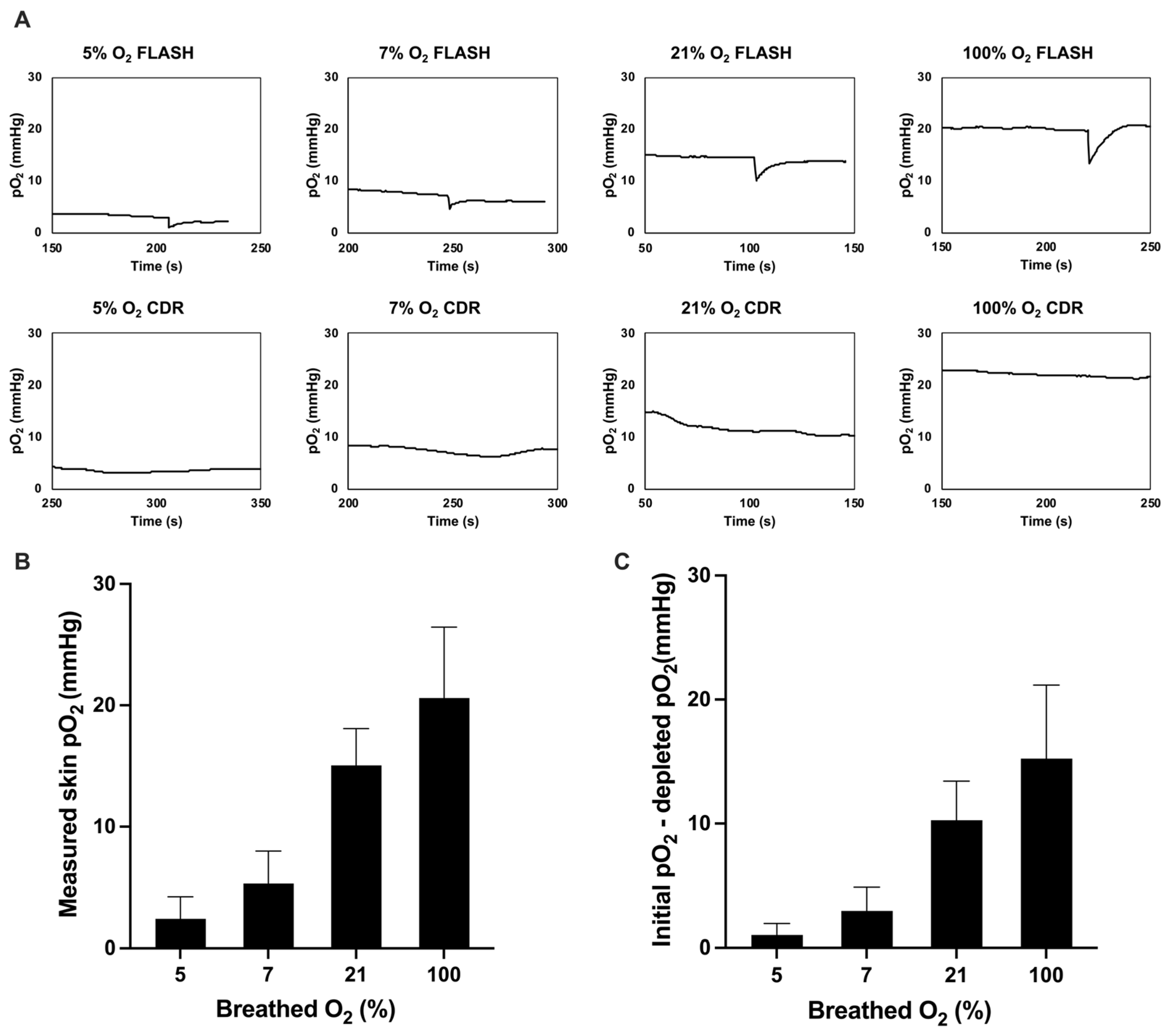
As seen in Figure 1, mean tissue pO2 strongly correlates with the percent oxygen (balance nitrogen) in the inhaled gas, increasing from approximately 15 mmHg in air-breathing mice to 22 mmHg in mice breathing 100% oxygen and decreasing to 4 mmHg in mice breathing 5% oxygen (Figure 1B). The variability in tissue pO2 between mice, as indicated by the confidence intervals, is likely due to both the mouse-to-mouse variability in pO2 and variability in the precise injection site of the oxygen probe. The rapid change in tissue pO2 status in mice breathing gas containing different oxygen concentrations is consistent with polarographically evaluated skin pO2 changes [33]. Differences in dermal and epidermal skin pO2 [36] are not resolved in these measurements.
Rapid FLASH oxygen depletion and the recovery from oxygen depletion is seen in Figure 1A. This figure shows that the quantity of FLASH-depleted oxygen depends on the pre-irradiation tissue oxygen concentration. In mice breathing 5% oxygen, there is substantially less depletable and depleted oxygen than in mice breathing gas containing more oxygen. Figure 1A also shows a rapid decrease in oxygenation during FLASH irradiations. There is no depletion observed during CDR irradiation. The average tissue pO2 remaining after FLASH irradiations is depicted in Figure 1C. Of note, the phosphorescence emission signal arises from approximately 100–150 mm3 of tissue [22], which does not resolve any potential heterogeneities in oxygenation and the percent of tissue at a specific pO2 value.

3.2. Skin Contraction Is Oxygen-Dependent, Increased at High Tissue pO2 and Reduced at Low pO2

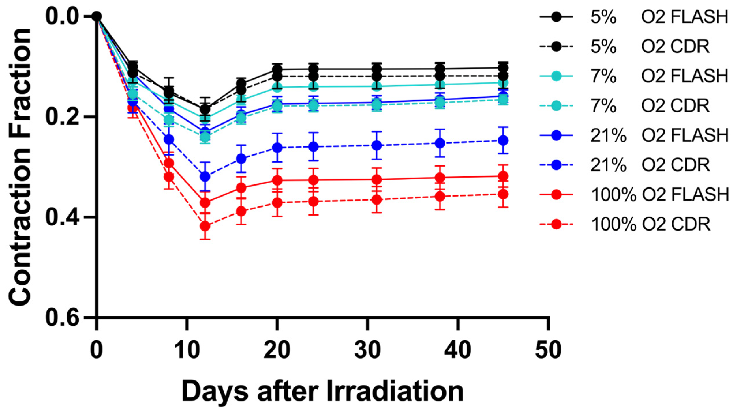
Figure 2 shows the skin contraction fraction in mice receiving 27 Gy CDR or FLASH irradiation while breathing 5% to 100% oxygen. For all oxygen concentrations, acute skin contraction was maximal approximately 12 days post-irradiation, followed by a recovery period and stabilization around day 25. For both CDR and FLASH irradiation, skin contraction increases with increasing tissue oxygen concentration. The most pronounced contraction is seen in CDR irradiated mice breathing 100% oxygen (red dashed curve). Minimal skin contraction is observed in mice breathing 5% oxygen, irrespective of dose rate (black dashed and solid curves). Differences between the FLASH and CDR contraction fractions did not change from day 25 through day 30–45 post-irradiation. Changes in skin contraction following various-sized doses of low-LET irradiation for up to a year post-irradiation reveal a slow gradual increase in contraction [37]. Skin contraction and histology experiments were therefore terminated between day 45 and 60 post-irradiation.

3.3. Dependence of FLASH Skin Sparing on Tissue Oxygenation and Hypoxic Conditions

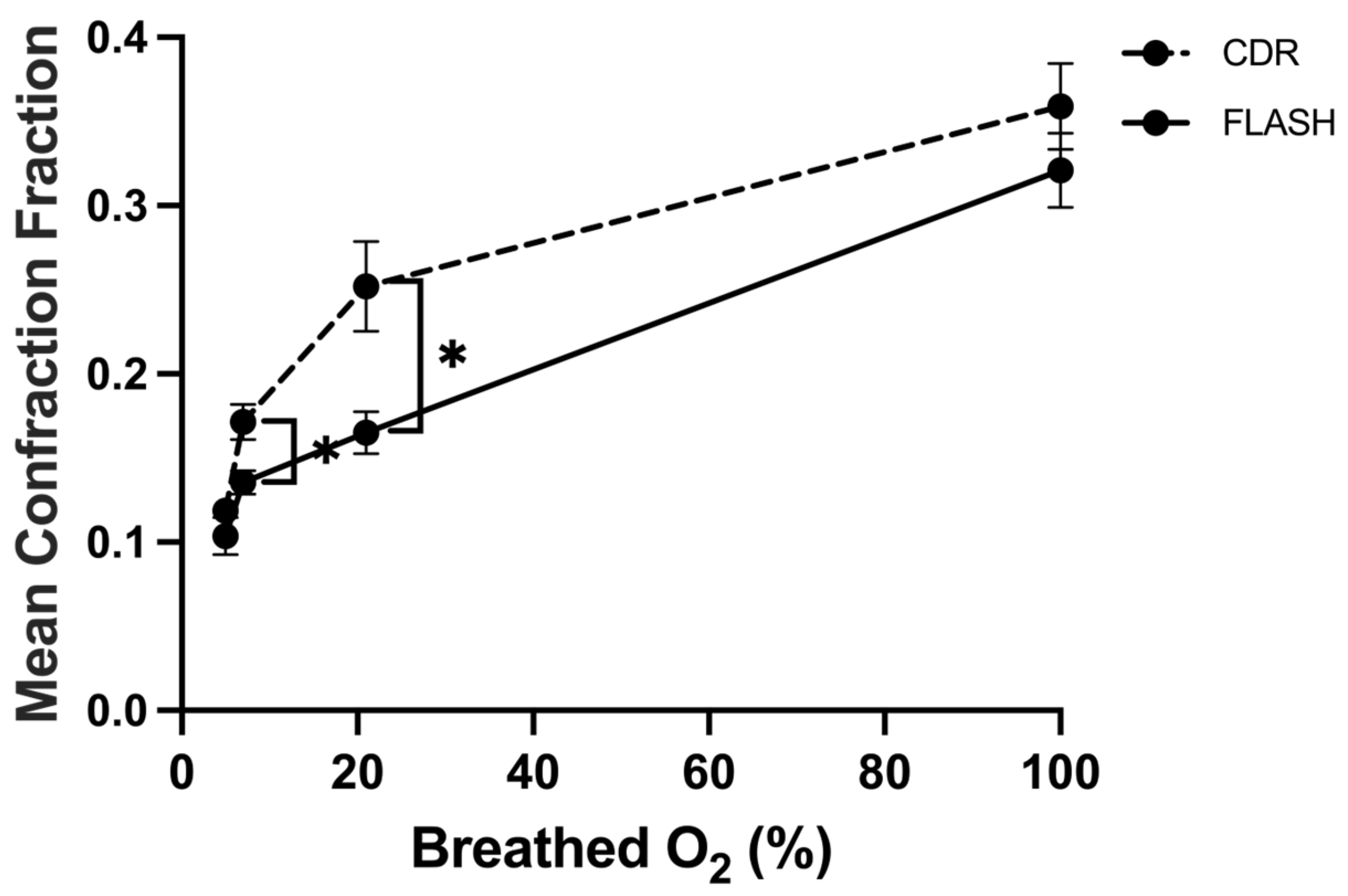
Figure 3 more clearly illustrates the relationship between oxygen concentration, skin contraction, and the FLASH effect. Skin contraction systematically increases with increasing inspired gas oxygen concentration, regardless of dose rate. At low oxygen concentrations, contraction is significantly reduced, as is the relative magnitude of the FLASH effect. In mice breathing 5% oxygen, a sparing effect due to FLASH irradiation is not observed (p = 0.26). FLASH sparing is most pronounced in mice breathing air (20.9% oxygen). The ratio of CDR to FLASH skin contraction decreased from approximately 1.5 under room-air breathing conditions, to approximately 1.25 under 7% oxygen breathing, and to approximately 1.0 under 5% oxygen breathing. FLASH sparing is most pronounced in mice breathing air (20.9% oxygen). In mice breathing 100% oxygen, FLASH sparing is not statistically significant (p = 0.27).
Figure 3 shows that 27 Gy induces significant but minimal contraction in 5% oxygen-breathing mice. Figure 4, which is reproduced from our previous study [34], compares the CDR vs. FLASH effect at 27 Gy and 45 Gy, which induces significantly more skin contraction than 27 Gy. The response of skin irradiated with 27 Gy and 45 Gy that was rendered hypoxic by tourniquet ligation 3 min prior to and during irradiation. Minimal contraction and no FLASH sparing were observed in mice following 27 Gy irradiation, consistent with what was observed following 5% oxygen breathing. A dose of 45 Gy gives rise to significant skin contraction following FLASH and CDR irradiation, but no FLASH protection was observed. Rather, there is a non-significant tendency toward greater contraction following FLASH irradiation (p > 0.05).

3.4. Histologic Changes Parallel the Reduction in Skin Contraction by FLASH vs. CDR Irradiation

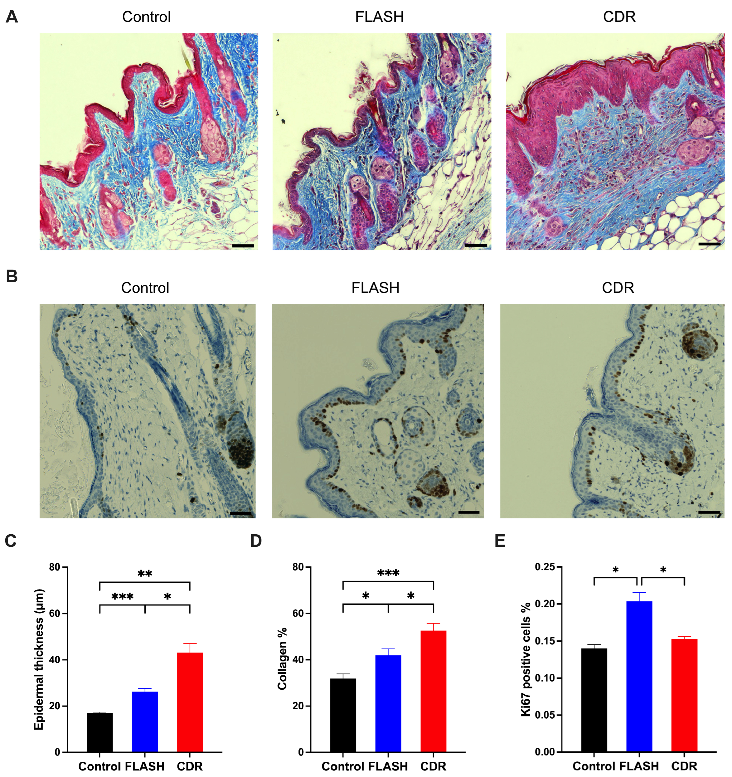
Radiation-induced histologic skin changes are shown in Figure 5. Both FLASH and CDR irradiation significantly increased epidermal thickness and dermal collagen density after 60 days following irradiation; however, FLASH irradiation significantly reduced the magnitude of the tissue changes (p = 0.02 and 0.03, for epidermal thickness and collagen deposition, respectively). Ki-67 immunostaining was performed to assess cellular proliferation at early time points after irradiation (Supplementary Figure S2). The proliferative activity of epidermal progenitor cells is markedly suppressed on day 4 but increases substantially by day 12 and remained elevated on day 18 for both irradiation modalities. Notably, FLASH irradiation preserves a higher fraction of actively proliferating cells compared with CDR. These proliferating cells are predominantly localized within the basal layer of the epidermis. The impact of altered tissue pO2 on histologically evident skin changes is also shown in Supplementary Figure S2. Breathing 5% oxygen significantly preserved epidermal thickness compared with breathing air following either FLASH (p = 0.002) or CDR irradiation (p = 0.002), with no significant difference observed between FLASH and CDR under hypoxic breathing conditions (p = 0.511). In contrast, breathing 100% oxygen significantly increased epidermal thickness following both FLASH (p = 0.011) and CDR irradiation (p = 0.019), and reduced proliferative activity after both FLASH (p = 0.005) and CDR irradiation (p = 0.026).

3.5. Impact of Oxygen Heterogeneity

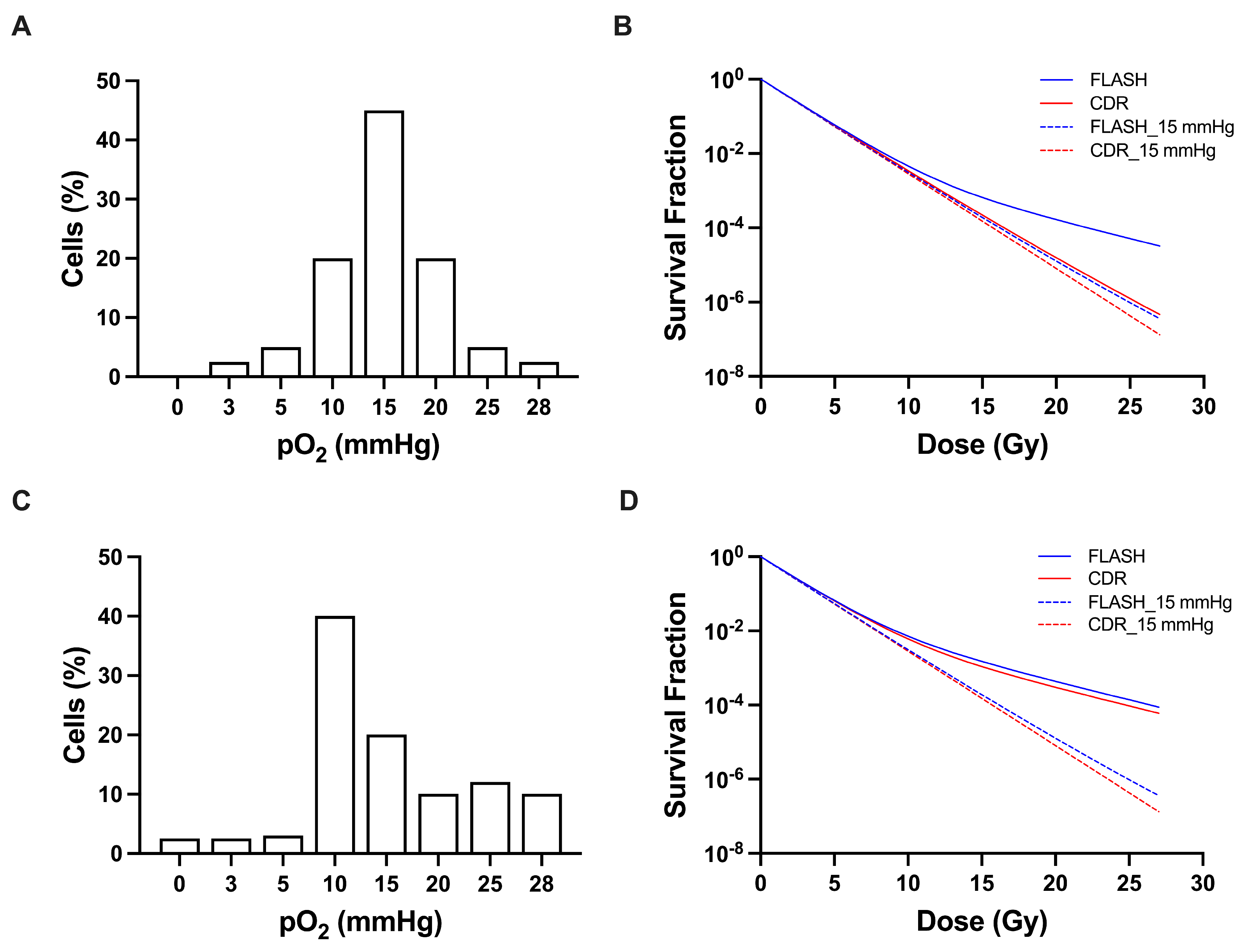
Figure 6 demonstrates the impact of tissue pO2 profiles vs. average tissue pO2 on the response to FLASH vs. CDR irradiation. Figure 6A,C shows two hypothetical pO2 frequency distributions consistent with the measured mean tissue pO2 of 15 mmHg. The distributions were constructed to satisfy the experimentally determined requirement that cells are present both above and below the mean pO2 value, with equal cumulative oxygen content (pO2 × frequency) on either side of the mean. The impact of 27 Gy irradiation on the survival of cells in the pO2 environment in Figure 6A is shown in Figure 6B. The survival of cells in the cell frequency distribution shown in Figure 6C is shown in Figure 6D. The response curves shown in Figure 6B,D were generated using the oxygen-dependent survival model described in the Methods section. These two pO2 distributions shown in Figure 6A,C illustrate how different cellular oxygen distributions can yield the same mean tissue pO2 while producing markedly different predicted responses to FLASH and CDR irradiation.
The presence of pO2 values above and below the mean pO2 are based on the change in skin contraction when tissue pO2 is raised and lowered prior to and during irradiation. The necessity for cells that determine response to reside above and below the mean pO2 value is not revealed by the mean tissue pO2. The decrease in skin contraction observed following the reduction in tissue pO2 by lowering the oxygen content of the breathed gas indicates that not all skin cells are fully hypoxic. Conversely, the increased skin response observed when tissue pO2 is elevated demonstrates that not all cells are fully oxygenated. The tissue contains cellular subpopulations existing at both higher and lower oxygen tensions, with the overall distribution yielding a mean tissue pO2 of approximately 15 mmHg.
The experimental findings are consistent with the distribution illustrated in Figure 6A, but not with the distribution shown in Figure 6C. The distribution in panel C contains anoxic cellular populations similar to those commonly observed in tumors [14,15].

4. Discussion

The increase in the skin sensitivity in mice breathing 100% oxygen and the decrease in sensitivity in mice breathing 7% and 5% oxygen versus air (20.9% oxygen) demonstrates that skin is neither fully oxygenated nor fully hypoxic. A dose of 27 Gy may be expected to deplete approximately 8–10 mmHg of intracellular oxygen [39,40,41]. As previously noted, sensitivity to radiation changes rapidly over the range of 0–10 mmHg, particularly over the 0–5 mmHg range mmHg pO2, with the half-maximum oxygen sensitization at ~3.5 mmHg. Over the pO2 range of ~15 mmHg to 760 mmHg (100% oxygen), radiation sensitivity is approximately three times greater than under very low or anoxic conditions, i.e., OER~3. Accordingly, if all cells resided in the mean tissue 15 mmHg pO2 environment, a FLASH tissue sparing effect would not be achieved by a dose of 27 Gy. It should be noted that the dose of radiation that deletes cellular oxygen is not dependent on the sensitivity of cells or the model which best fits the surviving fraction of cells. The surviving fraction of cells at the point when O2 is deleted is dependent on the cells’ radiation sensitivity. For cells residing in the sub-15 mmHg pO2 range, a dose of 27 Gy is sufficient to induce radiobiologic hypoxia.
As the OER relationship is independent of the response fitting model and the linear–quadratic model overestimates tissue response at doses greater than 10 Gy [43], a log-linear dose–response (hit-target) model was used to illustrate the impact of the different sub-15 mmHg pO2 distributions on cell response (Figure 6). Based on the average pO2 value of 15 mmHg, i.e., assuming all cells reside at the average pO2 value, there is essentially no difference in response to FLASH and CDR irradiation for a dose up 27 Gy, as seen in Figure 6B,D (dashed lines). However, if the pO2 distribution of the cells is as shown in Figure 6A, FLASH yields a significant tissue sparing effect vs. CDR irradiation (solid lines). On the other hand, the distribution in Figure 6C yields substantial but identical tissue sparing for both CDR and FLASH irradiation (solid lines) compared to all cells residing at the average of 15 mmHg pO2 (dashed lines). Thus, the pO2 distribution of cells in the 0–15 mmHg range, not the mean tissue pO2, governs the response of FLASH vs. CDR irradiation for doses at least up to 27 Gy. The presence of cells or tissue existing in the total absence of oxygen, as seen in Figure 6C, is more characteristic of tumors [14,15], for which a FLASH sparing effect has not been reported.
A significant finding of this study is that the percent decrease in the FLASH vs. CDR contraction in mice breathing 7% oxygen is smaller than in mice breathing 20.9% oxygen (p = 0.014). In mice breathing 5% oxygen, CDR contraction is minimal, ~11%, with no significant FLASH tissue sparing (p = 0.26). That is, in the absence or near absence of oxygen, there is no FLASH sparing effect. As seen in Figure 4, under conditions of tourniquet hypoxia, there is no FLASH sparing effect following a dose of 27 Gy or 45 Gy [34]. While the previously reported study suggests that metabolically induced hypoxia fails to yield a FLASH effect, presumably due to the absence of oxygen, the metabolic consequences of ligation and breathing low-oxygen-containing gas differ. Tourniquet ischemia not only blocks the supply of oxygen, but the sole remaining source of energy, i.e., glucose [44]. Additionally, the clearance of catabolites such as lactic acid and carbonic acid produced by the aerobic metabolism of residual tissue oxygen at the time of ligation is blocked, likely leading to intracellular acidification. The similarity in the skin contraction response to FLASH and CDR irradiation following 27 Gy irradiation under 5% oxygen breathing conditions, and tourniquet hypoxia, suggests that tourniquet-dependent metabolic changes do not abrogate the impact of oxygen deprivation. That suggests that the absence of a FLASH sparing effect following 45 Gy is also due to oxygen deprivation.
In addition to the quantitative dose response metric used in this study, i.e., skin contraction, histologic changes similarly reflect the impact of inspired oxygen concentration on skin response to radiation. Epidermal thickening is more pronounced in mice breathing oxygen, and less pronounced in mice breathing low-oxygen-content gas. In mice breathing oxygen-reduced gas and 100% oxygen, the differences between the FLASH and CDR groups did not achieve statistical significance. The histologic results reported here are consistent with other extensive studies [45], which found that, for an exposure of mouse legs to a single dose of 30 and 45 Gy, FLASH produced fewer severe toxicities and less epidermal skin thickening than CDR irradiation. RNA-seq analyses of murine skin and bone revealed pathways that were upregulated by CDR proton irradiation yet were left unaltered by FLASH irradiation, such as apoptosis signaling and keratinocyte differentiation in skin [45].
In related studies, Sesink et al. designed studies to evaluate the modifying effect of FLASH vs. CDR irradiation under air- and >90% oxygen-breathing conditions [27], and Hansen et al. evaluated skin response under air and clamp hypoxic conditions [28]. For the air and clamp hypoxia study, the endpoint was a specified level of acute skin damage up to 28 days post-irradiation. These carefully designed studies reported OER values of air breathing vs. clamp hypoxia of 1.38 for FLASH irradiation and 2.05 for CDR irradiation for the same acute skin toxicity score. The FLASH dose-modifying factor was 1.43 under normal air-breathing conditions and 0.96 under clamp hypoxic conditions. The results of those studies are complementary to the study presented here.
In the study by Sesink et al., abdominal irradiation lethality and skin toxicity were evaluated for up to 30 days post-irradiation [27]. Interestingly, the impact of altered oxygen breathing conditions appeared to differ between the skin and gastrointestinal studies, although the experiments were performed in different mouse strains. Oxygen breathing conditions did not appear to significantly affect the acute skin response.
In the study reported here for late skin contraction, a small FLASH effect under oxygen-breathing conditions was also observed, but it did not achieve statistical significance at a dose of 27 Gy. The results provide strong evidence that oxygen depletion is the mechanism of FLASH protection in mouse skin. At reduced tissue oxygenation, FLASH tissue sparing is reduced and, in the absence of oxygen, a FLASH sparing effect is not observed. This does not preclude other potential mechanisms of FLASH normal tissue sparing to play a role, for example, in other tissue, such as has been reported in the brain [46].
Montay-Gruel et al. [47] reported changes in the brains of mice exposed to FLASH and CDR irradiation, for up to 6 months following irradiation. Following 10 Gy CDR irradiation, substantial cognitive, behavioral, and morphological changes were observed, but not following FLASH irradiation. FLASH protective effect was also observed at 12 Gy, although not at 14 Gy. Interestingly, carbogen breathing eliminated FLASH sparing, suggesting a role of oxygen in the FLASH effect. The investigators further examined H2O2 production following CDR and FLASH irradiation in a cell-free system equilibrated with 4% oxygen. Upon exposure to 20–80 Gy in 10 Gy increments, the yield of H2O2 was lower by FLASH compared to CDR irradiation. However, no significant difference in H2O2 production was observed following 10 Gy FLASH and 10 Gy CDR irradiation. These results appear to substantially differ from the results of the current study in skin. As summarized by Bohlen [48], and in our previous skin studies [34], the minimum dose at which FLASH sparing becomes evident substantially exceeds 10 Gy. Moreover, the relationship between oxygen concentration and cell sensitivity is well known, and the results of the current study are quantitatively consistent with the well-established oxygen radiation sensitivity relationship. In the brain, if one assumes that FLASH tissue sparing is due to the reduced H2O2 production, then one might assume that brain sparing would be minimal at 10 Gy, a dose at which FLASH and CDR H2O2 production do not appear to differ, and become more pronounced at higher doses where the difference in H2O2 production is significant. However, such a relationship was not observed. The significant difference in the skin and brain threshold doses for a FLASH effect suggests the mechanisms of the FLASH effect in the brain and skin likely differ. A determination of the role of oxygen in FLASH brain sparing could become clearer if a reduction in brain pO2 reduced the magnitude of FLASH sparing.
Vaupel et al. reported median normal tissue pO2 values of 24 to 50 mmHg in liver, brain, skeletal muscle and gastric mucosa, with 7.5%, 10%, 8% and 0% of the recorded values in the 5–10 mmHg range, respectively [14,15]. Median pO2 values in normal human brain and subcutis (24 mmHg and 50 mmHg, respectively) substantially exceed the oxygen concentration that could be depleted by a clinically relevant dose of radiation. However, approximately 2% of recorded brain oxygenation values are less than 5 mmHg, and 2% of subcutaneous tissue values lie between 5 and 10 mmHg [14]. As demonstrated in [23], employing radiation sensitivity parameters of breast tissue, the presence of the 1% mildly hypoxic cells would increase the dose to yield the same level of cell survival from 20 Gy CDR to 27 Gy FLASH irradiation [23].
The long-established and well-defined quantitative relationship between pO2 and cell and tissue response to radiation, the presence of skin cells residing in a low pO2 environment, the rate of intracellular oxygen depletion in mammalian cells, and the absence of a FLASH effect in the absence of oxygen provide compelling evidence that FLASH oxygen depletion is the mechanism of normal tissue sparing in mouse skin.

5. Conclusions

Mean normal tissue pO2 values do not reveal the fraction of tissue on the threshold of hypoxia that may be rendered resistant by ultra-high dose-rate radiation at dose levels with clinical relevance. Variation in radiation-induced skin contraction in response to changes in skin oxygenation show that skin is neither fully radiobiologically hypoxic nor oxygenated. A reduction in mean tissue pO2 reduces skin sparing, and, under severe hypoxic conditions, the FLASH sparing effect is eliminated. An evaluation of the low-pO2 profiles of brain, heart and other normal tissues will be of value in determining if these tissues may exhibit radiation resistance upon exposure to FLASH irradiation.

Supplementary Materials

The following supporting information can be downloaded at: https://www.mdpi.com/article/10.3390/cancers18122011/s1, Figure S1: Mice setup, delivery parameters, and beam flatness; Figure S2: Histological alterations in mice breathing 5–100% oxygen following a single dose of 27 Gy irradiation at FLASH or CDR.

Author Contributions

Conceptualization, Q.H., L.G., P.H. and J.S.; methodology, Q.H., L.G. and P.H.; software, Q.H., E.C. and J.S.; validation: Q.H. and P.H.; formal analysis: Q.H. and L.G.; investigation, Q.H., L.G., P.H., E.C., B.R., T.R.G., J.P.S., M.A.C. and J.S.; resources, Q.H. and P.H.; data curation, Q.H. and P.H.; writing—original draft preparation, Q.H. and L.G.; writing—review and editing, Q.H., L.G., P.H., E.C., B.R. and J.S.; visualization, Q.H.; supervision, P.H., L.G. and J.S.; funding acquisition, J.S. All authors have read and agreed to the published version of the manuscript.

Funding

This work was funded by the NIH/NCI, grant number R01CA226419: “Using experimentally-guided multi-scale modeling to determine the mechanism of FLASH tissue sparing”.

Institutional Review Board Statement

The study was conducted in accordance with the Declaration of Helsinki, and approved by the IACUC (Institutional Animal Care and Use Committee) at Massachusetts General Hospital (protocol code 2019N000022 and 8 March 2019).

Data Availability Statement

The data presented in this study are available on request from the corresponding author.

Acknowledgments

We thank the Specialized Histopathology Core Facility of the Dana-Farber/Harvard Cancer Center for support (P30 CA06516). We also thank the Steele’s lab, Kebert Joseph, and Tsz Yui (Zoe) Chan for their support with the experiments. T.R.G. acknowledges funding from the European Union’s Horizon Europe Research and Innovation Programme under a Marie Skłodowska-Curie Postdoctoral Fellowship (Grant Agreement No. 101146938).

Conflicts of Interest

The authors declare no conflicts of interest.

Abbreviations

The following abbreviations are used in this manuscript:
FLASHultra-high dose-rate irradiation
CDRconventional dose-rate
OERoxygen enhancement ratio
LETlinear energy transfer
SOBPSpread-Out Bragg Peak
H&EHematoxylin and Eosin
CFcontraction fraction
SFsparing factors
DMFdose modifying factor

References

  1. Bryant, A.K.; Banegas, M.P.; Martinez, M.E.; Mell, L.K.; Murphy, J.D. Trends in Radiation Therapy among Cancer Survivors in the United States, 2000-2030. Cancer Epidemiol. Biomark. Prev. 2017, 26, 963–970. [Google Scholar] [CrossRef] [PubMed]
  2. Zeisberg, M.; Kalluri, R. Cellular mechanisms of tissue fibrosis. 1. Common and organ-specific mechanisms associated with tissue fibrosis. Am. J. Physiol. Cell Physiol. 2013, 304, C216–C225. [Google Scholar] [CrossRef] [PubMed]
  3. Authors on behalf of ICRP; Stewart, F.A.; Akleyev, A.V.; Hauer-Jensen, M.; Hendry, J.H.; Kleiman, N.J.; Macvittie, T.J.; Aleman, B.M.; Edgar, A.B.; Mabuchi, K.; et al. ICRP publication 118: ICRP statement on tissue reactions and early and late effects of radiation in normal tissues and organs–threshold doses for tissue reactions in a radiation protection context. Ann. ICRP 2012, 41, 1–322. [Google Scholar] [CrossRef] [PubMed]
  4. Cheng, J.; Dong, J.; Fang, Y.; Zhang, X.; Dang, X. Radiation-induced skin injury: A review of pathophysiology, assessment, management, and re-irradiation protocols. Front Oncol. 2025, 15, 1736717. [Google Scholar] [CrossRef] [PubMed]
  5. Favaudon, V.; Caplier, L.; Monceau, V.; Pouzoulet, F.; Sayarath, M.; Fouillade, C.; Poupon, M.-F.; Brito, I.; Hupé, P.; Bourhis, J.; et al. Ultrahigh dose-rate FLASH irradiation increases the differential response between normal and tumor tissue in mice. Sci. Transl. Med. 2014, 6, 245ra93. [Google Scholar] [CrossRef] [PubMed]
  6. Chow, J.C.L.; Ruda, H.E. Mechanisms of Action in FLASH Radiotherapy: A Comprehensive Review of Physicochemical and Biological Processes on Cancerous and Normal Cells. Cells 2024, 13, 835. [Google Scholar] [CrossRef] [PubMed]
  7. Ma, Y.; Zhang, W.; Zhao, Z.; Lv, J.; Chen, J.; Yan, X.; Lin, X.; Zhang, J.; Wang, B.; Gao, S.; et al. Current views on mechanisms of the FLASH effect in cancer radiotherapy. Natl. Sci. Rev. 2024, 11, nwae350. [Google Scholar] [CrossRef] [PubMed]
  8. Spitz, D.R.; Buettner, G.R.; Petronek, M.S.; St-Aubin, J.J.; Flynn, R.T.; Waldron, T.J.; Limoli, C.L. An integrated physico-chemical approach for explaining the differential impact of FLASH versus conventional dose rate irradiation on cancer and normal tissue responses. Radiother. Oncol. 2019, 139, 23–27. [Google Scholar] [CrossRef] [PubMed]
  9. Manring, H.R.; Fleming, J.L.; Meng, W.; Gamez, M.; Blakaj, D.M.; Chakravarti, A. FLASH Radiotherapy: From In Vivo Data to Clinical Translation. Hematol. Oncol. Clin. N. Am. 2025, 39, 237–255. [Google Scholar] [CrossRef] [PubMed]
  10. Alexander, P. On the mode of action of some treatments that influence the radiation sensitivity of cells. Trans. N. Y. Acad. Sci. 1962, 24, 966–978. [Google Scholar] [CrossRef] [PubMed]
  11. Grimes, D.R.; Partridge, M. A mechanistic investigation of the oxygen fixation hypothesis and oxygen enhancement ratio. BioMed. Phys. Eng. Express 2015, 1, 045209. [Google Scholar] [CrossRef] [PubMed]
  12. Tinganelli, W.; Sokol, O.; Quartieri, M.; Puspitasari, A.; Dokic, I.; Abdollahi, A.; Durante, M.; Haberer, T.; Debus, J.; Boscolo, D.; et al. Ultra-High Dose Rate (FLASH) Carbon Ion Irradiation: Dosimetry and First Cell Experiments. Int. J. Radiat. Oncol. Biol. Phys. 2022, 112, 1012–1022. [Google Scholar] [CrossRef] [PubMed]
  13. Yoshida, Y.; Suda, H.; Tashiro, M.; Yusa, K.; Nakao, M.; Ando, K.; Takahashi, A.; Ohno, T. Carbon ion FLASH irradiation reduces acute skin toxicity compared with conventional dose rate irradiation. Sci. Rep. 2025, 16, 2307. [Google Scholar] [CrossRef] [PubMed]
  14. Vaupel, P.; Kallinowski, F.; Okunieff, P. Blood flow, oxygen and nutrient supply, and metabolic microenvironment of human tumors: A review. Cancer Res. 1989, 49, 6449–6465. [Google Scholar] [PubMed]
  15. Vaupel, P.; Flood, A.B.; Swartz, H.M. Oxygenation Status of Malignant Tumors vs. Normal Tissues: Critical Evaluation and Updated Data Source Based on Direct Measurements with pO2 Microsensors. Appl. Magn. Reson. 2021, 52, 1451–1479. [Google Scholar] [CrossRef]
  16. Fouillade, C.; Curras-Alonso, S.; Giuranno, L.; Quelennec, E.; Heinrich, S.; Bonnet-Boissinot, S.; Beddok, A.; Leboucher, S.; Karakurt, H.U.; Bohec, M.; et al. FLASH Irradiation Spares Lung Progenitor Cells and Limits the Incidence of Radio-induced Senescence. Clin. Cancer Res. 2020, 26, 1497–1506. [Google Scholar] [CrossRef] [PubMed]
  17. Montay-Gruel, P.; Bouchet, A.; Jaccard, M.; Patin, D.; Serduc, R.; Aim, W.; Petersson, K.; Petit, B.; Bailat, C.; Bourhis, J.; et al. X-rays can trigger the FLASH effect: Ultra-high dose-rate synchrotron light source prevents normal brain injury after whole brain irradiation in mice. Radiother. Oncol. 2018, 129, 582–588. [Google Scholar] [CrossRef] [PubMed]
  18. Limoli, C.L.; Kramár, E.A.; Almeida, A.; Petit, B.; Grilj, V.; Baulch, J.E.; Ballesteros-Zebadua, P.; Loo, B.W.; Wood, M.A.; Vozenin, M.-C. The sparing effect of FLASH-RT on synaptic plasticity is maintained in mice with standard fractionation. Radiother. Oncol. 2023, 186, 109767. [Google Scholar] [CrossRef] [PubMed]
  19. Jansen, J.; Knoll, J.; Beyreuther, E.; Pawelke, J.; Skuza, R.; Hanley, R.; Brons, S.; Pagliari, F.; Seco, J. Does FLASH deplete oxygen? Experimental evaluation for photons, protons, and carbon ions. Med. Phys. 2021, 48, 3982–3990. [Google Scholar] [CrossRef] [PubMed]
  20. Boscolo, D.; Scifoni, E.; Durante, M.; Krämer, M.; Fuss, M.C. May oxygen depletion explain the FLASH effect? A chemical track structure analysis. Radiother. Oncol. 2021, 162, 68–75. [Google Scholar] [CrossRef] [PubMed]
  21. El Khatib, M.; Motlagh, A.O.; Beyer, J.N.; Troxler, T.; Allu, S.R.; Sun, Q.; Burslem, G.M.; Vinogradov, S.A. Direct Measurements of FLASH-Induced Changes in Intracellular Oxygenation. Int. J. Radiat. Oncol. Biol. Phys. 2024, 118, 781–789. [Google Scholar] [CrossRef] [PubMed]
  22. Cao, X.; Zhang, R.; Esipova, T.V.; Allu, S.R.; Ashraf, R.; Rahman, M.; Gunn, J.R.; Bruza, P.; Gladstone, D.J.; Williams, B.B.; et al. Quantification of Oxygen Depletion During FLASH Irradiation In Vitro and In Vivo. Int. J. Radiat. Oncol. Biol. Phys. 2021, 111, 240–248. [Google Scholar] [CrossRef] [PubMed]
  23. Zhu, H.; Schuemann, J.; Zhang, Q.; Gerweck, L.E. Modeling the impact of tissue oxygen profiles and oxygen depletion parameter uncertainties on biological response and therapeutic benefit of FLASH. Med. Phys. 2024, 51, 670–681. [Google Scholar] [CrossRef] [PubMed]
  24. Singers Sørensen, B.; Krzysztof Sitarz, M.; Ankjærgaard, C.; Johansen, J.; Andersen, C.E.; Kanouta, E.; Overgaard, C.; Grau, C.; Poulsen, P. In vivo validation and tissue sparing factor for acute damage of pencil beam scanning proton FLASH. Radiother. Oncol. 2022, 167, 109–115. [Google Scholar] [CrossRef] [PubMed]
  25. Tavakkoli, A.D.; Clark, M.A.; Kheirollah, A.; Sloop, A.M.; Soderholm, H.E.; Daniel, N.J.; Petusseau, A.F.; Huang, Y.H.; Thomas, C.R.; Jarvis, L.A.; et al. Anesthetic Oxygen Use and Sex Are Critical Factors in the FLASH Sparing Effect. Adv. Radiat. Oncol. 2024, 9, 101492. [Google Scholar] [CrossRef] [PubMed]
  26. Soto, L.A.; Casey, K.M.; Wang, J.; Blaney, A.; Manjappa, R.; Breitkreutz, D.; Skinner, L.; Dutt, S.; Ko, R.B.; Bush, K.; et al. FLASH Irradiation Results in Reduced Severe Skin Toxicity Compared to Conventional-Dose-Rate Irradiation. Radiat. Res. 2020, 194, 618–624. [Google Scholar] [CrossRef] [PubMed]
  27. Sesink, A.; Soutter, L.; Geyer, R.W.; Böhlen, T.T.; Bailat, C.; Grilj, V. Impact of increased oxygen concentration on the FLASH sparing effect in mice is tissue dependent. Br. J. Radiol. 2026, 99, 254–262. [Google Scholar] [CrossRef] [PubMed]
  28. Hansen, A.H.; Poulsen, P.R.; Kristensen, L.; Møller, V.V.; Sinha, P.M.; Johansen, J.G.; Sørensen, B.S. Impact of oxygen deprivation on the FLASH effect for skin toxicity in a murine model. Radiother. Oncol. 2026, 214, 111277. [Google Scholar] [CrossRef] [PubMed]
  29. Hall, E.J.; Giaccia, A.J. Radiobiology for the Radiologist, 8th ed.; Lippincott Williams & Wilkins (LWW): Philadelphia, PA, USA, 2018. [Google Scholar]
  30. Hachadorian, R.; Cascio, E.; Schuemann, J. Increased flexibility and efficiency of a double-scattering FLASH proton beamline configuration forin vivoSOBP radiotherapy treatments. Phys. Med. Biol. 2023, 68. [Google Scholar] [CrossRef] [PubMed]
  31. Cascio, E.W.; Gottschalk, B. Using a 3D printer for 2D beam profile measurements in proton radiotherapy. arXiv 2019, arXiv:1908.03763. [Google Scholar]
  32. Zhang, Q.; Cascio, E.; Li, C.; Yang, Q.; Gerweck, L.E.; Huang, P.; Gottschalk, B.; Flanz, J.; Schuemann, J. FLASH Investigations Using Protons: Design of Delivery System, Preclinical Setup and Confirmation of FLASH Effect with Protons in Animal Systems. Radiat. Res. 2020, 194, 656–664. [Google Scholar] [CrossRef] [PubMed]
  33. Gerweck, L.E.; Hetzel, F.W. PO2 in irradiated versus nonirradiated tumors of mice breathing oxygen at normal and elevated pressure. Int. J. Radiat. Oncol. Biol. Phys. 1995, 32, 695–701. [Google Scholar] [CrossRef] [PubMed]
  34. Zhang, Q.; Gerweck, L.E.; Cascio, E.; Yang, Q.; Huang, P.; Niemierko, A.; Bertolet, A.; Nesteruk, K.P.; McNamara, A.; Schuemann, J. Proton FLASH effects on mouse skin at different oxygen tensions. Phys. Med. Biol. 2023, 68, 055010. [Google Scholar] [CrossRef] [PubMed]
  35. Koch, C.J. Measurement of absolute oxygen levels in cells and tissues using oxygen sensors and 2-nitroimidazole EF5. Methods Enzymol. 2002, 352, 3–31. [Google Scholar] [CrossRef] [PubMed]
  36. Bedogni, B.; Welford, S.M.; Cassarino, D.S.; Nickoloff, B.J.; Giaccia, A.J.; Powell, M.B. The hypoxic microenvironment of the skin contributes to Akt-mediated melanocyte transformation. Cancer Cell 2005, 8, 443–454. [Google Scholar] [CrossRef] [PubMed]
  37. Ando, K.; Yoshida, Y.; Hirayama, R.; Koike, S.; Matsufuji, N. Dose- and LET-dependent changes in mouse skin contracture up to a year after either single dose or fractionated doses of carbon ion or gamma rays. J. Radiat. Res. 2022, 63, 221–229. [Google Scholar] [CrossRef] [PubMed]
  38. Bankhead, P.; Loughrey, M.B.; Fernández, J.A.; Dombrowski, Y.; McArt, D.G.; Dunne, P.D.; McQuaid, S.; Gray, R.T.; Murray, L.J.; Coleman, H.G.; et al. QuPath: Open source software for digital pathology image analysis. Sci. Rep. 2017, 7, 16878. [Google Scholar] [CrossRef] [PubMed]
  39. Ling, C.C.; Michaels, H.B.; Epp, E.R.; Peterson, E.C. Oxygen diffusion into mammalian cells following ultrahigh dose rate irradiation and lifetime estimates of oxygen-sensitive species. Radiat. Res. 1978, 76, 522–532. [Google Scholar] [CrossRef] [PubMed]
  40. Nias, A.H.; Swallow, A.J.; Keene, J.P.; Hodgson, B.W. Effects of pulses of radiation on the survival of mammalian cells. Br. J. Radiol. 1969, 42, 553. [Google Scholar] [CrossRef] [PubMed]
  41. Michaels, H.B.; Epp, E.R.; Ling, C.C.; Peterson, E.C. Oxygen sensitization of CHO cells at ultrahigh dose rates: Prelude to oxygen diffusion studies. Radiat. Res. 1978, 76, 510–521. [Google Scholar] [CrossRef] [PubMed]
  42. Bera, A.K.; Koley, M. A History of the Delta Method and Some New Results. Sankhya B 2023, 85, 272–306. [Google Scholar] [CrossRef]
  43. Kirkpatrick, J.P.; Meyer, J.J.; Marks, L.B. The Linear-Quadratic Model Is Inappropriate to Model High Dose per Fraction Effects in Radiosurgery. Semin Radiat. Oncol. 2008, 18, 240–243. [Google Scholar] [CrossRef] [PubMed]
  44. Gerweck, L.E.; Seneviratne, T.; Gerweck, K.K. Energy status and radiobiological hypoxia at specified oxygen concentrations. Radiat. Res. 1993, 135, 69–74. [Google Scholar] [CrossRef]
  45. Velalopoulou, A.; Karagounis, I.V.; Cramer, G.M.; Kim, M.M.; Skoufos, G.; Goia, D.; Hagan, S.; Verginadis, I.I.; Shoniyozov, K.; Chiango, J.; et al. FLASH Proton Radiotherapy Spares Normal Epithelial and Mesenchymal Tissues While Preserving Sarcoma Response. Cancer Res. 2021, 81, 4808–4821. [Google Scholar] [CrossRef] [PubMed]
  46. Montay-Gruel, P.; Acharya, M.M.; Gonçalves Jorge, P.; Petit, B.; Petridis, I.G.; Fuchs, P.; Leavitt, R.; Petersson, K.; Gondré, M.; Ollivier, J.; et al. Hypofractionated FLASH-RT as an Effective Treatment against Glioblastoma that Reduces Neurocognitive Side Effects in Mice. Clin. Cancer Res. 2021, 27, 775–784. [Google Scholar] [CrossRef] [PubMed]
  47. Montay-Gruel, P.; Acharya, M.M.; Petersson, K.; Alikhani, L.; Yakkala, C.; Allen, B.D.; Ollivier, J.; Petit, B.; Jorge, P.G.; Syage, A.R.; et al. Long-term neurocognitive benefits of FLASH radiotherapy driven by reduced reactive oxygen species. Proc. Natl. Acad. Sci. USA 2019, 116, 10943–10951. [Google Scholar] [CrossRef] [PubMed]
  48. Böhlen, T.T.; Germond, J.F.; Bourhis, J.; Vozenin, M.C.; Ozsahin, E.M.; Bochud, F.; Bailat, C.; Moeckli, R. Normal Tissue Sparing by FLASH as a Function of Single-Fraction Dose: A Quantitative Analysis. Int. J. Radiat. Oncol. Biol. Phys. 2022, 114, 1032–1044. [Google Scholar] [CrossRef] [PubMed]
Figure 1. Skin pO2 in mice treated with 27 Gy irradiation while breathing 5%, 7%, 21%, and 100% oxygen. (A) Skin pO2 of representative mice breathing 5%, 7%, 21%, and 100% oxygen following 27 Gy FLASH (top row) or CDR (bottom row) irradiation. Quantification of (B) initial pO2 and (C) initial pO2 minus depleted pO2 immediately following FLASH irradiation in mice breathing 5%, 7%, 21%, and 100% oxygen. The initial pO2 was the average pO2 for approximately 5 s prior to the rapid pO2 decrease associated with FLASH irradiation. n = 47. Data are presented as Mean ± SD.
Cancers 18 02011 g001
Figure 2. Skin contraction as a function of time following 27 Gy irradiation at FLASH and CDR dose rates in mice breathing 5%, 7%, 21%, and 100% oxygen. Solid curves are for FLASH irradiations, dashed curves are CDR irradiations. n = 165, data are presented as Mean ± SEM.
Cancers 18 02011 g002
Figure 3. Mean skin contraction values in mice breathing 5–100% oxygen at 30–45 days following a single dose of 27 Gy irradiation. Solid curves are for FLASH irradiations; dashed curves are CDR irradiations. n = 165, statistical analysis by unpaired two-tailed t-test, * p < 0.05. Data are presented as Mean ± SEM.
Cancers 18 02011 g003
Figure 4. Skin contraction following 27 and 45 Gy under tourniquet hypoxic conditions. Solid curves are for FLASH irradiations; dashed curves are CDR irradiations. n = 40, data are presented as Mean ± SEM.
Cancers 18 02011 g004
Figure 5. Histologic alterations in mice receiving 27 Gy FLASH or CDR irradiation. Representative images of Masson’s trichrome- (A) or Ki67- (B) stained skin tissue in mice breathing room air for the unirradiated control or irradiated with 27 Gy at FLASH vs. CDR dose rates, scale bar = 50 µm. Collagen is stained blue in the Masson’s trichrome sections, and Ki-67-positive nuclei are stained black in the Ki67 sections. Quantification of epidermal thickness (C), and collagen deposition (D) and proliferation (E). n = 6 for all panels, statistical analysis by one-way ANOVA, * p < 0.05; ** p < 0.01; *** p < 0.001. Data are presented as Mean ± SEM.
Cancers 18 02011 g005
Figure 6. Profiles of skin oxygenation (A,C) and corresponding effects of CDR and FLASH irradiation (B,D). Solid curves represent cell survival based on the full spectrum of cell pO2 values shown in panels (A,C), whereas dashed curves represent survival at the measured mean pO2 value of 15 mmHg following FLASH (blue) or CDR (red) irradiation.
Cancers 18 02011 g006
Disclaimer/Publisher’s Note: The statements, opinions and data contained in all publications are solely those of the individual author(s) and contributor(s) and not of MDPI and/or the editor(s). MDPI and/or the editor(s) disclaim responsibility for any injury to people or property resulting from any ideas, methods, instructions or products referred to in the content.