CD40 Agonism in Pancreatic Ductal Adenocarcinoma: Expression, Biology, and Therapeutic Targeting
 , ,
, ,  ,
,
Simple Summary
Abstract
1. Introduction
2. CD40 Expression Landscape in PDAC
2.1. CD40 Expression on Non-Immune PDAC Compartments
2.2. CD40 Expression on Immune Cells Within the PDAC Tumor Microenvironment
3. Biological Functions and Signaling Pathways of CD40 in PDAC
3.1. Core CD40 Signaling Pathways Shaping Immune Activation
3.2. The CD40-CD40L Axis In Vivo: Ligand Sources and Signaling Context
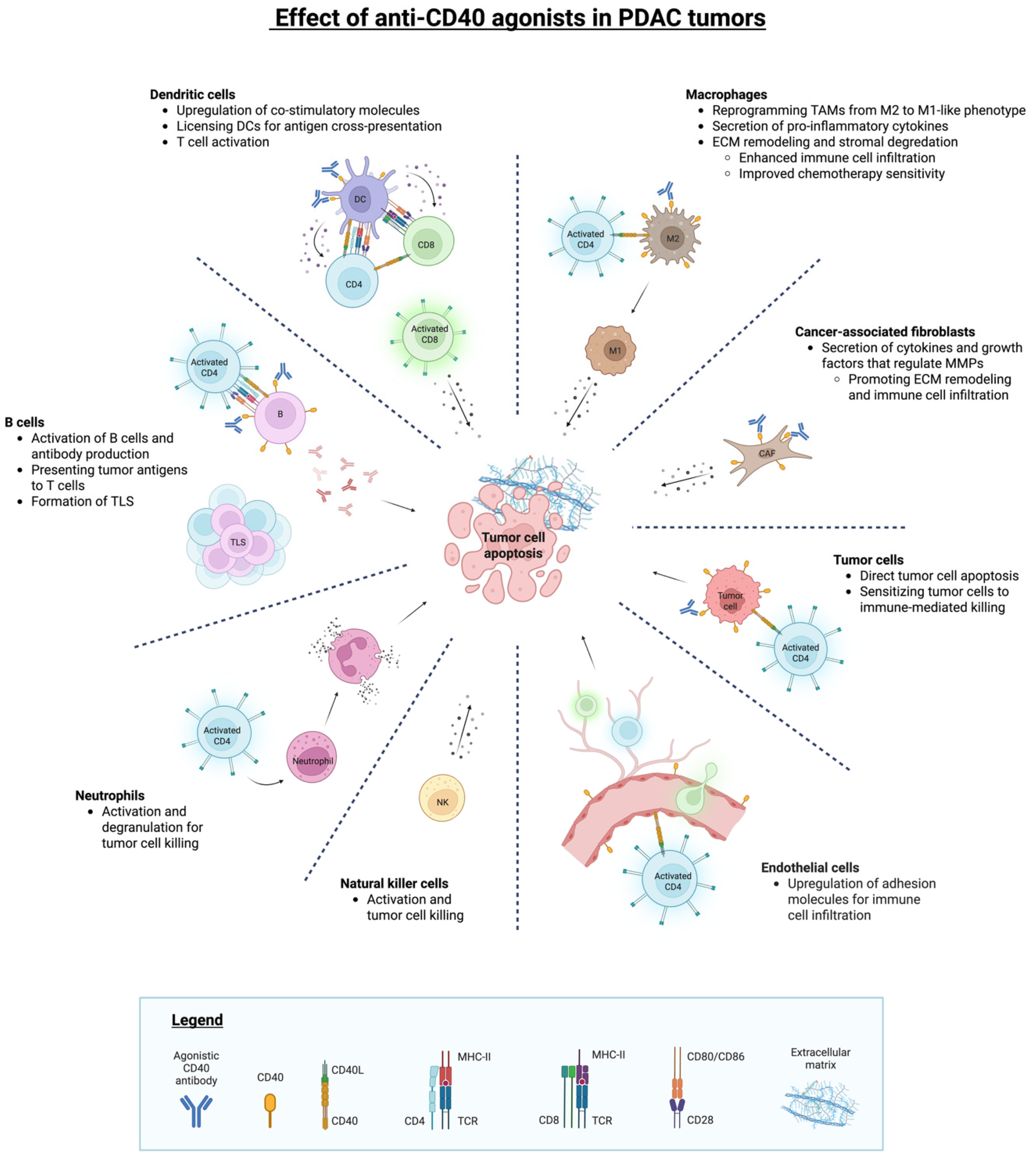
3.3. CD40-Driven Activation of Antigen-Presenting Cells
3.4. Reprogramming of Tumor-Associated Macrophages
3.5. Stromal and Vascular Remodeling Downstream of CD40 Activation
3.6. Tumor-Intrinsic CD40 Signaling in PDAC
4. Therapeutic Targeting of CD40 in PDAC
4.1. CD40 Agonistic Modalities and Design Principles
4.2. Combination Approaches with Chemotherapy
4.3. CD40 Agonism Combined with Immune Checkpoint Inhibition
4.4. Integration with Cancer Vaccines and Neoantigen-Directed Therapies
4.5. Neoadjuvant and Window-of-Opportunity Experience
4.6. Biomarkers: Prognostic Context and Predictors of Benefit
4.7. Safety Considerations and Toxicity Management
5. Challenges and Future Directions
5.1. Barriers to Consistent Clinical Benefit
5.2. Biomarkers and Patient Selection: Beyond Baseline CD40 Abundance
5.3. Optimization of Dosing, Scheduling, and Delivery
5.4. Context-Dependent Tumor-Intrinsic CD40 Signaling
5.5. Next-Generation CD40 Agonists and Rational Combinations
5.6. Standardization of CD40 Measurement and Reporting
6. Conclusions
Author Contributions
Funding
Data Availability Statement
Conflicts of Interest
References
- Bray, F.; Laversanne, M.; Sung, H.; Ferlay, J.; Siegel, R.L.; Soerjomataram, I.; Jemal, A. Global cancer statistics 2022: GLOBOCAN estimates of incidence and mortality worldwide for 36 cancers in 185 countries. CA Cancer J. Clin. 2024, 74, 229–263. [Google Scholar] [CrossRef] [PubMed]
- Rahib, L.; Smith, B.D.; Aizenberg, R.; Rosenzweig, A.B.; Fleshman, J.M.; Matrisian, L.M. Projecting cancer incidence and deaths to 2030: The unexpected burden of thyroid, liver, and pancreas cancers in the United States. Cancer Res. 2014, 74, 2913–2921. [Google Scholar] [CrossRef]
- Stoop, T.F.; Javed, A.A.; Oba, A.; Koerkamp, B.G.; Seufferlein, T.; Wilmink, J.W.; Besselink, M.G. Pancreatic cancer. Lancet 2025, 405, 1182–1202. [Google Scholar] [CrossRef] [PubMed]
- Park, W.; Chawla, A.; O’Reilly, E.M. Pancreatic Cancer: A Review. JAMA 2021, 326, 851–862. [Google Scholar] [CrossRef]
- Siegel, R.L.; Kratzer, T.B.; Wagle, N.S.; Sung, H.; Jemal, A. Cancer statistics, 2026. CA Cancer J. Clin. 2026, 76, e70043. [Google Scholar] [CrossRef]
- André, T.; Elez, E.; Lenz, H.J.; Jensen, L.H.; Touchefeu, Y.; Van Cutsem, E.; Garcia-Carbonero, R.; Tougeron, D.; Mendez, G.A.; Schenker, M.; et al. Nivolumab plus ipilimumab versus nivolumab in microsatellite instability-high metastatic colorectal cancer (CheckMate 8HW): A randomised, open-label, phase 3 trial. Lancet 2025, 405, 383–395. [Google Scholar] [CrossRef]
- Chalabi, M.; Fanchi, L.F.; Dijkstra, K.K.; Van den Berg, J.G.; Aalbers, A.G.; Sikorska, K.; Lopez-Yurda, M.; Grootscholten, C.; Beets, G.L.; Snaebjornsson, P.; et al. Neoadjuvant immunotherapy leads to pathological responses in MMR-proficient and MMR-deficient early-stage colon cancers. Nat. Med. 2020, 26, 566–576. [Google Scholar] [CrossRef]
- Mok, T.S.K.; Wu, Y.L.; Kudaba, I.; Kowalski, D.M.; Cho, B.C.; Turna, H.Z.; Castro, G., Jr.; Srimuninnimit, V.; Laktionov, K.K.; Bondarenko, I.; et al. Pembrolizumab versus chemotherapy for previously untreated, PD-L1-expressing, locally advanced or metastatic non-small-cell lung cancer (KEYNOTE-042): A randomised, open-label, controlled, phase 3 trial. Lancet 2019, 393, 1819–1830. [Google Scholar] [CrossRef] [PubMed]
- Spicer, J.D.; Garassino, M.C.; Wakelee, H.; Liberman, M.; Kato, T.; Tsuboi, M.; Lee, S.H.; Chen, K.N.; Dooms, C.; Majem, M.; et al. Neoadjuvant pembrolizumab plus chemotherapy followed by adjuvant pembrolizumab compared with neoadjuvant chemotherapy alone in patients with early-stage non-small-cell lung cancer (KEYNOTE-671): A randomised, double-blind, placebo-controlled, phase 3 trial. Lancet 2024, 404, 1240–1252. [Google Scholar] [CrossRef]
- Wolchok, J.D.; Chiarion-Sileni, V.; Rutkowski, P.; Cowey, C.L.; Schadendorf, D.; Wagstaff, J.; Queirolo, P.; Dummer, R.; Butler, M.O.; Hill, A.G.; et al. Final, 10-Year Outcomes with Nivolumab plus Ipilimumab in Advanced Melanoma. N. Engl. J. Med. 2025, 392, 11–22. [Google Scholar] [CrossRef]
- Janjigian, Y.Y.; Shitara, K.; Moehler, M.; Garrido, M.; Salman, P.; Shen, L.; Wyrwicz, L.; Yamaguchi, K.; Skoczylas, T.; Campos Bragagnoli, A.; et al. First-line nivolumab plus chemotherapy versus chemotherapy alone for advanced gastric, gastro-oesophageal junction, and oesophageal adenocarcinoma (CheckMate 649): A randomised, open-label, phase 3 trial. Lancet 2021, 398, 27–40. [Google Scholar] [CrossRef] [PubMed]
- Tannir, N.M.; Albigès, L.; McDermott, D.F.; Burotto, M.; Choueiri, T.K.; Hammers, H.J.; Barthélémy, P.; Plimack, E.R.; Porta, C.; George, S.; et al. Nivolumab plus ipilimumab versus sunitinib for first-line treatment of advanced renal cell carcinoma: Extended 8-year follow-up results of efficacy and safety from the phase III CheckMate 214 trial. Ann. Oncol. 2024, 35, 1026–1038. [Google Scholar] [CrossRef]
- Abou-Alfa, G.K.; Lau, G.; Kudo, M.; Chan, S.L.; Kelley, R.K.; Furuse, J.; Sukeepaisarnjaroen, W.; Kang, Y.K.; Van Dao, T.; De Toni, E.N.; et al. Tremelimumab plus Durvalumab in Unresectable Hepatocellular Carcinoma. NEJM Evid. 2022, 1, EVIDoa2100070. [Google Scholar] [CrossRef]
- Powles, T.; Valderrama, B.P.; Gupta, S.; Bedke, J.; Kikuchi, E.; Hoffman-Censits, J.; Iyer, G.; Vulsteke, C.; Park, S.H.; Shin, S.J.; et al. Enfortumab Vedotin and Pembrolizumab in Untreated Advanced Urothelial Cancer. N. Engl. J. Med. 2024, 390, 875–888. [Google Scholar] [CrossRef]
- Brahmer, J.R.; Tykodi, S.S.; Chow, L.Q.; Hwu, W.J.; Topalian, S.L.; Hwu, P.; Drake, C.G.; Camacho, L.H.; Kauh, J.; Odunsi, K.; et al. Safety and activity of anti-PD-L1 antibody in patients with advanced cancer. N. Engl. J. Med. 2012, 366, 2455–2465. [Google Scholar] [CrossRef]
- Renouf, D.J.; Loree, J.M.; Knox, J.J.; Topham, J.T.; Kavan, P.; Jonker, D.; Welch, S.; Couture, F.; Lemay, F.; Tehfe, M.; et al. The CCTG PA.7 phase II trial of gemcitabine and nab-paclitaxel with or without durvalumab and tremelimumab as initial therapy in metastatic pancreatic ductal adenocarcinoma. Nat. Commun. 2022, 13, 5020. [Google Scholar] [CrossRef]
- O’Reilly, E.M.; Oh, D.Y.; Dhani, N.; Renouf, D.J.; Lee, M.A.; Sun, W.; Fisher, G.; Hezel, A.; Chang, S.C.; Vlahovic, G.; et al. Durvalumab with or Without Tremelimumab for Patients with Metastatic Pancreatic Ductal Adenocarcinoma: A Phase 2 Randomized Clinical Trial. JAMA Oncol. 2019, 5, 1431–1438. [Google Scholar] [CrossRef]
- Ho, W.J.; Jaffee, E.M.; Zheng, L. The tumour microenvironment in pancreatic cancer—Clinical challenges and opportunities. Nat. Rev. Clin. Oncol. 2020, 17, 527–540. [Google Scholar] [CrossRef] [PubMed]
- Murakami, T.; Hiroshima, Y.; Matsuyama, R.; Homma, Y.; Hoffman, R.M.; Endo, I. Role of the tumor microenvironment in pancreatic cancer. Ann. Gastroenterol. Surg. 2019, 3, 130–137. [Google Scholar] [CrossRef]
- Dallal, R.M.; Christakos, P.; Lee, K.; Egawa, S.; Son, Y.I.; Lotze, M.T. Paucity of dendritic cells in pancreatic cancer. Surgery 2002, 131, 135–138. [Google Scholar] [CrossRef] [PubMed]
- Hegde, S.; Krisnawan, V.E.; Herzog, B.H.; Zuo, C.; Breden, M.A.; Knolhoff, B.L.; Hogg, G.D.; Tang, J.P.; Baer, J.M.; Mpoy, C.; et al. Dendritic Cell Paucity Leads to Dysfunctional Immune Surveillance in Pancreatic Cancer. Cancer Cell 2020, 37, 289–307. [Google Scholar] [CrossRef]
- Grewal, I.S.; Flavell, R.A. CD40 and CD154 in cell-mediated immunity. Annu. Rev. Immunol. 1998, 16, 111–135. [Google Scholar] [CrossRef]
- van Kooten, C.; Banchereau, J. CD40-CD40 ligand. J. Leukoc. Biol. 2000, 67, 2–17. [Google Scholar] [CrossRef]
- Vonderheide, R.H. Prospect of targeting the CD40 pathway for cancer therapy. Clin. Cancer Res. 2007, 13, 1083–1088. [Google Scholar] [CrossRef] [PubMed]
- Caux, C.; Massacrier, C.; Vanbervliet, B.; Dubois, B.; Van Kooten, C.; Durand, I.; Banchereau, J. Activation of human dendritic cells through CD40 cross-linking. J. Exp. Med. 1994, 180, 1263–1272. [Google Scholar] [CrossRef]
- Schoenberger, S.P.; Toes, R.E.; van der Voort, E.I.; Offringa, R.; Melief, C.J. T-cell help for cytotoxic T lymphocytes is mediated by CD40-CD40L interactions. Nature 1998, 393, 480–483. [Google Scholar] [CrossRef] [PubMed]
- Vonderheide, R.H. CD40 Agonist Antibodies in Cancer Immunotherapy. Annu. Rev. Med. 2020, 71, 47–58. [Google Scholar] [CrossRef]
- He, S.; Zhao, H.; Fei, M.; Wu, Y.; Wang, L.; Zhu, X.; Li, D. Expression of the co-signaling molecules CD40-CD40L and their growth inhibitory effect on pancreatic cancer in vitro. Oncol. Rep. 2012, 28, 262–268. [Google Scholar] [PubMed]
- Bates, K.M.; Vathiotis, I.; MacNeil, T.; Ahmed, F.S.; Aung, T.N.; Katlinskaya, Y.; Bhattacharya, S.; Psyrri, A.; Yea, S.; Parkes, A.; et al. Spatial characterization and quantification of CD40 expression across cancer types. BMC Cancer 2023, 23, 220. [Google Scholar] [CrossRef]
- Beatty, G.L.; Chiorean, E.G.; Fishman, M.P.; Saboury, B.; Teitelbaum, U.R.; Sun, W.; Huhn, R.D.; Song, W.; Li, D.; Sharp, L.L.; et al. CD40 agonists alter tumor stroma and show efficacy against pancreatic carcinoma in mice and humans. Science 2011, 331, 1612–1616. [Google Scholar] [CrossRef]
- Long, K.B.; Gladney, W.L.; Tooker, G.M.; Graham, K.; Fraietta, J.A.; Beatty, G.L. IFNγ and CCL2 Cooperate to Redirect Tumor-Infiltrating Monocytes to Degrade Fibrosis and Enhance Chemotherapy Efficacy in Pancreatic Carcinoma. Cancer Discov. 2016, 6, 400–413. [Google Scholar] [CrossRef] [PubMed]
- McVey, J.C.; Beatty, G.L. Facts and Hopes of CD40 Agonists in Cancer Immunotherapy. Clin. Cancer Res. 2025, 31, 2079–2087. [Google Scholar] [CrossRef] [PubMed]
- Mangsbo, S.M.; Broos, S.; Fletcher, E.; Veitonmäki, N.; Furebring, C.; Dahlén, E.; Norlén, P.; Lindstedt, M.; Tötterman, T.H.; Ellmark, P. The human agonistic CD40 antibody ADC-1013 eradicates bladder tumors and generates T-cell-dependent tumor immunity. Clin. Cancer Res. 2015, 21, 1115–1126. [Google Scholar] [CrossRef]
- Irenaeus, S.M.M.; Nielsen, D.; Ellmark, P.; Yachnin, J.; Deronic, A.; Nilsson, A.; Norlén, P.; Veitonmäki, N.; Wennersten, C.S.; Ullenhag, G.J. First-in-human study with intratumoral administration of a CD40 agonistic antibody, ADC-1013, in advanced solid malignancies. Int. J. Cancer 2019, 145, 1189–1199. [Google Scholar] [CrossRef]
- Moreno, V.; Perets, R.; Peretz-Yablonski, T.; Fourneau, N.; Girgis, S.; Guo, Y.; Hellemans, P.; Verona, R.; Pendás, N.; Xia, Q.; et al. A phase 1 study of intravenous mitazalimab, a CD40 agonistic monoclonal antibody, in patients with advanced solid tumors. Investig. New Drugs 2023, 41, 93–104. [Google Scholar] [CrossRef]
- Baxendale, A.J.; Dawson, C.W.; Stewart, S.E.; Mudaliar, V.; Reynolds, G.; Gordon, J.; Murray, P.G.; Young, L.S.; Eliopoulos, A.G. Constitutive activation of the CD40 pathway promotes cell transformation and neoplastic growth. Oncogene 2005, 24, 7913–7923. [Google Scholar] [CrossRef]
- Murugaiyan, G.; Martin, S.; Saha, B. CD40-induced countercurrent conduits for tumor escape or elimination? Trends Immunol. 2007, 28, 467–473. [Google Scholar] [CrossRef]
- Korniluk, A.; Kemona, H.; Dymicka-Piekarska, V. Multifunctional CD40L: Pro- and anti-neoplastic activity. Tumor Biol. 2014, 35, 9447–9457. [Google Scholar] [CrossRef]
- Batrla, R.; Linnebacher, M.; Rudy, W.; Stumm, S.; Wallwiener, D.; Gückel, B. CD40-expressing carcinoma cells induce down-regulation of CD40 ligand (CD154) and impair T-cell functions. Cancer Res. 2002, 62, 2052–2057. [Google Scholar] [PubMed]
- Khaliq, A.M.; Rajamohan, M.; Saeed, O.; Mansouri, K.; Adil, A.; Zhang, C.; Turk, A.; Carstens, J.L.; House, M.; Hayat, S.; et al. Spatial transcriptomic analysis of primary and metastatic pancreatic cancers highlights tumor microenvironmental heterogeneity. Nat. Genet. 2024, 56, 2455–2465. [Google Scholar] [CrossRef] [PubMed]
- Gong, R.; Chen, Y.; Li, C.; Zhang, H.; Liu, Z.; Yu, Q. Targeting myeloid cells in pancreatic ductal adenocarcinoma: From primary tumors to liver metastasis. Front. Immunol. 2025, 16, 1555036. [Google Scholar] [CrossRef]
- Byrne, K.T.; Betts, C.B.; Mick, R.; Sivagnanam, S.; Bajor, D.L.; Laheru, D.A.; Chiorean, E.G.; O’Hara, M.H.; Liudahl, S.M.; Newcomb, C.; et al. Neoadjuvant Selicrelumab, an Agonist CD40 Antibody, Induces Changes in the Tumor Microenvironment in Patients with Resectable Pancreatic Cancer. Clin. Cancer Res. 2021, 27, 4574–4586. [Google Scholar] [CrossRef]
- Karmann, K.; Hughes, C.C.; Schechner, J.; Fanslow, W.C.; Pober, J.S. CD40 on human endothelial cells: Inducibility by cytokines and functional regulation of adhesion molecule expression. Proc. Natl. Acad. Sci. USA 1995, 92, 4342–4346. [Google Scholar] [CrossRef]
- Urban, D.; Thanabalasingam, U.; Stibenz, D.; Kaufmann, J.; Meyborg, H.; Fleck, E.; Gräfe, M.; Stawowy, P. CD40/CD40L interaction induces E-selectin dependent leukocyte adhesion to human endothelial cells and inhibits endothelial cell migration. Biochem. Biophys. Res. Commun. 2011, 404, 448–452. [Google Scholar] [CrossRef]
- Richards, K.E.; Zeleniak, A.E.; Fishel, M.L.; Wu, J.; Littlepage, L.E.; Hill, R. Cancer-associated fibroblast exosomes regulate survival and proliferation of pancreatic cancer cells. Oncogene 2017, 36, 1770–1778. [Google Scholar] [CrossRef]
- Chakravarthy, A.; Khan, L.; Bensler, N.P.; Bose, P.; De Carvalho, D.D. TGF-β-associated extracellular matrix genes link cancer-associated fibroblasts to immune evasion and immunotherapy failure. Nat. Commun. 2018, 9, 4692. [Google Scholar] [CrossRef]
- Grout, J.A.; Sirven, P.; Leader, A.M.; Maskey, S.; Hector, E.; Puisieux, I.; Steffan, F.; Cheng, E.; Tung, N.; Maurin, M.; et al. Spatial Positioning and Matrix Programs of Cancer-Associated Fibroblasts Promote T-cell Exclusion in Human Lung Tumors. Cancer Discov. 2022, 12, 2606–2625. [Google Scholar] [CrossRef]
- Andersson, H.; Nyesiga, B.; Hermodsson, T.; Enell Smith, K.; Hägerbrand, K.; Lindstedt, M.; Ellmark, P. Next-generation CD40 agonists for cancer immunotherapy. Expert. Opin. Biol. Ther. 2024, 24, 351–363. [Google Scholar] [CrossRef]
- Xie, Z.; Fang, Y.; Zhang, X.; Fang, Y.; Li, R.; Guo, Y.; Yang, Y.; Yang, S.; Song, L. The emerging role of dendritic cells in the tumor microenvironment: From antigen presentation to targeted immunotherapy. Cell Death Dis. 2025, 16, 900. [Google Scholar] [CrossRef]
- Zuo, C.; Baer, J.M.; Knolhoff, B.L.; Belle, J.I.; Liu, X.; Alarcon De La Lastra, A.; Fu, C.; Hogg, G.D.; Kingston, N.L.; Breden, M.A.; et al. Stromal and therapy-induced macrophage proliferation promotes PDAC progression and susceptibility to innate immunotherapy. J. Exp. Med. 2023, 220, 20212062. [Google Scholar] [CrossRef]
- Lehmann, J.; Thelen, M.; Kreer, C.; Schran, S.; Garcia-Marquez, M.A.; Cisic, I.; Siepmann, K.; Hagen, E.M.; Eckel, H.N.C.; Lohneis, P.; et al. Tertiary Lymphoid Structures in Pancreatic Cancer are Structurally Homologous, Share Gene Expression Patterns and B-cell Clones with Secondary Lymphoid Organs, but Show Increased T-cell Activation. Cancer Immunol. Res. 2025, 13, 323–336. [Google Scholar] [CrossRef]
- Pan, P.Y.; Ma, G.; Weber, K.J.; Ozao-Choy, J.; Wang, G.; Yin, B.; Divino, C.M.; Chen, S.H. Immune stimulatory receptor CD40 is required for T-cell suppression and T regulatory cell activation mediated by myeloid-derived suppressor cells in cancer. Cancer Res. 2010, 70, 99–108. [Google Scholar] [CrossRef]
- Minaei, E.; Wade, S.J.; Penney, C.; Ranson, M.; Aghmesheh, M.; Sluyter, R.; Vine-Perrow, K.L. Localized Delivery of Anti-CD40/PD-1 Immunotherapy via a Polymeric Implant is Efficacious, Remodels the Tumor Immune Microenvironment and Improves Safety in a PDAC Mouse Model. Adv. Healthc. Mater. 2026, 15, e03780. [Google Scholar] [CrossRef]
- Baumann, D.; Hägele, T.; Mochayedi, J.; Drebant, J.; Vent, C.; Blobner, S.; Noll, J.H.; Nickel, I.; Schumacher, C.; Boos, S.L.; et al. Proimmunogenic impact of MEK inhibition synergizes with agonist anti-CD40 immunostimulatory antibodies in tumor therapy. Nat. Commun. 2020, 11, 2176. [Google Scholar] [CrossRef]
- Jyothi, M.D.; Khar, A. Regulation of CD40L expression on natural killer cells by interleukin-12 and interferon gamma: Its role in the elicitation of an effective antitumor immune response. Cancer Immunol. Immunother. 2000, 49, 563–572. [Google Scholar] [CrossRef]
- Turner, J.G.; Rakhmilevich, A.L.; Burdelya, L.; Neal, Z.; Imboden, M.; Sondel, P.M.; Yu, H. Anti-CD40 antibody induces antitumor and antimetastatic effects: The role of NK cells. J. Immunol. 2001, 166, 89–94. [Google Scholar] [CrossRef]
- Carbone, E.; Ruggiero, G.; Terrazzano, G.; Palomba, C.; Manzo, C.; Fontana, S.; Spits, H.; Kärre, K.; Zappacosta, S. A new mechanism of NK cell cytotoxicity activation: The CD40-CD40 ligand interaction. J. Exp. Med. 1997, 185, 2053–2060. [Google Scholar] [CrossRef]
- Terrazzano, G.; Pisanti, S.; Grimaldi, S.; Sica, M.; Fontana, S.; Carbone, E.; Zappacosta, S.; Ruggiero, G. Interaction between natural killer and dendritic cells: The role of CD40, CD80 and major histocompatibility complex class i molecules in cytotoxicity induction and interferon-gamma production. Scand. J. Immunol. 2004, 59, 356–362. [Google Scholar] [CrossRef]
- Foight, G.W.; Keating, A.E. Comparison of the peptide binding preferences of three closely related TRAF paralogs: TRAF2, TRAF3, and TRAF5. Protein Sci. 2016, 25, 1273–1289. [Google Scholar] [CrossRef]
- Ma, Y.; Ding, J.; Chen, Y. The effect of CD40 agonist antibody therapy on the pancreatic cancer microenvironment. Naunyn Schmiedebergs Arch. Pharmacol. 2026, 399, 1897–1915. [Google Scholar] [CrossRef]
- Sun, S.C. Non-canonical NF-κB signaling pathway. Cell Res. 2011, 21, 71–85. [Google Scholar] [CrossRef]
- Clatza, A.; Bonifaz, L.C.; Vignali, D.A.; Moreno, J. CD40-induced aggregation of MHC class II and CD80 on the cell surface leads to an early enhancement in antigen presentation. J. Immunol. 2003, 171, 6478–6487. [Google Scholar] [CrossRef]
- Cella, M.; Scheidegger, D.; Palmer-Lehmann, K.; Lane, P.; Lanzavecchia, A.; Alber, G. Ligation of CD40 on dendritic cells triggers production of high levels of interleukin-12 and enhances T cell stimulatory capacity: T-T help via APC activation. J. Exp. Med. 1996, 184, 747–752. [Google Scholar] [CrossRef]
- Gallagher, E.; Enzler, T.; Matsuzawa, A.; Anzelon-Mills, A.; Otero, D.; Holzer, R.; Janssen, E.; Gao, M.; Karin, M. Kinase MEKK1 is required for CD40-dependent activation of the kinases Jnk and p38, germinal center formation, B cell proliferation and antibody production. Nat. Immunol. 2007, 8, 57–63. [Google Scholar] [CrossRef]
- Tang, T.; Cheng, X.; Truong, B.; Sun, L.; Yang, X.; Wang, H. Molecular basis and therapeutic implications of CD40/CD40L immune checkpoint. Pharmacol. Ther. 2021, 219, 107709. [Google Scholar] [CrossRef]
- Zhao, X.; Li, D.C.; Zhu, X.G.; Gan, W.J.; Li, Z.; Xiong, F.; Zhang, Z.X.; Zhang, G.B.; Zhang, X.G.; Zhao, H. B7-H3 overexpression in pancreatic cancer promotes tumor progression. Int. J. Mol. Med. 2013, 31, 283–291. [Google Scholar] [CrossRef]
- Inamura, K.; Takazawa, Y.; Inoue, Y.; Yokouchi, Y.; Kobayashi, M.; Saiura, A.; Shibutani, T.; Ishikawa, Y. Tumor B7-H3 (CD276) Expression and Survival in Pancreatic Cancer. J. Clin. Med. 2018, 7, 172. [Google Scholar] [CrossRef]
- Ma, Y.; Yang, Y.; Huang, Y.; Fang, W.; Xue, J.; Meng, X.; Fan, Y.; Fu, S.; Wu, L.; Zheng, Y.; et al. A B7H3-targeting antibody-drug conjugate in advanced solid tumors: A phase 1/1b trial. Nat. Med. 2025, 31, 1949–1957. [Google Scholar] [CrossRef]
- Duan, J.; Sun, Y.; Wang, Q.; Xing, L.; Wu, L.; Han, L.; Chen, J.; Liu, B.; Lu, P.; Shi, H.; et al. HS-20093, a B7-H3-targeted antibody-drug conjugate in lung cancer: Results from the ARTEMIS-001 phase 1a/b trial. Cancer Cell 2026, 44, 846–857. [Google Scholar] [CrossRef]
- Hostager, B.S.; Bishop, G.A. CD40-Mediated Activation of the NF-κB2 Pathway. Front. Immunol. 2013, 4, 376. [Google Scholar] [CrossRef]
- Zhou, Y.; Richmond, A.; Yan, C. Harnessing the potential of CD40 agonism in cancer therapy. Cytokine Growth Factor Rev. 2024, 75, 40–56. [Google Scholar] [CrossRef]
- Hanissian, S.H.; Geha, R.S. Jak3 is associated with CD40 and is critical for CD40 induction of gene expression in B cells. Immunity 1997, 6, 379–387. [Google Scholar] [CrossRef]
- Revy, P.; Hivroz, C.; Andreu, G.; Graber, P.; Martinache, C.; Fischer, A.; Durandy, A. Activation of the Janus kinase 3-STAT5a pathway after CD40 triggering of human monocytes but not of resting B cells. J. Immunol. 1999, 163, 787–793. [Google Scholar] [CrossRef]
- Säemann, M.D.; Diakos, C.; Kelemen, P.; Kriehuber, E.; Zeyda, M.; Böhmig, G.A.; Hörl, W.H.; Baumruker, T.; Zlabinger, G.J. Prevention of CD40-triggered dendritic cell maturation and induction of T-cell hyporeactivity by targeting of Janus kinase 3. Am. J. Transplant. 2003, 3, 1341–1349. [Google Scholar] [CrossRef]
- Wattenberg, M.M.; Herrera, V.M.; Giannone, M.A.; Gladney, W.L.; Carpenter, E.L.; Beatty, G.L. Systemic inflammation is a determinant of outcomes of CD40 agonist-based therapy in pancreatic cancer patients. JCI Insight 2021, 6, e145389. [Google Scholar] [CrossRef]
- Yu, H.; Pardoll, D.; Jove, R. STATs in cancer inflammation and immunity: A leading role for STAT3. Nat. Rev. Cancer 2009, 9, 798–809. [Google Scholar] [CrossRef]
- Lesina, M.; Kurkowski, M.U.; Ludes, K.; Rose-John, S.; Treiber, M.; Klöppel, G.; Yoshimura, A.; Reindl, W.; Sipos, B.; Akira, S.; et al. Stat3/Socs3 activation by IL-6 transsignaling promotes progression of pancreatic intraepithelial neoplasia and development of pancreatic cancer. Cancer Cell 2011, 19, 456–469. [Google Scholar] [CrossRef]
- Corcoran, R.B.; Contino, G.; Deshpande, V.; Tzatsos, A.; Conrad, C.; Benes, C.H.; Levy, D.E.; Settleman, J.; Engelman, J.A.; Bardeesy, N. STAT3 plays a critical role in KRAS-induced pancreatic tumorigenesis. Cancer Res. 2011, 71, 5020–5029. [Google Scholar] [CrossRef]
- Fukuda, A.; Wang, S.C.; Morris, J.P.t.; Folias, A.E.; Liou, A.; Kim, G.E.; Akira, S.; Boucher, K.M.; Firpo, M.A.; Mulvihill, S.J.; et al. Stat3 and MMP7 contribute to pancreatic ductal adenocarcinoma initiation and progression. Cancer Cell 2011, 19, 441–455. [Google Scholar] [CrossRef]
- Scholz, A.; Heinze, S.; Detjen, K.M.; Peters, M.; Welzel, M.; Hauff, P.; Schirner, M.; Wiedenmann, B.; Rosewicz, S. Activated signal transducer and activator of transcription 3 (STAT3) supports the malignant phenotype of human pancreatic cancer. Gastroenterology 2003, 125, 891–905. [Google Scholar] [CrossRef]
- Wei, D.; Le, X.; Zheng, L.; Wang, L.; Frey, J.A.; Gao, A.C.; Peng, Z.; Huang, S.; Xiong, H.Q.; Abbruzzese, J.L.; et al. Stat3 activation regulates the expression of vascular endothelial growth factor and human pancreatic cancer angiogenesis and metastasis. Oncogene 2003, 22, 319–329. [Google Scholar] [CrossRef]
- Hurwitz, H.I.; Uppal, N.; Wagner, S.A.; Bendell, J.C.; Beck, J.T.; Wade, S.M., 3rd; Nemunaitis, J.J.; Stella, P.J.; Pipas, J.M.; Wainberg, Z.A.; et al. Randomized, Double-Blind, Phase II Study of Ruxolitinib or Placebo in Combination with Capecitabine in Patients with Metastatic Pancreatic Cancer for Whom Therapy with Gemcitabine Has Failed. J. Clin. Oncol. 2015, 33, 4039–4047. [Google Scholar] [CrossRef]
- Hurwitz, H.; Van Cutsem, E.; Bendell, J.; Hidalgo, M.; Li, C.P.; Salvo, M.G.; Macarulla, T.; Sahai, V.; Sama, A.; Greeno, E.; et al. Ruxolitinib + capecitabine in advanced/metastatic pancreatic cancer after disease progression/intolerance to first-line therapy: JANUS 1 and 2 randomized phase III studies. Investig. New Drugs 2018, 36, 683–695. [Google Scholar] [CrossRef]
- Beatty, G.L.; Shahda, S.; Beck, T.; Uppal, N.; Cohen, S.J.; Donehower, R.; Gabayan, A.E.; Assad, A.; Switzky, J.; Zhen, H.; et al. A Phase Ib/II Study of the JAK1 Inhibitor, Itacitinib, plus nab-Paclitaxel and Gemcitabine in Advanced Solid Tumors. Oncologist 2019, 24, 14-e10. [Google Scholar] [CrossRef]
- Parampalli Yajnanarayana, S.; Stübig, T.; Cornez, I.; Alchalby, H.; Schönberg, K.; Rudolph, J.; Triviai, I.; Wolschke, C.; Heine, A.; Brossart, P.; et al. JAK1/2 inhibition impairs T cell function in vitro and in patients with myeloproliferative neoplasms. Br. J. Haematol. 2015, 169, 824–833. [Google Scholar] [CrossRef]
- Klein, K.; Stoiber, D.; Sexl, V.; Witalisz-Siepracka, A. Untwining Anti-Tumor and Immunosuppressive Effects of JAK Inhibitors-A Strategy for Hematological Malignancies? Cancers 2021, 13, 2611. [Google Scholar] [CrossRef]
- Kubo, S.; Yamaoka, K.; Kondo, M.; Yamagata, K.; Zhao, J.; Iwata, S.; Tanaka, Y. The JAK inhibitor, tofacitinib, reduces the T cell stimulatory capacity of human monocyte-derived dendritic cells. Ann. Rheum. Dis. 2014, 73, 2192–2198. [Google Scholar] [CrossRef]
- Chand Dakal, T.; Dhabhai, B.; Agarwal, D.; Gupta, R.; Nagda, G.; Meena, A.R.; Dhakar, R.; Menon, A.; Mathur, R.; Mona; et al. Mechanistic basis of co-stimulatory CD40-CD40L ligation mediated regulation of immune responses in cancer and autoimmune disorders. Immunobiology 2020, 225, 151899. [Google Scholar] [CrossRef]
- Johnson, P.; Challis, R.; Chowdhury, F.; Gao, Y.; Harvey, M.; Geldart, T.; Kerr, P.; Chan, C.; Smith, A.; Steven, N.; et al. Clinical and biological effects of an agonist anti-CD40 antibody: A Cancer Research UK phase I study. Clin. Cancer Res. 2015, 21, 1321–1328. [Google Scholar] [CrossRef]
- Ino, Y.; Yamazaki-Itoh, R.; Shimada, K.; Iwasaki, M.; Kosuge, T.; Kanai, Y.; Hiraoka, N. Immune cell infiltration as an indicator of the immune microenvironment of pancreatic cancer. Br. J. Cancer 2013, 108, 914–923. [Google Scholar] [CrossRef]
- Fukunaga, A.; Miyamoto, M.; Cho, Y.; Murakami, S.; Kawarada, Y.; Oshikiri, T.; Kato, K.; Kurokawa, T.; Suzuoki, M.; Nakakubo, Y.; et al. CD8+ tumor-infiltrating lymphocytes together with CD4+ tumor-infiltrating lymphocytes and dendritic cells improve the prognosis of patients with pancreatic adenocarcinoma. Pancreas 2004, 28, e26–e31. [Google Scholar] [CrossRef]
- Caronni, N.; La Terza, F.; Vittoria, F.M.; Barbiera, G.; Mezzanzanica, L.; Cuzzola, V.; Barresi, S.; Pellegatta, M.; Canevazzi, P.; Dunsmore, G.; et al. IL-1β(+) macrophages fuel pathogenic inflammation in pancreatic cancer. Nature 2023, 623, 415–422. [Google Scholar] [CrossRef]
- Shakiba, M.; Tuveson, D.A. Macrophages and fibroblasts as regulators of the immune response in pancreatic cancer. Nat. Immunol. 2025, 26, 678–691. [Google Scholar] [CrossRef] [PubMed]
- Ichikawa, J.; Okuda, H.; Kawano, K.; Kato, S.; Sato, S.; Kuroishikawa, R.; Kurotaki, D.; Kawase, W.; Yoshida, H.; Hiroshima, Y.; et al. A Combinatorial Effect of Immune Checkpoint Inhibitors and CD40 Agonistic Antibody in Murine Pancreatic Cancer Model. Cancer Sci. 2026, 117, 8–18. [Google Scholar] [CrossRef]
- Byrne, K.T.; Vonderheide, R.H. CD40 Stimulation Obviates Innate Sensors and Drives T Cell Immunity in Cancer. Cell Rep. 2016, 15, 2719–2732. [Google Scholar] [CrossRef] [PubMed]
- Lau, S.P.; van Montfoort, N.; Kinderman, P.; Lukkes, M.; Klaase, L.; van Nimwegen, M.; van Gulijk, M.; Dumas, J.; Mustafa, D.A.M.; Lievense, S.L.A.; et al. Dendritic cell vaccination and CD40-agonist combination therapy licenses T cell-dependent antitumor immunity in a pancreatic carcinoma murine model. J. Immunother. Cancer 2020, 8, e000772. [Google Scholar] [CrossRef] [PubMed]
- Wagner, A.H.; Gebauer, M.; Pollok-Kopp, B.; Hecker, M. Cytokine-inducible CD40 expression in human endothelial cells is mediated by interferon regulatory factor-1. Blood 2002, 99, 520–525. [Google Scholar] [CrossRef]
- Antoniades, C.; Bakogiannis, C.; Tousoulis, D.; Antonopoulos, A.S.; Stefanadis, C. The CD40/CD40 ligand system: Linking inflammation with atherothrombosis. J. Am. Coll. Cardiol. 2009, 54, 669–677. [Google Scholar] [CrossRef]
- Mach, F.; Schönbeck, U.; Fabunmi, R.P.; Murphy, C.; Atkinson, E.; Bonnefoy, J.Y.; Graber, P.; Libby, P. T lymphocytes induce endothelial cell matrix metalloproteinase expression by a CD40L-dependent mechanism: Implications for tubule formation. Am. J. Pathol. 1999, 154, 229–238. [Google Scholar] [CrossRef]
- Luheshi, N.M.; Coates-Ulrichsen, J.; Harper, J.; Mullins, S.; Sulikowski, M.G.; Martin, P.; Brown, L.; Lewis, A.; Davies, G.; Morrow, M.; et al. Transformation of the tumour microenvironment by a CD40 agonist antibody correlates with improved responses to PD-L1 blockade in a mouse orthotopic pancreatic tumour model. Oncotarget 2016, 7, 18508–18520. [Google Scholar] [CrossRef]
- Melter, M.; Reinders, M.E.; Sho, M.; Pal, S.; Geehan, C.; Denton, M.D.; Mukhopadhyay, D.; Briscoe, D.M. Ligation of CD40 induces the expression of vascular endothelial growth factor by endothelial cells and monocytes and promotes angiogenesis in vivo. Blood 2000, 96, 3801–3808. [Google Scholar] [CrossRef]
- Reinders, M.E.; Sho, M.; Robertson, S.W.; Geehan, C.S.; Briscoe, D.M. Proangiogenic function of CD40 ligand-CD40 interactions. J. Immunol. 2003, 171, 1534–1541. [Google Scholar] [CrossRef]
- Chung, H.W.; Lim, J.B. Clinical significance of elevated serum soluble CD40 ligand levels as a diagnostic and prognostic tumor marker for pancreatic ductal adenocarcinoma. J. Transl. Med. 2014, 12, 102. [Google Scholar] [CrossRef]
- Flaxenburg, J.A.; Melter, M.; Lapchak, P.H.; Briscoe, D.M.; Pal, S. The CD40-induced signaling pathway in endothelial cells resulting in the overexpression of vascular endothelial growth factor involves Ras and phosphatidylinositol 3-kinase. J. Immunol. 2004, 172, 7503–7509. [Google Scholar] [CrossRef]
- Selvaraj, S.; Raundhal, M.; Patidar, A.; Saha, B. Anti-VEGF antibody enhances the antitumor effect of CD40. Int. J. Cancer 2014, 135, 1983–1988. [Google Scholar] [CrossRef]
- Kashyap, A.S.; Schmittnaegel, M.; Rigamonti, N.; Pais-Ferreira, D.; Mueller, P.; Buchi, M.; Ooi, C.H.; Kreuzaler, M.; Hirschmann, P.; Guichard, A.; et al. Optimized antiangiogenic reprogramming of the tumor microenvironment potentiates CD40 immunotherapy. Proc. Natl. Acad. Sci. USA 2020, 117, 541–551. [Google Scholar] [CrossRef]
- Shoji, Y.; Miyamoto, M.; Ishikawa, K.; Yoshioka, T.; Mishra, R.; Ichinokawa, K.; Matsumura, Y.; Itoh, T.; Shinohara, T.; Hirano, S.; et al. The CD40-CD154 interaction would correlate with proliferation and immune escape in pancreatic ductal adenocarcinoma. J. Surg. Oncol. 2011, 103, 230–238. [Google Scholar] [CrossRef] [PubMed]
- Schiele, P.; Japp, A.S.; Stark, R.; Sattelberg, J.J.; Nikolaou, C.; Kornhuber, G.; Abbasi, P.; Ding, N.; Rosnev, S.; Meinke, S.; et al. CD8(+) T cell-derived CD40L mediates noncanonical cytotoxicity in CD40-expressing cancer cells. Sci. Adv. 2025, 11, eadr9331. [Google Scholar] [CrossRef]
- Davies, C.C.; Mason, J.; Wakelam, M.J.; Young, L.S.; Eliopoulos, A.G. Inhibition of phosphatidylinositol 3-kinase- and ERK MAPK-regulated protein synthesis reveals the pro-apoptotic properties of CD40 ligation in carcinoma cells. J. Biol. Chem. 2004, 279, 1010–1019. [Google Scholar] [CrossRef] [PubMed]
- Elmetwali, T.; Young, L.S.; Palmer, D.H. CD40 ligand-induced carcinoma cell death: A balance between activation of TNFR-associated factor (TRAF) 3-dependent death signals and suppression of TRAF6-dependent survival signals. J. Immunol. 2010, 184, 1111–1120. [Google Scholar] [CrossRef] [PubMed]
- Fonsatti, E.; Maio, M.; Altomonte, M.; Hersey, P. Biology and clinical applications of CD40 in cancer treatment. Semin. Oncol. 2010, 37, 517–523. [Google Scholar] [CrossRef]
- Murugaiyan, G.; Agrawal, R.; Mishra, G.C.; Mitra, D.; Saha, B. Differential CD40/CD40L expression results in counteracting antitumor immune responses. J. Immunol. 2007, 178, 2047–2055. [Google Scholar] [CrossRef] [PubMed]
- Murgaski, A.; Kiss, M.; Van Damme, H.; Kancheva, D.; Vanmeerbeek, I.; Keirsse, J.; Hadadi, E.; Brughmans, J.; Arnouk, S.M.; Hamouda, A.E.I.; et al. Efficacy of CD40 Agonists Is Mediated by Distinct cDC Subsets and Subverted by Suppressive Macrophages. Cancer Res. 2022, 82, 3785–3801. [Google Scholar] [CrossRef] [PubMed]
- Dahan, R.; Barnhart, B.C.; Li, F.; Yamniuk, A.P.; Korman, A.J.; Ravetch, J.V. Therapeutic Activity of Agonistic, Human Anti-CD40 Monoclonal Antibodies Requires Selective FcγR Engagement. Cancer Cell 2016, 29, 820–831. [Google Scholar] [CrossRef]
- Beatty, G.L.; Winograd, R.; Evans, R.A.; Long, K.B.; Luque, S.L.; Lee, J.W.; Clendenin, C.; Gladney, W.L.; Knoblock, D.M.; Guirnalda, P.D.; et al. Exclusion of T Cells from Pancreatic Carcinomas in Mice Is Regulated by Ly6C(low) F4/80(+) Extratumoral Macrophages. Gastroenterology 2015, 149, 201–210. [Google Scholar] [CrossRef]
- Troiano, G.; Caponio, V.C.A.; Adipietro, I.; Tepedino, M.; Santoro, R.; Laino, L.; Lo Russo, L.; Cirillo, N.; Lo Muzio, L. Prognostic significance of CD68(+) and CD163(+) tumor associated macrophages in head and neck squamous cell carcinoma: A systematic review and meta-analysis. Oral Oncol. 2019, 93, 66–75. [Google Scholar] [CrossRef]
- López-Janeiro, Á.; Padilla-Ansala, C.; de Andrea, C.E.; Hardisson, D.; Melero, I. Prognostic value of macrophage polarization markers in epithelial neoplasms and melanoma. A systematic review and meta-analysis. Mod. Pathol. 2020, 33, 1458–1465. [Google Scholar] [CrossRef]
- Van Laethem, J.L.; Borbath, I.; Prenen, H.; Geboes, K.P.; Lambert, A.; Mitry, E.; Cassier, P.A.; Blanc, J.F.; Pilla, L.; Batlle, J.F.; et al. Combining CD40 agonist mitazalimab with mFOLFIRINOX in previously untreated metastatic pancreatic ductal adenocarcinoma (OPTIMIZE-1): A single-arm, multicentre phase 1b/2 study. Lancet Oncol. 2024, 25, 853–864. [Google Scholar] [CrossRef]
- Coveler, A.L.; Smith, D.C.; Phillips, T.; Curti, B.D.; Goel, S.; Mehta, A.N.; Kuzel, T.M.; Markovic, S.N.; Rixe, O.; Bajor, D.L.; et al. Phase 1 dose-escalation study of SEA-CD40: A non-fucosylated CD40 agonist, in advanced solid tumors and lymphomas. J. Immunother. Cancer 2023, 11, e005584. [Google Scholar] [CrossRef]
- Beatty, G.L.; Torigian, D.A.; Chiorean, E.G.; Saboury, B.; Brothers, A.; Alavi, A.; Troxel, A.B.; Sun, W.; Teitelbaum, U.R.; Vonderheide, R.H.; et al. A phase I study of an agonist CD40 monoclonal antibody (CP-870,893) in combination with gemcitabine in patients with advanced pancreatic ductal adenocarcinoma. Clin. Cancer Res. 2013, 19, 6286–6295. [Google Scholar] [CrossRef] [PubMed]
- Van Laethem, J.L.; Geboes, K.; Borbath, I.; Macarulla Mercade, T.; Lambert, A.; Cassier, P.; Prenen, H.; Mitry, E.; Blanc, J.F.; Pilla, L.; et al. CD40 agonist mitazalimab with mFOLFIRINOX in untreated metastatic pancreatic cancer: Biomarkers associated with outcomes from OPTIMIZE-1. Cell Rep. Med. 2025, 6, 102407. [Google Scholar] [CrossRef]
- O’Hara, M.H.; O’Reilly, E.M.; Varadhachary, G.; Wolff, R.A.; Wainberg, Z.A.; Ko, A.H.; Fisher, G.; Rahma, O.; Lyman, J.P.; Cabanski, C.R.; et al. CD40 agonistic monoclonal antibody APX005M (sotigalimab) and chemotherapy, with or without nivolumab, for the treatment of metastatic pancreatic adenocarcinoma: An open-label, multicentre, phase 1b study. Lancet Oncol. 2021, 22, 118–131. [Google Scholar] [CrossRef] [PubMed]
- Padrón, L.J.; Maurer, D.M.; O’Hara, M.H.; O’Reilly, E.M.; Wolff, R.A.; Wainberg, Z.A.; Ko, A.H.; Fisher, G.; Rahma, O.; Lyman, J.P.; et al. Sotigalimab and/or nivolumab with chemotherapy in first-line metastatic pancreatic cancer: Clinical and immunologic analyses from the randomized phase 2 PRINCE trial. Nat. Med. 2022, 28, 1167–1177. [Google Scholar] [CrossRef]
- Kucukcelebi, S.; van ‘t Land, F.R.; van der Burg, S.H.; Eskens, F.; Homs, M.Y.V.; Willemsen, M.; Onrust-van Schoonhoven, A.; Rozendaal, N.E.M.; Fellah, A.; Vadgama, D.; et al. REACtiVe-2: Phase I evaluation of dendritic cell vaccination and agonistic CD40 therapy following (m)FOLFIRINOX in metastatic pancreatic cancer. Nat. Commun. 2025, 16, 10609. [Google Scholar] [CrossRef]
- Winograd, R.; Byrne, K.T.; Evans, R.A.; Odorizzi, P.M.; Meyer, A.R.; Bajor, D.L.; Clendenin, C.; Stanger, B.Z.; Furth, E.E.; Wherry, E.J.; et al. Induction of T-cell Immunity Overcomes Complete Resistance to PD-1 and CTLA-4 Blockade and Improves Survival in Pancreatic Carcinoma. Cancer Immunol. Res. 2015, 3, 399–411. [Google Scholar] [CrossRef]
- Morrison, A.H.; Diamond, M.S.; Hay, C.A.; Byrne, K.T.; Vonderheide, R.H. Sufficiency of CD40 activation and immune checkpoint blockade for T cell priming and tumor immunity. Proc. Natl. Acad. Sci. USA 2020, 117, 8022–8031. [Google Scholar] [CrossRef] [PubMed]
- Burrack, A.L.; Rollins, M.R.; Spartz, E.J.; Mesojednik, T.D.; Schmiechen, Z.C.; Raynor, J.F.; Wang, I.X.; Kedl, R.M.; Stromnes, I.M. CD40 Agonist Overcomes T Cell Exhaustion Induced by Chronic Myeloid Cell IL-27 Production in a Pancreatic Cancer Preclinical Model. J. Immunol. 2021, 206, 1372–1384. [Google Scholar] [CrossRef]
- Sanborn, R.; Gabrail, N.; Carneiro, B.; O’Hara, M.; Bordoni, R.; Gordon, M.; Khalil, D.; Hauke, R.; Taglienti, C.; Rogalski, M.; et al. Abstract 596: Results from a phase 1 study of CDX-1140, a fully human anti-CD40 agonist monoclonal antibody (mAb), in combination with pembrolizumab. J. Immunother. Cancer 2022, 10. [Google Scholar] [CrossRef]
- Sanborn, R.E.; Gabrail, N.Y.; Bhardwaj, N.; Gordon, M.S.; O’Hara, M.; Khalil, D.; Hawthorne, T.; Gedrich, R.; Vitale, L.; Rogalski, M.; et al. Abstract LB-194: First-in-human Phase I study of the CD40 agonist mAb CDX-1140 and in combination with CDX-301 (rhFLT3L) in patients with advanced cancers: Interim results. Cancer Res. 2019, 79, LB-194. [Google Scholar] [CrossRef]
- Hogg, G.D.; Weinstein, A.G.; Kingston, N.L.; Liu, X.; Dres, O.M.; Kang, L.I.; Lander, V.E.; Kao, Y.L.; Ahmad, F.; Knolhoff, B.L.; et al. Combined Flt3L and CD40 agonism restores dendritic cell-driven T cell immunity in pancreatic cancer. Sci. Immunol. 2025, 10, eadp3978. [Google Scholar] [CrossRef] [PubMed]
- Fu, S. Abstract e14501: Early safety and efficacy from a phase I open-label clinical study of LVGN7409 (CD40 agonist antibody) in patients with advanced or metastatic malignancies. J. Clin. Oncol. 2022, 40, e14501. [Google Scholar] [CrossRef]
- Sanchez-Paulete, A.R.; Labiano, S.; Rodriguez-Ruiz, M.E.; Azpilikueta, A.; Etxeberria, I.; Bolaños, E.; Lang, V.; Rodriguez, M.; Aznar, M.A.; Jure-Kunkel, M.; et al. Deciphering CD137 (4-1BB) signaling in T-cell costimulation for translation into successful cancer immunotherapy. Eur. J. Immunol. 2016, 46, 513–522. [Google Scholar] [CrossRef]
- Schmiechen, Z.C.; Nanda, H.A.; Burrack, A.L.; Hickok, G.H.; Butler, J.Z.; Cruz-Hinojoza, E.; Maurice, N.J.; Geuenich, M.J.; Yu, C.; Tsai, A.K.; et al. IL-15 Complex Enhances Agonistic Anti-CD40 + Anti-PDL1 by Correcting the T-bet to Tox Ratio in CD8+ T cells Infiltrating Pancreatic Ductal Adenocarcinoma. Cancer Immunol. Res. 2025, 13, 847–866. [Google Scholar] [CrossRef]
- Pant, S.; Wainberg, Z.A.; Weekes, C.D.; Furqan, M.; Kasi, P.M.; Devoe, C.E.; Leal, A.D.; Chung, V.; Basturk, O.; VanWyk, H.; et al. Lymph-node-targeted, mKRAS-specific amphiphile vaccine in pancreatic and colorectal cancer: The phase 1 AMPLIFY-201 trial. Nat. Med. 2024, 30, 531–542. [Google Scholar] [CrossRef]
- Sethna, Z.; Guasp, P.; Reiche, C.; Milighetti, M.; Ceglia, N.; Patterson, E.; Lihm, J.; Payne, G.; Lyudovyk, O.; Rojas, L.A.; et al. RNA neoantigen vaccines prime long-lived CD8(+) T cells in pancreatic cancer. Nature 2025, 639, 1042–1051. [Google Scholar] [CrossRef]
- Yamauchi, T.; Hoki, T.; Oba, T.; Kajihara, R.; Attwood, K.; Cao, X.; Ito, F. CD40 and CD80/86 signaling in cDC1s mediate effective neoantigen vaccination and generation of antigen-specific CX3CR1(+) CD8(+) T cells. Cancer Immunol. Immunother. 2022, 71, 137–151. [Google Scholar] [CrossRef] [PubMed]
- Zhao, J.; Wen, X.; Tian, L.; Li, T.; Xu, C.; Wen, X.; Melancon, M.P.; Gupta, S.; Shen, B.; Peng, W.; et al. Irreversible electroporation reverses resistance to immune checkpoint blockade in pancreatic cancer. Nat. Commun. 2019, 10, 899. [Google Scholar] [CrossRef]
- Ishikawa, K.; Miyamoto, M.; Yoshioka, T.; Kato, T.; Kaji, M.; Ohbuchi, T.; Hirano, S.; Itoh, T.; Dosaka-Akita, H.; Kondo, S. Up-regulation of CD40 with juxtacrine activity in human nonsmall lung cancer cells correlates with poor prognosis. Cancer 2008, 113, 530–541. [Google Scholar] [CrossRef]
- Guo, J.; Xiao, J.J.; Zhang, X.; Fan, K.X. CD40 expression and its prognostic significance in human gastric carcinoma. Med. Oncol. 2015, 32, 63. [Google Scholar] [CrossRef]
- Werner, J.M.; Kuhl, S.; Ulrich, K.; Krischek, B.; Stavrinou, P.; Goldbrunner, R.; Timmer, M. Expression of CD40 Correlates Negatively with Overall and Progression-Free Survival of Low- and High-Grade Gliomas. World Neurosurg. 2019, 130, e17–e25. [Google Scholar] [CrossRef] [PubMed]
- Grazia, G.A.; Bastos, D.R.; Villa, L.L. CD40/CD40L expression and its prognostic value in cervical cancer. Braz. J. Med. Biol. Res. 2023, 56, e13047. [Google Scholar] [CrossRef]
- Uehara, Y.; Nishizaki, D.; Fujiwara, Y.; Miyashita, H.; Lee, S.; Pabla, S.; DePietro, P.; Ko, H.; Jensen, T.J.; Sicklick, J.K.; et al. CD40 transcriptomic expression patterns across malignancies: Implications for clinical trials of CD40 agonists. Cancer Immunol. Immunother. 2025, 74, 359. [Google Scholar] [CrossRef]
- Digomann, D.; Heiduk, M.; Reiche, C.; Glück, J.; Kahlert, C.; Mirtschink, P.; Klimova, A.; Bösch, F.; Tonn, T.; Gaedcke, J.; et al. Serum immune checkpoint profiling identifies soluble CD40 as a biomarker for pancreatic cancer. npj Precis. Oncol. 2023, 7, 104. [Google Scholar] [CrossRef]
- Vonderheide, R.H.; Flaherty, K.T.; Khalil, M.; Stumacher, M.S.; Bajor, D.L.; Hutnick, N.A.; Sullivan, P.; Mahany, J.J.; Gallagher, M.; Kramer, A.; et al. Clinical activity and immune modulation in cancer patients treated with CP-870,893, a novel CD40 agonist monoclonal antibody. J. Clin. Oncol. 2007, 25, 876–883. [Google Scholar] [CrossRef]
- Rüter, J.; Antonia, S.J.; Burris, H.A.; Huhn, R.D.; Vonderheide, R.H. Immune modulation with weekly dosing of an agonist CD40 antibody in a phase I study of patients with advanced solid tumors. Cancer Biol. Ther. 2010, 10, 983–993. [Google Scholar] [CrossRef]
- Richman, L.P.; Vonderheide, R.H. Role of crosslinking for agonistic CD40 monoclonal antibodies as immune therapy of cancer. Cancer Immunol. Res. 2014, 2, 19–26. [Google Scholar] [CrossRef]
- Tanaka, T.; Masuda, A.; Inoue, J.; Hamada, T.; Ikegawa, T.; Toyama, H.; Sofue, K.; Shiomi, H.; Sakai, A.; Kobayashi, T.; et al. Integrated analysis of tertiary lymphoid structures in relation to tumor-infiltrating lymphocytes and patient survival in pancreatic ductal adenocarcinoma. J. Gastroenterol. 2023, 58, 277–291. [Google Scholar] [CrossRef]
- Kinker, G.S.; Vitiello, G.A.F.; Diniz, A.B.; Cabral-Piccin, M.P.; Pereira, P.H.B.; Carvalho, M.L.R.; Ferreira, W.A.S.; Chaves, A.S.; Rondinelli, A.; Gusmão, A.F.; et al. Mature tertiary lymphoid structures are key niches of tumour-specific immune responses in pancreatic ductal adenocarcinomas. Gut 2023, 72, 1927–1941. [Google Scholar] [CrossRef]
- Yousuf, S.; Qiu, M.; Voith von Voithenberg, L.; Hulkkonen, J.; Macinkovic, I.; Schulz, A.R.; Hartmann, D.; Mueller, F.; Mijatovic, M.; Ibberson, D.; et al. Spatially Resolved Multi-Omics Single-Cell Analyses Inform Mechanisms of Immune Dysfunction in Pancreatic Cancer. Gastroenterology 2023, 165, 891–908. [Google Scholar] [CrossRef]
- Hwang, W.L.; Jagadeesh, K.A.; Guo, J.A.; Hoffman, H.I.; Yadollahpour, P.; Reeves, J.W.; Mohan, R.; Drokhlyansky, E.; Van Wittenberghe, N.; Ashenberg, O.; et al. Single-nucleus and spatial transcriptome profiling of pancreatic cancer identifies multicellular dynamics associated with neoadjuvant treatment. Nat. Genet. 2022, 54, 1178–1191. [Google Scholar] [CrossRef]
- Mi, H.; Sivagnanam, S.; Betts, C.B.; Liudahl, S.M.; Jaffee, E.M.; Coussens, L.M.; Popel, A.S. Quantitative Spatial Profiling of Immune Populations in Pancreatic Ductal Adenocarcinoma Reveals Tumor Microenvironment Heterogeneity and Prognostic Biomarkers. Cancer Res. 2022, 82, 4359–4372. [Google Scholar] [CrossRef]
- Vonderheide, R.H.; Glennie, M.J. Agonistic CD40 antibodies and cancer therapy. Clin. Cancer Res. 2013, 19, 1035–1043. [Google Scholar] [CrossRef]
- Liu, H.C.; Davila Gonzalez, D.; Viswanath, D.I.; Vander Pol, R.S.; Saunders, S.Z.; Di Trani, N.; Xu, Y.; Zheng, J.; Chen, S.H.; Chua, C.Y.X.; et al. Sustained Intratumoral Administration of Agonist CD40 Antibody Overcomes Immunosuppressive Tumor Microenvironment in Pancreatic Cancer. Adv. Sci. 2023, 10, e2206873. [Google Scholar] [CrossRef]
- Chen, S.; Lin, Y.; Li, D.; Zhou, X.; Sun, X.; Yang, C.; Liao, C. Development of a Tumor-Specific Multivalent CD40 Agonist Antibody FAPxCD40 for Cancer Therapy: Balancing Efficacy and Toxicity. Mol. Cancer Ther. 2025, 24, 1063–1074. [Google Scholar] [CrossRef]
- McIntyre, C.A.; Grimont, A.; Park, J.; Meng, Y.; Sisso, W.J.; Seier, K.; Jang, G.H.; Walch, H.; Aveson, V.G.; Falvo, D.J.; et al. Distinct clinical outcomes and biological features of specific KRAS mutants in human pancreatic cancer. Cancer Cell 2024, 42, 1614–1629. [Google Scholar] [CrossRef]
- Osorio, J.C.; Knorr, D.A.; Weitzenfeld, P.; Blanchard, L.; Yao, N.; Baez, M.; Sevilla, C.; DiLillo, M.; Rahman, J.; Sharma, V.P.; et al. Fc-optimized CD40 agonistic antibody elicits tertiary lymphoid structure formation and systemic antitumor immunity in metastatic cancer. Cancer Cell 2025, 43, 1902–1916 e1909. [Google Scholar] [CrossRef]
- Wang, L.M.; Yang, C.; Sun, X.; Tian, Y.; Zhang, J.; Wang, X.; Zhou, L.; Wang, Z.; Lin, Y.; Wu, T.; et al. SHR-2005, an FcγR-Dependent CD40 Agonistic Antibody with Potent Antitumor Activities and Favorable Safety Profile. Mol. Cancer Ther. 2025, 24, 1671–1684. [Google Scholar] [PubMed]
- Ceglia, V.; Zurawski, S.; Montes, M.; Bouteau, A.; Wang, Z.; Ellis, J.; Igyártó, B.Z.; Lévy, Y.; Zurawski, G. Anti-CD40 Antibodies Fused to CD40 Ligand Have Superagonist Properties. J. Immunol. 2021, 207, 2060–2076. [Google Scholar] [CrossRef] [PubMed]
- Salomon, R.; Dahan, R. Next Generation CD40 Agonistic Antibodies for Cancer Immunotherapy. Front. Immunol. 2022, 13, 940674. [Google Scholar] [CrossRef] [PubMed]
- Sum, E.; Rapp, M.; Fröbel, P.; Le Clech, M.; Dürr, H.; Giusti, A.M.; Perro, M.; Speziale, D.; Kunz, L.; Menietti, E.; et al. Fibroblast Activation Protein α-Targeted CD40 Agonism Abrogates Systemic Toxicity and Enables Administration of High Doses to Induce Effective Antitumor Immunity. Clin. Cancer Res. 2021, 27, 4036–4053. [Google Scholar] [CrossRef]
- Althobaiti, S.; Parajuli, P.; Luong, D.; Sau, S.; Polin, L.A.; Kim, S.; Ge, Y.; Iyer, A.K.; Gavande, N.S. Enhanced safety and efficacy profile of CD40 antibody upon encapsulation in pHe-triggered membrane-adhesive nanoliposomes. Nanomedicine 2025, 20, 155–166. [Google Scholar] [CrossRef] [PubMed]
- Rech, A.J.; Dada, H.; Kotzin, J.J.; Henao-Mejia, J.; Minn, A.J.; Twyman-Saint Victor, C.; Vonderheide, R.H. Radiotherapy and CD40 Activation Separately Augment Immunity to Checkpoint Blockade in Cancer. Cancer Res. 2018, 78, 4282–4291. [Google Scholar] [CrossRef] [PubMed]
- Maltez, V.I.; Arora, C.; Gribbin, K.P.; Caruso, B.; Haerr, M.E.; Sor, R.; Lian, Q.; Blise, K.E.; Sivagnanam, S.; Sears, R.C.; et al. Agonistic anti-CD40 antibody treatment converts resident regulatory T cells into activated type 1 effectors within the tumor microenvironment. Immunity 2026, 59, 1058–1074 e1057. [Google Scholar] [CrossRef]
- Steele, N.G.; Carpenter, E.S.; Kemp, S.B.; Sirihorachai, V.R.; The, S.; Delrosario, L.; Lazarus, J.; Amir, E.D.; Gunchick, V.; Espinoza, C.; et al. Multimodal Mapping of the Tumor and Peripheral Blood Immune Landscape in Human Pancreatic Cancer. Nat. Cancer 2020, 1, 1097–1112. [Google Scholar] [CrossRef] [PubMed]
- Karamitopoulou, E. Tumour microenvironment of pancreatic cancer: Immune landscape is dictated by molecular and histopathological features. Br. J. Cancer 2019, 121, 5–14. [Google Scholar] [CrossRef]


| Agent | Other Names | Developer | Antibody Class | Fc Engineering |
|---|---|---|---|---|
| Sotigalimab | APX005M | Apexigen/Pyxis Oncology | IgG1 | Enhanced FcγRIIB binding |
| Mitazalimab | JNJ-64457107/ADC-1013 | Alligator Bioscience | IgG1 | FcγR crosslinking-dependent; no specific Fc engineering reported |
| Selicrelumab | RG7876/CP-870893 | Pfizer/Genentech/Roche | IgG2 | None |
| CDX-1140 | - | Celldex Therapeutics | IgG2 | None |
| ChiLob 7/4 | - | University of Southampton | IgG1 | None |
| SEA-CD40 | - | Seagen | IgG1 | Non-fucosylated Fc; Enhanced FcγRIIIa binding |
| LVGN7409 | - | Lyvgen Biopharma | IgG1 | Selective FcγRIIB binding |
| Regimen | Design | Tumor Type | Treatment Scheme | Clinical Outcomes | Immunological Outcomes | Toxicity | Clinical Trial/References |
|---|---|---|---|---|---|---|---|
| ChiLob 7/4 | Phase I | CD40+ solid tumors and DLBCL, including 2 PDAC (n = 28) | ChiLob7/4 weekly × 4 doses | No objective responses; SD in 52%, with a median duration of 6 months | Immune activation and effector cell trafficking | Well-tolerated; 1 DLT; infusion reactions prevented with single-dose corticosteroid premedication | NCT01561911 [89] |
| SEA-CD40 | Phase I | Advanced solid tumors (n = 56) and lymphoma (n = 11), including 3 PDAC (n = 67) | SEA-CD40 monotherapy, 21-day cycle | 1 CR and 3 SD in seven lymphoma patients; no PDAC responses reported | Cytokine induction; T and NK cell activation | Acceptable; IHRs in 73%, primarily grade 1–2 | NCT02376699 [119] |
| CP-870893 (=selicrelumab) + gemcitabine | Phase I | Chemo-naive advanced PDAC (n = 22) | Gemcitabine weekly × 3 weeks + CP-870893 on day 3 of each 28-day cycle | ORR 19%; mPFS 5.2 months; mOS 8.4 months; 1-year OS 28.6% | Inflammatory cytokine increase; increased costimulatory molecules on B cells; Transient depletion of B cells | Well-tolerated; 1 DLT; grade 1–2 CRS most common | NCT00711191 [120] |
| Selicrelumab ± Gem/Nab | Phase I | Resectable PDAC (n = 16) | (1) neoadjuvant selicrelumab followed by surgery; (2) neoadjuvant Gem/Nab + selicrelumab followed by surgery; Adjuvant Gem/Nab + selicrelumab, up to 4 28-day cycles, in both arms | Combined: mOS 23.4 months; mDFS 13.9 months; (1) mOS 23.4 months; mDFS 9.8 months; 1-year DFS 49.9%; 1-year OS 81.8%; (2) mOS and mDFS not reached; 1-year DFS 75.0%; 1-year OS 100% | T cell enrichment in 82% of tumors; increased active and proliferative T cells; Reduced tumor fibrosis; decreased M2-like macrophages; increased mature intratumoral DCs; increased inflammatory cytokines | Acceptable; 3 SAEs in 2 patients; grade 3–4 AEs in 6 patients | NCT02588443 [42] |
| Mitazalimab + mFOLFIRNOX (OPTIMIZE-1) | Phase Ib/II | Chemo-naive mPDAC (n = 70) | Mitazalimab on day 1 (priming dose) and day 10, and mFOLFIRINOX on day 8; subsequent cycles: mFOLFIRINOX on day 1, mitazalimab on day 3 | ORR 40%; mPFS 7.7 months; mOS 14.3 months; 1-year PFS 34%; 1-year OS 59% | Activated myeloid, B cell, and T cell frequencies correlated with better outcomes; Intratumoral myeloid and T cell activation in objective responders | Manageable; 1 DLT; SAEs in 41%, not related to mitazalimab; Most common grade ≥3 AEs: neutropenia 26%, hypokalaemia 16%, anaemia and thrombocytopenia 11% | NCT04888312 [118,121] |
| Sotigalimab (APX005M) + Gem/Nab +/− nivolumab (PRINCE) | Phase Ib | First line mPDAC (n = 30) | (1) Nivolumab + Gem/Nab; (2) Sotigalimab + Gem/Nab; (3) Sotigalimab + nivolumab + Gem/Nab; Nivolumab on day 1 and 15; Sotigalimab on day 3 (2 days after chemotherapy), or day 10 if chemotherapy on day 8 | ORR 58%; mPFS 11.7 months; mOS 20.1 months | B cell shift to plasmablasts; increased CD141- myeloid DCs and pDC frequency; increased activated CD8+ and CD4+ T cells; decreased KRAS VAF in 86% | Tolerable; 2 DLTs; 47% treatment-related SAEs but unrelated to either sotigalimab or nivolumab; 93% grade 3–4 treatment-related AEs (mostly hematologic, transient); 2 Gem/Nab-related deaths; 1 death from an unknown cause 4 months after last study intervention | NCT03214250 [122] |
| Sotigalimab (APX005M) + Gem/Nab +/− nivolumab (PRINCE) | Phase II | First line mPDAC (n = 105) | Same 3 arms as phase Ib | (1) ORR 50%; mPFS 6.4 months; mOS 16.7 months; 1-year OS 57.7%; (2) ORR 33%; mPFS 7.3 months; mOS 11.4 months; 1-year OS 48.1%; (3) ORR 31%; mPFS 6.7 months; mOS 10.1 months; 1-year OS 41.3% | Not reported in this phase | 98% with ≥1 treatment-related AEs; Most common grade 3–4 treatment related AEs were hematologic and generally transient; 2 treatment-related deaths | NCT03214250 [123] |
| Mitazalimab + autologous DC vaccine (REACtiVe-2) | Phase I | mPDAC (n = 16) | 25 × 106 DCs (1/3 i.d. and 2/3 i.v.) co-administered with mitazalimab, bi-weekly for max. 5 administrations | No objective responses; SD in 50% after 3 administrations; In patients with non-PD at baseline: mPFS 2.76 months; mOS 12.1 months; 1-year PFS 13%; 1-year OS 50% | Increased vaccine-specific T cell responses; Increased intratumoral T cells; Decreased collagen deposition | Safe; well-tolerated; 1 transient DLT (grade 3 fever) | NCT05650918 [124] |
Disclaimer/Publisher’s Note: The statements, opinions and data contained in all publications are solely those of the individual author(s) and contributor(s) and not of MDPI and/or the editor(s). MDPI and/or the editor(s) disclaim responsibility for any injury to people or property resulting from any ideas, methods, instructions or products referred to in the content. |
© 2026 by the authors. Licensee MDPI, Basel, Switzerland. This article is an open access article distributed under the terms and conditions of the Creative Commons Attribution (CC BY) license.
Share and Cite
Kucukcelebi, S.; Diepen, A.E.v.; Vos-Geelen, J.d.; van Eijck, C.H.J.; Montfoort, N.v.; Eijck, C.W.F.v. CD40 Agonism in Pancreatic Ductal Adenocarcinoma: Expression, Biology, and Therapeutic Targeting. Cancers 2026, 18, 1743. https://doi.org/10.3390/cancers18111743
Kucukcelebi S, Diepen AEv, Vos-Geelen Jd, van Eijck CHJ, Montfoort Nv, Eijck CWFv. CD40 Agonism in Pancreatic Ductal Adenocarcinoma: Expression, Biology, and Therapeutic Targeting. Cancers. 2026; 18(11):1743. https://doi.org/10.3390/cancers18111743
Chicago/Turabian StyleKucukcelebi, Songul, Aniek E. van Diepen, Judith de Vos-Geelen, Casper H. J. van Eijck, Nadine van Montfoort, and Casper W. F. van Eijck. 2026. "CD40 Agonism in Pancreatic Ductal Adenocarcinoma: Expression, Biology, and Therapeutic Targeting" Cancers 18, no. 11: 1743. https://doi.org/10.3390/cancers18111743
APA StyleKucukcelebi, S., Diepen, A. E. v., Vos-Geelen, J. d., van Eijck, C. H. J., Montfoort, N. v., & Eijck, C. W. F. v. (2026). CD40 Agonism in Pancreatic Ductal Adenocarcinoma: Expression, Biology, and Therapeutic Targeting. Cancers, 18(11), 1743. https://doi.org/10.3390/cancers18111743