1. Introduction
Pain is among the most frequent presenting symptoms of peripheral nerve tumors (PNT) and should be systematically considered during diagnostic work-up and surgical planning [
1]. Patients commonly report pain in benign entities—including schwannomas and neurofibromas—while some lesions remain asymptomatic; clinical signs such as a positive Tinel sign are frequent [
2,
3]. In schwannomatosis, chronic pain is highly prevalent and often difficult to treat [
4,
5,
6]. Malignant peripheral nerve sheath tumors (MPNST) mostly present with severe pain, yet new or progressively worsening pain together with tumor growth constitutes a critical red flag for malignancy [
7,
8]. Pain is also a major driver of impaired health-related quality-of-life in this population [
9,
10]. Surgical resection of benign tumors is frequently associated with meaningful pain relief and satisfactory functional outcomes, supporting timely intervention in selected cases with substantial pain burden [
10,
11]. Together, the heterogeneity of pain presentations and neuropathic features across PNT subtypes supports prospective, structured assessment of the full pain spectrum to reduce diagnostic delays and guide patient-centered management [
1,
2,
4,
9,
10].
1.1. Preliminary Clinical PNTR Findings
In an interim analysis of clinical data from the multicenter Peripheral Nerve Tumor Registry (PNTR) [
12], pain was the most frequent presenting symptom among 280 patients with available 3-month follow-up data. Preoperatively, pain was routinely differentiated into rest pain and load-/activity-related pain. Postoperatively, we observed a significant reduction in overall pain (
p < 0.001, McNemar test). Immediately after surgery, neurological deficits (rest and load pain, motor, and sensory deficits) were transiently more frequent, consistent with early postoperative effects; however, these deficits decreased over time, with the greatest improvement observed for pain. These registry signals motivated the present, more detailed, symptom-focused subanalysis in a surgically treated cohort. These Findings are therefore provided for context and should be interpreted separately from the primary results of this study.
1.2. Rationale for the Prospective Pain Sub-Study
These registry observations underscored the need for a prospective, structured characterization of pain phenotypes in peripheral nerve tumors. We therefore implemented a standardized preoperative and follow-up protocol. This sub-study was designed to capture the full spectrum of pain features, including intensity, quality, distribution, temporal pattern, and neuropathic characteristics, as well as their postoperative evolution, while aligning patient-reported outcomes with histology, imaging, and surgical variables.
2. Materials and Methods
This prospective PNTR sub-study was conducted at two neurosurgical centers in Germany: the Department of Neurosurgery, University Hospital Essen (n = 19), and the Section for Peripheral Nerve Surgery, Department of Neurosurgery, BKH Günzburg, Ulm University (n = 72). All patients provided written informed consent. The study followed institutional guidelines and was approved by the local ethics committees of both centers, with overarching approval by the Ethics Committee of Ulm University for registry management (No. 249/17). Data extracted from the clinical registry and operative records included patient characteristics (demographics and comorbidities); tumor location; histopathological diagnosis; surgical management (biopsy vs. gross-total/complete resection); and clinical symptoms/signs, including motor deficits (paresis), sensory deficits, and, based on structured history, pain at rest and load-/activity-related pain. The Tinel–Hoffman (Tinel’s) sign was assessed systematically. Data on comorbidities and medication were available for 73 of 91 patients (80.2%). In patients with multiple tumors, the most recently operated tumor for which a prospective PainDETECT assessment was available was defined as the index lesion. Previous tumor resections were documented retrospectively from medical records but were not included as separate tumor-specific pain assessments. Each patient was included only once in the present analysis. This cohort only included patients with peripheral nerve tumors or associated lesions; patients with purely spinal manifestations were excluded. Patients were categorized into four groups according to final histopathology: benign PNSTs (Group 1), malignant tumors (Group 2), rare/other peripheral nerve-associated tumor entities (Group 3), and dropout lesions (Group 4). Group 4 comprised cases in which a peripheral nerve tumor was suspected preoperatively due to close nerve contact on imaging and/or intraoperative findings, but histopathology ultimately revealed non-PNST tissue, such as non-neoplastic soft tissue or other lesions. This group was retained as a clinically meaningful comparison cohort because these lesions can mimic PNSTs through their close relationship to a nerve and therefore represent a relevant control group for evaluating symptom patterns and diagnostic features. This approach reflects the real-world diagnostic setting in which the final distinction between true peripheral nerve sheath tumors and nerve-adjacent tumor mimics is often only possible after histopathological confirmation. A further mechanistic distinction between extrinsic nerve compression, displacement, stretching, intraneural infiltration, or nerve destruction was not required for inclusion in this prospective interim analysis.
2.1. Study Protocol
Preoperatively, validated patient-reported outcome measures were used to assess pain. These included the PainDETECT questionnaire, which quantifies pain characteristics to estimate the likelihood of neuropathic pain and ranges from 0 to 38, with higher scores indicating greater pain burden. The overall score refers to the final summed PainDETECT score, including the pain course pattern and radiation items, whereas the end score refers to the summed symptom score without these additional modifiers [
13,
14]. Health-related quality of life was assessed using the five-level EuroQoL-5D-5L (EQ-5D-5L), including the dimension for pain and discomfort, together with the EuroQoL visual analog scale (EQ-VAS) [
15]. Sleep quality was measured with the Pittsburgh Sleep Quality Index (PSQI) [
16]. Follow-up consisted of a clinical examination and magnetic resonance imaging (MRI) surveillance at 3–6 months postoperatively, at which time all three questionnaires were re-administered.
This interim analysis was conducted after the first year of recruitment for prespecified descriptive and clinical purposes. Based on clinical observations and questionnaire review indicating higher-than-expected pain intensities, including in benign peripheral nerve tumors, the analysis aimed to provide an early characterization of preoperative pain phenotypes, assess their clinical relevance, and identify potential subgroups with higher pain burden to inform refinement of the ongoing PNTR pain assessment protocol.
Because this publication reports preliminary results and follow-up data are not yet complete, only preoperative data are included in the present analysis. The full study protocol will be expanded within the PNTR framework, and the resulting data will be reported during the course of the study.
2.2. Statistical Analysis
All study data were analyzed descriptively. Categorical variables are presented as absolute frequencies and percentages, and continuous variables as mean, median, quartiles, and range. Because subgroup sizes were limited, exploratory hypothesis testing and correlation analyses were performed using nonparametric methods. Spearman’s rank correlation was used for correlation analysis, and the Mann–Whitney U test or the Kruskal–Wallis test for the comparison of median values between two or three groups, respectively. The two-sided, explorative type I error level was set to 5%. All analyses were performed using the R statistical software (version 4.5.0,
www.r-project.org).
Given the limited sample size and biological heterogeneity of several histopathological subgroups, particularly Groups 2–4, all subgroup comparisons should be interpreted as exploratory and hypothesis-generating.
3. Results
A total of 91 patients were included, comprising 56 males (61.5%) and 35 females (38.9%), with a mean age of 49 years (range: 12–82). Tumors were located in the upper extremity in 39 patients (42.8%), the lower extremity in 50 patients (54.9%), and the trunk in 2 patients (2.2%). Lesions were right-sided in 55 patients (60.4%) and left-sided in 36 patients (39.5%). The most frequently affected nerves were the peroneal nerve (n = 15, 16.4%), brachial plexus (n = 11, 12%), and lumbosacral plexus (n = 10, 10.9%). Mean height and weight were higher in males (180 cm (162–196), 85 kg (50–150)) than in females (164 cm (145–181), 68 kg (38–115)). Radiating pain was reported by 48 patients (52.7%).
3.1. General PainDETECT Scores
Most patients (49/91, 53.8%) had PainDETECT scores ≤ 12, indicating predominantly nociceptive pain. Twenty patients (21.9%) had scores ≥ 19, and 22 patients (24.1%) were in the ambiguous range (13–18). Although males were more frequent in the cohort (61.5%), mean PainDETECT scores were similar between males and females (11.8 vs. 12.1). Patients with obesity (BMI > 30 kg/m
2) had slightly higher mean pain scores (13.0). Most tumors were located in the lower extremity (54.9%), where PainDETECT scores were marginally higher than in the upper extremity group (mean = 12 in both groups, with more patients in the neuropathic range). These results are summarized in
Table 1.
3.2. Pain Profiles by Tumor Type and Syndromic Peripheral Nerve Tumors
According to histological grouping, benign tumors (Group 1) were most common (69/91, 75.8%) and had a mean PainDETECT score of 11.9 (range 0–29). Malignant tumors (Group 2) included 6/91 (6.5%) patients with the highest mean score (17; range 0–33) and comprised three MPNSTs and three sarcomas. Group 3 included six rare tumor entities (6.5%) with a mean PainDETECT score of 12.1 (range 1–25): perineurioma (
n = 3; mean 7, range 1–10), hemangioma (
n = 2, mean 23, range 21–25), and ganglioneuroma (
n = 1; end score 6). Group 4 (dropout lesions) included 10 patients (10.9%) with a mean score of 9.1 (range 0–21). Group 4 encompassed angioleiomyoma (
n = 2) and glomus tumors (
n = 1), which had the highest end scores in this subgroup (12, 19, and 21). Lipoma (
n = 2, end score 0, 9), hibernoma (
n = 1, end score 11), angiolipoma (
n = 1), hidradenoma (
n = 1, end score 7), and tumor-free soft tissue (
n = 2, end score 0, 4) were also part of the dropout lesions. Results are summarized in
Table 1.
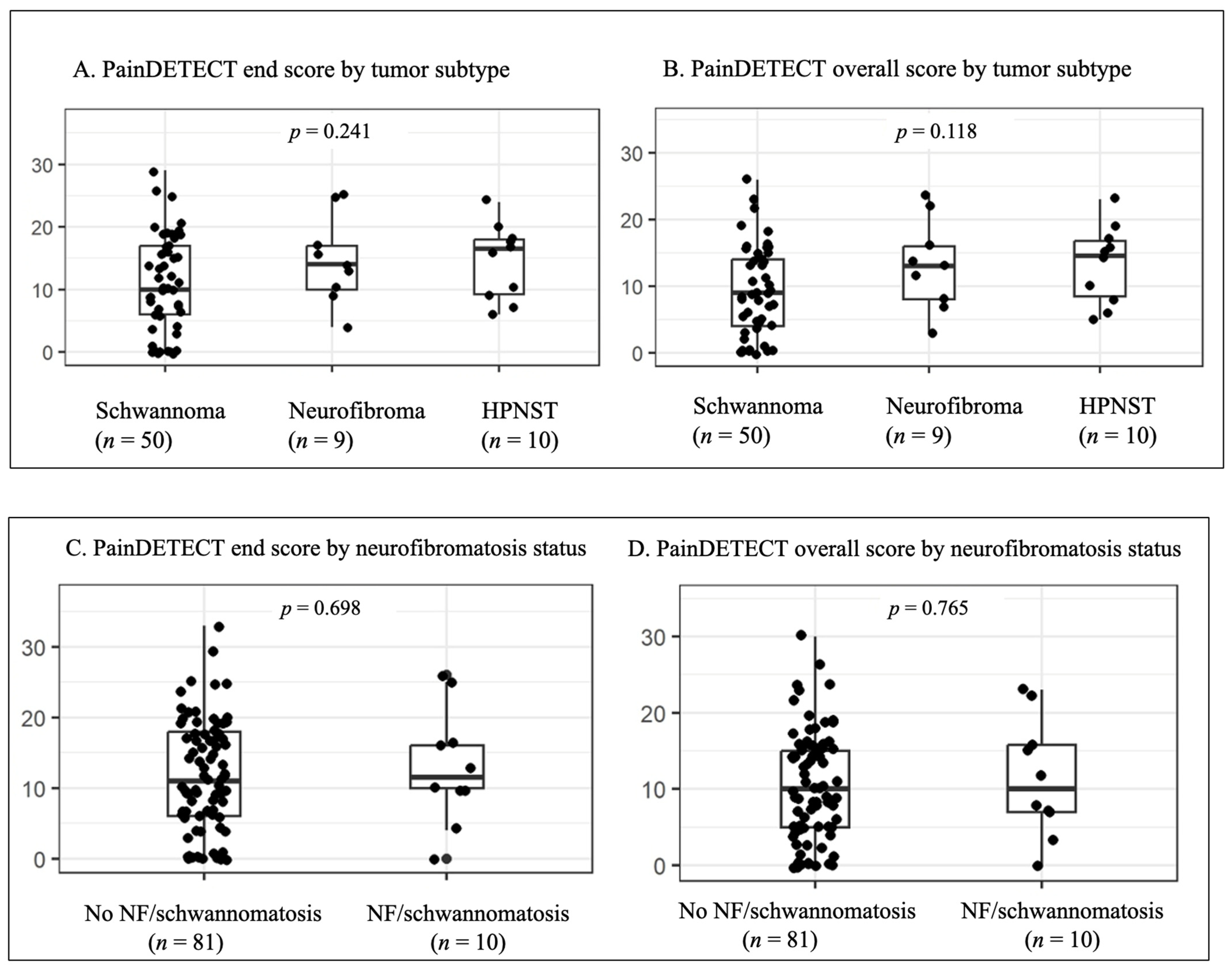
For subgroup analysis, Group 1 tumors were classified as schwannomas (
n = 50, 72.4%), Hybrid Peripheral Nerve Sheath Tumors (HPNST;
n = 10, 14.4%, all hybrid schwannoma/neurofibroma), or neurofibromas (
n = 9, 13.0%). HPNST and neurofibromas showed numerically higher PainDETECT end and overall scores than schwannomas; however, these differences were not statistically significant (
p = 0.241 and
p = 0.118, respectively;
Figure 1).
Similar results were observed in patients with a neurofibromatosis spectrum disorder (
n = 10, 10.9%), including six patients with neurofibromatosis type 1 (NF1), two with LZTR1-related schwannomatosis, and two with non-NF-associated schwannomatosis. PainDETECT end and overall scores did not differ significantly between patients with and without neurofibromatosis-spectrum disorders (
p = 0.698 and
p = 0.765, respectively;
Figure 1).
Eleven of 91 patients had undergone resection of more than one tumor (12.1%), whereas this information was unknown or not documented in 8 patients (8.8%). Among these 11 patients, the currently analyzed index lesions were schwannomas in 5 cases (45.5%), neurofibromas in 3 cases (27.3%), hybrid nerve sheath tumors in 2 cases (18.2%), and lipoma in 1 case (9.1%). Six patients had undergone surgery for three tumors (54.5%), and two patients had undergone surgery for more than five tumors (18.2%). Seven of the 11 patients with multiple tumor resections had an associated tumor predisposition syndrome (63.6%). In these patients, the PainDETECT questionnaire was completed only for the most recently operated tumor, which was defined as the index lesion. The mean PainDETECT overall score of these index lesions was 11.8, and the mean PainDETECT end score was 13.7.
3.3. Relationship Between PainDETECT Scores and Localization
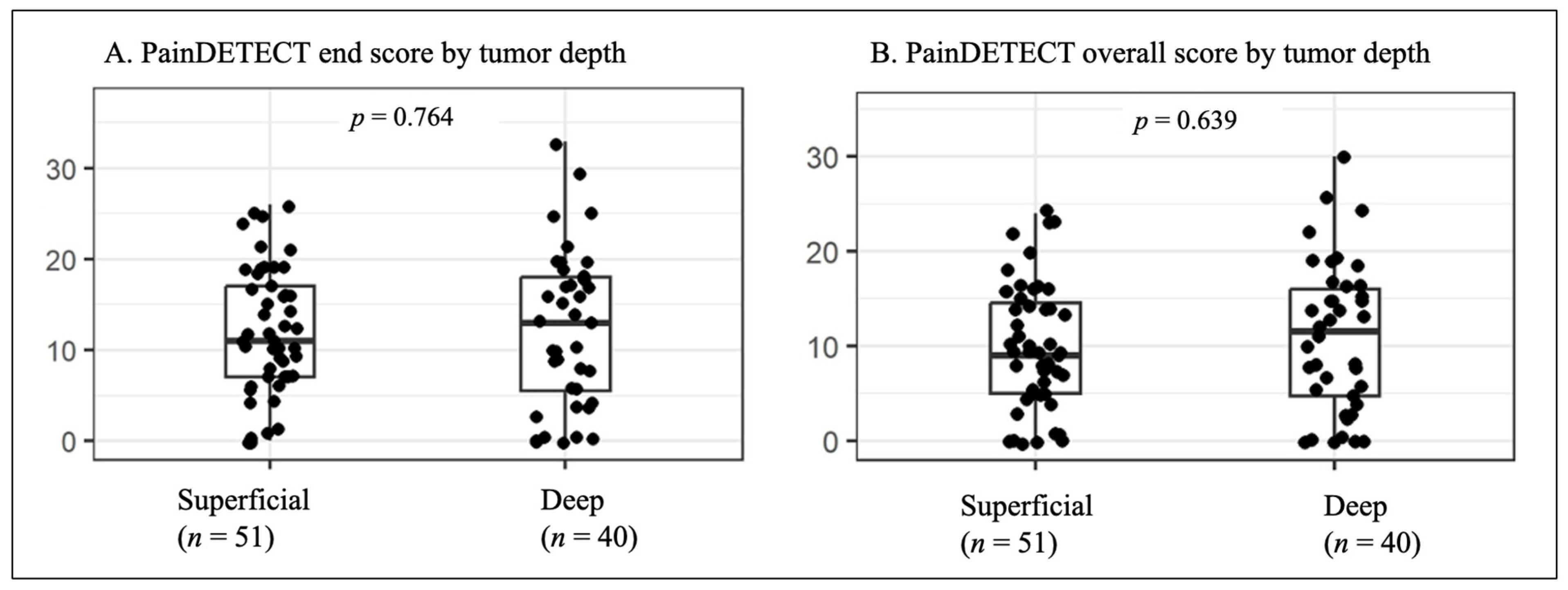
To assess the influence of tumor localization on pain, PainDETECT end and overall scores were analyzed according to anatomical localization and the most frequently affected nerves. Tumor depth was classified as superficial in 51 patients (56.0%) and deep in 40 patients (43.9%). Although the initial hypothesis suggested that superficial tumors would be associated with higher PainDETECT scores, the mean scores were slightly higher in deep tumors. This difference was not statistically significant (
p = 0.764 for end score,
p = 0.639 for overall score,
Figure 2).
Tumors involving the brachial plexus and sciatic nerve tumors had relatively high mean scores (14.6 (range 0–29) and 14.3 (range 4–20), respectively). In contrast, peroneal and lumbosacral plexus lesions had lower mean scores (12.1; range 0–25 and 12.1; range 0–33, respectively).
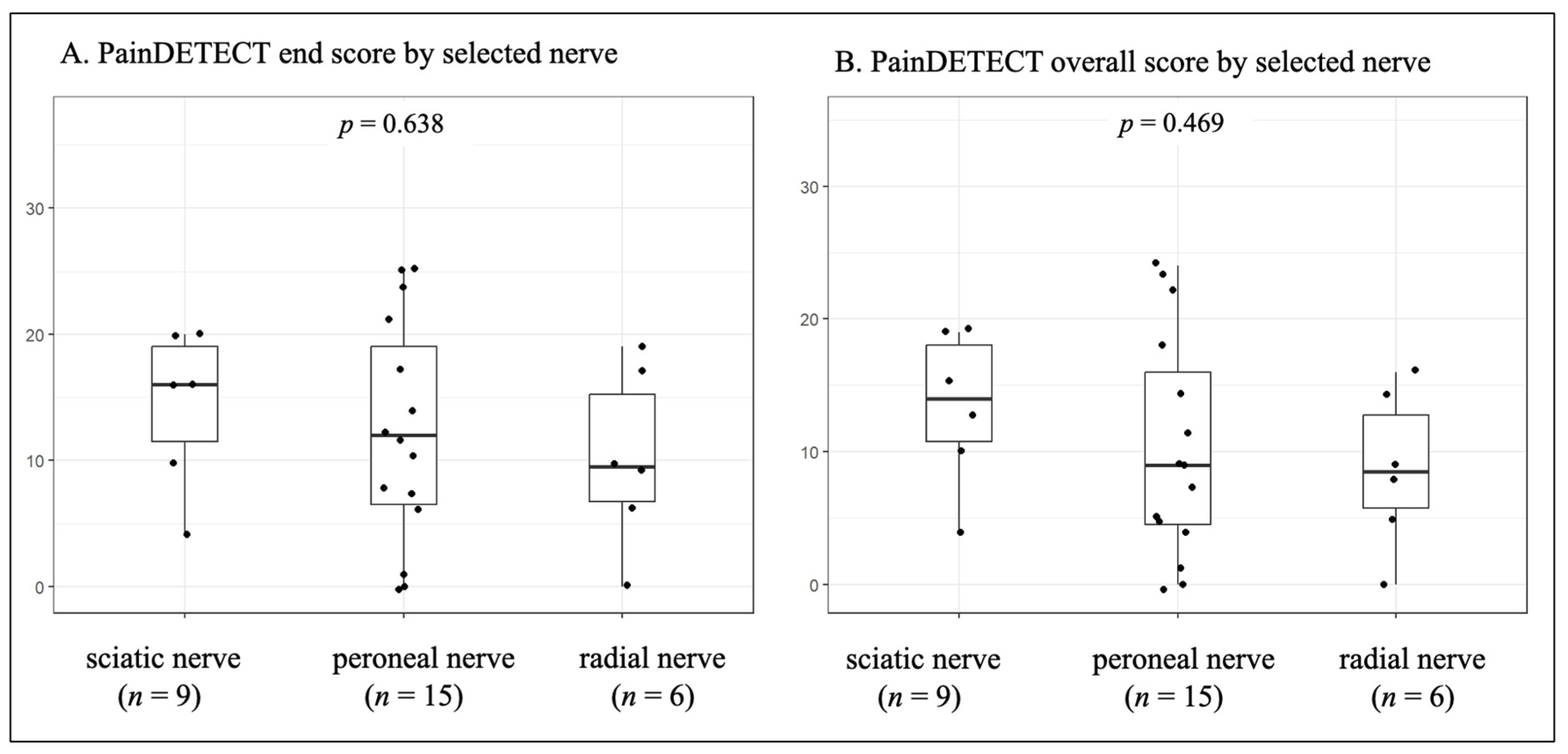
Figure 3 shows PainDETECT end and overall scores for tumors affecting the sciatic, peroneal, and radial nerves. Sciatic nerve tumors showed numerically higher scores; however, these differences were not statistically significant (
p = 0.638 for the end score,
p = 0.469 for the overall score).
3.4. Relationship Between PainDETECT Scores and Tumor Size
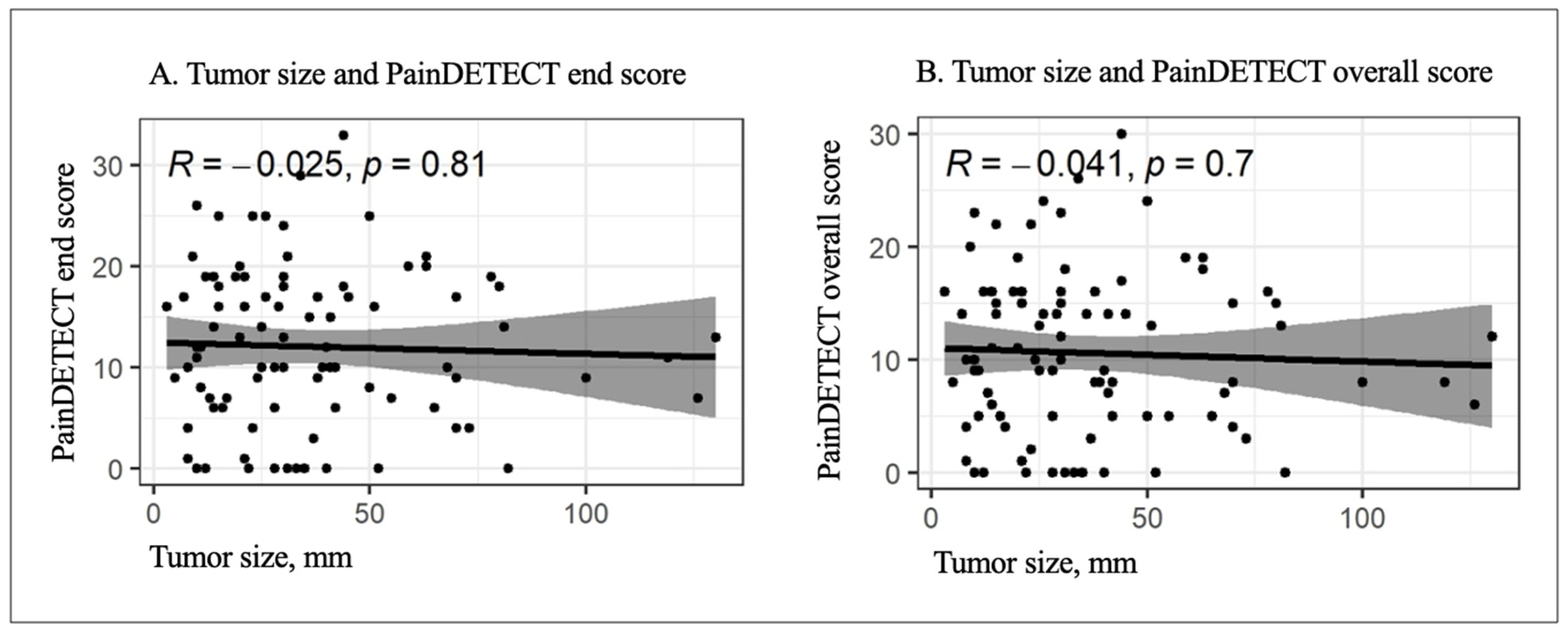
In the overall cohort, tumor size was not significantly correlated with PainDETECT scores (
Figure 4). Both the end score (rho = −0.025,
p = 0.81) and overall score (rho = −0.041,
p = 0.7) showed very weak, non-significant, negative correlations, indicating no association between tumor size and pain severity in this cohort.
3.5. Pain-Course Patterns
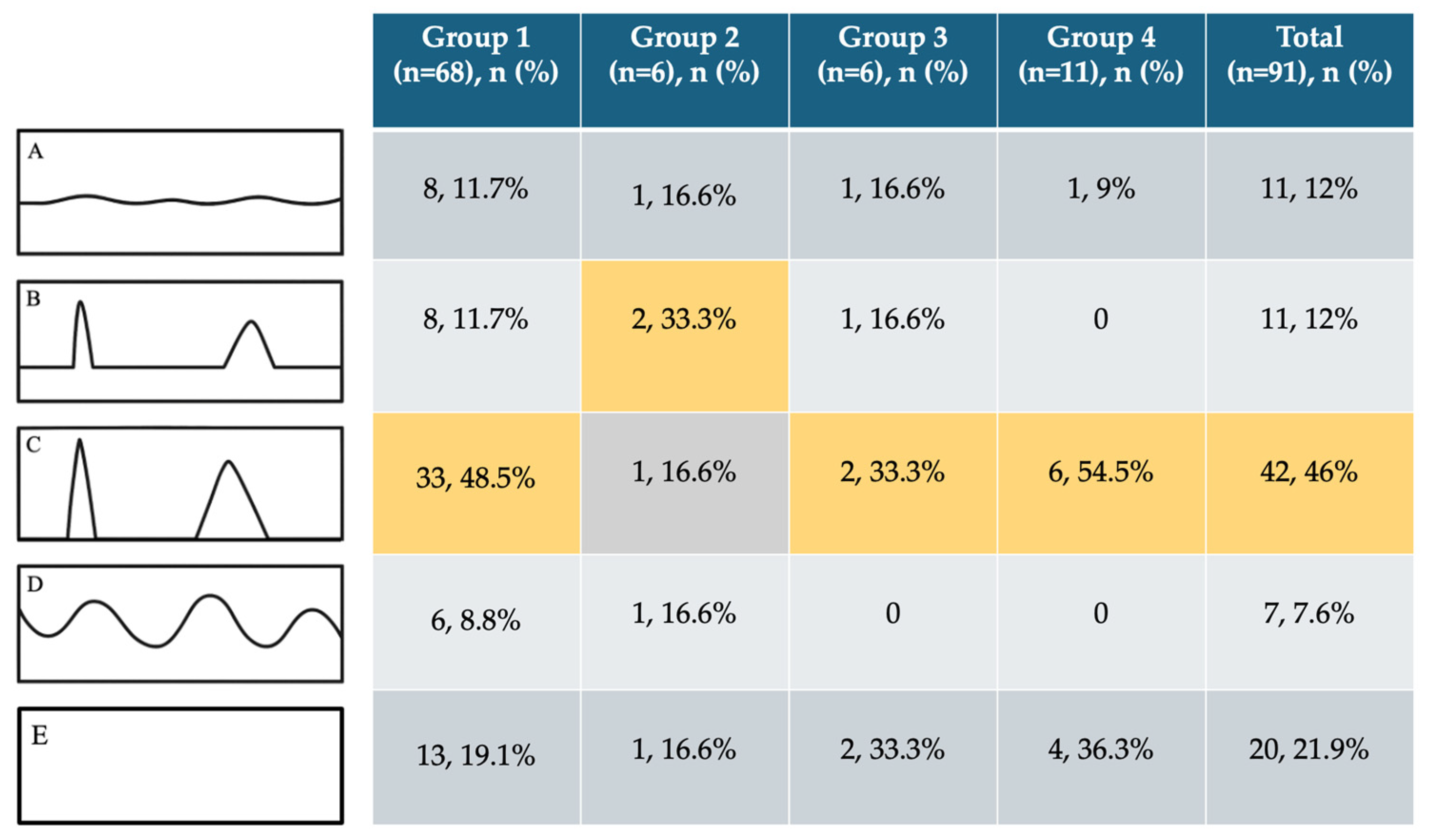
The most common pain-course pattern in the overall cohort was type C (pain attacks with pain-free intervals;
Figure 5), observed in 42 of 91 patients (46%). This pattern also predominated in nearly all histological subgroups, except for Group 2, in which type B (continuous pain with intermittent pain attacks) was most frequent (
n = 2). Pain-free patients (type E) accounted for 21% of the cohort, whereas continuous or fluctuating pain patterns (types A and D) were less common.
3.6. Numerical Rating Scale and Pain Dimensions
Pain intensity on the numerical rating scale (NRS, range 0–10; 10 indicating the most severe pain) was assessed as current, maximum, and average pain. Group 2 (malignant tumors) reported the highest pain levels, with a mean current NRS of 4.1, a maximum of 7.1, and an average of 5.5. In contrast, Groups 1 (benign), 3 (rare tumors), and 4 (dropout lesions) had lower mean NRS scores of 3.3, 2.6, and 2.8, respectively.
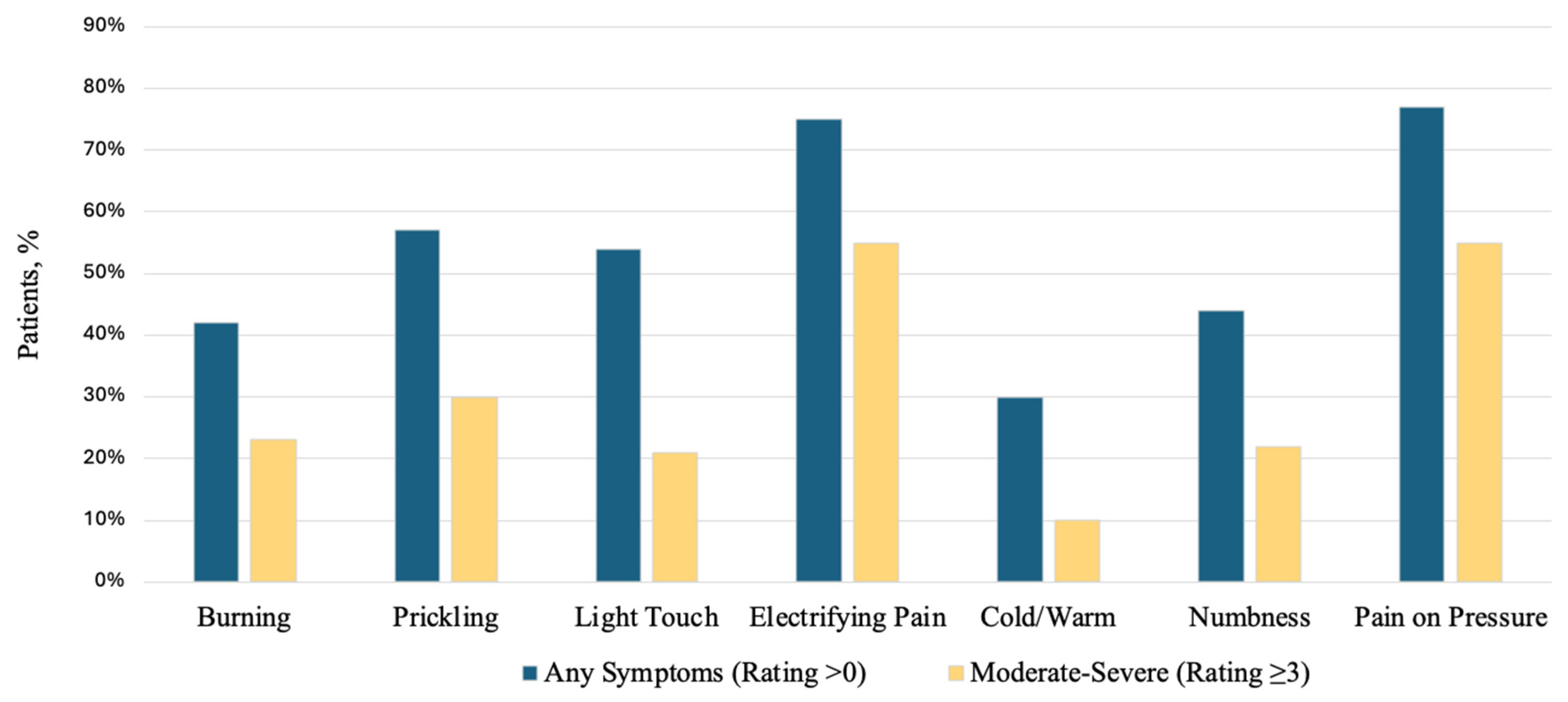
Across pain dimensions, the proportion of patients reporting any symptom (rating > 0) ranged from 30% for cold/warm symptoms to 77% for pressure pain. Pressure-related pain (77%) and electric-shock-like pain (75%) were the most frequent symptoms, and both were most often moderate-to-severe (rating ≥ 3; 55% each). Prickling/tingling (57% any symptom; 30% moderate-to-severe) and pain on light touch (54%; 21%) were also common but less frequently moderate-to-severe. Burning pain (42%; 23%) and numbness (44%; 22%) affected a substantial subset of patients, whereas cold/warm symptoms were least prevalent and rarely moderate-to-severe (30%; 10%). These Results are shown in
Figure 6. Consistent with these findings, 79 out of 91 patients (86.8%) had a positive Tinel sign on clinical examination.
3.7. Comorbidities, Previous Malignancy, and Pain Medication
Data on comorbidities and preoperative medication were available for 73 of 91 patients (80.2%); no data were available for 18 patients (19.8%). Thirty-five patients had no documented relevant comorbidity (35/73, 47.9%). The most frequent comorbidity was arterial hypertension requiring medication (14/73, 19.2%), followed by previous malignancy (8/73, 11.0%), depression (4/73, 5.5%), type 2 diabetes mellitus requiring medication (3/73, 4.1%), and hypercholesterolemia (3/73, 4.1%). A history of malignancy was documented in 8 patients (8/73, 11.0%), including optic glioma in 1 patient (1/73, 1.4%), malignant melanoma in 2 patients (2/73, 2.7%), prostate cancer in 2 patients (2/73, 2.7%), breast cancer in 2 patients (2/73, 2.7%), and testicular cancer in 1 patient (1/73, 1.4%). One patient had previously received chemotherapy with vincristine and carboplatin for optic glioma (1/73, 1.4%).
Preoperative pain medication was not required in most patients with available data (61/73, 83.6%), whereas 12 patients received analgesics (12/73, 16.4%). Medication included non-opioid analgesics, opioid or opioid-like analgesics, and drugs typically used for neuropathic pain, including gabapentin, pregabalin, duloxetine, amitriptyline, or amineurin. Only one patient with a previous malignancy received preoperative pain medication (1/8, 12.5%). Postoperatively, 63 of 73 patients (86.3%) received mainly ibuprofen and/or metamizole as needed for wound pain, which was discontinued according to clinical need.
4. Discussion
This study represents one of the first standardized, prospective assessments of preoperative pain symptomatology in syndromic and non-syndromic patients with PNTs using a validated instrument (PainDETECT) in a bicenter setting. The findings highlight pain as a relevant and underrecognized clinical symptom in PNTs, including benign subtypes. Although malignant tumors had the highest mean PainDETECT scores, a substantial proportion of patients with benign tumors, including schwannomas and HPNSTs, also reported moderate to high pain levels.
4.1. Malignancy and Syndromic Features
Pain is a common and clinically relevant feature of PNTs and can substantially impair quality of life. Previous studies suggest that approximately three-quarters of patients with a peripheral nerve sheath tumor experience pain in some form [
1]. In the present cohort, more than two-thirds of patients reported some degree of pain, whereas only 12% had a pain score of zero.
MPNSTs are aggressive sarcomas that frequently arise from pre-existing plexiform neurofibromas in patients with NF1, although they may occur sporadically [
17,
18,
19]. While MPNSTs are life-threatening malignancies, pain is often the earliest and most prominent clinical symptom. Sudden onset or marked increase in pain intensity, particularly in a previously stable neurofibroma, may indicate malignant transformation. Patients may also present with numbness or focal neurological deficits. Compared with benign peripheral nerve tumors, MPNSTs are typically associated with more severe and persistent pain profiles [
20].
The finding that more than 22% of patients had PainDETECT scores ≥ 19, suggesting likely neuropathic pain, supports the concept that pain in PNTs is not limited to malignancy. Even patients with perineurioma (
n = 3 in this cohort), an entity generally considered less painful [
21,
22], reported a mean end score of 7 (range, 1–10).
Syndromic patients frequently experience pain. In one PainDETECT-based study, approximately 6 of 20 patients with schwannomatosis scored in the range suggestive of neuropathic pain [
23]. Studies comparing painful vs. nonpainful schwannomas have suggested potential molecular mechanisms: painful schwannomas secrete higher levels of certain cytokines and growth factors, such as IL-6, CXCL1 and VEGF, which may sensitize sensory neurons [
24]. Systemic medical treatment options have also emerged; for example NF1 patients receiving selumetinib have reported lower pain intensity and reduced pain-related interference with daily activities [
6,
25]. Pain in non-NF2 schwannomatosis is often chronic, treatment-resistant, and associated with anxiety and depression [
26]. In NF1, up to 50–60% of patients with plexiform neurofibromas may experience tumor-associated neuropathic pain [
27].
Notably, HPNSTs and neurofibromas showed numerically higher pain scores than schwannomas, although these differences were not statistically significant. HPNSTs remain incompletely understood but are frequently associated with syndromic conditions such as neurofibromatosis and schwannomatosis [
28,
29].
4.2. Size and Location
The hypothesis that superficial tumors cause more pain was not confirmed. Deep lesions showed slightly higher mean scores, although this difference was not statistically significant. Although lower-extremity lesions showed a slightly higher proportion of patients within the neuropathic PainDETECT range, no robust association between anatomical location and specific pain quality was identified in this cohort. Similarly, tumor size was not correlated with pain intensity (rho = −0.025), challenging the assumption that larger tumors invariably generate more pain. Previous studies suggest that pain in PNSTs is heterogeneous and cannot be explained by tumor size or volume alone. Additional factors, including nerve involvement and tumor–nerve interactions, are likely to contribute to pain severity [
3,
24,
30]. Most previous studies have focused on syndromic patients, whereas data in non-syndromic patients remain limited.
The functional profile of the affected nerve is also relevant. In the present cohort, several affected nerves had mixed sensorimotor function, including the sciatic, peroneal, and ulnar nerves, as well as the brachial and lumbosacral plexuses. A strict classification into purely sensory and purely motor nerves was therefore not feasible.
Schwannoma-related pain is often episodic or movement-related rather than spontaneous. Patients may notice pain during specific movements, exercise, or palpation of the lesion, rather than severe nocturnal or resting pain. This pattern is consistent with our finding that pain attacks with pain-free intervals (type C) were common in benign tumors (48.5%) and supports the concept that benign schwannomas may cause pain mainly through nerve compression or stretch.
4.3. Clinical Implications
Pain is a major source of morbidity in patients with peripheral nerve tumors, and these prospective baseline data provide clinically relevant insights into the preoperative pain phenotype and patient-reported burden. Because benign tumors represent the largest subgroup, their pain symptoms should not be underestimated but actively addressed in clinical care. Although outcome analyses require complete follow-up, the current findings support timely surgical intervention in selected patients—even those without neurological deficits—when pain is significant, and imaging suggests operability. Future analyses, including follow-up and medication data, will clarify the extent and persistence of pain relief across tumor types. A patient reporting high pain intensity (e.g., NRS 7/10) and PainDETECT scores ≥ 19 may benefit from targeted neuropathic pain treatment. Schwannomas typically cause intermittent, stimulus-dependent pain, often tingling or shooting upon pressure, and usually respond well to surgical removal [
10,
31]; however, in schwannomatosis, multiple tumors make pain management more complex and frequently require combined medical and surgical strategies. Overall, improved characterization of pain mechanisms in these tumors may support more precise clinical assessment and management.
An additional clinically relevant observation concerns the dropout group. Although these lesions were not histologically confirmed as PNSTs, they had close anatomical nerve contact and clinically mimicked peripheral nerve tumors. Some of these entities, particularly angioleiomyoma and glomus tumors, showed high individual PainDETECT scores. This is consistent with the fact that especially benign subcutaneous tumors may cause severe pain [
32,
33,
34] and therefore frequently lead to surgical resection. Accurate preoperative differentiation from true peripheral nerve tumors remains essential [
35].
PainDETECT was selected as an established, standardized, and easy-to-use tool for capturing multiple pain qualities in routine clinical practice. However, elevated scores should be interpreted as neuropathic-like pain features rather than definitive evidence of nerve fiber dysfunction, particularly in benign tumors, where deep somatic pain, pressure effects, or mechanical nerve irritation may also contribute. Treatment decisions should therefore integrate clinical examination, imaging, and the individual pain phenotype.
4.4. Influence of Comorbidities, Previous Medical History, Medication, and Multiple Tumors
Comorbidities, previous medical history, and medication use are relevant confounders when interpreting PainDETECT and NRS scores. Data on comorbidities and medication were available for 73 of 91 patients (80.2%). Depression was documented in 4 patients (5.5%), type 2 diabetes mellitus in 3 (4.1%), and previous malignancy in 8 (11.0%). These factors, as well as previous oncological treatment, may influence pain perception and patient-reported outcomes independently of the peripheral nerve tumor itself. Because the subgroups were small and heterogeneous, adjusted statistical analyses were not feasible.
Preoperative pain medication was required in only 12 of 73 patients (16.4%), whereas most patients with available data did not require preoperative analgesics (61/73, 83.6%). Medication included non-opioid analgesics, opioid or opioid-like analgesics, and drugs commonly used for neuropathic pain. These treatments may have influenced PainDE-TECT and NRS scores. Therefore, low pain scores should not automatically be interpreted as evidence of an intrinsically painless lesion, whereas high scores may reflect insufficiently controlled pain, mixed nociceptive–neuropathic pain, or tumor-related nerve irritation. Postoperative medication consisted mainly of ibuprofen and/or metamizole as needed for wound pain in 63 of 73 patients (86.3%) and was therefore not interpreted as a standardized postoperative pain outcome.
Patients with multiple tumors represent an important subgroup, particularly in syndrome-associated peripheral nerve tumors. In this cohort, 11 of 91 patients had undergone more than one tumor resection (12.1%), and 7 of these 11 patients had an associated tumor predisposition syndrome (63.6%). In this interim analysis, PainDETECT was assessed prospectively only for the most recently operated index lesion, while previous tumor resections were documented retrospectively.
Future PNTR analyses will assess PainDETECT before each new tumor operation and separately for each tumor, aiming to identify which tumors, locations, or histological subtypes are associated with the highest pain burden in patients with multiple lesions. The final PNTR analysis is planned once at least 200 patients with histologically confirmed peripheral nerve tumors have been included, excluding the drop-out cohort, and complete follow-up data are available.
4.5. Limitations and Strengths
Limitations: This study has several limitations. First, the cohort predominantly comprised benign tumors, limiting generalizability to malignant and rare entities. Second, subgroup sizes were small; therefore, all histopathological comparisons should be interpreted as exploratory and hypothesis-generating. Third, postoperative follow-up data were incomplete, precluding analysis of longitudinal pain trajectories and postoperative neurological deficits. In the present analysis, postoperative deficits were not evaluated because the primary focus was on preoperative pain symptomatology. In the overall PNTR cohort, malignant tumors appear to be associated with more frequent preoperative pain, often accompanied by motor and sensory deficits that may persist; these data are not included in the present publication and will be analyzed separately. Fourth, comorbidities and medication use represent relevant confounders. Depression, diabetes mellitus, previous malignancy, prior chemo- or radiotherapy, chronic pain disorders, neuropathic pain conditions, and analgesic or neuropathic pain medication may influence PainDETECT and NRS scores independently of the tumor itself. Although these variables were reviewed descriptively, incomplete documentation and the limited sample size prevented adjusted analyses. Therefore, tumors should not be classified as intrinsically painful or painless based solely on PainDETECT scores. Finally, the lesion–nerve relationship, including extrinsic compression, displacement, stretching, infiltration, or destruction, could not be reliably classified in all cases.
Strengths: This study is among the first standardized, prospective assessments of preoperative pain symptomatology in patients with peripheral nerve tumors using a validated instrument (PainDETECT) in a bicenter setting. Its focus on pain, often the dominant patient-reported burden, adds substantial clinical relevance.



 ,
,