Article

Presence of Adipophilin-Positive Cancer-Associated Fibroblasts Is an Independent Poor Prognostic Indicator and Is Correlated with Immature-Type Desmoplastic Reaction in Patients with Colorectal Cancer

1
Department of Colorectal Surgery, Kansai Medical University, 2-5-1, Shinmachi, Hirakata 573-1010, Japan
2
Department of Pathology, Osaka Medical and Pharmaceutical University, 2-7, Daigaku-machi, Takatsuki 569-8686, Japan
*
Authors to whom correspondence should be addressed.
These authors contributed equally to this work.
Cancers 2025, 17(18), 3006; https://doi.org/10.3390/cancers17183006
Submission received: 27 July 2025 / Revised: 2 September 2025 / Accepted: 11 September 2025 / Published: 15 September 2025
(This article belongs to the Section Tumor Microenvironment)

Simple Summary

Immature-type desmoplastic reaction (IM-type DR) is a histopathological indicator for poor prognosis in colorectal carcinoma (CRC); however, the underlying mechanism has not been clarified. Recent studies showed that lipid metabolism in cancer-associated fibroblasts (CAFs) can have critical impact on cancer cell growth. Our findings demonstrated that the presence of lipid-laden CAFs was an independent indicator of poor prognosis and was significantly correlated with presence of IM-type DR in CRC. Additionally, adipophilin expression in carcinoma cells at the invasive front was significantly correlated with both IM-type DR and the presence of lipid-laden CAFs. The presence of lipid-laden CAFs in CRC may enhance cancer progression and metastasis, and may link a lipid-rich tumor microenvironment.

Abstract

Background/Objectives: Immature-type (IM type) desmoplastic reaction (DR) is a poor prognostic indicator of CRC; however, the underlying mechanism has not yet been clarified. Recent studies have demonstrated that energy metabolism, including lipid metabolism, in cancer-associated fibroblasts (CAFs) can critically impact cancer cell growth. The prognostic significance of lipid-laden CAFs in CRC has not yet been elucidated. This study aimed to clarify whether the presence of lipid-laden CAFs correlates with the DR type and prognosis in CRC. Methods: We enrolled patients with pT3 or pT4 CRC who underwent surgical resection without neoadjuvant chemoradiotherapy. Immunohistochemical staining for adipophilin (ADP) was performed to determine the presence of lipid-laden CAFs. Results: The cohort included 70 patients (49 men and 21 women) with a median age of 74 years. ADP-positive lipid-laden CAFs were observed in 37 patients (53%). The presence of lipid-laden CAFs and ADP expression in carcinoma cells at the invasive front was significantly correlated with IM-type DR (p < 0.0001 and p = 0.0063, respectively). The presence of lipid-laden CAFs was an independent poor prognostic indicator based on multivariate analysis (hazard ratio: 3.65; 95% confidence interval: 1.082–13.038; p = 0.0368). Conclusions: These results suggest that the presence of lipid-laden CAFs enhance cancer progression, invasion, and metastasis, and that IM-type DR might be related to the lipid-rich tumor microenvironment.

1. Introduction

Colorectal cancer (CRC) is a common cause of cancer-related deaths worldwide, and its incidence is steadily increasing [1]. Therefore, establishing effective and useful prognostic indicators for patients with CRC is critical for prognostic stratification and developing treatment strategies in oncology [1]. The histopathological parameters of resected CRC specimens can provide pivotal information for prognosis. For example, histological differentiation grade, lymphovascular invasion, perineural invasion, and tumor budding (TB) have been recognized as established prognostic indicators [2,3,4,5,6]. Desmoplastic reaction (DR) is a tissue reaction that occurs around carcinoma cell nests to varying extents, such as presence of myxoid material or thick collagen bundles [7]. Types of DR were first reported as significant prognostic indicators in patients with pT3 or pT4 CRC by Ueno et al. [8], and subsequent articles have addressed their prognostic significance in other types of carcinomas [9,10]. The type of DR may reflect the tumor microenvironment (TME), which plays a pivotal role in tumor progression, invasion, and metastasis [7,8,9,10,11]. DR is classified into three categories: immature (IM), intermediate, and mature types, and IM-type DR, which is histopathologically characterized by the presence of myxoid material (amorphous mucoid material) at the invasive front of the tumor by hematoxylin and eosin staining, indicates a significantly worse prognosis in patients with CRC, followed by the intermediate and mature types, respectively (the detailed histopathological definition is described in the Histopathological analysis section) [7,8,12,13,14,15]. IM-type DR is significantly correlated with the presence of lymphovascular invasion, lymph node metastasis, and higher TB [7,8,12,13,14,15]. However, the mechanisms underlying its development and the relationship between the presence of IM-type DR and poor prognosis have not yet been elucidated, although the TME might influence the development of DR [7,12].
It has been demonstrated that energy metabolism markedly changes in carcinoma cells, and metabolic reprogramming is considered a hallmark of cancer [16]. The reprogramming of energy metabolism, including glucose, amino acids, and lipids, plays crucial roles in tumor initiation, progression, and metastasis [16,17,18,19,20], and lipid metabolism is usually upregulated in carcinoma cells [17,20]. Previous studies have extensively analyzed changes in energy metabolism in carcinoma cells [16,17,18,19,20]. Additionally, metabolic changes in the TME, such as in cancer-associated fibroblasts (CAFs) and inflammatory cells, have recently received much attention because it has been demonstrated that these changes could have a critical impact on cancer cell growth and metastasis via crosstalk between carcinoma cells and the TME [19,21,22]. Recent studies have demonstrated that the upregulation of lipid metabolism in CAFs plays a pivotal role in tumor growth and progression [23,24,25,26]. For example, Liu et al. demonstrated that lipid synthesis was upregulated in CAFs of oral squamous cell carcinoma, and these lipid metabolites were taken up by carcinoma cells, leading to tumor proliferation, migration, and invasion [23]. Niu et al. showed that pancreatic cancer cells facilitated lipid synthesis in CAFs, and these lipids were transferred to carcinoma cells, leading to the proliferation of cancer cells using a transgenic mouse model [24]. These CAFs are termed lipid-laden CAFs because lipid synthesis is upregulated, and lipids are stored in the cytoplasm [24]. Rich lipid storage can be visualized by immunohistochemical staining for adipophilin (ADP), a lipid-regulating protein that coats the surface of cytoplasmic lipid droplets [27]. Although upregulating lipid metabolism in CAFs in CRC has also been reported in a few articles [25,26], the relationship between the presence of lipid-laden CAFs, the type of DR, and prognosis has not yet been analyzed. This study aimed to examine the relationship between the presence of lipid-laden CAFs and the type of DR and their prognostic significance and to elucidate the correlation between the type of DR and prognosis.

2. Materials and Methods

2.1. Patient Selection

We enrolled consecutive patients with CRC who underwent surgical resection at the Department of Surgery of Kansai Medical University Hospital between January and December 2016. Patients with pT1 or pT2 disease were excluded since DR is defined as pT3 or pT4 [7,8,12,13,14,15]. Patients with lower or middle rectal cancers were excluded because neoadjuvant chemotherapy and/or radiation therapy were performed in most of these patients in our hospital. Patients who underwent neoadjuvant chemotherapy and/or radiation therapy were excluded. We set the observation window to 5 years according to the Japanese Society for Cancer of the Colon and Rectum guideline [28]. Moreover, no data regarding KRAS or BRAF mutation, and mismatch repair proteins status, were available.
The study cohort partially overlapped with that of our previous studies [2,29]. In one study, we showed that IM-type DR was significantly correlated with the presence of tumor deposits, which are discrete macroscopic or microscopic carcinoma nodules located in the extramural fatty tissue, and without lymph node structures [29]. In another study, we proposed a prognostic scoring system for patients with CRC, which was based on indicators reflecting the TME, including DR [2]. The results of this study did not overlap with those of these previous studies [2,29].
This retrospective study was conducted in accordance with the principles of the Declaration of Helsinki. The study protocol was approved by the Institutional Review Board of Kansai Medical University Hospital (Approval #2021-197). All data were anonymized. The Institutional Review Board waived the requirement for informed consent, because this was the retrospective study, and medical records and archived samples were used, and there was no risk to the participants. Additionally, this study did not include children. Information regarding this study was provided through the institutional website (https://www.kmu.ac.jp/hirakata/hospital/2671t800001356c-att/a1642567101597.pdf) (accessed on 27 July 2025).

2.2. Histopathological Analysis

Surgically resected specimens were fixed in 10% buffered neutral formalin, sectioned, and stained with hematoxylin and eosin. Two researchers (T.K. and M.I.) blinded to the clinical features independently evaluated the histopathological features of all tumor slides. The staging of patients with CRC is currently described in the American Joint Committee on Cancer Staging Manual, Eighth edition [30], which is based on the depth of tumor invasion (pT) and tumor lymph node and/or distant metastases (pN and M).
DR was classified into the IM, intermediate, and mature types according to the definition by Ueno et al. in patients with pT3 or 4 CRC [7,8,12,13,14,15]. Briefly, the IM type was histopathologically defined as the presence of myxoid stroma (amorphous mucoid material) greater than the microscopic field of ×400 at the invasive front of the carcinoma. The intermediate type was defined as the presence of keloid-like collagen (thick bundles of collagen accompanying hyalinization) without myxoid stroma, whereas the absence of myxoid stroma and keloid-like collagen was regarded as the mature type. In this study, DR was sub-classified into IM and non-IM (intermediate and mature) types.
TB was evaluated using the same method as previously reported [31]. Tumors with 0–4 buds (defined as the presence of a single tumor cell or a cluster of up to four tumor cells) at the invasive front were classified as TB1, 5–9 buds as TB2, and >10 buds as TB3.

2.3. Immunohistochemical Analysis

A whole section of the most morphologically representative carcinoma regions with DR, identified by hematoxylin and eosin-stained slides, was used for immunohistochemical staining of each tumor. Immunohistochemical staining was performed using an autostainer (Discovery ULTRA system; Roche Diagnostics, Basel, Switzerland) according to the manufacturer’s instructions. An OptiView DAB Universal Kit (760-700) was used for immunostaining. Dual immunohistochemical staining was performed using an autostainer (Leica BOND-MAX; Leica Biosystems GmbH, Nußloch, Germany) according to the manufacturer’s instructions. BOND Polymer Refine Detection Kit (DS9800; Leica) and BOND Polymer Refine Red Detection Kit (DS9390; Leica) were used. A primary mouse monoclonal antibody against ADP (AP125, ×100 dilution, Progen Biotechnik, Heidelberg, Germany) and a primary mouse monoclonal antibody against alpha-smooth muscle actin (alpha-SMA) (1A4, ×800 dilution, DAKO, Agilent Technologies, Inc., Santa Clara, CA, USA) were used. Human sebaceous glands and the muscularis propria of the colon were used as positive controls for ADP and alpha-SMA staining, respectively. Two researchers (T.K. and M.I.) blinded to the clinical or histopathological features independently evaluated the immunostaining results. When there were discrepancies of the results of the evaluation, a final decision was made by reassessment and agreement by two researchers.
First, we performed a single immunohistochemical staining for ADP in all CRC samples. ADP expression was evaluated in both the carcinoma cells and CAFs. If the tumors contained ADP-positive spindle cells around the carcinoma cell nests by single immunohistochemical staining for ADP, then dual immunohistochemical staining for ADP and alpha-SMA were performed to clarify whether these ADP-expressing spindle cells were CAFs or not. Both ADP and alpha-SMA were expressed in spindle cells, and these cells were recognized as lipid-laden CAFs. ADP-positivity in the carcinoma cells were evaluated by the morphological features as well as alpha-SMA negativity. ADP expression in both carcinoma cells and CAFs was identified as positive when carcinoma cells or CAFs exhibited granular and/or globular cytoplasmic expression, as previously reported [32,33,34,35,36]. When at least one ADP-expressing CAF was present, the tumor was classified as the presence of lipid-laden CAFs. We counted the percentage of ADP-expressing carcinoma cells (%). We also evaluated the presence of ADP-expressing carcinoma cells in the invasive front region of the carcinoma.

2.4. Statistical Analyses

JMP® Student Edition version 18.2.1 (SAS Institute, Cary, NC, USA) was used for statistical analyses. Correlations between the two groups were analyzed using the chi-square test or Fisher’s exact test. Logistic regression analysis was performed to estimate the odds ratios (ORs) and 95% confidence intervals (CIs). Variables with a p-value < 0.20 in the univariate analysis were included to identify independent associations with histological features for multivariate logistic regression. To avoid overfitting in the multivariate Cox proportional hazards model, we selected up to three variables based on both clinical relevance and statistical significance in the univariate analysis, in accordance with the commonly recommended rule of at least 10 events per variable. Overall survival (OS) was analyzed using the Kaplan–Meier method, and survival differences between the groups were compared using the log-rank test. Statistical significance was set at p < 0.05.

3. Results

3.1. Clinicopathological Characteristics of the Study Cohort

Table 1 summarizes the clinicopathological features of the study cohort. This study included 70 patients (29 [41%] women and 41 [59%] men). The median age at the time of surgery was 74 years (range: 47–89 years). The tumor locations were as follows: right-sided colon 40 (57%), left-sided colon 26 (37%), and upper rectum four patients (6%). Regarding the pathological T stage, 46 (66%) and 24 (34%) patients were classified as pT3 and pT4, respectively. Lymph node metastases were observed in 36 patients (51%). Thirty-two (46%), 25 (36%), and 13 (19%) patients were classified into pStages II, III, and IV, respectively. Figure 1 shows the flowchart of the patient selection process. The median observation period was 56.5 months (range: 0–77 months).

3.2. Histopathological Characteristics

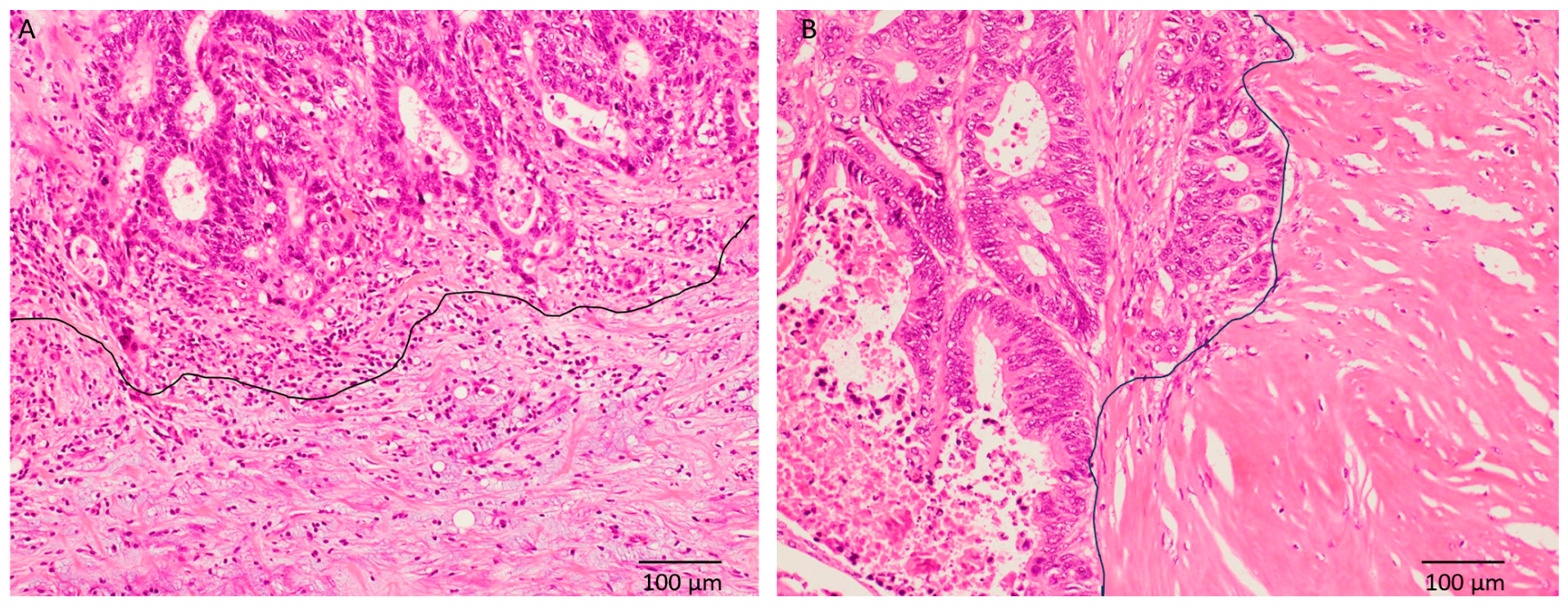
Thirty-five (50%) patients each were classified into IM and non-IM type DR, respectively. The typical histopathological features of IM-type DR and non-IM type DR (intermediate type) are shown in Figure 2. IM-type DR was significantly correlated with pT4, the presence of lymph node metastasis, and higher pStage (p = 0.0118, 0.0001, and 0.0006, respectively) (Table 1).

3.3. Immunohistochemical Characteristics

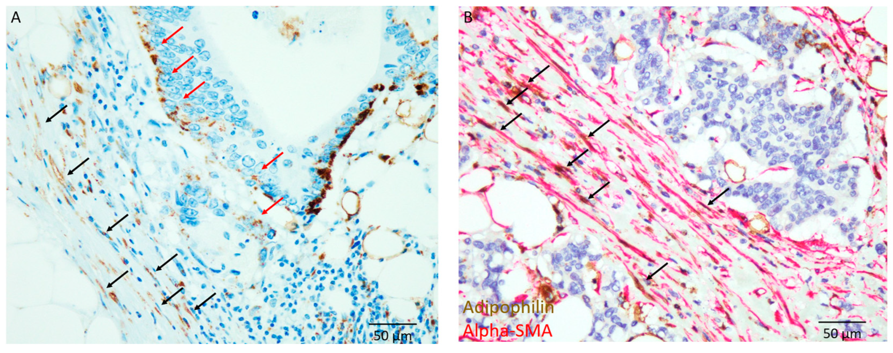
The typical features of ADP-positive lipid-laden CAFs at the invasive front are shown in Figure 3A. These ADP-positive lipid-laden spindle cells were also alpha-SMA-positive, according to dual immunohistochemical staining (Figure 3B). Therefore, these ADP-positive spindle cells at the invasive front were considered lipid-laden CAFs. Lipid-laden CAFs were observed in 37 patients (53%).
ADP expression was also observed in carcinoma cells. The median ratio of ADP-expressing carcinoma cells to total carcinoma cells was 10%; thus, we considered an ADP-high carcinoma if >10% of carcinoma cells showed positive immunoreactivity for ADP. Twenty-nine patients (41%) had ADP-high carcinomas (Table 1). ADP-high carcinomas were not significantly correlated with IM-type DR (p = 0.0894).
Additionally, ADP expression in carcinoma cells at the invasive front was observed in 51 patients (73%) (Figure 3A). ADP expression in carcinoma cells at the invasive front significantly correlated with IM-type DR (p = 0.0063).

3.4. Correlation Between Lipid-Laden CAFs and Clinicopathological Features

Table 2 summarizes the clinicopathological features of patients with or without lipid-laden CAFs. Lipid-laden CAFs were observed in 37 patients (53%) (28 and nine patients with IM-type and non-IM type DR, respectively), and the remaining 33 patients (47%) had no lipid-laden CAFs (seven and 26 patients with IM-type and non-IM type DR, respectively).
The presence of lipid-laden CAFs was significantly correlated with IM-type DR (p < 0.0001). It was also significantly correlated with the presence of lymph node metastasis, venous invasion, higher TB, and higher pStage (p = 0.0008, 0.0321, 0.0322, and 0.0035, respectively) but not with pT (p = 0.0946).
ADP-high carcinoma was not significantly correlated with the presence of lipid-laden CAFs (p = 0.873). However, ADP expression in carcinoma cells at the invasive front was significantly correlated with the presence of lipid-laden CAFs (p = 0.0014).

3.5. Prognostic Significance of the Type of DR and the Presence of Lipid-Laden CAFs

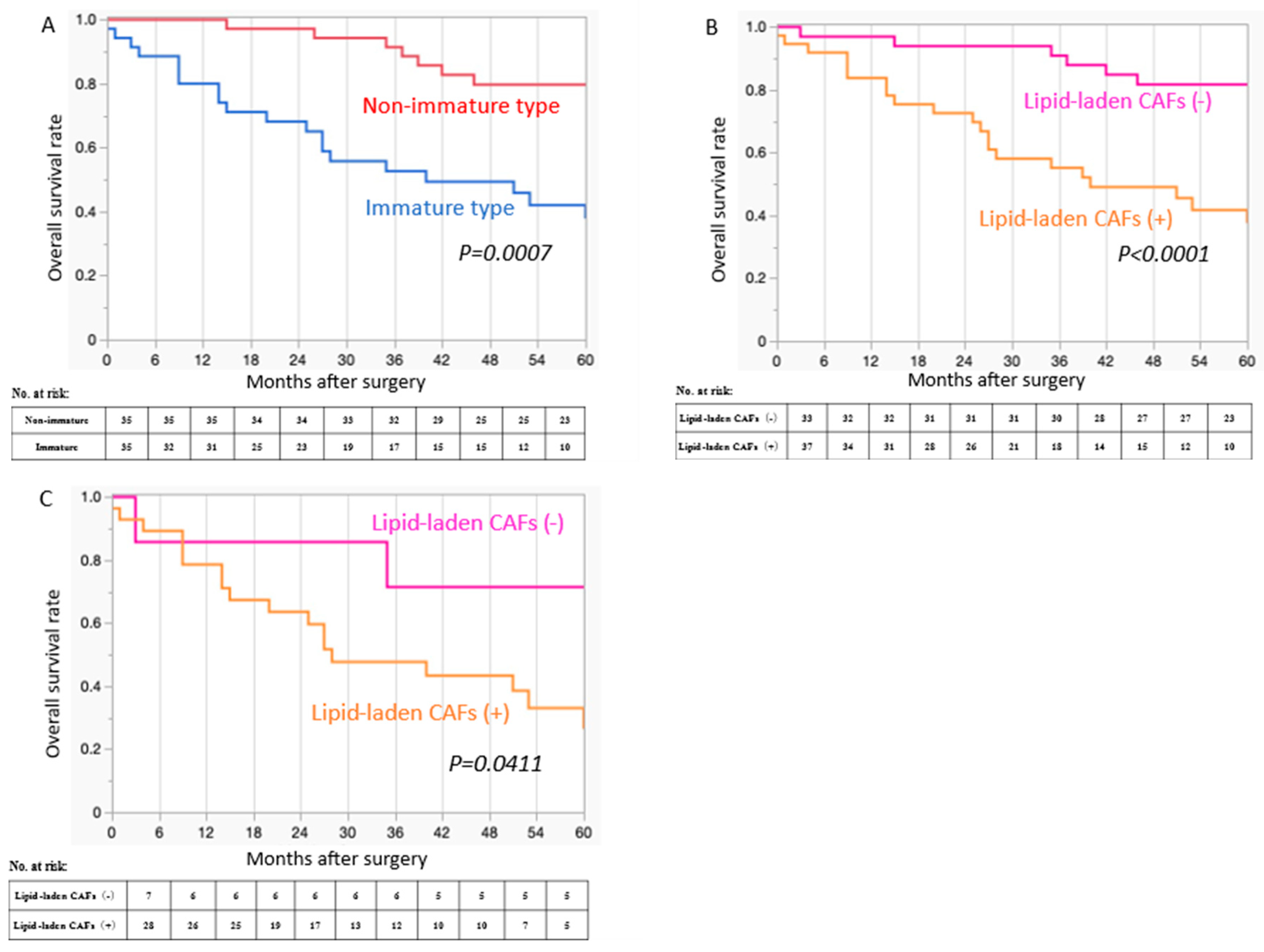
Figure 4A shows the Kaplan–Meier curves of OS according to DR type (IM vs. non-IM). The presence of IM-type DR significantly correlated with poor OS (p = 0.0007). The median 5-year OS in patients with IM-type DR was 35 months (range 0–77 months), whereas that in patients with non-IM-type DR was 61 months (range 15–75 months).
The presence of lipid-laden CAFs was significantly correlated with poor OS in all patient cohorts (p < 0.0001) (Figure 4B). The median 5-year OS in patients with lipid-laden CAFs was 36 months (range 0–77 months), whereas that in patients without lipid-laden CAFs was 61 months (range 3–77 months). In patients with IM-type DR, the presence of lipid-laden CAFs significantly correlated with poor OS (p = 0.0411) (Figure 4C). The median 5-year OS of IM-type DR patients with lipid-laden CAFs was 27 months (range 0–77 months), while that of patients without lipid-laden CAFs was 71 months (range 3–77 months).
ADP-high carcinomas and ADP expression in carcinoma cells at the invasive front were not correlated with OS p = 0.9175 and 0.2001, respectively.

3.6. Cox Proportional Hazards Regression Analysis for OS

Table 3 shows the results of the univariate and multivariate Cox proportional hazards regression analyses for OS. The univariate analysis revealed that IM-type DR, the presence of lymph node metastasis, and presence of lipid-laden CAFs were significantly associated with OS (p = 0.0038, 0.0071, and 0.0005, respectively).
To avoid model overfitting due to the limited number of events, only three variables were selected for the multivariate Cox regression model, based on clinical relevance and statistical significance in the univariate analysis. The multivariate analysis revealed that the presence of lipid-laden CAFs was an independent predictor of poor OS (hazard ratio: 3.65; 95% CI: 1.082–13.038; p = 0.0368). IM-type DR and the presence of lymph node metastasis showed a trend toward poorer OS, although they were not statistically significant in the multivariate model (HR: 1.744, p = 0.387; and HR: 2.057, p = 0.172, respectively).

4. Discussion

This study clearly demonstrated that the presence of lipid-laden CAFs was an independent indicator of poor prognosis and was correlated with IM-type DR in CRC. Additionally, ADP expression in carcinoma cells at the invasive front was significantly correlated with both IM-type DR and the presence of lipid-laden CAFs. These results indicate that IM-type DR might be linked to a lipid-rich TME, and this environment might lead to a poor prognosis in patients with CRC. To our knowledge, this is the first study to address the prognostic significance of lipid-laden CAFs and their correlation with IM-type DR in CRC.
It is well recognized that reprogramming lipid metabolism is crucial for cancer cell growth and proliferation because lipids are essential sources of cell membranes and energy [17,20]. Over the past decades, changes in lipid metabolism in carcinoma cells have been extensively studied, and it has been revealed that de novo lipogenesis and lipid uptake are usually upregulated in carcinoma cells to meet their needs for rapid proliferation; reprogramming of lipid metabolism is recognized as one of the most remarkable changes in cancer metabolism [17,20]. Additionally, the significance of the TME in cancer growth, progression, and metastasis is well recognized, and changes in energy metabolism in the TME have received much attention [19,21,22]. Most studies on the metabolic reprogramming of CAFs have focused on changes in glycolysis and amino acids, such as glutamine [37]. CAFs reportedly produce large amounts of lactate, which are enhanced by aerobic glycolysis and decreased oxidative phosphorylation, as well as glutamine [38,39]. Upregulating lactate and glutamine can increase cancer cell proliferation and metastatic potential [37,38,39]. However, the significance of changes in lipid metabolism in CAFs has recently been highlighted in only a few studies [23,24,25,26]. Liu et al. showed that interleukin-8 secreted from oral squamous cell carcinoma cells activates lipid synthesis in CAFs via ATP citrate lyase, which catalyzes the conversion of citrate and coenzyme B to oxaloacetate and acetyl-CoA. These lipids are stored within the cytoplasm of CAFs, namely lipid-laden CAFs, and are subsequently released and absorbed by carcinoma cells, leading to cell proliferation, migration, and invasion [23]. Niu et al. demonstrated that bone morphogenic protein 2 produced by carcinoma cells induces lipid synthesis and storage in CAFs, and these lipid-laden CAFs, visualized by ADP staining, provide lipids to carcinoma cells, enhancing tumor progression in a mouse pancreatic cancer model [24]. Gong et al. reported that the combination of upregulation of fatty acid synthase (FASN), a key enzyme in fatty acid synthesis, in CAFs, and CD36, a fatty acid transporter, in carcinoma cells enhanced the migration ability of CRC [25]. Additionally, CRC cell-derived exosomes alter lipid metabolism in CAFs, establishing a premetastatic niche in the liver and promoting liver metastasis of CRC [22]. Accordingly, these results suggest that CAFs are a lipid source for carcinoma cells because lipid synthesis and storage are upregulated in CAFs, and these lipid metabolites are absorbed into carcinoma cells [23,24,25,26]. Metabolic reprogramming in both carcinoma cells and CAFs is thought to promote cancer proliferation, invasion, and metastasis. Therefore, the presence of lipid-laden CAFs in the TME may indicate upregulated lipid synthesis in CAFs, suggesting an increase in lipid metabolism in carcinoma cells. The results of this study showed that the presence of lipid-laden CAFs is an independent indicator of poor prognosis in patients with CRC and is significantly correlated with the presence of ADP-positive carcinoma cells at the invasive front. Accordingly, the upregulation of lipid metabolism at the invasive front, in collaboration with CFAs and carcinoma cells, might be suitable for cancer proliferation and progression, leading to a poor prognosis in patients with CRC. Moreover, Hsu et al. reported that oncogenic KRAS mutation transforms CAFs into lipid-laden CAFs, which promotes angiogenesis and progression of colon carcinoma, using mouse models [40]. Although KRAS mutation status was not available in the present cohort, presence of lipid-laden CAFs might be related to genetic change of CRC.
Previous studies have shown that ADP expression in carcinoma cells is a significantly poor prognostic indicator in some types of carcinomas, such as lung adenocarcinoma, pancreatic ductal adenocarcinoma, triple-negative breast cancer, salivary duct carcinoma, and metastatic CRC in the liver [32,33,34,35,36]. These results suggest that the upregulation of lipid metabolism in carcinoma cells leads to enhanced cancer proliferation, progression, and poor prognosis. ADP expression in carcinoma cells significantly correlates with lower FASN expression in triple-negative breast cancer and salivary duct carcinoma [35,41]. Additionally, FASN was reportedly upregulated in lipid-laden CAFs of pancreatic cancer in a mouse model [24]. Although ADP-high carcinomas and ADP expression in carcinoma cells at the invasive front did not correlate with survival in patients with CRC in this study, ADP expression in carcinoma cells at the invasive front was significantly correlated with the presence of lipid-laden CAFs. Accordingly, the upregulation of lipid synthesis in CAFs, downregulation of lipid synthesis, and upregulation of lipid intake in carcinoma cells may promote cancer cell proliferation and progression. Taken together, lipid-laden CAFs and carcinoma cells at the invasive front lead to the formation of a lipid-rich TME and the promotion of cancer cell proliferation and progression.
It is well established that IM-type DR is significantly correlated with poor prognosis in patients with CRC, which corresponds with our findings in this study. The DR types may represent the TME, including CAFs [7,8,12]. Significantly lower microvessel counts, lower inflammatory cell infiltrates, and higher tenascin-C and fibronectin expression have been observed in IM-type DR than in non-IM-type DR in CRC [8,12]. Fibronectin is a component of the extracellular matrix that plays a role in cell adhesion and migration and is involved in tissue remodeling and fibrosis. Therefore, myofibroblast activation in the TME may be correlated with the formation of IM-type DR and its aggressive clinical behaviour [12]. It has been speculated that the population and/or characteristics of CAFs differ among DR types in CRC [7]; however, the characterization of CAFs in IM-type DR has not been established. In this study, we clearly demonstrated that IM-type DR was significantly correlated with the presence of lipid-laden CAFs and ADP expression in carcinoma cells at the invasive front but not in ADP-high carcinomas. This result indicates that IM-type DR may be linked to a lipid-rich TME. Not all patients with IM-type DR had lipid-laden CAFs; however, the presence of lipid-laden CAFs in patients with IM-type DR was significantly correlated with poor OS. Therefore, although IM-type DR is not directly associated with the presence of lipid-laden CAFs, the presence of lipid-laden CAFs may also play an important role in cancer proliferation and progression, leading to poor OS.
This study had some limitations. First, it was conducted at a single institute with a relatively small sample size. Although this study included 70 patients with pT3 or pT4 CRC, a selection bias cannot be ruled out. Patients with rectal cancer who received neoadjuvant therapy were excluded because DR and the tumor microenvironment could be affected by neoadjuvant therapy. This may have limited the generalizability of the findings. Moreover, external validation using an independent cohort was not performed. Therefore, our findings should be interpreted as hypothesis-generating and require further validation in larger, multicenter cohorts. Second, this study clearly showed that both the presence of lipid-laden CAFs and ADP expression in carcinoma cells at the invasive front were significantly correlated with IM-type DR. Therefore, IM-type DR may be linked to the lipid-rich TME. However, our findings did not reveal the main player involved in lipid synthesis, carcinoma cells, or CAFs. Therefore, further studies are required to clarify the metabolic changes in both carcinoma cells and CAFs, as well as the molecular alterations in carcinoma cells. Third, a direct demonstration of the presence of lipids in the TME of IM-type DR was not performed because lipids are dissolved during the preparation of formalin-fixed and paraffin-embedded specimens. Fourth, the present cohort showed relatively high frequency of venous invasion. This might be related to the inclusion criteria of the present study. Specifically, all patients had pT3 or pT4 CRC, and 13 patients (19%) had stage IV disease. Therefore, the present cohort represented a relatively advanced population of CRC patients, which likely contributed to the high incidence of venous invasion. Moreover, no genetic data of carcinoma cells, as well as immunological markers, were available in the present cohort. Presence of lipid-laden CAFs might be related to genetic changes because KRAS mutation status can transform CAFs into lipid-laden CAFs in mouse models [40]. Therefore, further studies are needed to clarify the genetic alterations in patients with lipid-laden CAFs.

5. Conclusions

This study clearly demonstrated that the presence of lipid-laden CAFs and ADP expression in carcinoma cells at the invasive front were significantly correlated with IM-type DR. The presence of lipid-laden CAFs may be linked to a lipid-rich TME, which might enhance cancer cell proliferation, invasion, and metastasis. Further studies are needed to clarify the mechanism underlying IM-type DR with respect to metabolic changes in both lipid-laden CAFs and carcinoma cells.

Author Contributions

Conceptualization, T.K. and M.I.; methodology, T.K. and M.I.; validation, T.K., M.I. and Y.H.; formal analysis, T.K.; investigation, T.K. and M.I.; resources, T.K., H.U., S.I., N.Y., Y.I., C.H., Y.M. and J.W.; data curation, T.K., M.I., H.U., S.I., N.Y., Y.I., C.H., Y.M. and J.W.; writing—original draft preparation, T.K. and M.I.; writing—review and editing, T.K., M.I., H.U., S.I., N.Y., Y.I., C.H., Y.M., Y.H., and J.W.; visualization, T.K. and M.I.; supervision, Y.H. and J.W.; project administration, T.K. and M.I.; funding acquisition, T.K. and M.I. All authors have read and agreed to the published version of the manuscript.

Funding

This research was funded by JSPS KAKENHI, grant number JP23K15509 (to T.K.) and JP22K06948, JP25K10291 (to M.I.) and the Osaka Community Foundation 2023 (to T.K.).

Institutional Review Board Statement

The study was conducted in accordance with the Declaration of Helsinki, and approved by the Institutional Review Board of Kansai Medical University Hospital (protocol no. 2021197; approved date 7 February 2025, Hirakata, Japan) for studies involving humans. After receiving official approval, this study was conducted as a retrospective analysis of database records based on the Guideline Research issued by the Ministry of Health and Welfare of Japan. All data were anonymized before analysis to protect patient privacy.

Informed Consent Statement

Informed consent was obtained from all patients using the opt-out methodology because of the retrospective study design, as medical records and archival samples were used with no risk to the participants.

Data Availability Statement

Due to the nature of this research, participants in this study could not be contacted about whether the findings could be shared publicity. Thus, supporting data are not available. The datasets generated and analysed during the current study are not publicly available due to the nature of the research but are available from the corresponding author on reasonable request.

Acknowledgments

The authors express their gratitude to Shizuka Ono, Yusuke Ohnishi, and Naoto Kohno (Department of Pathology, Osaka Medical and Pharmaceutical University) for their technical assistance with this study.

Conflicts of Interest

The authors declare no conflicts of interest.

Abbreviations

The following abbreviations are used in this manuscript:
ADPAdipophilin
alpha-SMAalpha-smooth muscle actin
CAFCancer-associated fibroblast
CRCColorectal cancer
DRDesmoplastic reaction
FASNFatty acid synthase
IM type DRImmature-type desmoplastic reaction
OSOverall survival
TBTumor budding
TMETumor microenvironment

References

  1. Dekker, E.; Tanis, P.J.; Vleugels, J.L.A.; Kasi, P.M.; Wallace, M.B. Colorectal cancer. Lancet 2019, 394, 1467–1480. [Google Scholar] [CrossRef]
  2. Kobayashi, T.; Ishida, M.; Miki, H.; Yamamoto, N.; Harino, T.; Yagyu, T.; Hori, S.; Hatta, M.; Hashimoto, Y.; Kotsuka, M.; et al. Prognostic scoring system based on indicators reflecting the tumor glandular differentiation and microenvironment for patients with colorectal cancer. Sci. Rep. 2024, 14, 14188. [Google Scholar] [CrossRef]
  3. Weiser, M.R.; Hsu, M.; Bauer, P.S.; Chapman, W.C., Jr.; González, I.A.; Chatterjee, D.; Lingam, D.; Mutch, M.G.; Keshinro, A.; Shia, J.; et al. Clinical calculator based on molecular and clinicopathologic characteristics predicts recurrence following resection of stage I-III colon cancer. J. Clin. Oncol. 2021, 39, 911–919. [Google Scholar] [CrossRef]
  4. Knijn, N.; van Exsel, U.E.M.; de Noo, M.E.; Nagtegaal, I.D. The value of intramural vascular invasion in colorectal cancer—A systematic review and meta-analysis. Histopathology 2018, 72, 721–728. [Google Scholar] [CrossRef] [PubMed]
  5. Knijn, N.; Mogk, S.C.; Teerenstra, S.; Simmer, F.; Nagtegaal, I.D. Perineural invasion is a strong prognostic factor in colorectal cancer: A systematic review. Am. J. Surg. Pathol. 2016, 40, 103–112. [Google Scholar] [CrossRef] [PubMed]
  6. Lugli, A.; Zlobec, I.; Berger, M.D.; Kirsch, R.; Nagtegaal, I.D. Tumour budding in solid cancers. Nat. Rev. Clin. Oncol. 2021, 18, 101–115. [Google Scholar] [CrossRef] [PubMed]
  7. Ueno, H.; Kajiwara, Y.; Ajioka, Y.; Sugai, T.; Sekine, S.; Ishiguro, M.; Takashima, A.; Kanemitsu, Y. Histopathological atlas of desmoplastic reaction characterization in colorectal cancer. Jpn. J. Clin. Oncol. 2021, 51, 1004–1012. [Google Scholar] [CrossRef]
  8. Ueno, H.; Jones, A.M.; Wilkinson, K.H.; Jass, J.R.; Talbot, I.C. Histological categorisation of fibrotic cancer stroma in advanced rectal cancer. Gut 2004, 53, 581–586. [Google Scholar] [CrossRef]
  9. Li, Z.W.; He, L.; Zheng, Z.; Zhang, Q.; Xu, Y.T.; Chen, J.Y.; Shi, J.; Huang, W.B.; Fan, X.S. Combined assessment of tumour cell nest size and desmoplastic reaction as an excellent prognostic predictor in oesophageal squamous cell carcinoma. Histopathology 2022, 80, 1112–1120. [Google Scholar] [CrossRef]
  10. Noda, Y.; Ishida, M.; Ueno, Y.; Fujisawa, T.; Iwai, H.; Tsuta, K. Novel pathological predictive factors for extranodal extension in oral squamous cell carcinoma: A retrospective cohort study based on tumor budding, desmoplastic reaction, tumor-infiltrating lymphocytes, and depth of invasion. BMC Cancer 2022, 22, 402. [Google Scholar] [CrossRef]
  11. Farc, O.; Cristea, V. An overview of the tumor microenvironment, from cells to complex networks (Review). Exp. The. Med. 2021, 21, 96. [Google Scholar] [CrossRef]
  12. Ueno, H.; Shinto, E.; Shimazaki, H.; Kajiwara, Y.; Sueyama, T.; Yamamoto, J.; Hase, K. Histologic categorization of desmoplastic reaction: Its relevance to the colorectal cancer microenvironment and prognosis. Ann. Surg. Oncol. 2015, 22, 1504–1512. [Google Scholar] [CrossRef] [PubMed]
  13. Ueno, H.; Ishiguro, M.; Nakatani, E.; Ishikawa, T.; Uetake, H.; Murotani, K.; Matsui, S.; Teramukai, S.; Sugai, T.; Ajioka, Y.; et al. Prognostic value of desmoplastic reaction characterisation in stage II colon cancer: Prospective validation in a Phase 3 study (SACURA Trial). Br. J. Cancer 2021, 124, 1088–1097. [Google Scholar] [CrossRef] [PubMed]
  14. Ueno, H.; Kanemitsu, Y.; Sekine, S.; Ishiguro, M.; Ito, E.; Hashiguchi, Y.; Kondo, F.; Shimazaki, H.; Mochizuki, S.; Kajiwara, Y.; et al. Desmoplastic pattern at the tumor front defines poor-prognosis subtypes of colorectal cancer. Am. J. Surg. Pathol. 2017, 41, 1506–1512. [Google Scholar] [CrossRef]
  15. Ueno, H.; Kanemitsu, Y.; Sekine, S.; Ishiguro, M.; Ito, E.; Hashiguchi, Y.; Kondo, F.; Shimazaki, H.; Kajiwara, Y.; Okamoto, K.; et al. A multicenter study of the prognostic value of desmoplastic reaction categorization in stage II colorectal cancer. Am. J. Surg. Pathol. 2019, 43, 1015–1022. [Google Scholar] [CrossRef] [PubMed]
  16. Hanahan, D. Hallmarks of Cancer: New Dimensions. Cancer Discov. 2022, 12, 31–46. [Google Scholar] [CrossRef]
  17. Vogel, F.C.E.; Chaves-Filho, A.B.; Schulze, A. Lipids as mediators of cancer progression and metastasis. Nat. Cancer 2024, 5, 16–29. [Google Scholar] [CrossRef]
  18. Boroughs, L.K.; DeBerardinis, R.J. Metabolic pathways promoting cancer cell survival and growth. Nat. Cell Biol. 2015, 17, 351–359. [Google Scholar] [CrossRef]
  19. Xia, L.; Oyang, L.; Lin, J.; Tan, S.; Han, Y.; Wu, N.; Yi, P.; Tang, L.; Pan, Q.; Rao, S.; et al. The cancer metabolic reprogramming and immune response. Mol. Cancer 2021, 20, 28. [Google Scholar] [CrossRef]
  20. Snaebjornsson, M.T.; Janaki-Raman, S.; Schulze, A. Greasing the wheels of the cancer machine: The role of lipid metabolism in cancer. Cell Metab. 2020, 31, 62–76. [Google Scholar] [CrossRef]
  21. Ganguly, D.; Chandra, R.; Karalis, J.; Teke, M.; Aguilera, T.; Maddipati, R.; Wachsmann, M.B.; Ghersi, D.; Siravegna, G.; Zeh, H.J., 3rd; et al. Cancer-associated fibroblasts: Versatile players in the tumor microenvironment. Cancers 2020, 12, 2652. [Google Scholar] [CrossRef] [PubMed]
  22. Zhang, C.; Wang, X.Y.; Zhang, P.; He, T.C.; Han, J.H.; Zhang, R.; Lin, J.; Fan, J.; Lu, L.; Zhu, W.W.; et al. Cancer-derived exosomal HSPC111 promotes colorectal cancer liver metastasis by reprogramming lipid metabolism in cancer-associated fibroblasts. Cell Death. Dis. 2022, 13, 57. [Google Scholar] [CrossRef] [PubMed]
  23. Liu, P.; Wang, Y.; Li, X.; Liu, Z.; Sun, Y.; Liu, H.; Shao, Z.; Jiang, E.; Zhou, X.; Shang, Z. Enhanced lipid biosynthesis in oral squamous cell carcinoma cancer-associated fibroblasts contributes to tumor progression: Role of IL8/AKT/p-ACLY axis. Cancer Sci. 2024, 115, 1433–1445. [Google Scholar] [CrossRef] [PubMed]
  24. Niu, N.; Shen, X.; Wang, Z.; Chen, Y.; Weng, Y.; Yu, F.; Tang, Y.; Lu, P.; Liu, M.; Wang, L.; et al. Tumor cell-intrinsic epigenetic dysregulation shapes cancer-associated fibroblasts heterogeneity to metabolically support pancreatic cancer. Cancer Cell 2024, 42, 869–884.e9. [Google Scholar] [CrossRef]
  25. Gong, J.; Lin, Y.; Zhang, H.; Liu, C.; Cheng, Z.; Yang, X.; Zhang, J.; Xiao, Y.; Sang, N.; Qian, X.; et al. Reprogramming of lipid metabolism in cancer-associated fibroblasts potentiates migration of colorectal cancer cells. Cell Death. Dis. 2020, 11, 267. [Google Scholar] [CrossRef]
  26. Wang, J.; Xi, M.J.; Lu, Q.; Xia, B.H.; Liu, Y.Z.; Yang, J.L. Single-cell and bulk transcriptomics identifies a tumor-specific CD36+ cancer-associated fibroblast subpopulation in colorectal cancer. Cancer Commun. 2024, 44, 495–498. [Google Scholar] [CrossRef]
  27. Li, F.Z.; Fang, S. Adipophilin: Roles in physiology and pathology. J. Clin. Pathol. 2023, 76, 98–102. [Google Scholar] [CrossRef]
  28. Hashiguchi, Y.; Muro, K.; Saito, Y.; Ito, Y.; Ajioka, Y.; Hamaguchi, T.; Hasegawa, K.; Hotta, K.; Ishida, H.; Ishiguro, M.; et al. Japanese Society for Cancer of the Colon and Rectum. Japanese Society for Cancer of the Colon and Rectum (JSCCR) guidelines 2019 for the treatment of colorectal cancer. Int. J. Clin. Oncol. 2020, 25, 1–42. [Google Scholar] [CrossRef]
  29. Kobayashi, T.; Ishida, M.; Miki, H.; Hatta, M.; Hamada, M.; Hirose, Y.; Sekimoto, M. Significance of desmoplastic reactions on tumor deposits in patients with colorectal cancer. Oncol. Lett. 2022, 25, 1. [Google Scholar] [CrossRef]
  30. Amin, M.B.; Greene, F.L.; Edge, S.B.; Compton, C.C.; Gershenwald, J.E.; Brookland, R.K.; Meyer, L.; Gress, D.M.; Byrd, D.R.; Winchester, D.P. The Eighth Edition AJCC Cancer Staging Manual: Continuing to build a bridge from a population-based to a more "personalized" approach to cancer staging. CA Cancer J. Clin. 2017, 67, 93–99. [Google Scholar] [CrossRef]
  31. Lugli, A.; Kirsch, R.; Ajioka, Y.; Bosman, F.; Cathomas, G.; Dawson, H.; El Zimaity, H.; Fléjou, J.F.; Hansen, T.P.; Hartmann, A.; et al. Recommendations for reporting tumor budding in colorectal cancer based on the International Tumor Budding Consensus Conference (ITBCC) 2016. Mod. Pathol. 2017, 30, 1299–1311. [Google Scholar] [CrossRef] [PubMed]
  32. Fujimoto, M.; Yoshizawa, A.; Sumiyoshi, S.; Sonobe, M.; Menju, T.; Hirata, M.; Momose, M.; Date, H.; Haga, H. Adipophilin expression in lung adenocarcinoma is associated with apocrine-like features and poor clinical prognosis: An immunohistochemical study of 328 cases. Histopathology 2017, 70, 232–241. [Google Scholar] [CrossRef] [PubMed]
  33. Hashimoto, Y.; Ishida, M.; Ryota, H.; Yamamoto, T.; Kosaka, H.; Hirooka, S.; Yamaki, S.; Kotsuka, M.; Matsui, Y.; Yanagimoto, H.; et al. Adipophilin expression is an indicator of poor prognosis in patients with pancreatic ductal adenocarcinoma: An immunohistochemical analysis. Pancreatology 2019, 19, 443–448. [Google Scholar] [CrossRef] [PubMed]
  34. Yoshikawa, K.; Ishida, M.; Yanai, H.; Tsuta, K.; Sekimoto, M.; Sugie, T. Adipophilin expression is an independent marker for poor prognosis of patients with triple-negative breast cancer: An immunohistochemical study. PLoS ONE 2020, 15, e0242563. [Google Scholar] [CrossRef]
  35. Hirai, H.; Tada, Y.; Nakaguro, M.; Kawakita, D.; Sato, Y.; Shimura, T.; Tsukahara, K.; Kano, S.; Ozawa, H.; Okami, K.; et al. The clinicopathological significance of the adipophilin and fatty acid synthase expression in salivary duct carcinoma. Virchows. Arch. 2020, 477, 291–299. [Google Scholar] [CrossRef]
  36. Lai, T.T.; Ishida, M.; Kosaka, H.; Matsui, K.; Matsushima, H.; Yamamoto, H.; Kiguchi, G.; Van Nguyen, K.; Inoue, K.; Takada, M.; et al. The prognostic impact of adipophilin expression on long-term survival following liver resection in patients with colorectal liver metastases. Cancers 2024, 16, 3827. [Google Scholar] [CrossRef]
  37. Wu, D.; Zhuo, L.; Wang, X. Metabolic reprogramming of carcinoma-associated fibroblasts and its impact on metabolic heterogeneity of tumors. Semin. Cell Dev. Biol. 2017, 64, 125–131. [Google Scholar] [CrossRef]
  38. Zhang, D.; Wang, Y.; Shi, Z.; Liu, J.; Sun, P.; Hou, X.; Zhang, J.; Zhao, S.; Zhou, B.P.; Mi, J. Metabolic reprogramming of cancer-associated fibroblasts by IDH3α downregulation. Cell Rep. 2015, 10, 1335–1348. [Google Scholar] [CrossRef]
  39. Ko, Y.H.; Lin, Z.; Flomenberg, N.; Pestell, R.G.; Howell, A.; Sotgia, F.; Lisanti, M.P.; Martinez-Outschoorn, U.E. Glutamine fuels a vicious cycle of autophagy in the tumor stroma and oxidative mitochondrial metabolism in epithelial cancer cells: Implications for preventing chemotherapy resistance. Cancer Biol. Ther. 2011, 12, 1085–1097. [Google Scholar] [CrossRef]
  40. Hsu, W.H.; LaBella, K.A.; Lin, Y.; Xu, P.; Lee, R.; Hsieh, C.E.; Yang, L.; Zhou, A.; Blecher, J.M.; Wu, C.J.; et al. Oncogenic KRAS drives lipofibrogenesis to promote angiogenesis and colon cancer progression. Cancer Discov. 2023, 13, 2652–2673. [Google Scholar] [CrossRef]
  41. Yoshikawa, K.; Ishida, M.; Yanai, H.; Tsuta, K.; Sekimoto, M.; Sugie, T. Association between fatty acid synthase and adipophilin expression in triple-negative breast cancer. Mol. Clin. Oncol. 2022, 16, 80. [Google Scholar] [CrossRef]
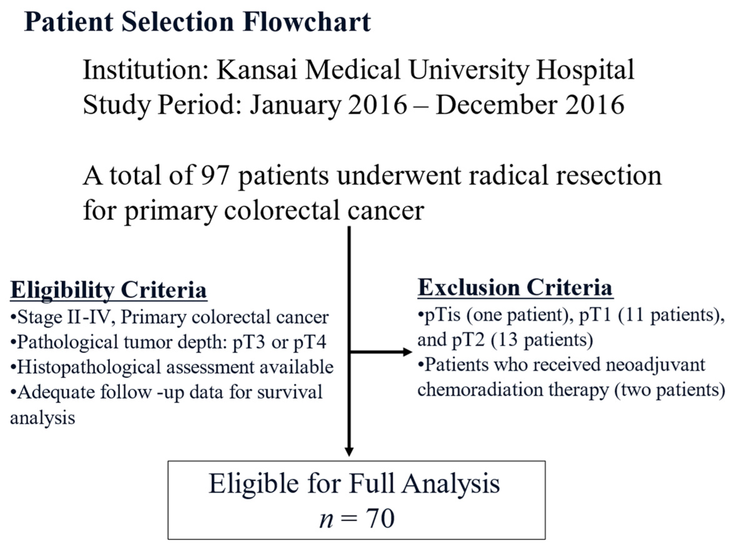
Figure 1. Flowchart of the patient selection process of the study cohort.
Cancers 17 03006 g001
Figure 2. Typical histopathological features of desmoplastic reactions of colorectal carcinoma. (A) Immature type is characterized by the presence of myxoid material around the carcinoma cell nests at the invasive front (under area of the black line). (B) Presence of thick bundles of collagen without myxoid material (right area of the black line) is characteristic feature of intermediate type (hematoxylin and eosin, ×200).
Cancers 17 03006 g002
Figure 3. Immunohistochemical features. (A) Presence of adipophilin-expressing spindle cells around the carcinoma nests at the invasive front (black arrows). Adipophilin expression is also noted in carcinoma cells (red arrows) (×400). (B) Dual immunohistochemical staining demonstrates that both adipophilin (brown) and alpha-smooth muscle actin (alpha-SMA) (red) are expressed in spindle cells around the carcinoma nests (arrows). These spindle cells are considered lipid-laden cancer-associated fibroblasts (×400).
Cancers 17 03006 g003
Figure 4. Overall survival curves. (A) The presence of immature-type desmoplastic reaction is a significant poor prognostic indicator compared to the non-immature type. (B) Presence of lipid-laden cancer-associated fibroblasts is a significant poor prognostic indicator in all patient cohorts. (C) Presence of lipid-laden cancer-associated fibroblasts is a significant poor prognostic indicator in patients with immature-type desmoplastic reaction.
Cancers 17 03006 g004
Table 1. Clinicopathological features between immature and non-immature-type desmoplastic reactions.
VariablesTotal Number (%)Immature, n = 35 (%)Non-Immature, n = 35 (%)p-Value
Median age, years (range)74 (47–89)73 (58–86)74 (47–89)0.976
Sex
Male41 (59)20 (57)21 (60)0.808
Female29 (41)15 (43)14 (40)
Location
Right-side40 (57)16 (46)24 (68)0.136
Left-side26 (37)16 (46)10 (29)
Rectum4 (6)3 (8)1 (3)
pT
pT346 (66)18 (51)28 (80)0.0118
pT424 (34)17 (49)7 (20)
pN
Negative34 (49)9 (26)25 (71)0.0001
Positive36 (51)26 (74)10 (29)
pStage
II32 (46)8 (23)24 (69)0.0006
III25 (36)17 (48)8 (23)
IV13 (18)10 (29)3 (8)
Tumour differentiation
Low-grade66 (94)33 (94)33 (94)
High-grade4 (6)2 (6)2 (6)0.99
Tumour budding
TB121 (30)9 (25)12 (34)
TB2+349 (70)26 (75)23 (66)0.433
Lymphatic invasion
Negative24 (34)12 (34)12 (34)
Positive46 (66)23 (66)23 (66)0.99
Venous invasion
Negative7 (10)2 (5)5 (14)0.232
Positive63 (90)33 (95)30 (86)
Lipid-laden cancer-associated fibroblasts
Absent33 (47)7 (20)26 (74)<0.0001
Present37 (53)28 (80)9 (26)
Adipophilin expression in total carcinoma cells (%) median (range)10 (0–90)15 (0–90)5 (0–90)0.1207
Adipophilin expression in carcinoma cells
Low (<10%)41 (58)17 (49)24 (69)0.0894
High (≧10%)29 (42)18 (51)11 (31)
Adipophilin expression in carcinoma cells at the invasive front
Absent19 (27)4 (11)15 (43)0.0063
Present51 (73)31 (89)20 (57)
Table 2. Correlation between clinicopathological features and lipid-laden cancer-associated fibroblasts.
Lipid-Laden Cancer-Associated Fibroblasts
VariablesPresent, n = 37 (%)Absent, n = 33 (%)p-Value
Median age, years (range)75 (58–89)71 (47–85)0.145
Sex
Male21 (57)20 (60)0.744
Female16 (43)13 (40)
Location
Right-side22 (60)18 (55)0.514
Left-side12 (32)14 (42)
Rectum3 (8)1 (3)
pT
pT321 (56)25 (76)0.0946
pT416 (43)8 (24)
pN
Negative11 (30)23 (70)0.0008
Positive26 (70)10 (30)
pStage
II10 (27)22 (66)0.0035
III17 (46)8 (24)
IV10 (27)3 (10)
Tumor differentiation
Low-grade35 (96)31 (94)0.906
High-grade2 (4)2 (6)
Tumor budding
TB17 (19)14 (42)
TB2+330 (81)19 (58)0.0322
Lymphatic invasion
Negative12 (32)12 (36)0.7294
Positive25 (68)21 (64)
Venous invasion
Negative1 (3)6 (18)0.0321
Positive36 (97)27 (82)
Desmoplastic reaction
Immature type28 (76)7 (21)<0.0001
Non-immature type9 (24)26 (79)
Adipophilin expression in carcinoma cells
Low (<10%)22 (59)19 (58)0.873
High (≧10%)15 (41)14 (42)
Adipophilin expression in carcinoma cells at the invasive front
Absent4 (11)15 (45)0.0014
Present33 (89)18 (55)
Table 3. Univariate and multivariate analyses of prognostic factors.
VariablesDead Patients n = 30 (%)Alive Patients n = 40 (%)Univariate p-ValueMultivariate p-ValueHR95% CI
pT414 (47)10 (25)0.0588---
Immature-type desmoplastic reaction21 (70)14 (35)0.00380.3871.7440.482–6.094
Presence of lymph node metastasis21 (70)15 (38)0.00710.1722.0570.631–6.671
Presence of lymphatic invasion21 (70)25 (63)0.513---
Presence of vascular invasion29 (97)34 (85)0.107---
Tumor differentiation (high-grade)3 (10)1 (2.5)0.181---
Tumor budding (TB) 2+323 (77)26 (65)0.291---
Adipophilin expression in carcinoma cells (more than 10%)14 (47)15 (38)0.441---
Presence of adipophilin expression in carcinoma cells at the invasive front24 (80)27 (68)0.244---
Presence of lipid-laden cancer-associated fibroblasts23 (77)14 (35)0.00050.03683.651.082–13.038
HR, hazard ratio; CI, confidence interval.
Disclaimer/Publisher’s Note: The statements, opinions and data contained in all publications are solely those of the individual author(s) and contributor(s) and not of MDPI and/or the editor(s). MDPI and/or the editor(s) disclaim responsibility for any injury to people or property resulting from any ideas, methods, instructions or products referred to in the content.