Review

The Role of Microbiota in Upper Gastrointestinal Cancers

1
IRCCS Azienda Ospedaliero Universitaria di Bologna, 40138 Bologna, Italy
2
Department of Medical and Surgical Sciences, University of Bologna, Via Massarenti, 9, 40138 Bologna, Italy
3
Department of Gastroenterology and Endoscopy, Fondazione Poliambulanza Istituto Ospedaliero, 25124 Brescia, Italy
*
Author to whom correspondence should be addressed.
These authors contributed equally to this work.
Cancers 2025, 17(10), 1719; https://doi.org/10.3390/cancers17101719
Submission received: 27 April 2025 / Revised: 19 May 2025 / Accepted: 19 May 2025 / Published: 21 May 2025
(This article belongs to the Special Issue Developments in the Management of Gastrointestinal Malignancies)

Simple Summary

The gut microbiota, the bacteria, fungi, and virus residing in our gastrointestinal tract, plays a key role in the development of upper gastrointestinal cancers, such as esophageal and gastric cancers. Changes in the microbial balance (dysbiosis) can promote cancer through inflammation, immune changes, and DNA damage. In esophageal cancer, potentially harmful bacteria are more common in precancerous conditions. In gastric cancer, it is known that Helicobacter pylori is a potent carcinogen, but a new field of study has shown that other gastric bacteria can contribute to cancer progression. Understanding these microbial changes could lead to better ways to diagnose patients and, eventually, treat or prevent cancer.

Abstract

The gut microbiota significantly impacts the development and progression of upper gastrointestinal (GI) cancers, including esophageal and gastric cancers. Microbial dysbiosis contributes to carcinogenesis through mechanisms such as inflammation, immune modulation, and direct DNA damage. Techniques for sampling oral, esophageal, and gastric microbiota vary, with standardization being essential for reliable results. Barrett’s esophagus (BE) and esophageal adenocarcinoma (EAC) are associated with an enrichment of Gram-negative bacteria, promoting inflammation and cancer progression. Esophageal squamous cell carcinoma (ESCC) also shows distinct microbial patterns, with reduced diversity and increased harmful bacteria like Porphyromonas gingivalis and Fusobacterium nucleatum. In gastric cancer (GC), Helicobacter pylori (HP) and non-HP gastric microbiota play significant roles, with diverse microbial communities contributing to cancer development through nitrate reduction, immune modulation, and inflammation. Emerging evidence highlights the role of non-HP bacteria in promoting carcinogenesis, with specific taxa like Fusobacterium nucleatum and Lactobacillus influencing tumor growth and immune evasion. Further research is needed to elucidate the complex interactions between gut microbiota and upper GI cancers, paving the way for novel diagnostic and therapeutic approaches. Understanding these microbial dynamics offers potential for microbiota-based interventions, improving the early detection, prognosis, and treatment of upper GI cancers. This comprehensive review summarizes the available evidence on the role of microbiota in upper GI oncology and the need for continued exploration in this field.

1. Introduction

Upper gastrointestinal cancers, including esophageal and gastric cancers, represent significant global health challenges due to their high incidence and mortality rates [1]. The human gastrointestinal tract is home to a diverse and dynamic microbiota, which has been increasingly recognized for its role in health and disease [2]. Digestive tract microbiota refers to the microorganisms inhabiting the human digestive tract, including the oral, gastric, esophageal, and intestinal microbiota. It is established at birth and further shaped during breastfeeding, becoming relatively stable by late childhood and continuing through adolescence into adulthood [2]. The esophagus, typically containing about 104 bacteria per gram of content, predominantly hosts Gram-positive bacteria, such as Streptococcus, Prevotella, and Lactobacillus, with less abundant genera like Peptostreptococcus, Neisseria, and Actinobacillus [3]. Dysbiosis, or microbial imbalance, in the esophagus can result from various factors, including diet, chemical exposure, alcohol, infections, or medications, leading to altered immune responses and chronic inflammation that may contribute to cancer development [4]. Esophageal cancer (EC) primarily comprises esophageal squamous cell carcinoma (ESCC) and esophageal adenocarcinoma (EAC), with ESCC being more prevalent in eastern countries and EAC in western countries [5]. EAC often develops from Barrett’s esophagus (BE), a condition characterized by the replacement of the normal squamous epithelium with columnar metaplasia due to chronic gastroesophageal reflux disease (GERD) [6,7,8]. The progression from GERD to BE and subsequently to EAC is linked to significant alterations in the local microbiota, with an enrichment of Gram-negative bacteria such as Fusobacteria, Enterobacteriaceae, Sphingomonas, Proteobacteria, and Campylobacter [9,10]. These bacteria can release lipopolysaccharides (LPSs) that activate inflammatory pathways, promoting cancer progression [11]. In gastric cancer (GC), the role of Helicobacter pylori (HP) is well established, but emerging evidence highlights the importance of non-HP gastric microbiota in carcinogenesis [12]. The gastric microbiota, influenced by intrinsic factors like low pH and antimicrobial enzymes and extrinsic factors such as diet and antibiotic use, undergoes significant changes during the progression from chronic gastritis to GC [13,14]. Also, nitrate-reducing bacteria, such as Escherichia, Shigella, and Lactobacillus, can increase concentrations of carcinogenic nitrite and N-nitroso compounds in the stomach [15,16]. Additionally, bacteria like Fusobacterium nucleatum can induce inflammatory responses and modulate the host immune system to favor tumor growth [17,18,19].
This review aims to provide a comprehensive overview of the current understanding of the gut microbiota’s role in upper GI cancers, exploring the mechanisms of microbial dysbiosis in carcinogenesis, potential biomarkers for early detection and prognosis, and emerging strategies for microbiota modulation as therapeutic interventions.

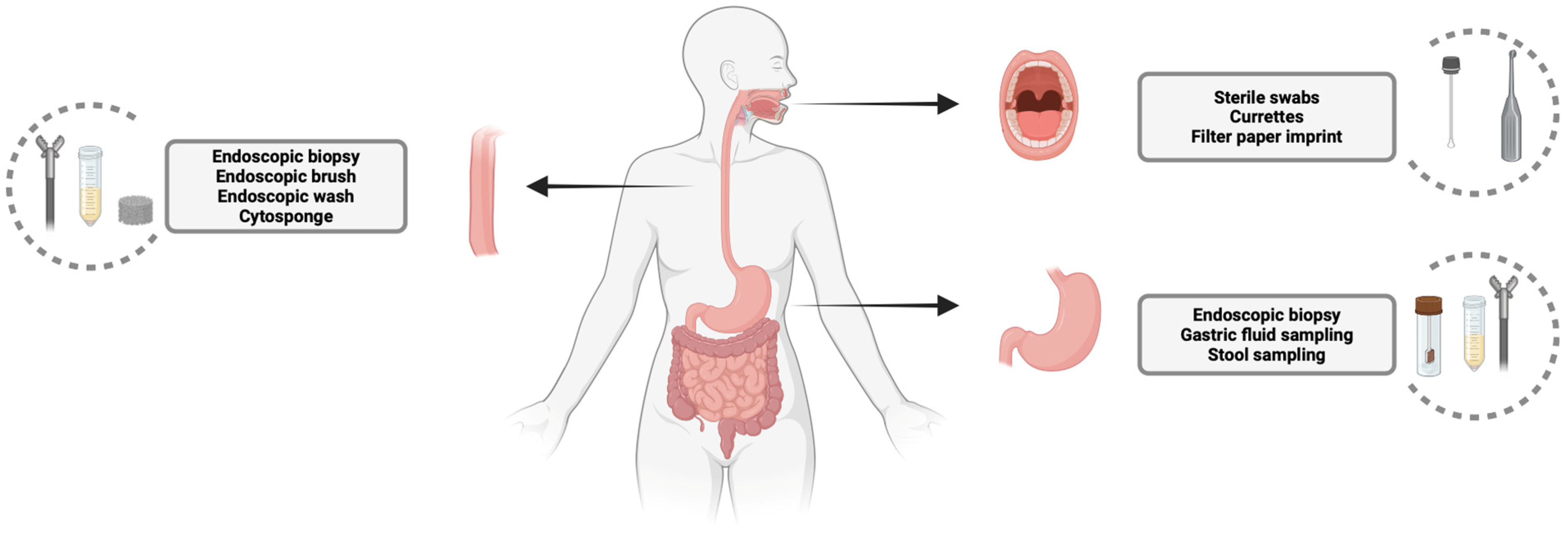
2. Sampling of Oral, Esophageal, and Gastric Microbiota: Techniques and Relevance

2.1. Oral Microbiota Sampling

The oral microbiota exhibits significant heterogeneity depending on the specific site sampled. Different regions of the oral cavity harbor distinct microbial niches. Before even selecting different techniques for sampling the oral microbiota, a challenge arises between using stimulated versus unstimulated saliva sampling methods. Stimulated salivation may cause dilution and introduce bias into the results. On the other hand, unstimulated saliva, collected after several mouth rinses, may result in a low total volume, limiting its use in multi-omics analyses [20]. In a well-designed study, Roca et al. compared the microbiome composition in patients using both stimulated and unstimulated saliva, showing notable differences within the same participants. This study demonstrated that stimulated saliva provided a more comprehensive representation of the oral microbiota [20].
The most common sampling techniques for oral microbiota analysis involve the use of sterile swabs for regions like the tongue dorsum, hard palate, palatine tonsils, and oropharynx, while sterile curettes are used for collecting supra- and subgingival plaques (Figure 1) [21]. These methods have been proposed as part of a standard protocol for oral cavity sampling and have been utilized to define the oral microbiome in healthy individuals using whole-genome sequencing (WGS) [22,23]. An innovative technique using a filter paper imprint has also been validated by comparing it with the swab method, showing comparable results for buccal mucosa sampling. The filter paper method offers advantages, such as calculating the microbial colonization density over a defined unit area and the improved analysis of Gram-negative bacteria due to its suction mechanism [24]. However, the validation study relied on a culture-based microbiota analysis rather than the more advanced next-generation sequencing techniques now commonly used. Therefore, the standardization of sampling techniques is essential to ensure reliable and reproducible testing results across studies and clinical applications.

2.2. Esophageal Microbiota Sampling

Esophageal microbiota sampling methods can be categorized into invasive and non-invasive techniques (Figure 1). Invasive methods, which require an endoscopy, are currently the gold standard for microbiota sampling and are often used as a comparator for newer techniques [25,26,27,28,29,30]. Endoscopic biopsies allow for the analysis of a mucosa-associated microbiome which is in direct contact with the mucosal cells. However, this method has several limitations, including the analysis of only a small tissue section (3–5 mm), being time-consuming, and carrying the risk of potential complications. Like biopsies, brushes are sometimes used to sample a wider surface area. However, it has been suggested that brushes mainly collect firmly attached microbes from the mucosal surface, potentially missing loosely attached microorganisms [25]. Additionally, the mechanical forces involved in brushing may alter the microbiota composition.
To target the loosely attached microbes, a non-invasive technique, though still requiring an endoscopy, has been introduced: endoscopic wash sampling [27]. A Japanese study found that in patients with esophageal cancer, the alpha diversity measures (the Shannon Index, Observed Features, and Simpson Index of Evenness) in esophageal wash samples significantly correlated with the microbiota from normal esophageal mucosal biopsies but not with the microbiota from cancerous tissue, suggesting that the wash method may be useful for analyzing normal esophageal microbiota [29]. However, an endoscopy is still required, and the risk of contamination from the oral microbiota remains.
A non-invasive cell-sampling device, the Cytosponge, has also been proposed. It is a safe and well-tolerated method in which a spherical mesh attached to a string is swallowed, expands in the stomach, and is then withdrawn, collecting cells from the esophagus [31]. In a case–control study, Elliott et al. demonstrated that the Cytosponge yielded more than ten times the microbial DNA compared to endoscopic brushes or biopsies, as measured by a quantitative PCR [30]. This could be due to its ability to sample a larger portion of the esophagus, although contamination from the stomach or oral cavity is still a potential concern. Similar findings have been confirmed in a more recent comparative study between the Cytosponge and brush sampling techniques [25]. A similar device, the esophageal string test, has been proposed for use in the pediatric population. It has demonstrated comparable microbial compositions to those found in matched esophageal biopsies, suggesting it could serve as a promising non-invasive method for studying the esophageal microbiome in children [28].

2.3. Gastric Microbiota Sampling

The two most used methods for sampling the gastric microbiota are biopsy and stool sampling (Figure 1). Similarly to the approach for esophageal sampling, biopsies involve an invasive procedure and are limited to specific target regions.
In contrast, stool sampling is a non-invasive, patient-friendly method that is cost-effective and yields sufficient biomass for the microbial analysis [32]. However, stool samples predominantly represent microbial communities from the terminal colon rather than the entire gut, providing a limited perspective. It has been demonstrated that the microbiota composition, viral activity, host proteome, and bile acid profiles vary substantially along the GI tract, affecting the accuracy of stool samples in capturing the full longitudinal and temporal variability of the microbiota [33].
An alternative approach to characterizing the gastric microbiota involves sampling gastric fluid, typically obtained during an endoscopy [34,35,36]. However, limitations of gastric juice sampling have been noted, including the potential detection of bystander microbes, the risk of contamination from endoscopic sampling methods, and a low bacterial load. To reduce the invasiveness, a novel gastric fluid sampling device utilizing a disintegrating polymeric blocking disk has been proposed [37]. Initial in vitro, in situ, and ex vivo animal studies demonstrated the device’s safety and accuracy, though no human models have been tested.
Additionally, Park et al. developed a capsule robot designed for sequential sampling at six sites within the gastrointestinal tract, including the stomach [38]. This device minimizes the cross-contamination between samples and is navigated via an electromagnet system, equipped with a camera to document sampling locations. To date, only ex vivo and in vivo tests on porcine models have been conducted, highlighting the need for further research to evaluate its efficacy in humans.

3. Microbiota in Esophageal Cancer

Esophageal cancer (EC) is a significant global health challenge, ranking as the eighth leading cause of cancer-related deaths worldwide [5]. EC mainly comprises esophageal squamous cell carcinoma (ESCC) and esophageal adenocarcinoma (EAC), with ESCC being predominant in eastern countries and EAC in western countries [5]. EAC typically develops from Barrett’s esophagus (BE) at the distal esophagus, while ESCC can initiate in any part of the esophagus usually due to the direct exposure to carcinogens, such as chemicals, cigarette smoke, and alcohol [1]. Symptoms of EC are closely linked to disease progression. In the initial stages, patients may be asymptomatic or suffer from mild dysphagia, while advanced stages show worsening dysphagia with persistent chest or back pain and considerable weight loss [1]. As far as therapeutic management is concerned, for early-stage disease, endoscopic therapy is a common approach for removing the tumor tissue infiltrating the mucosa or submucosa while chemotherapy and radiation therapy are performed either before or after a surgical resection for locally advanced disease [39]. For metastatic disease, instead, treatment options include chemotherapy, radiation, targeted therapy, immunotherapy, or their combination [39].
Upper digestive microbiota has been hypothesized as a contributing factor in EC development and progression. Typically, the esophagus contains about 104 bacteria per gram of content, mainly Gram-positives [40]. The most common identified bacteria include Streptococcus, Prevotella, and Lactobacillus, with Peptostreptococcus, Neisseria, and Actinobacillus being less abundant and rarely reported [3]. Furthermore, a relatively low abundance of viruses, such a Betaherpesvirus and Papillomavirus, and fungi, such as Candida glabrata, have been found [41]. Although most studies on esophageal microbial communities have been conducted on the lower esophageal mucosa, it has been shown that the microbiota composition is also similar in the proximal and medial esophagus, with a substantial overlap with the microbial composition of the mouth, pharynx, and stomach [42,43]. The esophageal microbiota can be altered by diet, chemical exposure, alcohol, infections, or medications that disrupt the normal balance between bacterial geni and between microbiota and the esophageal immune system. This microbial imbalance leads to an altered immune response and the chronic inflammation of the esophageal mucosa, which may contribute to the development and progression of cancer [4].

3.1. Microbial Alterations in Barrett’s Esophagus

Barrett’s esophagus is a condition in which columnar metaplasia replaces the normal squamous cell epithelium of the esophagus. The major risk factor for BE is gastroesophageal reflux disease (GERD). A chronic exposure to stomach acid may cause the inflammation of the esophagus and initiate the cellular changes that characterize this disease [6,7,8]. The exact molecular mechanism for this shift is not yet known; however, it was demonstrated that the chronic exposure of the esophageal mucosa to gastric acid results in increased intercellular spaces which allow hydrochloric acid molecules to permeate down to the stem cells in the basal layer, modifying their differentiation [6,7,8]. GERD, BE, and EAC define a sequence that begins with GERD and progresses with BE development, followed by dysplasia (first low-grade and then high-grade), and finally EAC development [8].
Recent data have emerged on the local gastroesophageal microbiota’s composition being altered in neoplastic and pre-neoplastic lesions and playing a crucial role in the pathological process from BE to adenocarcinoma. The first observed key feature of the esophageal microbiota in GERD and BE is a relative enrichment of Gram-negative bacteria, particularly Fusobacteria, Enterobacteriaceae, Sphingomonas, Proteobacteria, and Campylobacter [9,10]. Subsequent studies comparing BE and healthy controls confirmed the Gram-negative shift but showed fewer taxonomic differences, such as increased relative abundances of Leptotrichia [44,45], Capnocytophaga, Gemella, Veillonella, and Streptococcus sanguinis [45]. Although these studies showed interesting results and the selection criteria were stringent, it should be noted that the population assessed was rather small. Regarding further differences in the characteristics of BE, a prospective study assessed the microbial population of 74 patients, including 34 patients with BE and 40 patients without BE, finding that as Barrett’s length increased, multiple organisms, such as Corynebacterium, Dialister, Gemella, Haemophilus, Leptotrichia, Neisseria, Prevotella, Rothia, Streptococcus, and Veillonella, were less likely to be detected [46]. A possible pathophysiological mechanism linking Gram-negative bacteria to the esophageal adenocarcinoma cascade involves lipopolysaccharides (LPSs). In fact, LPSs released by Gram-negative bacteria can activate the NF-κB pathway, leading to IL-8 expression in immune cells, which in turn activates toll-like receptors (TLRs) and nucleotide-binding oligomeric domain (NOD)-like receptors, triggering chronic inflammation and facilitating cancer progression [11]. Similarly, higher levels of the proinflammatory cytokine IL-18 were found in subjects colonized by Campylobacter, a Gram-negative bacterium, in the context of BE [47]. Furthermore, Gram-negative bacteria-derived LPSs may relax the lower esophageal sphincter and postpone gastric emptying, contributing to the increase in acid reflux and the molecular changes in the esophageal epithelium caused by acid reflux [48].
Finally, Gram-negative bacteria, such as Escherichia coli and Campylobacter, directly induce DNA damage via the production of alkylating agents as well as reactive oxidative species, promoting cancer development [49]. The role of HP is still debated, although various recent meta-analyses [50,51] have shown a reduced rate of EAC in patients colonized by HP. The main microbial alterations found in Barrett’s esophagus and esophageal cancers are summarized in Figure 2.

3.2. Microbial Alterations in Esophageal Cancer

In the setting of EAC, the esophageal microbiota is distinct from that of the healthy esophagus. Actinobacteria, Lactobacillus salivarius, Moryella, Streptococcus infantis, and Staphylococcus aureus have all been reported to be more represented in this population [52]. A fundamental shared trait of these bacteria is lactic acid generation. Long-term elevated lactate levels, in addition to the pathophysiological changes brought by Gram-negatives, have been shown to enhance carcinogenesis and progression by promoting angiogenesis, immunological evasion, cell migration, and metastasis [52]. Moreover, a sustained acidic environment inhibits the growth of other potentially beneficial bacteria, creating a self-sustained harmful environment [53]. In a pivotal case–control study, oral microbiome samples from 17 control patients, 16 patients with BE without dysplasia, 6 with low-grade dysplasia (LGD), 5 with high-grade dysplasia (HGD), and 5 with esophageal adenocarcinoma (EAC) have shown that the most significant change in the microbiome happened between the stages of LGD and HGD [11]. In fact, when compared to the group of patients with BE or LGD, patients with HGD or EAC exhibited a substantial decrease in the Firmicutes phylum and an increase in Enterobacteriaceae, a family of Gram-negative bacteria. Additionally, a lower microbial alpha-diversity, a measurement of the local bacterial variability, was found in EAC compared with BE [11]. In this study, the recent use of antibiotics was considered as an exclusion criterion; however, other significant confounding factors, such as the type of diet, use of proton pump inhibitors, or oral hygiene status, were not taken into consideration.
Regarding ESCC, a case–control study on 18 patients with ESCC and 11 controls has shown a reduction in microbial diversity with respect to healthy controls, with increased Porphyromonas (P.) gingivalis and Fusobacterium nucleatum [54]. Although limited by the small sample size, this study confirmed the hypothesized role of Porphyromonas gingivalis, a Gram-negative bacterium linked to periodontal disease, as capable of promoting ESCC progression by activating the NF-κB and IL-6/STAT3 pathways. Studies show P. gingivalis enhances tumor growth, chemotherapy resistance, and is associated with a poor prognosis in ESCC patients [54]. Additionally, Fusobacterium nucleatum can stimulate chemokines, such as CCL20, known to aid tumor invasion [52].

3.3. Microbiota and Esophageal Cancer Screening and Prognosis

Identifying precise biomarkers for early BE or EC detection is crucial to improve clinical outcomes. The standard of care for people with non-dysplastic BE includes an upper endoscopy every three years to appropriately screen for the development of EAC [8]. This method is limited by the need for repeated invasive procedures, the associated healthcare costs, and the unclear benefits in terms of the lower EAC incidence or mortality [55]. Non-invasive techniques, such as a microbiota analysis via oral swabs and brushings from the distal esophagus, have been tested in the previous years.
In the context of EAC, for example, a model using the relative abundance of Lautropia, Streptococcus, and Bacteroidales in the saliva was developed to distinguish BE from controls with a high accuracy (AUROC 0.94, 95% confidence interval (CI): 0.85–1.00) [11]. Likewise, in ESCC, other models including Actinomyces, Fusobacterium (F.) nucleatum, Haemophilus haemolyticus, Porphyromonas gingivalis, and Streptococcus australis extracted by a salivary swab have been proven to be feasible to detect early ESCC in a total of 449 specimens collected from 349 participants with different esophageal diseases (AUROC 0.722, 95% CI: 0.621–0.823) [56]. Although promising, these tests are still limited by the low number of enrolled patients, the availability of tests, and the need for validation.
Microbiota show potential not only for diagnosing EC but also as prognostic markers. Among microbial candidates, F. nucleatum seems to be the most promising. High F. nucleatum levels are correlated to a larger tumor size, advanced EC stages, and a worse prognosis, particularly in recurrent cases [57]. Similarly, Porphyromonas gingivalis was detected more frequently in the oral microbiome of patients with ESCC than in healthy controls (57% vs. 24% p = 0.0034) and was associated with advanced clinical stages and a worse prognosis (Hazard Ratio = 2.14, p = 0.014) [58]. Moreover, the esophageal microbiota sampling in lower esophageal biopsies showed an increased Streptococcus abundance in patients with advanced ESCC stages (T3/T4) with respect to more confined disease (T1/T2) [59]. These findings highlight the importance of the gut microbiota in early EC detection and prognosis, with further clinical trials needed to translate these findings into clinical practice.

3.4. Microbiota Modulation in Esophageal Cancer

A new field of study that shows potential for enhancing preventative and therapeutic approaches is microbiota manipulation in esophageal cancer. Proton pump inhibitors (PPIs) are widely used medications to treat GERD and are known to actively alter the gut microbiota [60]. In a case–control study conducted in the United States on 45 patients with BE it was found that after an 8-week course of PPIs, PPI users exhibited higher relative abundances of Streptococcus and lower relative abundances of Gram-negative bacteria as compared to controls, which are known to be markers of neoplastic progression. However, these results should be interpreted with caution since the PPI users were a mix of BE and non-BE patients [61]. Although direct studies are lacking, it could be speculated that PPI therapy may help patients with precancerous esophageal lesions by reversing the Gram-negative shift seen in the BE to EAC cascade [62]. More data are available on the role of dental hygiene in EC. A recent analysis on 163 patients who underwent surgery with perioperative oral care showed that poor oral hygiene, evaluated via radiological measures and classified by dentists into three groups of severity, was a significant independent prognostic factor for poor overall survival, making oral hygiene a simple and affordable preventive target for EC [63]. Finally, targeted dietary interventions may be used as new EC preventive strategies. For example, a meta-analysis encompassing 15 studies involving 16,885 subjects, showed that subjects with higher daily fiber intakes had a decreased risk of EAC (for an increment of 10 g/day, RR = 0.79, 95% CI: 0.69–0.92), although this was limited by the lack of specific daily fiber quantities [64]. In fact, the effect of fiber is linked to its ability to reduce the relative abundance of esophageal Gram-negative bacteria which contribute to the adenocarcinoma cascade [64,65].
In conclusion, esophageal cancer is a significant global health issue. Factors such as diet and chemical exposure may disturb the healthy esophageal microbiota, leading to dysbiosis and, hence, chronic inflammation and neoplastic changes. Indeed, several unique microbial communities have been defined in the healthy esophagus, BE, and progressive stages of esophageal adenocarcinoma. Although an increase in Gram-negative bacteria is commonly observed, the precise determination of the causal role of microbiota in cancer formation is still lacking. In esophageal squamous cell carcinoma, there is a decrease in microbial diversity and an increase in the harmful bacteria, such as Porphyromonas gingivalis, associated with lower overall survival. Although the available evidence remains scant, mainly due to the limited sample size evaluated and the missing evaluation of several confounding factors, identifying changes in the microbiota and potentially modifying it could help the early detection and treatment of esophageal cancer.

4. Microbiota in Gastric Cancer

4.1. Microbiota in Gastric Cancer

The human stomach, historically considered a sterile environment due to its acidic conditions and digestive enzymes, is now acknowledged to harbor a diverse and dynamic microbiota. Advances in high-throughput sequencing and microbial culture techniques have unveiled a complex community of microorganisms residing in the gastric mucosa and lumen [66]. While Helicobacter pylori (HP) is a well-established pathogen linked to GC and mucosal-associated lymphoid tissue (MALT) lymphoma, emerging evidence highlights the significant role of non-HP gastric microbiota in gastric carcinogenesis [12]. Not enough data are available on the role of non-HP gastric microbiota in MALT-lymphoma; therefore, we aimed to summarize the available data on gastric adenocarcinoma.
Early studies utilizing the temperature gradient gel electrophoresis of 16S rRNA amplicons identified three predominant bacterial phyla in the stomach: Proteobacteria, Firmicutes, and Actinobacteria [67,68]. Subsequent research employing G2 Phylo-Chip and 16S sequencing expanded this composition, revealing up to 44 bacterial phyla, with Actinobacteria, Firmicutes, Bacteroidetes, Proteobacteria, and Fusobacteria being dominant [69]. Common genera include Streptococcus (Firmicutes), Neisseria and Haemophilus (Proteobacteria), and Prevotella and Porphyromonas (Bacteroidetes) [70].
The gastric microbiota is influenced by intrinsic factors such as a low pH, antimicrobial enzymes like pepsin, immunoglobulins (e.g., IgA), and antimicrobial peptides [71]. Extrinsic factors include diet, antibiotic use, proton pump inhibitors (PPIs), geographic location, and surgical interventions [13,14]. Long-term PPI use elevates the gastric pH, facilitating bacterial overgrowth and translocation, particularly of oral bacteria like Peptostreptococcus stomatis and Streptococcus anginosus [72]. Surgical procedures such as subtotal gastrectomy alter the gastric environment, promoting the growth of bacteria such as Prevotella and Streptococcus [73]. Moreover, recent investigations have demonstrated that patients with GC often display a marked decrease in the overall bacterial diversity in their gastric microbiota, with a significant enrichment of genera such as Clostridium and Fusobacterium, as reported by Hsieh et al. [74].
This dysbiosis pattern can vary according to factors like the geographic location and the presence of H. pylori, highlighting the multifaceted interactions within the gastric ecosystem.

4.2. Non-Helicobacter Pylori Microbiota Alteration in Gastric Cancer

The composition and functionality of the gastric microbiota undergo significant changes during the progression from chronic gastritis to GC, a process often described by the Correa cascade. This progression is accompanied by a state of microbial dysbiosis, which is characterized by a reduced overall diversity but an increased abundance of specific pathogenic taxa in the gastric mucosa [66,75].
Microbiota imbalance is not merely a secondary effect of the gastric pathology but actively contributes to carcinogenesis through several mechanisms. Nitrate-reducing bacteria can increase the concentrations of nitrite and N-nitroso compounds in the stomach, which are potent carcinogens [15]. As a matter of fact, Ferreira et al. retrospectively evaluated the differences in the gastric microbiota of 54 patients with gastric carcinoma and 81 patients with chronic gastritis by 16S rRNA gene profiling, using next-generation sequencing, finding a significant increase in nitrate reductase and nitrite reductase activities, promoting the generation of nitrite and nitric oxide, leading to DNA damage [67]. Escherichia, Shigella, Lactobacillus, and Nitrospirae, known for nitrate/nitrite metabolism, are enriched in GC tissues [16].
Moreover, non-HP bacteria can induce inflammatory responses by activating immune cells and signaling pathways. Propionibacterium acnes has been recently identified as a strong risk factor for GC development [76]. It activates the natural killer group 2 member D (NKG2D) system and upregulates proinflammatory cytokines, like interleukin-15, leading to persistent inflammation and promoting carcinogenesis [71,77]. F. nucleatum, commonly implicated in colorectal cancer, has also been detected in gastric cancer, where it fosters a proinflammatory microenvironment conducive to tumor progression and modulates the host immune response in favor of oncogenesis [18]. In fact, F. nucleatum produces adhesins such as Fap2, which bind to immune receptors on natural killer (NK) and T cells, inhibiting their cytotoxic activity and allowing the immune evasion of cancer cells [19]. Another adhesin secreted by F. nucleatum, FadA, can instead bind to E-cadherin, thereby activating the Wnt/β-catenin pathway and driving malignant transformation [74,78]. Moreover, F. nucleatum releases endotoxins that can suppress immune responses, upregulate transcription factors such as NF-κB, and stimulate the secretion of proinflammatory cytokines (IL-1β, IL-6, IL-8, TNF), which lead to an oncogenic environment [79,80]. For these reasons, higher relative abundances of F. nucleatum have been linked to an advanced disease stage and poorer prognosis [81]. Finally, Propionibacterium acnes and F. nucleatum, besides inducing inflammation and immune evasion, can directly trigger DNA damage responses in host cells, contributing to genomic instability and promoting cellular proliferation and invasiveness [81,82].
In the context of advanced gastric lesions in mice models, it has been demonstrated that the gastric colonization by a restricted consortium of commensal bacteria—specifically Clostridium sp. ASF356, Lactobacillus murinus ASF361, and Bacteroides sp. ASF519—induces gastritis, atrophy, and dysplasia to a degree comparable to that driven by a more diverse microbiota, independently of Helicobacter pylori infections [83]. This suggests that even commensal bacteria can promote gastric carcinogenesis in the context of gastric inflammation and atrophy. For instance, Lactobacillus species, while often considered beneficial, have been found to be enriched in GC patients and may influence the gastric environment to favor carcinogenesis [77]. In a meta-analysis involving 1642 gastric biopsy samples and 394 stool samples, researchers found that Lactobacillus and Streptococcus were consistently enriched in GC patients [84]. Their abundance was associated with the progression of gastric carcinogenesis, and their combination in diagnostic models showed a high accuracy and reproducibility across different cohorts. These events promote a persistent inflammatory milieu and foster tumor cell proliferation, invasion, and survival. The main microbial alterations found in gastric cancer are summarized in Figure 2.

4.3. Microbiota and Gastric Cancer Screening and Prognosis

Mounting evidence indicates that alterations in the gastric microbiota may serve as valuable biomarkers for the early detection of GC and inform the development of novel therapeutic strategies. Indeed, multiple studies have identified distinct microbial signatures in patients with GC compared to those with non-malignant gastric conditions, underscoring the potential for these profiles to refine diagnostic accuracy and guide personalized interventions. Huang et al. aimed to characterize the salivary microbiota in patients at different progressive histological stages of gastric carcinogenesis in order to identify relevant microbial markers for gastric cancer detection in 293 patients with superficial gastritis [number (n)= 101], atrophic gastritis (n = 93), and gastric cancer (n = 99). A distinct salivary microbiota in patients with GC compared to superficial and atrophic gastritis was found. Although not externally validated, this bacterial signature was found to have a remarkable ability to distinguish GC from non-malignant gastric diseases with a high accuracy (AUC of 91%) [85]. These findings underscore the potential of using the salivary microbiota as a non-invasive biomarker for gastric malignancy. In addition, other studies emphasize the potential utility of the salivary microbiota—particularly the relative abundance of certain oral-derived bacteria—as a non-invasive screening tool for GC, reaching predictive accuracies as high as 91% [85].
Additionally, alterations in stomach-associated microbial communities have been linked to histological stages of gastric carcinogenesis, revealing that certain bacterial genera, such as Veillonella and Enterobacteriaceae, may be enriched in cancerous tissues compared to normal gastric mucosa [36,86]. For instance, Kwon et al. demonstrated that the transplantation of the human gastric microbiota into germ-free mice recapitulated premalignant changes, further implying the microflora’s active role in tumorigenesis [87]. Likewise, Liu et al. mapped key differences across various gastric microhabitats in GC patients, revealing specialized microbial niches linked to the cancer stage and immune modulation [77].

4.4. Microbiota Modulation in Gastric Cancer

HP eradication is crucial for preventing gastric cancer, and meta-analyses along with randomized trials suggest that a successful eradication reduces the risk by approximately 33–47%, especially in areas with a high HP prevalence, such as East Asia [88]. Nonetheless, up to 30–40% of patients may still face progression to malignancy if notable atrophy, metaplasia, or other advanced precancerous lesions are already established prior to bacterial clearance.
PPIs are commonly employed in eradication protocols because their potent acid suppression enhances antibiotic stability. However, large observational studies have raised concerns that prolonged or intensive PPI therapy may negatively affect gastric physiology, particularly in individuals with a history of HP infections [89,90]. In one retrospective cohort of more than 63,000 HP-eradicated individuals, extended PPI use (exceeding 12 months) was linked to a two- to eightfold increase in the subsequent incidence of gastric cancer [91].
Probiotic supplementation has been suggested both to augment HP eradication rates by 8–10% and to support gastrointestinal health following treatment [92]. A recent meta-analysis [93] including 140 studies on adults and a total of 20,215 patients evaluated the role of probiotics in addition to classic eradication therapies of different durations and lines. The supplementation of probiotics led to higher eradication rates compared to the control group, 84.1 vs. 70.5, respectively (Risk Ratio (RR) 1.17, 95% CI 1.15–1.18); the authors additionally reported that Lactobacillus acidopilus, Saccharomyces boulardii, Lactobacillus + Bifidobacterium + Enterococcus, and Lactobacillus + Bifidobacterium + Bacillus cereus showed a higher efficacy in increasing eradication rates when added to a 10-day triple therapy, whereas only the first three mentioned probiotic mixtures showed a benefit in a 14-day triple therapy.
Beyond the application of probiotics in HP eradication, their use has recently been tested in different steps of GC treatment. For example, in patients with advanced GC undergoing gastric surgery, it was found that the oral administration of Clostridium butyricum could decrease postoperative inflammation [94]. Moreover, in a recent phase II trial, the microbiota from healthy obese donors was transplanted to cachectic patients with metastatic GC prior to chemotherapy, improving the response and overall survival [95]. Additional evidence in the context of chemotherapy shows that butyrate-producing bacteria increase the efficacy of oxaliplatin by activating the IL-12 signaling pathway in both mice and humans [95]. A further possible field of application of microbiota modulation in GC is the use of probiotics before or during immunotherapy. Indeed, it has been reported that H. pylori can elude the immune response by inducing the up-regulation of PD-L1 in gastric epithelial cells, which in turn can cause the apoptosis of T cells. However, a negative correlation between responses to immunotherapy and H. pylori infections has been highlighted, showing a microbiota-related response to therapy [96]. The role of non-H. pylori gastric microbiota modulation in the context of immunotherapy is a field of great interest that will have a fundamental role in future research.
In summary, H. pylori eradication significantly reduces GC risk, even though patients with existing advanced precancerous lesions may still develop cancer post-eradication. Preliminary data showed that long-term proton pump inhibitors may increase the gastric cancer risk, while probiotics can enhance HP. Beyond eradication, microbial modulations have shown potential in various steps of GC treatment. However, no sufficient data are currently available to suggest a definite role for microbial modulation in gastric cancer treatment.

5. Conclusions

The gut microbiota significantly impacts the development and progression of esophageal and gastric cancers. Microbial imbalance contributes to carcinogenesis through mechanisms such as inflammation, immune modulation, and direct DNA damage. In the context of esophageal cancer, different microbial compositions have been found in all the steps of the cancer cascade, from Barrett’s esophagus to esophageal adenocarcinoma, which are characterized by the relative increase in Gram-negative bacteria in the first phases followed by the enrichment of lactic acid producers. Specific microbial signatures have also been found in esophageal squamous cell carcinoma, with a prominent role of Porphyromonas gingivalis, Fusobacterium nucleatum, and Streptococci. Emerging evidence highlights the role of non-HP bacteria in GC carcinogenesis, with a specific enrichment of nitrate-reducing bacteria other than Propionibacterium acnes, Fusobacterium nucleatum, Lactobacilli, and Streptococci. Further research is needed to elucidate the interactions between the gut microbiota and upper gastrointestinal cancers, with the aim of improving the early detection, prognosis, and treatment of upper GI cancers.

Author Contributions

G.M. and G.B. designed the review; G.M., L.C., D.S., A.B. and C.C. (Cecilia Capelli) performed the literature search; G.M., L.C., D.S., A.B., C.C. (Cecilia Capelli), E.D., M.R.B., C.C. (Cesare Cremon), V.S. and G.B. drafted the manuscript, critically revised, and approved the final version of the manuscript. All authors have read and agreed to the published version of the manuscript.

Funding

This research received no external funding.

Institutional Review Board Statement

Ethical review and approval were waived for this study due to the use of already available published data.

Informed Consent Statement

Informed consent was obtained from all subjects involved in the study by the investigator of each published study included in the present review.

Data Availability Statement

The data presented in this study are openly available in Medline and Embase.

Conflicts of Interest

The authors declare no conflicts of interest.

References

  1. Smyth, E.C.; Lagergren, J.; Fitzgerald, R.C.; Lordick, F.; Shah, M.A.; Lagergren, P.; Cunningham, D. Oesophageal Cancer. Nat. Rev. Dis. Primers 2017, 3, 17048. [Google Scholar] [CrossRef]
  2. McCallum, G.; Tropini, C. The Gut Microbiota and Its Biogeography. Nat. Rev. Microbiol. 2024, 22, 105–118. [Google Scholar] [CrossRef] [PubMed]
  3. Liu, N.; Ando, T.; Ishiguro, K.; Maeda, O.; Watanabe, O.; Funasaka, K.; Nakamura, M.; Miyahara, R.; Ohmiya, N.; Goto, H. Characterization of Bacterial Biota in the Distal Esophagus of Japanese Patients with Reflux Esophagitis and Barrett’s Esophagus. BMC Infect. Dis. 2013, 13, 130. [Google Scholar] [CrossRef]
  4. Radani, N.; Metwaly, A.; Reitmeier, S.; Baumeister, T.; Ingermann, J.; Horstmann, J.; Anand, A.; Gatz, I.; Kohlmayer, F.; Janssen, K.P.; et al. Analysis of Fecal, Salivary, and Tissue Microbiome in Barrett’s Esophagus, Dysplasia, and Esophageal Adenocarcinoma. Gastro Hep Adv. 2022, 1, 755–766. [Google Scholar] [CrossRef] [PubMed]
  5. Bray, F.; Laversanne, M.; Sung, H.; Ferlay, J.; Siegel, R.L.; Soerjomataram, I.; Jemal, A. Global Cancer Statistics 2022: GLOBOCAN Estimates of Incidence and Mortality Worldwide for 36 Cancers in 185 Countries. CA Cancer J. Clin. 2024, 74, 229–263. [Google Scholar] [CrossRef] [PubMed]
  6. Zhou, J.; Sun, S.; Luan, S.; Xiao, X.; Yang, Y.; Mao, C.; Chen, L.; Zeng, X.; Zhang, Y.; Yuan, Y. Gut Microbiota for Esophageal Cancer: Role in Carcinogenesis and Clinical Implications. Front. Oncol. 2021, 11, 71242. [Google Scholar] [CrossRef]
  7. Cook, M.B.; Coburn, S.B.; Lam, J.R.; Taylor, P.R.; Schneider, J.L.; Corley, D.A. Cancer Incidence and Mortality Risks in a Large US Barrett’s Oesophagus Cohort. Gut 2018, 67, 418–429. [Google Scholar] [CrossRef]
  8. Sharma, P. Barrett Esophagus. JAMA 2022, 328, 663–671. [Google Scholar] [CrossRef]
  9. Yang, L.; Lu, X.; Nossa, C.W.; Francois, F.; Peek, R.M.; Pei, Z. Inflammation and Intestinal Metaplasia of the Distal Esophagus Are Associated with Alterations in the Microbiome. Gastroenterology 2009, 137, 588–597. [Google Scholar] [CrossRef]
  10. Dan, W.; Peng, L.; Yan, B.; Li, Z.; Pan, F. Human Microbiota in Esophageal Adenocarcinoma: Pathogenesis, Diagnosis, Prognosis and Therapeutic Implications. Front. Microbiol. 2022, 12, 791214. [Google Scholar] [CrossRef]
  11. Snider, E.J.; Compres, G.; Freedberg, D.E.; Giddins, M.J.; Khiabanian, H.; Lightdale, C.J.; Nobel, Y.R.; Toussaint, N.C.; Uhlemann, A.C.; Abrams, J.A. Barrett’s Esophagus Is Associated with a Distinct Oral Microbiome. Clin. Transl. Gastroenterol. 2018, 9, e135. [Google Scholar] [CrossRef] [PubMed]
  12. Castaño-Rodríguez, N.; Goh, K.L.; Fock, K.M.; Mitchell, H.M.; Kaakoush, N.O. Dysbiosis of the Microbiome in Gastric Carcinogenesis. Sci. Rep. 2017, 7, 15957. [Google Scholar] [CrossRef] [PubMed]
  13. Murphy, J.D.; Gadalla, S.M.; Anderson, L.A.; Rabkin, C.S.; Cardwell, C.R.; Song, M.; Camargo, M.C. Autoimmune Conditions and Gastric Cancer Risk in a Population-Based Study in the United Kingdom. Br. J. Cancer 2024, 131, 138–148. [Google Scholar] [CrossRef] [PubMed]
  14. Chen, Y.C.; Malfertheiner, P.; Yu, H.T.; Kuo, C.L.; Chang, Y.Y.; Meng, F.T.; Wu, Y.X.; Hsiao, J.L.; Chen, M.J.; Lin, K.P.; et al. Global Prevalence of Helicobacter Pylori Infection and Incidence of Gastric Cancer Between 1980 and 2022. Gastroenterology 2024, 166, 605–619. [Google Scholar] [CrossRef]
  15. Li, J.; Perez Perez, G.I. Is There a Role for the Non-Helicobacter Pylori Bacteria in the Risk of Developing Gastric Cancer? Int. J. Mol. Sci. 2018, 19, 1353. [Google Scholar] [CrossRef]
  16. Zhou, S.; Li, C.; Liu, L.; Yuan, Q.; Miao, J.; Wang, H.; Ding, C.; Guan, W. Gastric Microbiota: An Emerging Player in Gastric Cancer. Front. Microbiol. 2023, 14, 1130001. [Google Scholar] [CrossRef]
  17. Yang, Y.; Weng, W.; Peng, J.; Hong, L.; Yang, L.; Toiyama, Y.; Gao, R.; Liu, M.; Yin, M.; Pan, C.; et al. Fusobacterium Nucleatum Increases Proliferation of Colorectal Cancer Cells and Tumor Development in Mice by Activating Toll-Like Receptor 4 Signaling to Nuclear Factor-ΚB, and Up-Regulating Expression of MicroRNA-21. Gastroenterology 2017, 152, 851–866.e24. [Google Scholar] [CrossRef]
  18. Rubinstein, M.R.; Wang, X.; Liu, W.; Hao, Y.; Cai, G.; Han, Y.W. Fusobacterium Nucleatum Promotes Colorectal Carcinogenesis by Modulating E-Cadherin/β-Catenin Signaling via Its FadA Adhesin. Cell Host Microbe 2013, 14, 195–206. [Google Scholar] [CrossRef]
  19. Gur, C.; Ibrahim, Y.; Isaacson, B.; Yamin, R.; Abed, J.; Gamliel, M.; Enk, J.; Bar-On, Y.; Stanietsky-Kaynan, N.; Coppenhagen-Glazer, S.; et al. Binding of the Fap2 Protein of Fusobacterium Nucleatum to Human Inhibitory Receptor TIGIT Protects Tumors from Immune Cell Attack. Immunity 2015, 42, 344–355. [Google Scholar] [CrossRef]
  20. Roca, C.; Alkhateeb, A.A.; Deanhardt, B.K.; Macdonald, J.K.; Chi, D.L.; Wang, J.R.; Wolfgang, M.C. Saliva Sampling Method Influences Oral Microbiome Composition and Taxa Distribution Associated with Oral Diseases. PLoS ONE 2024, 19, e0301016. [Google Scholar] [CrossRef]
  21. Lu, H.; Zou, P.; Zhang, Y.; Zhang, Q.; Chen, Z.; Chen, F. The Sampling Strategy of Oral Microbiome. iMeta 2022, 1, e23. [Google Scholar] [CrossRef]
  22. Caselli, E.; Fabbri, C.; D’Accolti, M.; Soffritti, I.; Bassi, C.; Mazzacane, S.; Franchi, M. Defining the Oral Microbiome by Whole-Genome Sequencing and Resistome Analysis: The Complexity of the Healthy Picture. BMC Microbiol. 2020, 20, 120. [Google Scholar] [CrossRef]
  23. Brzychczy-Sroka, B.; Talaga-Ćwiertnia, K.; Sroka-Oleksiak, A.; Gurgul, A.; Zarzecka-Francica, E.; Ostrowski, W.; Kąkol, J.; Drożdż, K.; Brzychczy-Włoch, M.; Zarzecka, J. Standardization of the Protocol for Oral Cavity Examination and Collecting of the Biological Samples for Microbiome Research Using the Next-Generation Sequencing (NGS): Own Experience with the COVID-19 Patients. Sci. Rep. 2024, 14, 3717. [Google Scholar] [CrossRef]
  24. Rusanen, P.; Siikala, E.; Uittamo, J.; Richardson, M.; Rautemaa, R. A Novel Method for Sampling the Microbiota from the Oral Mucosa. Clin. Oral. Investig. 2009, 13, 243–246. [Google Scholar] [CrossRef]
  25. Xue, X.; Wang, S.; Li, Y.; Liu, Z.; Zhang, J.; Hu, Z.; Fan, C.; Zhang, X.; Li, H.; Li, J. A Comparative Study of Sampling Methods in the Detection of Esophageal Cancer-Related Microbiota. Microbiol. Spectr. 2024, 12, e0038924. [Google Scholar] [CrossRef]
  26. Yu, G.; Gail, M.H.; Shi, J.; Klepac-Ceraj, V.; Paster, B.J.; Dye, B.A.; Wang, G.Q.; Wei, W.Q.; Fan, J.H.; Qiao, Y.L.; et al. Association between Upper Digestive Tract Microbiota and Cancer Predisposing States in the Esophagus and Stomach. Cancer Epidemiol. Biomark. Prev. 2014, 23, 735. [Google Scholar] [CrossRef]
  27. Zhu, H.; Yip, H.C.; Cheung, M.K.; Chan, H.C.; Ng, C.; Lau, E.H.L.; Yeung, Z.W.C.; Wong, E.W.Y.; Leung, L.; Qu, X.; et al. Convergent Dysbiosis of Upper Aerodigestive Microbiota between Patients with Esophageal and Oral Cavity Squamous Cell Carcinoma. Int. J. Cancer 2023, 152, 1903–1915. [Google Scholar] [CrossRef]
  28. Fillon, S.A.; Harris, J.K.; Wagner, B.D.; Kelly, C.J.; Stevens, M.J.; Moore, W.; Fang, R.; Schroeder, S.; Masterson, J.C.; Robertson, C.E.; et al. Novel Device to Sample the Esophageal Microbiome—The Esophageal String Test. PLoS ONE 2012, 7, e42938. [Google Scholar] [CrossRef]
  29. Shijimaya, T.; Tahara, T.; Yamazaki, J.; Kobayashi, S.; Matsumoto, Y.; Nakamura, N.; Takahashi, Y.; Tomiyama, T.; Fukui, T.; Shibata, T.; et al. Microbiome of Esophageal Endoscopic Wash Samples Is Associated with Resident Flora in the Esophagus and Incidence of Cancer. Sci. Rep. 2024, 14, 19525. [Google Scholar] [CrossRef]
  30. Elliott, D.R.F.; Walker, A.W.; O’Donovan, M.; Parkhill, J.; Fitzgerald, R.C. A Non-Endoscopic Device to Sample the Oesophageal Microbiota: A Case-Control Study. Lancet Gastroenterol. Hepatol. 2017, 2, 32–42. [Google Scholar] [CrossRef]
  31. Januszewicz, W.; Tan, W.K.; Lehovsky, K.; Debiram-Beecham, I.; Nuckcheddy, T.; Moist, S.; Kadri, S.; di Pietro, M.; Boussioutas, A.; Shaheen, N.J.; et al. Safety and Acceptability of Esophageal Cytosponge Cell Collection Device in a Pooled Analysis of Data From Individual Patients. Clin. Gastroenterol. Hepatol. 2019, 17, 647–656.e1. [Google Scholar] [CrossRef]
  32. Tang, Q.; Jin, G.; Wang, G.; Liu, T.; Liu, X.; Wang, B.; Cao, H. Current Sampling Methods for Gut Microbiota: A Call for More Precise Devices. Front. Cell. Infect. Microbiol. 2020, 10, 493817. [Google Scholar] [CrossRef] [PubMed]
  33. Shalon, D.; Culver, R.N.; Grembi, J.A.; Folz, J.; Treit, P.V.; Shi, H.; Rosenberger, F.A.; Dethlefsen, L.; Meng, X.; Yaffe, E.; et al. Profiling the Human Intestinal Environment under Physiological Conditions. Nature 2023, 617, 581–591. [Google Scholar] [CrossRef] [PubMed]
  34. Article, O.; Sung, J.; Kim, N.; Kim, J.; Jo, H.J.; Park, J.H.; Nam, R.H.; Seok, Y.-J.; Kim, Y.-R.; Lee, D.H.; et al. Comparison of Gastric Microbiota Between Gastric Juice and Mucosa by Next Generation Sequencing Method. J. Cancer Prev. 2016, 21, 60–65. [Google Scholar] [CrossRef]
  35. Delgado, S.; Cabrera-Rubio, R.; Mira, A.; Suárez, A.; Mayo, B. Microbiological Survey of the Human Gastric Ecosystem Using Culturing and Pyrosequencing Methods. Microb. Ecol. 2013, 65, 763–772. [Google Scholar] [CrossRef]
  36. Park, J.Y.; Seo, H.; Kang, C.S.; Shin, T.S.; Kim, J.W.; Park, J.M.; Kim, J.G.; Kim, Y.K. Dysbiotic Change in Gastric Microbiome and Its Functional Implication in Gastric Carcinogenesis. Sci. Rep. 2022, 12, 4285. [Google Scholar] [CrossRef]
  37. Mandsberg, N.K.; Moro, G.; Ghavami, M.; Andersen, S.B.; de Visser, E.N.; Bertelsen, M.F.; Mortensen, M.S.; Licht, T.R.; Boisen, A. Ingestible Device for Gastric Fluid Sampling. Adv. Mater. Technol. 2024, 9, 2400434. [Google Scholar] [CrossRef]
  38. Park, S.; Hoang, M.C.; Kim, J.; Park, S. Multiple Sampling Capsule Robot for Studying Gut Microbiome. Adv. Intell. Syst. 2024, 2300625. [Google Scholar] [CrossRef]
  39. Obermannová, R.; Alsina, M.; Cervantes, A.; Leong, T.; Lordick, F.; Nilsson, M.; van Grieken, N.C.T.; Vogel, A.; Smyth, E.C. Oesophageal Cancer: ESMO Clinical Practice Guideline for Diagnosis, Treatment and Follow-Up. Ann. Oncol. 2022, 33, 992–1004. [Google Scholar] [CrossRef]
  40. Zou, Q.; Feng, L.; Cai, X.; Qian, Y.; Xu, L. Esophageal Microflora in Esophageal Diseases. Front. Cell. Infect. Microbiol. 2023, 13, 1145791. [Google Scholar] [CrossRef]
  41. Rajendra, S.; Wang, B.; Snow, E.T.; Sharma, P.; Pavey, D.; Merrett, N.; Ball, M.J.; Brain, T.; Fernando, R.; Robertson, I.K. Transcriptionally Active Human Papillomavirus Is Strongly Associated with Barrett’s Dysplasia and Esophageal Adenocarcinoma. Am. J. Gastroenterol. 2013, 108, 1082–1093. [Google Scholar] [CrossRef]
  42. Norder Grusell, E.; Dahlén, G.; Ruth, M.; Ny, L.; Quiding-Järbrink, M.; Bergquist, H.; Bove, M. Bacterial Flora of the Human Oral Cavity, and the Upper and Lower Esophagus. Dis. Esophagus 2013, 26, 84–90. [Google Scholar] [CrossRef] [PubMed]
  43. Gall, A.; Fero, J.; McCoy, C.; Claywell, B.C.; Sanchez, C.A.; Blount, P.L.; Li, X.; Vaughan, T.L.; Matsen, F.A.; Reid, B.J.; et al. Bacterial Composition of the Human Upper Gastrointestinal Tract Microbiome Is Dynamic and Associated with Genomic Instability in a Barrett’s Esophagus Cohort. PLoS ONE 2015, 10, e0129055. [Google Scholar] [CrossRef] [PubMed]
  44. Lopetuso, L.R.; Severgnini, M.; Pecere, S.; Ponziani, F.R.; Boskoski, I.; Larghi, A.; Quaranta, G.; Masucci, L.; Ianiro, G.; Camboni, T.; et al. Esophageal Microbiome Signature in Patients with Barrett’s Esophagus and Esophageal Adenocarcinoma. PLoS ONE 2020, 15, e0231789. [Google Scholar] [CrossRef] [PubMed]
  45. Deshpande, N.P.; Riordan, S.M.; Castaño-Rodríguez, N.; Wilkins, M.R.; Kaakoush, N.O. Signatures within the Esophageal Microbiome Are Associated with Host Genetics, Age, and Disease. Microbiome 2018, 6, 227. [Google Scholar] [CrossRef] [PubMed]
  46. Okereke, I.C.; Miller, A.L.; Jupiter, D.C.; Hamilton, C.F.; Reep, G.L.; Krill, T.; Andersen, C.R.; Pyles, R.B. Microbiota Detection Patterns Correlate With Presence and Severity of Barrett’s Esophagus. Front. Cell. Infect. Microbiol. 2021, 11, 555072. [Google Scholar] [CrossRef]
  47. Blackett, K.L.; Siddhi, S.S.; Cleary, S.; Steed, H.; Miller, M.H.; MacFarlane, S.; MacFarlane, G.T.; Dillon, J.F. Oesophageal Bacterial Biofilm Changes in Gastro-Oesophageal Reflux Disease, Barrett’s and Oesophageal Carcinoma: Association or Causality? Aliment. Pharmacol. Ther. 2013, 37, 1084–1092. [Google Scholar] [CrossRef]
  48. Calatayud, S.; García-Zaragozá, E.; Hernández, C.; Quintana, E.; Felipo, V.; Esplugues, J.V.; Barrachina, M.D. Downregulation of NNOS and Synthesis of PGs Associated with Endotoxin-Induced Delay in Gastric Emptying. Am. J. Physiol. Gastrointest. Liver Physiol. 2002, 283, G1360–G1367. [Google Scholar] [CrossRef]
  49. Wilson, M.R.; Jiang, Y.; Villalta, P.W.; Stornetta, A.; Boudreau, P.D.; Carrá, A.; Brennan, C.A.; Chun, E.; Ngo, L.; Samson, L.D.; et al. The Human Gut Bacterial Genotoxin Colibactin Alkylates DNA. Science 2019, 363, eaar7785. [Google Scholar] [CrossRef]
  50. Malik, A.; Nadeem, M.; Naseem, K.; Amjad, W.; Zahid, R.; Haider, M.; Farooq, U.; Sohail, A. S464 Helicobacter Pylori Infection and Risk of Esophageal Carcinoma: A Systematic Review and Meta-Analysis. Am. J. Gastroenterol. 2021, 116, S205. [Google Scholar] [CrossRef]
  51. Du, Y.L.; Duan, R.Q.; Duan, L.P. Helicobacter Pylori Infection Is Associated with Reduced Risk of Barrett’s Esophagus: A Meta-Analysis and Systematic Review. BMC Gastroenterol. 2021, 21, 459. [Google Scholar] [CrossRef]
  52. Zhou, J.; Shrestha, P.; Qiu, Z.; Harman, D.G.; Teoh, W.C.; Al-Sohaily, S.; Liem, H.; Turner, I.; Ho, V. Distinct Microbiota Dysbiosis in Patients with Non-Erosive Reflux Disease and Esophageal Adenocarcinoma. J. Clin. Med. 2020, 9, 2162. [Google Scholar] [CrossRef]
  53. Tözün, N.; Vardareli, E. Gut Microbiome and Gastrointestinal Cancer Les Liaisons Dangereuses. J. Clin. Gastroenterol. 2016, 50, S191–S196. [Google Scholar] [CrossRef]
  54. Yang, W.; Chen, C.H.; Jia, M.; Xing, X.; Gao, L.; Tsai, H.T.; Zhang, Z.; Liu, Z.; Zeng, B.; Yeung, S.C.J.; et al. Tumor-Associated Microbiota in Esophageal Squamous Cell Carcinoma. Front. Cell Dev. Biol. 2021, 9, 641270. [Google Scholar] [CrossRef]
  55. Guccione, C.; Yadlapati, R.; Shah, S.; Knight, R.; Curtius, K. Challenges in Determining the Role of Microbiome Evolution in Barrett’s Esophagus and Progression to Esophageal Adenocarcinoma. Microorganisms 2021, 9, 2003. [Google Scholar] [CrossRef] [PubMed]
  56. Li, M.; Shao, D.; Fan, Z.; Qin, J.; Xu, J.; Huang, Q.; Li, X.; Hua, Z.; Li, J.; Hao, C.; et al. Non-Invasive Early Detection on Esophageal Squamous Cell Carcinoma and Precancerous Lesions by Microbial Biomarkers Combining Epidemiological Factors in China. J. Gastroenterol. 2024, 59, 531–542. [Google Scholar] [CrossRef] [PubMed]
  57. Yamamura, K.; Izumi, D.; Kandimalla, R.; Sonohara, F.; Baba, Y.; Yoshida, N.; Kodera, Y.; Baba, H.; Goel, A. Intratumoral Fusobacterium Nucleatum Levels Predict Therapeutic Response to Neoadjuvant Chemotherapy in Esophageal Squamous Cell Carcinoma. Clin. Cancer Res. 2019, 25, 6170–6179. [Google Scholar] [CrossRef]
  58. Chen, M.-F.; Lu, M.-S.; Hsieh, C.-C.; Chen, W.-C. Porphyromonas Gingivalis Promotes Tumor Progression in Esophageal Squamous Cell Carcinoma. Cell. Oncol. 2020, 44, 373–384. [Google Scholar] [CrossRef] [PubMed]
  59. Liu, Y.; Lin, Z.; Lin, Y.; Chen, Y.; Peng, X.-E.; He, F.; Liu, S.; Yan, S.; Huang, L.; Lu, W.; et al. Streptococcus and Prevotella Are Associated with the Prognosis of Oesophageal Squamous Cell Carcinoma. J. Med. Microbiol. 2018, 67, 1058–1068. [Google Scholar] [CrossRef]
  60. Kiecka, A.; Szczepanik, M. Proton Pump Inhibitor-Induced Gut Dysbiosis and Immunomodulation: Current Knowledge and Potential Restoration by Probiotics. Pharmacol. Rep. 2023, 75, 791–804. [Google Scholar] [CrossRef]
  61. Snider, E.J.; Compres, G.; Freedberg, D.E.; Khiabanian, H.; Nobel, Y.R.; Stump, S.; Uhlemann, A.C.; Lightdale, C.J.; Abrams, J.A. Alterations to the Esophageal Microbiome Associated with Progression from Barrett’s Esophagus to Esophageal Adenocarcinoma. Cancer Epidemiol. Biomark. Prev. 2019, 28, 1687–1693. [Google Scholar] [CrossRef] [PubMed]
  62. Amir, I.; Konikoff, F.M.; Oppenheim, M.; Gophna, U.; Half, E.E. Gastric Microbiota Is Altered in Oesophagitis and Barrett’s Oesophagus and Further Modified by Proton Pump Inhibitors. Environ. Microbiol. 2014, 16, 2905–2914. [Google Scholar] [CrossRef]
  63. Watanabe, T.; Sohda, M.; Kim, M.; Saito, H.; Ubukata, Y.; Nakazawa, N.; Kuriyama, K.; Hara, K.; Sano, A.; Sakai, M.; et al. Preoperative Evaluation of Oral Hygiene May Predict the Overall Survival of Patients with Esophageal Cancer. Esophagus 2023, 20, 99–108. [Google Scholar] [CrossRef] [PubMed]
  64. Sun, L.; Zhang, Z.; Xu, J.; Xu, G.; Liu, X. Dietary Fiber Intake Reduces Risk for Barrett’s Esophagus and Esophageal Cancer. Crit. Rev. Food Sci. Nutr. 2017, 57, 2749–2757. [Google Scholar] [CrossRef]
  65. Nobel, Y.R.; Snider, E.J.; Compres, G.; Freedberg, D.E.; Khiabanian, H.; Lightdale, C.J.; Toussaint, N.C.; Abrams, J.A. Increasing Dietary Fiber Intake Is Associated with a Distinct Esophageal Microbiome. Clin. Transl. Gastroenterol. 2018, 9, e199. [Google Scholar] [CrossRef] [PubMed]
  66. Zhang, L.; Zhao, M.; Fu, X. Gastric Microbiota Dysbiosis and Helicobacter Pylori Infection. Front. Microbiol. 2023, 14, 1153269. [Google Scholar] [CrossRef]
  67. Ferreira, R.M.; Pereira-Marques, J.; Pinto-Ribeiro, I.; Costa, J.L.; Carneiro, F.; MacHado, J.C.; Figueiredo, C. Gastric Microbial Community Profiling Reveals a Dysbiotic Cancer-Associated Microbiota. Gut 2018, 67, 226–236. [Google Scholar] [CrossRef] [PubMed]
  68. Castellarin, M.; Warren, R.L.; Freeman, J.D.; Dreolini, L.; Krzywinski, M.; Strauss, J.; Barnes, R.; Watson, P.; Allen-Vercoe, E.; Moore, R.A.; et al. Fusobacterium Nucleatum Infection Is Prevalent in Human Colorectal Carcinoma. Genome Res. 2012, 22, 299–306. [Google Scholar] [CrossRef]
  69. Li, Y.; Hu, Y.; Zhan, X.; Song, Y.; Xu, M.; Wang, S.; Huang, X.; Xu, Z.Z. Meta-Analysis Reveals Helicobacter Pylori Mutual Exclusivity and Reproducible Gastric Microbiome Alterations during Gastric Carcinoma Progression. Gut Microbes 2023, 15, 2197835. [Google Scholar] [CrossRef]
  70. Andersson, A.F.; Lindberg, M.; Jakobsson, H.; Bäckhed, F.; Nyrén, P.; Engstrand, L. Comparative Analysis of Human Gut Microbiota by Barcoded Pyrosequencing. PLoS ONE 2008, 3, e2836. [Google Scholar] [CrossRef]
  71. Suzuki, K.; Meek, B.; Doi, Y.; Muramatsu, M.; Chiba, T.; Honjo, T.; Fagarasan, S. Aberrant Expansion of Segmented Filamentous Bacteria in IgA-Deficient Gut. Proc. Natl. Acad. Sci. USA 2004, 101, 1981–1986. [Google Scholar] [CrossRef]
  72. Parsons, B.N.; Ijaz, U.Z.; D’Amore, R.; Burkitt, M.D.; Eccles, R.; Lenzi, L.; Duckworth, C.A.; Moore, A.R.; Tiszlavicz, L.; Varro, A.; et al. Comparison of the Human Gastric Microbiota in Hypochlorhydric States Arising as a Result of Helicobacter Pylori-Induced Atrophic Gastritis, Autoimmune Atrophic Gastritis and Proton Pump Inhibitor Use. PLoS Pathog. 2017, 13, e1006653. [Google Scholar] [CrossRef]
  73. Tseng, C.H.; Lin, J.T.; Ho, H.J.; Lai, Z.L.; Wang, C.B.; Tang, S.L.; Wu, C.Y. Gastric Microbiota and Predicted Gene Functions Are Altered after Subtotal Gastrectomy in Patients with Gastric Cancer. Sci. Rep. 2016, 6, 20701. [Google Scholar] [CrossRef]
  74. Hsieh, Y.Y.; Tung, S.Y.; Pan, H.Y.; Yen, C.W.; Xu, H.W.; Lin, Y.J.; Deng, Y.F.; Hsu, W.T.; Wu, C.S.; Li, C. Increased Abundance of Clostridium and Fusobacterium in Gastric Microbiota of Patients with Gastric Cancer in Taiwan. Sci. Rep. 2018, 8, 158. [Google Scholar] [CrossRef]
  75. Lei, L.; Zhao, L.-Y.; Cheng, R.; Zhang, H.; Xia, M.; Chen, X.-L.; Kudriashov, V.; Liu, K.; Zhang, W.-H.; Jiang, H.; et al. Distinct Oral-Associated Gastric Microbiota and Helicobacter Pylori Communities for Spatial Microbial Heterogeneity in Gastric Cancer. mSystems 2024, 9, e0008924. [Google Scholar] [CrossRef]
  76. Aloliqi, A.A. Towards Identification of Therapeutics against Multi-Infections and Cancers Causing Propionibacterium Acnes: Molecular Modeling and Dynamics Simulation Investigation. J. Mol. Liq. 2024, 415, 126373. [Google Scholar] [CrossRef]
  77. Liu, X.; Shao, L.; Liu, X.; Ji, F.; Mei, Y.; Cheng, Y.; Liu, F.; Yan, C.; Li, L.; Ling, Z. Alterations of Gastric Mucosal Microbiota across Different Stomach Microhabitats in a Cohort of 276 Patients with Gastric Cancer. eBioMedicine 2018, 40, 336–348. [Google Scholar] [CrossRef]
  78. Raoul, P.; Maccauro, V.; Cintoni, M.; Scarpellini, E.; Ianiro, G.; Gasbarrini, A.; Mele, M.C.; Rinninella, E. Microbiota–Gastric Cancer Interactions and the Potential Influence of Nutritional Therapies. Int. J. Mol. Sci. 2024, 25, 1679. [Google Scholar] [CrossRef]
  79. Kostic, A.D.; Chun, E.; Robertson, L.; Glickman, J.N.; Gallini, C.A.; Michaud, M.; Clancy, T.E.; Chung, D.C.; Lochhead, P.; Hold, G.L.; et al. Fusobacterium Nucleatum Potentiates Intestinal Tumorigenesis and Modulates the Tumor-Immune Microenvironment. Cell Host Microbe 2013, 14, 207–215. [Google Scholar] [CrossRef]
  80. Hsieh, Y.Y.; Tung, S.Y.; Pan, H.Y.; Chang, T.S.; Wei, K.L.; Chen, W.M.; Deng, Y.F.; Lu, C.K.; Lai, Y.H.; Wu, C.S.; et al. Fusobacterium Nucleatum Colonization Is Associated with Decreased Survival of Helicobacter Pylori-Positive Gastric Cancer Patients. World J. Gastroenterol. 2021, 27, 7311–7323. [Google Scholar] [CrossRef]
  81. Dicksved, J.; Lindberg, M.; Rosenquist, M.; Enroth, H.; Jansson, J.K.; Engstrand, L. Molecular Characterization of the Stomach Microbiota in Patients with Gastric Cancer and in Controls. J. Med. Microbiol. 2009, 58, 509–516. [Google Scholar] [CrossRef] [PubMed]
  82. Brennan, C.A.; Garrett, W.S. Fusobacterium Nucleatum—Symbiont, Opportunist and Oncobacterium. Nat. Rev. Microbiol. 2018, 17, 156–166. [Google Scholar] [CrossRef] [PubMed]
  83. Lertpiriyapong, K.; Whary, M.T.; Muthupalani, S.; Lofgren, J.L.; Gamazon, E.R.; Feng, Y.; Ge, Z.; Wang, T.C.; Fox, J.G. Gastric Colonisation with a Restricted Commensal Microbiota Replicates the Promotion of Neoplastic Lesions by Diverse Intestinal Microbiota in the Helicobacter Pylori INS-GAS Mouse Model of Gastric Carcinogenesis. Gut 2014, 63, 54–63. [Google Scholar] [CrossRef]
  84. Wang, Y.; Wang, Y.; Han, W.; Han, M.; Liu, X.; Dai, J.; Dong, Y.; Sun, T.; Xu, J. Intratumoral and Fecal Microbiota Reveals Microbial Markers Associated with Gastric Carcinogenesis. Front. Cell. Infect. Microbiol. 2024, 14, 1397466. [Google Scholar] [CrossRef]
  85. Huang, K.; Gao, X.; Wu, L.; Yan, B.; Wang, Z.; Zhang, X.; Peng, L.; Yu, J.; Sun, G.; Yang, Y. Salivary Microbiota for Gastric Cancer Prediction: An Exploratory Study. Front. Cell. Infect. Microbiol. 2021, 11, 640309. [Google Scholar] [CrossRef]
  86. Deng, Y.; Ding, X.; Song, Q.; Zhao, G.; Han, L.; Ding, B.; Wang, X.; Hao, X.; Li, H. Alterations in Mucosa-Associated Microbiota in the Stomach of Patients with Gastric Cancer. Cell. Oncol. 2021, 44, 701–714. [Google Scholar] [CrossRef]
  87. Kwon, S.K.; Park, J.C.; Kim, K.H.; Yoon, J.; Cho, Y.; Lee, B.; Lee, J.J.; Jeong, H.; Oh, Y.; Kim, S.H.; et al. Human Gastric Microbiota Transplantation Recapitulates Premalignant Lesions in Germ-Free Mice. Gut 2021, 71, 1266–1276. [Google Scholar] [CrossRef] [PubMed]
  88. Mao, L.Q.; Zhou, Y.L.; Wang, S.S.; Chen, L.; Hu, Y.; Yu, L.M.; Xu, J.M.; Lyu, B. Impact of Helicobacter Pylori Eradication on the Gastric Microbiome. Gut Pathog. 2021, 13, 60. [Google Scholar] [CrossRef]
  89. Bruno, G.; Rocco, G.; Zaccari, P.; Porowska, B.; Mascellino, M.T.; Severi, C. Helicobacter Pylori Infection and Gastric Dysbiosis: Can Probiotics Administration Be Useful to Treat This Condition? Can. J. Infect. Dis. Med. Microbiol. 2018, 2018, 6237239. [Google Scholar] [CrossRef]
  90. Cheung, K.S.; Leung, W.K. Long-Term Use of Proton-Pump Inhibitors and Risk of Gastric Cancer: A Review of the Current Evidence. Ther. Adv. Gastroenterol. 2019, 12, 1756284819834511. [Google Scholar] [CrossRef]
  91. Yuan, Z.; Xiao, S.; Li, S.; Suo, B.; Wang, Y.; Meng, L.; Liu, Z.; Yin, Z.; Xue, Y.; Zhou, L. The Impact of Helicobacter Pylori Infection, Eradication Therapy, and Probiotics Intervention on Gastric Microbiota in Young Adults. Helicobacter 2021, 26, e12848. [Google Scholar] [CrossRef]
  92. Yang, D.; Meng, X.; Wang, Y.; Zhang, J.; Zhao, Y.; Zheng, Z.; Zhang, T. Effects of Probiotics on Gastric Cancer-Related Inflammation: A Systematic Review and Meta-Analysis. J. Food Biochem. 2022, 46, e14034. [Google Scholar] [CrossRef] [PubMed]
  93. Wang, F.; Feng, J.; Chen, P.; Liu, X.; Ma, M.; Zhou, R.; Chang, Y.; Liu, J.; Li, J.; Zhao, Q. Probiotics in Helicobacter Pylori Eradication Therapy: Systematic Review and Network Meta-Analysis. Clin. Res. Hepatol. Gastroenterol. 2017, 41, 466–475. [Google Scholar] [CrossRef] [PubMed]
  94. Cao, W.; Zheng, C.; Xu, X.; Jin, R.; Huang, F.; Shi, M.; He, Z.; Luo, Y.; Liu, L.; Liu, Z.; et al. Clostridium Butyricum Potentially Improves Inflammation and Immunity through Alteration of the Microbiota and Metabolism of Gastric Cancer Patients after Gastrectomy. Front. Immunol. 2022, 13, 1076245. [Google Scholar] [CrossRef] [PubMed]
  95. He, Y.; Fu, L.; Li, Y.; Wang, W.; Gong, M.; Zhang, J.; Dong, X.; Huang, J.; Wang, Q.; Mackay, C.R.; et al. Gut Microbial Metabolites Facilitate Anticancer Therapy Efficacy by Modulating Cytotoxic CD8+ T Cell Immunity. Cell Metab. 2021, 33, 988–1000.e7. [Google Scholar] [CrossRef]
  96. Zeng, R.; Gou, H.; Lau, H.C.H.; Yu, J. Stomach Microbiota in Gastric Cancer Development and Clinical Implications. Gut 2024, 73, 2062–2073. [Google Scholar] [CrossRef]
Figure 1. Main diagnostic tests for analyzing microbiota across different sections of the upper gastrointestinal tract.
Cancers 17 01719 g001
Figure 2. Main microbial alterations found in neoplastic and pre-neoplastic conditions of the upper gastrointestinal tract; ↑, increase; ↓, decrease.
Cancers 17 01719 g002
Disclaimer/Publisher’s Note: The statements, opinions and data contained in all publications are solely those of the individual author(s) and contributor(s) and not of MDPI and/or the editor(s). MDPI and/or the editor(s) disclaim responsibility for any injury to people or property resulting from any ideas, methods, instructions or products referred to in the content.

