Review

Application of Nanoparticles for Magnetic Hyperthermia for Cancer Treatment—The Current State of Knowledge

by
Marzena Szwed
* and
Agnieszka Marczak
Department of Medical Biophysics, Institute of Biophysics, Faculty of Biology and Environmental Protection, University of Lodz, Pomorska 141/143 St, 90-236 Lodz, Poland
*
Author to whom correspondence should be addressed.
Cancers 2024, 16(6), 1156; https://doi.org/10.3390/cancers16061156
Submission received: 21 February 2024 / Revised: 9 March 2024 / Accepted: 12 March 2024 / Published: 14 March 2024
(This article belongs to the Special Issue Cancer Smart Nanomedicine (2nd Edition))

Abstract

:

Simple Summary

Hyperthermia (HT) is a commonly used technique applied as an effective sensitizer during cancer therapy. However, the localized heating of the tumor due to supraphysiological temperature may cause serious side effects towards normal tissues. Thus, new methods are needed to improve the precision of HT. Nanotechnology has allowed for the development of many promising tools to revolutionize traditional thermotherapy. Here, we showed that magnetic nanoparticles (MNPs) following activation by altered magnetic field not only destroy tumor cells but also cause increased blood flow to and oxygenation of cancer tissue. Moreover, we present the current state of knowledge regarding the combination of MNPs-based hyperthermia with traditional and innovative cancer therapies as well perspectives on its implementation within clinics.

Abstract

Hyperthermia (HT) is an anti-cancer therapy commonly used with radio and chemotherapies based on applying heat (39–45 °C) to inhibit tumor growth. However, controlling heat towards tumors and not normal tissues is challenging. Therefore, nanoparticles (NPs) are used in HT to apply heat only to tumor tissues to induce DNA damage and the expression of heat shock proteins, which eventually result in apoptosis. The aim of this review article is to summarize recent advancements in HT with the use of magnetic NPs to locally increase temperature and promote cell death. In addition, the recent development of nanocarriers as NP-based drug delivery systems is discussed. Finally, the efficacy of HT combined with chemotherapy, radiotherapy, gene therapy, photothermal therapy, and immunotherapy is explored.

1. Introduction

Cancer is a generic term for various types of potentially malignant neoplasms resulting from genetic or epigenetic alterations to somatic cells. All cancers combined accounted for nearly 10 million deaths worldwide in 2020 [1]. Current cancer treatments include surgery, chemotherapy (CT), and radiotherapy (RT). However, these strategies are limited by systemic toxicity, the induction of multidrug resistance, and low efficacy. Moreover, highly toxic CT agents can cause hypoxia and lower pH, thereby promoting the proliferation of tumor cells [2]. Hence, novel and more effective anticancer agents are urgently needed to prolong disease remission and improve quality of life [3].
Among recently proposed non-invasive cancer therapies, hyperthermia (HT) has attracted considerable interest in oncology research [4]. Although the use of heat for cancer treatment dates back to 5000 BC, clinical studies were first reported in 1891 by William Coley who induced heat with extracts of Streptococcus pyogenes, later called “Coley’s toxin” [5]. Defined as the directional application of heat energy, HT is a rediscovered technique that can be combined with CT and RT. HT of solid tumors involves increasing the tissue temperature to 40–45 °C to initiate the coagulation of proteins and damage to other biological macromolecules in order to induce apoptosis. Also, HT can be used to trigger the activation of the immune response and improve the blood flow and oxygenation of tumor tissues, which have two-fold greater blood flow than normal tissues [6]. In contrast to HT, thermal ablation, which is typically performed at temperatures greater than 60 °C [7], is not suitable for maintaining the temperature of different areas of the tumor and can result in coagulative necrosis [8].
Even though a controlled method is lacking, HT is still a viable therapeutic option by incrementally increasing temperature with the use of nanoparticles (NPs). The physico-chemical properties of NPs are highly persistent in the tumor environment. However, most NPs used for the induction of HT are not biodegradable and, thus, cannot be removed from the system with other metabolic products [6]. Therefore, biodegradable NPs that can maintain their activities in biological fluids and various nano-delivery systems have been developed to prolong the half-life of transported materials, such as polymeric NPs for the distribution thermo-therapeutic nanosubstances within tumor tissues. HT can be induced by applying an altered magnetic field (AMF) with iron oxide NPs encapsulated in polymeric nanovesicles [9]. Locally generated HT facilitates the disintegration of nanocarriers, which have the ability to encapsulate a chemotherapeutic agent or radionuclide to eliminate cancer cells with minimal risk of damage to normal cells.

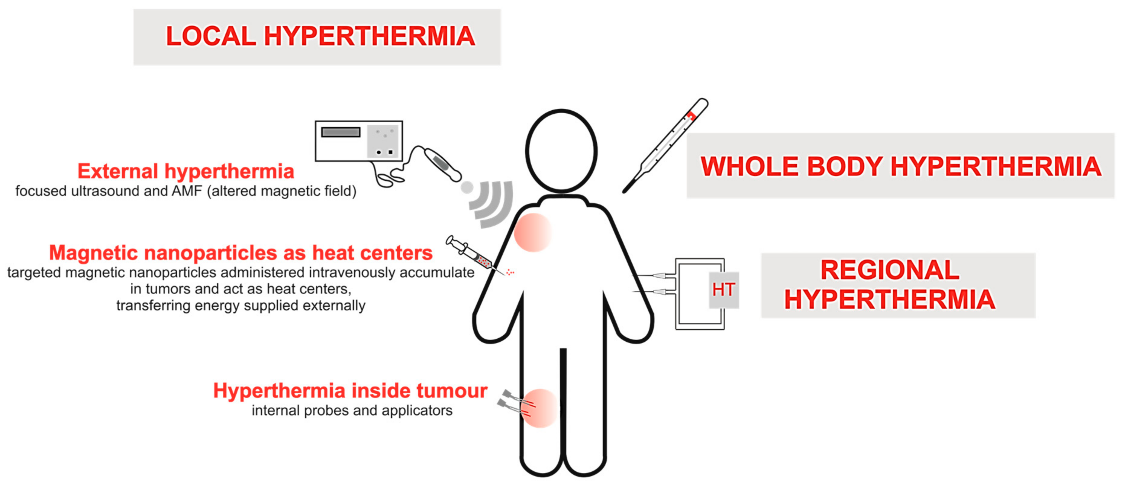
2. Types of Conventional HT Methods

Although HT has been applied for clinical cancer treatment, local control of temperature remains challenging. To date, three main types of HT have been applied in clinical practice (Figure 1): whole-body, regional, and local. Whole-body HT, which is often used as an adjuvant for metastatic disease, is applied with heating blankets and thermal chambers. Regional HT involves perfusion of the peritoneal cavity with heated fluids with anticancer drugs. However, the clinical application of both whole-body and regional HT is limited due to severe side effects, especially gastrointestinal symptoms (diarrhea, nausea, and vomiting) and cardiac complications (thrombosis, myocardial ischemia, and even myocardial failure) [10].
Local HT, a tumor-focused technique that can be applied at different stages of invasiveness, is classified into three types: external, luminal, and interstitial. External HT is characterized by surface-induced increases in temperature, so that the heat generated at the cutaneous layers reaches the superficial tumor. External heat generation can be achieved using microwaves, laser, radio-irradiation, or high-intensity ultrasound. However, the tumor and normal tissues are heated in a heterogeneous manner, as the temperature can range from 39 to 42 °C to lethal temperatures of 43–45 °C, which is a major disadvantage of this method. Luminal HT, which is primarily applied for the treatment of colorectal cancer, uses special probes placed as close as possible to the tumor located in the rectal lumen. Interstitial HT is based on heat generated via a metal antenna made of ferromagnetic material to increase the temperature of tumor tissues [11,12]. However, this method is highly invasive, painful, and can lead to necrosis as far as 1–2 cm from the applied heat source [13]. Since heat generated by conventional HT methods is distributed in a chaotic manner, improved methods are needed to minimize thermal damage to surrounding healthy tissues. Thus, novel HT methods based on magnetic NPs (MNPs) and carriers are discussed in the following sections.

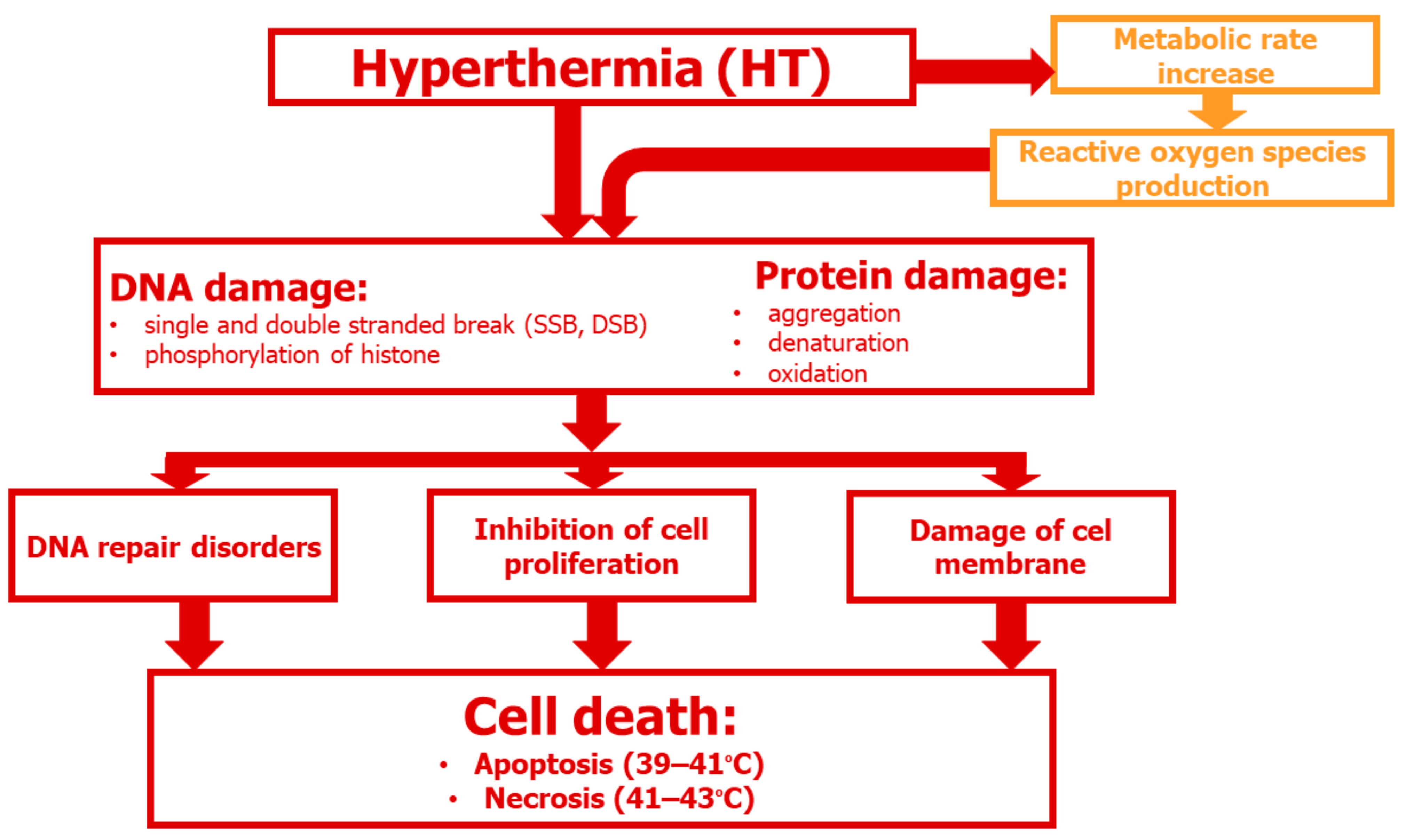
3. Cellular and Molecular Aspects of HT

HT causes many changes within cells, resulting in the loss of cellular homeostasis and subsequent cell death [14,15]. The type and extent of cell damage is determined by temperature. Moderate temperatures of 39–42 °C are generally non-lethal, whereas temperatures > 42 °C can kill cells in a time-dependent manner [16]. Higher temperatures cause damage at the cellular level by facilitating unfolding of proteins, thereby exposing hydrophobic groups, resulting in protein aggregation (Figure 2). Proteins damaged by HT can also form aggregates with normal proteins. In addition, degradation of aggregated/misfolded proteins through the proteasomal and lysosomal pathways leads to compression within the nuclear matrix and irreversible changes to chromatin.
Heat-induced unfolding and aggregation of proteins also impact the nucleus, which contains large amounts of proteins and DNA. The direct cytotoxic effects of HT are due to the denaturation, aggregation, and degradation of specific proteins involved in DNA synthesis (DNA polymerases-α and -β), DNA repair, transcription, RNA processing, and translation, ultimately leading to cell cycle arrest and cell death [17,18,19]. Other effects of HT include disruption of the cytoskeleton, dysregulation of membrane permeability, and metabolic changes (e.g., uncoupling of oxidative phosphorylation) that lead to decreased energy production and increased intracellular levels of Na+, H+, and Ca2+ [20,21,22].
HT is a viable strategy for cancer treatment by either directly killing cancer cells or induction of sensitization to RT or CT. Although normal cells are more tolerant to heat, tumor cells exhibit stronger thermal cytotoxicity and can be selectively targeted due to the chaotic architecture of the vasculature and hypoxic and low pH regions of solid tumors [23]. Hence, HT is considered a noninvasive strategy that does not adversely affect normal cells [24,25]. HT can also trigger immune responses via several mechanisms. The goal of HT is to mimic a state of fever and activate the immune response to destroy cancer cells. At around 38.5 °C, the immune response is activated, while antitumor immunity is activated at 39–43 °C. Therefore, HT can be used in combination therapy and as an adjuvant immunotherapy [26,27,28]. Also, HT can indirectly modulate the innate and adaptive immune responses to target the tumor microenvironment by increasing the production of proinflammatory cytokines and heat shock proteins (HSPs), which activate antigen-presenting cells (APCs), such as dendritic cells (DCs) and macrophages [29,30,31].
HSPs have been implicated in the difference in sensitivity to HT between neoplastic and normal cells. HSPs are produced in response to stress, including heat [32]. Using special metallic culture vessels for the immediate and accurate regulation of temperature, Imashiro et al. [23] observed that, after increasing the temperature to 43 °C for 30 min, HSP72 expression was higher in normal human dermal fibroblasts than breast cancer (MCF-7) cells. In addition, HSP72 was localized in the nuclei of normal cells, which is associated with the development of thermotolerance, as compared to the cytoplasm of tumor cells, which results in apoptosis [33].

3.1. DNA Damage

HT can directly and indirectly damage DNA. As direct damage, HT induces breaks to single-stranded as well as double-stranded DNA, phosphorylates the C-terminal serine residues of histone H2AX and ataxia–telangiectasia-mutated protein, and downregulates the activities of DNA polymerases and topoisomerases. Indirectly, HT increases production of reactive oxygen species (ROS), arrests the cell cycle, and inhibits DNA replication, resulting in cell death. Moreover, HT promotes DNA damage in cancer stem cells, which can be beneficial against cancer because these cells are resistant to most classical treatment strategies [34]. In addition, HT causes irreversible DNA damage that can complement the effects of CT and RT [35]. Notably, exosomes extracted from heat-stressed tumor cells induce a bystander effect that can also cause DNA damage to tumor cells not exposed to heat stress [36]. Nonetheless, mapping of tumor-specific genetic aberrations by whole-exosome sequencing, in silico gene prediction, mass spectrometry, and T cell assays would be beneficial for identifying novel antigens [37].

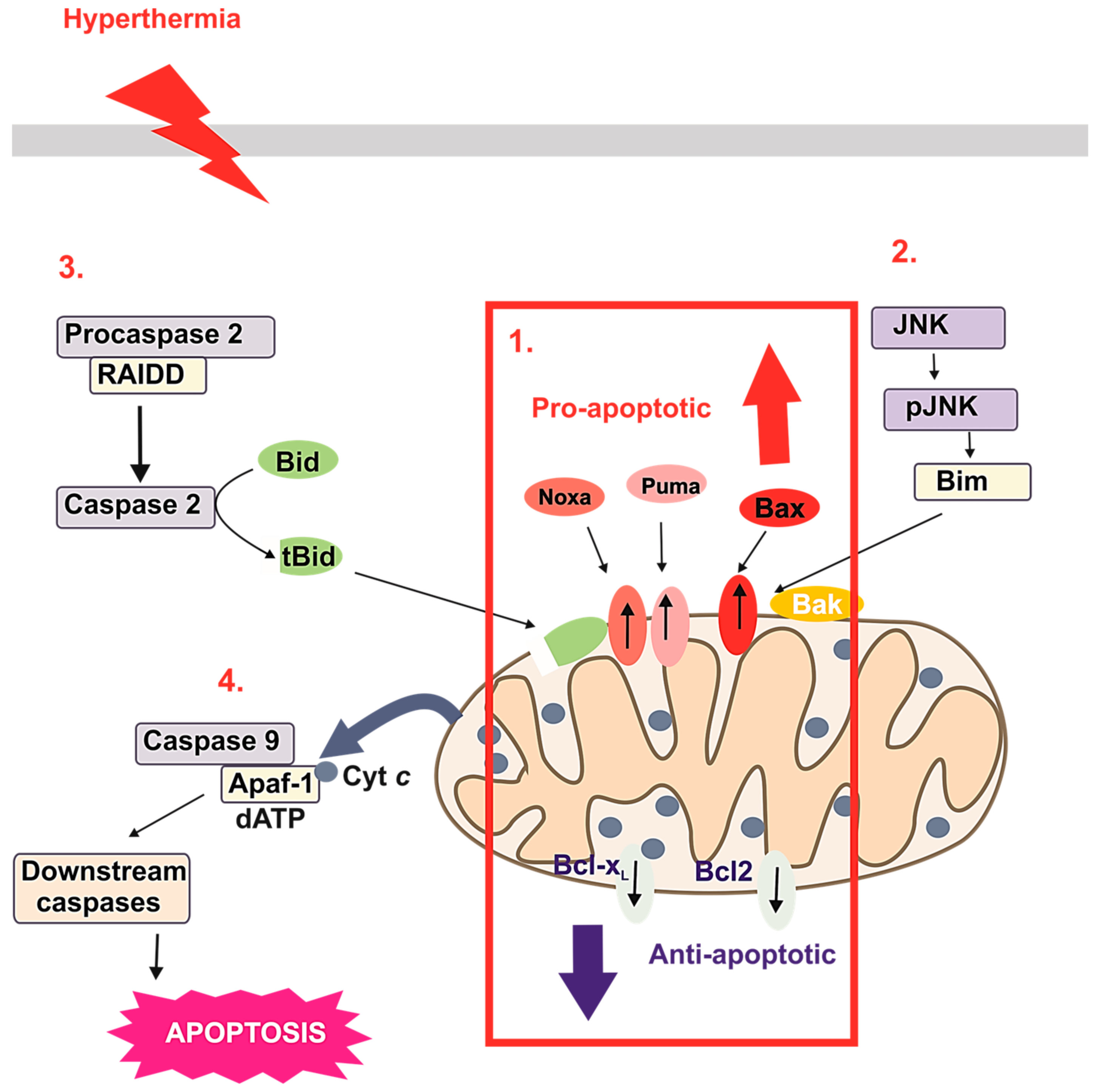
3.2. HT-Induced Apoptosis

Apoptosis is a natural biological process which can control the proper development, homeostasis, and proliferation of new cells with constant removal of damaged and unnecessary cells. HT at 42–44 °C is reported to stimulate apoptosis of cervical cancer (HeLa) and leukemic cells [38]. However, some cell types are more tolerant to heat stress than others [39]. Major apoptotic pathways resulting in caspase activation include the extrinsic (death receptor mediated), intrinsic (mitochondrial mediated), and endoplasmic reticulum stress-mediated pathways (Figure 3). In the intrinsic signaling pathway, which is considered the primary pathway involved in HT the pro-apoptotic protein Bid (BH3 interacting-domain death agonist) regulates the translocation of the pro-apoptotic protein Bax (bcl-2-like protein 4) to the mitochondrial membrane. The ratio of endogenous pro- to anti-apoptotic proteins largely determines cell fate [40]. An elevated temperature alters the balance of pro- and anti-apoptosis Bcl-2 family proteins. Experiments performed with HeLa cells confirmed that HT at 42–43 °C led to the decreased expression of the anti-apoptotic proteins Bcl-2 and Bcl-xL (B-cell lymphoma-extra-large protein), and the increased expression of the pro-apoptotic proteins Bax, Bak (BCL2 antagonist/killer 1 protein), Puma (p53 upregulated modulator of apoptosis), and Noxa (phorbol-12-myristate-13-acetate-induced protein 1). HT also activates procaspase 9, the initiator of the intrinsic apoptosis pathway, and caspase 3, which promotes chromatin condensation. The significance of HT and the roles of Bcl-2 family proteins in apoptosis are demonstrated by reversal of the decreased translocation of Bax, Noxa, and Puma to the mitochondria and inhibition of cytochrome c release in cells tolerant to 40 °C. The Bcl-2/Bcl-xL inhibitor ABT-737 was reported to sensitize cells to apoptosis, indicating that Bcl-2 family proteins are involved in HT-induced apoptosis [40]. Also, HT in the range of 40 to 45 °C was shown to induce apoptosis of tumor cells via the intrinsic pathway, whereas higher temperatures resulted in necrosis [41]. During heat-induced cell death, caspase 2 forms a complex with the specific adaptor protein RIP-Associated Protein with the A Death Domain, which activates caspase 2 and cleaves Bid to tBid, causing changes to the mitochondrial outer membrane potential with the subsequent release of cytochrome c, resulting in the formation of apoptosomes consisting of cytochrome c, Apaf1 (apoptotic protease activating factor 1), and caspase 9 [25]. Moreover, HT activates the pro-apoptotic protein Bim (Bcl-2-interacting mediator of cell death), which induces apoptosis through a Bax/Bak-dependent pathway [42].
HT is also a strong activator of c-Jun N-terminal kinases (JNKs), which phosphorylate Bim to enhance pro-apoptotic activity [43]. HT-induced ROS production triggers apoptosis of various cell types [25]. HT can also trigger the extrinsic apoptosis pathways via activation of cell surface receptors (Figure 3). HT-induced apoptosis is partially dependent on activation of the Fas ligand, TNF-α (tumor necrosis factor α), and TRAIL (TNF-related apoptosis-inducing ligand) [39].

4. Use of MNPs with Locally Induced HT

Regardless of the type of HT, the use of different heat sources is accompanied by the generation of a specific temperature gradient within tumor tissues. Commonly used external heat sources, such as ultrasound and infrared radiation, have a narrow window of efficacy and could burn the skin surface before the temperature within the tumor reaches a satisfactory therapeutic level. Therefore, MNPs present an extremely promising tool for HT to penetrate the tumor while avoiding damage to healthy tissues [6,44]. The physicochemical properties of MNPs also support their use in HT. MNPs have a diameter of approximately 100 nm, with a large surface area to mass ratio, and are highly reactive. As an additional advantage, the irregular structure of MNPs facilitates the penetration of tumors [45].
The phenomenon of enhanced permeability and retention relies on the increased permeability of the blood and lymphatic vessels in the tumor environment. Thus, the large diameters of the intercellular spaces of tumors favor free localization, local accumulation, and prolonged retention of NPs. Localization near the vascularizing blood system of the tumor also favors the use of NPs for local HT. When a temperature gradient is created, maximum heat is induced at the vessel wall and decreases with distance from the perivascular space, which facilitates the destruction of the blood vessel network, reduces angiogenesis, and inhibits tumor metastasis. Since the main drawback of conventional HT is the lack of selectivity when heating the tissues, the application of NPs prevents the application of heat to healthy tissues situated along the path of external radiation [6]. NPs can absorb heat energy originating from an external source to enhance the effects of HT. In addition, NPs are the primary source of heat and reverse the direction of heat loss (inside-out HT). In this type of HT, NPs focus heat from an external source onto the tumor to induce localized thermal destruction, while minimizing damage to collateral tissues [3]. Thus, iron oxide NPs, gold NPs, and carbon nanotubes can be applied with inside-out HT (Figure 4).
For non-invasive HT, MNPs can localize heat generated with an AMF. As the most promising “thermo-sensitizer”, the use of iron oxide NPs in HT is supported by the size, ease of functionalization with both organic and inorganic compounds, biocompatibility, minimal toxicity, and ease of excretion [46]. In addition, the ferromagnetic properties of MNPs can be exploited for simultaneous therapeutic and diagnostic applications. Iron oxide NPs are characterized by theranostic action. MNPs are essential components of diagnostic systems used to monitor disease progression and responses to therapy. In addition, MNPs are used as components of complex drug delivery systems for anticancer drugs, immunomodulators, and nucleic acids [47].
When exposed to a magnetic field, the oscillatory vibrations of MNPs are accompanied by a local increase in temperature and the release of cargo, such as anticancer drugs [48,49].
In addition, MNPs are activated by an external magnetic field, through the magnetic coupling between their magnetic moment and the magnetic component of the field [50]. The energy from this coupling process is absorbed by MNPs and then released as heat. The heating capacity of MNPs follows from their magnetic properties [51]. It means that the heat generation is associated with dynamic hysteresis losses resulting from the magnetic moment relaxation of single domain nanoparticles. For this magnetic hyperthermia, biological tissues display no significant energy deposition, and this technique is safe for non-cancer cells [52].
Recent studies have focused on the magnetic dipoles of iron oxide-based NPs, such as superparamagnetic iron oxide nanoparticles (SPIONs). During AMF amplification, SPIONs align with the direction of the field and the resultant magnetic susceptibility is several folds greater than that of standard paramagnetic materials [53]. The synthesis of SPIONs usually involves precipitation of iron salts (mainly by photochemical methods) in the presence of ammonia, sodium nitrate, and sodium hydroxide [54]. Nonorganic SPION-type nanostructures are rapidly eliminated from the body. Therefore, various polymeric modifications are used to improve the biological performance of SPIONs, such as the combination of polyethylene glycol (PEG), β-cyclodextran, the non-ionic detergent Pluronic F127, and chitosan [55,56]. Surface modifications of SPIONs also involve the attachment of ligands, which act as vectors to increase the likelihood of direct delivery into the tumor niche. For example, folic acid–SPION conjugates can be attached to surface receptors for delivery of folate to tumors in the brain, breast, and liver [57]
The surface of SPIONs can also be modified with colloidal gold. Briefly, a thin layer of gold is heated by AMF to enhance the HT effect of the MNPs. The heat release is several folds greater with gold-coated SPIONs as compared to unmodified SPIONs. HT mediated by SPIONs can be combined with other cancer treatment strategies [58]. For example, SPIONs coated with thermosensitive polymers can be loaded with anti-cancer drugs and delivered into the tumor microenvironment. The application of an AMF increases the temperature within the tumor causing the destruction of SPIONs and the subsequent release of the drug [59].
The mode of intracellular transport influences the induced cytotoxic mechanisms of MNPs. Biocompatibility studies with human umbilical vein endothelial cells (HUVECs) showed that endocytosis is the main mechanism of the intracellular transport of dextran- and citric acid-coated SPIONs. Additionally, SPIONs were found to inhibit the migration and induce apoptosis of HUVECs [60]. An in vivo animal study revealed that the pharmacokinetics and biodistribution of MNPs were dependent on the mode of administration, hydrodynamic diameter, and surface charge. Iron oxide MNPs are preferentially accumulated in the liver and spleen, rather than the brain, heart, kidney, and lung. Moreover, the site of distribution is strongly correlated with the size of the MNPs, as large MNPs (up to 4 μm) are removed by the reticuloendothelial system (RES) of the liver, while those that are 200–250 nm are usually filtered by the spleen [44]. As the intravenous administration of MNPs accelerates their rapid elimination from the body, SPIONs should be loaded into cells ex vivo and infused directly within the tumor, as confirmed with neuronal progenitor cells loaded with SPIONs and transplanted into mice with induced melanomas. The progenitor cells penetrated the tumors and subsequent exposure to AMF resulted in significant tumor regression and prolonged survival [61]. Drug delivery systems are developed to maximize drug efficacy and minimize side effects of targeted therapies. Encapsulation of therapeutics or biologically incompatible particles in nanocarriers can increase the solubility and stability of delivered molecules, thereby increasing their half-life and bioavailability. Although significant progress has been made in the field of drug nanocarriers, widespread clinical use remains uncertain. Their interaction with living cells changes the surface reactivity of nanomaterials, which could lead to undesirable and unforeseen physiological consequences. Also, the physicochemical properties of drug delivery systems, such as size, shape, surface charge, and coating, determine the biocompatibility of nanotransporters [62]. The phenomenon of protein adsorption onto macromolecule surfaces, as described by Leo Vroman in 1962 [63], which is currently known as the “protein corona effect”, defines the biological identity of nanocarriers and may be important in the systemic biological response generated by NPs [64].
As the first nanocarriers approved by the U.S. Food and Drug Administration (FDA) for clinical use, liposomes are circular vesicles composed of a lipid bilayer surrounding an aqueous core [65]. Liposomal doxorubicin (DOX), marketed under the trade name Doxil® in the USA and Caelyx® in the European Union (Jonson and Jonson, New Brunswick, NJ, USA), was initially developed for the treatment of Kaposi sarcoma and multiple myeloma. As compared to non-encapsulated DOX, Doxil® has greater efficacy and lower cardiotoxicity. However, the low stability of Doxil® and the phenomenon of intravenous leakage, described as the uncontrolled release of DOX from liposomes into the bloodstream, significantly limited its widespread clinical use [66]. Hence, biodegradable polymeric NPs, which were developed as an alternative to liposomes, can improve therapeutic efficacy, while reducing the risk of adverse side effects. Nanocarriers based on polyelectrolytes are biocompatible and dissociate into polyanions and polycations in aqueous solutions. Biomacromolecules, such as nucleic acids, proteins, and polysaccharides, are classified as polyelectrolytes [67].
Various types of magnetic materials, including metal NPs, metal oxide NPs, and core–shell MNPs [68], are used for magnetic hyperthermia (MHT). The features of MNPs are strictly related to size, shape, composition, and structure, which are controlled by polymeric modifications during synthesis [49,69].
The polymer used as the coating material or as a component of the nanocarrier protects MNPs from oxidation and aggregation, while allowing for further functionalization in the body. However, even slight modification of the polymer could influence the adsorption of blood plasma proteins (opsonization) and their ability to bind to the membranes of macrophages and other cell types [70]. Opsonization usually leads to receptor-mediated phagocytosis and rapid clearance of MNPs from the blood, and facilitates the formation of a corona of plasma proteins, which increases the diameter of MNPs. For instance, MNPs with diameters larger than 200 nm are rapidly taken up by the RES and accumulate in the liver and spleen, while MNPs with diameters smaller than 6 nm are filtered by the kidneys [71]. Nevertheless, MNPs with diameters of 10–100 nm are pharmacokinetically ideal for in vivo applications [72,73]. MNPs can be encapsulated in a polymeric core shell or coated with suitable polyelectrolytes to form a protective layer against protein attachment. For example, iron oxide NPs coated with dimercaptosuccinic acid avoid opsonization and clearance by the RES, while reducing cell toxicity [70]. Moreover, surface functionalization with a hydrophilic PEG polymer can extend the half-life of iron oxide NPs to 12 h [74]. Polyvinylpyrrolidone and zwitterionic materials, such as dopamine sulfonate and poly(amino acids), as novel polymers, can provide a longer blood circulation time than PEG [75].

5. Bioconjugates of MNPs as Potential Passive or Targeted Delivery Systems

Modifications to and the size of the polymers within MNPs determine the mode of uptake by cancer cells. MNPs with diameters greater than 10 nm can extravasate and accumulate in tumor tissues, but not in normal tissues, due to natural differences between the defective and leaky vasculature of tumors and regular openings of normal vessels in healthy tissue [76]. The slower lymphatic clearance and venous return of the tumor microenvironment help to retain MNPs in tumor tissues [77]. The enhanced permeability and low clearance of solid tumors [68] can inhibit the targeted accumulation of MNPs [78], which is mostly due to the phagocytic activity of specialized cells of the RES [61]. In contrast to passive targeting, active targeting ensures accumulation of MNPs in solid tumor tissues via the overexpression of surface ligands specific to cancer cells [79]. Active targeting can facilitate the efficient internalization of targeted MNPs by receptor-mediated endocytosis [80] and has been described as a main factor affecting the binding of MNPs to cells in vitro [81]. Adhesion to the target cell is fully dependent on recognition of the targeting moiety of the MNPs [82]. Thus, the targeted internalization and accumulation of MNPs in cancer cells can avoid damage to normal cells. Antibodies, peptides, and ligands are the most commonly used molecules for targeting cancer cells.
The amount of MNPs delivered by active targeting could be insufficient to generate adequate heating at the tumor site [83]. However, many recent in vitro and in vivo studies [84,85] support the superiority of targeted versus non-targeted MHT. For example, the folate receptor (FR) facilitates the high-affinity binding of folate and related conjugates to target cells via receptor-mediated endocytosis [86]. A study by Li et al. [87] showed that the FR is required to mediate drug accumulation in targeted malignant cells.
Folic acid conjugates bind to the FR on the surface of malignant cells and form endosomes that are internalized to intracellular compartments [88]. Folic acid conjugation along with pH-sensitive linkers can increase the rate of drug secretion at pH 5.0 within growing cancer cells [89]. However, a linker is not necessary, as described by Bonvin et al. [90], who developed a folic acid-based theragnostic platform for simultaneous diagnosis and treatment of metastatic prostate cancer by targeting the lymph nodes [90]. Folate-conjugated SPIONs were reportedly taken up by FR-positive HeLa cells and significantly decreased the intensity of non-specific signals, thereby improving the accuracy of magnetic resonance imaging (MRI) [91]. While the FR is overexpressed by various cancer cells and minimally expressed by normal cells [92], the receptor tyrosine–protein kinase erbB-2 (HER2) is specifically expressed in some aggressive types of breast and lung cancers [93]. MNPs are used as initial nanocarriers and modified via PEGylation followed by the immobilization of trastuzumab (TRA), a monoclonal antibody against HER2, to target various types of breast cancer cells. Recently, Hamzehalipour et al. [94] described a novel drug delivery system (MNP-PEG-TRA) for the targeting of SK-BR-3 breast cancer cells in a mouse model of 7,12–dimethylbenz(a)anthracene (DMBA)-induced breast cancer. As compared to non-labelled MNPs, the proposed MNP-PEG-AMF drug delivery system remarkably enriched the effect of HT in cultured SK-BR-3 cells in vitro as well as DMBA tumor-bearing mice in vivo. The dosage of MNP-PEG-TRA was four-fold greater at the tumor site as compared to other organs, confirming considerable potential for treatment of breast cancer [94]. A cell-targeting function could also be added to thermally responsive core–shell MNPs by grafting a monoclonal antibody to target overexpressed HER receptors on mouse bladder tumor cells. In fact, the viability of mouse bladder tumor cells treated with the combination of fluorouracil, a cell-targeting agent, and an AMF was reduced by 50%. Interestingly, the same treatment, but with a non-specific targeting ligand (immunoglobulin G) or free fluorouracil, did not reduce cell viability as significantly [95]. Epidermal growth factor receptor (EGFR) is another surface protein that mediates cellular responses to various growth factors and is highly expressed on the surface of non-small cell lung cancer cells in almost 80% of patients [96]. EGFR is a useful diagnostic and therapeutic biomarker. In addition, EGFR-targeted SPIONs exhibited increased retention in tumor cells and moderately suppressed the growth of lung tumors [97].
Since membrane proteins, such as EGFR, can be targeted with antibodies and other molecules, it should be possible to entice the cell to internalize the antibody–antigen–MNP complex [98]. After the application of an AMF, MNPs are endocytosed and may interact with different membrane compartments, which (1) internalize MNPs from the plasma membrane, (2) recycle the MNPs back to the surface via early and recycled endosomes, or (3) degrade the MNPs via late endosomes and lysosomes. Increasing subcellular temperatures may induce cell death through lysosomal death pathways, suggesting potential applications of MNPs to induce death of apoptosis-resistant cancer cells. Clerc et al. [99] reported that the induction of magnetic intralysosomal hyperthermia caused cell death through a non-apoptotic signaling pathway by locally increasing temperature. Specifically, gastrin-grafted MNPs delivered to lysosomes induced generation of free radicals via the Fenton reaction. Subsequently, magnetic intralysosomal hyperthermia triggered permeabilization of the lysosomal membrane, resulting in the leakage of lysosomal enzymes into the cytosol, including cathepsin B, which activated non-apoptotic caspase 1. In contrast, Domenech et al. [100] reported that the application of an AMF induced the accumulation of EGFR-targeted MNPs resulting in the subsequent disruption of lysosomes, but no local increase in temperature, although ROS production was increased and cell viability was reduced. Shah et al. [101] developed a magnetic core–shell NP to deliver a mitochondria-targeting pro-apoptotic amphipathic tail-anchoring peptide to cancer cells combined with increased localized temperatures, which significantly enhanced apoptosis due to a synergistic effect on mitochondrial dysfunction in cancer cells [101].

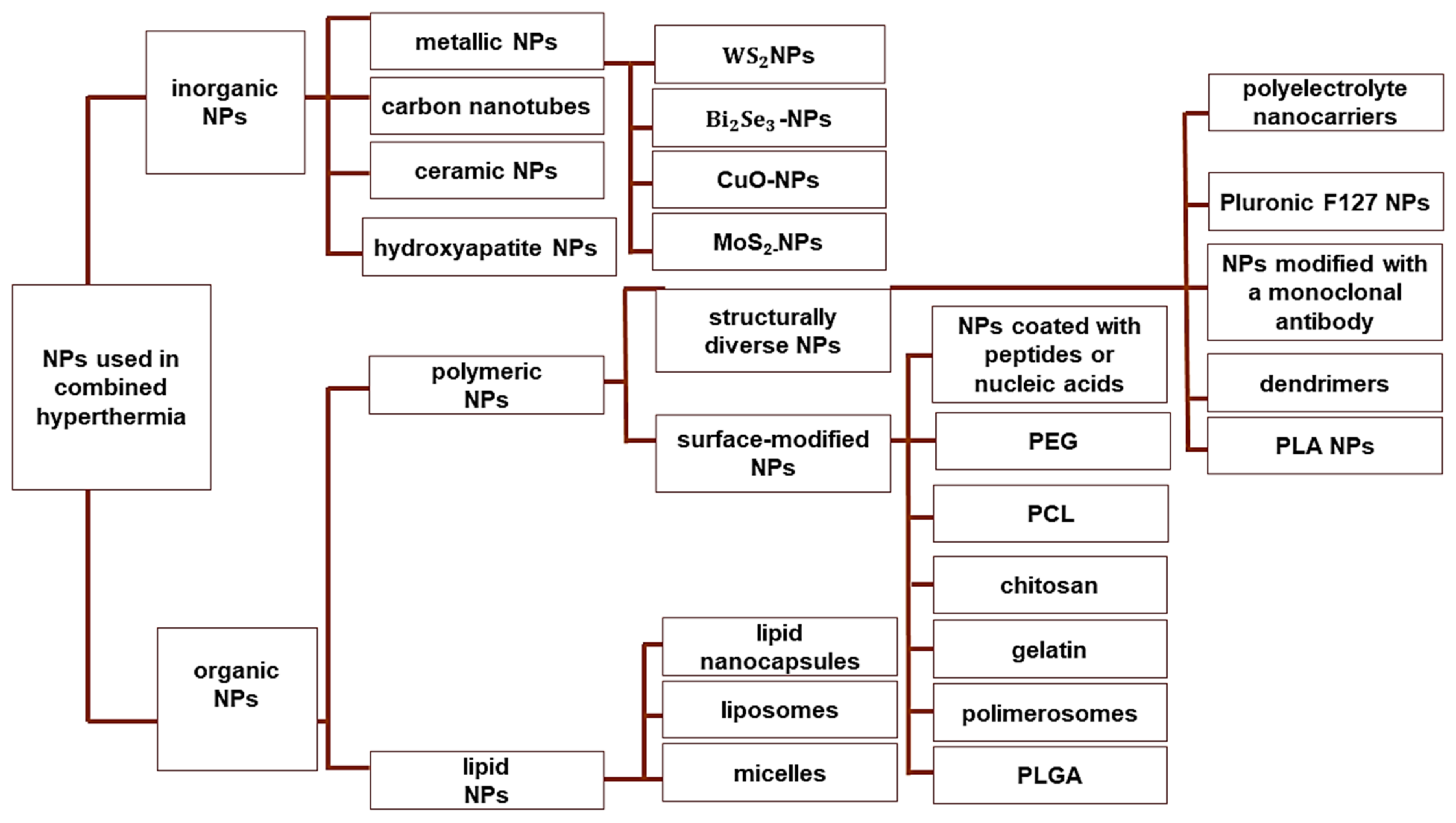
6. Combination Therapies Using NP-Based MHT

One of the most clinically significant factors concerning the application of MHT is the effect of increased temperature on blood flow within the tumor and the surrounding healthy tissues [51]. The blood vessels are among the first barriers to interact with MNPs. Thus, increased permeability of vascular walls or blood vessel dilation is a natural consequence of constant exposure of non-cancer endothelial cells to elevated temperatures (41–46 °C). This issue can be addressed by the synergistic application of MHT together with CT, RT, photothermal therapy, or gene therapy, which are also more effective when combined with other modalities [9].

6.1. CT

MHT in conjunction with CT provides several advantages, including increased intracellular drug concentrations, inhibition of DNA repair, and a reduction in the proportion of apoptosis-resistant cancer cells [102]. Local application of MNPs with MHT can increase blood flow to effectively accelerate intracellular drug delivery and release in cancer tissues. In this scenario, the initial application of MHT will augment the CT-triggered death rate of already dying cancer cells. Ideal polymers for development of MNPs should be controlled by pH, temperature, or AMF to release loaded therapeutics [103]. Polymeric MNPs can be loaded with various classes of anti-cancer drugs, including hydrophilic agents, such as the anthracycline DOX, which is water-soluble at a mildly acidic or neutral pH [104]. The major molecular mechanisms underlying the anticancer activities of DOX include intercalation into DNA, p53-induced apoptosis, ROS production, and mitochondrial dysregulation. However, under continuous oxidative stress, DOX can also act on normal, noncancerous tissues, thereby impairing the function of healthy organs, especially the heart, liver, and kidney [105]. Although approved by the FDA for cancer therapy, the liposomal nanoformulation Doxil® did not significantly improve the quality of life of cancer patients [106]. Mai et al. [107] engineered magnetic thermoresponsive iron oxide nanocubes (TR-cubes) for use with MHT for heat-mediated drug delivery. Iron oxide-based NPs were selected due to their outstanding stability and performance with MHT. Copper-mediated polymerization with ultraviolet light increased the polymerization rate and prevented aggregation of the TR-cubes. Moreover, the TR-cubes were sufficient for the delivery of DOX, while maintaining thermo-responsiveness. The results of an in vivo study showed that DOX-loaded TR-cubes achieved complete tumor regression and the highest survival rate of animals exposed to an AMF. Polymeric MNP-based complexes can not only be applied for DOX delivery systems, but also possess additional features as promising tools for imaging. Thirunavukkarasu et al. [108] developed magnetic field-inducible drug-eluting nanoparticles (MIDENs) by encapsulating superparamagnetic iron oxide NPs and DOX in a temperature-responsive poly(lactic-co-glycolic acid) (PLGA) nanomatrix that, when exposed to an external AMF, generated heat at >42 °C, which subsequently triggered the controlled release of DOX from the nanomatrix. The results of an in vitro study showed that MIDENs exposed to an AMF effectively killed CT26 colon cancer cells. Moreover, the results of an in vivo T2-weighted MRI study indicated that the use of MIDENs exposed to an AMF suppressed the growth of malignant tumors [108].
Hybrid nanogels are also attractive nanocarriers for biomedical applications. These nanostructures are composed of thermoresponsive polymers and superparamagnetic NPs that can take up and release large amounts of DOX. Cazares-Cortes et al. [109] developed biocompatible, pH-responsive, magnetoresponsive, and thermoresponsive nanogels (MagNanoGels), composed of polymer complexes of maghemite (γ-Fe2O3) MNPs loaded with DOX. In PC3 prostate cancer cells, not only did the DOX-MagNanoGels efficiently internalize DOX, but also intracellular release of DOX could be remotely triggered by an AMF, thus improving the cytotoxic properties of DOX [109].
Since DOX is a key chemotherapeutic agent, different types of porous magnetite nanospheres (PMNS) conjugated with DOX have been evaluated for the treatment of early and advanced breast cancer. For instance, Sharifi et al. [110] reported that lactoferrin-DOX-PMNS significantly suppressed the proliferation of 4T1 breast cancer cells and reduced tumor weight by prolonging drug availability and potential drug loading in cancer cells. Other antineoplastic agents for breast cancer therapy can also be effectively encapsulated in magnetic-based drug delivery systems [111]. Polymeric micelles are multifunctional MNPs that allow for control of drug release via an external AMF and prevent early clearance by the RES. Zheng et al. [112] synthesized hyaluronic acid-C16 copolymers via a peptide formation process with subsequent co-encapsulation of the therapeutic agent docetaxel and SPIONs to form multifunctional micelles with specific targeting capability based on CD44 receptor-mediated endocytosis that was enhanced in the presence of an external magnetic field. The cytotoxicity of the anti-breast cancer drug mitoxantrone was improved by linking it to magnetic thermoresponsive copolymer NPs. Li et al. [113] reported the superb paramagnetic behavior of magnetic thermoresponsive copolymer NPs for controlled drug release, which improved the anti-tumor efficacy of mitoxantrone with fewer side effects.
Polymeric MNPs have been applied for successful targeted delivery of various anticancer drugs and many natural substances that possess antineoplastic properties for potential use with MHT. For instance, curcumin (CUR) has been shown to effectively inhibit tumor growth and has been classified as “Generally Recognized as Safe” by the FDA [114]. Senturk et al. [115] developed multi-functionalized NPs composed of SPIONs coated with a PLGA-PEG di-block copolymer and loaded with CUR that exhibited high antitumor activity against glioblastoma cells and confirmed that these MNPs generated heat at ~42–45 °C by exposure to an AMF for 15 min.
Novel polymeric magnetic nanosystems have been developed to deliver CUR alone and in combination with specific inhibitors. Sudame et al. [116] successfully encapsulated CUR and the calcium channel blocker nifedipine within d-ɑ-tocopheryl-based polymeric magnetic NPs to improve the magnetothermal effects and effectively release drugs in HepG2 cancer cells, which significantly reduced tumor growth [116].

6.2. RT

RT can damage the DNA of tumor cells but does not reduce the risk of metastasis [117]. MNPs can act as both HT agents and radiosensitizers [3,118]. Rezaie et al. [119] found that the combination of RT (2 Gy at 6 MV) and HT (43 °C for 1 h) with iododeoxyuridine-loaded PCL-PEG-coated MNPs significantly reduced colony formation of U87MG glioblastoma spheroids in culture. Grauer et al. [57] assessed the efficacy of SPIONs combined with HT (six 1-h courses of SPION-mediated HT) and stereotactic RT (39.6 Gy) for treatment of recurrent glioblastoma multiforme and found that intracavitary HT in combination with RT induced a powerful antitumor immune response, mostly around the resection cavity, and achieved long-term stabilization [57]. The combination of MNP-HT and RT has also been successfully applied for different types of prostate cancer. For example, Attaluri et al. [120] reported that RT at 5 Gy in combination with MHT with polymeric bionised nanoferrite NPs for 1 h improved efficacy against both LAPC-4 and PC3 prostate cancer cells as compared to either RT or HT alone. Also, outstanding results were achieved against PC3 and LAPC-4 cells using mouse models of human prostate cancer. Another animal study found that HT (43 °C for 20 min) plus RT (5 Gy) extended survival by two-fold as compared to the control group. Moreover, Jiang et al. [121] developed gadolinium-doped iron oxide NPs with improved superparamagnetic properties and higher specific absorption rates than ordinary iron oxide, which successfully delayed the progression of prostate C1 tumor growth by 10 days in a transgenic mouse model of adenocarcinoma treated with MHT and RT as compared to mice treated with only MHT (2.5 days) or RT (4.5 days). Moreover, immunohistochemical staining revealed diminished hypoxia with vascular disruption, suggesting reduced resistance to radiation [121].

6.3. Gene Therapy

Polymeric MNPs are promising tools for the targeted delivery of foreign genetic material (oligonucleotides, genes, or gene segments). The many advantages of MNPs include improved solubility, pharmacology, and stability [122]. In addition, gene delivery with MNPs can restore normal cellular functions and mediate programmed cell death [123]. MNPs have also been successfully used to deliver therapeutics and reporter genes via a high-field, high-gradient magnetic field to suppress specific genes [122].
MNPs are associated with nucleic acid complexes, added to cell-growing media and subsequently onto the cell surface by applying a magnetic force [124]. Developed by Stephanie Huth and coworkers [125] magnetofection was first used for MNP–naked DNA complexes or MNP–viral vector complexes that were attracted to the bottom by a magnet, placed close below the bottom of a dish. Subsequently, nucleic acid-bound MNPs could be introduced into the cells after their exposition to AMF [126].
Several crucial issues must be addressed when using polymeric MNPs to deliver nucleic acids to cancer cells. First, an AMF should be applied to MNP-DNA conjugates in cell culture to improve the rate of transfection. Second, for in vivo studies, a magnetic field should be applied for the targeted delivery of therapeutic genes. Hence, an AMF can be applied intravenously to target polymeric MNP-DNA complexes in the bloodstream. Once the NPs reach the target site, the genes are released from the particles by either enzymatic cleavage of the crosslinking polymers, pH-dependent reactions, or relapse of the polymer matrix [122]. However, even a slight increase in temperature can result in the overexpression of HSPs both in vitro and in vivo [127]. Therefore, various vectors for gene therapy with the application of an AMF have been constructed using expression systems under the control of an HSP promoter [127]. For example, Tang et al. [128] developed a polymeric Mn-Zn ferrite NP and vector-encoding β-galactosidase (β-gal) driven by a HSP70 promoter that highly expressed β-gal in tumor cells upon activation with an AMF. Even though β-gal expression did not induce anti-oncogenic activities, the results confirmed the potential of using inducible promoters together with MHT [129]. In another study, Yin et al. [130] reported that the binding of HSPs to the promoter acted as a switch to induce the expression of TNF-α in human lung adenocarcinoma A549 cells transfected with a TNF-α-specific construct with magnetite cationic liposomes and exposed to an AMF (30.6 kA/m, 118 kHz), while the temperature was controlled at 45 °C. Subsequently, TNF-α production was confirmed in cells transfected with the plasmid and exposed to an AMF, indicating that the HSP70B promoter was activated by increasing the temperature. Furthermore, the overproduction of TNF-α significantly decreased the viability of transfected A549 cells exposed to an AMF. In the same study, exposure to an AMF for 30 min significantly diminished the tumor volume of transgenic mice [130]. Interestingly, the combination of gene silencing with small interfering RNA (siRNA) and MHT has been used as a diagnostic tool [131,132]. Polymeric magnetic iron oxide NPs conjugated with low molecular weight protamine were used to deliver siRNA directly into tumor cells. Upon application of an external magnetic field, the siRNA concentration was decreased by almost seven-fold but achieved the same diagnostic effect [133].

6.4. Photothermal Therapy

The heat of some MNPs is relatively lower due to lower concentrations in the tumor tissue, thus increasing the internal temperature via photothermal therapy was explored as an alternative source [134]. There are two arguments supporting the application of MHT with photothermal therapy. First, although MNP-MHT can be achieved by direct intertumoral injection, particle distribution is significantly limited. Second, the addition of photothermal therapy can reduce the required dosage of irradiation. A synergistic effect was reportedly achieved by combining polymeric MNPs and photothermal nanomaterials with an AMF and laser irradiation [135,136]. For instance, Zhang et al. [137] developed iron-gallic acid (GA) network-based NPs for MRI-guided chemo-photothermal synergistic therapy of tumors. The spatial location and size of the tumor were accurately determined by T1-weighted MRI based on a Fe-GA network. The results of an in vitro study revealed that the Fe-GA network-based NPs were actively endocytosed and induced apoptosis in T1-grade and HepG2 cancer cells. Moreover, the NPs exhibited significant tumor-targeting capability and achieved the highest accumulation in tumor cells after 24 h of incubation [137]. Meanwhile, Ma et al. [138] designed Fe3O4–Pd Janus NPs (JNPs) with dual-mode MRI/photoacoustic imaging properties for simultaneous magnetic-photo HT and chemodynamic therapy, which achieved relatively higher temperatures through the synchronized application of AMF and laser irradiation. Impressive free radical generation was observed due to the increased magnetic–photo heating of Fe3O4–Pd JNPs. In addition, ROS production was correlated with the antitumor effects of the Fe3O4–Pd JNPs in a mouse model of 4T1 orthotopic breast cancer. The Fe3O4–Pd JNPs with the application of an AMF plus laser irradiation completely inhibited tumor growth without significant adverse effects [138].

6.5. Immunotherapy

MNPs have also been applied to enhance the accumulation of active agents to not only kill tumor cells but also sensitize the immune system [5]. The efficacy of MNP-HT combined with immune checkpoint inhibitors has been intensively investigated [26,139]. Interactions with the immune system are influenced by the shape, surface charge, outer shell composition, and aggregation capacity of MHPs. Since the immune system detects extracellular components, partly based on size, NPs are designed to avoid the host immune response. Relatively large carriers are captured by APCs, while smaller particles (<200 nm) can circulate freely in the venous and lymphatic systems for much longer [140]. MNPs trigger the release of various cytokines, both locally and systemically, and recruit immune cells, including neutrophils, DCs, macrophages, natural killer cells, and B and T cells. DCs, for example, present antigens to T cells, which initiate the immune response by activation of CD4+ and CD8+ T cells. Some MNPs can alter the cytotoxic profiles of certain immune cells. Iron oxide NPs shift the polarity of macrophages from the anti-inflammatory M2 phenotype, which promotes uncontrolled tumor growth, to the pro-inflammatory M1 phenotype, which limits tumor growth [141,142]. Notably, most tumor-associated macrophages are the M2 phenotype, which promotes tumor invasion [143]. Regarding the influence of elevated temperature on the tumor environment, HT can induce both apoptotic and necrotic cell death [5]. Necrotic cells release danger-associated molecular patterns (DAMPs) and inflammatory cytokines into the tumor niche. DCs take up tumor antigens and tumor antigen–DAMP complexes for presentation to T cells, which results in the expansion of T cells and the activation of the adaptive anti-tumor immune response via macrophages and natural killer cells [144]. Production of DAMPs can be triggered by dextran-coated SPIONs and the MRI contrast agent FeridexTM (manufactured in Berlex Laboratories (Hanover, NJ, USA)). Specifically, dextran-coated SPIONs activate complement through both the lectin pathway and immunoglobulin M-dependent pathway [145]. Accumulation of SPIONs in U937 monocytes was reported to induce stimulation of the pro-inflammatory Th1 immune response [146]. These discoveries led to the discontinuation of FeridexTM and also highlighted the influence of surface-modified SPIONs on the immune system. For instance, PEG-coated iron oxide NPs induced the expression of interleukin 1β and TNF-α, and caused free radical-dependent toxicity, but not polarization of THP-1 macrophages [147]. On the other hand, polyethylenimine-coated SPIONs were reported to induce M1 polarization of macrophages via Toll-like receptor (TLR)4-mediated signaling pathways and ROS production [148].
The immunostimulatory effects of MNPs have been found to be beneficial against specific types of cancers, such as melanoma [149]. Duval et al. [150] reported that MHT of B16 melanoma cells triggered the overexpression of various immunogenic genes, such as Hsp-70 and CXCR3, as well as the innate immune activators TLR3 and TLR4. Interestingly, MHT combined with other immunotherapeutic approaches might have a synergistic therapeutic effect against oral melanoma [151,152,153]. Immunotherapy combined with HT increased immune cell infiltration and reduced the growth rate of melanoma [154]. The synergistic effect of immunoadjuvants and SPIONs provides a good example of the bimodal activity of immunotherapy and MHT [154]. As an immunoadjuvant, cytosin–-phosphate–guanine (CpG) oligodeoxynucleotides (ODNs) have been implemented in immunotherapy to activate TLR9 signaling pathways to increase the secretion of cytokines that promote the maturation of APCs and improve the Th1 immune response [155]. However, the clinical application of CpG ODNs is limited by nuclease susceptibility, unsatisfactory biodistribution, and systemic adverse effects [156]. Thus, Guo et al. [154] designed magnetic-responsive immunostimulatory NPs (MINPs) with FDA-approved drugs for MRI and anti-cancer immunotherapy. The MINPs were prepared with superparamagnetic iron oxide NPs and CpG encapsulated in monomethoxy PEG-PLGA-poly-L-lysine triblock copolymers. The authors demonstrated that, under an external magnetic field, the MINPs exhibited a magnetic-targeting ability, leading to a high accumulation of SPIONs and CpG ODNs in the tumors. After activation, the MINPs triggered strong anti-tumor immune responses, including DC maturation, cytokine secretion, and tumor infiltration by CD8+ T cells. An immune response was generated by the elimination of primary cancer cells and the inhibition of metastasis [154]. This strategy may potentially be relevant for precise diagnosis and individualized therapy for various tumors.

7. Conclusions and Future Perspectives

The main focus of this review was to summarize the applications of NP-mediated MHT for cancer therapy. A brief description of the current strategies used to improve the physicochemical properties and biological activities of MNPs combined with MHT was presented. Various factors, such as the size and shape of polymeric MNPs, viscosity of the medium, and parameters of the applied magnetic field, are crucial for the heating efficiency of MNPs. Furthermore, applications of polymeric NPs for MHT were described. Potential molecular disturbances at the cellular level in response to local increases in the temperature of tumor tissues with the use of MNPs were addressed. In addition, the synergistic effects of MNP-MHT combined with CT, RT, photothermal therapy, gene therapy and immunotherapy were investigated to improve the effectiveness of anticancer treatments. Recent in vitro and in vivo studies demonstrated the high capacity of MNPs for local drug delivery to reduce the viability of cancer cells and inhibit tumor growth. Moreover, the results of cellular and animal studies of MNP-MHT therapy were introduced into clinical trials for the treatment of prostate cancer (clinical trial NCT02033447). Magnetic thermoablation activated by an AMF was shown to effectively destroy cancer cells, while limiting the damage to surrounding healthy tissues. A study completed in January 2015 on 12 male patients with bladder or prostate cancer treated with radical cystoprostatectomy was conducted to assess the location of MNPs because their movement to other areas could have very serious consequences if heat is applied to sensitive, non-cancerous structures around the prostate (back-passage, bladder, sphincter muscle controlling urine flow, and nerves controlling erections). In addition, the fabrication process of MNPs was reviewed to assess the cost effectiveness.
Overall, multi-modal MNPs for drug delivery combined with RT and imaging improved the efficacy of anticancer therapy. Nonetheless, further studies are needed to investigate the biological mechanisms underlying nanoscale heating from the single cell to the whole-body level.

Author Contributions

Conceptualization, M.S. and A.M. writing—original draft preparation, M.S. and A.M.; writing—review and editing, M.S.; visualization, M.S. and A.M.; supervision, M.S. All authors have read and agreed to the published version of the manuscript.

Funding

This review received no external funding.

Conflicts of Interest

The authors declare no conflicts of interest. The funders had no role in the design of the study; in the collection, analyses, or interpretation of data; in the writing of the manuscript; or in the decision to publish the results.

References

  1. Sung, H.; Ferlay, J.; Siegel, R.L.; Laversanne, M.; Soerjomataram, I.; Jemal, A.; Bray, F. Global Cancer Statistics 2020: GLOBOCAN Estimates of incidence and mortality worldwide for 36 cancers in 185 countries. CA Cancer J. Clin. 2021, 71, 209–249. [Google Scholar] [CrossRef]
  2. Jing, X.; Yang, F.; Shao, C.; Wei, K.; Xie, M.; Shen, H.; Shu, Y. Role of hypoxia in cancer therapy by regulating the tumor microenvironment. Mol. Cancer 2019, 18, 157. [Google Scholar] [CrossRef]
  3. Kang, J.K.; Kim, J.C.; Shin, Y.; Han, S.M.; Won, W.R.; Her, J.; Park, J.Y.; Oh, K.T. Principles and applications of nanomaterial-based hyperthermia in cancer therapy. Arch. Pharm. Res. 2020, 43, 46–57. [Google Scholar] [CrossRef]
  4. Bucharskaya, A.B.; Khlebtsov, N.G.; Khlebtsov, B.N.; Maslyakova, G.N.; Navolokin, N.A.; Genin, V.D.; Tuchin, V.V. Photothermal and Photodynamic Therapy of tumors with plasmonic nanoparticles: Challenges and prospects. Materials 2022, 15, 1606. [Google Scholar] [CrossRef]
  5. Hader, M.; Frey, B.; Fietkau, R.; Hecht, M.; Gaipl, U.S. Immune biological rationales for the design of combined radio- and immunotherapies. Cancer Immunol. Immunother. 2020, 69, 293–306. [Google Scholar] [CrossRef]
  6. Farzin, L.; Saber, R.; Sadjadi, S.; Mohagheghpour, E.; Sheini, A. Nanomaterials-based hyperthermia: A literature review from concept to applications in chemistry and biomedicine. J. Therm. Biol. 2022, 104, 103201. [Google Scholar] [CrossRef]
  7. Kok, H.P.; Cressman, E.N.K.; Ceelen, W.; Brace, C.L.; Ivkov, R.; Grüll, H.; Ter Haar, G.; Wust, P.; Crezee, J. Heating technology for malignant tumors: A review. Int. J. Hyperth. 2020, 37, 711–741. [Google Scholar] [CrossRef] [PubMed]
  8. Takahashi, E.A.; Kinsman, K.A.; Schmit, G.D.; Atwell, T.D.; Schmitz, J.J.; Welch, B.T.; Callstrom, M.R.; Geske, J.R.; Kurup, A.N. Thermal ablation of intrahepatic cholangiocarcinoma: Safety, efficacy, and factors affecting local tumor progression. Abdom. Radiol. 2018, 43, 3487–3492. [Google Scholar] [CrossRef] [PubMed]
  9. Wang, S.; Hou, Y. New types of magnetic nanoparticles for stimuli-responsive theranostic nanoplatforms. Adv. Sci. 2024, 11, e2305459. [Google Scholar] [CrossRef] [PubMed]
  10. Liebl, C.M.; Kutschan, S.; Dörfler, J.; Käsmann, L.; Hübner, J. Systematic review about complementary medical hyperthermia in oncology. Clin. Exp. Med. 2022, 22, 519–565. [Google Scholar] [CrossRef] [PubMed]
  11. Ribeiro, T.P.; Moreira, J.A.; Monteiro, F.J.; Laranjeira, M.S. Nanomaterials in cancer: Reviewing the combination of hyperthermia and triggered chemotherapy. J. Control Release 2022, 347, 89–103. [Google Scholar] [CrossRef]
  12. Toro-Córdova, A.; Llaguno-Munive, M.; Jurado, R.; Garcia-Lopez, P. The therapeutic potential of chemo/thermotherapy with magnetoliposomes for cancer treatment. Pharmaceutics 2022, 14, 2443. [Google Scholar] [CrossRef]
  13. Eltigani, F.; Ahmed, S.; Yahya, M.; Ahmed, M. Modeling of interstitial microwave hyperthermia for hepatic tumors using floating sleeve antenna. Phys. Eng. Sci. Med. 2022, 45, 569–575. [Google Scholar] [CrossRef] [PubMed]
  14. Roti Roti, J.L. Cellular responses to hyperthermia (40–46 °C): Cell killing and molecular events. Int. J. Hyperth. 2008, 24, 3–15. [Google Scholar] [CrossRef] [PubMed]
  15. Souiade, L.; Domingo-Diez, J.; Alcaide, C.; Gámez, B.; Gámez, L.; Ramos, M.; Serrano Olmedo, J.J. Improving the efficacy of magnetic nanoparticle-mediated hyperthermia using trap-ezoidal pulsed electromagnetic fields as an in vitro anticancer treatment in melanoma and glioblastoma multiforme cell lines. Int. J. Mol. Sci. 2023, 24, 15933. [Google Scholar] [CrossRef]
  16. Dewey, W.C.; Hopwood, L.E.; Sapareto, S.A.; Gerweck, L.E. Cellular responses to combinations of hyperthermia and radiation. Radiology 1977, 123, 463–474. [Google Scholar] [CrossRef] [PubMed]
  17. Coss, R.A.; Dewey, W.C.; Bamburg, J.R. Effects of hyperthermia (41.5 degrees) on Chinese hamster ovary cells analyzed in motisis. Cancer Res. 1979, 39 Pt 1, 1911–1918. [Google Scholar]
  18. Lepock, J.R. How do cells respond to their thermal environment? Int. J. Hyperth. 2005, 21, 681–687. [Google Scholar] [CrossRef]
  19. Mackey, M.A.; Ianzini, F. Enhancement of radiation-induced mitotic catastrophe by moderate hyperthermia. Int. J. Radiat. Biol. 2000, 76, 273–280. [Google Scholar]
  20. Richter, K.; Haslbeck, M.; Buchner, J. The heat shock response: Life on the verge of death. Mol. Cell 2010, 40, 253–266. [Google Scholar] [CrossRef]
  21. Bettaieb, A.; Averill-Bates, D.A. Thermotolerance induced at a mild temperature of 40 degrees C protects cells against heat shock-induced apoptosis. J. Cell Physiol. 2005, 205, 47–57. [Google Scholar] [CrossRef] [PubMed]
  22. Sonna, L.A.; Fujita, J.; Gaffin, S.L.; Lilly, C.M. Invited review: Effects of heat and cold stress on mammalian gene expression. J. Appl. Physiol. 2002, 92, 1725–1742. [Google Scholar] [CrossRef] [PubMed]
  23. Imashiro, C.; Takeshita, H.; Morikura, T.; Miyata, S.; Takemura, K.; Komotori, J. Development of accurate temperature regulation culture system with metallic culture vessel demonstrates different thermal cytotoxicity in cancer and normal cells. Sci. Rep. 2021, 11, 21466. [Google Scholar] [CrossRef] [PubMed]
  24. Kase, K.; Hahn, G.M. Differential heat response of normal and transformed human cells in tissue culture. Nature 1975, 255, 228–230. [Google Scholar] [CrossRef] [PubMed]
  25. Ahmed, K.; Tabuchi, Y.; Kondo, T. Hyperthermia: An effective strategy to induce apoptosis in cancer cells. Apoptosis 2015, 20, 1411–1419. [Google Scholar] [CrossRef]
  26. Pan, J.; Xu, Y.; Wu, Q.; Hu, P.; Shi, J. Mild magnetic hyperthermia-activated innate immunity for liver cancer therapy. J. Am. Chem. Soc. 2021, 143, 8116–8128. [Google Scholar] [CrossRef]
  27. Gao, G.; Jiang, Y.W.; Sun, W.; Guo, Y.; Jia, H.R.; Yu, X.W.; Pan, G.Y.; Wu, F.G. Molecular targeting-mediated mild-temperature photothermal therapy with a smart albumin-based nanodrug. Small 2019, 15, e1900501. [Google Scholar] [CrossRef]
  28. Yagawa, Y.; Tanigawa, K.; Kobayashi, Y.; Yamamoto, M. Cancer immunity and therapy using hyperthermia with immunotherapy, radiotherapy, chemotherapy, and surgery. J. Cancer Metastasis Treat. 2017, 3, 218. [Google Scholar] [CrossRef]
  29. Frey, B.; Weiss, E.M.; Rubner, Y.; Wunderlich, R.; Ott, O.J.; Sauer, R.; Fietkau, R.; Gaipl, U.S. Old and new facts about hyperthermia-induced modulations of the immune system. Int. J. Hyperth. 2012, 28, 528–542. [Google Scholar] [CrossRef] [PubMed]
  30. Covarrubias, G.; Lorkowski, M.E.; Sims, H.M.; Loutrianakis, G.; Rahmy, A.; Cha, A.; Abenojar, E.; Wickramasinghe, S.; Moon, T.F.; Samia, A.C.S.; et al. Hyperthermia-mediated changes in the tumor immune microenvironment using iron oxide nanoparticles. Nanoscale Adv. 2021, 3, 5890–5899. [Google Scholar] [CrossRef] [PubMed]
  31. Mikucki, M.E.; Fisher, D.T.; Ku, A.W.; Appenheimer, M.M.; Muhitch, J.B.; Evans, S.S. Preconditioning thermal therapy: Flipping the switch on IL-6 for anti-tumour immunity. Int. J. Hyperth. 2013, 29, 464–473. [Google Scholar] [CrossRef]
  32. Wu, J.; Liu, T.; Rios, Z.; Mei, Q.; Lin, X.; Cao, S. Heat shock proteins and cancer. Trends Pharmacol. Sci. 2017, 38, 226–256. [Google Scholar] [CrossRef]
  33. Watanabe, M.; Suzuki, K.; Kodama, S.; Sugahara, T. Normal human cells at confluence get heat resistance by efficient accumulation of hsp72 in nucleus. Carcinogenesis 1995, 16, 2373–2380. [Google Scholar] [CrossRef]
  34. Oei, A.L.; Vriend, L.E.M.; Krawczyk, P.M.; Horsman, M.R.; Franken, N.A.P.; Crezee, J. Targeting therapy-resistant cancer stem cells by hyperthermia. Int. J. Hyperth. 2017, 33, 419–427. [Google Scholar] [CrossRef]
  35. Hurwitz, M.; Stauffer, P. Hyperthermia, radiation and chemotherapy: The role of heat in multidisciplinary cancer care. Semin. Oncol. 2014, 41, 714–729. [Google Scholar] [CrossRef] [PubMed]
  36. Bewicke-Copley, F.; Mulcahy, L.A.; Jacobs, L.A.; Samuel, P.; Akbar, N.; Pink, R.C.; Carter, D.R.F. Extracellular vesicles released following heat stress induce bystander effect in unstressed populations. J. Extracell. Vesicles 2017, 6, 1340746. [Google Scholar] [CrossRef] [PubMed]
  37. Schumacher, T.N.; Scheper, W.; Kvistborg, P. Cancer neoantigens. Annu. Rev. Immunol. 2019, 37, 173–200. [Google Scholar] [CrossRef]
  38. Poe, B.S.; O’Neill, K.L. Inhibition of protein synthesis sensitizes thermotolerant cells to heat shock induced apoptosis. Apoptosis 1997, 2, 510–517. [Google Scholar] [CrossRef] [PubMed]
  39. Bettaieb, A.; Averill-Bates, D.A. Thermotolerance induced at a fever temperature of 40 °C protects cells against hyperthermia-induced apoptosis mediated by death receptor signalling. Biochem. Cell Biol. 2008, 86, 521–538. [Google Scholar] [CrossRef] [PubMed]
  40. Signore, M.; Ricci-Vitiani, L.; De Maria, R. Targeting apoptosis pathways in cancer stem cells. Cancer Lett. 2013, 332, 374–382. [Google Scholar] [CrossRef]
  41. Moriyama-Gonda, N.; Igawa, M.; Shiina, H.; Urakami, S.; Shigeno, K.; Terashima, M. Modulation of heat-induced cell death in PC-3 prostate cancer cells by the antioxidant inhibitor diethyldithiocarbamate. BJU Int. 2002, 90, 317–325. [Google Scholar] [CrossRef] [PubMed]
  42. Kuwana, T.; Bouchier-Hayes, L.; Chipuk, J.E.; Bonzon, C.; Sullivan, B.A.; Green, D.R.; Newmeyer, D.D. BH3 domains of BH3-only proteins differentially regulate Bax-mediated mitochondrial membrane permeabilization both directly and indirectly. Mol. Cell 2005, 17, 525–535. [Google Scholar] [CrossRef] [PubMed]
  43. Cui, Z.G.; Piao, J.L.; Rehman, M.U.; Ogawa, R.; Li, P.; Zhao, Q.L.; Kondo, T.; Inadera, H. Molecular mechanisms of hyperthermia-induced apoptosis enhanced by withaferin A. Eur. J. Pharmacol. 2014, 723, 99–107. [Google Scholar] [CrossRef] [PubMed]
  44. Mortezaee, K.; Narmani, A.; Salehi, M.; Bagheri, H.; Farhood, B.; Haghi-Aminjan, H.; Masoud, N. Synergic effects of nanoparticles-mediated hyperthermia in radiotherapy/chemotherapy of cancer. Life Sci. 2021, 269, 119020. [Google Scholar] [CrossRef] [PubMed]
  45. Rezaei, B.; Yari, P.; Sanders, S.M.; Wang, H.; Chugh, V.K.; Liang, S.; Mostufa, S.; Xu, K.; Wang, J.P.; Gómez-Pastora, J.; et al. Magnetic nanoparticles: A review on synthesis, characterization, functionalization, and biomedical applications. Small 2024, 20, e2304848. [Google Scholar] [CrossRef] [PubMed]
  46. Beik, J.; Abed, Z.; Ghoreishi, F.S.; Hosseini-Nami, S.; Mehrzadi, S.; Shakeri-Zadeh, A.; Kamrava, S.K. Nanotechnology in hyperthermia cancer therapy: From fundamental principles to advanced applications. J. Control Release 2016, 235, 205–221. [Google Scholar] [CrossRef]
  47. Yusuf, A.; Almotairy, A.R.Z.; Henidi, H.; Alshehri, O.Y.; Aldughaim, M.S. Nanoparticles as drug delivery systems: A review of the implication of nanoparticles’ physicochemical properties on responses in biological systems. Polymers 2023, 15, 1596. [Google Scholar] [CrossRef]
  48. Rahban, D.; Doostan, M.; Salimi, A. Cancer therapy; prospects for application of nanoparticles for magnetic-based hyperthermia. Cancer Investig. 2020, 38, 507–521. [Google Scholar] [CrossRef]
  49. Lanier, O.L.; Korotych, O.I.; Monsalve, A.G.; Wable, D.; Savliwala, S.; Grooms, N.W.F.; Nacea Ch Tuitt, O.R.; Dobson, J. Evaluation of magnetic nanoparticles for magnetic fluid hyperthermia. Int. J. Hyperth. 2019, 36, 687–701. [Google Scholar] [CrossRef]
  50. Di Corato, R.; Espinosa, A.; Lartigue, L.; Tharaud, M.; Chat, S.; Pellegrino, T.; Ménager, C.; Gazeau, F.; Wilhelm, C. Magnetic hyperthermia efficiency in the cellular environment for different nanoparticle designs. Biomaterials 2014, 35, 6400–6411. [Google Scholar] [CrossRef]
  51. Usov, N.A.; Gubanova, E.M. Application of magnetosomes in magnetic hyperthermia. Nanomaterials 2020, 10, 1320. [Google Scholar] [CrossRef]
  52. Snigdhadev, C.; Gokul, N.; Gunaseelan, M.; Srestha, R.; Muruga, L.; Jayesh, G.; Priyankan, D.; Pallab, S.M.; Basudev, R. Facets of optically and magnetically induced heating in ferromagnetically doped-NaYF4 particle. J. Phys. Commun. 2022, 7, 065008. [Google Scholar]
  53. Vangijzegem, T.; Lecomte, V.; Ternad, I.; Van Leuven, L.; Muller, R.N.; Stanicki, D.; Laurent, S. Superparamagnetic iron oxide nanoparticles (SPION): From fundamentals to state of the art Innovative applications for cancer therapy. Pharmaceutics 2023, 15, 236. [Google Scholar] [CrossRef]
  54. Schemberg, J.; Abbassi, A.E.; Lindenbauer, A.; Chen, L.Y.; Grodrian, A.; Nakos, X.; Apte, G.; Khan, N.; Kraupner, A.; Nguyen, T.H.; et al. Synthesis of biocompatible superparamagnetic iron oxide nanoparticles (SPION) under different microfluidic regimes. ACS Appl. Mater. Interfaces 2022, 14, 48011–48028. [Google Scholar] [CrossRef]
  55. Carrera Espinoza, M.J.; Lin, K.S.; Weng, M.T.; Kunene, S.C.; Lin, Y.S.; Wu, C.M. Synthesis and characterization of supermagnetic nanocomposites coated with pluronic F127 as a contrast agent for biomedical applications. Pharmaceutics 2023, 15, 740. [Google Scholar] [CrossRef] [PubMed]
  56. Pucci, C.; Degl’Innocenti, A.; Belenli Gümüş, M.; Ciofani, G. Superparamagnetic iron oxide nanoparticles for magnetic hyperthermia: Recent advancements, molecular effects, and future directions in the omics era. Biomater. Sci. 2022, 10, 2103–2121. [Google Scholar] [CrossRef] [PubMed]
  57. Grauer, O.; Jaber, M.; Hess, K.; Weckesser, M.; Schwindt, W.; Maring, S.; Wölfer, J.; Stummer, W. Combined intracavitary thermotherapy with iron oxide nanoparticles and radiotherapy as local treatment modality in recurrent glioblastoma patients. J. Neurooncol. 2019, 141, 83–94. [Google Scholar] [CrossRef] [PubMed]
  58. Sharma, A.R.; Lee, Y.H.; Bat-Ulzii, A.; Bhattacharya, M.; Chakraborty, C.; Lee, S.S. Recent advances of metal-based nanoparticles in nucleic acid delivery for therapeutic applications. J. Nanobiotechnol. 2022, 20, 501. [Google Scholar] [CrossRef] [PubMed]
  59. Vassallo, M.; Martella, D.; Barrera, G.; Celegato, F.; Coïsson, M.; Ferrero, R.; Olivetti, E.S.; Troia, A.; Sözeri, H.; Parmeggiani, C.; et al. Improvement of hyperthermia properties of iron oxide nanoparticles by surface coating. ACS Omega 2023, 8, 2143–2154. [Google Scholar] [CrossRef] [PubMed]
  60. Zhang, L.; Hajebrahimi, S.; Tong, S.; Gao, X.; Cheng, H.; Zhang, Q.; Hinojosa, D.T.; Jiang, K.; Hong, L.; Huard, J.; et al. Force-Mediated Endocytosis of iron oxide nanoparticles for magnetic targeting of stem cells. ACS Appl. Mater. Interfaces, 2023; ahead of print. [Google Scholar] [CrossRef]
  61. Dias, A.M.M.; Courteau, A.; Bellaye, P.S.; Kohli, E.; Oudot, A.; Doulain, P.E.; Petitot, C.; Walker, P.M.; Decréau, R.; Collin, B. Superparamagnetic iron oxide nanoparticles for immunotherapy of cancers through macrophages and magnetic hyperthermia. Pharmaceutics 2022, 14, 2388. [Google Scholar] [CrossRef]
  62. Savage, D.T.; Hilt, J.Z.; Dziubla, T.D. In Vitro methods for assessing nanoparticle toxicity. Methods Mol. Biol. 2019, 1894, 1–29. [Google Scholar] [PubMed]
  63. Vroman, L. Effect of absorbed proteins on the wettability of hydrophilic and hydrophobic solids. Nature 1962, 196, 476–477. [Google Scholar] [CrossRef] [PubMed]
  64. Cedervall, T.; Lynch, I.; Lindman, S.; Berggård, T.; Thulin, E.; Nilsson, H.; Dawson, K.A.; Linse, S. Understanding the nanoparticle-protein corona using methods to quantify exchange rates and affinities of proteins for nanoparticles. Proc. Natl. Acad. Sci. USA 2007, 104, 2050–2055. [Google Scholar] [CrossRef]
  65. Akkewar, A.; Mahajan, N.; Kharwade, R.; Gangane, P. Liposomes in the targeted gene therapy of cancer: A critical review. Curr. Drug Deliv. 2023, 20, 350–370. [Google Scholar] [CrossRef] [PubMed]
  66. Fulton, M.D.; Najahi-Missaoui, W. Liposomes in cancer therapy: How did we start and where are we now. Int. J. Mol. Sci. 2023, 24, 6615. [Google Scholar] [CrossRef] [PubMed]
  67. Ivanov, A.S.; Pershina, L.V.; Nikolaev, K.G.; Skorb, E.V. Recent progress of layer-by-layer assembly, free-standing film and hydrogel based on polyelectrolytes. Macromol. Biosci. 2021, 21, e2100117. [Google Scholar] [CrossRef] [PubMed]
  68. Das, P.; Colombo, M.; Prosperi, D. Recent advances in magnetic fluid hyperthermia for cancer therapy. Colloids Surf. B Biointerfaces 2019, 174, 42–55. [Google Scholar] [CrossRef] [PubMed]
  69. Vilas-Boas, V.; Carvalho, F.; Espiña, B. Magnetic Hyperthermia for cancer treatment: Main parameters affecting the outcome of in vitro and in vivo studies. Molecules 2020, 25, 2874. [Google Scholar] [CrossRef]
  70. Sohail, A.; Ahmad, Z.; Bég, O.A.; Arshad, S.; Sherin, L. A review on hyperthermia via nanoparticle-mediated therapy. Bull. Cancer 2017, 104, 452–461. [Google Scholar] [CrossRef]
  71. Kudr, J.; Haddad, Y.; Richtera, L.; Heger, Z.; Cernak, M.; Adam, V.; Zitka, O. Magnetic nanoparticles: From design and synthesis to real world applications. Nanomaterials 2017, 7, 243. [Google Scholar] [CrossRef] [PubMed]
  72. Spirou, S.V.; Costa Lima, S.A.; Bouziotis, P.; Vranješ-Djurić, S.; Efthimiadou, E.; Laurenzana, A.; Barbosa, A.I.; Garcia-Alonso, I.; Jones, C.; Jankovic, D.; et al. Recommendations for in vitro and in vivo testing of magnetic nanoparticle hyperthermia combined with radiation therapy. Nanomaterials 2018, 8, 306. [Google Scholar] [CrossRef] [PubMed]
  73. Hoshyar, N.; Gray, S.; Han, H.; Bao, G. The effect of nanoparticle size on in vivo pharmacokinetics and cellular interaction. Nanomedicine 2016, 11, 673–692. [Google Scholar] [CrossRef] [PubMed]
  74. Dulińska-Litewka, J.; Łazarczyk, A.; Hałubiec, P.; Szafrański, O.; Karnas, K.; Karewicz, A. Superparamagnetic iron oxide nanoparticles-current and prospective medical applications. Materials 2019, 12, 617. [Google Scholar] [CrossRef]
  75. Arami, H.; Khandhar, A.; Liggitt, D.; Krishnan, K.M. In vivo delivery, pharmacokinetics, biodistribution and toxicity of iron oxide nanoparticles. Chem. Soc. Rev. 2015, 44, 8576–8607. [Google Scholar] [CrossRef]
  76. Shi, Y.; van der Meel, R.; Chen, X.; Lammers, T. The EPR effect and beyond: Strategies to improve tumor targeting and cancer nanomedicine treatment efficacy. Theranostics 2020, 10, 7921–7924. [Google Scholar] [CrossRef]
  77. Takada, K.; Kashiwagi, S.; Asano, Y.; Goto, W.; Kouhashi, R.; Yabumoto, A.; Morisaki, T.; Shibutani, M.; Takashima, T.; Fujita, H. Prediction of lymph node metastasis by tumor-infiltrating lymphocytes in T1 breast cancer. BMC Cancer 2020, 20, 598. [Google Scholar] [CrossRef]
  78. Danhier, F. To exploit the tumor microenvironment: Since the EPR effect fails in the clinic, what is the future of nanomedicine? J. Control Release 2016, 244 Pt A, 108–121. [Google Scholar] [CrossRef]
  79. Ahmed, A.; Sarwar, S.; Hu, Y.; Munir, M.U.; Nisar, M.F.; Ikram, F.; Asif, A.; Rahman, S.U.; Chaudhry, A.A.; Rehman, I.U. Surface-modified polymeric nanoparticles for drug delivery to cancer cells. Expert Opin. Drug Deliv. 2021, 18, 1–24. [Google Scholar] [CrossRef] [PubMed]
  80. Zhang, Y.; Li, L.; Wang, J. Role of ligand distribution in the cytoskeleton-associated endocytosis of ellipsoidal nanoparticles. Membranes 2021, 11, 993. [Google Scholar] [CrossRef] [PubMed]
  81. Ndong, C.; Tate, J.A.; Kett, W.C.; Batra, J.; Demidenko, E.; Lewis, L.D.; Hoopes, P.J.; Gerngross, T.U.; Griswold, K.E. Affiliations expand. Tumor cell targeting by iron oxide nanoparticles is dominated by different factors in vitro versus in vivo. PLoS ONE 2015, 10, e0115636. [Google Scholar] [CrossRef]
  82. Abdelkhaliq, A.; van der Zande, M.; Punt, A.; Helsdingen, R.; Boeren, S.; Vervoort, J.J.M.; Rietjens, I.M.C.M.; Bouwmeester, H. Impact of nanoparticle surface functionalization on the protein corona and cellular adhesion, uptake and transport. J. Nanobiotechnol. 2018, 16, 70. [Google Scholar] [CrossRef] [PubMed]
  83. Salunkhe, A.B.; Khot, V.M.; Pawar, S.H. Magnetic hyperthermia with magnetic nanoparticles: A status review. Curr. Top. Med. Chem. 2014, 14, 572–594. [Google Scholar] [CrossRef] [PubMed]
  84. Manigandan, A.; Handi, V.; Sundaramoorthy, N.S.; Dhandapani, R.; Radhakrishnan, J.; Sethuraman, S.; Subramanian, A. Responsive nanomicellar theranostic cages for metastatic breast cancer. Bioconjug. Chem. 2018, 29, 275–286. [Google Scholar] [CrossRef] [PubMed]
  85. Wang, X.; Yang, R.; Yuan, C.; An, Y.; Tang, Q.; Chen, D. Preparation of folic acid-targeted temperature-sensitive magnetoliposomes and their antitumor effects in vitro and in vivo. Target Oncol. 2018, 13, 481–494. [Google Scholar] [CrossRef]
  86. Khan, M.M.; Madni, A.; Filipczak, N.; Pan, J.; Rehman, M.; Rai, N.; Attia, S.A.; Torchilin, V.P. Folate targeted lipid chitosan hybrid nanoparticles for enhanced anti-tumor efficacy. Nanomedicine 2020, 28, 102228. [Google Scholar] [CrossRef] [PubMed]
  87. Li, K.; Liang, N.; Yang, H.; Liu, H.; Li, S. Temozolomide encapsulated and folic acid decorated chitosan nanoparticles for lung tumor targeting: Improving therapeutic efficacy both in vitro and in vivo. Oncotarget 2017, 8, 111318–111332. [Google Scholar] [CrossRef] [PubMed]
  88. Merzel, R.L.; Frey, C.; Chen, J.; Garn, R.; van Dongen, M.; Dougherty, C.A.; Kandaluru, A.K.; Low, P.S.; Marsh, E.N.G.; Banaszak Holl, M.M. Conjugation dependent interaction of folic acid with folate binding protein. Bioconjug. Chem. 2017, 28, 2350–2360. [Google Scholar] [CrossRef]
  89. Narmani, A.; Rezvani, M.; Farhood, B.; Darkhor, P.; Mohammadnejad, J.; Amini, B.; Refahi, S.; Abdi Goushbolagh, N. Folic acid functionalized nanoparticles as pharmaceutical carriers in drug delivery systems. Drug Dev. Res. 2019, 80, 404–424. [Google Scholar] [CrossRef]
  90. Bonvin, D.; Bastiaansen, J.A.M.; Stuber, M.; Hofmann, H.; Mionić Ebersold, M. Folic acid on iron oxide nanoparticles: Platform with high potential for simultaneous targeting, MRI detection and hyperthermia treatment of lymph node metastases of prostate cancer. Dalton Trans. 2017, 46, 12692–12704. [Google Scholar] [CrossRef]
  91. Liu, F.; Le, W.; Mei, T.; Wang, T.; Chen, L.; Lei, Y.; Cui, S.; Chen, B.; Cui, Z.; Shao, C. In Vitro and in vivo targeting imaging of pancreatic cancer using a Fe3O4@SiO2 nanoprobe modified with anti-mesothelin antibody. Int. J. Nanomed. 2016, 11, 2195–2207. [Google Scholar]
  92. Kasprzak, A.; Grudzinski, I.P.; Bamburowicz-Klimkowska, M.; Parzonko, A.; Gawlak, M.; Poplawska, M. New insight into the synthesis and Biological activity of the polymeric materials consisting of folic acid and β-Cyclodextrin. Macromol. Biosci. 2018, 18. [Google Scholar] [CrossRef] [PubMed]
  93. Modi, S.; Saura, C.; Yamashita, T.; Park, Y.H.; Kim, S.B.; Tamura, K.; Andre, F.; Iwata, H.; Ito, Y.; Tsurutani, J.; et al. Trastuzumab deruxtecan in previously treated HER2-positive breast cancer. N. Engl. J. Med. 2020, 382, 610–621. [Google Scholar] [CrossRef] [PubMed]
  94. Hamzehalipour Almaki, J.; Nasiri, R.; Idris, A.; Nasiri, M.; Abdul Majid, F.A.; Losic, D. Trastuzumab-decorated nanoparticles for in vitro and in vivo tumor-targeting hyperthermia of HER2+ breast cancer. J. Mater. Chem. B 2017, 5, 7369–7383. [Google Scholar] [CrossRef] [PubMed]
  95. Yoo, D.; Jeong, H.; Noh, S.H.; Lee, J.H.; Cheon, J. Magnetically triggered dual functional nanoparticles for resistance-free apoptotic hyperthermia. Angew. Chem. Int. Ed. Engl. 2013, 52, 13047–13051. [Google Scholar] [CrossRef] [PubMed]
  96. Zhang, Q.; Liu, Q.; Du, M.; Vermorken, A.; Cui, Y.; Zhang, L.; Guo, L.; Ma, L.; Chen, M. Cetuximab and doxorubicin loaded dextran-coated Fe3O4 magnetic nanoparticles as novel targeted nanocarriers for non-small cell lung cancer. J. Magn. Magn. Mater. 2019, 481, 122–128. [Google Scholar] [CrossRef]
  97. Sadhukha, T.; Wiedmann, T.S.; Panyam, J. Inhalable magnetic nanoparticles for targeted hyperthermia in lung cancer therapy. Biomaterials 2013, 34, 5163–5171. [Google Scholar] [CrossRef]
  98. Irvine, D.J.; Dane, E.L. Enhancing cancer immunotherapy with nanomedicine. Nat. Rev. Immunol. 2020, 20, 321–334. [Google Scholar] [CrossRef]
  99. Clerc, P.; Jeanjean, P.; Hallali, N.; Gougeon, M.; Pipy, B.; Carrey, J.; Fourmy, D.; Gigoux, V. Targeted magnetic intra-lysosomal hyperthermia produces lysosomal reactive oxygen species and causes Caspase-1 dependent cell death. J. Control Release 2018, 270, 120–134. [Google Scholar] [CrossRef]
  100. Domenech, M.; Marrero-Berrios, I.; Torres-Lugo, M.; Rinaldi, C. Lysosomal membrane permeabilization by targeted magnetic nanoparticles in alternating magnetic fields. ACS Nano 2013, 7, 5091–5101. [Google Scholar] [CrossRef]
  101. Shah, B.P.; Pasquale, N.; De, G.; Tan, T.; Ma, J.; Lee, K.B. Core-shell nanoparticle-based peptide therapeutics and combined hyperthermia for enhanced cancer cell apoptosis. ACS Nano 2014, 8, 9379–9387. [Google Scholar] [CrossRef]
  102. Phung, D.C.; Nguyen, H.T.; Phuong Tran, T.T.; Jin, S.G.; Yong, C.S.; Truong, D.H.; Tran, T.H.; Kim, J.O. Combined hyperthermia and chemotherapy as a synergistic anticancer treatment. J. Pharm. Investig. 2019, 49, 519–526. [Google Scholar] [CrossRef]
  103. Cazares-Cortes, E.; Cabana, S.; Boitard, C.; Nehlig, E.; Griffete, N.; Fresnais, J.; Wilhelm, C.; Abou-Hassan, A.; Ménager, C. Recent insights in magnetic hyperthermia: From the “hot-spot” effect for local delivery to combined magneto-photo-thermia using magneto-plasmonic hybrids. Adv. Drug Deliv. Rev. 2019, 138, 233–246. [Google Scholar] [CrossRef]
  104. Mertz, D.; Sandre, O.; Bégin-Colin, S. Drug releasing nanoplatforms activated by alternating magnetic fields. Biochim. Biophys. Acta Gen. Subj. 2017, 1861, 1617–1641. [Google Scholar] [CrossRef]
  105. Wigner, P.; Zielinski, K.; Labieniec-Watala, M.; Marczak, A.; Szwed, M. Doxorubicin-transferrin conjugate alters mitochondrial homeostasis and energy metabolism in human breast cancer cells. Sci. Rep. 2021, 11, 4544. [Google Scholar] [CrossRef]
  106. Bavli, Y.; Winkler, I.; Chen, B.M.; Roffler, S.; Cohen, R.; Szebeni, J.; Barenholz, Y. Doxebo (doxorubicin-free Doxil-like liposomes) is safe to use as a pre-treatment to prevent infusion reactions to PEGylated nanodrugs. J. Control Release 2019, 306, 138–148. [Google Scholar] [CrossRef] [PubMed]
  107. Mai, B.T.; Balakrishnan, P.B.; Barthel, M.J.; Piccardi, F.; Niculaes, D.; Marinaro, F.; Fernandes, S.; Curcio, A.; Kakwere, H.; Autret, G.; et al. Thermoresponsive iron oxide nanocubes for an effective clinical translation of magnetic hyperthermia and heat-mediated chemotherapy. ACS Appl. Mater. Interfaces 2019, 11, 5727–5739. [Google Scholar] [CrossRef]
  108. Thirunavukkarasu, G.K.; Cherukula, K.; Lee, H.; Jeong, Y.Y.; Park, I.K.; Lee, J.Y. Magnetic field-inducible drug-eluting nanoparticles for image-guided thermo-chemotherapy. Biomaterials 2018, 180, 240–252. [Google Scholar] [CrossRef] [PubMed]
  109. Cazares-Cortes, E.; Espinosa, A.; Guigner, J.M.; Michel, A.; Griffete, N.; Wilhelm, C.; Ménager, C. Doxorubicin intracellular remote release from biocompatible oligo(ethylene glycol) methyl ether methacrylate-based magnetic nanogels triggered by magnetic hyperthermia. ACS Appl. Mater. Interfaces 2017, 9, 25775–25788. [Google Scholar] [CrossRef]
  110. Sharifi, M.; Hasan, A.; Nanakali, N.M.Q.; Salihi, A.; Qadir, F.A.; Muhammad, H.A.; Shekha, M.S.; Aziz, F.M.; Amen, K.M.; Najafi, F.; et al. Combined chemo-magnetic field-photothermal breast cancer therapy based on porous magnetite nanospheres. Sci. Rep. 2020, 10, 5925. [Google Scholar] [CrossRef] [PubMed]
  111. Chaurawal, N.; Misra, C.; Raza, K. Lipid-based nanocarriers loaded with taxanes for the management of breast cancer: Promises and challenges. Curr. Drug Targets 2022, 23, 544–558. [Google Scholar] [CrossRef] [PubMed]
  112. Zheng, S.; Han, J.; Jin, Z.; Kim, C.S.; Park, S.; Kim, K.P.; Park, J.O.; Choi, E. Dual tumor-targeted multifunctional magnetic hyaluronic acid micelles for enhanced MR imaging and combined photothermal-chemotherapy. Colloids Surf. B Biointerfaces 2018, 164, 424–435. [Google Scholar] [CrossRef]
  113. Li, Z.; Zhang, J.; Guo, X.; Guo, X.; Zhang, Z. Multi-functional magnetic nanoparticles as an effective drug carrier for the controlled anti-tumor treatment. J. Biomater. Appl. 2018, 32, 967–976. [Google Scholar] [CrossRef]
  114. Kumar, A.; Harsha, C.; Parama, D.; Girisa, S.; Daimary, U.D.; Mao, X.; Kunnumakkara, A.B. Current clinical developments in curcumin-based therapeutics for cancer and chronic diseases. Phytother. Res. 2021, 35, 6768–6801. [Google Scholar] [CrossRef] [PubMed]
  115. Senturk, F.; Cakmak, S.; Kocum, I.C.; Gumusderelioglu, M.; Ozturk, G.G. GRGDS-conjugated and curcumin-loaded magnetic polymeric nanoparticles for the hyperthermia treatment of glioblastoma cells. Colloids Surf. A Physicochem. Eng. Asp. 2021, 622, 126648. [Google Scholar] [CrossRef]
  116. Sudame, A.; Kandasamy, G.; Singh, D.; Tomy, C.V.; Maity, D. Symbiotic thermo-chemotherapy for enhanced HepG2 cancer treatment via magneto-drugs encapsulated polymeric nanocarriers. Colloids Surf. A Physicochem. Eng. Asp. 2020, 606, 125355. [Google Scholar] [CrossRef]
  117. Baccelli, I.; Schneeweiss, A.; Riethdorf, S.; Stenzinger, A.; Schillert, A.; Vogel, V.; Klein, C.; Saini, M.; Bäuerle, T.; Wallwiener, M.; et al. Identification of a population of blood circulating tumor cells from breast cancer patients that initiates metastasis in a xenograft assay. Nat. Biotechnol. 2013, 31, 539–544. [Google Scholar] [CrossRef]
  118. Shestovskaya, M.V.; Luss, A.L.; Bezborodova, O.A.; Makarov, V.V.; Keskinov, A.A. Iron oxide nanoparticles in cancer treatment: Cell responses and the potency to improve radiosensitivity. Pharmaceutics 2023, 15, 2406. [Google Scholar] [CrossRef]
  119. Rezaie, P.; Khoei, S.; Khoee, S.; Shirvalilou, S.; Mahdavi, S.R. Evaluation of combined effect of hyperthermia and ionizing radiation on cytotoxic damages induced by IUdR-loaded PCL-PEG-coated magnetic nanoparticles in spheroid culture of U87MG glioblastoma cell line. Int. J. Radiat. Biol. 2018, 94, 1027–1037. [Google Scholar] [CrossRef] [PubMed]
  120. Attaluri, A.; Kandala, S.K.; Wabler, M.; Zhou, H.; Cornejo, C.; Armour, M.; Hedayati, M.; Zhang, Y.; DeWeese, T.L.; Herman, C.; et al. Magnetic nanoparticle hyperthermia enhances radiation therapy: A study in mouse models of human prostate cancer. Int. J. Hyperth. 2015, 31, 359–374. [Google Scholar] [CrossRef] [PubMed]
  121. Jiang, P.S.; Tsai, H.Y.; Drake, P.; Wang, F.N.; Chiang, C.S. Gadolinium-doped iron oxide nanoparticles induced magnetic field hyperthermia combined with radiotherapy increases tumour response by vascular disruption and improved oxygenation. Int. J. Hyperth. 2017, 33, 770–778. [Google Scholar] [CrossRef] [PubMed]
  122. Denkbas, E.B.; Çelik, E.; Erdal, E.; Kavaz Da Akbal, Ö.; Kara, G.; Bayram, C. Magnetically based nanocarriers in drug delivery. Nanobiomater. Drug Deliv. 2016, 9, 285–331. [Google Scholar]
  123. Moros, M.; Idiago-López, J.; Asín, L.; Moreno-Antolín, E.; Beola, L.; Grazú, V.; Fratila, R.M.; Gutiérrez, L.; de la Fuente, J.M. Triggering antitumoural drug release and gene expression by magnetic hyperthermia. Adv. Drug Deliv. Rev. 2019, 138, 326–343. [Google Scholar] [CrossRef]
  124. Majidi, S.; Sehrig, F.Z.; Samiei, M.; Milani, M.; Abbasi, E.; Dadashzadeh, K.; Akbarzadeh, A. Magnetic nanoparticles: Applications in gene delivery and gene therapy. J. Mater. Chem. B 2021, 9, 4267–4286. [Google Scholar] [CrossRef] [PubMed]
  125. Huth, S.; Lausier, J.; Gersting, S.W.; Carsten, R.; Plank Ch Welsch, U.; Rosenecker, J. Insights into the mechanism of magnetofection using PEI-based magnetofectins for gene transfer. J. Gene Med. 2004, 6, 923–936. [Google Scholar] [CrossRef]
  126. Rarokar, N.; Yadav, S.; Saoji, S.; Bramhe, P.; Agade, R.; Gurav, S.; Khedekar, P.; Subramaniyan, V.; Wong, L.S.; Kumarasamy, V. Magnetic nanosystem a tool for targeted delivery and diagnostic application: Current challenges and recent advancement. Int. J. Pharm. X 2024, 7, 100231. [Google Scholar] [CrossRef]
  127. Saini, J.; Sharma, P.K. Clinical, Prognostic and therapeutic significance of heat shock proteins in cancer. Curr. Drug Targets 2018, 19, 1478–1490. [Google Scholar] [CrossRef]
  128. Tang, Q.S.; Zhang, D.S.; Cong, X.M.; Wan, M.L.; Jin, L.Q. Using thermal energy produced by irradiation of Mn-Zn ferrite magnetic nanoparticles (MZF-NPs) for heat-inducible gene expression. Biomaterials 2008, 29, 2673–2679. [Google Scholar] [CrossRef]
  129. Marinozzi, M.R.; Pandolfi, L.; Malatesta, M.; Colombo, M.; Collico, V.; Lievens, P.M.; Tambalo, S.; Lasconi, C.; Vurro, F.; Boschi, F.; et al. Innovative approach to safely induce controlled lipolysis by superparamagnetic iron oxide nanoparticles-mediated hyperthermic treatment. Int. J. Biochem. Cell Biol. 2017, 93, 62–73. [Google Scholar] [CrossRef]
  130. Yin, P.T.; Shah, S.; Pasquale, N.J.; Garbuzenko, O.B.; Minko, T.; Lee, K.B. Stem cell-based gene therapy activated using magnetic hyperthermia to enhance the treatment of cancer. Biomaterials 2016, 81, 46–57. [Google Scholar] [CrossRef]
  131. Zhang, Z.Q.; Kim, Y.M.; Song, S.C. Injectable and quadruple-functional hydrogel as an alternative to intravenous delivery for enhanced tumor targeting. ACS Appl. Mater. Interfaces 2019, 11, 34634–34644. [Google Scholar] [CrossRef]
  132. Chen, Z.; Peng, Y.; Li, Y.; Xie, X.; Wei, X.; Yang, G.; Zhang, H.; Li, N.; Li, T.; Qin, X.; et al. Aptamer-dendrimer functionalized magnetic nano-octahedrons: Theranostic drug/gene delivery platform for near-infrared/magnetic resonance imaging-guided magnetochemotherapy. ACS Nano 2021, 15, 16683–16696. [Google Scholar] [CrossRef] [PubMed]
  133. Sadhasivam, J.; Sugumaran, A. Magnetic nanocarriers: Emerging tool for the effective targeted treatment of lung cancer. J. Drug Deliv. Sci. Technol. 2020, 55, 101493. [Google Scholar] [CrossRef]
  134. Wen, C.; Cheng, R.; Gong, T.; Huang, Y.; Li, D.; Zhao, X.; Yu, B.; Su, D.; Song, Z.; Liang, W. β-Cyclodextrin-cholic acid-hyaluronic acid polymer coated Fe3O4-graphene oxide nanohybrids as local chemo-photothermal synergistic agents for enhanced liver tumor therapy. Colloids Surf. B Biointerfaces 2021, 199, 111510. [Google Scholar] [CrossRef] [PubMed]
  135. Espinosa, A.; Di Corato, R.; Kolosnjaj-Tabi, J.; Flaud, P.; Pellegrino, T.; Wilhelm, C. Duality of iron oxide nanoparticles in cancer therapy: Amplification of heating efficiency by magnetic hyperthermia and photothermal bimodal treatment. ACS Nano 2016, 10, 2436–2446. [Google Scholar] [CrossRef] [PubMed]
  136. Yan, H.; Shang, W.; Sun, X.; Zhao, L.; Wang, J.; Xiong, Z.; Yuan, J.; Zhang, R.; Huang, Q.; Wang, K.; et al. “All-in-One” nanoparticles for trimodality imaging-guided intracellular photo-magnetic hyperthermia therapy under intravenous administration. Adv. Funct. Mater. 2018, 28, 1705710. [Google Scholar] [CrossRef]
  137. Zhang, C.; Li, J.; Yang, C.; Gong, S.; Jiang, H.; Sun, M.; Qian, C. A pH-sensitive coordination polymer network-based nanoplatform for magnetic resonance imaging-guided cancer chemo-photothermal synergistic therapy. Nanomedicine 2020, 23, 102071. [Google Scholar] [CrossRef]
  138. Ma, X.; Wang, Y.; Liu, X.-L.; Ma, H.; Li, G.; Li, Y.; Gao, F.; Peng, M.; Fan, H.M.; Liang, X.J. Fe3O4–Pd Janus nanoparticles with amplified dual-mode hyperthermia and enhanced ROS generation for breast cancer treatment. Nanoscale Horiz. 2019, 4, 1450–1459. [Google Scholar] [CrossRef]
  139. Li, Z.; Deng, J.; Sun, J.; Ma, Y. Hyperthermia targeting the tumor microenvironment facilitates immune checkpoint inhibitors. Front. Immunol. 2020, 11, 595207. [Google Scholar] [CrossRef]
  140. Jiang, Z.; Zhang, W.; Zhang, J.; Liu, T.; Xing, J.; Zhang, H.; Tang, D. Nanomaterial-based drug delivery systems: A new weapon for cancer immunotherapy. Int. J. Nanomed. 2022, 17, 4677–4696. [Google Scholar] [CrossRef]
  141. Yuan, A.; Hsiao, Y.J.; Chen, H.Y.; Chen, H.W.; Ho, C.C.; Chen, Y.Y.; Liu, Y.C.; Hong, T.H.; Yu, S.L.; Chen, J.J.W.; et al. Opposite effects of M1 and M2 macrophage subtypes on lung cancer progression. Sci. Rep. 2015, 5, 14273. [Google Scholar] [CrossRef]
  142. Mills, C.D. Anatomy of a discovery: M1 and M2 macrophages. Front. Immunol. 2015, 6, 212. [Google Scholar] [CrossRef]
  143. Hu, Q.; Huang, Z.; Duan, Y.; Fu, Z.; Bin, L. Reprogramming tumor microenvironment with photothermal therapy. Bioconjug. Chem. 2020, 31, 1268–1278. [Google Scholar] [CrossRef]
  144. Pondman, K.; Le Gac, S.; Kishore, U. Nanoparticle-induced immune response: Health risk versus treatment opportunity? Immunobiology 2023, 228, 152317. [Google Scholar] [CrossRef] [PubMed]
  145. Banda, N.K.; Mehta, G.; Chao, Y.; Wang, G.; Inturi, S.; Fossati-Jimack, L.; Botto, M.; Wu, L.P.; Moghimi, S.M.; Simberg, D. Mechanisms of complement activation by dextran-coated superparamagnetic iron oxide (SPIO) nanoworms in mouse versus human serum. Part Fibre Toxicol. 2014, 11, 64. [Google Scholar] [CrossRef]
  146. Park, E.J.; Oh, S.Y.; Lee, S.J.; Lee, K.; Kim, Y.; Lee, B.S.; Kim, J.S. Chronic pulmonary accumulation of iron oxide nanoparticles induced Th1-type immune response stimulating the function of antigen-presenting cells. Environ. Res. 2015, 143 Pt A, 138–147. [Google Scholar] [CrossRef]
  147. Escamilla-Rivera, V.; Uribe-Ramírez, M.; González-Pozos, S.; Lozano, O.; Lucas, S.; De Vizcaya-Ruiz, A. Protein corona acts as a protective shield against Fe3O4-PEG inflammation and ROS-induced toxicity in human macrophages. Toxicol. Lett. 2016, 240, 172–184. [Google Scholar] [CrossRef] [PubMed]
  148. Mulens-Arias, V.; Rojas, J.M.; Pérez-Yagüe, S.; Morales, M.P.; Barber, D.F. Polyethylenimine-coated SPIONs trigger macrophage activation through TLR-4 signaling and ROS production and modulate podosome dynamics. Biomaterials 2015, 52, 494–506. [Google Scholar] [CrossRef] [PubMed]
  149. Shevtsov, M.; Kaesler, S.; Posch, C.; Multhoff, G.; Biedermann, T. Magnetic nanoparticles in theranostics of malignant melanoma. EJNMMI Res. 2021, 11, 127. [Google Scholar] [CrossRef] [PubMed]
  150. Duval, K.E.A.; Vernice, N.A.; Wagner, R.J.; Fiering, S.N.; Petryk, J.D.; Lowry, G.J.; Tau, S.S.; Yin, J.; Houde, G.R.; Chaudhry, A.S.; et al. Immunogenetic effects of low dose (CEM43 30) magnetic nanoparticle hyperthermia and radiation in melanoma cells. Int. J. Hyperth. 2019, 36 (Suppl. S1), 37–46. [Google Scholar] [CrossRef] [PubMed]
  151. Moy, A.J.; Tunnell, J.W. Combinatorial immunotherapy and nanoparticle mediated hyperthermia. Adv. Drug Deliv. Rev. 2017, 114, 175–183. [Google Scholar] [CrossRef]
  152. Stephen, Z.R.; Zhang, M. Recent progress in the synergistic combination of nanoparticle-mediated hyperthermia and immunotherapy for treatment of cancer. Adv. Healthc. Mater. 2021, 10, e2001415. [Google Scholar] [CrossRef]
  153. Hoopes, P.J.; Wagner, R.J.; Duval, K.; Kang, K.; Gladstone, D.J.; Moodie, K.L.; Crary-Burney, M.; Ariaspulido, H.; Veliz, F.A.; Steinmetz, N.F.; et al. Treatment of canine oral melanoma with nanotechnology-based immunotherapy and radiation. Mol. Pharm. 2018, 15, 3717–3722. [Google Scholar] [CrossRef] [PubMed]
  154. Guo, Y.; Ran, Y.; Wang, Z.; Cheng, J.; Cao, Y.; Yang, C.; Liu, F.; Ran, H. Magnetic-responsive and targeted cancer nanotheranostics by PA/MR bimodal imaging-guided photothermally triggered immunotherapy. Biomaterials 2019, 219, 119370. [Google Scholar] [CrossRef] [PubMed]
  155. Krieg, A.M. Therapeutic potential of Toll-like receptor 9 activation. Nat. Rev. Drug Discov. 2006, 5, 471–484. [Google Scholar] [CrossRef] [PubMed]
  156. Zhang, Y.; Zhang, G.; Wang, G.; Wu, L.; Monteiro-Riviere, N.A.; Li, Y. The synergistic strategies for the immuno-oncotherapy with photothermal nanoagents. Wiley Interdiscip. Rev. Nanomed. Nanobiotechnol. 2021, 13, e1717. [Google Scholar] [CrossRef]
Figure 1. Types of HT used in cancer therapy. Abbreviations: alternating magnetic field (AMF), hyperthermia (HT).
Cancers 16 01156 g001
Figure 2. Multidimensional effects of HT at the cellular level.
Cancers 16 01156 g002
Figure 3. HT disrupts the balance of pro- and anti-apoptosis Bcl-2 family proteins by (1) inducing translocation of the pro-apoptotic proteins Bax, Puma, and Noxa to the mitochondria and decreasing levels of the anti-apoptotic proteins Bcl-2 and Bcl-xL. (2) HT activates JNKs, which phosphorylate Bim to enhance pro-apoptotic activity. (3) Caspase 2 forms a complex with the specific adaptor protein (RAIDD), which activates caspase 2 and cleaves BH3 interacting-domain death agonist (Bid) to tBid. (4) Processes (13) trigger cytochrome c release and formation of an apoptosome complex composed of Apaf1 and caspase 9, which activates caspase 9 and initiates a cascade of events leading to apoptosis. Abbreviations: phorbol-12-myristate-13-acetate-induced protein 1 (Noxa), p53 upregulated modulator of apoptosis (Puma), bcl-2-like protein 4 (Bax), B-cell lymphoma-extra-large protein (Bcl -x), B-cell lymphoma 2 (Bcl2), c-Jun N-terminal kinases (JNK), phospho c-Jun N-terminal kinases (p-JNK), Bcl-2-interacting mediator of cell death (Bim), RIP-Associated Protein with A Death Domain (RAIDD), BH3 interacting-domain death agonist (Bid), C-terminal BH3 interacting-domain death agonist (t-Bid), apoptotic protease activating factor 1 (Apaf-1), cytochrome c (cyt c), deoxyadenosine triphosphate (dATP).
Cancers 16 01156 g003
Figure 4. Classification of nanosystems used for HT. Abbreviations: nanoparticles (NPs), tungsten disulfide quantum dots (WS2NPs), bismuth selenide nanoparticles (Bi2Se3-NPs), cupric oxide nanoparticles (CuO-NPs), molybdenum disulfide nanoparticles (MoS2-NPs), polyethylene glycol (PEG), poly (caprolactone) (PCL), poly(lactic-co-glycolic acid) (PLGA), Poly(D,L-lactic acid) (PLA).
Cancers 16 01156 g004
Disclaimer/Publisher’s Note: The statements, opinions and data contained in all publications are solely those of the individual author(s) and contributor(s) and not of MDPI and/or the editor(s). MDPI and/or the editor(s) disclaim responsibility for any injury to people or property resulting from any ideas, methods, instructions or products referred to in the content.

