
The Influence of Diet and Its Components on the Development and Prevention of Hepatocellular Carcinoma (HCC)


Abstract
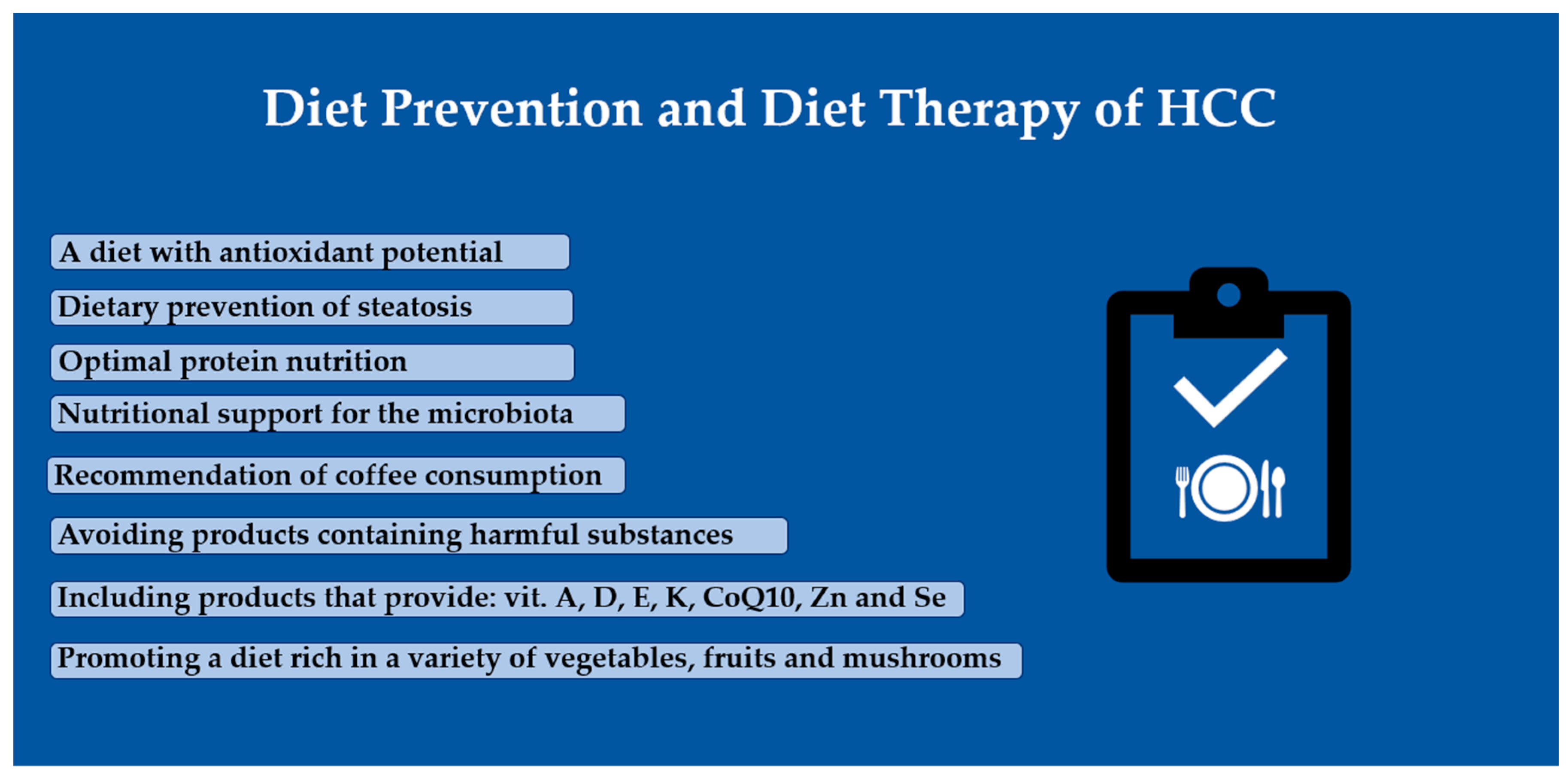
Simple Summary
Abstract
1. Introduction
2. Materials and Methods
3. Nutrition-Dependent Metabolic Dysfunction-Associated Steatotic Liver Disease (MASLD) as a Risk Factor for HCC
4. Potential of Fasting-Activated Autophagy
5. Malnutrition as a Condition Requiring Intervention in the Prevention and Treatment of HCC
6. Nutritional Support for the Microbiota
7. Vitamins and Trace Elements as Supportive Elements in the Prevention of, and Therapy for, HCC
7.1. Fat-Soluble Vitamins
7.2. Coenzyme Q10
7.3. Trace Elements
8. Plant Hepatoprotective Compounds
9. Selected Food Products with Hepatoprotective Properties
9.1. Coffee
9.2. Honey
9.3. Chlorella
9.4. Interesting Potential of Mushrooms in HCC Prevention and Treatment
10. Foods and Food Compounds of Which Consumption Should Be Avoided
10.1. Alcohol
10.2. Iron Overload in At-Risk Groups
10.3. Aflatoxins
11. Conclusions
- Implementation of a diet preventing liver steatosis, enabling the attainment of proper body composition parameters, with antioxidant potential;
- Optimal nourishment of the body, with a particular emphasis on an appropriate intake of amino acids;
- Nutritional support for the microbiota through the provision of probiotics and food-based prebiotics;
- Consumption of products rich in fat-soluble vitamins, CoQ10, zinc, and selenium;
- Adoption of a diet rich in diverse vegetables, fruits, and mushrooms, as sources of bioactive compounds;
- Inclusion of coffee, honey, and hepatoprotective superfoods in one’s daily diet;
- Avoiding the consumption of products that may increase the risk of supply substances harmful to the liver.
Author Contributions
Funding
Conflicts of Interest
References
- Fang, J.; Celton-Morizur, S.; Desdouets, C. NAFLD-Related HCC: Focus on the Latest Relevant Preclinical Models. Cancers 2023, 15, 3723. [Google Scholar] [CrossRef]
- Romualdo, G.R.; Heidor, R.; Bacil, G.P.; Moreno, F.S.; Barbisan, L.F. Past, present, and future of chemically induced hepatocarcinogenesis rodent models: Perspectives concerning classic and new cancer hallmarks. Life Sci. 2023, 330, 121994. [Google Scholar] [CrossRef]
- Yang, J.D.; Hainaut, P.; Gores, G.J.; Amadou, A.; Plymoth, A.; Roberts, L.R. A global view of hepatocellular carcinoma: Trends, risk, prevention and management. Nature reviews. Gastroenterol. Hepatol. 2019, 16, 589–604. [Google Scholar] [CrossRef]
- Carlberg, C. Nutrigenomics in the context of evolution. Redox Biol. 2023, 62, 102656. [Google Scholar] [CrossRef]
- Buzzetti, E.; Linden, A.; Best, L.M.; Madden, A.M.; Roberts, D.; Chase, T.J.G.; Freeman, S.C.; Cooper, N.J.; Sutton, A.J.; Fritche, D.; et al. Lifestyle modifications for nonalcohol-related fatty liver disease: A network meta-analysis. Cochrane Database Syst. Rev. 2021, 6, CD013156. [Google Scholar] [CrossRef] [PubMed]
- Huang, D.Q.; Singal, A.G.; Kono, Y.; Tan, D.J.H.; El-Serag, H.B.; Loomba, R. Changing global epidemiology of liver cancer from 2010 to 2019: NASH is the fastest growing cause of liver cancer. Cell Metab. 2022, 34, 969–977.e2. [Google Scholar] [CrossRef] [PubMed]
- Banerjee, A.; Sriramulu, S.; Catanzaro, R.; He, F.; Chabria, Y.; Balakrishnan, B.; Hari, S.; Ayala, A.; Muñoz, M.; Pathak, S.; et al. Natural Compounds as Integrative Therapy for Liver Protection against Inflammatory and Carcinogenic Mechanisms: From Induction to Molecular Biology Advancement. Curr. Mol. Med. 2023, 23, 216–231. [Google Scholar] [CrossRef] [PubMed]
- Omar, M.A.; Omran, M.M.; Farid, K.; Tabll, A.A.; Shahein, Y.E.; Emran, T.M.; Petrovic, A.; Lucic, N.R.; Smolic, R.; Kovac, T.; et al. Biomarkers for Hepatocellular Carcinoma: From Origin to Clinical Diagnosis. Biomedicines 2023, 11, 1852. [Google Scholar] [CrossRef] [PubMed]
- Riazi, K.; Azhari, H.; Charette, J.H.; Underwood, F.E.; King, J.A.; Afshar, E.E.; Swain, M.G.; Congly, S.E.; Kaplan, G.G.; Shaheen, A. The prevalence and incidence of NAFLD worldwide: A systematic review and meta-analysis. Lancet Gastroenterol. Hepatol. 2022, 7, 851–861. [Google Scholar] [CrossRef] [PubMed]
- Zhu, B.; Wu, H.; Li, K.S.; Eisa-Beygi, S.; Singh, B.; Bielenberg, D.R.; Huang, W.; Chen, H. Two sides of the same coin: Non-alcoholic fatty liver disease and atherosclerosis. Vasc. Pharmacol. 2023, 154, 107249. [Google Scholar] [CrossRef] [PubMed]
- Janota, B.; Adamek, B.; Szczepańska, E.; Biernacki, K.; Janczewska, E. Lifestyle and Quality of Life of Women Diagnosed with Hypothyroidism in the Context of Non-Alcoholic Fatty Liver. Metabolites 2023, 13, 1174. [Google Scholar] [CrossRef]
- Zhao, S.; Jang, C.; Liu, J.; Uehara, K.; Gilbert, M.; Izzo, L.; Zeng, X.; Trefely, S.; Fernandez, S.; Carrer, A.; et al. Dietary fructose feeds hepatic lipogenesis via microbiota-derived acetate. Nature 2020, 579, 586–591. [Google Scholar] [CrossRef]
- Bhat, N.; Mani, A. Dysregulation of Lipid and Glucose Metabolism in Nonalcoholic Fatty Liver Disease. Nutrients 2023, 15, 2323. [Google Scholar] [CrossRef]
- DeBlauw, J.A.; Churchill, A.I.; Yunda, B.C.; Kotarsky, C.J.; Caldwell, A.; Ives, S.J. The effects of short-term caloric restriction on cardiometabolic health in overweight/obese men and women: A single-arm trial. Physiol. Rep. 2023, 11, e15856. [Google Scholar] [CrossRef]
- Saitta, C.; Pollicino, T.; Raimondo, G. Obesity and liver cancer. Ann. Hepatol. 2019, 18, 810–815. [Google Scholar] [CrossRef]
- Haigh, L.; Kirk, C.; El Gendy, K.; Gallacher, J.; Errington, L.; Mathers, J.C.; Anstee, Q.M. The effectiveness and acceptability of Mediterranean diet and calorie restriction in non-alcoholic fatty liver disease (NAFLD): A systematic review and meta-analysis. Clin. Nutr. 2022, 41, 1913–1931. [Google Scholar] [CrossRef]
- Del Bo’, C.; Perna, S.; Allehdan, S.; Rafique, A.; Saad, S.; AlGhareeb, F.; Rondanelli, M.; Tayyem, R.F.; Marino, M.; Martini, D.; et al. Does the Mediterranean Diet Have Any Effect on Lipid Profile, Central Obesity and Liver Enzymes in Non-Alcoholic Fatty Liver Disease (NAFLD) Subjects? A Systematic Review and Meta-Analysis of Randomized Control Trials. Nutrients 2023, 15, 2250. [Google Scholar] [CrossRef] [PubMed]
- George, E.S.; Reddy, A.; Nicoll, A.J.; Ryan, M.C.; Itsiopoulos, C.; Abbott, G.; Johnson, N.A.; Sood, S.; Roberts, S.K.; Tierney, A.C. Impact of a Mediterranean diet on hepatic and metabolic outcomes in non-alcoholic fatty liver disease: The MEDINA randomised controlled trial. Liver Int. Off. J. Int. Assoc. Study Liver 2022, 42, 1308–1322. [Google Scholar] [CrossRef] [PubMed]
- Koh, J.H.; Wang, M.; Suzuki, H.; Muthiah, M.; Ng, C.H.; Huang, D.Q. NAFLD and NAFLD-related HCC in Asia: Burden and Surveillance. J. Clin. Exp. Hepatol. 2023, 14, 101213. [Google Scholar] [CrossRef] [PubMed]
- Qian, H.; Chao, X.; Williams, J.; Fulte, S.; Li, T.; Yang, L.; Ding, W.X. Autophagy in liver diseases: A review. Mol. Asp. Med. 2024, 82, 100973. [Google Scholar] [CrossRef] [PubMed]
- Hong, F.; Gao, Y.; Li, Y.; Zheng, L.; Xu, F.; Li, X. Inhibition of HIF1A-AS1 promoted starvation-induced hepatocellular carcinoma cell apoptosis by reducing HIF-1α/mTOR-mediated autophagy. World J. Surg. Oncol. 2020, 18, 113. [Google Scholar] [CrossRef] [PubMed]
- Ma, Y.N.; Jiang, X.; Tang, W.; Song, P. Influence of intermittent fasting on autophagy in the liver. Biosci. Trends 2023, 17, 335–355. [Google Scholar] [CrossRef] [PubMed]
- Minciuna, I.; Gallage, S.; Heikenwalder, M.; Zelber-Sagi, S.; Dufour, J.F. Intermittent fasting-the future treatment in NASH patients? Hepatology 2023, 78, 1290–1305. [Google Scholar] [CrossRef] [PubMed]
- Krstic, J.; Reinisch, I.; Schindlmaier, K.; Galhuber, M.; Riahi, Z.; Berger, N.; Kupper, N.; Moyschewitz, E.; Auer, M.; Michenthaler, H.; et al. Fasting improves therapeutic response in hepatocellular carcinoma through p53-dependent metabolic synergism. Sci. Adv. 2022, 8, eabh2635. [Google Scholar] [CrossRef]
- Geladari, E.; Alexopoulos, T.; Kontogianni, M.D.; Vasilieva, L.; Mani, I.; Alexopoulou, A. Mechanisms of sarcopenia in liver cirrhosis and the role of myokines. Ann. Gastroenterol. 2023, 36, 392–404. [Google Scholar] [CrossRef] [PubMed]
- Janota, B.; Krupowicz, A.; Noras, K.; Janczewska, E. Evaluation of the nutritional status of patients with liver cirrhosis. World J. Hepatol. 2023, 15, 914–924. [Google Scholar] [CrossRef] [PubMed]
- Zhang, C.Y.; Liu, S.; Yang, M. Nutrition deprivation affects the cytotoxic effect of CD8 T cells in hepatocellular carcinoma. World J. Gastrointest. Oncol. 2022, 14, 1887–1891. [Google Scholar] [CrossRef] [PubMed]
- Qin, R.; Zhao, C.; Wang, C.J.; Xu, W.; Zhao, J.Y.; Lin, Y.; Yuan, Y.Y.; Lin, P.C.; Li, Y.; Zhao, S.; et al. Tryptophan potentiates CD8+ T cells against cancer cells by TRIP12 tryptophanylation and surface PD-1 downregulation. J. Immunother. Cancer 2021, 9, e002840. [Google Scholar] [CrossRef]
- Wang, W.; Guo, M.N.; Li, N.; Pang, D.Q.; Wu, J.H. Glutamine deprivation impairs function of infiltrating CD8+ T cells in hepatocellular carcinoma by inducing mitochondrial damage and apoptosis. World J. Gastrointest. Oncol. 2022, 14, 1124–1140. [Google Scholar] [CrossRef]
- Revoredo, S.; Del Fabbro, E. Hepatocellular carcinoma and sarcopenia: A narrative review. Ann. Palliat. Med. 2023, 12, 1295–1309. [Google Scholar] [CrossRef]
- Hepburn, C.; von Roenn, N. Nutrition in Liver Disease—A Review. Curr. Gastroenterol. Rep. 2023, 25, 242–249. [Google Scholar] [CrossRef] [PubMed]
- Ruiz-Margáin, A.; Román-Calleja, B.M.; Moreno-Guillén, P.; González-Regueiro, J.A.; Kúsulas-Delint, D.; Campos-Murguía, A.; Flores-García, N.C.; Macías-Rodríguez, R.U. Nutritional therapy for hepatocellular carcinoma. World J. Gastrointest. Oncol. 2021, 13, 1440–1452. [Google Scholar] [CrossRef] [PubMed]
- Sideris, G.A.; Tsaramanidis, S.; Vyllioti, A.T.; Njuguna, N. The Role of Branched-Chain Amino Acid Supplementation in Combination with Locoregional Treatments for Hepatocellular Carcinoma: Systematic Review and Meta-Analysis. Cancers 2023, 15, 926. [Google Scholar] [CrossRef] [PubMed]
- Álvarez, J.; Fernández Real, J.M.; Guarner, F.; Gueimonde, M.; Rodríguez, J.M.; Saenz de Pipaon, M.; Sanz, Y. Gut microbes and health. Microbiota Intest. Salud. Gastroenterol. Hepatol. 2021, 44, 519–535. [Google Scholar] [CrossRef]
- Spanu, D.; Pretta, A.; Lai, E.; Persano, M.; Donisi, C.; Mariani, S.; Dubois, M.; Migliari, M.; Saba, G.; Ziranu, P.; et al. Hepatocellular carcinoma and microbiota: Implications for clinical management and treatment. World J. Hepatol. 2022, 14, 1319–1332. [Google Scholar] [CrossRef] [PubMed]
- Zhou, A.; Tang, L.; Zeng, S.; Lei, Y.; Yang, S.; Tang, B. Gut microbiota: A new piece in understanding hepatocarcinogenesis. Cancer Lett. 2020, 474, 15–22. [Google Scholar] [CrossRef] [PubMed]
- Mungamuri, S.K.; Vijayasarathy, K. Role of the Gut Microbiome in Nonalcoholic Fatty Liver Disease Progression. Crit. Rev. Oncog. 2020, 25, 57–70. [Google Scholar] [CrossRef]
- Ponziani, F.R.; Bhoori, S.; Castelli, C.; Putignani, L.; Rivoltini, L.; Del Chierico, F.; Sanguinetti, M.; Morelli, D.; Paroni Sterbini, F.; Petito, V.; et al. Hepatocellular Carcinoma Is Associated with Gut Microbiota Profile and Inflammation in Nonalcoholic Fatty Liver Disease. Hepatology 2019, 69, 107–120. [Google Scholar] [CrossRef]
- Thilakarathna, W.P.D.W.; Rupasinghe, H.P.V.; Ridgway, N.D. Mechanisms by Which Probiotic Bacteria Attenuate the Risk of Hepatocellular Carcinoma. Int. J. Mol. Sci. 2021, 22, 2606. [Google Scholar] [CrossRef]
- Illikoud, N.; Mantel, M.; Rolli-Derkinderen, M.; Gagnaire, V.; Jan, G. Dairy starters and fermented dairy products modulate gut mucosal immunity. Immunol. Lett. 2022, 251–252, 91–102. [Google Scholar] [CrossRef]
- Yuan, Y.; Yang, Y.; Xiao, L.; Qu, L.; Zhang, X.; Wei, Y. Advancing Insights into Probiotics during Vegetable Fermentation. Foods 2023, 12, 3789. [Google Scholar] [CrossRef]
- Nduti, N.; McMillan, A.; Seney, S.; Sumarah, M.; Njeru, P.; Mwaniki, M. Investigating probiotic yoghurt to reduce an aflatoxin B1 biomarker among school children in eastern Kenya: Preliminary study. Int. Dairy J. 2016, 63, 124–129. [Google Scholar] [CrossRef]
- Lange, N.F.; Radu, P.; Dufour, J.F. Prevention of NAFLD-associated HCC: Role of lifestyle and chemoprevention. J. Hepatol. 2021, 75, 1217–1227. [Google Scholar] [CrossRef] [PubMed]
- Xue, L.; He, J.; Gao, N.; Lu, X.; Li, M.; Wu, X.; Liu, Z.; Jin, Y.; Liu, J.; Xu, J.; et al. Probiotics may delay the progression of nonalcoholic fatty liver disease by restoring the gut microbiota structure and improving intestinal endotoxemia. Sci. Rep. 2017, 7, 45176. [Google Scholar] [CrossRef] [PubMed]
- Chan, M.; Larsen, N.; Baxter, H.; Jespersen, L.; Ekinci, E.I.; Howell, K. The impact of botanical fermented foods on metabolic syndrome and type 2 diabetes: A systematic review of randomised controlled trials. Nutr. Res. Rev. 2023, 1–20, Advance online publication. [Google Scholar] [CrossRef] [PubMed]
- Gulbicka, P.; Kanikowska, A.; Marciniak, M.; Grzymisławski, M. Współczesne terapie niealkoholowej stłuszczeniowej choroby wątroby. The modern therapies of non-alcoholic fatty liver disease. Forum Zaburzeń Metab. 2018, 9, 168–174. [Google Scholar]
- Tutunchi, H.; Arefhosseini, S.; Ebrahimi-Mameghani, M. Clinical effectiveness of α-lipoic acid, myo-inositol and propolis supplementation on metabolic profiles and liver function in obese patients with NAFLD: A randomized controlled clinical trial. Clin. Nutr. ESPEN 2023, 54, 412–420. [Google Scholar] [CrossRef] [PubMed]
- Kosmalski, M.; Frankowski, R.; Ziółkowska, S.; Różycka-Kosmalska, M.; Pietras, T. What’s New in the Treatment of Non-Alcoholic Fatty Liver Disease (NAFLD). J. Clin. Med. 2023, 12, 1852. [Google Scholar] [CrossRef] [PubMed]
- Grinberg, L.; Dabbah Assadi, F.; Baum, G.; Zemel, R.; Tur-Kaspa, R.; Shochat, C.; Karasik, D.; Karpuj, M.V. Beneficial Effect of Vitamin D on Non-Alcoholic Fatty Liver Disease (NAFLD) Progression in the Zebrafish Model. Nutrients 2023, 15, 1362. [Google Scholar] [CrossRef]
- Pereira, F.; Fernández-Barral, A.; Larriba, M.J.; Barbáchano, A.; González-Sancho, J.M. From molecular basis to clinical insights: A challenging future for the vitamin D endocrine system in colorectal cancer. FEBS J. 2023. Advance online publication. [Google Scholar] [CrossRef]
- Brustad, M.; Meyer, H.E. Vitamin D—A scoping review for Nordic nutrition recommendations 2023. Food Nutr. Res. 2023, 67. [Google Scholar] [CrossRef]
- Płudowski, P.; Kos-Kudła, B.; Walczak, M.; Fal, A.; Zozulińska-Ziółkiewicz, D.; Sieroszewski, P.; Peregud-Pogorzelski, J.; Lauterbach, R.; Targowski, T.; Lewiński, A.; et al. Guidelines for Preventing and Treating Vitamin D Deficiency: A 2023 Update in Poland. Nutrients 2023, 15, 695. [Google Scholar] [CrossRef]
- Matsuda, A.; Ishiguro, K.; Yan, I.K.; Patel, T. Therapeutic Efficacy of Vitamin D in Experimental c-MET-β-Catenin-Driven Hepatocellular Cancer. Gene Expr. 2019, 19, 151–159. [Google Scholar] [CrossRef] [PubMed]
- Zhang, Y.; Jiang, X.; Li, X.; Găman, M.A.; Kord-Varkaneh, H.; Rahmani, J.; Salehi-Sahlabadi, A.; Day, A.S.; Xu, Y. Serum Vitamin D Levels and Risk of Liver Cancer: A Systematic Review and Dose-Response Meta-Analysis of Cohort Studies. Nutr. Cancer 2021, 73, 1–9. [Google Scholar] [CrossRef] [PubMed]
- Huang, Y.; Yuan, Y.; Chen, S.; Xu, D.; Xiao, L.; Wang, X.; Qin, W.; Liu, B. Identifying potential pharmacological targets and mechanisms of vitamin D for hepatocellular carcinoma and COVID-19. Front. Immunol. 2022, 13, 985781. [Google Scholar] [CrossRef] [PubMed]
- Lyytinen, A.T.; Linneberg, A. Vitamin K—A scoping review for Nordic Nutrition Recommendations 2023. Food Nutr. Res. 2023, 67. [Google Scholar] [CrossRef]
- Jarosz, M.; Rychlik, E.; Stoś, K.; Charzewska, J. Normy żywienia dla Populacji Polski i ich Zastosowanie; Narodowy Instytut Zdrowia Publicznego-Państwowy Zakład Higieny: Warsaw, Poland, 2020.
- Haruna, Y.; Yakushijin, T.; Kawamoto, S. Efficacy and safety of sorafenib plus vitamin K treatment for hepatocellular carcinoma: A phase II, randomized study. Cancer Med. 2021, 10, 914–922. [Google Scholar] [CrossRef]
- Ciudad-Mulero, M.; Domínguez, L.; Morales, P.; Fernández-Ruiz, V.; Cámara, M. A Review of Foods of Plant Origin as Sources of Vitamins with Proven Activity in Oxidative Stress Prevention according to EFSA Scientific Evidence. Molecules 2023, 28, 7269. [Google Scholar] [CrossRef] [PubMed]
- Pervez, M.A.; Khan, D.A.; Mirza, S.A.; Slehria, A.U.R.; Nisar, U.; Aamir, M. Comparison of delta-tocotrienol and alpha-tocopherol effects on hepatic steatosis and inflammatory biomarkers in patients with non-alcoholic fatty liver disease: A randomized double-blind active-controlled trial. Complement. Ther. Med. 2022, 70, 102866. [Google Scholar] [CrossRef] [PubMed]
- Diaz-Vegas, A.; Madsen, S.; Cooke, K.C.; Carroll, L.; Khor, J.X.Y.; Turner, N.; Lim, X.Y.; Astore, M.A.; Morris, J.C.; Don, A.S.; et al. Mitochondrial electron transport chain, ceramide, and coenzyme Q are linked in a pathway that drives insulin resistance in skeletal muscle. eLife 2023, 12, RP87340. [Google Scholar] [CrossRef]
- Liaghat, M.; Yaghoubzad-Maleki, M.; Nabi-Afjadi, M.; Fathi, Z.; Zalpoor, H.; Heidari, N.; Bahreini, E. A Review of the Potential Role of CoQ10 in the Treatment of Hepatocellular Carcinoma. Biochem. Genet. 2023. Advance online publication. [Google Scholar] [CrossRef] [PubMed]
- Akbari, A.; Mobini, G.R.; Agah, S.; Morvaridzadeh, M.; Omidi, A.; Potter, E.; Fazelian, S.; Ardehali, S.H.; Daneshzad, E.; Dehghani, S. Coenzyme Q10 supplementation and oxidative stress parameters: A systematic review and meta-analysis of clinical trials. Eur. J. Clin. Pharmacol. 2020, 76, 1483–1499. [Google Scholar] [CrossRef] [PubMed]
- Tamai, Y.; Iwasa, M.; Eguchi, A.; Shigefuku, R.; Sugimoto, K.; Hasegawa, H.; Takei, Y. Serum copper, zinc and metallothionein serve as potential biomarkers for hepatocellular carcinoma. PLoS ONE 2020, 15, e0237370. [Google Scholar] [CrossRef] [PubMed]
- Strand, T.A.; Mathisen, M. Zinc—A scoping review for Nordic Nutrition Recommendations 2023. Food Nutr. Res. 2023, 67. [Google Scholar] [CrossRef] [PubMed]
- Hosui, A.; Kimura, E.; Abe, S.; Tanimoto, T.; Onishi, K.; Kusumoto, Y.; Sueyoshi, Y.; Matsumoto, K.; Hirao, M.; Yamada, T.; et al. Long-Term Zinc Supplementation Improves Liver Function and Decreases the Risk of Developing Hepatocellular Carcinoma. Nutrients 2018, 10, 1955. [Google Scholar] [CrossRef]
- Zhao, J.; Zou, H.; Huo, Y.; Wei, X.; Li, Y. Emerging roles of selenium on metabolism and type 2 diabetes. Front. Nutr. 2022, 9, 1027629. [Google Scholar] [CrossRef] [PubMed]
- Hsiao, Y.F.; Cheng, S.B.; Lai, C.Y.; Liu, H.T.; Huang, S.C.; Huang, Y.C. The Prognostic Role of Glutathione and Its Related Antioxidant Enzymes in the Recurrence of Hepatocellular Carcinoma. Nutrients 2021, 13, 4071. [Google Scholar] [CrossRef]
- Qiao, L.; Men, L.; Yu, S.; Yao, J.; Li, Y.; Wang, M.; Yu, Y.; Wang, N.; Ran, L.; Wu, Y.; et al. Hepatic deficiency of selenoprotein S exacerbates hepatic steatosis and insulin resistance. Cell Death Dis. 2022, 13, 275. [Google Scholar] [CrossRef]
- Zhou, S.; Li, M.; Zhou, C.; Jiang, Z.; Wen, X.; Cong, W.; Ni, Y.; Zhang, F. Przepływ selenu w łańcuchu „gleba-uprawa-żywność-człowiek”. Nauka O Jedzeniu 2023, 44, 231–244. [Google Scholar] [CrossRef]
- Cheng, K.; Sun, Y.; Liu, B.; Ming, J.; Wang, L.; Xu, C.; Xiao, Y.; Zhang, C.; Shang, L. Selenium Modification of Natural Products and Its Research Progress. Foods 2023, 12, 3773. [Google Scholar] [CrossRef]
- Sun, Y.; Wang, Z.; Gong, P.; Yao, W.; Ba, Q.; Wang, H. Review on the health-promoting effect of adequate selenium status. Front. Nutr. 2023, 10, 1136458. [Google Scholar] [CrossRef]
- Wu, B.K.; Chen, Q.H.; Pan, D.; Chang, B.; Sang, L.X. A novel therapeutic strategy for hepatocellular carcinoma: Immunomodulatory mechanisms of selenium and/or selenoproteins on a shift towards anti-cancer. Int. Immunopharmacol. 2021, 96, 107790. [Google Scholar] [CrossRef] [PubMed]
- Gong, Y.; Dong, F.; Geng, Y.; Zhuang, H.; Ma, Z.; Zhou, Z.; Huang, B.; Sun, Z.; Hou, B. Selenium concentration, dietary intake and risk of hepatocellular carcinoma—A systematic review with meta-analysis. Concentración de selenio, ingesta dietética y riesgo de carcinoma hepatocelular: Revisión sistemática con metaanálisis. Nutr. Hosp. 2019, 36, 1430–1437. [Google Scholar] [CrossRef] [PubMed]
- Kiruthiga, C.; Devi, K.P.; Nabavi, S.M.; Bishayee, A. Autophagy: A Potential Therapeutic Target of Polyphenols in Hepatocellular Carcinoma. Cancers 2020, 12, 562. [Google Scholar] [CrossRef] [PubMed]
- Cheng, K.C.; Wang, C.J.; Chang, Y.C.; Hung, T.W.; Lai, C.J.; Kuo, C.W.; Huang, H.P. Mulberry fruits extracts induce apoptosis and autophagy of liver cancer cell and prevent hepatocarcinogenesis in vivo. J. Food Drug Anal. 2020, 28, 84–93. [Google Scholar] [CrossRef] [PubMed]
- Guariglia, M.; Saba, F.; Rosso, C.; Bugianesi, E. Molecular Mechanisms of Curcumin in the Pathogenesis of Metabolic Dysfunction Associated Steatotic Liver Disease. Nutrients 2023, 15, 5053. [Google Scholar] [CrossRef] [PubMed]
- Liao, S.C.; Hsu, W.H.; Huang, Z.Y.; Chuang, K.L.; Lin, K.T.; Tseng, C.L.; Tsai, T.H.; Dao, A.H.; Su, C.L.; Huang, C.F. Bioactivity Evaluation of a Novel Formulated Curcumin. Nutrients 2019, 11, 2982. [Google Scholar] [CrossRef] [PubMed]
- Badroon, N.A.; Abdul Majid, N.; Alshawsh, M.A. Antiproliferative and Apoptotic Effects of Cardamonin against Hepatocellular Carcinoma HepG2 Cells. Nutrients 2020, 12, 1757. [Google Scholar] [CrossRef]
- Machado, I.F.; Miranda, R.G.; Dorta, D.J.; Rolo, A.P.; Palmeira, C.M. Targeting Oxidative Stress with Polyphenols to Fight Liver Diseases. Antioxidants 2023, 12, 1212. [Google Scholar] [CrossRef]
- Wu, R.; Xiong, J.; Zhou, T.; Zhang, Z.; Huang, Z.; Tian, S.; Wang, Y. Quercetin/Anti-PD-1 Antibody Combination Therapy Regulates the Gut Microbiota, Impacts Macrophage Immunity and Reshapes the Hepatocellular Carcinoma Tumor Microenvironment. Front. Biosci. 2023, 28, 327. [Google Scholar] [CrossRef]
- Novi, S.; Vestuto, V.; Campiglia, P.; Tecce, N.; Bertamino, A.; Tecce, M.F. Anti-Angiogenic Effects of Natural Compounds in Diet-Associated Hepatic Inflammation. Nutrients 2023, 15, 2748. [Google Scholar] [CrossRef]
- Fernández-Palanca, P.; Fondevila, F.; Méndez-Blanco, C.; Tuñón, M.J.; González-Gallego, J.; Mauriz, J.L. Antitumor Effects of Quercetin in Hepatocarcinoma In Vitro and In Vivo Models: A Systematic Review. Nutrients 2019, 11, 2875. [Google Scholar] [CrossRef]
- Wang, N.; Feng, Y.; Zhu, M.; Tsang, C.M.; Man, K.; Tong, Y.; Tsao, S.W. Berberine induces autophagic cell death and mitochondrial apoptosis in liver cancer cells: The cellular mechanism. J. Cell. Biochem. 2010, 111, 1426–1436. [Google Scholar] [CrossRef]
- Tsang, C.M.; Cheung, K.C.; Cheung, Y.C.; Man, K.; Lui, V.W.; Tsao, S.W.; Feng, Y. Berberine suppresses Id-1 expression and inhibits the growth and development of lung metastases in hepatocellular carcinoma. Biochim. Biophys. Acta 2015, 1852, 541–551. [Google Scholar] [CrossRef]
- Guo, W.; Tan, H.Y.; Li, S.; Wang, N.; Feng, Y. Glutamic-Pyruvic Transaminase 1 Facilitates Alternative Fuels for Hepatocellular Carcinoma Growth-A Small Molecule Inhibitor, Berberine. Cancers 2020, 12, 1854. [Google Scholar] [CrossRef] [PubMed]
- Wei, R.R.; Lin, Q.Y.; Adu, M.; Huang, H.L.; Yan, Z.H.; Shao, F.; Zhong, G.Y.; Zhang, Z.L.; Sang, Z.P.; Cao, L.; et al. The sources, properties, extraction, biosynthesis, pharmacology, and application of lycopene. Food Funct. 2023, 14, 9974–9998. [Google Scholar] [CrossRef]
- Mekuria, A.N.; Tura, A.K.; Hagos, B.; Sisay, M.; Abdela, J.; Mishore, K.M.; Motbaynor, B. Anti-Cancer Effects of Lycopene in Animal Models of Hepatocellular Carcinoma: A Systematic Review and Meta-Analysis. Front. Pharmacol. 2020, 11, 1306. [Google Scholar] [CrossRef] [PubMed]
- Alkandahri, M.Y.; Pamungkas, B.T.; Oktoba, Z.; Shafirany, M.Z.; Sulastri, L.; Arfania, M.; Anggraeny, E.N.; Pratiwi, A.; Astuti, F.D.; Indriyani Dewi, S.Y.; et al. Hepatoprotective Effect of Kaempferol: A Review of the Dietary Sources, Bioavailability, Mechanisms of Action, and Safety. Adv. Pharmacol. Pharm. Sci. 2023, 2023, 1387665. [Google Scholar] [CrossRef] [PubMed]
- Sojoodi, M.; Wei, L.; Erstad, D.J.; Yamada, S.; Fujii, T.; Hirschfield, H.; Kim, R.S.; Lauwers, G.Y.; Lanuti, M.; Hoshida, Y.; et al. Epigallocatechin Gallate Induces Hepatic Stellate Cell Senescence and Attenuates Development of Hepatocellular Carcinoma. Cancer Prev. Res. 2020, 13, 497–508. [Google Scholar] [CrossRef] [PubMed]
- Yua, J.; Liang, D.; Li, J.; Liu, Z.; Zhou, F.; Wang, T.; Ma, S.; Wang, G.; Chen, B.; Chen, W. Coffee, Green Tea Intake, and the Risk of Hepatocellular Carcinoma: A Systematic Review and Meta-Analysis of Observational Studies. Nutr. Cancer 2023, 75, 1295–1308. [Google Scholar] [CrossRef] [PubMed]
- Yin, X.; He, X.; Wu, L.; Yan, D.; Yan, S. Chlorogenic Acid, the Main Antioxidant in Coffee, Reduces Radiation-Induced Apoptosis and DNA Damage via NF-E2-Related Factor 2 (Nrf2) Activation in Hepatocellular Carcinoma. Oxidative Med. Cell. Longev. 2022, 2022, 4566949. [Google Scholar] [CrossRef]
- Kennedy, O.J.; Roderick, P.; Buchanan, R.; Fallowfield, J.A.; Hayes, P.C.; Parkes, J. Coffee, including caffeinated and decaffeinated coffee, and the risk of hepatocellular carcinoma: A systematic review and dose-response meta-analysis. BMJ Open 2017, 7, e013739. [Google Scholar] [CrossRef]
- Deng, Y.; Huang, J.; Wong, M.C.S. Associations between six dietary habits and risk of hepatocellular carcinoma: A Mendelian randomization study. Hepatol. Commun. 2022, 6, 2147–2154. [Google Scholar] [CrossRef]
- Ghramh, H.A.; Alrumman, S.A.; Ahmad, I.; Kalam, A.; Elbehairi, S.E.I.; Alfaify, A.M.; Mohammed, M.E.A.; Al-Sehemi, A.G.; Alfaifi, M.; Al-Shehri, B.M.; et al. Chemical Characterization of Honey and Its Effect (Alone as well as with Synthesized Silver Nanoparticles) on Microbial Pathogens’ and Human Cancer Cell Lines’ Growth. Nutrients 2023, 15, 684. [Google Scholar] [CrossRef]
- Abdel-Rahman Mohamed, A.; El-Kholy, S.S.; Dahran, N.; El Bohy, K.M.; Moustafa, G.G.; Saber, T.M.; Metwally, M.M.M.; Gaber, R.A.; Alqahtani, L.S.; Mostafa-Hedeab, G.; et al. Scrutinizing pathways of nicotine effect on renal Alpha-7 nicotinic acetylcholine receptor and Mitogen-activated protein kinase (MAPK) expression in Ehrlich ascites carcinoma-bearing mice: Role of Chlorella vulgaris. Gene 2022, 837, 146697. [Google Scholar] [CrossRef] [PubMed]
- Sawasdee, N.; Jantakee, K.; Wathikthinnakon, M.; Panwong, S.; Pekkoh, J.; Duangjan, K.; Yenchitsomanus, P.T.; Panya, A. Microalga Chlorella sp. extract induced apoptotic cell death of cholangiocarcinoma via AKT/mTOR signaling pathway. Biomed. Pharmacother. 2023, 160, 114306. [Google Scholar] [CrossRef] [PubMed]
- Hamouda, R.A.; Abd El Latif, A.; Elkaw, E.M.; Alotaibi, A.S.; Alenzi, A.M.; Hamza, H.A. Assessment of Antioxidant and Anticancer Activities of Microgreen Alga Chlorella vulgaris and Its Blend with Different Vitamins. Molecules 2022, 27, 1602. [Google Scholar] [CrossRef] [PubMed]
- Moradi, M.N.; Behrouj, H.; Alipoor, B.; Kheiripour, N.; Ghasemi, H.; Ghasemi, H. Chlorella vulgaris is an effective supplement in counteracting non-alcoholic fatty liver disease-related complications through modulation of dyslipidemia, insulin resistance, and inflammatory pathways. J. Food Biochem. 2021, 45, e13914. [Google Scholar] [CrossRef]
- Fontes, A.; Alemany-Pagès, M.; Oliveira, P.J.; Ramalho-Santos, J.; Zischka, H.; Azul, A.M. Antioxidant Versus Pro-Apoptotic Effects of Mushroom-Enriched Diets on Mitochondria in Liver Disease. Int. J. Mol. Sci. 2019, 20, 3987. [Google Scholar] [CrossRef]
- Song, M.; Li, Z.H.; Gu, H.S.; Tang, R.Y.; Zhang, R.; Zhu, Y.L.; Liu, J.L.; Zhang, J.J.; Wang, L.Y. Ganoderma lucidum Spore Polysaccharide Inhibits the Growth of Hepatocellular Carcinoma Cells by Altering Macrophage Polarity and Induction of Apoptosis. J. Immunol. Res. 2021, 2021, 6696606. [Google Scholar] [CrossRef]
- Wang, N.; Liu, H.; Liu, G.; Li, M.; He, X.; Yin, C.; Tu, Q.; Shen, X.; Bai, W.; Wang, Q.; et al. Yeast β-D-glucan exerts antitumour activity in liver cancer through impairing autophagy and lysosomal function, promoting reactive oxygen species production and apoptosis. Redox Biol. 2020, 32, 101495. [Google Scholar] [CrossRef]
- Zhang, J.; Fan, J.; Luo, H.; Liang, Z.; Guan, Y.; Lei, X.; Bo, N.; Zhao, M. Alleviation of Alcoholic Fatty Liver by Dendrobium officinale Flower Extracts due to Regulation of Gut Microbiota and Short-Chain Fatty Acids in Mice Exposed to Chronic Alcohol. Foods 2023, 12, 1428. [Google Scholar] [CrossRef]
- Petroni, M.L.; Brodosi, L.; Marchignoli, F.; Musio, A.; Marchesini, G. Moderate Alcohol Intake in Non-Alcoholic Fatty Liver Disease: To Drink or Not to Drink? Nutrients 2019, 11, 3048. [Google Scholar] [CrossRef] [PubMed]
- Åberg, F.; Puukka, P.; Salomaa, V.; Männistö, S.; Lundqvist, A.; Valsta, L.; Perola, M.; Färkkilä, M.; Jula, A. Risks of Light and Moderate Alcohol Use in Fatty Liver Disease: Follow-Up of Population Cohorts. Hepatology 2020, 71, 835–848. [Google Scholar] [CrossRef] [PubMed]
- Tsuchiya, H. Iron-Induced Hepatocarcinogenesis-Preventive Effects of Nutrients. Front. Oncol. 2022, 12, 940552. [Google Scholar] [CrossRef] [PubMed]
- Chen, T.; Liu, J.; Li, Y.; Wei, S. Burden of Disease Associated with Dietary Exposure to Aflatoxins in China in 2020. Nutrients 2020, 14, 1027. [Google Scholar] [CrossRef]
- Alamir, J.; Almaiman, L.; Alrujib, Y.; Alhamidi, R.; Alowais, B.; Alhussain, S.; Aldakheelallah, A.; Alkhalaf, M.; Bineid, M. Aflatoxins in food products consumed in the Kingdom of Saudi Arabia: A preliminary dietary risk assessment. Food Sci. Nutr. 2023, 11, 5948–5958. [Google Scholar] [CrossRef]


| Type of Food | Components | Functions | References |
|---|---|---|---|
| Fermented vegetables and dairy | Lactobacillus spp. and Bifidobacterium spp. |
| [39,42,43,44,45] |
| Meat, dairy, eggs | High-protein products |
| [27,28,29,30,31,32,33] |
| Egg yolks, fish, dairy, seafood | D3 |
| [53,54,55] |
| Dairy, eggs, meat | K2 |
| [58] |
| Nuts, almonds, plant oils, sprouts, olive oil | Vitamin E |
| [59,60] |
| Meat, liver, egg yolks, fish | CoQ10 |
| [62,63] |
| Grains, legume seeds, eggs, meat, dairy products | Zinc |
| [66] |
| Whole grains, nuts, meat, eggs, dairy products | Selenium |
| [72,73] |
| White mulberry | Polyphenols |
| [76] |
| Turmeric | Curcumin |
| [78,80] |
| Cardamom | Cardamonin |
| [79] |
| Apples, grapes, citrus fruits | Quercetin |
| [82,83] |
| Barberry fruits | Berberine |
| [84,85,86] |
| Tomatoes and tomato products | Lycopene |
| [82] |
| Pumpkin, chives, spinach, kale, dill | Kaempferol |
| [89] |
| Green tea leaves | Epigallocatechin gallate |
| [90] |
| Cabbage, cauliflower, broccoli | Sulforaphane |
| [82] |
| Coffee | Chlorogenic acid |
| [93] |
| Honey | Proteins, enzymes, polyphenols |
| [95] |
| Chlorella | Algae |
| [97,98,99] |
| Shiitake, reishi, yeast, pine mushroom, king oyster mushroom | Various bioactive compounds, including β-D-glucan |
| [100,101,102] |
Disclaimer/Publisher’s Note: The statements, opinions and data contained in all publications are solely those of the individual author(s) and contributor(s) and not of MDPI and/or the editor(s). MDPI and/or the editor(s) disclaim responsibility for any injury to people or property resulting from any ideas, methods, instructions or products referred to in the content. |
© 2024 by the authors. Licensee MDPI, Basel, Switzerland. This article is an open access article distributed under the terms and conditions of the Creative Commons Attribution (CC BY) license (https://creativecommons.org/licenses/by/4.0/).
Share and Cite
Janota, B.; Szymanek, B. The Influence of Diet and Its Components on the Development and Prevention of Hepatocellular Carcinoma (HCC). Cancers 2024, 16, 1030. https://doi.org/10.3390/cancers16051030
Janota B, Szymanek B. The Influence of Diet and Its Components on the Development and Prevention of Hepatocellular Carcinoma (HCC). Cancers. 2024; 16(5):1030. https://doi.org/10.3390/cancers16051030
Chicago/Turabian StyleJanota, Barbara, and Barbara Szymanek. 2024. "The Influence of Diet and Its Components on the Development and Prevention of Hepatocellular Carcinoma (HCC)" Cancers 16, no. 5: 1030. https://doi.org/10.3390/cancers16051030
APA StyleJanota, B., & Szymanek, B. (2024). The Influence of Diet and Its Components on the Development and Prevention of Hepatocellular Carcinoma (HCC). Cancers, 16(5), 1030. https://doi.org/10.3390/cancers16051030