A Novel Polymer-Encapsulated Multi-Imaging Modality Fiducial Marker with Positive Signal Contrast for Image-Guided Radiation Therapy
by
 , , ,
, , ,
Li Wang
1, 
Jeremiah Sanders
2,
John F. Ward
3,
Stephen R. Lee
4,
Falk Poenisch
5,
David Michael Swanson
6,
Narayan Sahoo
5,
Xiaorong Ronald Zhu
5,
Jingfei Ma
2,
Rajat J. Kudchadker
5,
Seungtaek L. Choi
7,
Quynh-Nhu Nguyen
7,
Lauren L. Mayo
7,
Shalin J. Shah
7 and
Steven J. Frank
7,* 1
Department of Experimental Radiation Oncology, The University of Texas MD Anderson Cancer Center, Houston, TX 77030, USA
2
Department of Imaging Physics, The University of Texas MD Anderson Cancer Center, Houston, TX 77030, USA
3
Department of Urology, The University of Texas MD Anderson Cancer Center, Houston, TX 77030, USA
4
Department of Interventional Radiology, The University of Texas MD Anderson Cancer Center, Houston, TX 77030, USA
5
Department of Radiation Physics, The University of Texas MD Anderson Cancer Center, Houston, TX 77030, USA
6
Department of Biostatistics, The University of Texas MD Anderson Cancer Center, Houston, TX 77030, USA
7
Department of Radiation Oncology, The University of Texas MD Anderson Cancer Center, Houston, TX 77030, USA
*
Author to whom correspondence should be addressed.
Cancers 2024, 16(3), 625; https://doi.org/10.3390/cancers16030625
Submission received: 4 January 2024
/
Revised: 22 January 2024
/
Accepted: 28 January 2024
/
Published: 31 January 2024
(This article belongs to the Special Issue MRI-Assisted Radiosurgery (MARS))
Simple Summary
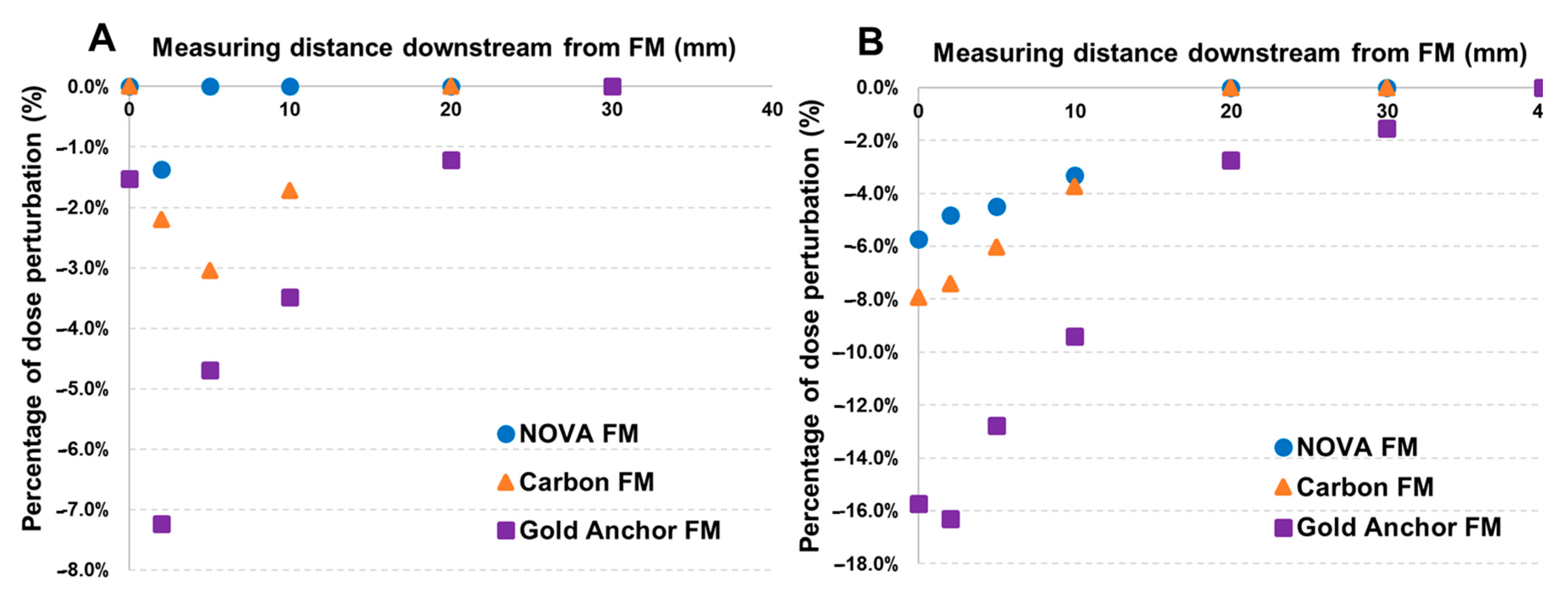
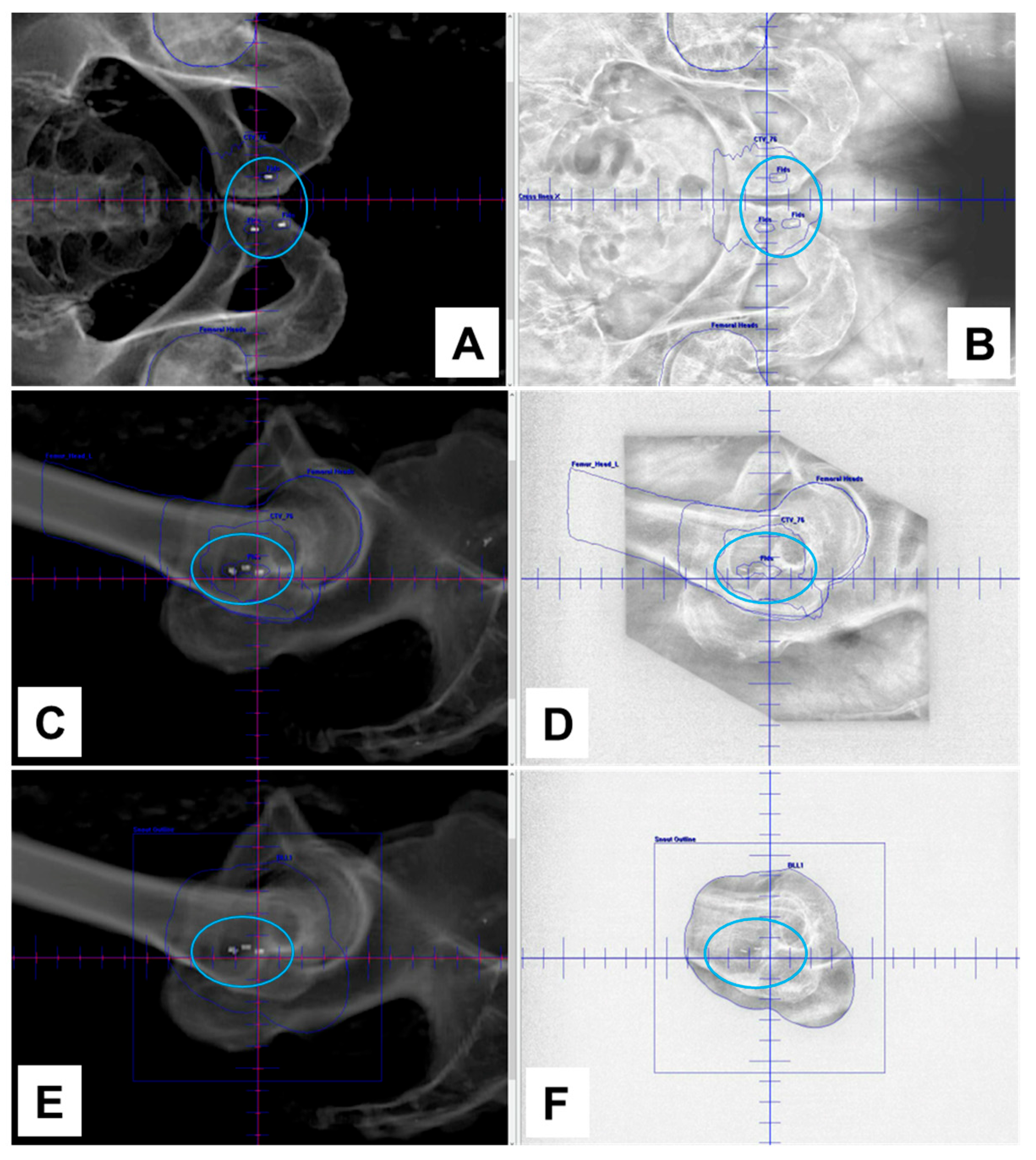
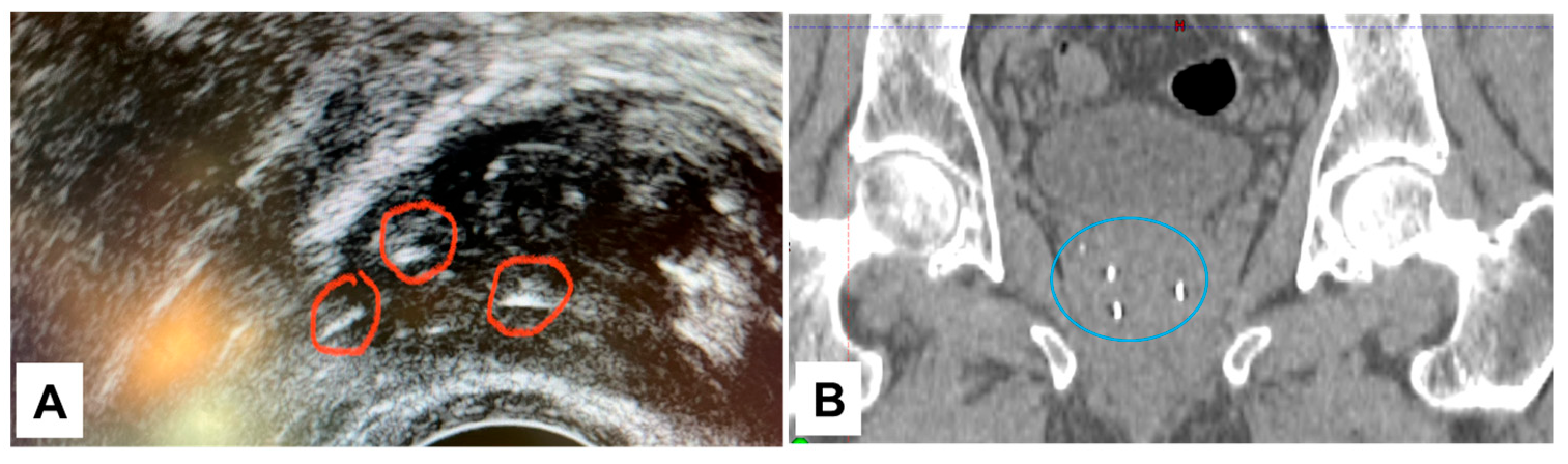
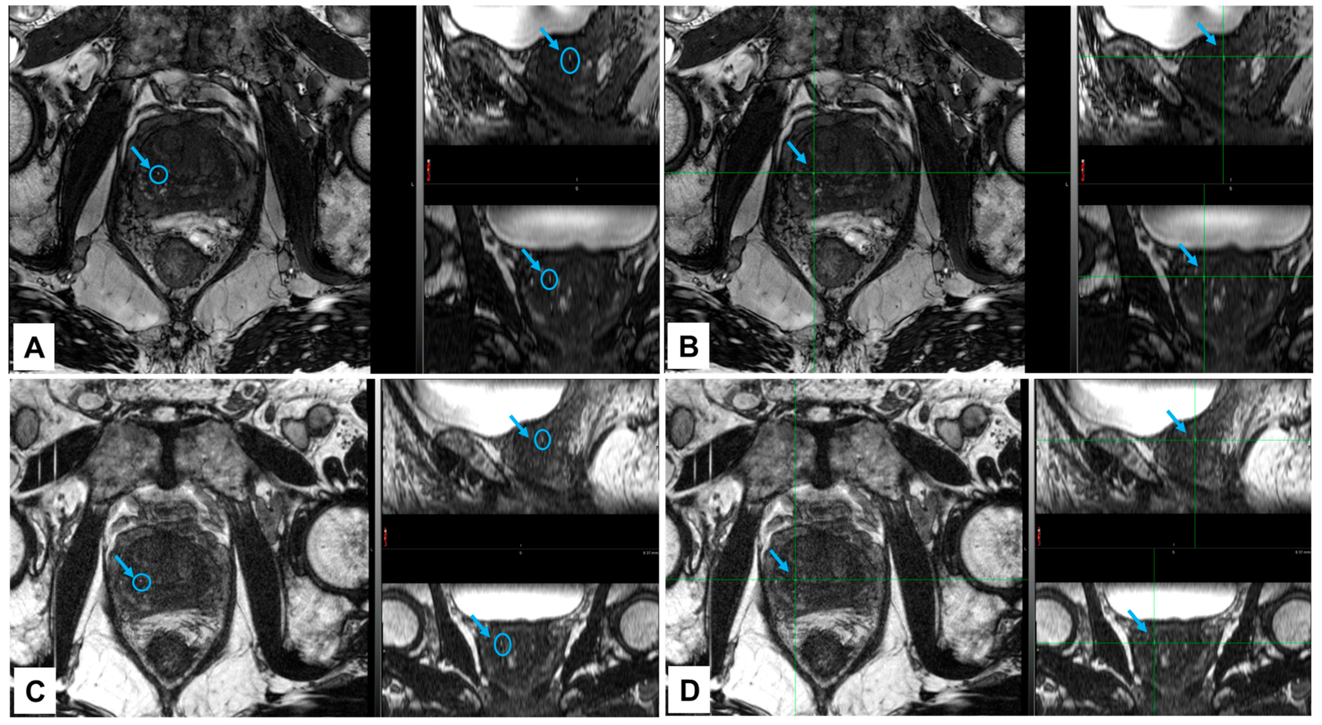
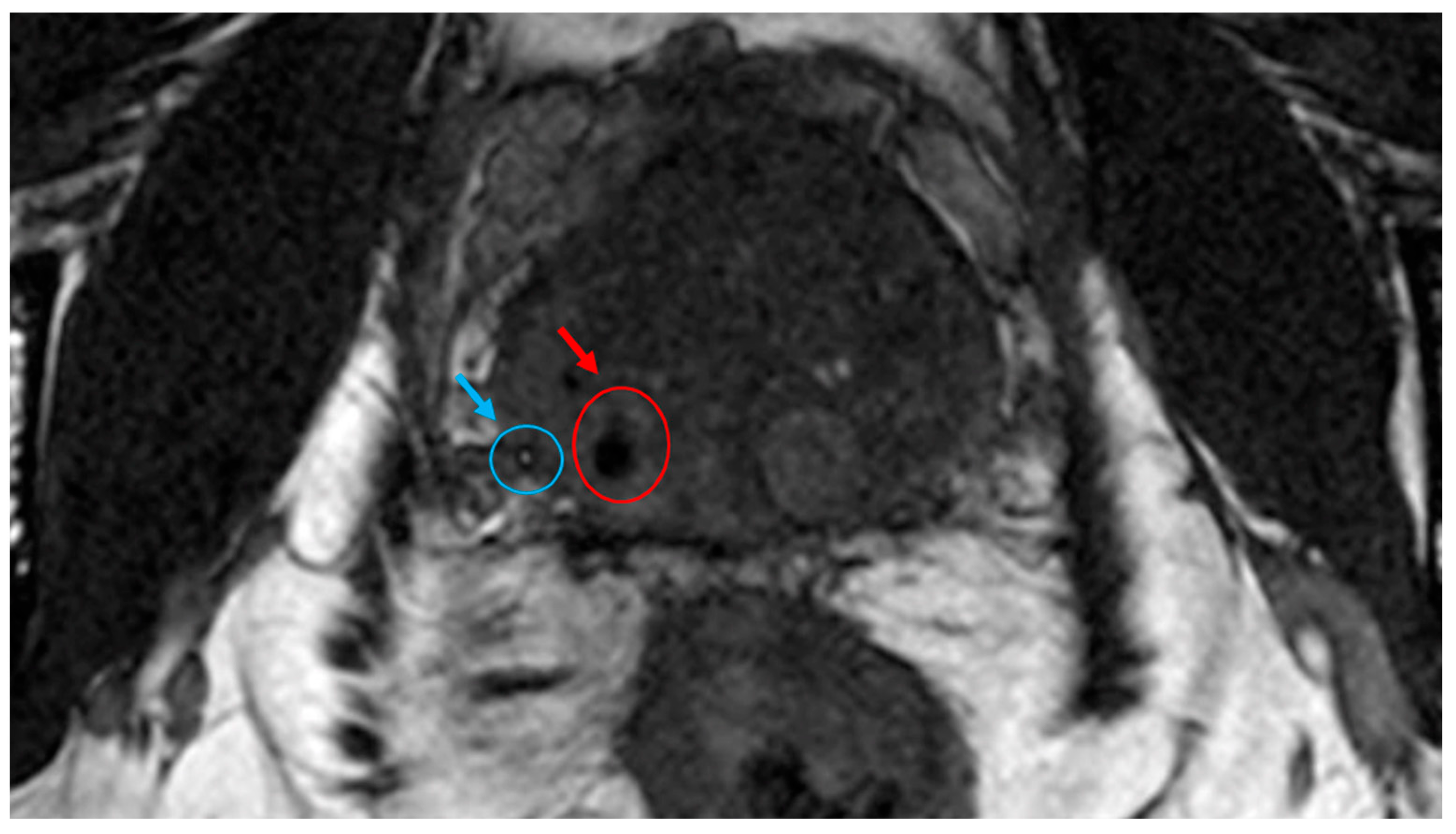
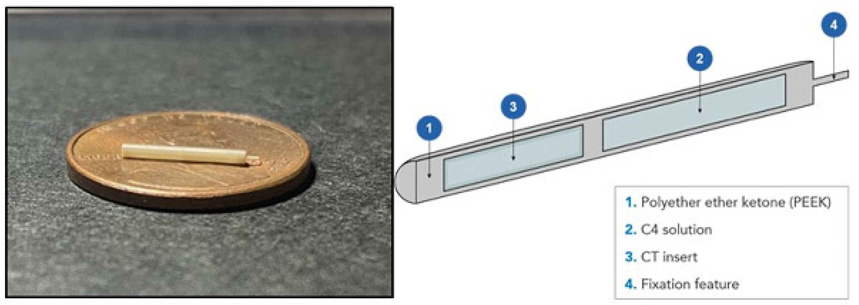
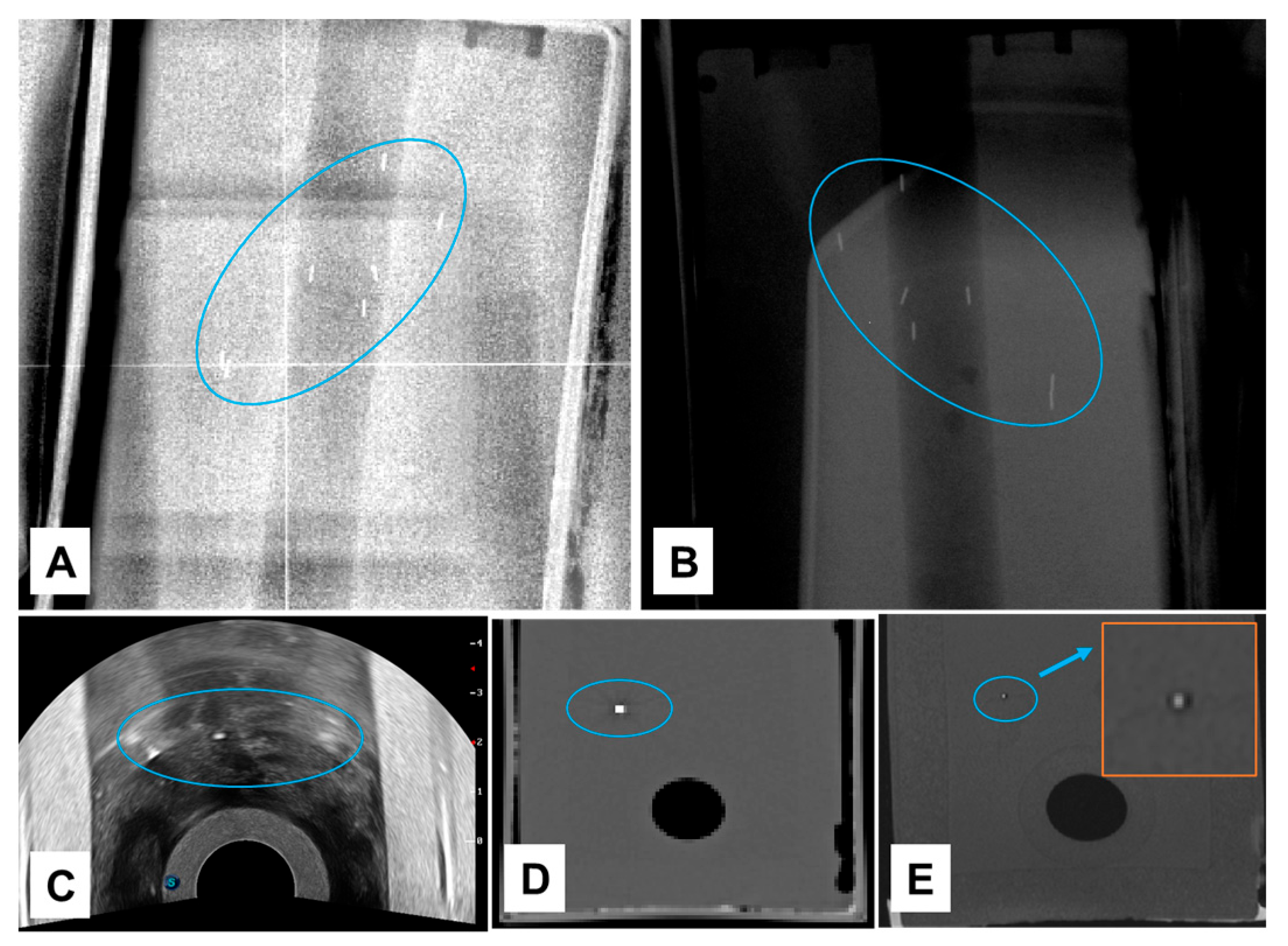
External-beam radiotherapy (EBRT) is the standard of care for prostate cancer (PCa). Precision is critical to target the intraprostatic dominant lesion(s) and limit treatment-related toxicity from unnecessary irradiation of surrounding normal tissues. Currently, to achieve this goal, fiducial markers (FMs) are used in EBRT planning and treatment delivery. However, some of the features of current FMs (e.g., poor visibility on MRI, substantial CT and MR artifacts, radiation dose perturbations) affect treatment accuracy. The aim of our study was to evaluate the performance of a novel FM (NOVA) in phantoms and in a patient. We found that NOVA FMs were positively visualized on ultrasound (US), kV X-ray, CT, and MR images, had reduced CT and MR artifacts, and caused less proton dose perturbations than Gold Anchor and BiomarC (carbon) FMs. Our findings indicate the value of NOVA FMs in daily treatment positioning, MRI/CT co-image registration, and MRI-based treatment-plan generation for patients with PCa undergoing EBRT.
Abstract
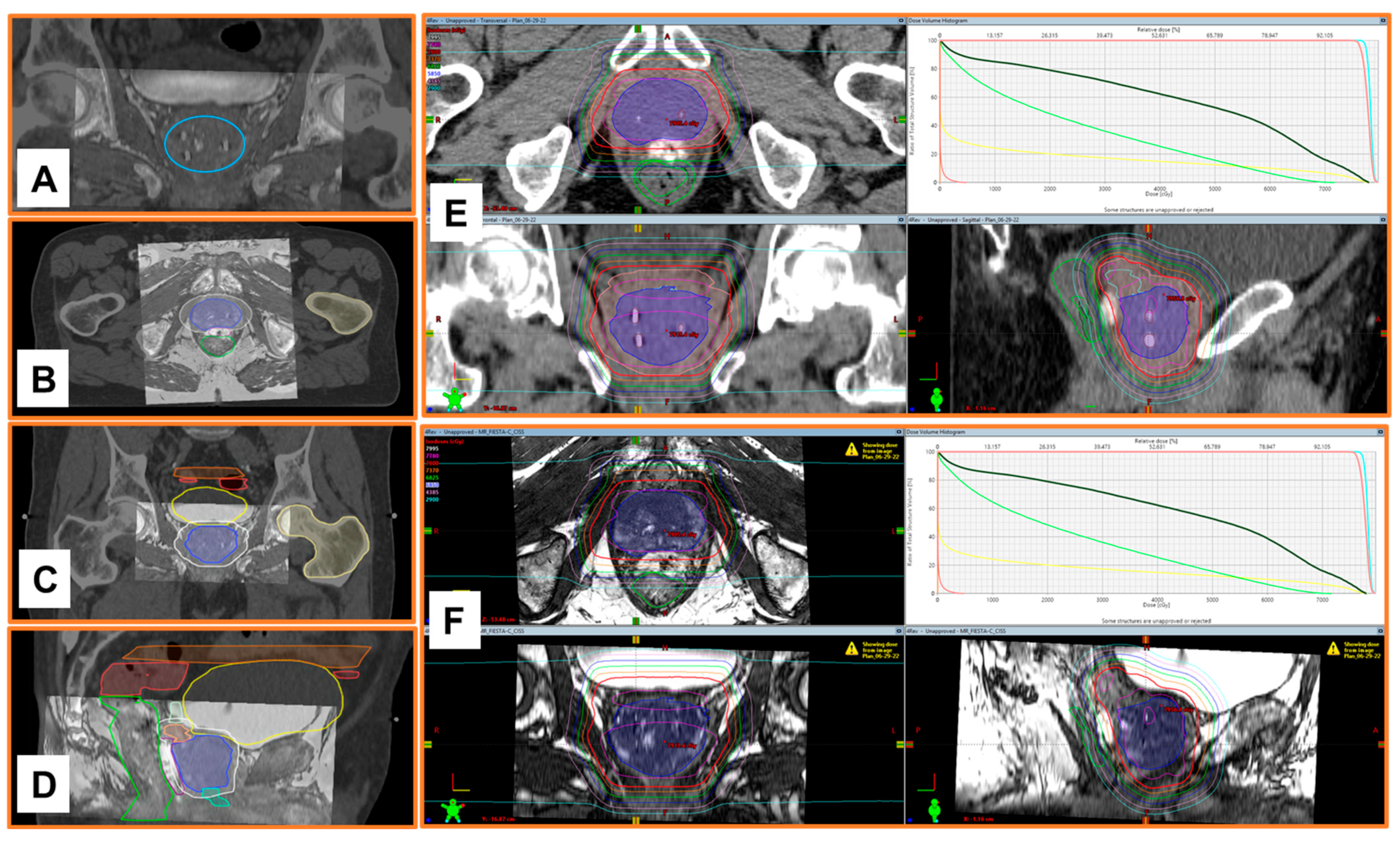
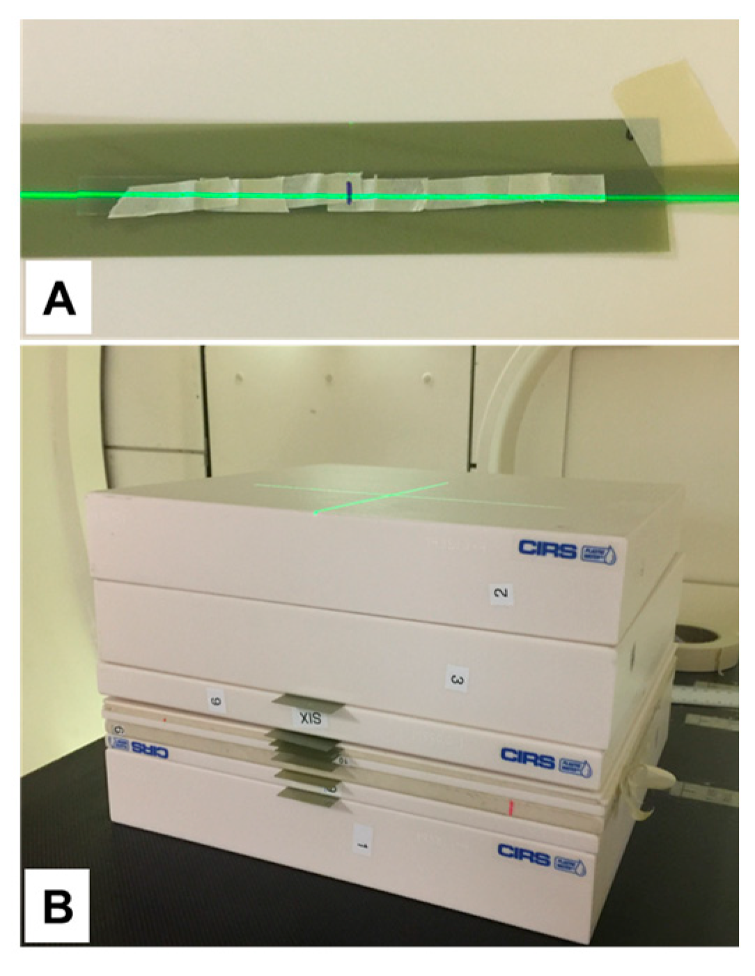
Background: Current fiducial markers (FMs) in external-beam radiotherapy (EBRT) for prostate cancer (PCa) cannot be positively visualized on magnetic resonance imaging (MRI) and create dose perturbation and significant imaging artifacts on computed tomography (CT) and MRI. We report our initial experience with clinical imaging of a novel multimodality FM, NOVA. Methods: We tested Gold Anchor [G-FM], BiomarC [carbon, C-FM], and NOVA FMs in phantoms imaged with kilovoltage (kV) X-rays, transrectal ultrasound (TRUS), CT, and MRI. Artifacts of the FMs on CT were quantified by the relative streak artifacts level (rSAL) metric. Proton dose perturbations (PDPs) were measured with Gafchromic EBT3 film, with FMs oriented either perpendicular to or parallel with the beam axis. We also tested the performance of NOVA-FMs in a patient. Results: NOVA-FMs were positively visualized on all 4 imaging modalities tested. The rSAL on CT was 0.750 ± 0.335 for 2-mm reconstructed slices. In F-tests, PDP was associated with marker type and depth of measurement (p < 10−6); at 5-mm depth, PDP was significantly greater for the G-FM (12.9%, p = 10−6) and C-FM (6.0%, p = 0.011) than NOVA (4.5%). EBRT planning with MRI/CT image co-registration and daily alignments using NOVA-FMs in a patient was feasible and reproducible. Conclusions: NOVA-FMs were positively visible and produced less PDP than G-FMs or C-FMs. NOVA-FMs facilitated MRI/CT fusion and identification of regions of interest.