


The Anticancer Potential of Kaempferol: A Systematic Review Based on In Vitro Studies

 ,
,  , , , , and
Simple Summary
Abstract
1. Introduction
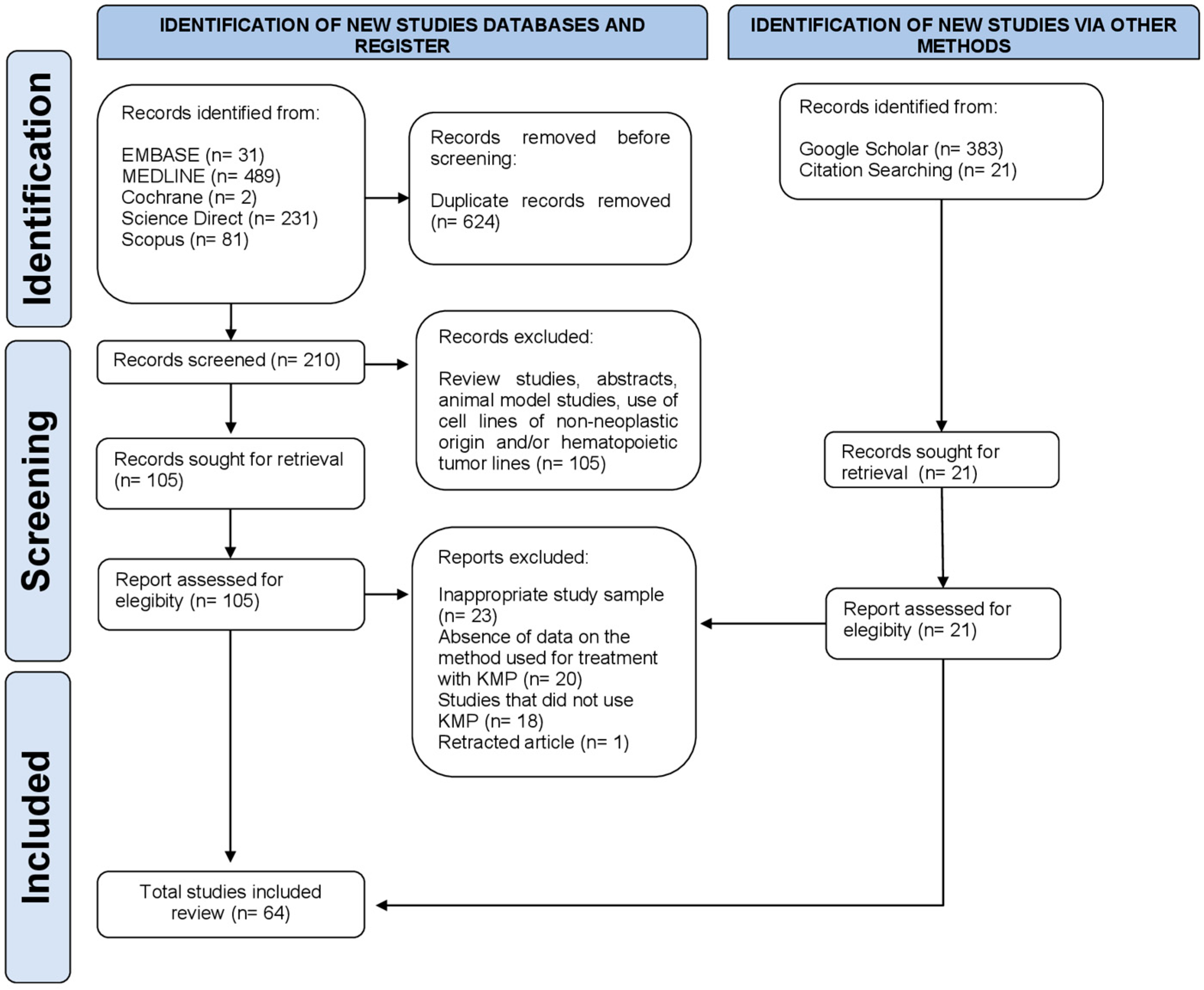
2. Materials and Methods
2.1. Search Strategy
2.2. Study Selection and Selection Criteria
2.3. Data Extraction, Analysis, and Risk of Bias Assessment
3. Results
3.1. Study Characteristics
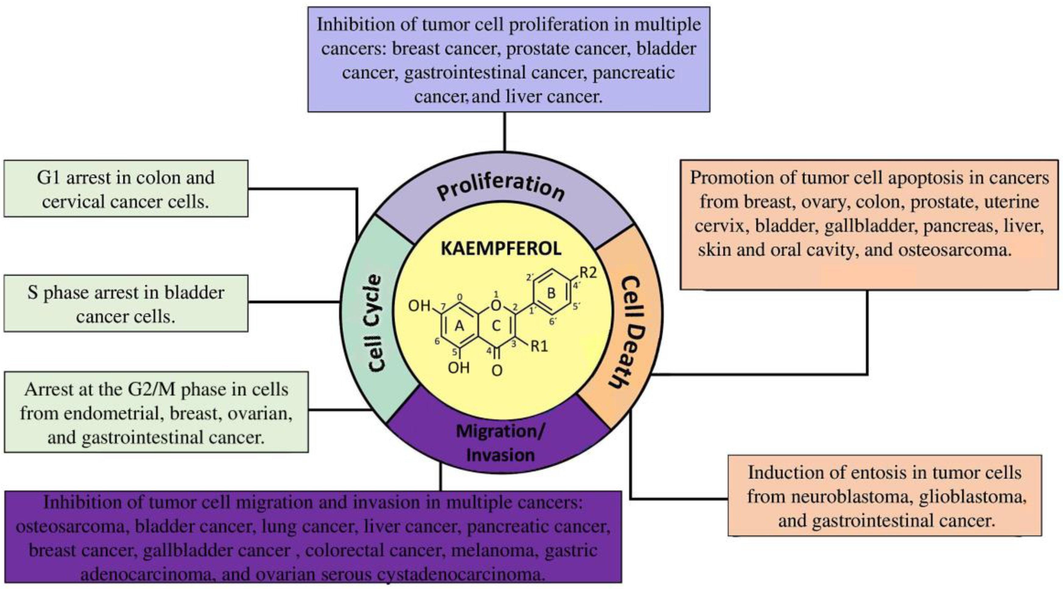
3.2. KMP Inhibits Cell Proliferation, Invasion, and Migration and Promotes Cell Death
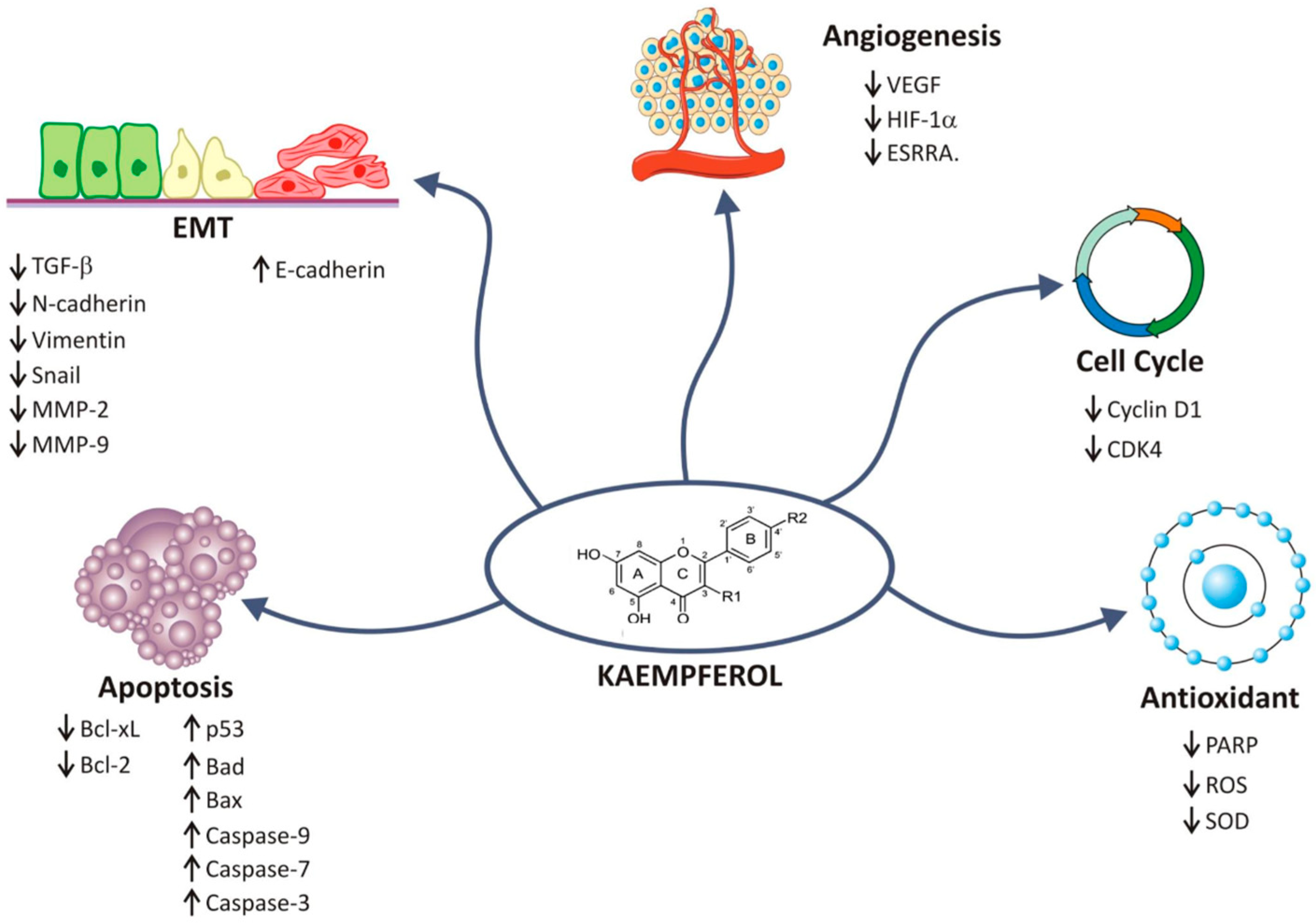
3.3. KMP Modulates the Expression of Cancer Biomarkers
3.4. KMP Sensitizes Cancer Cells to Chemotherapy
3.5. Risk of Bias
4. Discussion
5. Conclusions
Supplementary Materials
Author Contributions
Funding
Institutional Review Board Statement
Informed Consent Statement
Data Availability Statement
Conflicts of Interest
Abbreviations
| 5-FU | 5-Fluorouracil |
| AHR | Aryl hydrocarbon receptor |
| AP-1 | Activator protein-1 transcription factor |
| BAD | BCL2-associated agonist of cell death |
| BAX | BCL2-associated X |
| BCL2 | B-cell lymphoma 2 |
| CDC2 | Cyclin-dependent kinase 2 |
| CDC25C | Cell Division Cycle 25C |
| CDK2 | Cyclin D-dependent kinase 2 |
| CDK4 | Cyclin D-dependent kinase 4 |
| CDKN1A | Cyclin-dependent kinase inhibitor 1A |
| Chk2 | Checkpoint kinase 2 |
| DNA | Deoxyribonucleic Acid |
| EGFR | Epidermal growth factor receptor |
| EMT | Epithelial–mesenchymal transition |
| FADD | Fas-associated death domain |
| GRADE | Grading of Recommendations Assessment, Development, and Evaluation |
| HIF | Hypoxia-inducible factor |
| JNK | c-Jun N-terminal kinase |
| KMP | Kaempferol |
| MAPK | Mitogen-activated protein kinase |
| MMPs | Matrix metalloproteinases |
| mRNA | Messenger ribonucleic acid |
| mTOR | Mammalian target of rapamycin |
| Nrf2 | Nuclear factor erythroid 2-related factor 2 |
| PKB | Protein kinase B, also known as AKT |
| ROS | Reactive oxygen species |
| Smad2 | Mothers against decapentaplegic homolog 2 |
| Smad4 | Mothers against decapentaplegic homolog 4 |
| STAT | Signal transducer and activator of transcription |
| TFEB | Transcription factor EB |
| TGF-β1 | Transforming growth factor-β1 |
| VEGF | Vascular endothelial growth factor |
Appendix A

| Database | Strategy |
|---|---|
| PubMed/Medline | (“Kaempferol” OR “3,5,7-trihydroxy-2-(4-hydroxyphenyl)-4H-1-benzopyran-4-one” [Title/Abstract]) (“Kaempferol” OR “3,5,7-trihydroxy-2-(4-hydroxyphenyl)-4H-1-benzopyran-4-one”[Title/Abstract]) AND “Cancer”[Title/Abstract]) (“Kaempferol” OR “3,5,7-trihydroxy-2-(4-hydroxyphenyl)-4H-1-benzopyran-4-one”[Title/Abstract]) AND (“cell lines”[Title/Abstract] OR “cancer cell lines”[Title/Abstract] OR “neoplastic cell lines”[Title/Abstract] OR “malignancy”[Title/Abstract]) (“Kaempferol” OR “3,5,7-trihydroxy-2-(4-hydroxyphenyl)-4H-1-benzopyran-4-one”[Title/Abstract]) AND (“cell lines”[Title/Abstract] OR “cancer cell lines”[Title/Abstract] OR “neoplastic cell lines”[Title/Abstract] OR “malignancy”[Title/Abstract]) AND “Treatment”[Title/Abstract]) |
| Science Direct | “Kaempferol” OR “3,5,7-trihydroxy-2-(4-hydroxyphenyl)-4H-1-benzopyran-4-one” “Kaempferol” OR “3,5,7-trihydroxy-2-(4-hydroxyphenyl)-4H-1-benzopyran-4-one” AND “cell lines” OR “cancer cell lines” OR “neoplastic cell lines” OR “malignancy” “Kaempferol” OR “3,5,7-trihydroxy-2-(4-hydroxyphenyl)-4H-1-benzopyran-4-one” AND “cell lines” OR “cancer cell lines” OR “neoplastic cell lines” OR “malignancy” AND “Treatment” |
| Scopus | TITLE-ABS-KEY((“Kaempferol” OR “3,5,7-trihydroxy-2-(4-hydroxyphenyl)-4H-1-benzopyran-4-one”)) TITLE-ABS-KEY((“Kaempferol” OR “3,5,7-trihydroxy-2-(4-hydroxyphenyl)-4H-1-benzopyran-4-one”) AND (“cell lines” OR “cancer cell lines” OR “neoplastic cell lines” OR “malignancy”)) TITLE-ABS-KEY((“Kaempferol” OR “3,5,7-trihydroxy-2-(4-hydroxyphenyl)-4H-1-benzopyran-4-one”) AND (“cell lines” OR “cancer cell lines” OR “neoplastic cell lines” OR “malignancy”) AND (“Treatment”)) |
| EMBASE | (‘Kaempferol’ OR ‘3,5,7-trihydroxy-2-(4-hydroxyphenyl)-4H-1-benzopyran-4-one’) AND [embase]/lim NOT ([embase]/lim AND [medline]/lim) (‘Kaempferol’ OR ‘3,5,7-trihydroxy-2-(4-hydroxyphenyl)-4H-1-benzopyran-4-one’) AND (‘cell lines’ OR ‘cancer cell lines’ OR ‘neoplastic cell lines’ OR ‘malignancy’) AND [embase]/lim NOT ([embase]/lim AND [medline]/lim) (‘Kaempferol’ OR ‘3,5,7-trihydroxy-2-(4-hydroxyphenyl)-4H-1-benzopyran-4-one’) AND (‘cell lines’ OR ‘cancer cell lines’ OR ‘neoplastic cell lines’ OR ‘malignancy’) AND (‘Treatment’) AND [embase]/lim NOT ([embase]/lim AND [medline]/lim) |
| Cochrane Collaboration Library | “Kaempferol” OR “3,5,7-trihydroxy-2-(4-hydroxyphenyl)-4H-1-benzopyran-4-one” “Kaempferol” OR “3,5,7-trihydroxy-2-(4-hydroxyphenyl)-4H-1-benzopyran-4-one” AND “cell lines” OR “cancer cell lines” OR “neoplastic cell lines” OR “malignancy” “Kaempferol” OR “3,5,7-trihydroxy-2-(4-hydroxyphenyl)-4H-1-benzopyran-4-one” AND “cell lines” OR “cancer cell lines” OR “neoplastic cell lines” OR “malignancy” AND “Treatment” |
| Google Scholar | “Kaempferol” OR “3,5,7-trihydroxy-2-(4-hydroxyphenyl)-4H-1-benzopyran-4-one” “Kaempferol” OR “3,5,7-trihydroxy-2-(4-hydroxyphenyl)-4H-1-benzopyran-4-one” AND “cell lines” OR “cancer cell lines” OR “neoplastic cell lines” OR “malignancy” “Kaempferol” OR “3,5,7-trihydroxy-2-(4-hydroxyphenyl)-4H-1-benzopyran-4-one” AND “cell lines” OR “cancer cell lines” OR “neoplastic cell lines” OR “malignancy” AND “Treatment”. |
Appendix B
| Author (Year) | Title and DOI/PMID | Reason for Exclusion |
|---|---|---|
| Alkandahri et al. [98] | Hepatoprotective Effect of Kaempferol: A Review of the Dietary Sources, Bioavailability, Mechanisms of Action, and Safety. DOI: 10.1155/2023/1387665. | Study did not evaluate cancer cells |
| Ayob et al. [99] | Cytotoxicity Activities in Local Justicia gendarussa Crude Extracts against Human Cancer Cell Lines. DOI: 10.11113/jt.v64.2043 | Study did not evaluate cancer cells |
| Bandyopadhyay et al. [100] | Kaempferol and quercetin stimulate granulocyte-macrophage colony-stimulating factor secretion in human prostate cancer cells. DOI: 10.1016/j.mce.2008.01.015 | No information about kaempferol source |
| Bezdieniezhnykh et al. [101] | Establishment And Characterization of New Breast And Ovarian Cancer Cell Lines As A Model For Studying Cellular Plasticity In Vitro. PMID: 27356577 | Kaempferol was not assessed |
| Boadi et al. [102] | Effect of Quercetin, Genistein and Kaempferol on Glutathione and Glutathione-Redox Cycle Enzymes in 3T3-L1 Preadipocytes. DOI: 10.3109/01480545.2015.1082135 | Study did not evaluate cancer cells |
| Boadi et al. [103] | Flavonoids Reduce Lipid Peroxides and Increase Glutathione Levels in Pooled Human Liver Microsomes (HLMs). DOI: 10.4236/abc.2021.116019 | Study did not evaluate cancer cells |
| Budisan et al. [104] | Inhibitory effect of CAPE and kaempferol in colon cancer cell lines-possible implications in new therapeutic strategies. DOI: 0.3390/ijms20051199 | No information about kaempferol source and/or dilution |
| Catalán et al. [105] | Kaempferol Induces Cell Death and Sensitizes Human Head and Neck Squamous Cell Carcinoma Cell Lines to Cisplatin. DOI: 10.1007/5584_2020_603 | No information about kaempferol source and/or dilution |
| Chen et al. [106] | Dietary flavonoids as proteasome inhibitors and apoptosis inducers in human leukemia cells. DOI: 0.1016/j.bcp.2005.02.022 | Study did not evaluate cells from solid tumor |
| Chen et al. [107] | Biological Evaluation of Selected Flavonoids as Inhibitors of MNKs Targeting Acute Myeloid Leukemia. DOI: 10.1021/acs.jnatprod.0c00516 | Study did not evaluate cells from solid tumor |
| Chien et al. [108] | Kaempferol suppresses cell migration through the activation of the ERK signaling pathways in ARPE-19 cells. DOI: 10.1002/tox.22686 | Study did not evaluate cancer cells |
| Da et al. [109] | Kaempferol Promotes Apoptosis While Inhibiting Cell Proliferation via Androgen-Dependent Pathway and Suppressing Vasculogenic Mimicry and Invasion in Prostate Cancer. DOI: 10.1155/2019/1907698 | Study did not evaluate cancer cells |
| De Sousa et al. [110] | Hypoglycemic effect and antioxidant potential of kaempferol-3,7-O-(alpha)-dirhamnoside from Bauhinia forficata leaves. DOI: 10.1021/np030513u | Study did not evaluate cancer cells |
| Dimas et al. [111] | Cytotoxic activity of kaempferol glycosides against human leukaemic cell lines in vitro. DOI: 10.1006/phrs.1999.0562 | No information about kaempferol source and no application of solid cancer cells |
| Halimah et al. [112] | Induction of caspase cascade pathway by kaempferol-3-O-rhamnoside in LNCaP prostate cancer cell lines. DOI: 10.3892/br.2014.385 | No information about kaempferol source and/or dilution |
| Han et al. [113] | Kaempferol suppresses proliferation but increases apoptosis and autophagy by up-regulating microRNA-340 in human lung cancer cells. DOI: 10.1016/j.biopha.2018.09.087 | Retracted article |
| Huang et al. [114] | Cytotoxicity of Kaempferol-3-O-rhamnoside against nasopharyngeal cancer via inhibition of EGFR-TK | No information about kaempferol source and/or dilution |
| Jeong et al. [115] | Kaempferol Induces Cell Death Through ERK and Akt-Dependent Down-Regulation of XIAP and Survivin in Human Glioma Cells. DOI: 10.1007/s11064-008-9868-5 | No information about kaempferol source and/or dilution |
| Jin et al. [116] | Kaempferol attenuates diquat-induced oxidative damage and apoptosis in intestinal porcine epithelial cells. DOI: 10.1039/D1FO00402F | Study did not evaluate cancer cells |
| Jin et al. [117] | Kaempferol, a potential neuroprotective agent in neurodegenerative diseases: From chemistry to medicine. DOI: 10.1016/j.biopha.2023.115215. | Study did not evaluate cancer cells |
| Jo et al. [48] | Analysis of Inhibitory Effects of Kaempferol on Migration and Epithelial-mesenchymal Transition in Human Lung Cancer. DOI: 10.1016/j.neo.2015.06.004 | No information about kaempferol source and/or dilution |
| Joe et al. [118] | Engineering of flavonoid O-methyltransferase for a novel regioselectivity. DOI: 10.1007/s10059-010-0098-8 | Study did not evaluate cancer cells |
| Jokar et al. [119] | A comparative study of anti-leukemic effects of kaempferol and epigallocatechin-3-gallate (EGCG) on human leukemia HL-60 cells. DOI: 10.22038/AJP.2021.17604 | Study did not evaluate cells from solid tumor |
| Kang et al. [120] | Downregulation of PLK-1 expression in kaempferol-induced apoptosis of MCF-7 cells. DOI: 10.1016/j.ejphar.2009.03.068 | No information about kaempferol source and no application of cancer cells |
| Kang et al. [121] | Antiproliferation and Redifferentiation in Thyroid Cancer Cell Lines by Polyphenol Phytochemicals. DOI: 10.3346/jkms.2011.26.7.893 | Kaempferol was not assessed |
| Kim et al. [122] | Isolation of flavonol rhamnosides fromloranthus tanakae and cytotoxic effect of them on human tumor cell lines. DOI: 10.1007/BF02980044 | Kaempferol was not assessed |
| Kim et al. [123] | Treatment with kaempferol suppresses breast cancer cell growth caused by estrogen and triclosan in cellular and xenograft breast cancer models. DOI: 10.1016/j.jnutbio.2015.09.027 | No information about kaempferol source |
| Kluska et al. [124] | Kaempferol derivatives isolated from Lens culinaris Medik. reduce DNA damage induced by etoposide in peripheral blood mononuclear cells. DOI: 10.1039/c9tx00176j | Study did not evaluate cancer cells |
| Kuntz et al. [125] | Comparative analysis of the effects of flavonoids on proliferation, cytotoxicity, and apoptosis in human colon cancer cell lines. DOI: 10.1007/s003940050054 | No information about kaempferol source and/or dilution |
| Lee et al. [126] | Kaempferol protects HIT-T15 Pancreatic Beta Cells from 2-deoxy-D-ribose-induced Oxidative Damage. DOI: 10.1002/ptr.2983 | Study did not evaluate cancer cells |
| Lee et al. [127] | Kaempferol Isolated from Nelumbo nucifera Inhibits Lipid Accumulation and Increases Fatty Acid Oxidation Signaling in Adipocytes. DOI: 10.1089/jmf.2015.3457 | Study did not evaluate cancer cells |
| Li et al. [128] | Kaempferol induces apoptosis in human HCT116 colon cancer cells via the Ataxia-Telangiectasia Mutated-p53 pathway with the involvement of p53 Upregulated Modulator of Apoptosis. DOI: 10.1016/j.cbi.2008.10.048 | Study did not evaluate cancer cells |
| Lin et al. [129] | Kaempferol enhances the suppressive function of Treg cells by inhibiting FOXP3 phosphorylation. DOI: 10.1016/j.intimp.2015.03.044 | Study did not evaluate cancer cells |
| Lin et al. [130] | Isolation and identification of antiproliferative compounds from the roots of Tetrastigma hemsleyanum against MDA-MB-435S cell lines. PMID: 27393430 | No information about kaempferol source and/or dilution |
| Luo et al. [131] | Kaempferol inhibits VEGF expression and in vitro angiogenesis through a novel ERK-NFκB-cMyc-p21 pathway. DOI: 10.1016/j.foodchem.2011.07.045 | No information about kaempferol source and/or dilution |
| Luo et al. [132] | Kaempferol attenuates streptozotocin-induced diabetic nephropathy by downregulating TRAF6 expression: The role of TRAF6 in diabetic nephropathy. DOI: 10.1016/j.jep.2020.113553 | Study did not evaluate cancer cells |
| Meng et al. [133] | A kaempferol-3-O-β-d-glucoside, intervention effect of astragalin on estradiol metabolism. DOI: 10.1016/j.steroids.2019.05.005 | Study did not evaluate cancer cells |
| Nejabati et al. [134] | Kaempferol: A potential agent in the prevention of colorectal cancer. DOI: 10.14814/phy2.15488 | Review |
| Park et al. [135] | Enzymatic preparation of kaempferol from green tea seed and its antioxidant activity. DOI: 10.1021/jf052900a | Study did not evaluate cancer cells |
| Raghavan et al. [136] | Kaempferol mediated synthesis of gold nanoparticles and their cytotoxic effects on MCF-7 cancer cell line. DOI: 10.1016/j.procbio.2015.08.003 | Study did not evaluate cancer cells |
| Rahul et al. [137] | Effect of kaempferol on the transgenic Drosophila model of Parkinson’s disease. DOI: 10.1038/s41598-020-70236-2 | Study did not evaluate cancer cells |
| Riahi-Chebbi et al. [95] | The Phenolic compound Kaempferol overcomes 5-fluorouracil resistance in human resistant LS174 colon cancer cells. DOI: 10.1038/s41598-018-36808-z | No information about kaempferol source and/or dilution |
| Roth et al. [138] | Phytoestrogen kaempferol (3,4′,5,7-tetrahydroxyflavone) protects PC12 and T47D cells from β-amyloid–induced toxicity. PMID: 10412031 | Study did not evaluate cancer cells |
| Saraei et al. [139] | Kaempferol sensitizes tumor necrosis factor-related apoptosis-inducing ligand-resistance chronic myelogenous leukemia cells to apoptosis. DOI: 10.1007/s11033-021-06778-z | Study did not evaluate cells from solid tumor |
| Sengupta et al. [15] | Anticancer Properties of Kaempferol on Cellular Signaling Pathways. DOI: 10.2174/1568026622666220907112822 | Review |
| Sharma et al. [140] | Kaempferol induces apoptosis in glioblastoma acells through oxidative stress. DOI: 10.1158/1535-7163.MCT-06-0788 | Study did not evaluate cancer cells |
| Sharma et al. [141] | Kaempferol attenuates diabetic nephropathy by inhibiting RhoA/Rho-kinase mediated inflammatory signalling. DOI: 10.1016/j.biopha.2018.10.195 | Study did not evaluate cancer cells |
| Stapel et al. [142] | Polyphenol compounds with anti-carcinogenic qualities: Effects of quercetin (flavonol), chrysin (flavon), kaempferol (flavanol), naringenin (flavanon) and hesperidin (flavanoid) on in vitro breast cancer. DOI: 10.5897/JMPR12.5126 | No information about kaempferol source and/or dilution |
| Swanson et al. [143] | Impact of apigenin and kaempferol on human head and neck squamous cell carcinoma. DOI: 10.1016/j.oooo.2013.10.012 | Study did not evaluate cancer cells |
| Tu et al. [144] | The mechanism of kaempferol induced apoptosis and inhibited proliferation in human cervical cancer SiHa cell: From macro to nano. DOI: 10.1002/sca.21312 | No information about kaempferol source and/or dilution |
| Tu et al. [145] | Synthesis, characterization and anticancer activity of kaempferol-zinc(II) complex. DOI: 10.1016/j.bmcl.2016.03.091 | No information about kaempferol source |
| Wang et al. [146] | The mechanism of anticancer action and potential clinical use of kaempferol in the treatment of breast cancer. DOI: 10.1016/j.biopha.2019.109086 | Review |
| Wang et al. [147] | Kaempferol protects mice from d-GalN/LPS-induced acute liver failure by regulating the ER stress-Grp78-CHOP signaling pathway. DOI: 10.1016/j.biopha.2018.12.105 | Study did not evaluate cancer cells |
| Wang et al. [148] | Kaempferol-Driven Inhibition of Listeriolysin O Pore Formation and Inflammation Suppresses Listeria monocytogenes Infection. DOI: 10.1128/spectrum.01810-22 | Study did not evaluate cancer cells |
| Wu et al. [149] | Kaempferol protects mitochondria and alleviates damages against endotheliotoxicity induced by doxorubicin. DOI: 10.1016/j.biopha.2020.110040 | Study did not evaluate cancer cells |
| Xiao et al. [150] | Old wine in new bottles: Kaempferol is a promising agent for treating the trilogy of liver diseases. DOI: 10.1016/j.phrs.2021.106005 | Study did not evaluate cancer cells |
| Xie et al. [151] | Kaempferol promotes apoptosis in human bladder cancer cells by inducing the tumor suppressor, PTEN. DOI: 10.3390/ijms141121215 | Study did not evaluate cancer cells |
| Yang et al. [152] | Exploration in the Mechanism of Kaempferol for the Treatment of Gastric Cancer Based on Network Pharmacology. DOI: 10.1155/2020/5891016 | Study based in bioinformatics only |
| Yusof et al. [153] | Hypolipidemic effects of quercetin and kaempferol in human hepatocellular carcinoma (HepG2) cells. * | No information about kaempferol source and/or dilution |
| Zeng et al. [154] | Kaempferol ameliorates in-vitro and in-vivo postovulatory oocyte ageing in mice. DOI: 10.1016/j.rbmo.2022.07.005 | Animal model study |
| Zhang et al. [155] | Kaempferol suppresses human gastric cancer SNU-216 cell proliferation, promotes cell autophagy, but has no influence on cell apoptosis. DOI: 10.1590/1414-431x20187843 | Retracted article |
| Zheng et al. [156] | Molecular Mechanism Investigation on Monomer Kaempferol of the Traditional Medicine Dingqing Tablet in Promoting Apoptosis of Acute Myeloid Leukemia HL-60 Cells. DOI: 10.1155/2022/8383315. | Study did not evaluate cells from solid tumor |
Appendix C
| Author | Country | Cell Lines | Origin of Cell Lines | Dosages of KMP | IC50 | Main Results |
|---|---|---|---|---|---|---|
| Wang et al. [10] | China | HepG2, CT26, B16F1 | Human hepatocellular carcinoma, mouse colon cancer, mouse melanoma | 10–100 µM | 30.92–88.02 µM | Inhibited proliferation and AKT phosphorylation and promoted the cleavage of caspase-9, caspase-7, caspase-3, and PARP. |
| Li et al. [12] | The United States | HcT-8, HcT-116 | Human colorectal cancer | 0–200 μM | 350 μM | Synergistic effect with 5-FU by inhibiting cell proliferation and inducing apoptosis. |
| Park et al. [14] | Republic of Korea | HT29, HCT116 | Human colon cancer | 20–40 µM | NI | Inhibited AP-1 in oxaliplatin-resistant cells. |
| Zheng et al. [17] | China | A375, B16F10 | Human and mouse melanoma | 2–64 μM | NI | Inhibited migration and invasion. |
| Nguyen et al. [22] | Republic of Singapore | A549 | Human lung cancer | 17.5–70 µM | NI | Induced apoptosis via activation of AKT-1, Bcl-2 family of proteins, and MAPK signaling. |
| Ackland et al. [23] | Australia | PMC42, HuTu-80, Caco-2 | Human breast cancer, duodenal adenocarcinoma, colon adenocarcinoma | 1–10 µM | NI | Reduced proliferation. |
| Nakamura et al. [24] | Japan | HCT116, KNC | Human colon cancer | 2.5–20 µM | NI | Promoted expression and phosphorylation of connexin 43 via a STAT3-dependent mechanism. |
| Campbell et al. [25] | The United States | LNCaP | Human prostate cancer | 50 mM | NI | Inhibited proliferation in a dose-dependent manner, without cytotoxicity. |
| Oh et al. [26] | Republic of Korea | MCF-7 | Human breast cancer | 10−7–10−4 M | NI | Antiproliferative effects are dependent of estrogen receptor activation. |
| Leung et al. [27] | Taiwan | H460 | Human lung carcinoma | 50–80 μM | NI | Induced apoptosis and MnSOD levels. |
| Li et al. [28] | China | BEL-7402, HeLa, 95-D, A375, MKN-45, A431 | Human liver cancer, cervical carcinoma, melanoma, gastric cancer, skin cancer | 100–400 μg/mL | 43–110 g/mL | Induced cell cycle arrest at G1 and apoptosis. |
| Choi et al. [29] | Republic of Korea | MDA-MB-453 | Human breast cancer | 1–200 μM | NI | Inhibited proliferation, induced cell cycle arrest at G2/M phase, downregulated CDK1, cyclin A, and cyclin B, and induced apoptosis which was associated with p53 upregulation. |
| Kim et al. [30] | Republic of Korea | MCF-7, T47D, MDA-MB-231, HC-11 | Human breast cancer | 20 μM | NI | Induced apoptosis via activation of MAPK, MEK1, and ELK1. |
| Siegelin et al. [31] | Germany | U87, U251, U373 | Human glioblastoma | 5–200 µM | NI | Promoted apoptosis after suppression of survivin. |
| Yoshida et al. [32] | Japan | SW480, DLD-1, PC3 | Human colon cancer, prostate cancer | 2.5–40 µM | NI | Upregulated TRAIL receptors (DR5 and DR4). |
| Zhang et al. [33] | The United States | MIA PaCa-2, Panc-1 | Human pancreatic cancer | 17.5–35 μM | NI | Inhibited proliferation and induced apoptosis. |
| Luo et al. [34] | The United States | OVCAR-3, A2780/CP70 | Human ovarian cancer | 5–80 µM | NI | Inhibited angiogenesis and VEGF expression via HIF-dependent (AKT/HIF) and HIF-independent pathways. |
| Huang et al. [35] | Taiwan | U-2 OS, HOB, 143B | Human osteosarcoma | 25–200 mM | NI | Reduced cell viability and regulated the expression of proteins involved in the endoplasmic reticulum stress pathway and the mitochondrial signaling pathway. |
| Kang et al. [36] | Republic of Korea | SCC-1483, SCC-QLL1, SCC-25 | Human oral squamous cell carcinoma | 20–80 μM | NI | Inhibited proliferation and induced apoptosis, which was dependent on caspase-3. |
| Luo et al. [37] | The United States | OVCAR-3 | Human ovarian cancer | 20 μM | NI | Enhanced cisplatin effects on promoting cell death. |
| Macpherson et al. [38] | Canada | T-47D, BT-549, MDA-MB-231 | Human breast cancer | 0.1–10 μM | NI | Induced cell death. |
| Mylonis et al. [39] | Greece | Huh7 | Human hepatocellular carcinoma | 1–100 μM | 5.16 μM | Promoted cell death and inhibited MAPK and HIF-1 activity. |
| Luo et al. [40] | The United States | OVCAR-3, A2780/CP70, A2780/wt | Human ovarian cancer | 20–160 μM | NI | Kaempferol induces apoptosis in ovarian cancer cells by regulating pro-apoptotic and anti-apoptotic protein expressions in the intrinsic apoptosis pathways and is a good candidate for the chemoprevention of ovarian cancers in humans. |
| Luo et al. [41] | The United States | A2780/CP70, OVCAR-3 | Human ovarian cancer | 10–25 µM | NI | Induced apoptosis. |
| Chen et al. [42] | Japan | U-2 OS | Human osteosarcoma | 25–100 μM | NI | Inhibited invasion, migration, and adhesion, reduced activities of MMP-2, MMP-9, and uPA, and altered phosphorylation of MAPK, p38, and JNK and decreased the DNA binding activity of AP-1. |
| Cho et al. [43] | Republic of Korea | HT-29 | Human colon adenocarcinoma | 60 μmol/L | NI | Decreased viability and proliferation and induced G1 cell cycle arrest in 6 h and G2/M arrest in 12 h. Inhibited CDK2, CDK4, cyclin D1, cyclin E, cyclin A, phosphorylation of Rb, Cdc25C, Cdc2, cyclin B1, and Cdc2. |
| Wang et al. [44] | China | HeLa, HepG2, A549 | Human cervical cancer, liver cancer, lung cancer | 5–20 μM | NI | Antagonized activities of estrogen-related receptors alpha and gamma (ERRα and ERRγ). |
| Jaramillo-Carmona et al. [45] | Spain | HCT-116 | Human colon carcinoma | 0–200 μM | 75 μM | The combination of quercetin and kaempferol exhibited greater cytotoxic efficacy than quercetin or kaempferol alone, and this effect was related to cell cycle arrest at G2/M. |
| Dang et al. [46] | China | 5637, T24, T24L | Human bladder cancer | 50–150 mM | NI | Induced apoptosis and cell cycle arrest. |
| Hang et al. [47] | China | A549 | Lung cancer | 10–60 μM | 72 μM | Inhibited proliferation and migration in a dose-dependent manner and modulated the expression of E-cadherin and vimentin. |
| Jo et al. [48] | Republic of Korea | A549 | Human lung cancer | 50 μM | NI | Blocked TGF-β1-induced migration, MMP-2 activity, and EMT |
| Kuo et al. [49] | Taiwan | A-549 | Human lung cancer | 7–112 μM | NI | Increased cell death. |
| Li et al. [50] | China | MDA-MB-231 | Human breast cancer | 10–40 μM | 204.7 mol/L. | Inhibited invasion by blocking the PKC/MAPK/AP-1 cascade and subsequent MMP-9 expression and activity. |
| Song et al. [51] | China | MKN28. SGC7901 | Human gastric cancer | 25–200 µM | 124.86 μM | Inhibited proliferation and promoted G2/M cell cycle arrest and apoptosis. |
| Lee et al. [52] | Republic of Korea | Miapaca-2, Panc-1, SNU-213 | Human pancreatic cancer | 0–200 μM | NI | Decreased viability and increased apoptosis, which were mediated by inhibition of EGFR-related Src, MAPK, and AKT pathways. |
| Liao et al. [53] | China | MCF-7, SGC-7901, Hela, A549 | Human breast cancer, gastric cancer, cervical cancer, lung cancer | 25–100 μg/mL | NI | Inhibited proliferation and induced apoptosis. |
| Han et al. [54] | China | HCC, HepG2, Huh-7, BEL7402 SMMC, Pri-1/-2/-3 | Human hepatocellular carcinoma | 5–100 μM | 25–50 μM | Induced AMPK, which led to Ulk1 phosphorylation, mTOR 1 complex inhibition, and cellular autophagy. |
| Kashafi et al. [55] | Iran | HeLa | Human cervical adenocarcinoma | 12–100 mM | 10.48 mM | Decreased viability and induced apoptosis through downregulation of PI3K/AKT and hTERT pathways. |
| Chuwa et al. [56] | Japan | HEC-265/HEC-108, HEC-180 | Human endometrial carcinoma | 36–72 µM | 83.65 μM | Induced sub-G1 cell accumulation and apoptosis. |
| Gao et al. [57] | The United States | A2780/CP70 | Human ovarian endometrioid adenocarcinoma | 40μmol/L | NI | Induced G2/M cell cycle arrest via Chk2/Cdc25C/Cdc2 pathway and Chk2/p21/Cdc2 pathway and apoptosis and p53 upregulation. |
| Kim et al. [58] | Republic of Korea | AGS, SNU-216, NCI-N87, SNU-638, MKN-74 | Human gastric cancer | 25–100 μM | NI | Promoted autophagy and cell death. |
| Lu et al. [59] | China | HCT116, HT29, YB5 | Human colorectal cancer | 1.25–5 M | NI | Regulated proliferation and migration. |
| Pham et al. [60] | Australia | A2780, H460, A431, MIA PaCa-2, Du145, HT29, MCF-7, BE2-C, SJ-G2, U87, SMA | Human ovarian cancer, lung cancer, skin cancer, pancreas cancer, prostate cancer, colon cancer, breast cancer, neuroblastoma, glioblastoma | 0–50 µM | 19–50 µM | Promoted cytotoxicity. |
| Thangavel et al. [61] | Republic of Korea | A549 | Human lung cancer | 25–150 μM | 10 μM | Inhibited proliferation. |
| Wu et al. [62] | China | EJ | Human bladder cancer | 10–320 µM | NI | Induced apoptosis and cell cycle arrest at S phase. |
| Yang et al. [63] | China | A375 | Human melanoma | 10–80 µM | 20 µM | Induced apoptosis and G2/M cell cycle arrest and inhibited migration, with downregulation of mTOR, pmTOR, PI3K, p-PI3K, and AKT. |
| Abdullah et al. [64] | India | IMR32, Neuro2A | Human and mouse neuroblastoma | 25–100 μM | NI | Reduced proliferation and increased apoptosis along with induction of neuritogenesis. |
| Zhu et al. [65] | China | BT474, MDA-MB231 | Human breast cancer | 50 μmol/L | 43, >100 μmol/L | Induced apoptosis and DNA damage and increased the expression levels of H2AX, cleaved caspase-9, cleaved caspase-3, and p-ATM. |
| Chen et al. [66] | China | U87 MG, U251 | Human glioblastoma | 20–120 μM | 97.2, 79.2 μM | Suppressed proliferation, increased ROS, and decreased mitochondrial membrane potential. |
| Gutierrez-Uribe et al. [67] | Mexico | RKO | Human colon cancer | 0.01–1 μM | NI | miR31, miR92a, KRAS, and c-MYC were downregulated, whereas AMPK and APC were upregulated. |
| Kitakaze et al. [68] | Japan | HepG2 | Human hepatocellular carcinoma | 30 μM | NI | In combination with luteolin, inhibited the expression of drug-metabolizing enzymes. |
| Nair et al. [69] | India | HepG2, N1S1 | Human hepatocellular carcinoma | 6.25–50 µM | 9.61 µM | Increased the effect of sub-toxic concentrations of sorafenib. |
| Fouzder et al. [70] | India | A549, NCIH460 | Human colon adenocarcinoma | NI | NI | Induced ROS accumulation and apoptosis, which was related to modulation of Nrf2, GST, and NQO1. |
| Inden et al. [71] | Japan | N2A | Mouse neuroblastoma | 5 mM | NI | Induced autophagy by increasing lysosome biogenesis and directly blocked the formation of α-Syn amyloid fibrils. |
| Ju et al. [72] | Taiwan | Huh-7, SK-Hep-1 | Human hepatocellular carcinoma | 25–100 μM | NI | Reduced invasion, migration, MMP-9 expression and activity, and AKT phosphorylation. |
| Li et al. [73] | China | A549 | Human lung cancer | 0–200 μM | NI | Reduced cell viability, after cytoskeleton collapse and mitochondrial dysfunction. |
| Liu et al. [74] | China | SGC996, GBC-SD | Human gallbladder cancer | 100–200 μg/mL | NI | Promoted apoptosis due to cytochrome C release, activation of caspase-3 and caspase-9, and increase in BAX expression. |
| Wang et al. [75] | China | Mia PaCa-2, PANC-1 | Human pancreatic cancer | 0–1000 µM/L | 78.75, 79.07 μM | Induced ROS-dependent apoptosis via TGM2-mediated AKT/mTOR signaling. |
| Wu et al. [76] | China | HCT-116, DLD1 | Human colon cancer | 20–160 μM | 63.0, 98.3 μM | Inhibited glycolysis and proliferation by modulating miR-339–5p-hnRNPA1/PTBP1–PKM2 axis. |
| Yang et al. [77] | China | Huh-7, Huh-1, HepG2, HepG2.2.15, SK-Hep-1, PLC/PRF/5, HLE, HLF, Hep3B | Human liver cancer | 10–40 µM | 40 µM | Inhibited proliferation, migration, and invasion. |
| Zhang et al. [78] | China | BxPC-3, PANC-1 | Human pancreatic cancer | 6–192 µM | 155.21, 108.47 µM | Increased the effect of erlotinib via inhibiting PI3K/AKT and EGFR signaling. |
| Zhang et al. [79] | China | AGS, SKOV3IP1, MDA-MB-231 | Human gastric adenocarcinoma, ovarian cystadenocarcinoma, breast cancer | 0.5–4 μM | 6.83, 9.6 μM 11.12 μM | Reduced cell proliferation and inhibited TGF-β-induced cell migration, invasion, and EMT markers. |
| Ma et al. [80] | China | A549 | Non-small-cell lung cancer | 5–120 µM | 72.8 μM/L | Inhibited proliferation, migration, and invasion and promoted apoptosis. |
| Zhang et al. [81] | China | 22Rv1, PC-3, DU145 | Human prostate cancer | 10–40 µM | NI | Inhibited proliferation of androgen-dependent and androgen-independent prostate cancer cells. |
Appendix D
| Author (Year) | Study Limitation | Inconsistency | Indirectness | Imprecision | Publication Bias | Overall Quality |
|---|---|---|---|---|---|---|
| Wang et al. [10] | Yes | Yes | No | Yes | Yes | +++ |
| Li et al. [12] | Yes | Yes | Yes | Yes | Yes | ++++ |
| Park et al. [14] | Yes | Yes | Yes | No | Yes | +++ |
| Zheng et al. [17] | Yes | Yes | Yes | No | Yes | +++ |
| Nguyen et al. [22] | Yes | Yes | Yes | No | Yes | +++ |
| Ackland et al. [23] | Yes | Yes | Yes | No | Yes | +++ |
| Nakamura et al. [24] | Yes | Yes | Yes | No | Yes | +++ |
| Campbell et al. [25] | Yes | Yes | Yes | No | Yes | +++ |
| Oh et al. [26] | Yes | Yes | Yes | No | Yes | +++ |
| Leung et al. [27] | Yes | Yes | Yes | No | Yes | +++ |
| Li et al. [28] | Yes | Yes | No | Yes | Yes | +++ |
| Choi et al. [29] | Yes | Yes | Yes | No | Yes | +++ |
| Kim et al. [30] | Yes | Yes | Yes | No | Yes | +++ |
| Siegelin et al. [31] | Yes | Yes | Yes | No | Yes | +++ |
| Yoshida et al. [32] | Yes | Yes | Yes | No | Yes | +++ |
| Zhang et al. [33] | Yes | Yes | Yes | No | Yes | +++ |
| Luo et al. [34] | Yes | Yes | Yes | No | Yes | +++ |
| Huang et al. [35] | Yes | Yes | Yes | No | Yes | +++ |
| Kang et al. [36] | Yes | Yes | Yes | No | Yes | +++ |
| Luo et al. [37] | Yes | Yes | Yes | No | Yes | +++ |
| Macpherson et al. [38] | Yes | Yes | Yes | No | Yes | +++ |
| Mylonis et al. [39] | Yes | Yes | Yes | Yes | Yes | ++++ |
| Luo et al. [40] | Yes | Yes | Yes | No | Yes | +++ |
| Luo et al. [41] | Yes | Yes | Yes | No | Yes | +++ |
| Chen et al. [42] | Yes | Yes | Yes | No | Yes | +++ |
| Cho et al. [43] | Yes | Yes | Yes | No | Yes | +++ |
| Wang et al. [44] | Yes | Yes | Yes | No | Yes | +++ |
| Jaramillo-Carmona et al. [45] | Yes | Yes | Yes | Yes | Yes | ++++ |
| Dang et al. [46] | Yes | Yes | Yes | No | Yes | +++ |
| Hang et al. [47] | Yes | Yes | Yes | Yes | Yes | ++++ |
| Jo et al. [48] | Yes | Yes | Yes | No | Yes | +++ |
| Kuo et al. [49] | Yes | Yes | Yes | No | Yes | +++ |
| Li et al. [50] | Yes | Yes | Yes | Yes | Yes | ++++ |
| Song et al. [51] | Yes | Yes | Yes | Yes | Yes | ++++ |
| Lee et al. [52] | Yes | Yes | Yes | No | Yes | +++ |
| Liao et al. [53] | Yes | Yes | No | No | Yes | ++ |
| Han et al. [54] | Yes | Yes | Yes | Yes | Yes | ++++ |
| Kashafi et al. [55] | Yes | Yes | Yes | Yes | Yes | ++++ |
| Chuwa et al. [56] | Yes | Yes | Yes | Yes | Yes | ++++ |
| Gao et al. [57] | Yes | Yes | Yes | No | Yes | +++ |
| Kim et al. [58] | Yes | Yes | Yes | No | Yes | +++ |
| Lu et al. [59] | Yes | Yes | Yes | No | Yes | +++ |
| Pham et al. [60] | Yes | Yes | Yes | Yes | Yes | ++++ |
| Thangavel et al. [61] | Yes | Yes | Yes | Yes | Yes | ++++ |
| Wu et al. [62] | Yes | Yes | Yes | No | Yes | +++ |
| Yang et al. [63] | Yes | Yes | Yes | Yes | Yes | ++++ |
| Abdullah et al. [64] | Yes | Yes | Yes | No | Yes | +++ |
| Zhu et al. [65] | Yes | Yes | Yes | Yes | Yes | ++++ |
| Chen et al. [66] | Yes | Yes | Yes | Yes | Yes | ++++ |
| Gutierrez-Uribe et al. [67] | Yes | Yes | Yes | No | Yes | +++ |
| Kitakaze et al. [68] | Yes | Yes | Yes | No | Yes | +++ |
| Nair et al. [69] | Yes | Yes | No | Yes | Yes | +++ |
| Fouzder et al. [70] | Yes | Yes | Yes | No | Yes | ++++ |
| Inden et al. [71] | Yes | Yes | Yes | No | Yes | +++ |
| Ju et al. [72] | Yes | Yes | Yes | No | Yes | +++ |
| Li et al. [73] | Yes | Yes | Yes | No | Yes | +++ |
| Liu et al. [74] | Yes | Yes | Yes | No | Yes | +++ |
| Wang et al. [75] | Yes | Yes | Yes | Yes | Yes | ++++ |
| Wu et al. [76] | Yes | Yes | Yes | Yes | Yes | ++++ |
| Yang et al. [77] | Yes | Yes | No | Yes | Yes | +++ |
| Zhang et al. [78] | Yes | Yes | Yes | Yes | Yes | ++++ |
| Zhang et al. [79] | Yes | Yes | Yes | Yes | Yes | ++++ |
| Ma et al. [80] | Yes | Yes | Yes | Yes | Yes | ++++ |
| Zhang et al. [81] | Yes | Yes | Yes | No | Yes | +++ |
References
- Sung, H.; Ferlay, J.; Siegel, R.L.; Laversanne, M.; Soerjomataram, I.; Jemal, A.; Bray, F. Global Cancer Statistics 2020: GLOBOCAN Estimates of Incidence and Mortality Worldwide for 36 Cancers in 185 Countries. CA Cancer J. Clin. 2021, 71, 209–249. [Google Scholar] [CrossRef] [PubMed]
- Debela, D.T.; Muzazu, S.G.; Heraro, K.D.; Ndalama, M.T.; Mesele, B.W.; Haile, D.C.; Kitui, S.K.; Manyazewal, T. New approaches and procedures for cancer treatment: Current perspectives. SAGE Open Med. 2021, 9, 20503121211034366. [Google Scholar] [CrossRef]
- Papież, M.A.; Krzyściak, W. Biological therapies in the treatment of cancer-update and new directions. Int. J. Mol. Sci. 2021, 22, 11694. [Google Scholar] [CrossRef] [PubMed]
- Wang, X.; Zhang, H.; Chen, X. Drug resistance and combating drug resistance in cancer. Cancer Drug Resist. 2019, 2, 141–160. [Google Scholar] [CrossRef] [PubMed]
- Wang, X.J.; Chen, J.Y.; Fu, L.Q.; Yan, M.J. Recent advances in natural therapeutic approaches for the treatment of cancer. J. Chemother. 2020, 332, 53–65. [Google Scholar] [CrossRef]
- Talib, W.H.; Awajan, D.; Hamed, R.A.; Azzam, A.O.; Mahmod, A.I.; Al-Yasari, I.H. Combination anticancer therapies using selected phytochemicals. Molecules 2022, 27, 5452. [Google Scholar] [CrossRef]
- Talib, W.H.; Alsayed, A.R.; Barakat, M.; Abu-Taha, M.I.; Mahmod, A.I. Targeting drug chemo-resistance in cancer using natural products. Biomedicines 2021, 9, 1353. [Google Scholar] [CrossRef]
- Hashem, S.; Ali, T.A.; Akhtar, S.; Nisar, S.; Sageena, G.; Ali, S.; Al-Mannai, S.; Therachiyil, L.; Mir, R.; Elfaki, I.; et al. Targeting cancer signaling pathways by natural products: Exploring promising anti-cancer agents. Biomed. Pharmacother. 2022, 150, 113054. [Google Scholar] [CrossRef]
- Nabavi, S.M.; Šamec, D.; Tomczyk, M.; Milella, L.; Russo, D.; Habtemariam, S.; Suntar, I.; Rastrelli, L.; Daglia, M.; Xiao, J.; et al. Flavonoid biosynthetic pathways in plants: Versatile targets for metabolic engineering. Biotechnol. Adv. 2020, 38, 107316. [Google Scholar] [CrossRef]
- Wang, J.; Fang, X.; Ge, L.; Cao, F.; Zhao, L.; Wang, Z.; Xiao, W. Antitumor, antioxidant and anti-inflammatory activities of kaempferol and its corresponding glycosides and the enzymatic preparation of kaempferol. PLoS ONE 2018, 13, e0197563. [Google Scholar] [CrossRef]
- Imran, M.; Salehi, B.; Sharifi-Rad, J.; Aslam Gondal, T.; Saeed, F.; Imran, A.; Shahbaz, M.; Tsouh Fokou, P.V.; Umair Arshad, M.; Khan, H.; et al. Kaempferol: A key emphasis to its anticancer potential. Molecules 2019, 24, 2277. [Google Scholar] [CrossRef]
- Li, Q.; Wei, L.; Lin, S.; Chen, Y.; Lin, J.; Peng, J. Synergistic effect of kaempferol and 5-fluorouracil on the growth of colorectal cancer cells by regulating the PI3K/Akt signaling pathway. Mol. Med. Rep. 2019, 20, 728–734. [Google Scholar] [CrossRef]
- Zhang, L.; Guo, Z.; Wang, Y.; Geng, J.; Han, S. The protective effect of kaempferol on heart via the regulation of Nrf2, NF-κβ, and PI3K/Akt/GSK-3β signaling pathways in isoproterenol-induced heart failure in diabetics rats. Drug Dev. Res. 2019, 80, 294–309. [Google Scholar] [CrossRef]
- Park, J.; Lee, G.E.; An, H.J.; Lee, C.J.; Cho, E.S.; Kang, H.C.; Lee, J.Y.; Lee, H.S.; Choi, J.S.; Kim, D.J.; et al. Kaempferol sensitizes cell proliferation inhibition in oxaliplatin-resistant colon cancer cells. Arch. Pharm. Res. 2021, 44, 1091–1108. [Google Scholar] [CrossRef]
- Sengupta, B.; Biswas, P.; Roy, D.; Lovett, J.; Simington, L.; Fry, D.R.; Travis, K. Anticancer properties of kaempferol on cellular signaling pathways. Curr. Top. Med. Chem. 2022, 2, 2474–2482. [Google Scholar] [CrossRef]
- Wu, H.; Du, J.; Li, C.; Li, H.; Guo, H.; Li, Z. Kaempferol can reverse the 5-FU resistance of colorectal cancer cells by inhibiting PKM2-mediated glycolysis. Int. J. Mol. Sci. 2022, 23, 3544. [Google Scholar] [CrossRef] [PubMed]
- Zheng, X.; Pan, Y.; Yang, G.; Liu, Y.; Zou, J.; Zhao, H.; Yin, G.; Wu, Y.; Li, X.; Wei, Z.; et al. Kaempferol impairs aerobic glycolysis against melanoma metastasis via inhibiting the mitochondrial binding of HK2 and VDAC1. Eur. J. Pharmacol. 2022, 931, 175226. [Google Scholar] [CrossRef]
- Page, M.J.; McKenzie, J.E.; Bossuyt, P.M.; Boutron, I.; Hoffmann, T.C.; Mulrow, C.D.; Shamseer, L.; Tetzlaff, J.M.; Akl, E.A.; Brennan, S.E.; et al. The PRISMA 2020 statement: An updated guideline for reporting systematic reviews. BMJ 2021, 372, 71. [Google Scholar] [CrossRef] [PubMed]
- Guyatt, G.; Oxman, A.D.; Akl, E.A.; Kunz, R.; Vist, G.; Brozek, J.; Norris, S.; Falck-Ytter, Y.; Glasziou, P.; DeBeer, H.; et al. GRADE guidelines: 1. Introduction-GRADE evidence profiles and summary of findings tables. J. Clin. Epidemiol. 2011, 64, 383–394. [Google Scholar] [CrossRef] [PubMed]
- Huguet, A.; Hayden, J.A.; Stinson, J.; McGrath, P.J.; Chambers, C.T.; Tougas, M.E.; Wozney, L. Judging the quality of evidence in reviews of prognostic factor research: Adapting the GRADE framework. Syst. Rev. 2013, 2, 71. [Google Scholar] [CrossRef]
- Pavan, L.M.; Rêgo, D.F.; Elias, S.T.; De Luca Canto, G.; Guerra, E.N. In vitro anti-tumor effects of statins on head and neck squamous cell carcinoma: A systematic review. PLoS ONE 2015, 10, e0130476. [Google Scholar] [CrossRef]
- Nguyen, T.T.; Tran, E.; Ong, C.K.; Lee, S.K.; Do, P.T.; Huynh, T.T.; Nguyen, T.H.; Lee, J.J.; Tan, Y.; Ong, C.S.; et al. Kaempferol-induced growth inhibition and apoptosis in A549 lung cancer cells is mediated by activation of MEK-MAPK. J. Cell Physiol. 2003, 197, 110–121. [Google Scholar] [CrossRef] [PubMed]
- Ackland, M.L.; van de Waarsenburg, S.; Jones, R. Synergistic antiproliferative action of the flavonols quercetin and kaempferol in cultured human cancer cell lines. In Vivo 2005, 19, 69–76. [Google Scholar] [PubMed]
- Nakamura, Y.; Chang, C.C.; Mori, T.; Sato, K.; Ohtsuki, K.; Upham, B.L.; Trosko, J.E. Augmentation of differentiation and gap junction function by kaempferol in partially differentiated colon cancer cells. Carcinogenesis 2005, 26, 665–671. [Google Scholar] [CrossRef] [PubMed]
- Campbell, J.K.; King, J.L.; Harmston, M.; Lila, M.A.; Erdman, J.W. Synergistic effects of flavonoids on cell proliferation in Hepa-1c1c7 and LNCaP cancer cell lines. J. Food Sci. 2006, 71, S358–S363. [Google Scholar] [CrossRef]
- Oh, S.M.; Kim, Y.P.; Chung, K.H. Biphasic effects of kaempferol on the estrogenicity in human breast cancer cells. Arch. Pharm. Res. 2006, 29, 354–362. [Google Scholar] [CrossRef]
- Leung, H.W.; Lin, C.J.; Hour, M.J.; Yang, W.H.; Wang, M.Y.; Lee, H.Z. Kaempferol induces apoptosis in human lung non-small carcinoma cells accompanied by an induction of antioxidant enzymes. Food Chem. Toxicol. 2007, 45, 2005–2013. [Google Scholar] [CrossRef] [PubMed]
- Li, Y.L.; Gan, G.P.; Zhang, H.Z.; Wu, H.Z.; Li, C.L.; Huang, Y.P.; Liu, Y.W.; Liu, J.W. A flavonoid glycoside isolated from Smilax china L. rhizome in vitro anticancer effects on human cancer cell lines. J. Ethnopharmacol. 2007, 113, 115–124. [Google Scholar] [CrossRef]
- Choi, E.J.; Ahn, W.S. Kaempferol induced the apoptosis via cell cycle arrest in human breast cancer MDA-MB-453 cells. Nutr. Res. Pract. 2008, 2, 322–325. [Google Scholar] [CrossRef]
- Kim, B.W.; Lee, E.R.; Min, H.M.; Jeong, H.S.; Ahn, J.Y.; Kim, J.H.; Choi, H.Y.; Choi, H.; Kim, E.Y.; Park, S.P.; et al. Sustained ERK activation is involved in the kaempferol-induced apoptosis of breast cancer cells and is more evident under 3-D culture condition. Cancer Biol. Ther. 2008, 7, 1080–1089. [Google Scholar] [CrossRef]
- Siegelin, M.D.; Reuss, D.E.; Habel, A.; Herold-Mende, C.; von Deimling, A. The flavonoid kaempferol sensitizes human glioma cells to TRAIL-mediated apoptosis by proteasomal degradation of survivin. Mol. Cancer Ther. 2008, 7, 3566–3574. [Google Scholar] [CrossRef] [PubMed]
- Yoshida, T.; Konishi, M.; Horinaka, M.; Yasuda, T.; Goda, A.E.; Taniguchi, H.; Yano, K.; Wakada, M.; Sakai, T. Kaempferol sensitizes colon cancer cells to TRAIL-induced apoptosis. Biochem. Biophys. Res. Commun. 2008, 375, 129–133. [Google Scholar] [CrossRef]
- Zhang, Y.; Chen, A.Y.; Li, M.; Chen, C.; Yao, Q. Ginkgo biloba extract kaempferol inhibits cell proliferation and induces apoptosis in pancreatic cancer cells. J. Surg. Res. 2008, 148, 17–23. [Google Scholar] [CrossRef] [PubMed]
- Luo, H.; Rankin, G.O.; Liu, L.; Daddysman, M.K.; Jiang, B.H.; Chen, Y.C. Kaempferol inhibits angiogenesis and VEGF expression through both HIF dependent and independent pathways in human ovarian cancer cells. Nutr. Cancer. 2009, 61, 554–563. [Google Scholar] [CrossRef] [PubMed]
- Huang, W.W.; Chiu, Y.J.; Fan, M.J.; Lu, H.F.; Yeh, H.F.; Li, K.H.; Chen, P.Y.; Chung, J.G.; Yang, J.S. Kaempferol induced apoptosis via endoplasmic reticulum stress and mitochondria-dependent pathway in human osteosarcoma U-2 OS cells. Mol. Nutr. Food Res. 2010, 54, 1585–1595. [Google Scholar] [CrossRef] [PubMed]
- Kang, J.W.; Kim, J.H.; Song, K.; Kim, S.H.; Yoon, J.H.; Kim, K.S. Kaempferol and quercetin, components of Ginkgo biloba extract (EGb 761), induce caspase-3-dependent apoptosis in oral cavity cancer cells. Phytother. Res. 2010, 24, S77–S82. [Google Scholar] [CrossRef]
- Luo, H.; Daddysman, M.K.; Rankin, G.O.; Jiang, B.H.; Chen, Y.C. Kaempferol enhances cisplatin’s effect on ovarian cancer cells through promoting apoptosis caused by down regulation of cMyc. Cancer Cell Int. 2010, 10, 16. [Google Scholar] [CrossRef]
- MacPherson, L.; Matthews, J. Inhibition of aryl hydrocarbon receptor-dependent transcription by resveratrol or kaempferol is independent of estrogen receptor α expression in human breast cancer cells. Cancer Lett. 2010, 299, 119–129. [Google Scholar] [CrossRef]
- Mylonis, I.; Lakka, A.; Tsakalof, A.; Simos, G. The dietary flavonoid kaempferol effectively inhibits HIF-1 activity and hepatoma cancer cell viability under hypoxic conditions. Biochem. Biophys. Res. Commun. 2010, 398, 74–78. [Google Scholar] [CrossRef]
- Luo, H.; Rankin, G.O.; Li, Z.; Depriest, L.; Chen, Y.C. Kaempferol induces apoptosis in ovarian cancer cells through activating p53 in the intrinsic pathway. Food Chem. 2011, 128, 513–519. [Google Scholar] [CrossRef]
- Luo, H.; Jiang, B.; Li, B.; Li, Z.; Jiang, B.H.; Chen, Y.C. Kaempferol nanoparticles achieve strong and selective inhibition of ovarian cancer cell viability. Int. J. Nanomed. 2012, 7, 3951–3959. [Google Scholar] [CrossRef]
- Chen, H.J.; Lin, C.M.; Lee, C.Y.; Shih, N.C.; Peng, S.F.; Tsuzuki, M.; Amagaya, S.; Huang, W.W.; Yang, J.S. Kaempferol suppresses cell metastasis via inhibition of the ERK-p38-JNK and AP-1 signaling pathways in U-2 OS human osteosarcoma cells. Oncol. Rep. 2013, 30, 925–932. [Google Scholar] [CrossRef]
- Cho, H.J.; Park, J.H. Kaempferol induces cell cycle arrest in HT-29 human colon cancer cells. J. Cancer Prev. 2013, 18, 257–263. [Google Scholar] [CrossRef]
- Wang, H.; Gao, M.; Wang, J. Kaempferol inhibits cancer cell growth by antagonizing estrogen-related receptor α and γ activities. Cell Biol. Int. 2013, 37, 1190–1196. [Google Scholar] [CrossRef]
- Jaramillo-Carmonal, S.; Rocio Abia, S.L.; Rodriguez-Arcos, R.; Jimenez, A.; Guillen, R.; Muriana, F.J.G. Combination of quercetin and kaempferol enhances in vitro cytotoxicity on human colon cancer (HCT-116) cells. Rec. Nat. Prod. 2014, 8, 262–271. [Google Scholar]
- Dang, Q.; Song, W.; Xu, D.; Ma, Y.; Li, F.; Zeng, J.; Zhu, G.; Wang, X.; Chang, L.S.; He, D.; et al. Kaempferol suppresses bladder cancer tumor growth by inhibiting cell proliferation and inducing apoptosis. Mol. Carcinog. 2015, 54, 831–840. [Google Scholar] [CrossRef]
- Hang, M.; Zhao, F.; Chen, S.B.; Sun, Q.; Zhang, C.X. Kaempferol modulates the metastasis of human non-small cell lung cancer cells by inhibiting epithelial-mesenchymal transition. Bangladesh J. Pharmacol. 2015, 10, 267–270. [Google Scholar] [CrossRef]
- Jo, E.; Park, S.J.; Choi, Y.S.; Jeon, W.K.; Kim, B.C. Kaempferol suppresses transforming growth factor-β1-induced epithelial-to-mesenchymal transition and migration of A549 lung cancer cells by inhibiting Akt1-mediated phosphorylation of Smad3 at threonine-179. Neoplasia 2015, 17, 525–537. [Google Scholar] [CrossRef] [PubMed]
- Kuo, W.T.; Tsai, Y.C.; Wu, H.C.; Ho, Y.J.; Chen, Y.S.; Yao, C.H.; Yao, C.H. Radiosensitization of non-small cell lung cancer by kaempferol. Oncol. Rep. 2015, 34, 2351–2356. [Google Scholar] [CrossRef] [PubMed]
- Li, C.; Zhao, Y.; Yang, D.; Yu, Y.; Guo, H.; Zhao, Z.; Zhang, B.; Yin, X. Inhibitory effects of kaempferol on the invasion of human breast carcinoma cells by downregulating the expression and activity of matrix metalloproteinase-9. Biochem. Cell Biol. 2015, 93, 16–27. [Google Scholar] [CrossRef] [PubMed]
- Song, H.; Bao, J.; Wei, Y.; Chen, Y.; Mao, X.; Li, J.; Yang, Z.; Xue, Y. Kaempferol inhibits gastric cancer tumor growth: An in vitro and in vivo study. Oncol. Rep. 2015, 33, 868–874. [Google Scholar] [CrossRef]
- Lee, J.; Kim, J.H. Kaempferol Inhibits pancreatic cancer cell growth and migration through the blockade of EGFR-related pathway in vitro. PLoS ONE 2016, 11, e0155264. [Google Scholar] [CrossRef]
- Liao, W.; Chen, L.; Ma, X.; Jiao, R.; Li, X.; Wang, Y. Protective effects of kaempferol against reactive oxygen species-induced hemolysis and its antiproliferative activity on human cancer cells. Eur. J. Med. Chem. 2016, 114, 24–32. [Google Scholar] [CrossRef]
- Han, B.; Yu, Y.Q.; Yang, Q.L.; Shen, C.Y.; Wang, X.J. Kaempferol induces autophagic cell death of hepatocellular carcinoma cells via activating AMPK signaling. Oncotarget 2017, 16, 86227–86239. [Google Scholar] [CrossRef]
- Kashafi, E.; Moradzadeh, M.; Mohamadkhani, A.; Erfanian, S. Kaempferol increases apoptosis in human cervical cancer HeLa cells via PI3K/AKT and telomerase pathways. Biomed. Pharmacother. 2017, 89, 573–577. [Google Scholar] [CrossRef]
- Chuwa, A.H.; Sone, K.; Oda, K.; Tanikawa, M.; Kukita, A.; Kojima, M.; Oki, S.; Fukuda, T.; Takeuchi, M.; Miyasaka, A.; et al. Kaempferol, a natural dietary flavonoid, suppresses 17β-estradiol-induced survivin expression and causes apoptotic cell death in endometrial cancer. Oncol. Lett. 2018, 16, 6195–6201. [Google Scholar] [CrossRef] [PubMed]
- Gao, Y.; Yin, J.; Rankin, G.O.; Chen, Y.C. Kaempferol induces G2/M cell cycle arrest via checkpoint kinase 2 and promotes apoptosis via death receptors in human ovarian carcinoma A2780/CP70 cells. Molecules 2018, 23, 1095. [Google Scholar] [CrossRef] [PubMed]
- Kim, T.W.; Lee, S.Y.; Kim, M.; Cheon, C.; Ko, S.G. Kaempferol induces autophagic cell death via IRE1-JNK-CHOP pathway and inhibition of G9a in gastric cancer cells. Cell Death Dis. 2018, 9, 875. [Google Scholar] [CrossRef] [PubMed]
- Lu, L.; Wang, Y.; Ou, R.; Feng, Q.; Ji, L.; Zheng, H.; Guo, Y.; Qi, X.; Kong, A.N.; Liu, Z. DACT2 epigenetic stimulator exerts dual efficacy for colorectal cancer prevention and treatment. Pharmacol. Res. 2018, 129, 318–328. [Google Scholar] [CrossRef] [PubMed]
- Pham, H.N.T.; Sakoff, J.A.; Vuong, Q.V.; Bowyer, M.C.; Scarlett, C.J. Comparative cytotoxic activity between kaempferol and gallic acid against various cancer cell lines. Data Brief. 2018, 21, 1033–1036. [Google Scholar] [CrossRef] [PubMed]
- Thangavel, P.; Viswanath, B.; Kim, S. Synthesis and characterization of kaempferol-based ruthenium (II) complex: A facile approach for superior anticancer application. Mater. Sci. Eng. C Mater. Biol. Appl. 2018, 89, 87–94. [Google Scholar] [CrossRef]
- Wu, P.; Meng, X.; Zheng, H.; Zeng, Q.; Chen, T.; Wang, W.; Zhang, X.; Su, J. Kaempferol attenuates ROS-induced hemolysis and the molecular mechanism of its induction of apoptosis on bladder cancer. Molecules 2018, 23, 2592. [Google Scholar] [CrossRef]
- Yang, J.; Xiao, P.; Sun, J.; Guo, L. Anticancer effects of kaempferol in A375 human malignant melanoma cells are mediated via induction of apoptosis, cell cycle arrest, inhibition of cell migration and downregulation of m-TOR/PI3K/AKT pathway. J. BUON 2018, 23, 218–223. [Google Scholar]
- Abdullah, A.; Talwar, P.; d’Hellencourt, C.L.; Ravanan, P. IRE1α is critical for Kaempferol-induced neuroblastoma differentiation. FEBS J. 2019, 286, 1375–1392. [Google Scholar] [CrossRef]
- Zhu, L.; Xue, L. Kaempferol suppresses proliferation and induces cell cycle arrest, apoptosis, and DNA damage in breast cancer cells. Oncol. Res. 2019, 27, 629–634. [Google Scholar] [CrossRef] [PubMed]
- Chen, S.; Ma, J.; Yang, L.; Teng, M.; Lai, Z.Q.; Chen, X.; He, J. Anti-glioblastoma activity of kaempferol via programmed cell death induction: Involvement of autophagy and pyroptosis. Front. Bioeng. Biotechnol. 2020, 8, 614419. [Google Scholar] [CrossRef] [PubMed]
- Gutierrez-Uribe, J.A.; Salinas-Santander, M.; Serna-Guerrero, D.; Serna-Saldivar, S.R.O.; Rivas-Estilla, A.M.; Rios-Ibarra, C.P. Inhibition of miR31 and miR92a as oncological biomarkers in RKO colon cancer cells treated with kaempferol-3-O-glycoside isolated from black bean. J. Med. Food. 2020, 23, 50–55. [Google Scholar] [CrossRef] [PubMed]
- Kitakaze, T.; Makiyama, A.; Nakai, R.; Kimuraa, Y.; Ashida, H. Kaempferol modulates TCDD- and t-BHQ-induced drug-metabolizing enzymes and luteolin enhances this effect. Food Funct. 2020, 11, 3668–3680. [Google Scholar] [CrossRef] [PubMed]
- Nair, B.; Anto, R.J.; Sabitha, M.; Lekshmi, R.N. Kaempferol-mediated sensitization enhances chemotherapeutic efficacy of sorafenib against hepatocellular carcinoma: An in silico and in vitro approach. Adv. Pharm. Bull. 2020, 10, 472–476. [Google Scholar] [CrossRef] [PubMed]
- Fouzder, C.; Mukhuty, A.; Kundu, R. Kaempferol inhibits Nrf2 signalling pathway via downregulation of Nrf2 mRNA and induces apoptosis in NSCLC cells. Arch. Biochem. Biophys. 2021, 697, 108700. [Google Scholar] [CrossRef] [PubMed]
- Inden, M.; Takagi, A.; Kitai, H.; Ito, T.; Kurita, H.; Honda, R.; Kamatari, Y.O.; Nozaki, S.; Wen, X.; Hijioka, M.; et al. Kaempferol has potent protective and antifibrillogenic effects for α-synuclein neurotoxicity in vitro. Int. J. Mol. Sci. 2021, 22, 11484. [Google Scholar] [CrossRef] [PubMed]
- Ju, P.C.; Ho, Y.C.; Chen, P.N.; Lee, H.L.; Lai, S.Y.; Yang, S.F.; Yeh, C.B. Kaempferol inhibits the cell migration of human hepatocellular carcinoma cells by suppressing MMP-9 and Akt signaling. Environ. Toxicol. 2021, 36, 1981–1989. [Google Scholar] [CrossRef] [PubMed]
- Li, Y.; Yu, X.; Wang, Y.; Zheng, X.; Chu, Q. Kaempferol-3-O-rutinoside, a flavone derived from Tetrastigma hemsleyanum, suppresses lung adenocarcinoma via the calcium signaling pathway. Food Funct. 2021, 12, 8351–8365. [Google Scholar] [CrossRef] [PubMed]
- Liu, Z.Q.; Yao, G.L.; Zhai, J.M.; Hu, D.W.; Fan, Y.G. Kaempferol suppresses proliferation and induces apoptosis and DNA damage in human gallbladder cancer cells through the CDK4/CDK6/cyclin D1 pathway. Eur. Rev. Med. Pharmacol. Sci. 2021, 25, 1311–1321. [Google Scholar] [CrossRef] [PubMed]
- Wang, F.; Wang, L.; Qu, C.; Chen, L.; Geng, Y.; Cheng, C.; Yu, S.; Wang, D.; Yang, L.; Meng, Z.; et al. Kaempferol induces ROS-dependent apoptosis in pancreatic cancer cells via TGM2-mediated Akt/mTOR signaling. BMC Cancer 2021, 21, 396. [Google Scholar] [CrossRef] [PubMed]
- Wu, H.; Cui, M.; Li, C.; Li, H.; Dai, Y.; Cui, K.; Li, Z. Kaempferol reverses aerobic glycolysis via mir-339-5p-mediated pkm alternative splicing in colon cancer cells. J. Agric. Food Chem. 2021, 69, 3060–3068. [Google Scholar] [CrossRef] [PubMed]
- Yang, G.; Xing, J.; Aikemu, B.; Sun, J.; Zheng, M. Kaempferol exhibits a synergistic effect with doxorubicin to inhibit proliferation, migration, and invasion of liver cancer. Oncol. Rep. 2021, 45, 32. [Google Scholar] [CrossRef]
- Zhang, Z.; Guo, Y.; Chen, M.; Chen, F.; Liu, B.; Shen, C. Kaempferol potentiates the sensitivity of pancreatic cancer cells to erlotinib via inhibition of the PI3K/AKT signaling pathway and epidermal growth factor receptor. Inflammopharmacology 2021, 29, 1587–1601. [Google Scholar] [CrossRef]
- Zhang, Z.; Qiao, Y.; Yang, L.; Chen, Z.; Li, T.; Gu, M.; Li, C.; Liu, M.; Li, R. Kaempferol 3-O-gentiobioside, an ALK5 inhibitor, affects the proliferation, migration, and invasion of tumor cells via blockade of the TGF-β/ALK5/Smad signaling pathway. Phytother. Res. 2021, 35, 6310–6323. [Google Scholar] [CrossRef]
- Ma, Y.; Liu, J.; Cui, X.; Hou, J.; Yu, F.; Wang, J.; Wang, X.; Chen, C.; Tong, L. Hyaluronic acid modified nanostructured lipid carrier for targeting delivery of kaempferol to NSCLC: Preparation, optimization, characterization, and performance evaluation in vitro. Molecules 2022, 27, 4553. [Google Scholar] [CrossRef]
- Zhang, Y.; Chen, J.; Fang, W.; Liang, K.; Li, X.; Zhang, F.; Pang, Y.; Fang, G.; Wang, X. Kaempferol suppresses androgen-dependent and androgen-independent prostate cancer by regulating Ki67 expression. Mol. Biol. Rep. 2022, 49, 4607–4617. [Google Scholar] [CrossRef]
- Dick, M.; Jamal, H.; Liu, Y.R.; Celli, J.P.; Lilge, L. On the need for standardized reporting of photophysical parameters of in vitro photodynamic therapy studies. Photodiagnosis Photodyn. Ther. 2023, 41, 103263. [Google Scholar] [CrossRef]
- Dobrzynska, M.; Napierala, M.; Florek, E. Flavonoid nanoparticles: A promising approach for cancer therapy. Biomolecules 2020, 10, 1268. [Google Scholar] [CrossRef]
- Yang, L.; Gao, Y.; Bajpai, V.K.; El-Kammar, H.A.; Simal-Gandara, J.; Cao, H.; Cheng, K.W.; Wang, M.; Arroo, R.R.J.; Zou, L.; et al. Advance toward isolation, extraction, metabolism and health benefits of kaempferol, a major dietary flavonoid with future perspectives. Crit. Rev. Food Sci. Nutr. 2023, 63, 2773–2789. [Google Scholar] [CrossRef]
- Zamora-Ros, R.; Sacerdote, C.; Ricceri, F.; Weiderpass, E.; Roswall, N.; Buckland, G.; St-Jules, D.E.; Overvad, K.; Kyrø, C.; Fagherazzi, G.; et al. Flavonoid and lignan intake in relation to bladder cancer risk in the European Prospective Investigation into Cancer and Nutrition (EPIC) study. Br. J. Cancer 2014, 111, 1870–1880. [Google Scholar] [CrossRef] [PubMed]
- Sabharwal, S.S.; Schumacker, P.T. Mitochondrial ROS in cancer: Initiators, amplifiers or an Achilles’ heel? Nat. Rev. Cancer 2014, 14, 709–721. [Google Scholar] [CrossRef] [PubMed]
- Moloney, J.N.; Cotter, T.G. ROS signaling in the biology of cancer. Semin. Cell Dev. Biol. 2018, 80, 50–64. [Google Scholar] [CrossRef] [PubMed]
- Donadelli, M.; Dando, I.; Zaniboni, T.; Costanzo, C.; Dalla Pozza, E.; Scupoli, M.T.; Scarpa, A.; Zappavigna, S.; Marra, M.; Abbruzzese, A.; et al. Gemcitabine/cannabinoid combination triggers autophagy in pancreatic cancer cells through a ROS-mediated mechanism. Cell Death Dis. 2011, 2, e152. [Google Scholar] [CrossRef] [PubMed]
- Martinez-Useros, J.; Li, W.; Cabeza-Morales, M.; Garcia-Foncillas, J. Oxidative stress: A new target for pancreatic cancer prognosis and treatment. J. Clin. Med. 2017, 6, 29. [Google Scholar] [CrossRef]
- Zhang, Q.; Cheng, G.; Qiu, H.; Zhu, L.; Ren, Z.; Zhao, W.; Zhang, T.; Liu, L.; Liu, L. The p53-inducible gene 3 involved in flavonoid-induced cytotoxicity through the reactive oxygen species-mediated mitochondrial apoptotic pathway in human hepatoma cells. Food Funct. 2015, 6, 1518–1525. [Google Scholar] [CrossRef]
- Sun, Z.L.; Dong, J.L.; Wu, J. Juglanin induces apoptosis and autophagy in human breast cancer progression via ROS/JNK promotion. Biomed. Pharmacother. 2017, 85, 303–312. [Google Scholar] [CrossRef] [PubMed]
- Lin, C.W.; Chen, P.N.; Chen, M.K.; Yang, W.E.; Tang, C.H.; Yang, S.F.; Hsieh, Y.S. Kaempferol reduces matrix metalloproteinase-2 expression by down-regulating ERK1/2 and the activator protein-1 signaling pathways in oral cancer cells. PLoS ONE 2013, 8, e80883. [Google Scholar] [CrossRef]
- Yao, S.; Wang, X.; Li, C.; Zhao, T.; Jin, H.; Fang, W. Kaempferol inhibits cell proliferation and glycolysis in esophagus squamous cell carcinoma via targeting EGFR signaling pathway. Tumor Biol. 2016, 37, 10247–10256. [Google Scholar] [CrossRef]
- Pearson, G.W. Control of invasion by epithelial-to-mesenchymal transition programs during metastasis. J. Clin. Med. 2019, 8, 646. [Google Scholar] [CrossRef]
- Riahi-Chebbi, I.; Souid, S.; Othman, H.; Haoues, M.; Karoui, H.; Morel, A.; Srairi-Abid, N.; Essafi, M.; Essafi-Benkhadir, K. The phenolic compound Kaempferol overcomes 5-fluorouracil resistance in human resistant LS174 colon cancer cells. Sci. Rep. 2019, 9, 195. [Google Scholar] [CrossRef]
- Chen, A.Y.; Chen, Y.C. A review of the dietary flavonoid, kaempferol on human health and cancer chemoprevention. Food Chem. 2013, 138, 2099–2107. [Google Scholar] [CrossRef]
- Bangar, S.P.; Chaudhary, V.; Sharma, N.; Bansal, V.; Ozogul, F.; Lorenzo, J.M. Kaempferol: A flavonoid with wider biological activities and its applications. Crit. Rev. Food Sci. Nutr. 2023, 63, 9580–9604. [Google Scholar] [CrossRef] [PubMed]
- Alkandahri, M.Y.; Pamungkas, B.T.; Oktoba, Z.; Shafirany, M.Z.; Sulastri, L.; Arfania, M.; Anggraeny, E.N.; Pratiwi, A.; Astuti, F.D.; Indriyani; et al. Hepatoprotective Effect of Kaempferol: A Review of the Dietary Sources, Bioavailability, Mechanisms of Action, and Safety. Adv. Pharmacol. Pharm. Sci. 2023, 2023, 1387665. [Google Scholar] [CrossRef]
- Ayob, Z.; Mohd Bohari, S.P.; Abd Samad, A.; Jamil, S. Cytotoxic Activities against Breast Cancer Cells of Local Justicia gendarussa Crude Extracts. Evid. Based Complement. Altern. Med. 2014, 2014, 732980. [Google Scholar] [CrossRef] [PubMed]
- Bandyopadhyay, S.; Romero, J.R.; Chattopadhyay, N. Kaempferol and quercetin stimulate granulocyte-macrophage colony-stimulating factor secretion in human prostate cancer cells. Mol. Cell. Endocrinol. 2008, 287, 57–64. [Google Scholar] [CrossRef]
- Bezdieniezhnykh, N.; Lykhova, A.; Semesiuk, N.; Okhrimenko, R.; Kudryavets, Y. Establishment and characterization of new breast and ovarian cancer cell lines as a model for studying cellular plasticity in vitro. Exp. Oncol. 2016, 38, 94–100. [Google Scholar] [CrossRef]
- Boadi, W.Y.; Amartey, P.K.; Lo, A. Effect of quercetin, genistein and kaempferol on glutathione and glutathione-redox cycle enzymes in 3T3-L1 preadipocytes. Drug Chem. Toxicol. 2016, 39, 239–247. [Google Scholar] [CrossRef]
- Boadi, W.Y.; Stevenson, C.; Johnson, D.; Mohamed, M.A. Flavonoids Reduce Lipid Peroxides and Increase Glutathione Levels in Pooled Human Liver Microsomes (HLMs). Adv. Biol. Chem. 2021, 11, 283–295. [Google Scholar] [CrossRef]
- Budisan, L.; Gulei, D.; Jurj, A.; Braicu, C.; Zanoaga, O.; Cojocneanu, R.; Pop, L.; Raduly, L.; Barbat, A.; Moldovan, A.; et al. Inhibitory Effect of CAPE and Kaempferol in Colon Cancer Cell Lines-Possible Implications in New Therapeutic Strategies. Int. J. Mol. Sci 2019, 20, 1199. [Google Scholar] [CrossRef] [PubMed]
- Catalán, M.; Rodríguez, C.; Olmedo, I.; Carrasco-Rojas, J.; Rojas, D.; Molina-Berríos, A.; Díaz-Dosque, M.; Jara, J.A. Kaempferol Induces Cell Death and Sensitizes Human Head and Neck Squamous Cell Carcinoma Cell Lines to Cisplatin. Adv. Exp. Med. Biol. 2021, 1326, 95–109. [Google Scholar] [CrossRef]
- Chen, D.; Daniel, K.G.; Chen, M.S.; Kuhn, D.J.; Landis-Piwowar, K.R.; Dou, Q.P. Dietary flavonoids as proteasome inhibitors and apoptosis inducers in human leukemia cells. Biochem. Pharmacol. 2005, 69, 1421–1432. [Google Scholar] [CrossRef] [PubMed]
- Chen, L.C.; Huang, H.L.; HuangFu, W.C.; Yen, S.C.; Ngo, S.T.; Wu, Y.W.; Lin, T.E.; Sung, T.Y.; Lien, S.T.; Tseng, H.J.; et al. Biological Evaluation of Selected Flavonoids as Inhibitors of MNKs Targeting Acute Myeloid Leukemia. J. Nat. Prod. 2020, 83, 2967–2975. [Google Scholar] [CrossRef]
- Chien, H.W.; Wang, K.; Chang, Y.Y.; Hsieh, Y.H.; Yu, N.Y.; Yang, S.F.; Lin, H.W. Kaempferol suppresses cell migration through the activation of the ERK signaling pathways in ARPE-19 cells. Environ. Toxicol. 2019, 34, 312–318. [Google Scholar] [CrossRef]
- Da, J.; Xu, M.; Wang, Y.; Li, W.; Lu, M.; Wang, Z. Kaempferol Promotes Apoptosis While Inhibiting Cell Proliferation via Androgen-Dependent Pathway and Suppressing Vasculogenic Mimicry and Invasion in Prostate Cancer. Anal. Cell. Pathol. (Amst). 2019, 2019, 1907698. [Google Scholar] [CrossRef]
- de Sousa, E.; Zanatta, L.; Seifriz, I.; Creczynski-Pasa, T.B.; Pizzolatti, M.G.; Szpoganicz, B.; Silva, F.R. Hypoglycemic effect and antioxidant potential of kaempferol-3,7-O-(alpha)-dirhamnoside from Bauhinia forficata leaves. J. Nat. Prod. 2004, 67, 829–832. [Google Scholar] [CrossRef] [PubMed]
- Dimas, K.; Demetzos, C.; Mitaku, S.; Marselos, M.; Tzavaras, T.; Kokkinopoulos, D. Cytotoxic activity of kaempferol glycosides against human leukaemic cell lines in vitro. Pharmacol. Res. 2000, 41, 85–88. [Google Scholar] [CrossRef]
- Halimah, E.; Diantini, A.; Destiani, D.P.; Pradipta, I.S.; Sastramihardja, H.S.; Lestari, K.; Subarnas, A.; Abdulah, R.; Koyama, H. Induction of caspase cascade pathway by kaempferol-3-O-rhamnoside in LNCaP prostate cancer cell lines. Biomed. Rep. 2015, 3, 115–117. [Google Scholar] [CrossRef]
- Han, X.; Liu, C.F.; Gao, N.; Zhao, J.; Xu, J. RETRACTED: Kaempferol suppresses proliferation but increases apoptosis and autophagy by up-regulating microRNA-340 in human lung cancer cells. Biomed. Pharmacother. 2018, 108, 809–816. [Google Scholar] [CrossRef]
- Huang, D.; Yang, S.; Dai, Z.; Li, W.; Wang, R. Cytotoxicity of Kaempferol-3-O-rhamnoside against nasopharyngeal cancer via inhibition of EGFR-TK. Int. J. Clin. Exp. Pathol. 2017, 10, 5462–5470. [Google Scholar]
- Jeong, J.C.; Kim, M.S.; Kim, T.H.; Kim, Y.K. Kaempferol induces cell death through ERK and Akt-dependent down-regulation of XIAP and survivin in human glioma cells. Neurochem. Res. 2009, 34, 991–1001. [Google Scholar] [CrossRef]
- Jin, Y.; Zhai, Z.; Jia, H.; Lai, J.; Si, X.; Wu, Z. Kaempferol attenuates diquat-induced oxidative damage and apoptosis in intestinal porcine epithelial cells. Food Funct. 2021, 12, 6889–6899. [Google Scholar] [CrossRef] [PubMed]
- Jin, S.; Zhang, L.; Wang, L. Kaempferol, a potential neuroprotective agent in neurodegenerative diseases: From chemistry to medicine. Biomed. Pharmacother. 2023, 165, 115215. [Google Scholar] [CrossRef] [PubMed]
- Joe, E.J.; Kim, B.G.; An, B.C.; Chong, Y.; Ahn, J.H. Engineering of flavonoid O-methyltransferase for a novel regioselectivity. Mol. Cells 2010, 30, 137–141. [Google Scholar] [CrossRef] [PubMed]
- Jokar, M.H.; Sedighi, S.; Moradzadeh, M. A comparative study of anti-leukemic effects of kaempferol and epigallocatechin-3-gallate (EGCG) on human leukemia HL-60 cells. Avicenna J. Phytomed. 2021, 11, 314–323. [Google Scholar] [CrossRef] [PubMed]
- Kang, G.Y.; Lee, E.R.; Kim, J.H.; Jung, J.W.; Lim, J.; Kim, S.K.; Cho, S.G.; Kim, K.P. Downregulation of PLK-1 expression in kaempferol-induced apoptosis of MCF-7 cells. Eur. J. Pharmacol. 2009, 611, 17–21. [Google Scholar] [CrossRef]
- Kang, H.J.; Youn, Y.K.; Hong, M.K.; Kim, L.S. Antiproliferation and redifferentiation in thyroid cancer cell lines by polyphenol phytochemicals. J. Korean Med Sci. 2011, 26, 893–899. [Google Scholar] [CrossRef]
- Kim, Y.K.; Kim, Y.S.; Choi, S.U.; Ryu, S.Y. Isolation of flavonol rhamnosides from Loranthus tanakae and cytotoxic effect of them on human tumor cell lines. Arch. Pharmacal Res. 2004, 27, 44–47. [Google Scholar] [CrossRef] [PubMed]
- Kim, S.H.; Hwang, K.A.; Choi, K.C. Treatment with kaempferol suppresses breast cancer cell growth caused by estrogen and triclosan in cellular and xenograft breast cancer models. J. Nutr. Biochem. 2016, 28, 70–82. [Google Scholar] [CrossRef] [PubMed]
- Kluska, M.; Juszczak, M.; Wysokiński, D.; Żuchowski, J.; Stochmal, A.; Woźniak, K. Kaempferol derivatives isolated from Lens culinaris Medik. reduce DNA damage induced by etoposide in peripheral blood mononuclear cells. Toxicol. Res. 2019, 8, 896–907. [Google Scholar] [CrossRef]
- Kuntz, S.; Wenzel, U.; Daniel, H. Comparative analysis of the effects of flavonoids on proliferation, cytotoxicity, and apoptosis in human colon cancer cell lines. Eur. J. Nutr. 1999, 38, 133–142. [Google Scholar] [CrossRef] [PubMed]
- Lee, Y.J.; Suh, K.S.; Choi, M.C.; Chon, S.; Oh, S.; Woo, J.T.; Kim, S.W.; Kim, J.W.; Kim, Y.S. Kaempferol protects HIT-T15 pancreatic beta cells from 2-deoxy-D-ribose-induced oxidative damage. Phytother. Res. 2010, 24, 419–423. [Google Scholar] [CrossRef] [PubMed]
- Lee, B.; Kwon, M.; Choi, J.S.; Jeong, H.O.; Chung, H.Y.; Kim, H.R. Kaempferol Isolated from Nelumbo nucifera Inhibits Lipid Accumulation and Increases Fatty Acid Oxidation Signaling in Adipocytes. J. Med. Food 2015, 18, 1363–1370. [Google Scholar] [CrossRef] [PubMed]
- Li, W.; Du, B.; Wang, T.; Wang, S.; Zhang, J. Kaempferol induces apoptosis in human HCT116 colon cancer cells via the Ataxia-Telangiectasia Mutated-p53 pathway with the involvement of p53 Upregulated Modulator of Apoptosis. Chem. Biol. Interact. 2009, 177, 121–127. [Google Scholar] [CrossRef]
- Lin, F.; Luo, X.; Tsun, A.; Li, Z.; Li, D.; Li, B. Kaempferol enhances the suppressive function of Treg cells by inhibiting FOXP3 phosphorylation. Int. Immunopharmacol. 2015, 28, 859–865. [Google Scholar] [CrossRef]
- Lin, Z.; Chen, L.; Qiu, Q.; Guo, S. Isolation and identification of antiproliferative compounds from the roots of Tetrastigma hemsleyanum against MDA-MB-435S cell lines. Pak. J. Pharm. Sci. 2016, 29, 1171–1175. [Google Scholar]
- Luo, H.; Rankin, G.O.; Juliano, N.; Jiang, B.H.; Chen, Y.C. Kaempferol inhibits VEGF expression and in vitro angiogenesis through a novel ERK-NFκB-cMyc-p21 pathway. Food Chem. 2012, 130, 321–328. [Google Scholar] [CrossRef]
- Luo, W.; Chen, X.; Ye, L.; Chen, X.; Jia, W.; Zhao, Y.; Samorodov, A.V.; Zhang, Y.; Hu, X.; Zhuang, F.; et al. Kaempferol attenuates streptozotocin-induced diabetic nephropathy by downregulating TRAF6 expression: The role of TRAF6 in diabetic nephropathy. J. Ethnopharmacol. 2021, 268, 113553. [Google Scholar] [CrossRef] [PubMed]
- Meng, X.; Zhang, A.; Wang, X.; Sun, H. A kaempferol-3-O-β-d-glucoside, intervention effect of astragalin on estradiol metabolism. Steroids 2019, 149, 108413. [Google Scholar] [CrossRef] [PubMed]
- Nejabati, H.R.; Roshangar, L. Kaempferol: A potential agent in the prevention of colorectal cancer. Physiol. Rep. 2022, 10, e15488. [Google Scholar] [CrossRef]
- Park, J.S.; Rho, H.S.; Kim, D.H.; Chang, I.S. Enzymatic preparation of kaempferol from green tea seed and its antioxidant activity. J. Agric. Food Chem. 2006, 54, 2951–2956. [Google Scholar] [CrossRef] [PubMed]
- Raghavan, B.S.; Kondath, S.; Anantanarayanan, R.; Rajaram, R. Kaempferol mediated synthesis of gold nanoparticles and their cytotoxic effects on MCF-7 cancer cell line. Process Biochem. 2015, 50, 1966–1976. [Google Scholar] [CrossRef]
- Rahul, F.N.; Jyoti, S.; Siddique, Y.H. Effect of kaempferol on the transgenic Drosophila model of Parkinson’s disease. Sci. Rep. 2020, 10, 13793. [Google Scholar] [CrossRef] [PubMed]
- Roth, A.; Schaffner, W.; Hertel, C. Phytoestrogen kaempferol (3,4’,5,7-tetrahydroxyflavone) protects PC12 and T47D cells from beta-amyloid-induced toxicity. J. Neurosci. Res. 1999, 57, 399–404. [Google Scholar] [CrossRef]
- Saraei, R.; Rahman, H.S.; Soleimani, M.; Asghari-Jafarabadi, M.; Naimi, A.; Hassanzadeh, A.; Solali, S. Kaempferol sensitizes tumor necrosis factor-related apoptosis-inducing ligand-resistance chronic myelogenous leukemia cells to apoptosis. Mol. Biol. Rep. 2022, 49, 19–29. [Google Scholar] [CrossRef]
- Sharma, V.; Joseph, C.; Ghosh, S.; Agarwal, A.; Mishra, M.K.; Sen, E. Kaempferol induces apoptosis in glioblastoma cells through oxidative stress. Mol. Cancer Ther. 2007, 6, 2544–2553. [Google Scholar] [CrossRef]
- Sharma, D.; Gondaliya, P.; Tiwari, V.; Kalia, K. Kaempferol attenuates diabetic nephropathy by inhibiting RhoA/Rho-kinase mediated inflammatory signalling. Biomed. Pharmacother. 2019, 109, 1610–1619. [Google Scholar] [CrossRef]
- Stapel, J.; Oppermann, C.; Richter, D.U.; Ruth, W.; Briese, V. Polyphenol compounds with anti-carcinogenic qualities: Effects of quercetin (flavonol), chrysin (flavon), kaempferol (flavanol), naringenin (flavanon) and hesperidin (flavanoid) on in vitro breast cancer. J. Med. Plants Res. 2013, 7, 2187–2196. [Google Scholar] [CrossRef]
- Swanson, H.I.; Choi, E.Y.; Helton, W.B.; Gairola, C.G.; Valentino, J. Impact of apigenin and kaempferol on human head and neck squamous cell carcinoma. Oral Surg Oral Med. Oral Pathol. Oral Radiol. 2014, 117, 214–220. [Google Scholar] [CrossRef]
- Tu, L.Y.; Bai, H.H.; Cai, J.Y.; Deng, S.P. The mechanism of kaempferol induced apoptosis and inhibited proliferation in human cervical cancer SiHa cell: From macro to nano. Scanning 2016, 38, 644–653. [Google Scholar] [CrossRef]
- Tu, L.Y.; Pi, J.; Jin, H.; Cai, J.Y.; Deng, S.P. Synthesis, characterization and anticancer activity of kaempferol-zinc(II) complex. Bioorg. Med. Chem. Lett. 2016, 26, 2730–2734. [Google Scholar] [CrossRef] [PubMed]
- Wang, X.; Yang, Y.; An, Y.; Fang, G. The mechanism of anticancer action and potential clinical use of kaempferol in the treatment of breast cancer. Biomed. Pharmacother. 2019, 117, 109086. [Google Scholar] [CrossRef] [PubMed]
- Wang, H.; Chen, L.; Zhang, X.; Xu, L.; Xie, B.; Shi, H.; Duan, Z.; Zhang, H.; Ren, F. Kaempferol protects mice from d-GalN/LPS-induced acute liver failure by regulating the ER stress-Grp78-CHOP signaling pathway. Biomed. Pharmacother. 2019, 111, 468–475. [Google Scholar] [CrossRef]
- Wang, T.; Liu, B.; Zhang, C.; Fang, T.; Ding, Y.; Song, G.; Zhang, S.; Shi, L.; Deng, X.; Wang, J. Kaempferol-Driven Inhibition of Listeriolysin O Pore Formation and Inflammation Suppresses Listeria monocytogenes Infection. Microbiol. Spectr. 2022, 10, e0181022. [Google Scholar] [CrossRef]
- Wu, W.; Yang, B.; Qiao, Y.; Zhou, Q.; He, H.; He, M. Kaempferol protects mitochondria and alleviates damages against endotheliotoxicity induced by doxorubicin. Biomed. Pharmacother. 2020, 126, 110040. [Google Scholar] [CrossRef]
- Xiao, X.; Hu, Q.; Deng, X.; Shi, K.; Zhang, W.; Jiang, Y.; Ma, X.; Zeng, J.; Wang, X. Old wine in new bottles: Kaempferol is a promising agent for treating the trilogy of liver diseases. Pharmacol. Res. 2022, 175, 106005. [Google Scholar] [CrossRef]
- Xie, F.; Su, M.; Qiu, W.; Zhang, M.; Guo, Z.; Su, B.; Liu, J.; Li, X.; Zhou, L. Kaempferol promotes apoptosis in human bladder cancer cells by inducing the tumor suppressor, PTEN. Int. J. Mol. Sci. 2013, 14, 21215–21226. [Google Scholar] [CrossRef] [PubMed]
- Yang, L.; Li, H.; Yang, M.; Zhang, W.; Li, M.; Xu, Y.; Li, J.; Kang, J.; Zhang, J.; Guo, S. Exploration in the Mechanism of Kaempferol for the Treatment of Gastric Cancer Based on Network Pharmacology. Biomed. Res. Int. 2020, 2020, 5891016. [Google Scholar] [CrossRef] [PubMed]
- Yusof, H.M.; Sarah, N.M.L.; Lam, T.W.; Kassim, M.N.I. Hypolipidemic effects of quercetin and kaempferol in human hepatocellular carcinoma (HepG2) cells. Int. Food Res. J. 2017, 25, 241–245. [Google Scholar]
- Zeng, Z.C.; Jiang, J.; Wang, X.J.; Wei, K.N.; Liang, H.S.; Zeng, L.X.; Xu, Y.; Xie, S.J.; Meng, Z.; Yang, X.J.; et al. Kaempferol ameliorates in-vitro and in-vivo postovulatory oocyte ageing in mice. Reprod. Biomed. Online 2022, 45, 1065–1083. [Google Scholar] [CrossRef]
- Zhang, F.; Ma, C. Kaempferol suppresses human gastric cancer SNU-216 cell proliferation, promotes cell autophagy, but has no influence on cell apoptosis. Braz. J. Med Biol. Res. 2019, 52, e7843. [Google Scholar] [CrossRef]
- Zheng, D.; Zhou, Y.; Liu, Y.; Ma, L.; Meng, L. Molecular Mechanism Investigation on Monomer Kaempferol of the Traditional Medicine Dingqing Tablet in Promoting Apoptosis of Acute Myeloid Leukemia HL-60 Cells. Evid. Based Complement. Altern. Med. 2022, 2022, 8383315. [Google Scholar] [CrossRef]


| Cancer Type | Cell Lines | Dosage of KMP | Time on Treatment | References |
|---|---|---|---|---|
| Bladder cancer | 5637, T24, T24L, EJ | 10–320 mM | 72 h | [46,62] |
| Bone cancer | U-2, HOB, 143B | 25–200 µM | 24–48 h | [35,42] |
| Breast cancer | PMC42, HuTu-80, MDA-MB-453, MCF-7, T47D, MDA-MB-231, HC-11, BT-549, BT474 | 0.5–200 µM | 1–336 h | [23,26,29,30,38,49,50,53,60,65,79] |
| Cervical cancer | HeLa, SGC-7901 | 12–100 mM 100–400 μg/mL | 24–168 h | [28,44,53,55] |
| Colon cancer | HT-29, RKO, HCT-116, HCT-8, HcT-116, YB5, KNC, HCT-116, SW480, DLD-1, CT26 | 0.01–200 µM | 24–168 h | [10,12,14,24,32,43,45,59,60,67,76] |
| Endometrial cancer | HEC-265, HEC-108, HEC-180 | 36–72 µM | 48 h | [56] |
| Gastrointestinal cancer | AGS, SNU-216, Caco-2, NCI-N87, SNU-638, and MKN-74, MKN-45, MKN28, SGC7901 | 4–100 µM 100–400 μg/mL | 4–168 h | [23,28,33,51,58,79] |
| Head and neck cancer | SCC-25, SCC-4, SCC-1483), Eca-109, PCI-13, FaDu | 20–80 µM | 24 h | [36] |
| Liver cancer | HCC, HepG2, Huh-7, BEL7402 SMMC, Pri-1/-2/-3, SK-Hep-1, Huh7, N1S1, Huh-1, PLC/PRF/5, HLE, HLF, Hep3B | 5–200 µM 100–400 μg/mL | 24–168 h | [10,22,28,39,44,54,64,68,69,72,77] |
| Lung cancer | A549, NCIH460, H460, 95-D | 5–200 μM 100–400 μg/mL | 2–168 h | [22,27,28,44,47,48,53,60,61,70,71,73,80] |
| Nervous system cancer | IMR32, N2A, U87 MG, U251, U373 | 5–120 µM | 24–96 h | [31,60,64,66] |
| Ovarian cancer | A2780/CP70, OVCAR-3, A2780/wt, SKOV3IP1 | 4–160 µM | 24–48 h | [34,37,40,41,57,60,79] |
| Pancreatic cancer | Miapaca-2, Panc-1, and SNU-213, BxPC-3, PANC-1 | 0–1000 µM | 24–336 h | [52,60,75,78] |
| Prostate cancer | LNCaP, 22Rv1, PC-3, DU145 | 50 mM | 72 h | [25,60,81] |
| Skin cancer | A375, A431, B16F10 | 10–400 μg/mL 2–10–80 µM | 24–168 h | [10,17,28,60,63] |
| Others | SGC996, GBC-SD | 0–200 μg/mL | 24–48 h | [74] |
Disclaimer/Publisher’s Note: The statements, opinions and data contained in all publications are solely those of the individual author(s) and contributor(s) and not of MDPI and/or the editor(s). MDPI and/or the editor(s) disclaim responsibility for any injury to people or property resulting from any ideas, methods, instructions or products referred to in the content. |
© 2024 by the authors. Licensee MDPI, Basel, Switzerland. This article is an open access article distributed under the terms and conditions of the Creative Commons Attribution (CC BY) license (https://creativecommons.org/licenses/by/4.0/).
Share and Cite
de Morais, E.F.; de Oliveira, L.Q.R.; Farias Morais, H.G.d.; Souto Medeiros, M.R.d.; Freitas, R.d.A.; Rodini, C.O.; Coletta, R.D. The Anticancer Potential of Kaempferol: A Systematic Review Based on In Vitro Studies. Cancers 2024, 16, 585. https://doi.org/10.3390/cancers16030585
de Morais EF, de Oliveira LQR, Farias Morais HGd, Souto Medeiros MRd, Freitas RdA, Rodini CO, Coletta RD. The Anticancer Potential of Kaempferol: A Systematic Review Based on In Vitro Studies. Cancers. 2024; 16(3):585. https://doi.org/10.3390/cancers16030585
Chicago/Turabian Stylede Morais, Everton Freitas, Lilianny Querino Rocha de Oliveira, Hannah Gil de Farias Morais, Maurília Raquel de Souto Medeiros, Roseana de Almeida Freitas, Camila Oliveira Rodini, and Ricardo D. Coletta. 2024. "The Anticancer Potential of Kaempferol: A Systematic Review Based on In Vitro Studies" Cancers 16, no. 3: 585. https://doi.org/10.3390/cancers16030585
APA Stylede Morais, E. F., de Oliveira, L. Q. R., Farias Morais, H. G. d., Souto Medeiros, M. R. d., Freitas, R. d. A., Rodini, C. O., & Coletta, R. D. (2024). The Anticancer Potential of Kaempferol: A Systematic Review Based on In Vitro Studies. Cancers, 16(3), 585. https://doi.org/10.3390/cancers16030585