Perfusion Patterns of Peripheral Pulmonary Metastasis Using Contrast-Enhanced Ultrasound (CEUS) and Their Correlation with Immunohistochemically Detected Vascularization Pattern
by
 , , , , ,
Johannes Kroenig
1,2, 
Christian Görg
2,
Helmut Prosch
3,
Lara Von Schumann
2,
Christina C. Westhoff
4,
Amjad Alhyari
2,
Felix R. M. Koenig
3,
Hajo Findeisen
5 and
Ehsan Safai Zadeh
2,3,* 1
Lung Center Mainz, Clinic for Pneumology, Center for Thoracic Diseases, University Medical Center Mainz, 55131 Mainz, Germany
2
Interdisciplinary Center of Ultrasound Diagnostics, Gastroenterology, Endocrinology, Metabolism and Clinical Infectiology, University Hospital Giessen and Marburg, Philipp University of Marburg, Baldingerstraße, 35033 Marburg, Germany
3
Department of Biomedical Imaging and Image-Guided Therapy, Medical University of Vienna, 1090 Wien, Austria
4
Institute of Pathology, University Hospital Giessen and Marburg, Philipps University Marburg, Baldingerstraße, 35043 Marburg, Germany
5
Department for Internal Medicine, Red Cross Hospital Bremen, 28209 Bremen, Germany
*
Author to whom correspondence should be addressed.
Cancers 2024, 16(19), 3365; https://doi.org/10.3390/cancers16193365
Submission received: 29 August 2024
/
Revised: 23 September 2024
/
Accepted: 27 September 2024
/
Published: 1 October 2024
(This article belongs to the Special Issue Advances in Lung Ultrasound in Cancer Patients)
Simple Summary
This study investigated how pulmonary metastases in the lungs can be assessed using a special imaging technique called contrast-enhanced ultrasound (CEUS). We analyzed data from 54 patients with confirmed cancer spread to the lungs. Our goal was to identify patterns of blood flow in the lung tumors and to compare these with the findings from tissue samples stained to highlight blood vessels. The results showed that most lung tumors had a blood supply from the bronchial arteries, with a few showing a supply from the pulmonary arteries. Additionally, most tumors showed a rapid reduction in contrast enhancement. This research helps improve the understanding of blood flow in lung tumors and could aid in developing better diagnostics strategies.
Abstract
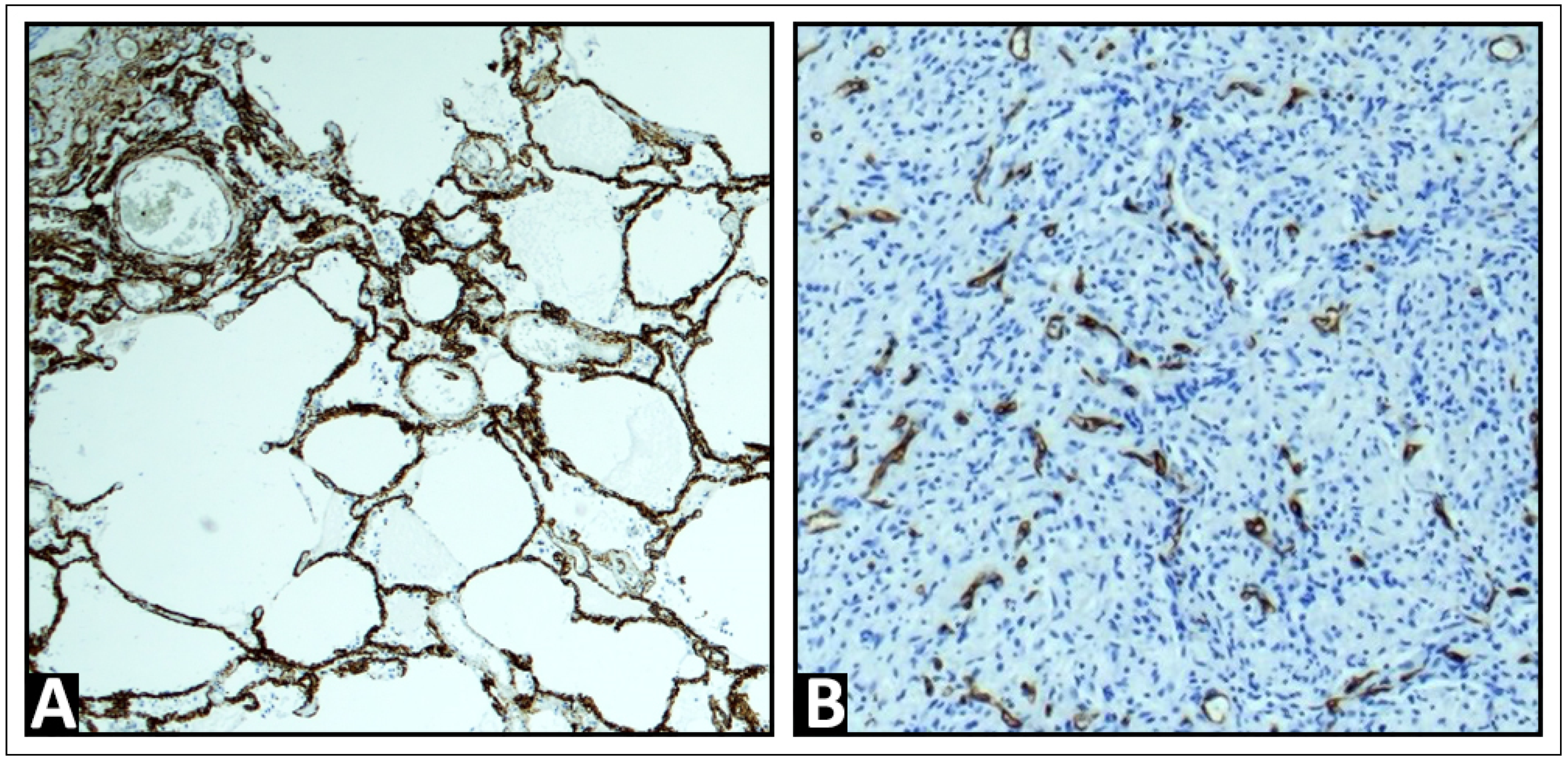
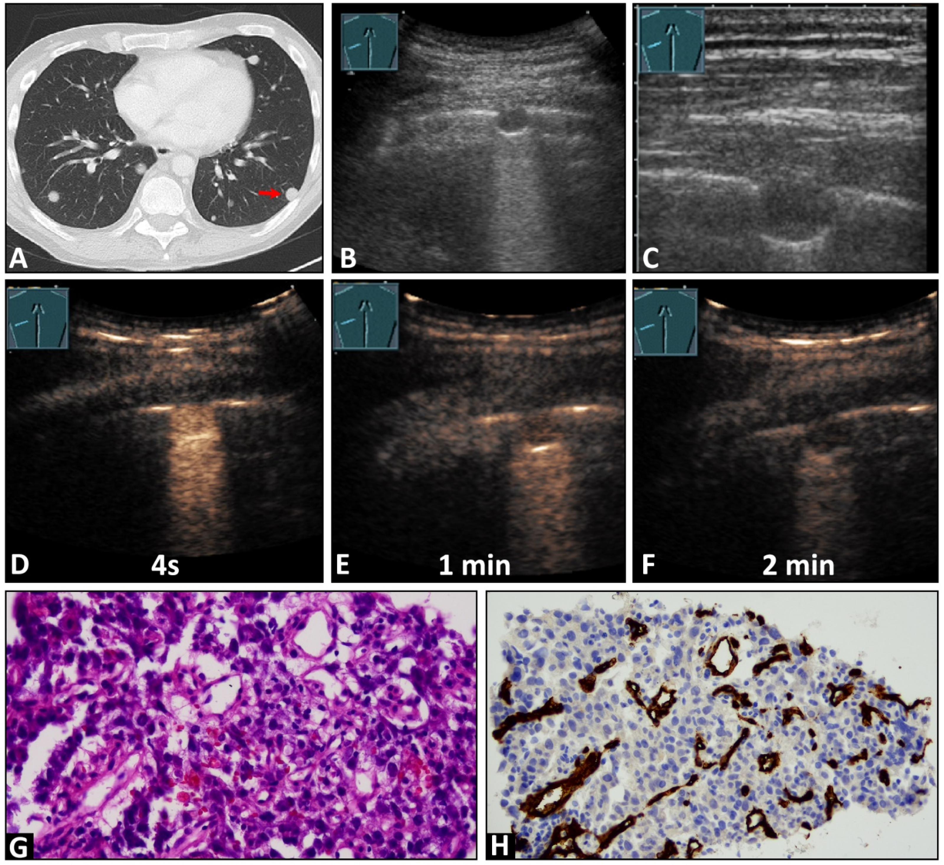
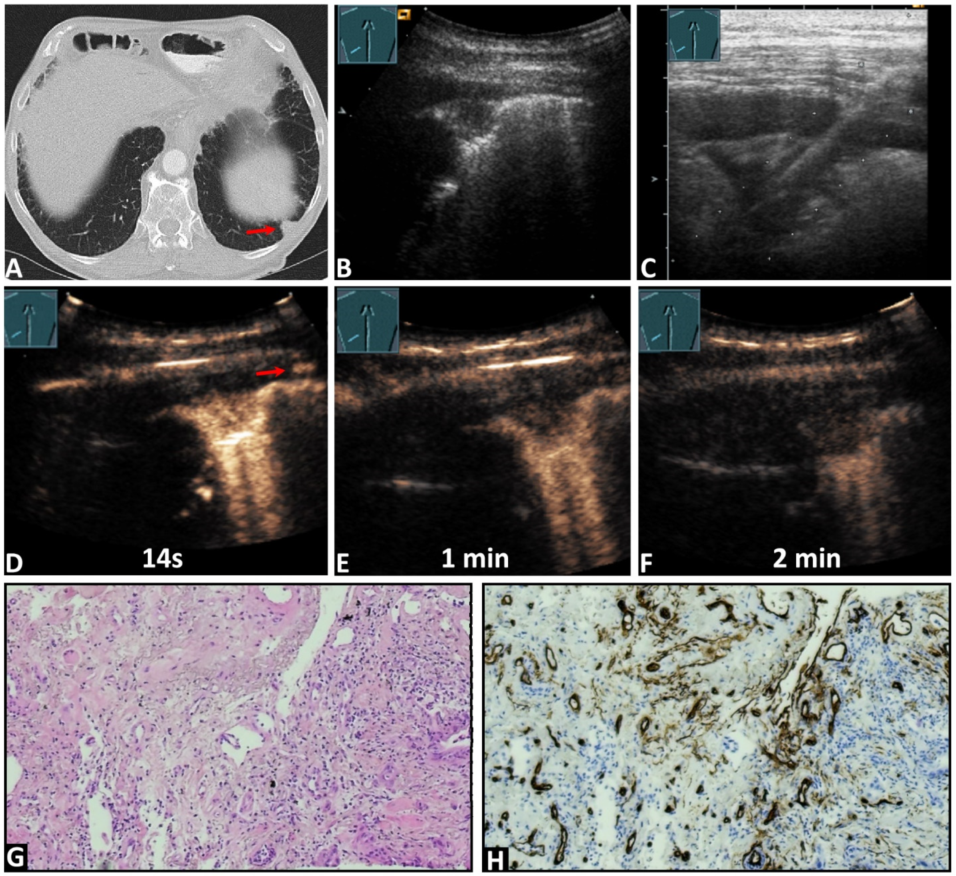
Purpose: Description of the perfusion of pulmonary metastasis by contrast-enhanced ultrasound (CEUS) and their correlation with vascularization patterns represented by immunohistochemical CD34 endothelial staining. Patients and methods: The data of 54 patients with histologic proven peripheral pulmonary metastasis, investigated between 2004 and 2023 by CEUS. These CEUS parameters were evaluated: time to enhancement (TE), categorized as early pulmonary-arterial (PA) or delayed bronchial-arterial (BA) patterns; extent of enhancement (EE), either marked or reduced; homogeneity of enhancement (HE), homogeneous or inhomogeneous; and decrease of enhancement (DE), rapid washout (<120 s) or late washout (≥120 s). Additionally, tissue samples in 45 cases (83.3%) were stained with CD34 antibody for immunohistochemical analysis. Results: In total, 4 lesions (7.4 %) exhibited PA enhancement, and 50 lesions (92.6%) demonstrated BA enhancement. Furthermore, 37 lesions (68.5%) showed marked enhancement, while 17 lesions (31.5%) exhibited reduced enhancement. The enhancement was homogeneous in 28 lesions (51.86%) and inhomogeneous in 26 lesions (48.14%). Additionally, 53 lesions (98.1%) displayed a rapid washout. A chaotic vascular pattern indicative of a bronchial arterial blood supply was identified in all cases (45/45, 100%), including all 4 lesions with PA enhancement. Conclusion: Pulmonary metastases in CEUS predominantly reveal bronchial arterial enhancement and a rapid washout. Regarding EE and HE, pulmonary metastases show heterogeneous perfusion patterns. A PA enhancement in CEUS does not exclude BA neoangiogenesis.