Review

18F-Fluoroethyl-L Tyrosine Positron Emission Tomography Radiomics in the Differentiation of Treatment-Related Changes from Disease Progression in Patients with Glioblastoma

by
Begoña Manzarbeitia-Arroba
1,
Marina Hodolic
2,
Robert Pichler
3,
Olga Osipova
3,
Ángel Maria Soriano-Castrejón
1 and
Ana María García-Vicente
1,*
1
Nuclear Medicine Department, University Hospital of Toledo, 45007 Toledo, Spain
2
Nuclear Medicine Department, Faculty of Medicine and Dentistry, Palacky University, 779 00 Olomouc, Czech Republic
3
Institute of Nuclear Medicine Kepler University Hospital—Neuromed Campus, 4020 Linz, Austria
*
Author to whom correspondence should be addressed.
Cancers 2024, 16(1), 195; https://doi.org/10.3390/cancers16010195
Submission received: 27 October 2023 / Revised: 10 December 2023 / Accepted: 23 December 2023 / Published: 30 December 2023
(This article belongs to the Special Issue FET PET Radiomics in Neuro-Oncology)

Abstract

:

Simple Summary

18F-Fluoroethyl-L tyrosine radiomics are useful in the differentiation of true progression from treatment-related changes in patients with glioblastoma, offering relevant complementary information with respect to the reference standard magnetic resonance imaging.

Abstract

The follow-up of glioma patients after therapeutic intervention remains a challenging topic, as therapy-related changes can emulate true progression in contrast-enhanced magnetic resonance imaging. 18F-fluoroethyl-tyrosine (18F-FET) is a radiopharmaceutical that accumulates in glioma cells due to an increased expression of L-amino acid transporters and, contrary to gadolinium, does not depend on blood–brain barrier disruption to reach tumoral cells. It has demonstrated a high diagnostic value in the differentiation of tumoral viability and pseudoprogression or any other therapy-related changes, especially when combining traditional visual analysis with modern radiomics. In this review, we aim to cover the potential role of 18F-FET positron emission tomography in everyday clinical practice when applied to the follow-up of patients after the first therapeutical intervention, early response evaluation, and the differential diagnosis between therapy-related changes and progression.

1. Introduction

Despite new molecular concepts introduced in the last decade, the diagnostic approach of patients with glioblastoma multiforme (GBM) is still conventional, mainly through the maintenance of contrast-enhanced magnetic resonance imaging (MRI) as the reference imaging technique for diagnosis, biopsy guidance, and treatment planning as well as treatment monitoring. So, despite the developments of new sequences for diffusion-weighted imaging (that assess the movement of water molecules and provide information on the cellularity of the tumor) and perfusion imaging (that provides information on tissue perfusion and permeability), no significant modifications in prognosis have been detected in the last decades. In addition, the inability of current treatments to achieve disease control explains why about 80% of GBM relapses occur within a 2 cm margin from the enhancing tumor location [1].
Positron emission tomography (PET) with labeled amino acids has been recommended to guide GBM resection and to delineate GBM extent by the Response Assessment in Neuro-Oncology (RANO) Working Group [2,3], based on a higher accuracy compared to other radiotracers and MRI. However, PET usually serves as a second-line diagnostic modality in neuro-oncology, performed only on the recommendation of a multidisciplinary tumor board in a minority of cases, mainly during the disease course, when patients present uncertain MRI features or an equivocal clinical course after or during treatment.
18F-fluoroethyl-L tyrosine (18F-FET) is an amino acid PET radiotracer that accumulates in glioma cells due to an increased expression of L-amino acid transporters (LAT) 1 and 2 as well as an increased tumor perfusion [4]. Moreover, a disruption of the blood–brain barrier (BBB) can also lead to a passive influx of the radiotracer to the tumoral tissue, although this is not a prerequisite for the intratumoral accumulation of 18F-FET [1].
In Europe, commercialized 18F-FET (IASOglio®), only authorized in France and Poland, includes the following indications of use in patients with glioma: characterization of brain lesions suggestive of glioma and selecting the best biopsy site in them, noninvasive grading of glioma, pretherapeutic delineation of viable glioma tissue, and after treatment, for the detection of viable tumor tissue in case of the suspicion of a persistent or recurrent glioma [5].
However, the differential diagnosis of pseudoprogression (PsP) from true progression (TP) is a big challenge in clinical practice for patients with a prior history of radical treatment and sometimes can only be made retrospectively based on an MRI follow-up. 18F-FET has demonstrated high efficacy in the diagnosis of PsP when compared to other PET radiopharmaceuticals and even to MRI [2,4].
Radiomics is the extraction of quantitative characteristics from medical images using advanced data-characterization algorithms. Radiomic features derived from PET have been described as an effective tool in the prediction of molecular tumor characteristics and patient outcome in several tumors [6]. Textural variables (first-, second-, and third-order parameters) that define the relations between voxel uptake and other measures of a more global spatial heterogeneity, such as the coefficient of variation that analyzes the spatial dispersion of gray intensity levels in voxels, have been defined. In addition, the SUV mean offers more integrated information of the radiotracer uptake in tumor voxels than the maximum standardized uptake value (SUVmax), and the SUVmean/SUVmax ratio can be described as an additional variable showing a direct relationship with homogeneity. On the other hand, tumor shape features, such as sphericity, seem to refer to the infiltrative tumor capacity of the tumor, so a less spherical lesion can be associated to a more aggressive molecular pattern. Moreover, other variables that inform about the molecular or metabolic tumor burden can be addressed by PET as the metabolic (or biological) tumor volume (MTV), that is, the volume of interest (VOI) after segmentation and total lesion activity (TLA) defined as the product of SUVmean by MTV.
In addition, radiomics extracted from 18F-FET PET have been found to add valuable diagnostic information in the prediction of the isocitrate dehydrogenase (IDH) enzyme genotype, the differential diagnosis between PsP and TP, and the prognosis prediction in newly diagnosed gliomas [7].
In the following sections, methodological, clinical aspects and controversies are described, referring to the most relevant and recent literature. Some illustrative cases are presented in Figure 1, Figure 2 and Figure 3.

2. Methodological Aspects

2.1. Imaging Acquisition and Analysis

The current protocol for 18F-FET PET/CT includes both static and dynamic acquisition [2]. A static scan is usually obtained 20 min after 18F-FET injection. However, a study by Verburg et al. [8] suggests performing 18F-FET PET 60–90 min after tracer injection in order to obtain a higher diagnostic accuracy. Dynamic acquisition is recommended and should be started right after the radiotracer injection, using 30–60 s frames within the first 10 min and 5 min frames within the next 40–50 min. The evolution of 18F-FET uptake as a VOI in a function of time can be presented as a time–activity curve (TAC).
A static study can be visually analyzed, although the obtention of the semiquantitative data of 18F-FET as the SUVmax or SUVmean is very informative. Based on a faint physiologic uptake in healthy brain, an increased radiotracer uptake can be described also using the tumor-to-background ratio (TBR). For that reason, a VOI delineating the tumor, and any area of healthy brain, preferable on the contralateral hemisphere, is selected. Afterwards, the SUVmax or SUVmean of tumor tissue is divided by the SUVmean of healthy brain.
The accurate tumor delineation of 18F-FET PET is crucial for interpretation. The usual threshold for the definition of the biological tumor volume (BTV) is 1.6 in static studies [9,10]. However, there is not a validated threshold and either standardization regarding the tumor and background VOIs. The largest reported FET-PET-credentialing study detected considerable variability in BTV delineation and image interpretation, even using the fixed value of 1.6. The discordances were explained by the manual adjustment of the segmented volume to remove any obvious non-tumor structures. However, despite these discrepancies, TBRmax and TBRmean were robust variables [11].
Several reports have shown that some radiomic features are sensitive to variations in several factors, including image acquisition, image reconstruction, and tumor segmentation, as well as test–retest imaging [11,12]. Zounek et al. [13] quantified the sensitivity of radiomic features derived from 18F-FET PET images of glioma patients with respect to variations in image reconstruction settings and tumor segmentation methods. The overall results showed that PET radiomic features, especially those shape-derived, were highly sensitive to the choice of image segmentation methods. On the other hand, Gutsche et al. [14] defined that first-order features extracted from the image histogram showed the highest repeatability. They also found a correlation between tumor volume and feature repeatability (for tumor volumes larger than 4 mL, more than 50% of features showed high repeatability), and a comparable repeatability was found between IDH-wild-type and IDH-mutant gliomas (repeatable features, 63% vs. 52%, respectively).

2.2. 18F-FET PET Interpretation

18F-FET scans should be fused with a CT or recent T2-weighted and T2/FLAIR-weighted MRI for a proper analysis of the lesions. The interpretation must be both visual and semiquantitative (such as TAC or TBR), as the sum of both analyses increases diagnostic accuracy and ensures intra-and inter-individual comparability.
The curve pattern of the dynamic acquisition is important, especially for lesion characterization. Three patterns have been described: 18F-FET uptake without an identifiable peak uptake (pattern I); 18F-FET uptake peaking at a midpoint (>20–40 min) followed by a plateau or a small descent (pattern II); and 18F-FET uptake peaking early (≤20 min) followed by a constant descent (pattern III) [15]. Pattern III is characteristic for high-grade gliomas (HGGs) or areas of malignant transformation in low-grade gliomas (LGGs) [16].
However, 18F-FET uptake is not always specific for neoplastic tissue, and pitfalls have been reported for brain abscesses, demyelinating processes, in the proximity of cerebral ischemic lesions, hematomas, and even in areas of reactive astrogliosis after high-dose brachytherapy [17,18]. In dexamethasone treatment, a possible increase in 18F-FET uptake by normal brain tissue has been described. In addition, a temporary increase in 18F-FET gyral uptake in the peri-ictal period of epilepsy may mimic a focal lesion [19].

3. Differentiation of Tumor Progression and Therapy-Related Changes

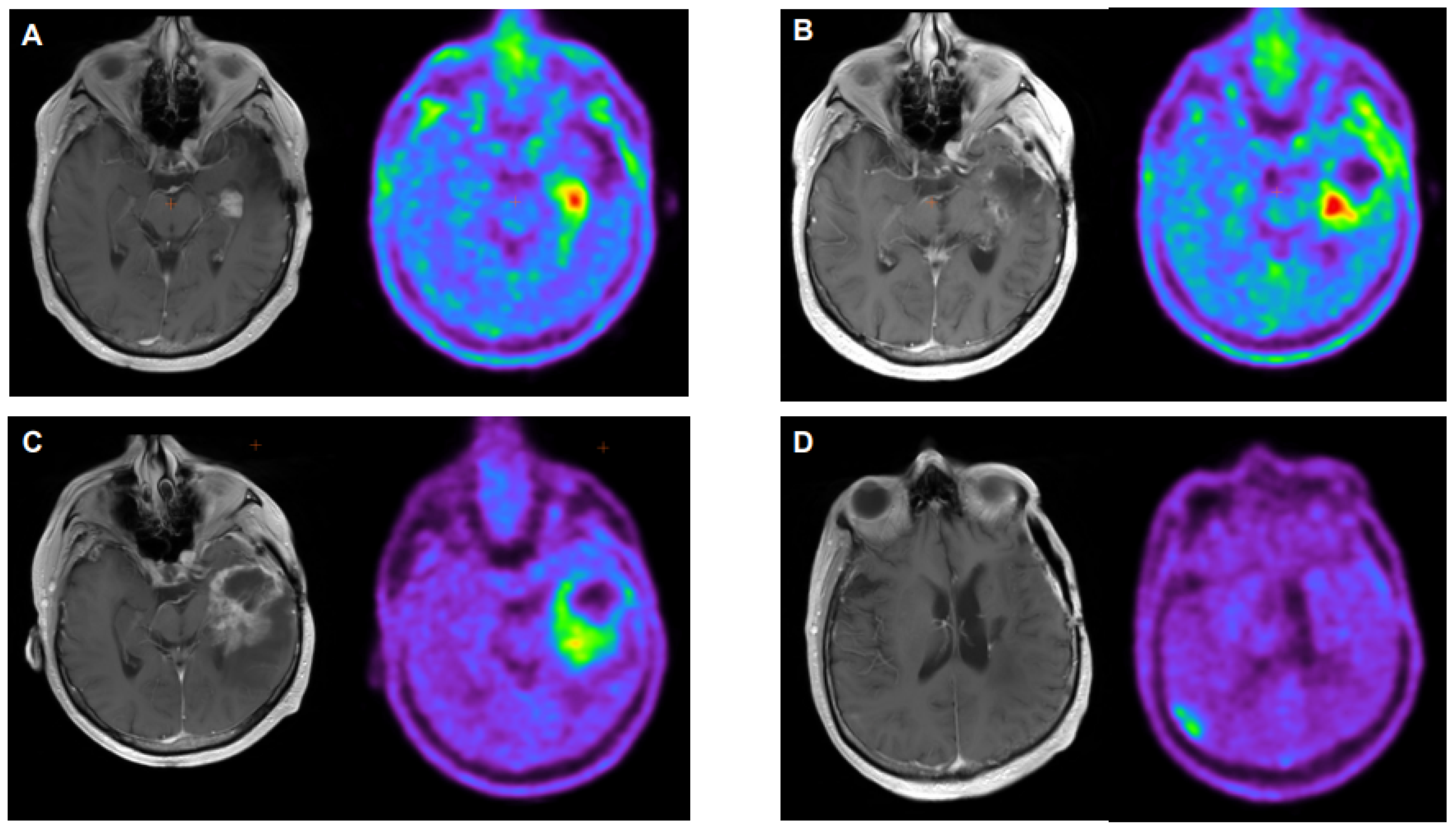
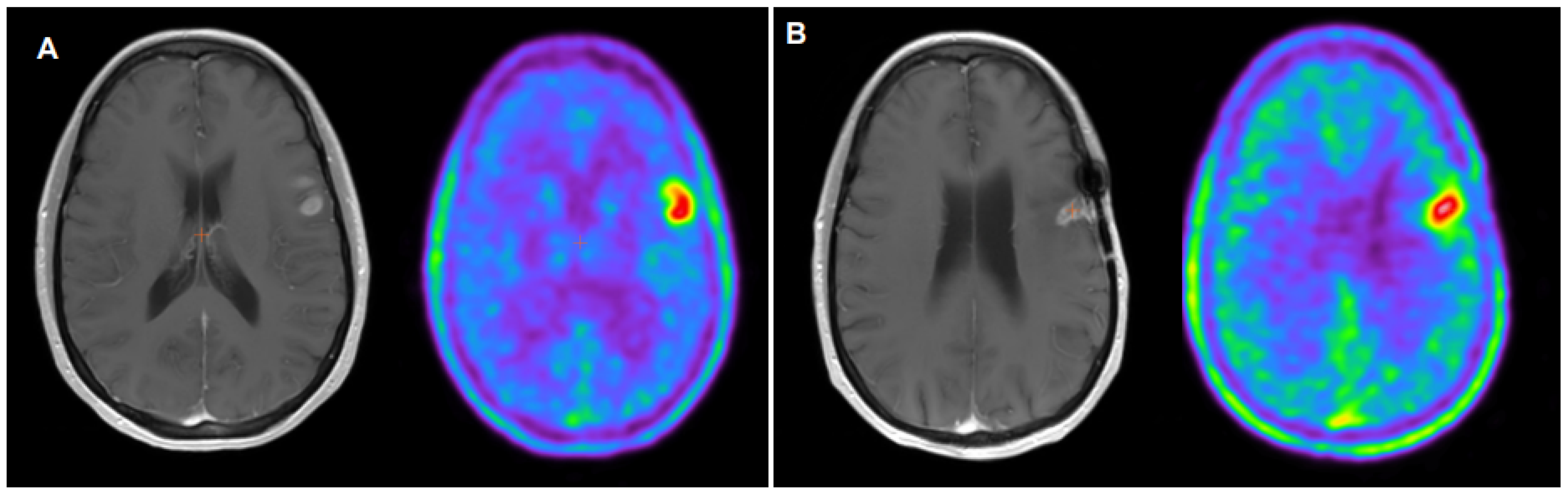
3.1. Pseudoprogression

In clinical practice, PsP is of considerable importance, representing approximately one-third of patients with GBM, usually within the first 12 weeks after radiochemotherapy with temozolomide (RCT-TMZ) [20,21]. PsP is a consequence of treatment-related local tissue reactions which comprise inflammation, oedema, and an increased permeability of the BBB, thereby resembling TP. PsP is associated with a better outcome, so if it is suspected, treatment should not be stopped, as it improves survival in patients with PsP [22,23,24]. Thus, a correct diagnosis is essential to avoid terminating an effective therapy. In patients with PsP, 18F-FET uptake is significantly lower than in patients with TP [25].
Magnetic resonance imaging using the RANO criteria, which relies heavily on findings such as contrast enhancement, T2-weighted and FLAIR changes, and further characterizes measurable versus non-measurable disease, is currently the reference standard in the diagnosis and follow-up of glioma patients. In the post-therapy setting, PsP appears as an enlarged area of contrast enhancement on MRI, and it cannot be effectively identified on a single MRI [24], being necessary sequential studies to demonstrate its subsequent improvement or stabilization without treatment [15]. Advancements in MRI, particularly perfusion-weighted imaging (PWI) using regional cerebral blood volume (rCBV) or diffusion-weighted imaging (DWI), can contribute to the differentiation of TP and therapy-related changes. However, treatment-related inflammation increases rCBV, and radiation necrosis may show as diffusion restriction due to oedema and leukocyte infiltrates in the transition zones [26,27].
In a Bayesian network meta-analysis including different PET radiotracers and MRI for recurrent glioma, 18F-FET showed the highest sensitivity, specificity, positive predictive value, and accuracy [4]. However, when a simultaneous imaging of 18F-FET PET and MRI obtained by a hybrid PET–MRI system comes into play, no significant differences seem to exist, probably explained by an increase in the interpretation confidence of both joined techniques [28,29].
The advantage of metabolic imaging using 18F-FET is explained because in tissue affected by post-therapeutic changes, LAT expression is normal or even downregulated (contrary to glioma tissue, which presents an increased number of LAT). However, an increased uptake of 18F-FET in PsP tissue can also appear, as a consequence of the passive influx of 18F-FET through a disrupted BBB, although with a lower intensity than TP [30].
In a recent meta-analysis, the pooled sensitivity and specificity of 18F-FET using a TBRmax of 1.9–2.3, for the differentiation of glioma recurrence from treatment-related changes, were 91% (95% CI, 74–97%) and 84% (95% CI, 69–93%), respectively [31]. Furthermore, an optimal cut-off value of 2.3 (accuracy 96%, area under curve of 0.94 ± 0.06; p < 0.001) has been described for identifying PsP, being significantly predictive of a longer overall survival (OS median 23 vs. 12 months; p = 0.046). Conversely, it seems advisable to assume late PsP when TBRmax is below 1.0. On the other hand, values between 1.0 and 2.3 should be interpreted with caution, as there is an overlap of final diagnoses [15].
In addition, dynamic 18F-FET PET may be helpful in the differentiation between TP and treatment-related changes or gliosis [32]. PsP, reactive gliosis, and benign tissues are associated with a TAC pattern of a steadily increasing 18F-FET uptake without an identifiable peak. In addition, a TAC pattern with an early or midpoint time to peak (TTP) uptake followed by a constant decline or plateau has been described as being highly specific of TP [10,25].
In current clinical practice, static 18F-FET PET parameters (TBRmean and TBRmax) seem to have a superior diagnostic performance in comparison to dynamic parameters in the detection of recurrence, although a combination of static and dynamic 18F-FET PET is the most valuable [33,34]. Additionally, other radiomics, informative of the heterogeneity of voxel radiotracer distribution, are associated with TP instead of PsP [5].
Regarding textural features on 18F-FET, small zone/low gray emphasis may be helpful in predicting the time to progression in patients with recurrent GBM undergoing re-irradiation [35].
IDH mutation status may also be taken into account when performing 18F-FET PET, as it has been described that the diagnostic accuracy when differentiating PsP and TP may depend on the mutation status, with higher accuracies observed in those with IDH-wild-type GBM [36,37].

3.2. Pseudoresponse

Antiangiogenic chemotherapeutics, such as bevacizumab (BEV), work by reducing the vascular permeability of immature blood vessels supplying the tumor and repairing the BBB. This results in a decrease in contrast enhancement in the peritumoral edema, regardless of the tumor’s sensitivity to the drug, which limits the diagnostic capability of MRI, hindering the differentiation between a good response to treatment from treatment-induced change [38].
18FET PET has demonstrated a better accuracy for disease monitoring than conventional MRI in BEV-treated patients with glioma [39,40]. Furthermore, adding 18F-FET PET to MRI can increase the rate of correct diagnoses by 41% [32]. However, the response criteria are not standardized in 18F-FET PET. Galldiks et al. [41] used the following criteria to identify responders in dynamic 18F-FET PET: (i) an increase in TTP from the baseline to the follow up PET scan greater than or equal to 10 min, (ii) a baseline TTP greater than 25 min, and (iii) a kinetic pattern of either an SUV peak at the end of the study or a TTP in the middle of the study followed by a plateau or slow descent. Other authors identified a decrease in 18F-FET PET/CT tumor volumes as a sign of BEV response [42].
However, with independence of the different criteria used, a response to treatment identified on 18F-FET PET seems to predict an increase in OS and progression free survival (PFS) of treated patients [41,42]. On the other hand, 18FET PET may detect tumor progression during antiangiogenic treatment earlier than MRI. Wirsching et al. [43] described that high 18FET-TBR of non-contrast-enhancing tumor portions during BEV therapy was associated with inferior OS on multivariate analysis (HR 5.97; CI, 1.16–30.8).

3.3. Early Response Evaluation

Previous studies have defined that MTV changes are predictive for the early identification of metabolic responders in patients undergoing adjuvant TMZ chemotherapy or lomustine-based chemotherapy in recurrent gliomas [14,44,45].
Suchorska et al. [46] proposed the following classification scheme for the evaluation of treatment response. In patients with non-contrast-enhancing glioma, a responsive disease was defined when a decrease in either BTV ≥ 25% and/or TBRmax ≥ 10% occurred; an increase in BTV ≥ 25% and/or TBRmax increase > 10% characterized a progressive disease, and minor changes ±25% for BTV and ±10% for TBRmax were regarded as a stable disease. Using the previous criteria, patients with a responsive disease had the longest time to treatment failure, while there was no significant difference between patients with a stable disease and progressive disease. On the contrary, a T2-volume-based assessment was not associated with outcome. Thus, in contrast to gadolinium-volume changes in MRI, changes in 18F-FET PET may be a valuable parameter to assess treatment response in GBM and predict survival [47].

3.4. Molecular Dependence

O6-methylguanine DNA methyltransferase (MGMT) gene promoter methylation and IDH mutation status allow for the stratification into biologically and prognostically distinct subgroups of glioma patients, PsP being more frequent [48]. Additionally, mutation status can be predicted by unifying dynamic and static features using TTP combined with the TBR max or TAC slope [49]. In patients with an MGMT methylated status, RCT-TMZ is effective in controlling GBM cells in the tumoral bed but not in controlling distant recurrence. In MGMT unmethylated patients, the rate of in-field recurrences is higher, which leaves room for a dose escalation with modern radiation techniques [50]. After the administration of TMZ concomitant with and adjuvant to RT in patients with GBM, the relapse pattern determined by 18F-FET PET/CT has been associated with the MGMT methylation status, with a higher PFS and ex-field recurrence rate in MGMT methylated patients, which might be a sign of better local control [51].
In addition, radiomic textural features seem to outperform the traditional 18F-FET parameters, such as TBRmax or TBRpeak, in the prediction of mutation status and for the differentiation of treatment-related changes from TP in GBM with enough confidence [52,53].

4. Comparison with Other PET Radiotracers

Many PET radiopharmaceuticals have been studied for brain tumor management. However, the consideration of a wide spectrum of primary brain tumors as a single pathological entity may prevent us from discovering the full potential of each radiopharmaceutical at our disposal. Taking these differences into account may allow us to reach more solid conclusions.

4.1. Fluorodeoxyglucose (FDG)

18F-FDG has a high physiological uptake in brain tissue and a relatively low uptake in some specific histological subtypes, such a lower-grade gliomas, which brings about a low lesion-to-background contrast. Delayed images can increase the target-to-background contrast, increasing the diagnostic accuracy. However, 18F-FET outperforms 18F-FDG in the differential diagnosis between radiation necrosis and recurrent disease in irradiated diffuse gliomas, with a sensitivity of 82–91% and 70–84%, respectively, and a specificity of 78–95% for FET and 70–88% for 18F-FDG [54].

4.2. Choline Analogues

The in vitro kinetic uptake of 18F-fluorocholine (18F-FCH) and 18F-FET is quite similar, only demonstrating a difference in uptake velocity: 18F-FET shows a more rapid initial uptake up to 40 min, and 18F-FCH shows a more progressive, continuous rise reaching a maximum activity plateau after 90 min [55]. However, the cellular transport mechanism of choline analogs differs with respect to amino acid tracers. First, 18F-FET uptake is mediated by LAT, whereas 18F-FCH uptake correlates with choline transporter-like 1 expression [8]. Second, 18F-FCH metabolism is very fast, with the parent fraction of the tracer decreasing in 15 min to 27% [8], compared to 87% in 120 min for 18F-FET, resulting in a better tracer availability [8,34]. Third, capillary density has also been described as influential in 18F-FCH uptake but not in 18F-FET uptake [56], and finally, the dependency of 18F-FET uptake on the breakdown of the BBB is less than that of 18F-FCH, with a high 18F-FET uptake also seen in tumor regions outside the area of contrast enhancement [57].
Moreover, the presence of reparative changes after therapy acts in a different manner in both radiotracers, with a higher affinity of 18F-FCH to inflammatory cells and an up-regulation of choline-kinase and/or choline transporters and a lower upregulation of LAT caused by radiation [56,58].

4.3. Other Amino Acid Tracers

Different tracer kinetics in malignant and benign tissues appear to be a special property of 18F-FET and have not been observed for other amino acid tracers such as 11C-methyl-L-methionine (11C-MET) or 3,4-dihydroxy-6-[18F]-fluoro-l-phenylalanine (18F-FDOPA) [59,60].
11C-MET and 18F-FDOPA provide comparable diagnostic information compared to 18F-FET for the differentiation between residual or recurrent tumor and treatment-related changes/PsP, as well as the delimitation of gliomas, although 18F-FET shows higher SUVs and a TBR mean for HGG than 18F-DOPA [61,62].

4.4. Prostate-Specific Membrane Antigen Ligands

Preliminary clinical results showed significantly high values of the in vivo uptake of prostate-specific membrane antigen (PSMA) ligands into HGG [63]. In a prospective study using 68Ga-PSMA-617 in a small sample of patients with recurrent glioma, 68Ga-PSMA-617 accumulated in large parts of the tumor that extended beyond the 18F-FET-avid margins, suggesting that PSMA ligands target a complementary biological process and might be a useful diagnostic marker to delineate parts of the recurrent tumor that are neoangiogenic but not extremely metabolically active yet. In addition, 68Ga-PSMA-617 had a higher TBR than 18F-FET, suggesting a better tumor specificity of the former [64].

5. Future Directions and Conclusions

Although MRI remains the standard of care, given the lack of alternatives available in current clinical practice, 18F-FET provides complementary information regarding the treatment response after chemoradiation, in terms of the prognostication of recurrence and patient survival. However, some pending issues deserve consideration.
The supplemental value of 18F-FET with respect to standard MRI must be addressed in prospective studies. So, we expect that the ongoing 18FET PET in glioblastoma (FIG) study [65], designed to determine the accuracy and management impact of 18F-FET PET in several clinical settings, reveals robust results.
The standardization and criteria harmonization in the imaging evaluation and interpretation of 18FET PET is mandatory, so credentialing studies are necessary to increase the expertise level across study sites.
Regarding radiomic features, before their implementation in clinical practice, it is essential to ensure their reproducibility and robustness. Thus, to properly translate radiomic models into clinical routine, they should be validated on large datasets that preferably include data from multiple centers.
The diagnostic impact of 18F-FET PET attending to the different molecular tumor profiles should be addressed based on their expected interdependency.
The clinical value of MTV must be demonstrated, in order to include volumetric assessment in consensus guidelines and recommendations.
Summarizing, regarding clinical decision making, 18F-FET radiomics may offer relevant information for patients with glioma, being useful in the differentiation of TP from therapy-related changes, overcoming the limitations of conventional MRI.

Author Contributions

Conceptualization, A.M.G.-V. and B.M.-A.; methodology, A.M.G.-V.; validation, M.H. and A.M.G.-V.; investigation, B.M.-A.; writing—original draft preparation, A.M.G.-V., B.M.-A., R.P. and O.O.; writing—review and editing, M-H. and A.M.G.-V.; visualization, R.P. and O.O.; supervision, R.P.; project administration, M-H, Á.M.S.-C. and A.M.G.-V. All authors have read and agreed to the published version of the manuscript.

Funding

This research received no external funding.

Data Availability Statement

Data sharing not applicable.

Conflicts of Interest

The authors declare no conflicts of interest.

References

  1. Langen, K.J.; Hamacher, K.; Weckesser, M.; Floeth, F.; Stoffels, G.; Bauer, D.; Conen, H.H.; Pauleit, D. O-(2-[18F] fluoroethyl)-L-tyrosine: Uptake mechanisms and clinical applications. Nucl. Med. Biol. 2006, 33, 287–294. [Google Scholar] [CrossRef] [PubMed]
  2. Law, I.; Albert, N.L.; Arbizu, J.; Boellaard, R.; Drzezga, A.; Galldiks, N.; la Fougère, C.; Langen, K.J.; Lopci, E.; Lowe, W.; et al. Joint EANM/EANO/RANO practice guidelines/SNMMI procedure standars for imagine of gliomas using PET with radiolabelled amino acids and [18F]FDG. Nucl. Med. Mol. Imaging 2019, 6, 540–557. [Google Scholar] [CrossRef] [PubMed]
  3. Stockhammer, F.; Plotkin, M.; Amthauer, H.; van Landeghem, F.K.H.; Woiciechowsky, C. Correlation of F-18-fluoro-ethyl-tyrosin uptake with vascular and cell density in non-contrast-enhancing gliomas. J. Neurooncol. 2008, 88, 205–210. [Google Scholar] [CrossRef] [PubMed]
  4. Xiaoxue, T.; Yinzhong, W.; Meng, Q.; Lu, X.; Junqiang, L. Diagnostic value of PET with different radiotracers and MRI for recurrent glioma: A Bayesian network meta-analysis. BMJ Open 2023, 13, e062555. [Google Scholar] [CrossRef] [PubMed]
  5. IASOglio Data-Sheet. Available online: https://curium-austria.com/products/iasoglio (accessed on 22 September 2023).
  6. Dai, J.; Wang, H.; Xu, Y.; Chen, X.; Tian, R. Clinical application of AI-based PET images in oncological patients. Semin. Cancer Biol. 2023, 91, 124–142. [Google Scholar] [CrossRef] [PubMed]
  7. Lohmann, P.; Elahmadaway, M.; Gutsche, R.; Werner, J.M.; Bauer, E.K.; Ceccon, G.; Kocher, M.; Lerche, C.W.; Rapp, M.; Fink, G.R.; et al. FET PET Radiomics for Differentiating Pseudoprogression from Early Tumor Progression in Glioma Patients Post-Chemotherapy. Cancers 2020, 12, 3835. [Google Scholar] [CrossRef]
  8. Verburg, N.; Koopman, T.; Yaqub, M.; Hoekstra, O.T.; Lammertsma, A.; Schwarte, L.A.; Barkhof, F.; Pouwels, P.J.W.; Heimans, J.J.; Reijneveld, J.C.; et al. Direct comparison of [11C] choline and [18F] FET PET to detect glioma infiltration: A diagnostic accuracy study in eight patients. EJNMMI Res. 2019, 9, 57. [Google Scholar] [CrossRef]
  9. Verger, A.; Filss, C.P.; Lohmann, P.; Stoffles, G.; Sabel, M.; Wittsack, H.J.; Kops, E.R.; Galldiks, N.; Fink, G.R.; Shah, N.J.; et al. Comparison of O-(2-18F-fluoroethyl)-l-tyrosine positron emission tomography and perfusion-weighted magnetic resonance imaging in the diagnosis of patients with progressive and recurrent glioma: A hybrid positron emission tomography/magnetic resonance study. World Neurosurg. 2018, 113, e727–e737. [Google Scholar] [CrossRef]
  10. Rausch, I.; Zitterl, A.; Berroterrán-Infante, N.; Rischka, L.; Prayer, D.; Fenchel, M.; Sareshgi, R.A.; Haug, A.R.; Hacker, M.; Beyer, R.; et al. Dynamic [18F]FET-PET/MRI using standard MRI-based attenuation correction methods. Eur. Radiol. 2019, 29, 4276–4285. [Google Scholar] [CrossRef]
  11. Barry, N.; Francis, R.J.; Evert, M.A.; Koh, E.-S.; Rowshanfarzad, P.; Hassan, G.M.; Kendrick, J.; Gan, H.K.; Lee, S.T.; Lau, E.; et al. Delineation and agreement of FET PET biological volumes in glioblastoma: Results of the nuclear medicine credentialing program from the prospective, multicentre trial evaluating FET PET In Glioblastoma (FIG) study—TROG 18. Eur. J. Nuc Med. Mol. Imaging 2023, 50, 3970–3981. [Google Scholar] [CrossRef]
  12. Gutsche, R.; Lowis, C.; Ziemons, K.; Kocher, M.; Ceccon, G.; Brambilla, C.R.; Shah, N.J.; Langen, K.J.; Galldiks, N.; Isensee, F.; et al. Automated Brain Tumor Detection and Segmentation for Treatment Response Assessment Using Amino Acid PET. J. Nucl. Med. 2023, 64, 1594–1602. [Google Scholar] [CrossRef] [PubMed]
  13. Zounek, A.J.; Albert, N.L.; Holzgreve, A.; Unterrainer, M.; Brosch-Lenz, J.; Lindner, S.; Bollenbacher, A.; Boening, G.; Rupprecht, R.; Brendel, M.; et al. Feasibility of radiomic feature harmonization for pooling of [18F] FET or [18F]GE-180 PET images of gliomas. Z. Med. Phys. 2023, 33, 91–102. [Google Scholar] [CrossRef] [PubMed]
  14. Gutsche, R.; Scheins, J.; Kocher, M.; Bousabarah, K.; Fink, G.R.; Shah, N.J.; Langen, K.-J.; Galldiks, N.; Lohmann, P. Evaluation of FET PET Radiomics Feature Repeatability in Glioma Patients. Cancers 2021, 13, 647. [Google Scholar] [CrossRef] [PubMed]
  15. Galldiks, N.; Dunkl, V.; Stoffels, G.; Hutterer, M.; Rapp, M.; Sabel, M.; Reifenberger, G.; Kebir, S.; Dorn, F.; Blau, T.; et al. Diagnosis of pseudoprogression in patients with glioblastoma using O-(2-[18F]fluoroethyl)-L-tyrosine PET. Eur. J. Nucl. Med. Mol. Imaging 2015, 42, 685–695. [Google Scholar] [CrossRef] [PubMed]
  16. Kunz, M.; Thon, N.; Eigenbrod, D.; Hartmann, C.; Egensperger, R.; Hermes, J.; Geisler, J.; la Fougère, C.; Lutz, J.; Linn, J. Hot spots in dynamic 18FET-PET delineate malignant tumor parts within suspected WHO grade II gliomas. Neuro Oncol. 2011, 13, 307–316. [Google Scholar] [CrossRef] [PubMed]
  17. Floeth, F.W.; Pauleit, D.; Sabel, M.; Reifenberger, G.; Stoffles, G.; Stummer, W.; Rommel, F.; Hamacher, K.; Langen, K.J. 18F-FET PET differentiation of ring-enhancing brain lesions. J. Nucl. Med. 2006, 47, 776–782. [Google Scholar] [PubMed]
  18. Hutterer, M.; Galldiks, N.; Hau, P.; Langen, K.J. Pitfalls in der [18F]FET-PET-Diagnostik von Hirntumoren. Nuklearmediziner 2015, 38, 295–303. [Google Scholar] [CrossRef]
  19. Suchorska, B.; Jansen, N.L.; Linn, J.; Kretzschmar, H.; Janssen, H.; Eigenbrod, S.; Simon, M.; Pöpperl, G.; Kreth, F.W.; la Fougere, C.; et al. Biological tumor volume in 18FET-PET before radiochemotherapy correlates with survival in GBM. Neurology 2015, 84, 710–719. [Google Scholar] [CrossRef]
  20. Young, R.J.; Gupta, A.; Shah, A.D.; Graber, J.J.; Zhang, Z.; Shi, W.; Holodny, A.I.; Omuro, A.M.P. Potential utility of conventional MRI signs in diagnosing pseudoprogression in glioblastoma. Neurology 2011, 76, 1918–1924. [Google Scholar] [CrossRef]
  21. Brandsma, D.; van den Bent, M.J. Pseudoprogression and pseudoresponse in the treatment of gliomas. Curr. Opin. Neurol. 2009, 22, 633–638. [Google Scholar] [CrossRef]
  22. Brandes, A.A.; Franceschi, E.; Tosoni, A.; Blatt, V.; Pession, A.; Tallini, G.; Bertorelle, R.; Bartolini, S.; Calbucci, F.; Andreoli, A.; et al. MGMT promoter methylation status can predict the incidence and outcome of pseudoprogression after concomitant radiochemotherapy in newly diagnosed glioblastoma patients. J. Clin. Oncol. 2008, 26, 2192–2197. [Google Scholar] [CrossRef] [PubMed]
  23. Hegi, M.E.; Diserens, A.C.; Gorila, T.; Hamou, M.F.; de Tribolet, N.; Weller, M.; Kros, J.M.; Hainfeller, J.A.; Mason, W.; Mariani, L.; et al. MGMT gene silencing and benefit from temozolomide in glioblastoma. N. Engl. J. Med. 2005, 352, 997–1003. [Google Scholar] [CrossRef] [PubMed]
  24. Abbasi, A.W.; Westerlaan, H.E.; Holtman, G.A.; Aden, K.M.; van Laar, P.J.; van der Hoorn, A. Incidence of tumor progression and pseudoprogression in high-grade gilomas: A systematic review and meta-analysis. Clin. Neuroradiol. 2018, 28, 401–411. [Google Scholar] [CrossRef] [PubMed]
  25. Kebir, S.; Fimmers, R.; Galldicks, N.; Schäfer, N.; Mack, F.; Schaub, C.; Stuplich, M.; Niessen, M.; Tzaridis, T.; Simon, M.; et al. Late Pseudoprogression in Glioblastoma: Diagnostic Value of Dynamic O-(2-[18F]fluoroethyl)-L-Tyrosine PET. Clin. Cancer Res. 2016, 22, 2190–2196. [Google Scholar] [CrossRef] [PubMed]
  26. Yoshii, Y. Pathological review of late cerebral radionecrosis. Brain Tumor Pathol. 2008, 25, 51–58. [Google Scholar] [CrossRef] [PubMed]
  27. Asao, C.; Korogi, Y.; Kitajima, M.; Hirai, T.; Baba, Y.; Makino, K.; Kochi, M.; Morishita, S.; Yamashita, Y. Diffusion-weighted imaging of radiation-induced brain injury for differentiation from tumor recurrence. Am. J. Neuroradiol. 2005, 26, 1455–1460. [Google Scholar]
  28. Henssen, D.; Leitjen, L.; Meijer, F.J.A.; van der Kolk, A.; Arens, A.I.J.; ter Laan, M.; Smeenk, R.J.; Gijtenbeek, A.; van de Giessen, E.M.; Tolboom, N.; et al. Head-To-Head Comparison of PET and Perfusion Weighted MRI Techniques to Distinguish Treatment Related Abnormalities from Tumor Progression in Glioma. Cancers 2023, 15, 2631. [Google Scholar] [CrossRef]
  29. Smith, N.J.; Deaton, T.K.; Territo, W.; Graner, B.; Gauger, A.; Snyder, S.E.; Schulte, M.L.; Green, M.A.; Hutchins, G.D.; Veronesi, M.C. Hybrid 18F-Fluoroethyltyrosine PET and MRI with Perfusion to Distinguish Disease Progression from Treatment-Related Change in Malignant Brain Tumors: The Quest to Beat the Toughest Cases. J. Nucl. Med. 2023, 64, 1087–1092. [Google Scholar] [CrossRef]
  30. Ouyang, Z.Q.; Zheng, G.R.; Duan, X.R.; Zhang, Z.R.; Ke, T.F.; Bao, S.S.; Yang, J.; He, B.; Liao, C.D. Diagnostic accuracy of glioma pseudoprogression identification with positron emission tomography imaging: A systematic review and meta-analysis. Quant. Imaging Med. Surg. 2023, 13, 4943–4959. [Google Scholar] [CrossRef]
  31. Singnurkar, A.; Poon, R.; Detsky, J. 18F-FET-PET imaging in high-grade gliomas and brain metastases: A systematic review and meta-analysis. Neuro Oncol. 2023, 161, 1–12. [Google Scholar] [CrossRef]
  32. Heinzel, A.; Müller, D.; Langen, K.J.; Blaum, M.; Verburg, F.A.; Mottaghy, F.M.; Galldiks, N. The use of O-(2-18F-fluoroethyl)-L tyrosine PET for treatment management of bevacizumab and irinotecan in patients with recurrent high-grade glioma: A cost-effectiveness analysis. J. Nucl. Med. 2013, 54, 1217–1222. [Google Scholar] [CrossRef] [PubMed]
  33. Pyka, T.; Hiob, D.; Preibisch, C.; Gempt, J.; Wiestler, B.; Schlegel, J.; Straube, C.; Zimmer, C. Diagnosis of glioma recurrence using multiparametric dynamic 18F-fluoroethyl-tyrosine PET-MRI. Eur. J. Radiol. 2018, 103, 32–37. [Google Scholar] [CrossRef] [PubMed]
  34. Werner, J.M.; Stoffels, G.; Lichtenstein, T.; Borggrefe, J.; Lohmann, P.; Ceccon, G.; Shah, N.J.; Fink, G.R.; Langen, K.J.; Kabbasch, C.; et al. Differentiation of treatment-related changes from tumor progression: A direct comparison between dynamic FET PET and ADC values obtained from DWI MRI. Eur. J. Nucl. Med. Mol. Imaging 2019, 46, 1889–1901. [Google Scholar] [CrossRef] [PubMed]
  35. Carles, M.; Popp, I.; Stark, M.M.; Mix, M.; Urbach, H.; Schimek-Jasch, T.; Eckert, F.; Niyazi, M.; Baltas, D.; Grosu, A.L. FETPET radiomics in recurrent glioblastoma: Prognostic value for outcome after reirradiation? Radiat. Oncol. 2021, 16, 46. [Google Scholar] [CrossRef] [PubMed]
  36. Maurer, G.D.; Brucker, D.P.; Stoffels, G.; Filipski, K.; Filss, C.P.; Mottaghy, F.M.; Galldiks, N.; Steinbach, J.P.; Hattingen, E.; Langen, K.J. 18F-FET PET Imaging in Differentiating Glioma Progression from Treatment-Related Changes: A Single-Center Experience. J. Nucl. Med. 2020, 61, 505–511. [Google Scholar] [CrossRef] [PubMed]
  37. Kebir, S.; Schmidt, T.; Weber, M.; Lazaridis, L.; Galldiks, N.; Langen, K.J.; Kleinschnitz, C.; Hattingen, E.; Herrlinger, U.; Lohmann, P.; et al. A Preliminary Study on Machine Learning-Based Evaluation of Static and Dynamic FET-PET for the Detection of Pseudoprogression in Patients with IDH-Wildtype Glioblastoma. Cancers 2020, 12, 3080. [Google Scholar] [CrossRef] [PubMed]
  38. Hughes, K.L.; O’Neal, C.M.; Andrews, B.J.; Westrup, A.M.; Battiste, J.D.; Glenn, C.A. A systematic review of the utility of amino acid PET in assessing treatment response to bevacizumab in recurrent high-grade glioma. Neurooncol. Adv. 2021, 3, vdab003. [Google Scholar] [CrossRef]
  39. George, E.; Kijewski, M.F.; Dubey, S.; Belanger, A.P.; Reardon, D.A.; Wen, P.Y.; Kesari, S.; Horky, L.; Park, M.-A.; Huang, R.Y. Voxel-wise analysis of fluoroethyltyrosine PET and MRI in the assessment of recurrent glioblastoma during antiangiogenic therapy. Am. J. Roentgenol. 2018, 211, 1342–1347. [Google Scholar] [CrossRef]
  40. Galldiks, N.; Dunkl, V.; Ceccon, G.; Tscherpel, C.; Stoffels, G.; Law, I.; Henriksen, O.M.; Muhic, A.; Poulsen, H.S.; Steger, J.; et al. Early treatment response evaluation using FET PET compared to MRI in glioblastoma patients at first progression treated with bevacizumab plus lomustine. Eur. J. Nucl. Med. Mol. Imaging 2018, 45, 2377–2386. [Google Scholar] [CrossRef]
  41. Galldiks, N.; Rapp, M.; Stoffels, G.; Fink, G.R.; Shah, N.J.; Coenen, H.H.; Sabel, M.; Langen, K.J. Response assessment of bevacizumab in patients with recurrent malignant glioma using [18F]Fluoroethyl-L-tyrosine PET in comparison to MRI. Eur. J. Nucl. Med. Mol. Imaging 2013, 40, 22–33. [Google Scholar] [CrossRef]
  42. Hutterer, M.; Nowosielski, M.; Putzer, D.; Waitz, D.; Tinkhauser, G.; Kostron, H.; Muigg, A.; Virgolini, I.J.; Staffen, W.; Trika, E.; et al. O-(2-18F-fluoroethyl)-L tyrosine PET predicts failure of antiangiogenic treatment in patients with recurrent high-grade glioma. J. Nucl. Med. 2011, 52, 856–864. [Google Scholar] [CrossRef]
  43. Wirsching, H.G.; Roelcke, U.; Weller, J.; Hundsberger, T.; Hottinger, A.F.; von Moos, R.; Caparrotti, F.; Conen, K.; Remonda, L.; Roth, P.; et al. MRI and 18FET-PET Predict Survival Benefit from Bevacizumab Plus Radiotherapy in Patients with Isocitrate Dehydrogenase Wild-type Glioblastoma: Results from the Randomized ARTE trial. Clin. Cancer Res. 2021, 27, 179–188. [Google Scholar] [CrossRef] [PubMed]
  44. Ceccon, G.; Lohmann, P.; Werner, J.M.; Tscherpel, C.; Dunkl, V.; Stoffels, G.; Rosen, J.; Rapp, M.; Sabel, M.; Herrlinger, U.; et al. Early treatment response assessment using 18F-FET PET compared with contrast-enhanced MRI in glioma patients after adjuvant temozolomide chemotherapy. J. Nucl. Med. 2021, 62, 918–925. [Google Scholar] [CrossRef] [PubMed]
  45. Wollring, M.M.; Werner, J.M.; Bauer, E.K.; Tscherpel, C.; Ceccon, G.S.; Lohmann, P.; Stoffels, G.; Kabbasch, C.; Goldbrunner, R.; Fink, G.R.; et al. Prediction of response to lomustinebased chemotherapy in glioma patients at recurrence using MRI and FET PET. Neuro Oncol. 2023, 25, 984–994. [Google Scholar] [CrossRef] [PubMed]
  46. Suchorska, B.; Unterrainer, M.; Biczok, A.; Sosnova, M.; Forbrig, R.; Bartenstein, P.; Tonn, J.C.; Albert, N.L.; Kreth, F.W. 18F-FET-PET as a biomarker for therapy response in non-contrast enhancing glioma following chemotherapy. J. Neurooncol. 2018, 139, 721–730. [Google Scholar] [CrossRef] [PubMed]
  47. Galldiks, N.; Langen, K.J.; Holy, R.; Pinkawa, M.; Stoffels, G.; Nolte, K.W.; Kaiser, H.J.; Filss, C.P.; Fink, G.R.; Coenen, H.H.; et al. Assessment of treatment response in patients with glioblastoma using O-(2-18F-fluoroethyl)-L-tyrosine PET in comparison to MRI. J. Nucl. Med. 2012, 53, 1048–1057. [Google Scholar] [CrossRef]
  48. Weller, M.; Felsberg, J.; Hartmann, C.; Berger, H.; Steinbach, J.P.; Schramm, J.; Westphal, M.; Schackert, G.; Simon, M.; Tonn, J.C.; et al. Molecular predictors of progressionfree and overall survival in patients with newly diagnosed glioblastoma: A prospective translational study of the German Glioma Network. J. Clin. Oncol. 2009, 27, 5743–5750. [Google Scholar] [CrossRef] [PubMed]
  49. Verger, A.; Stoffels, G.; Bauer, E.K.; Lohmann, P.; Blau, T.; Fink, G.R.; Neumaier, B.; Shah, N.J.; Langen, K.J.; Galldiks, N. Static and dynamic 18F–FET PET for the characterization of gliomas defined by IDH and 1p/19q status. Eur. J. Nucl. Med. Mol. Imaging 2018, 45, 443–451. [Google Scholar] [CrossRef]
  50. Amelio, D.; Lorentini, S.; Schwarz, M.; Amichetti, M. Intensity-modulated radiation therapy in newly diagnosed glioblastoma: A systematic review on clinical and technical issues. Radiother. Oncol. 2010, 97, 361–369. [Google Scholar] [CrossRef]
  51. Niyazi, M.; Schnell, O.; Suchorska, B.; Schwarz, S.B.; Ganswindt, U.; Geisler, J.; Bartenstein, P.; Kreth, F.W.; Tonn, J.C.; Eigenbrod, S.; et al. FET-PET assessed recurrence pattern after radio-chemotherapy in newly diagnosed patients with glioblastoma is influenced by MGMT methylation status. Radiother. Oncol. 2012, 104, 78–82. [Google Scholar] [CrossRef]
  52. Lohmann, P.; Lerche, C.; Bauer, E.K.; Steger, J.; Stoffels, G.; Blau, T.; Dunkl, V.; Kocher, V.; Viswanathan, S.; Filss, C.P.; et al. Predicting IDH genotype in gliomas using FET PET radiomics. Sci. Rep. 2018, 8, 13328. [Google Scholar] [CrossRef]
  53. Müller, M.; Winz, O.; Gutsche, R.; Leijenaar, R.T.H.; Kocher, M.; Lerche, C.; Filss, C.P.; Stoffels, F.; Steidl, E.; Hattingen, E.; et al. Static FET PET radiomics for the differentiation of treatment-related changes from glioma progression. J. Neurooncol. 2022, 159, 519–529. [Google Scholar] [CrossRef] [PubMed]
  54. Ninatti, G.; Pini, C.; Gelardi, F.; Sollini, M.; Chiti, A. The Role of PET Imaging in the Differential Diagnosis between Radiation Necrosis and Recurrent Disease in Irradiated Adult-Type Diffuse Gliomas: A Systematic Review. Cancers 2023, 15, 364. [Google Scholar] [CrossRef] [PubMed]
  55. Bansal, A.; Shuyan, W.; Hara, T.; Harris, R.B.; Degrado, T.R. Biodisposition and metabolism of [18F]fluorocholine in 9L glioma cells and 9L glioma-bearing fisher rats. Eur. J. Nucl. Med. Mol. Imaging 2008, 35, 1192–1203. [Google Scholar] [CrossRef] [PubMed]
  56. Spaeth, N.; Wyss, M.T.; Weber, B.; Scheidegger, S.; Lutz, A.; Verwey, J.; Radovanovic, I.; Pahnke, J.; Wild, D.; Wester, G.; et al. Uptake of 18Ffluorocholine, 18F-fluoroethyl-L- tyrosine, and 18F-FDG in acute cerebral radiation injury in the rat: Implications for separation of radiation necrosis from tumor recurrence. J. Nucl. Med. 2004, 45, 1931–1938. [Google Scholar] [PubMed]
  57. Pasi, F.; Persico, M.G.; Buroni, F.E.; Aprile, C.; Hodolic, M.; Corbella, F.; Nano, R.; Facoetti, A.; Lodola, L. 18F-FET and 18F-FCH uptake in human glioblastoma T98G cell lines after Irradiation with Photons or Carbon Ions. Contrast Media Mol. Imaging 2017, 2017, 6491674. [Google Scholar] [CrossRef] [PubMed]
  58. Taguchi, C.; Inazu, M.; Saiki, I.; Yara, M.; Hara, N.; Yamanaka, T.; Uchino, H. Functional analysis of [methyl-3H]choline uptake in glioblastoma cells: Influence of anti-cancer and central nervous system drugs. Biochem. Pharmacol. 2014, 88, 303–312. [Google Scholar] [CrossRef]
  59. Moulin-Romsée, G.; D’Hondt, E.; de Groot, T.; Goffin, J.; Scott, R.; Mortelmans, L.; Menten, J.; Bormans, G.; Van Laere, K. Non-invasive grading of brain tumors using dynamic amino acid PET imaging: Does it work for 11Cmethionine? Eur. J. Nucl. Med. Mol. Imaging 2007, 34, 2082–2087. [Google Scholar] [CrossRef]
  60. Kratochwil, C.; Combs, S.E.; Leotta, K.; Afshar-Oromieh, A.; Rieken, S.; Debus, J.; Haberkorn, U.; Giesel, F.L. Intra-individual comparison of 18F-FET and 18F-DOPA in PET imaging of recurrent brain tumors. Neuro Oncol. 2014, 16, 434–440. [Google Scholar] [CrossRef]
  61. Grosu, A.L.; Astner, S.T.; Riedel, E.; Nieder, C.; Wiedenmann, N.; Heinemann, F.; Schwaiger, M.; Molls, M.; Wester, H.J.; Weber, W.A. An interindividual comparison of O-(2-[18F]fluoroethyl)-L-tyrosine (FET)- and L-[methyl-11C]methionine (MET)-PET in patients with brain gliomas and metastases. Int. J. Radiat. Oncol. Biol. Phys. 2011, 81, 1049–1058. [Google Scholar] [CrossRef]
  62. Lapa, C.; Linsemann, T.; Monoranu, C.M.; Samnick, S.; Buck, A.K.; Bluemel, C.; Chernihiv, J.; Kessler, A.F.; Hmola, G.A.; Ernestus, R.I.; et al. Comparison of the amino acid tracers 18F-FET and 18F-dopa in high-grade glioma patients. J. Nuclear Med. 2014, 55, 1611. [Google Scholar] [CrossRef] [PubMed]
  63. Wang, S.; Wang, J.; Liu, D.; Yang, D. The Value of 68Ga-PSMA-617 PET/CT in Differential Diagnosis between Low-Grade and High-Grade Gliomas. J. Nucl. Med. 2018, 59, 146. [Google Scholar]
  64. Brighi, C.; Puttick, S.; Woods, A.; Keall, P.; Tooney, P.A.; Waddington, D.E.J.; Sproule, V.; Rose, S.; Fay, M. Comparison between [68Ga]Ga-PSMA-617 and [18F]FET PET as Imaging Biomarkers in Adult Recurrent Glioblastoma. Int. J. Mol. Sci. 2023, 24, 6208. [Google Scholar] [CrossRef] [PubMed]
  65. Koh, E.-W.; Gan, K.H.; Senko, C.; Francis, R.J.; Ebert, M.; Lee, S.T.; Lau, E.; Khasraw, M.; Nowak, A.K.; Bailey, D.L.; et al. [18F]-fluoroethyl-L-tyrosine (FET) in glioblastoma (FIG) TROG 18.06 study: Protocol for a prospective, multicentre PET/CT trial. BMJ Open 2023, 13, e071327. [Google Scholar] [CrossRef]
Figure 1. 45-year-old female diagnosed with an IDH1-2 wild-type GBM (WHO grade 4, TERT mutation, EGFR amplification, promotor MGMT unmethylated). Preoperative MRI and 18F-FET PET (A) showed an increased 18F-FET uptake in a contrast-enhanced left frontal lesion on MRI with a TBR max and mean of 3.2 and a 2.4, respectively. A complete surgical resection was performed followed by Stupp protocol and adjuvant temozolomide. Thirteen months later, 18F-FET PET (B) was performed to rule out tumor relapse after a previous suspicious MRI, showing an increased radiotracer uptake in the left frontal lobe, close to the postsurgical changes, with a TBRmax of 1.9 and a TBRmean of 1.7, consistent with recurrent disease. The patient denied any specific tumor therapy, and palliative corticoid therapy was initiated.
Cancers 16 00195 g001
Figure 2. 45-year-old male diagnosed with a grade II glioma by stereotactic biopsy. Preoperative MRI (FLAIR) and 18F-FET PET (A) showed an increased radiotracer uptake in a non-contrast-enhanced lesion on the left temporal lobe, with a TBRmax and mean of 2.4 and 2.0, respectively. Therapy with temozolomide and additional radiotherapy was administered. (B) Control MRI showed a stable disease with signs of a partial response to 18F-FET PET (TBRmax and TBRmean of 1.0 and 0.9, respectively). Following MRI was suspicious of progression and a new 18F-FET PET was performed (C), showing an increased 18F-FET uptake in the left temporal lobe, more intense and broader than in any previous study (TBR max and TBR mean of 3.8 and 3.1, respectively) and consistent with disease progression. A second-line therapy with temozolomide was scheduled; however, a progressive disease was detected on the last follow-up MRI, considered a probable transformation to a high-grade glioma based on disease evolution.
Cancers 16 00195 g002
Figure 3. 63-year-old male diagnosed with a left temporal glioblastoma (WHO grade IV, IDH wild type, no EGFR amplification, MGMT promoter unmethylated). The patient was treated with a tumor resection and posterior chemoradiotherapy with adjuvant temozolomide. Three years after the initial diagnosis, and with a suspicion-of-recurrence MRI, the patient underwent 18F-FET PET, showing an increased radiotracer uptake in the left temporal lobe (TBRmax and TBRmean of 2.5 and 2.3, respectively), suspicious of neoplastic tissue (A). Surgery was performed, confirming the relapse. A year later, an MRI showed signs of tumor regrowth but partially undistinguishable from radiation necrosis. 18F-FET PET (B) showed uptake in the left temporal lobe, adjacent to the surgical cavity (TBRmax and TBRmean of 2.6 and 2.3, respectively), consistent with disease progression. The patient underwent additional radiotherapy. Six months later, tumor regrowth appeared in the follow-up MRI with doubts of pseudoprogression, whereas 18F-FET PET showed signs of progressive disease with an increased ring-like shape uptake (TBRmax and TBRmean 3.4 of 2.8, respectively) in the left temporal lobe (C) and a new focal uptake (TBRmax and TBRmean of 2.5 and 2.1, respectively) on the contralateral parietal lobe (D), not observed in a contrast-enhanced MRI. A new therapeutic line with bevacizumab was initiated.
Cancers 16 00195 g003
Disclaimer/Publisher’s Note: The statements, opinions and data contained in all publications are solely those of the individual author(s) and contributor(s) and not of MDPI and/or the editor(s). MDPI and/or the editor(s) disclaim responsibility for any injury to people or property resulting from any ideas, methods, instructions or products referred to in the content.

