Review

Two Faces of Glutaminase GLS2 in Carcinogenesis

Department of Neurotoxicology, Mossakowski Medical Research Institute, Polish Academy of Sciences, 5 Pawińskiego Str., 02-106 Warsaw, Poland
*
Author to whom correspondence should be addressed.
Cancers 2023, 15(23), 5566; https://doi.org/10.3390/cancers15235566
Submission received: 31 October 2023 / Revised: 17 November 2023 / Accepted: 20 November 2023 / Published: 24 November 2023
(This article belongs to the Special Issue Glutamine Metabolism in the Onset and Progression of Tumorigenesis)

Abstract

:

Simple Summary

Glutaminase GLS2 is an enzyme that metabolizes glutamine, one of the main sources of energy and building blocks in tumor cells. In some types of cancer, this enzyme is overexpressed and fuels malignancy; however, there are some types of cancer in which GLS2 plays a role opposite to that of a tumor suppressor. The mechanisms behind the GLS2-mediated inhibition of tumorigenesis are complex and context-dependent. In this review, we report both the pro- and anti-tumorigenic properties of this protein.

Abstract

In rapidly proliferating cancer cells, glutamine is a major source of energy and building blocks. Increased glutamine uptake and enhanced glutaminolysis are key metabolic features of many cancers. Glutamine is metabolized by glutaminase (GA), which is encoded by two genes: GLS and GLS2. In contrast to isoforms arising from the GLS gene, which clearly act as oncoproteins, the role of GLS2 products in tumorigenesis is far from well understood. While in some cancer types GLS2 is overexpressed and drives cancer development, in some other types it is downregulated and behaves as a tumor suppressor gene. In this review, we describe the essential functions and regulatory mechanisms of human GLS2 and the cellular compartments in which GLS2 has been localized. Furthermore, we present the context-dependent oncogenic and tumor-suppressor properties of GLS2, and delve into the mechanisms underlying these phenomena.

Graphical Abstract

1. Introduction

Enhanced growth and the proliferation of cancer cells lead to increased demand for energy and biomacromolecules. To meet this requirement, cancer cells rewire their metabolism. One of the classical examples of the metabolic pathway being altered by cancer is the Warburg effect, defined as enhanced glucose uptake and lactate production, carried out regardless of oxygen availability [1]. In addition to glycolysis, cancer cells metabolize large amounts of glutamine (Gln) to sustain proliferation and grow unhindered. Gln is catabolized to glutamate (Glu) and further converted to α-ketoglutarate (α-KG), which enters the tricarboxylic acid (TCA) cycle to generate energy and provide precursors for proteins, nucleotides, and lipids. Gln-derived Glu is used for glutathione (GSH) synthesis to maintain redox homeostasis. Additionally, Gln is involved in the regulation of cell signaling. The aforementioned pleiotropic role of Gln in carcinogenesis has been exhaustively addressed in several reviews [2,3]. A growing body of evidence shows that Gln deprivation inhibits the survival of cancer cells [4,5,6,7,8,9] and indicates that Gln addiction is an important therapeutic target in a wide variety of cancer types.
Gln is hydrolyzed to Glu and ammonia by glutaminase (GA, EC 3.5.1.2). Human GAs are encoded by two genes, GLS and GLS2, located in chromosome 2 and chromosome 12, respectively [10]. The GLS gene codes for kidney-type GA (KGA) and glutaminase C (GAC) originate through alternative splicing of a single pre-mRNA [11]. GAC is identical to KGA except for the C-terminal region encoded by exon 15, which is missing in KGA (Figure 1) [12,13,14]. Of the two isozymes, GAC shows higher enzymatic activity, which is most likely related to its unique C terminus [15]. In fact, overexpression of the GLS gene has been documented in several cancer types of different origin and, in most cases, GAC is the predominant GLS isoform [15,16,17,18,19,20,21,22,23,24]; moreover, the silencing of GLS expression reversed the malignant phenotype of cancer cells both in vitro and in vivo, proving the important role GLS products play in promoting tumorigenesis [25,26,27,28,29,30]. These findings clearly indicate that GLS proteins are attractive therapeutic targets. Indeed, several GLS inhibitors have demonstrated efficacy in preclinical cancer models and some of them have entered clinical trials [31,32].
In contrast to GLS isoforms, which clearly act as oncoproteins, the role of GLS2 products in tumorigenesis is far from well understood. While in some cancer types GLS2 is overexpressed and drives cancer development, in some other types it is downregulated and behaves as a tumor suppressor gene. In this article, we briefly discuss the essential functions and regulatory mechanisms of human GLS2. Furthermore, we review its tumor promoting and suppressing properties, and delve into the mechanisms underlying these phenomena.

2. The GLS2 Gene and Its Products

The two Gls2 products, both protein and cDNA, were first isolated from rat liver [33,34,35], which is why this glutaminase was named liver-type glutaminase (LGA). Compared to the enzymes encoded by the Gls gene, rat hepatic LGA protein has a higher Michaelis constant (Km) for Gln and is fully activated at lower concentrations of inorganic phosphate. Furthermore, it is not inhibited by Glu and is activated by ammonia [33,34].
The human GLS2 gene spans more than 18.1 kb and consists of 18 exons [10,36]. To date, two transcripts arising from this gene have been identified. The canonical transcript contains all 18 exons and has an 1806-base open reading frame (ORF) encoding a 602-residue protein. It was cloned for the first time from human breast cancer cells [37] and later termed GAB in order to distinguish it from the LGA isoform encoded by a short transcript of the same gene [38]. The LGA transcript has a 1698-base ORF encoding a putative protein of 565 amino acids; it arises from an alternative transcription initiation occurring at an alternative promoter located in intron 1 (Figure 1).
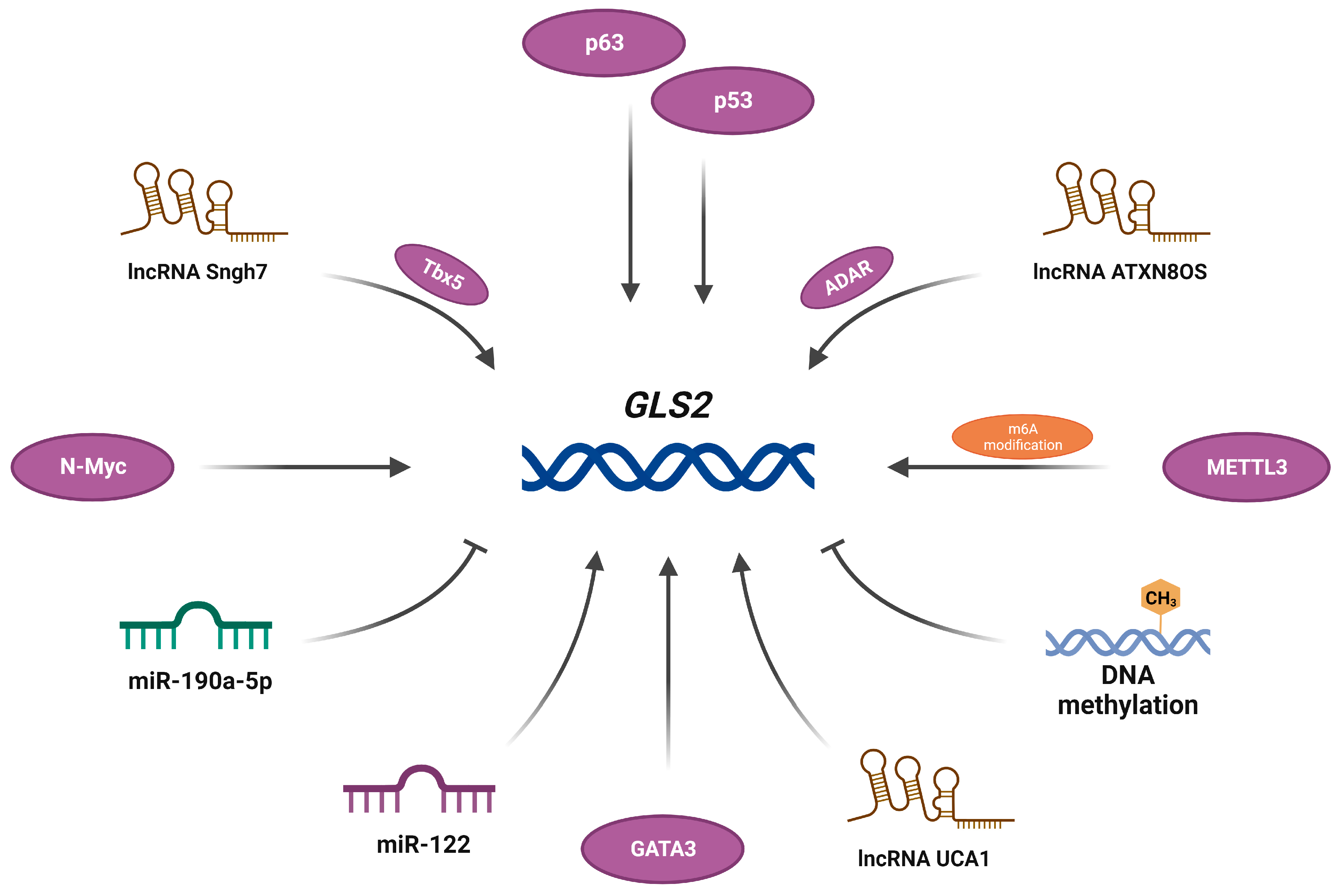
Both canonical and alternative GLS2 promoters contain CAAT- and TATA-like boxes, a Specificity protein 1 (Sp1) site and a tumor protein 53 (p53) site [36,39,40]. Indeed, p53 has been shown to induce GLS2 expression in human lung cancer and glioblastoma cells [39,41]. It is noteworthy that the transcription factor p63 belonging to the p53 family binds directly to the p53/p63 consensus DNA-binding sequence within the GLS2 promoter region and induces transcription of this gene [42]; moreover, Xiao and colleagues identified a Myc-binding site within the first intron of GLS2 and documented a direct activation of the transcription of this gene by N-Myc in MYCN-amplified neuroblastoma cells [43]. Despite the similarities noted above, canonical and alternative promoters differ with regard to other regulatory elements: the GAB promoter has an RREB site and an extremely high content of CpG islands, while the LGA promoter contains binding sites for Activator protein 1 (AP-1) and GATA-binding factor 1 (GATA-1), and it lacks a CpG island [36,40]. In fact, GLS2 promoter methylation, leading to a deficit in its product, was observed in liver and colon cancer [44,45], glioblastoma [46], and basal-subtype breast tumors [47]. Interestingly, in luminal-subtype breast cancer cells, GLS2 expression is upregulated via GATA-binding factor 3 (GATA3) [47], indicating that its expression pattern differs not only between tissues but, also, between cancer subtypes affecting the same tissue. As well as DNA methylation, RNA methylation is another epigenetic mechanism implicated in the regulation of GLS2 expression. Methyltransferase-like protein 3 (METTL3) is a component of a methyltransferase complex that catalyzes the addition of N6-methyladenosine (m6A) to mRNA [48]. Recently, METTL3-mediated m6A modification has been proven to enhance the stability of the GLS2 transcript in esophageal squamous cell carcinoma cells [49]. In addition, accumulating evidence suggests the influence of non-coding RNA on GLS2 expression. Long non-coding RNA urothelial carcinoma-associated 1 (lncRNA UCA1) regulates the expression of GLS2 by interfering with miR-16 in bladder cancer cells [50]. It has been proven that miR-190a-5p directly targets GLS2 in human cardiomyocytes [51]. In addition, an indirect impact of lncRNA small nucleolar RNA host gene 7 (Snhg7) on the expression of GLS2, through T-box transcription factor 5 (Tbx5), has been documented [52]. In glioblastoma cells, another lncRNA, ATXN8 Opposite Strand (ATXN8OS), has been shown to stabilize the GLS2 transcript by recruiting the RNA-binding protein (RBP) ADAR [53]. It is noteworthy that, in human hepatocellular carcinoma, GLS2 expression positively correlates with the level of miR-122, which inhibits GLS expression; moreover, Gls2 expression is lower in liver tissues from miR-122 knockout mice compared to wild-type animals [54]. Molecules which have been identified as regulators of GLS2 transcription are presented in Figure 2.
Although GLS2 was originally thought to be expressed exclusively in adult liver [37], several later studies proved its expression in extrahepatic tissues such as brain, pancreas, and human cancer cells, and cells of the immune system [17,37,55,56]. It is worth noting that western blot analysis of human neuroblastoma SHSY-5Y cell lysates revealed two protein bands with molecular masses compatible with GAB and LGA, confirming the existence of two GLS2 products [40]. Interestingly, human GAB expressed in baculovirus presented low affinity for Gln and low phosphate dependence, as expected for a GLS2 protein, but it was inhibited by Glu, demonstrating a kinetic behavior typical of GLS isoforms. Furthermore, human GAB was scarcely activated by ammonia, unlike the GLS2 enzyme isolated previously from liver [57]. It should be emphasized that the GAB enzyme contains the sequence encoded by exon 1 and the first six amino acids of exon 2, which is not present in the LGA enzyme. It is therefore tempting to speculate that this region is essential for Glu inhibition and ammonia activation; moreover, it may be important for the trafficking of GAB to a particular cellular compartment.
The N-terminal region of GAB contains a mitochondrial targeting sequence [37] and, indeed, GLS2 protein was detected in the mitochondrial fractions of human breast cancer cells, both wild-type an ectopically expressing GLS2 [47]. Similarly, mitochondrial localization of the exogenous GLS2 protein was found in human lung cancer, hepatoma, and neuroblastoma cells transfected with GLS2-expressing vector [39,58]. Importantly, in several human tumor cells, GLS2 was detected mainly in mitochondria but, in approximately 20% of these cells, it was localized in nuclei; moreover, a significant increase in the amount of GLS2 in nuclei was observed in either SH-SY5Y or HepG2 cells treated with the differentiation agent phorbol 12-myristate 13-acetate (PMA) or T98G glioblastoma cells transfected with the full GLS2 ORF [58]. These findings are consistent with previously reported nuclear localization of GLS2 in the rodent and monkey brain [59,60]. To date, the mechanism by which this protein enters the nucleus has not been identified. The GLS2 sequence lacks a classical nuclear localization signal, but has some other motifs that could be essential for the nuclear import. It possesses the sequence LGDLL in exon 1, which conforms to the LXXLL motif that facilitates the interaction of different proteins with nuclear receptors [59]. Furthermore, GLS2 was shown to interact with some PDZ domain-containing proteins through the ESMV motif in the C-terminus [61]. Given that several PDZ domain-containing proteins have been reported to localize to the nucleus, GLS2 might reach this compartment through interactions with such proteins [59,62]. Additionally, ankyrin repeats have been detected in the C-terminus of GLS2. Ankyrin-repeat motifs are crucial for the nuclear localization of several proteins; therefore, it is possible that they substitute for a classical nuclear localization signal in GLS2 [62]. Interestingly, in PMA-treated HepG2 cells, a perinuclear GLS2-positive staining has been detected. The perinuclear zone was enriched with vesicle-associated membrane protein 8 (VAMP8)-containing transport vesicles, which overlapped with the GLS2 staining, suggesting that the nuclear transport of GLS2 could be vesicle mediated [58]. It cannot be ruled out that GLS2 enters the cell nucleus through hypusination, a rare post-translational modification that converts lysine into hypusine (Nε-(4-amino-2-hydroxybutyl)lysine), so far identified only in eukaryotic translation initiation factor 5A (eIF5A). The significance of this modification is still far from well understood, but increasing evidence points to its role in protein localization and cellular signaling [63]. Hypusinated lysine was detected in the unfolded segment of GLS2, and the location of this modified residue suggests that it is exposed to the external medium [58]. Given that such exposed hypusine has proven to be necessary for the interaction between eIF5A and Xpo4 exportin, and evidently enables nucleocytoplasmic shuttling [64], this modification might be involved in the nuclear import of GLS2.
It is noteworthy that, in human neutrophils, GLS2 has been shown to be present in the cell surface. In intact neutrophils, GLS2 was secreted from the secondary granules and bound to the cell surface. Following PMA stimulation, GLS2 was removed from the membrane fractions and released to the extracellular culture medium [56].
The three cellular localizations identified so far, as well as several structural motifs that could enable interactions with other proteins, suggest that GLS2 is a multifaceted protein, which may be involved in other functions besides the generation of Glu [62]. Given the cell surface localization, it cannot be ruled out that this protein could interact with macromolecular components and/or regulate Gln levels in the extracellular space. Furthermore, GLS2 could be one enzyme that controls in situ Gln levels in the nucleoplasm and, hence, indirectly modulates the expression of Gln-regulated genes. This hypothesis is supported by the observation that nuclear GLS2 is catalytically active [59]. Additionally, it is also possible that GLS2 forms part of a transcriptional complex as a co-regulator. The few reports indicating the influence of ectopic GLS2 expression on the transcription process will be discussed in detail in Section 4. Finally, as mentioned above, GLS2 has been shown to interact with two PDZ domain-containing proteins: α1-syntrophin (SNTA1) and a glutaminase-interacting protein (GIP) [61]. SNT1A belongs to a family of membrane-associated adaptor proteins and is involved in cytoskeletal organization and localization and/or regulation of many transduction proteins [65]. GIP, also known as Tax-interacting protein 1 (TIP-1), plays pivotal roles in many aspects of cellular signaling, protein scaffolding, and the modulation of tumor growth [66]. Although their significance remains unknown, interactions between GLS2 and GIP or SNTA1 strengthen the hypothesis that GLS2 is a moonlight protein, i.e., one that carries out multiple unrelated functions in a cell or organism [67].
Regardless of its potential regulatory functions, GLS2 is primarily an enzyme that plays a key role in the altered metabolism of cancer cells. Recently, the structure of the human glutaminase domain of the GLS2 protein, and the functional characterization of the residues critical for its activity, was described [68]. The glutaminase domain comprises residues Ile154 to Gly479, common to both the GAB and LGA isoforms. Accessibility to the active site is controlled by two loops: the “lid” and “activation” loop, the latter being a drug target [68]. It should be noted that GLS2 and GLS proteins are highly similar in their catalytic sites. Despite this similarity, several compounds have been identified that inhibit the activity of specific glutaminase isoforms. Some of them have exhibited promising antitumor properties in different types of cancer [69]. Clearly, inhibition of GLS2 activity may reduce the survival of those cells in which GLS protein acts as a tumor promoter. It should be emphasized that, in some cancer types, GLS2 seems to play a tumor suppressor role. These two faces of GLS2 isoforms, tumor promoter and tumor suppressor, in cancer pathogenesis, will be discussed below.

3. GLS2 as a Promoter of Tumorigenesis

The analysis of data from the Oncomine database has revealed the overexpression of GLS2 compared to non-tumorigenic tissues in bladder, colon, rectum, head-and-neck, peritoneum, and lung cancers [70]. Among them, the latter is the one in which the potential role of GLS2 is, so far, best documented. An increased level of GLS2 transcript and protein was detected in epidermal growth factor receptor (EGFR)-mutated non-small cell lung cancer (NSCLC) tissues compared to EGFR wild-type tumors [68]. It is worth mentioning that EGFR-tyrosine kinase inhibitors (EGFR-TKI) prolong progression-free survival in NSCLC patients, but many patients eventually develop disease progression due to TKI-acquired resistance [71]. A recent study has indicated that Gln is a critical nutrient in acquired-EGFR-TKI-resistant lung cancer cell lines, which show higher Gln dependency compared with their parental cells [72]. Interestingly, the silencing of GLS2 diminished the viability of Gln-independent lung squamous-cell carcinoma QG56 cells and suppressed the activity of the mammalian target of rapamycin complex 1 (mTORC1) [73]. mTORC1 is a protein complex in which mTOR, a phosphoinositide 3-kinase-related protein kinase, plays a catalytic role. mTORC1 promotes translation, ribosome biogenesis, and autophagy and, thus, regulates cell growth. It acts as an effector of further oncogenic pathways, i.e., the Ras/Raf/Mek/Erk or PI3K/Akt pathways, which are often changed in the course of cancer which, in turn, results in the hyperactivation of mTORC1. In light of this, mTORC1 (and generally mTOR) inhibitors are promising cancer drugs [74]. In this context, GLS2 seems to exert a pro-oncogenic function through the regulation of mTORC1 signaling.
In the other model of NSCLC, A549 cells, GLS2 silencing decreased the cell proliferation; a similar effect was observed when the cells were treated with an alkyl benzoquinone 1-(5-methoxy-3,6-dioxocyclohexa-1,4-dien-1-yl)pentadecan-2-yl acetate (AV-1) isolated from Ardisia virens, which preferentially inhibits GLS2 rather than GLS enzymes [75]. These authors have also found that, in human hepatoma HepG2 cells, AV-1 induces autophagy via the activation of AMP-activated protein kinase (AMPK) and inhibition of the phosphatidylinositol 3-kinase (PI3K)/AKT pathway which, in turn, suppresses mTORC1 activity. It should be noted that, in this study, GLS2 silencing decreased the proliferation of HepG2 cells, which contrasts with several other papers documenting the tumor-suppressive role of GLS2 in hepatoma. All these reports will be presented in the following section of this review.
Xiao and colleagues reported a pro-oncogenic function of GLS2 in neuroblastoma [43]. In this study, an elevated expression of GLS2, and a reduced expression of GLS, was detected in the MYCN-amplified group when compared with non-amplified tumors. High GLS2 expression was significantly correlated with poor survival, while GLS expression was negatively associated with the prognosis of neuroblastoma patients. The induction of MYCN expression in SHEP MYCN-ER neuroblastoma cells, which carry a single copy of the MYCN gene, led to a time-dependent activation of GLS2 but not GLS. This finding indicates that N-Myc selectively induces GLS2 expression in MYCN-amplified neuroblastoma cells [43]. It should be emphasized that MYCN, a member of the MYC proto-oncogene family, is overexpressed in many types of cancer, including neuroblastoma, and is responsible for promoting proliferation and the survival of tumor cells [76]. Thus, N-Myc-dependent GLS2 activation may result in the reprogramming of cellular metabolism to maintain the viability and anaplerosis of the TCA cycle, ultimately leading to the Gln dependence of neuroblastoma cells. Indeed, further analyses have revealed that GLS2 silencing substantially reduced proliferation and clonogenic potential in two MYCN-amplified neuroblastoma cell lines, Kelly and BE-2C, and attenuated their ability to form tumors in vivo. The silencing of GLS2 induced apoptosis, which was suppressed when the cells were treated with dimethyl α-KG, a cell-permeable α-KG analog, indicating that these cells rely on GLS2-mediated glutaminolysis to refill TCA cycle intermediates necessary for proliferation. Furthermore, compared to the controls, GLS2-silenced cells presented diminished Gln consumption and Glu production, as well as decreased α-KG and adenosine triphosphate (ATP) generation. In addition, GLS2 depletion decreased GSH content and increased the level of reactive oxygen species (ROS), but also inhibited aerobic glycolysis. Collectively, the results noted above indicate that GLS2 co-ordinates both Gln-dependent anaplerosis and aerobic glycolysis to sustain the proliferation and survival of MYCN-amplified neuroblastoma cells [43].
Cervical cancer is another malignancy in which the expression of GLS2 is upregulated. Xiang and colleagues performed an immunohistochemical analysis of 58 cases of radioresistant tumor tissues and 86 cases of radiosensitive tumor tissues as well as 15 cases of adjacent normal cervical tissues [77]. A lack of GLS2 staining was found in the normal tissues, while cancer tissues contained considerable amounts of this protein. Notably, the GLS2 level in the tumor tissues of radioresistant patients was much higher than in those of radiosensitive patients. Similarly, GLS2 content was elevated in radioresistant cervical cancer cells HeLaR compared to the parental HeLa cells. GLS2 silencing decreased the production of GSH and the other molecules involved in the cellular antioxidant defense, including nicotinamide adenine dinucleotide hydrogen (NADH) and nicotinamide adenine dinucleotide phosphate hydrogen (NADPH). It also increased the level of intracellular ROS, which translated into the enhanced radiosensitivity of HeLaR cells in vitro and in vivo [77]. These results corroborate the above-mentioned findings, indicating the role of GLS2 in the modulation of redox homeostasis in neuroblastoma [43].
The other neoplasm in which GLS2 plays a pro-oncogenic role is in luminal-subtype breast cancer. Analysis of data from the Cancer Genome Atlas (TCGA) has shown substantially higher levels of GLS2 mRNA in luminal A (LumA) and luminal B (LumB) tumors than in basal and human epidermal growth factor receptor-2 (HER2+) tumors. Furthermore, receptor-positive breast tumors, the majority of which belonged to the luminal subtype, displayed significantly elevated level of GLS2 protein in comparison with normal tissue. In contrast, only a few receptor-negative breast cancer tissues, classified as basal subtype, contained large amount of this protein. A similar expression pattern was observed in breast cancer cell lines, with luminal-subtype cells showing high GLS2 content and basal-subtype cells showing low [47]. The level of GLS2 protein in breast cancer cell lines correlated with that of the GATA3 transcription factor; a positive correlation between the GLS2 and GATA3 transcript levels was found in human breast cancer tissues. Functional studies using MDA-MB-453 and T-47D luminal-subtype cells demonstrated that GATA3 binds to the promoter of the GLS2 gene and drives its expression [47]. Given that GATA3 regulates luminal cell differentiation in the mammary gland [78], a high expression of GLS2 may be inextricably linked to the luminal phenotype. Interestingly, in basal-subtype breast cancer cells, GLS2 expression proved to be repressed by promoter methylation [47]. The same group documented a mitochondrial localization of GLS2 in MDA-MB-453 cells, wherein this enzyme was metabolically active and critical for glutamine-mediated anaplerosis. Abrogation of GLS2 expression using shRNA significantly inhibited the proliferation of luminal-subtype cells in vitro and in vivo, but only slightly reduced this parameter in basal-subtype cells. On the contrary, neither GLS silencing, nor treatment with GLS-selective inhibitors, such as bis-2-(5-phenylacetamido-1,3,4-thiadiazol-2-yl)ethyl sulfide (BPTES) or CB-839 (Telaglenastat), affected the proliferation of luminal-subtype cells; however, these interventions decreased proliferation in basal-subtype cells. It should be noted that supplementation with dimethyl α-KG reversed the effect of GLS2 or GLS knockdown, indicating that diminished proliferation resulted from disturbed TCA cycle anaplerosis. Notably, this study revealed that a small molecule, 968, a GLS inhibitor, targeted the activity of both GA isozymes with a moderate selectivity for GLS2. Treatment with this compound suppressed TCA cycle anaplerosis and proliferation in BPTES-resistant breast cancer cells; moreover, it reduced the tumorigenic potential of MDA-MB-453 cells, supporting a view of GLS2 as a druggable target in luminal-subtype breast cancers [47].
In a near-simultaneously published study, Dias and colleagues detected considerable amounts of GLS2 protein in 14 triple-negative (TN) breast cancer cell lines, which included basal, luminal, and mesenchymal subtypes, while a significantly lower content of GLS2 was found in four non-TN cell lines [79]. Interestingly, most cell lines also expressed GLS in addition to GLS2, but the lowest overall GA levels were detected in non-TN cell lines. TN cells lines presented higher GA activity and sensitivity to Gln withdrawal, which translated into increased sensitivity to CB-839 treatment compared to non-TN cells. GLS2 knockdown diminished cell proliferation and increased oxidative stress. Similarly, either pharmacological or genetic GLS inhibition reduced proliferation, induced oxidative stress, and impaired TCA anaplerosis, while an ectopic expression of GLS2 rescued this phenotype. Furthermore, in comparison with the control cells, the cells overexpressing GLS2 presented an elevated invasion capacity as well as vimentin and actin stress fibers considered to be markers of the epithelial-to-mesenchymal transition (EMT), a process that plays a key role in cancer progression [80]. Finally, the authors examined the role of GLS2 in an in-vivo model in which MDA-MB-231 cells, transduced with either a GLS2-expressing or empty vector, were injected into mice. Astonishingly, the pro-tumorigenic effect, assessed as tumor volume and the number of lung metastases, was more pronounced when GLS2 overexpression was accompanied by GLS knockdown. It is noteworthy that, in three cohorts of breast cancer patients, the expression of GLS2 was significantly higher in tumors than in normal tissues; analysis of publicly available datasets (TCGA and the International Cancer Genome Consortium) revealed a correlation between high GLS2 expression and decreased overall, disease-free, and distant metastasis-free survival [79]. By contrast, when the TCGA pan-cancer patients with five years of follow-up were stratified into those who express and those who do not express GLS2, the former group had a higher survival rate than the latter [81]. This discrepancy, probably resulting from differences in the criteria used, hints at the complexity of the role of GLS2 in breast cancer, which is largely due to the heterogeneity of these tumors. In fact, while the paper by Dias and colleagues clearly indicates that GLS2 overexpression elevates mesenchymal markers in several breast cancer cell lines [79], a study by Ramirez-Peña and colleagues demonstrated that GLS2 expression is inversely correlated with EMT in breast cancer [81]. Firstly, analysis of the breast cancer patient samples from TCGA revealed an inverse correlation between GLS2 and EMT-signature gene expression. While the highly metastatic basal subtype was enriched with GLS2 deletions, the less aggressive subtypes were enriched in amplifications of these genes. Furthermore, an inverse correlation between GLS2 and EMT has also been observed in experimental models. The level of the GLS2 transcript was lower in mesenchymal breast cancer cell lines than in epithelial cells. Immortalized human mammary epithelial HMLE cells, genetically modified to express EMT-inducing transcription factors, which results in differentiation into the mesenchymal state, presented a diminished expression of the GLS2 gene compared to the control epithelial cells. This suppression of GLS2 correlated with decreased mitochondrial activity and Gln dependence. The inhibition of EMT restored GLS2 expression and Gln utilization. Additionally, an ectopic expression of GLS2 in SUM159 mesenchymal breast cancer cells enhanced mitochondrial activity and Gln consumption; it also inhibited mammosphere formation, which is a component of cancer-stem-cell potential, although it did not affect the proliferation of these cells. The findings of this study indicate that epithelial breast cancer cells rely on Gln and that cells that undergo EMT become Gln independent. Furthermore, EMT inhibition results in an elevated GLS2 expression and a subsequent metabolic shift in mesenchymal cells, which may sensitize them to chemotherapy [81]. The results of experimental studies documenting the tumor-promoting properties of GLS2 are summarized in Table 1.
The pro-oncogenic functions of GLS2 discussed above indicate that this protein may serve as a valuable therapeutic target in some cancers; however, this idea has not received much attention until recently, and the overwhelming majority of research has focused on targeting GLS isoforms. In this context, both Gln analogues and small molecule allosteric inhibitors have been tested. Gln analogs, such as 6-diazo-5-oxo-L-nor-leucine (DON), acivicin, or azaserine, have demonstrated anticancer activity in vitro and in vivo, but failed in the early phases of clinical trials due to side effects resulting from a lack of selectivity. It is worth mentioning that Gln analogs inhibit other enzymes that hydrolyze this amino acid, which results in off-target effects. The possibility of specific GA inhibition has advanced significantly with the development of allosteric inhibitors which, as already mentioned, are now mainly directed against GLS isoforms; the most promising is CB-839, which is currently being tested in several clinical trials. The current state of knowledge about the inhibitors of Gln metabolism have been discussed comprehensively in two very recent reviews [69,82]. The following section is limited to the description of the allosteric inhibitors that exhibit at least partial specificity for GLS2 (Table 2).
To date, the only known molecular scaffolds that potently and selectively target GLS2 are alkyl benzoquinones, isolated from A. virens. Among them, the above-mentioned AV, also known as ardisianone, shows high selectivity against GLS2 over GLS, with a half-maximal inhibitory concentration (IC50) of 0.28 μM for GLS2 and 2.1 μM for GLS [75]. The use of homologous modeling, docking, and mutagenesis studies have identified a potential binding site for AV-1 in GAB as well as residues crucial for the selectivity of this compound towards GAB. It is worth noting that two other natural alkyl benzoquinones, referred to as AV-2 and AV-8, exhibited only slightly higher IC50 values for GLS2 and GLS than AV-1. The keto/hydroxyl substituents at positions 1 and 4 in the benzoquinone core, as well as the acetate group at position 2, proved to be essential for potency towards each of the enzymes [75]. AV-1 exhibited anticancer properties against human hormone-refractory prostate cancer cells PC-3 and DU-145 [83] and acute myeloid leukemia cells HL-60 [84]. None of the studies noted above, however, have analyzed AV-1’s potential to inhibit the enzymatic activity of GA isoforms.
Another compound showing an ability to inhibit the catalytic activity of GLS2 is 986, a member of the benzophenanthridinone family, which was originally identified as a GAC inhibitor with anticancer properties in human breast cancer cells MDA-MB231 and SKBR3, as well as in a B-cell lymphoma model in vitro and in vivo [27]. In a more recent study, 968 was found to be a pan-GA inhibitor, with threefold selectivity for GLS2 over GLS [47]. This compound showed anti-proliferative activity in several breast cancer cell lines, including DU4475 cells that display intrinsic resistance to BPTES, a selective GLS inhibitor [85]. Consistent with the partial selectivity of 968 for GLS2 over GLS, the inhibition of GLS2-mediated TCA cycle anaplerosis was more pronounced in MDA-MB-453 cells containing a significant amount of GLS2 than in MDA-MB-231 cells, in which only traces of this protein were found. Furthermore, contrary to BPTES, 968 inhibited the growth of tumors formed by MDA-MB-453 cells in mice [47]. Aside from the above-mentioned cancers, 968 displayed anticancer activity in NSCLC cells A549, H23, H1299, and Spc-A1 [86], ovarian cell lines HEY, SKOV3, IGROV-1 [87], endometrial cancer cells Ishikawa and HEC-1B [88], as well as HCC cells LM3 and, to a lesser extent, 7402 and HepG2 [89]. In all these studies, 968 was considered to perform as a GLS inhibitor, while its potential effect on GLS2 has not been investigated; however, considering the pro-oncogenic role of GLS2 in some types of cancer, e.g., lung cancer [73,75], it cannot be ruled out that the observed reduction in survival is, at least partially, also due to GLS2 inhibition.
Finally, a group of thiazolidine-2,4-dione derivatives exhibited inhibitory activity against both GLS and GLS2. They diminished the clonogenic potential of human pancreas adenocarcinoma AsPC-1 cells and reduced the tumor size formed by AsPC-1 in mice; however, even though some of them inhibited GLS2 (e.g., compound 6 had an IC50 value of 102 nM), they were more selective against GLS (IC50 value of 50 nM) [90], clearly indicating that further studies are required to develop GLS2-specific inhibitors.

4. GLS2 as a Suppressor of Tumorigenesis

Twenty years ago, Turner and McGivan documented, for the first time, co-expression of the GLS and GLS2 genes in a single cancer cell type. The results of the northern blot analysis allowed the authors to speculate that the level of GLS2 mRNA was consistently higher in slow-growing colorectal adenoma AA/C1 cells than in the rapidly proliferating colorectal carcinoma HT29 cells [55]. Subsequently, Perez-Gomez and colleagues have confirmed the co-expression of both genes in tumor cells of different origin. According to their study, HepG2 cells contain much more GLS2 transcript than GLS although, at the protein level, this difference is not apparent [17]. The results of the measurement of the enzymatic activity indicates that GLS protein, not GLS2, is mainly responsible for GA activity in HepG2 cells, which is consistent with a previous report [91]. Taking into account that normal, non-proliferating hepatocytes express GLS2 [35], it is possible that GLS isoforms become upregulated during malignant transformation. The same group detected both GLS and GLS2 transcripts in human leukemia cells, but only the latter transcript in mature, non-proliferating lymphocytes from the medullar blood of a patient suffering aplasia. Based on these results, the authors hypothesized that GLS2 expression is a characteristic feature of resting or quiescent cells, while the overexpression of GLS is associated with elevated proliferation rates [17].
As mentioned in Section 3, Lee and colleagues documented the inhibitory effect of GLS2 silencing on the proliferation of HepG2 cells derived from hepatocellular carcinoma (HCC) [75]; nevertheless, this is one of the few reports, if not the only one, suggesting a pro-oncogenic role for GLS2 in this tumor type. Indeed, the immunohistochemical analysis of the GLS2 level in 112 HCC and 111 adjacent non-tumor tissues (NT) revealed significantly lower GLS2 staining in tumors than in NT tissues; a lack of GLS2 staining correlated with a short survival time [23]. Interestingly, an inverse pattern in GLS staining was also observed, with an elevated level of this protein in HCC, which indicates that GLS and GLS2 play opposing roles in HCC. Furthermore, the authors examined the expression of both GA isoforms in a set of liver tissues mimicking HCC transformation. The intensity of GLS2 staining was high in NT and low-grade fibrotic liver tissues, and low in HCC. On the contrary, GLS staining was weak in NT tissues but gradually increased in parallel with disease progression, at its most intense in HCC [23]. These findings allow for speculation that, during HCC transformation, Gln metabolism switches from GLS2- to GLS-dependent; they are consistent with an earlier study on MYC-induced mouse liver tumors [92].
Several other reports have confirmed diminished GLS2 expression in HCC compared to healthy tissues [39,44,45,93] and pointed to promoter methylation as the mechanism responsible for this phenomenon [44,45]. Furthermore, numerous studies have documented the onco-suppressive function of GLS2 in multiple HCC models in vitro and in vivo. For instance, an ectopic expression of GLS2 decreased the ability of HepG2 cells to form colonies. On the other hand, ablation of GLS2 increased the production of reactive oxygen species (ROS) and sensitized HepG2 cells to H2O2-induced apoptosis; it also diminished the α-ketoglutarate and ATP levels and oxygen consumption. Notably, the GLS2 transcription in HepG2 cells proved to be induced by p53 [39], a tumor suppressor whose role in the prevention of HCC has been widely documented [94]. It is therefore tempting to speculate that, as a p53 target, GLS2 might contribute to the cancer-suppressive properties of p53 in HCC. In another study, the overexpression of GLS2 markedly inhibited the growth of xenograft tumors formed by two HCC cell lines, Huh1 and Huh7, compared with tumors formed by the control cells transduced with an empty vector. Conversely, the knockdown of GLS2 in PLC/PRF/5 HCC cells containing traces of the GLS2 transcript significantly enhanced anchorage-independent cell growth and the formation of tumors in vivo. A subsequent functional analysis revealed that GLS2 negatively regulates the PI3K/AKT signaling pathway, which contributes to the tumor-suppressive properties exerted by GLS2 in HCC [45]. In another study, GLS2 proved to be critical for the inhibition of HCC cell migration and invasion [93]. Firstly, a negative correlation between the amount of GLS2 protein and the ability of different HCC cell lines to migrate and invade was observed. Secondly, an ectopic expression of GLS2 in Mahlavu and Huh7 cells, which contain barely detectable endogenous GLS2 protein, diminished migration and invasion; the opposite effect was obtained when GLS2 was silenced in HCC36 and HepG2 cells containing a considerable amount of GLS2. Further analysis provided compelling evidence that GLS2 represses EMT [93]. Compared to the controls, Mahlavu cells in which GLS2 was overexpressed presented more epithelial morphology, an elevated level of epithelial marker E-cadherin, and lowered levels of mesenchymal markers, vimentin, and fibronectin; the opposite effect was obtained by GLS2 silencing in HepG2 cells. The aforementioned modification diminished the level of Snail, a transcription factor which plays a crucial role in tumor metastasis [95]. It is particularly worth noting that overexpression of SNAI1, the gene encoding Snail, reversed the GLS2-mediated phenotype in vitro, and restored both intrahepatic and lung metastasis inhibited by GLS2 in the orthotopic model. In addition, further mechanistic studies have shown that GLS2 functionally binds to DICER, a protein involved in the production of miRNAs [96], in a non-enzymatic activity manner. This interaction stabilizes DICER and promotes the maturation of miR-34a which, in turn, inhibits SNAI1 expression [93]. Notably, DICER is a central player in the miRNA and RNAi pathways, and alterations in its level and activity can lead to several diseases, including cancer, while the promotion of its function may be beneficial [97]. Clearly, further investigations are needed to elucidate whether DICER stabilization by GLS2 can also modulate other miRNAs/RNAi, as well as how it influences the cancer cell phenotype.
Yet another protein that interacts with GLS2, at least in HCC cells, is Ras-related C3 botulinum toxin substrate 1 (Rac1), a member of the Rho GTPase family, involved in the regulation of tumor angiogenesis, invasion, and metastasis; these processes underlie malignant transformation. Rac1 hyperactivation and overexpression correlates with a malignant phenotype in several tumors of different origin [97]. The interaction between GLS2 and Rac1 has been observed not only in Huh-1 cells transduced with vectors expressing GLS2 and RAC1, but also in wild-type Huh-1 and HepG2 cells [98]. Interestingly, the C-terminus of GLS2, which does not contain the GA catalytic domain, proved to be necessary and sufficient for the interaction between this protein and Rac1. GLS2 binding inhibits the interaction of Rac1 with its activators, Tiam1 and VAV1. This negative regulation of Rac1 contributed to the inhibitory effect of GLS2 on the migration and invasion of several HCC cells in vitro as well as on their metastatic potential in vivo. Last but not least, the finding of this study is that GLS2 mediates the role of p53 in inhibiting metastasis. The silencing of GLS2 or TP53 markedly elevated the migration and invasion of Huh-1 and HepG2 cells in vitro and their metastasis in mice, whereas the simultaneous knockdown of both genes did not present an additive effect on either the migration/invasion of these cells or their ability to metastasize. Furthermore, a single knockdown of either GLS2 or TP53 significantly activated Rac1, while their simultaneous silencing did not present an additive effect on the activity of Rac1. The overexpression of GLS2 abolished the promoting influence of TP53 knockdown on Rac1 activity, suggesting that GLS2 mediates p53 function in inhibiting Rac1 activity [98]. It should be emphasized that Rac1 is a pleiotropic regulator of many cellular processes (i.e., proliferation, ROS-mediated cell death, and cell cycle progression), and has multiple effectors [97]. It is therefore tempting to speculate that the interaction between GLS2 and Rac1, which inhibits Rac1 activity, modulates the action of many molecules (not yet identified), which translates into the suppression of the malignant phenotype.
In a very recent study, Suzuki and colleagues not only confirmed the tumor-suppressive activity of GLS2 in HCC, but also shed new light on the mechanism underlying this phenomenon [99]. By 120 weeks of age, all Gls2 knockout mice included in the experiment developed either B-cell lymphomas, HCC, or both tumor types. Conversely, none of the wild-type mice exhibited any tumor. In addition, the animals were subjected to the hepatocarcinogenic Stelic Animal Model (STAM) protocol, which reflects non-alcoholic steatohepatitis closely associated with HCC [100]. A lack of Gls2 resulted in the earlier development of HCC tumors that were larger in size than those produced in wild-type mice. Compared to WT-STAM animals, KO-STAM mice displayed a lowered level of ferroptosis markers in both normal liver and HCC. Furthermore, the level of malondialdehyde, an indicator of ferroptosis, was reduced in the livers of knockout mice compared with wild-type mice; hepatocytes from knockout animals proved to be less sensitive to inducers of ferroptosis. This resistance to ferroptosis was attenuated by an ectopic expression of Gls2, indicating that GLS2 is necessary for the induction of ferroptosis in hepatocytes. These findings extend to human HCC cells, in which modifications of GLS2 levels resulted in a correspondingly altered extent of ferroptosis. GLS2 enhanced the production of lipid ROS through the conversion of Glu to α-KG which, in turn, promoted ferroptosis. Likewise, compared to the controls, GLS2-overexpressing cells formed significantly smaller tumors after injection into the severe combined immunodeficient (SCID) mice, which was accompanied by an increase in the expression of ferroptosis markers. Notably, in this study, the GA core domain, located between positions 177 and 463, was necessary for the tumor-suppressive functions of GLS2 and its ability to promote ferroptosis [99]. Interestingly, the enzymatic activity of GLS2 may be regulated by the general control of amino acid synthesis 5-like 1 (GCN5L1) protein, as demonstrated in mice hepatocytes. Mitochondrial-enriched GCN5L1 suppresses, while the absence of GCN5L1 increases GLS2 activity; this modulation is mediated in part by the change in acetylation of lysine 279, located in the putative catalytic domain of GLS2. The activation of GLS2 promotes hepatic glutaminolysis, which further leads to the activation of mTORC1 signaling [101]. The same mode of regulation of the enzymatic activity of GLS2 was observed in HCC. The deletion of GCN5L1 enhances Gln metabolism and promotes HCC cell proliferation and mTORC1 activity. Mechanistically, GCN5L1 promotes the acetylation and inactivation of both GLS2 and GLS, and increases enzyme oligomerisation. It is worth noting that an inverse correlation has been observed between GCN5L1 and mTORC1 levels and GA activity in HCC patients [102].
Collectively, the overwhelming majority of data collected to date clearly indicate that, in HCC, GLS2 is a bona fide tumor suppressor. This conclusion comes from observations made on several cohorts of patients as well as from numerous in vitro and in vivo studies, which have shed new light on the mechanism of GLS2 action. It is worth noting that GLS2 seems to inhibit tumorigenesis in other types of gastrointestinal cancer. Thus, GLS2 expression proved to be diminished in colon tumor tissues compared to adjacent non-tumor tissues and, as in the case of HCC, promoter methylation was found to be a key mechanism underlying GLS2 silencing in colon cancer. Even though the analysis was performed on a small number (n = 5) of tissues, and the level of GLS2 protein was not examined, the results of studies on human colon cancer cells (HCT116) revealed that an ectopic expression of GLS2 markedly reduced the viability and number of cell colonies. Additionally, an ectopic expression of GLS2 led to a significant increase in the number of cells in the G2/M phase and a reduction of the levels of p21 and cyclin D1 [44]. More recently, the downregulation of GLS2 expression was noted in a cohort of 36 gastric cancer tissues compared to the adjacent non-cancer tissues. GLS2 overexpression suppressed the viability of gastric cancer MGC-803 cells and their ability to migrate; it also induced apoptosis [103]. Both of the studies noted above were performed on only one cell line and do not include functional studies; therefore, further in-depth analysis is necessary to elucidate the significance of reduced GLS2 expression in colon and gastric cancer.
Another type of neoplasm in which GLS2 appears to exert suppressive properties is glioblastoma, an astrocytic tumor with the highest malignancy grade according to WHO classification (WHO IV) [104]. GLS2 expression in these tumors is abolished or significantly lower than in non-tumorigenic brain tissues, which was first demonstrated by examining groups of several patients [16,46] or by analyzing data from TCGA [70]. Nevertheless, a similar observation was also made in a recent study conducted on a relatively large cohort of 153 patients with astrocytomas of different grades of malignancy. Compared to non-tumorigenic brain tissues, pilocytic astrocytomas (WHO grade I) display a significantly lower expression of GLS2, which is further reduced with an increasing grade of malignancy [105]. Notably, promoter methylation has proved to be a crucial mechanism responsible for the downregulation of GLS2 in glioblastoma [46]. Interestingly, the expression of GLS is upregulated in glioblastoma compared to non-tumorigenic brain tissues [16], reinforcing the idea that GLS and GLS2 play opposing roles in certain cancer types. An ectopic expression of GLS2 decreased the viability and proliferation of glioblastoma T98G, U87MG and LN229 cells, as well as their ability to form colonies and migrate [106,107]. Microarray analysis revealed alterations in the expression of several genes, some of which are relevant to malignancy. One of the downregulated genes, MGMT, codes for the repair protein O6-methylguanine-DNA methyltransferase (MGMT), which reverses the alkylation process, thereby contributing to resistance to the alkylating agent temozolomide (TMZ), used in chemotherapy. Since DNA methylation contributes to MGMT silencing, the methylation status of the MGMT promoter is considered to be a biomarker of a patient’s response to TMZ treatment [108]. Diminished MGMT expression in T98G cells transfected with GLS2 translated into a decreased level of MGMT protein and activity which, in turn, resulted in increased sensitivity to TMZ. The overexpression of GLS2 did not change the methylation status of MGMT; therefore, the mechanism by which GLS2 diminishes MGMT transcription remains unknown [109]. Similar to T98G cells, two other glioblastoma cell lines, U87MG and LN229, also became more sensitive upon transfection with the GLS2 sequence. Given that U87MG cells do not express MGMT [110], a mechanism unrelated to the modulation of the MGMT level must have been responsible for the increase in sensitivity to TMZ. It could, for example, be related to oxidative stress induced by TMZ [111]. Indeed, an ectopic GLS2 expression sensitized all three cell lines to treatment with oxidate agents hydrogen peroxide (H2O2) and arsenic trioxide (ATO) [29,107]. Upon treatment with H2O2, GLS2-transfected cells presented a reduction in the phosphorylation of PDK1, PI3K, and AKT. Pre-treatment with PDGF-BB, an activator of AKT phosphorylation, prevented H2O2-evoked cell death in GLS2-transfectants, indicating that the downregulation of the PI3K/AKT pathway contributes to increased sensitivity to H2O2 [107]. Notably, both T98G and LN229 cells transfected with GLS2 displayed a reduced activity of catalase (CAT). Additionally, a diminished activity of superoxide dismutase (SOD) was found in LN229 transfectants, while it was elevated in GLS2-transfected T98G cells. Furthermore, total antioxidant capacity increased and lipid peroxidation decreased in T98G and LN229 cells upon transfection with GLS2, supporting the antioxidant effect of GLS2 [112]. It is noteworthy that this study identified a series of miRNAs whose expression was changed by an ectopic expression of GLS2. Thus, miRNA-140-3p, miRNA-1246, miRNA-1260a, miRNA-21-5p, and miRNA-146a-5p were overexpressed in both T98G and LN229 after an overexpression of GLS2. One more miRNA, miRNA-92a-3p, was additionally upregulated in LN229 cells transfected with GLS2. Although further functional analysis is necessary to clarify the significance of the changes described above in the miRNome of GLS2-transfected cells, it is worth noting that the included molecules have been linked to carcinogenesis in different tumor models [112].
Interestingly, relative to the controls, T98G cells transfected with the GLS2 sequence presented diminished proliferation when cultured in a medium containing adult bovine serum [58]. Such a procedure makes it possible to avoid non-physiological levels of cysteine in the culture medium, which induces increased reliance on Gln anaplerosis [113]. Compared to WT T98G cells, GLS2-transfected T98G cells showed an increased level of p53 in nuclei and decreased level of c-Myc in cytoplasm. Likewise, an elevated level of GLS2 observed in the nuclei of PMA-treated neuroblastoma SH-SY5Y cells was accompanied by significantly increased amounts of p53, p21, and cell cycle arrest at the G2/M stage. Since the treatment of SH-SY5Y cells with PMA reduces their proliferation, as does the transfection of T98G cells with the GLS2 sequence, it is tempting to speculate that the nuclear localization of GLS2 plays an important role in the inhibition of proliferation and is mediated, at least partially, by p53-dependent mechanisms [58].
As mentioned above, an ectopic GLS2 expression sensitized glioblastoma cells to treatment with TMZ [107]. In a very recent study, Luo and colleagues showed that TMZ-resistant glioblastoma cells (U251TR) contain a lesser amount of GLS2 than U251 cells [53]. An ectopic GLS2 expression in U251TR cells diminished proliferation and migration, and both parameters were further decreased upon TMZ treatment. This modification increased the lipid ROS level, which was reversed by Ferrostatin-1, an inhibitor of ferroptosis; it also increased the level of E-cadherin and decreased the level of vimentin. These results suggest that GLS2 can drive ferroptosis and repress EMT, which is consistent with the previous observations made in HCC cells [93,99]. Similar results were obtained in the intracranial tumor model, indicating, for the first time, that GLS2 exerts anti-glioma effects in in vivo settings [53].
In this regard, it is worth noting the report by Kim and colleagues documenting the correlation between the level of GLS2 and the regional heterogeneity of 5-aminolevulinic acid (5-ALA) fluorescence in glioblastoma [114]. Fluorescence-guided surgery using 5-ALA has been introduced in the treatment of high-grade gliomas, as it enables the intraoperative visualization of malignant tissue and differentiates it from normal brain [115]. Inter- and intra-tumoral molecular, cellular, and metabolic heterogeneity significantly contributes to therapy failure in glioblastoma [116]. Interestingly, the 5-ALA fluorescence intensity in glioblastoma shows regional heterogeneity, which may result from differences in cell density and/or intra-tumoral metabolic heterogeneity [117]. Kim and colleagues identified GLS2 as a key gene in the regulation of 5-ALA metabolism in glioblastoma. Each of the three glioblastoma cell lines used in this study, namely T98G, U87MG, and LN18, transduced with a GLS2 sequence, displayed significantly attenuated fluorescence intensity after 5-ALA treatment compared to their WT counterparts. The ratios of NADPH/NADP and GSH/GSSG were significantly reduced after 5-ALA treatment, and GLS2 expression elevated the levels of NADPH/NADP, which was most likely linked with an increased capacity for 5-ALA metabolism. Furthermore, 5-ALA fluorescence was inversely correlated with GLS2 expression and the NADPH level in glioblastoma tissues. Collectively, these results allow for a conclusion that regional metabolic heterogeneity in glioblastoma leads to low GLS2 expression and low levels of NADPH and GSH levels in the malignant area, while high GLS2 expression, together with high levels of NADPH and GSH, is found in the lower grade area [114]. The results of the above experiments, which suggest that GLS2 behaves as a tumor suppressor, are summarized in Table 3.

5. Conclusions and Future Directions

Impaired metabolism is generally considered to be a hallmark of cancer. Increased Gln uptake and enhanced glutaminolysis are key metabolic features of many cancers. Consequently, neoplasms of different origin display an elevated expression of GA, which correlates with the growth rate and malignancy of tumors. Targeting Gln, for instance through the inhibition of GA, appears to be a promising approach in treating Gln-addicted tumors.
In some cancers, GLS2 expression is regulated by oncoproteins and clearly contributes to tumorigenesis by supporting cancer cell proliferation and tumor growth. Nevertheless, current knowledge revises the traditional view of the function of GLS2 as an enzyme that drives carcinogenesis; rather, it hints that this protein acts as a molecule-suppressing neoplastic phenotype. This appears to be particularly true for HCC. Recent studies have conclusively proven that GLS2 is a bona fide tumor suppressor at an organismal level, but mounting evidence indicates that a similar situation also occurs in glioblastoma. So far, single reports suggest that GLS2 protein may also inhibit the development of other cancers.
The precise mechanism underlying the tumor-suppressing function of GLS2 is yet to be fully elucidated, but it is believed to involve the regulation of cellular metabolism and redox balance. It should be emphasized that, in several research models, the suppressor role of GLS2 proved to be independent of the enzymatic activity of this protein. Although progress has been made in identifying the pathways triggered by GLS2, as well as its functional protein partners, there are still many unanswered questions. Future research should focus on determining whether the mechanism described in HCC is universal and also applies to other types of cancer; moreover, further work is needed to investigate the effects of simultaneous GLS silencing and GLS2 overexpression. Extensive research in this area may pave the way for effective cancer treatment strategies.

Author Contributions

Conceptualization, M.S.; investigation, M.S. and J.B.; writing—original draft preparation, M.S.; writing—review and editing, M.S. and J.B.; visualization, J.B.; supervision, M.S.; project administration, M.S.; funding acquisition, M.S. All authors have read and agreed to the published version of the manuscript.

Funding

This research was funded by the National Science Centre Poland, grant number 2021/41/B/NZ5/01883.

Conflicts of Interest

The authors declare no conflict of interests.

References

  1. Warburg, O. On the Origin of Cancer Cells. Science 1956, 123, 309–314. [Google Scholar] [CrossRef] [PubMed]
  2. Altman, B.J.; Stine, Z.E.; Dang, C.V. From Krebs to Clinic: Glutamine Metabolism to Cancer Therapy. Nat. Rev. Cancer 2016, 16, 619–634. [Google Scholar] [CrossRef] [PubMed]
  3. Yang, L.; Venneti, S.; Nagrath, D. Glutaminolysis: A Hallmark of Cancer Metabolism. Annu. Rev. Biomed. Eng. 2017, 19, 163–194. [Google Scholar] [CrossRef]
  4. Willems, L.; Jacque, N.; Jacquel, A.; Neveux, N.; Trovati Maciel, T.; Lambert, M.; Schmitt, A.; Poulain, L.; Green, A.S.; Uzunov, M.; et al. Inhibiting Glutamine Uptake Represents an Attractive New Strategy for Treating Acute Myeloid Leukemia. Blood 2013, 122, 3521–3532. [Google Scholar] [CrossRef] [PubMed]
  5. Leone, R.D.; Zhao, L.; Englert, J.M.; Sun, I.-M.; Oh, M.-H.; Sun, I.-H.; Arwood, M.L.; Bettencourt, I.A.; Patel, C.H.; Wen, J.; et al. Glutamine Blockade Induces Divergent Metabolic Programs to Overcome Tumor Immune Evasion. Science 2019, 366, 1013–1021. [Google Scholar] [CrossRef] [PubMed]
  6. Sun, N.; Liang, Y.; Chen, Y.; Wang, L.; Li, D.; Liang, Z.; Sun, L.; Wang, Y.; Niu, H. Glutamine Affects T24 Bladder Cancer Cell Proliferation by Activating STAT3 through ROS and Glutaminolysis. Int. J. Mol. Med. 2019, 44, 2189–2200. [Google Scholar] [CrossRef]
  7. Jin, H.; Wang, S.; Zaal, E.A.; Wang, C.; Wu, H.; Bosma, A.; Jochems, F.; Isima, N.; Jin, G.; Lieftink, C.; et al. A Powerful Drug Combination Strategy Targeting Glutamine Addiction for the Treatment of Human Liver Cancer. eLife 2020, 9, 56749. [Google Scholar] [CrossRef]
  8. Yamashita, A.S.; da Costa Rosa, M.; Stumpo, V.; Rais, R.; Slusher, B.S.; Riggins, G.J. The Glutamine Antagonist Prodrug JHU-083 Slows Malignant Glioma Growth and Disrupts MTOR Signaling. Neurooncol. Adv. 2021, 3, 149. [Google Scholar] [CrossRef]
  9. Spada, M.; Piras, C.; Diana, G.; Leoni, V.P.; Frau, D.V.; Serreli, G.; Simbula, G.; Loi, R.; Noto, A.; Murgia, F.; et al. Glutamine Starvation Affects Cell Cycle, Oxidative Homeostasis and Metabolism in Colorectal Cancer Cells. Antioxidants 2023, 12, 683. [Google Scholar] [CrossRef]
  10. Aledo, J.C.; Gómez-Fabre, P.M.; Olalla, L.; Márquez, J. Identification of Two Human Glutaminase Loci and Tissue-Specific Expression of the Two Related Genes. Mamm. Genome 2000, 11, 1107–1110. [Google Scholar] [CrossRef]
  11. Elgadi, K.M.; Meguid, R.A.; Qian, M.; Souba, W.W.; Abcouwer, S.F. Cloning and Analysis of Unique Human Glutaminase Isoforms Generated by Tissue-Specific Alternative Splicing. Physiol. Genom. 1999, 1, 51–62. [Google Scholar] [CrossRef]
  12. Porter, L.D.; Ibrahim, H.; Taylor, L.; Curthoys, N.P. Complexity and Species Variation of the Kidney-Type Glutaminase Gene. Physiol. Genom. 2002, 9, 157–166. [Google Scholar] [CrossRef]
  13. Márquez, J.; Matés, J.M.; Campos-Sandoval, J.A. Glutaminases. In The Glutamate/GABA-Glutamine Cycle, Advances in Neurobiology 13, 1st ed.; Schousboe, A., Sonnewald, U., Eds.; Springer International Publishing: Cham, Switzerland, 2016; pp. 133–171. [Google Scholar] [CrossRef]
  14. Campos-Sandoval, J.A.; Martín-Rufián, M.; Cardona, C.; Lobo, C.; Peñalver, A.; Márquez, J. Glutaminases in Brain: Multiple Isoforms for Many Purposes. Neurochem. Int. 2015, 88, 1–5. [Google Scholar] [CrossRef]
  15. Cassago, A.; Ferreira, A.P.S.; Ferreira, I.M.; Fornezari, C.; Gomes, E.R.M.; Greene, K.S.; Pereira, H.M.; Garratt, R.C.; Dias, S.M.G.; Ambrosio, A.L.B. Mitochondrial Localization and Structure-Based Phosphate Activation Mechanism of Glutaminase C with Implications for Cancer Metabolism. Proc. Natl. Acad. Sci. USA 2012, 109, 1092–1097. [Google Scholar] [CrossRef]
  16. Szeliga, M.; Sidoryk, M.; Matyja, E.; Kowalczyk, P.; Albrecht, J. Lack of Expression of the Liver-Type Glutaminase (LGA) MRNA in Human Malignant Gliomas. Neurosci. Lett. 2005, 374, 171–173. [Google Scholar] [CrossRef]
  17. Pérez-Gómez, C.; Campos-Sandoval, J.A.; Alonso, F.J.; Segura, J.A.; Manzanares, E.; Riuz-Sánchez, P.; González, M.E.; Márquez, J.; Matés, J.M. Co-Expression of Glutaminase K and L Isoenzymes in Human Tumour Cells. Biochem. J. 2005, 386, 535–542. [Google Scholar] [CrossRef]
  18. Szeliga, M.; Matyja, E.; Obara, M.; Grajkowska, W.; Czernicki, T.; Albrecht, J. Relative Expression of MRNAS Coding for Glutaminase Isoforms in CNS Tissues and CNS Tumors. Neurochem. Res. 2008, 33, 808–813. [Google Scholar] [CrossRef]
  19. van den Heuvel, A.P.J.; Jing, J.; Wooster, R.F.; Bachman, K.E. Analysis of Glutamine Dependency in Non-Small Cell Lung Cancer. Cancer Biol. Ther. 2012, 13, 1185–1194. [Google Scholar] [CrossRef]
  20. Kim, S.; Jung, W.H.; Koo, J.S. The Expression of Glutamine-Metabolism-Related Proteins in Breast Phyllodes Tumors. Tumor Biol. 2013, 34, 2683–2689. [Google Scholar] [CrossRef]
  21. Huang, F.; Zhang, Q.; Ma, H.; Lv, Q.; Zhang, T. Expression of Glutaminase Is Upregulated in Colorectal Cancer and of Clinical Significance. Int. J. Clin Exp. Pathol. 2014, 7, 1093–1100. [Google Scholar] [PubMed]
  22. Pan, T.; Gao, L.; Wu, G.; Shen, G.; Xie, S.; Wen, H.; Yang, J.; Zhou, Y.; Tu, Z.; Qian, W. Elevated Expression of Glutaminase Confers Glucose Utilization via Glutaminolysis in Prostate Cancer. Biochem. Biophys. Res. Commun. 2015, 456, 452–458. [Google Scholar] [CrossRef]
  23. Yu, D.; Shi, X.; Meng, G.; Chen, J.; Yan, C.; Jiang, Y.; Wei, J.; Ding, Y. Kidney-Type Glutaminase (GLS1) Is a Biomarker for Pathologic Diagnosis and Prognosis of Hepatocellular Carcinoma. Oncotarget 2015, 6, 7619–7631. [Google Scholar] [CrossRef]
  24. Kim, H.M.; Lee, Y.K.; Koo, J.S. Expression of Glutamine Metabolism-Related Proteins in Thyroid Cancer. Oncotarget 2016, 7, 53628–53641. [Google Scholar] [CrossRef]
  25. Lobo, C.; Ruiz-Bellido, M.A.; Aledo, J.C.; Márquez, J.; Núñez De Castro, I.; Alonso, F.J. Inhibition of Glutaminase Expression by Antisense MRNA Decreases Growth and Tumourigenicity of Tumour Cells. Biochem. J. 2000, 348 Pt 2, 257–261. [Google Scholar] [CrossRef]
  26. Zhang, J.; Mao, S.; Guo, Y.; Wu, Y.; Yao, X.; Huang, Y. Inhibition of GLS Suppresses Proliferation and Promotes Apoptosis in Prostate Cancer. Biosci. Rep. 2019, 39, 1826. [Google Scholar] [CrossRef]
  27. Wang, J.-B.; Erickson, J.W.; Fuji, R.; Ramachandran, S.; Gao, P.; Dinavahi, R.; Wilson, K.F.; Ambrosio, A.L.B.; Dias, S.M.G.; Dang, C.V.; et al. Targeting Mitochondrial Glutaminase Activity Inhibits Oncogenic Transformation. Cancer Cell 2010, 18, 207–219. [Google Scholar] [CrossRef]
  28. Cheng, T.; Sudderth, J.; Yang, C.; Mullen, A.R.; Jin, E.S.; Matés, J.M.; DeBerardinis, R.J. Pyruvate Carboxylase Is Required for Glutamine-Independent Growth of Tumor Cells. Proc. Natl. Acad. Sci. USA 2011, 108, 8674–8679. [Google Scholar] [CrossRef]
  29. Martín-Rufián, M.; Nascimento-Gomes, R.; Higuero, A.; Crisma, A.R.; Campos-Sandoval, J.A.; Gómez-García, M.C.; Cardona, C.; Cheng, T.; Lobo, C.; Segura, J.A.; et al. Both GLS Silencing and GLS2 Overexpression Synergize with Oxidative Stress against Proliferation of Glioma Cells. J. Mol. Med. 2014, 92, 277–290. [Google Scholar] [CrossRef]
  30. Jacque, N.; Ronchetti, A.M.; Larrue, C.; Meunier, G.; Birsen, R.; Willems, L.; Saland, E.; Decroocq, J.; Maciel, T.T.; Lambert, M.; et al. Targeting Glutaminolysis Has Antileukemic Activity in Acute Myeloid Leukemia and Synergizes with BCL-2 Inhibition. Blood 2015, 126, 1346–1356. [Google Scholar] [CrossRef]
  31. Wang, D.; Li, X.; Gong, G.; Lu, Y.; Guo, Z.; Chen, R.; Huang, H.; Li, Z.; Bian, J. An Updated Patent Review of Glutaminase Inhibitors (2019–2022). Expert Opin. Ther. Pat. 2023, 33, 17–28. [Google Scholar] [CrossRef] [PubMed]
  32. Chen, Y.; Tan, L.; Gao, J.; Lin, C.; Wu, F.; Li, Y.; Zhang, J. Targeting Glutaminase 1 (GLS1) by Small Molecules for Anticancer Therapeutics. Eur. J. Med. Chem. 2023, 252, 115306. [Google Scholar] [CrossRef]
  33. Patel, M.; McGivan, J.D. Partial Purification and Properties of Rat Liver Glutaminase. Biochem. J. 1984, 220, 583–590. [Google Scholar] [CrossRef]
  34. Smith, E.M.; Watford, M. Rat Hepatic Glutaminase: Purification and Immunochemical Characterization. Arch. Biochem. Biophys. 1988, 260, 740–751. [Google Scholar] [CrossRef]
  35. Smith, E.M.; Watford, M. Molecular Cloning of a CDNA for Rat Hepatic Glutaminase. Sequence Similarity to Kidney-Type Glutaminase. J. Biol. Chem. 1990, 265, 10631–10636. [Google Scholar] [CrossRef]
  36. Pérez-Gómez, C.; Matés, J.M.; Gómez-Fabre, P.M.; Castillo-Olivares, A.D.; Alonso, F.J.; Márquez, J. Genomic Organization and Transcriptional Analysis of the Human L-Glutaminase Gene. Biochem. J. 2003, 370, 771–784. [Google Scholar] [CrossRef]
  37. Gómez-Fabre, P.M.; Aledo, J.C.; Del Castillo-Olivares, A.; Alonso, F.J.; Núñez De Castro, I.; Campos, J.A.; Márquez, J. Molecular Cloning, Sequencing and Expression Studies of the Human Breast Cancer Cell Glutaminase. Biochem. J. 2000, 345 Pt 2, 365–375. [Google Scholar] [CrossRef]
  38. de la Rosa, V.; Campos-Sandoval, J.A.; Martín-Rufián, M.; Cardona, C.; Matés, J.M.; Segura, J.A.; Alonso, F.J.; Márquez, J. A Novel Glutaminase Isoform in Mammalian Tissues. Neurochem. Int. 2009, 55, 76–84. [Google Scholar] [CrossRef]
  39. Hu, W.; Zhang, C.; Wu, R.; Sun, Y.; Levine, A.; Feng, Z. Glutaminase 2, a Novel P53 Target Gene Regulating Energy Metabolism and Antioxidant Function. Proc. Natl. Acad. Sci. USA 2010, 107, 7455–7460. [Google Scholar] [CrossRef]
  40. Martín-Rufián, M.; Tosina, M.; Campos-Sandoval, J.A.; Manzanares, E.; Lobo, C.; Segura, J.A.; Alonso, F.J.; Matés, J.M.; Márquez, J. Mammalian Glutaminase Gls2 Gene Encodes Two Functional Alternative Transcripts by a Surrogate Promoter Usage Mechanism. PLoS ONE 2012, 7, e38380. [Google Scholar] [CrossRef]
  41. Suzuki, S.; Tanaka, T.; Poyurovsky, M.V.; Nagano, H.; Mayama, T.; Ohkubo, S.; Lokshin, M.; Hosokawa, H.; Nakayama, T.; Suzuki, Y.; et al. Phosphate-Activated Glutaminase (GLS2), a P53-Inducible Regulator of Glutamine Metabolism and Reactive Oxygen Species. Proc. Natl. Acad. Sci. USA 2010, 107, 7461–7466. [Google Scholar] [CrossRef]
  42. Arianna, G.; Bongiorno-Borbone, L.; Bernassola, F.; Terrinoni, A.; Markert, E.; Levine, A.J.; Fen, Z.; Agostini, M.; Zolla, L.; Finazzi Agro’, A.; et al. P63 Regulates Glutaminase 2 Expression. Cell Cycle 2013, 12, 1395–1405. [Google Scholar] [CrossRef]
  43. Xiao, D.; Ren, P.; Su, H.; Yue, M.; Xiu, R.; Hu, Y.; Liu, H.; Qing, G. Myc Promotes Glutaminolysis in Human Neuroblastoma through Direct Activation of Glutaminase 2. Oncotarget 2015, 6, 40655–40666. [Google Scholar] [CrossRef]
  44. Zhang, J.; Wang, C.; Chen, M.; Cao, J.; Zhong, Y.; Chen, L.; Shen, H.-M.; Xia, D. Epigenetic Silencing of Glutaminase 2 in Human Liver and Colon Cancers. BMC Cancer 2013, 13, 601. [Google Scholar] [CrossRef]
  45. Liu, J.; Zhang, C.; Lin, M.; Zhu, W.; Liang, Y.; Hong, X.; Zhao, Y.; Young, K.H.; Hu, W.; Feng, Z. Glutaminase 2 Negatively Regulates the PI3K/AKT Signaling and Shows Tumor Suppression Activity in Human Hepatocellular Carcinoma. Oncotarget 2014, 5, 2635–2647. [Google Scholar] [CrossRef]
  46. Szeliga, M.; Bogacińska-Karaś, M.; Kuźmicz, K.; Rola, R.; Albrecht, J. Downregulation of GLS2 in Glioblastoma Cells Is Related to DNA Hypermethylation but Not to the P53 Status. Mol. Carcinog. 2016, 55, 1309–1316. [Google Scholar] [CrossRef]
  47. Lukey, M.J.; Cluntun, A.A.; Katt, W.P.; Lin, M.J.; Druso, J.E.; Ramachandran, S.; Erickson, J.W.; Le, H.H.; Wang, Z.-E.; Blank, B.; et al. Liver-Type Glutaminase GLS2 Is a Druggable Metabolic Node in Luminal-Subtype Breast Cancer. Cell Rep. 2019, 29, 76–88.e7. [Google Scholar] [CrossRef]
  48. Zhang, M.; Song, J.; Yuan, W.; Zhang, W.; Sun, Z. Roles of RNA Methylation on Tumor Immunity and Clinical Implications. Front. Immunol. 2021, 12, 641507. [Google Scholar] [CrossRef]
  49. Chen, X.; Huang, L.; Yang, T.; Xu, J.; Zhang, C.; Deng, Z.; Yang, X.; Liu, N.; Chen, S.; Lin, S. METTL3 Promotes Esophageal Squamous Cell Carcinoma Metastasis Through Enhancing GLS2 Expression. Front. Oncol. 2021, 11, 667451. [Google Scholar] [CrossRef]
  50. Li, H.-J.; Li, X.; Pang, H.; Pan, J.-J.; Xie, X.-J.; Chen, W. Long Non-Coding RNA UCA1 Promotes Glutamine Metabolism by Targeting MiR-16 in Human Bladder Cancer. Jpn. J. Clin. Oncol. 2015, 45, 1055–1063. [Google Scholar] [CrossRef]
  51. Zhou, X.; Zhuo, M.; Zhang, Y.; Shi, E.; Ma, X.; Li, H. MiR-190a-5p Regulates Cardiomyocytes Response to Ferroptosis via Directly Targeting GLS2. Biochem. Biophys. Res. Commun. 2021, 566, 9–15. [Google Scholar] [CrossRef]
  52. Zhang, Q.; Song, C.; Zhang, M.; Liu, Y.; Wang, L.; Xie, Y.; Qi, H.; Ba, L.; Shi, P.; Cao, Y.; et al. Super-Enhancer-Driven LncRNA Snhg7 Aggravates Cardiac Hypertrophy via Tbx5/GLS2/Ferroptosis Axis. Eur. J. Pharmacol. 2023, 953, 175822. [Google Scholar] [CrossRef]
  53. Luo, J.; Bai, R.; Liu, Y.; Bi, H.; Shi, X.; Qu, C. Long Non-Coding RNA ATXN8OS Promotes Ferroptosis and Inhibits the Temozolomide-Resistance of Gliomas through the ADAR/GLS2 Pathway. Brain Res. Bull. 2022, 186, 27–37. [Google Scholar] [CrossRef]
  54. Sengupta, D.; Cassel, T.; Teng, K.; Aljuhani, M.; Chowdhary, V.K.; Hu, P.; Zhang, X.; Fan, T.W.-M.; Ghoshal, K. Regulation of Hepatic Glutamine Metabolism by MiR-122. Mol. Metab. 2020, 34, 174–186. [Google Scholar] [CrossRef]
  55. TURNER, A.; McGIVAN, J.D. Glutaminase Isoform Expression in Cell Lines Derived from Human Colorectal Adenomas and Carcinomas. Biochem. J. 2003, 370, 403–408. [Google Scholar] [CrossRef]
  56. Castell, L.; Vance, C.; Abbott, R.; Marquez, J.; Eggleton, P. Granule Localization of Glutaminase in Human Neutrophils and the Consequence of Glutamine Utilization for Neutrophil Activity. J. Biol. Chem. 2004, 279, 13305–13310. [Google Scholar] [CrossRef]
  57. Campos-Sandoval, J.A.; López de la Oliva, A.R.; Lobo, C.; Segura, J.A.; Matés, J.M.; Alonso, F.J.; Márquez, J. Expression of Functional Human Glutaminase in Baculovirus System: Affinity Purification, Kinetic and Molecular Characterization. Int. J. Biochem. Cell Biol. 2007, 39, 765–773. [Google Scholar] [CrossRef]
  58. López de la Oliva, A.R.; Campos-Sandoval, J.A.; Gómez-García, M.C.; Cardona, C.; Martín-Rufián, M.; Sialana, F.J.; Castilla, L.; Bae, N.; Lobo, C.; Peñalver, A.; et al. Nuclear Translocation of Glutaminase GLS2 in Human Cancer Cells Associates with Proliferation Arrest and Differentiation. Sci. Rep. 2020, 10, 2259. [Google Scholar] [CrossRef]
  59. Olalla, L.; Gutiérrez, A.; Campos, J.A.; Khan, Z.U.; Alonso, F.J.; Segura, J.A.; Márquez, J.; Aledo, J.C. Nuclear Localization of L-Type Glutaminase in Mammalian Brain. J. Biol. Chem. 2002, 277, 38939–38944. [Google Scholar] [CrossRef]
  60. Cardona, C.; Sánchez-Mejías, E.; Dávila, J.C.; Martín-Rufián, M.; Campos-Sandoval, J.A.; Vitorica, J.; Alonso, F.J.; Matés, J.M.; Segura, J.A.; Norenberg, M.D.; et al. Expression of Gls and Gls2 Glutaminase Isoforms in Astrocytes. Glia 2015, 63, 365–382. [Google Scholar] [CrossRef]
  61. Olalla, L.; Aledo, J.C.; Bannenberg, G.; Márquez, J. The C-terminus of Human Glutaminase L Mediates Association with PDZ Domain-containing Proteins. FEBS Lett. 2001, 488, 116–122. [Google Scholar] [CrossRef]
  62. Márquez, J.; López de la Oliva, A.R.; Matés, J.M.; Segura, J.A.; Alonso, F.J. Glutaminase: A Multifaceted Protein Not Only Involved in Generating Glutamate. Neurochem. Int. 2006, 48, 465–471. [Google Scholar] [CrossRef]
  63. Sfakianos, A.P.; Raven, R.M.; Willis, A.E. The Pleiotropic Roles of EIF5A in Cellular Life and Its Therapeutic Potential in Cancer. Biochem. Soc. Trans. 2022, 50, 1885–1895. [Google Scholar] [CrossRef]
  64. Aksu, M.; Trakhanov, S.; Görlich, D. Structure of the Exportin Xpo4 in Complex with RanGTP and the Hypusine-Containing Translation Factor EIF5A. Nat. Commun. 2016, 7, 11952. [Google Scholar] [CrossRef]
  65. Bhat, H.F.; Adams, M.E.; Khanday, F.A. Syntrophin Proteins as Santa Claus: Role(s) in Cell Signal Transduction. Cell. Mol. Life Sci. 2013, 70, 2533–2554. [Google Scholar] [CrossRef]
  66. Mohanty, S.; Ovee, M.; Banerjee, M. PDZ Domain Recognition: Insight from Human Tax-Interacting Protein 1 (TIP-1) Interaction with Target Proteins. Biology 2015, 4, 88–103. [Google Scholar] [CrossRef]
  67. Singh, N.; Bhalla, N. Moonlighting Proteins. Annu. Rev. Genet. 2020, 54, 265–285. [Google Scholar] [CrossRef]
  68. Ferreira, I.M.; Quesñay, J.E.N.; Bastos, A.C.; Rodrigues, C.T.; Vollmar, M.; Krojer, T.; Strain-Damerell, C.; Burgess-Brown, N.A.; von Delft, F.; Yue, W.W.; et al. Structure and Activation Mechanism of the Human Liver-Type Glutaminase GLS2. Biochimie 2021, 185, 96–104. [Google Scholar] [CrossRef]
  69. Nguyen, T.-T.T.; Katt, W.P.; Cerione, R.A. Alone and Together: Current Approaches to Targeting Glutaminase Enzymes as Part of Anti-Cancer Therapies. Future Drug Discov. 2022, 4, FDD79. [Google Scholar] [CrossRef]
  70. Saha, S.; Islam, S.M.; Abdullah-AL-Wadud, M.; Islam, S.; Ali, F.; Park, K. Multiomics Analysis Reveals That GLS and GLS2 Differentially Modulate the Clinical Outcomes of Cancer. J. Clin. Med. 2019, 8, 355. [Google Scholar] [CrossRef]
  71. Díaz-Serrano, A.; Gella, P.; Jiménez, E.; Zugazagoitia, J.; Paz-Ares Rodríguez, L. Targeting EGFR in Lung Cancer: Current Standards and Developments. Drugs 2018, 78, 893–911. [Google Scholar] [CrossRef]
  72. Kim, S.; Jeon, J.S.; Choi, Y.J.; Baek, G.H.; Kim, S.K.; Kang, K.W. Heterogeneity of Glutamine Metabolism in Acquired-EGFR-TKI-Resistant Lung Cancer. Life Sci. 2022, 291, 120274. [Google Scholar] [CrossRef]
  73. Ye, X.; Zhou, Q.; Matsumoto, Y.; Moriyama, M.; Kageyama, S.; Komatsu, M.; Satoh, S.; Tsuchida, M.; Saijo, Y. Inhibition of Glutaminolysis Inhibits Cell Growth via Down-Regulating Mtorc1 Signaling in Lung Squamous Cell Carcinoma. Anticancer Res. 2016, 36, 6021–6030. [Google Scholar] [CrossRef]
  74. Saxton, R.A.; Sabatini, D.M. MTOR Signaling in Growth, Metabolism, and Disease. Cell 2017, 168, 960–976. [Google Scholar] [CrossRef]
  75. Lee, Y.-Z.; Yang, C.-W.; Chang, H.-Y.; Hsu, H.-Y.; Chen, I.-S.; Chang, H.-S.; Lee, C.-H.; Lee, J.; Kumar, C.R.; Qiu, Y.-Q.; et al. Discovery of Selective Inhibitors of Glutaminase-2, Which Inhibit MTORC1, Activate Autophagy and Inhibit Proliferation in Cancer Cells. Oncotarget 2014, 5, 6087–6101. [Google Scholar] [CrossRef]
  76. Deng, Z.; Richardson, D.R. The Myc Family and the Metastasis Suppressor NDRG1: Targeting Key Molecular Interactions with Innovative Therapeutics. Pharmacol. Rev. 2023, 75, 1007–1035. [Google Scholar] [CrossRef]
  77. Xiang, L.; Xie, G.; Liu, C.; Zhou, J.; Chen, J.; Yu, S.; Li, J.; Pang, X.; Shi, H.; Liang, H. Knock-down of Glutaminase 2 Expression Decreases Glutathione, NADH, and Sensitizes Cervical Cancer to Ionizing Radiation. Biochim. Biophys. Acta (BBA)—Mol. Cell Res. 2013, 1833, 2996–3005. [Google Scholar] [CrossRef]
  78. Kouros-Mehr, H.; Slorach, E.M.; Sternlicht, M.D.; Werb, Z. GATA-3 Maintains the Differentiation of the Luminal Cell Fate in the Mammary Gland. Cell 2006, 127, 1041–1055. [Google Scholar] [CrossRef]
  79. Dias, M.M.; Adamoski, D.; dos Reis, L.M.; Ascenção, C.F.R.; de Oliveira, K.R.S.; Mafra, A.C.P.; da Silva Bastos, A.C.; Quintero, M.; de, G. Cassago, C.; Ferreira, I.M.; et al. GLS2 Is Protumorigenic in Breast Cancers. Oncogene 2020, 39, 690–702. [Google Scholar] [CrossRef]
  80. Thiery, J.P.; Acloque, H.; Huang, R.Y.J.; Nieto, M.A. Epithelial-Mesenchymal Transitions in Development and Disease. Cell 2009, 139, 871–890. [Google Scholar] [CrossRef]
  81. Ramirez-Peña, E.; Arnold, J.; Shivakumar, V.; Joseph, R.; Vidhya Vijay, G.; den Hollander, P.; Bhangre, N.; Allegakoen, P.; Prasad, R.; Conley, Z.; et al. The Epithelial to Mesenchymal Transition Promotes Glutamine Independence by Suppressing GLS2 Expression. Cancers 2019, 11, 1610. [Google Scholar] [CrossRef]
  82. Yang, W.-H.; Qiu, Y.; Stamatatos, O.; Janowitz, T.; Lukey, M.J. Enhancing the Efficacy of Glutamine Metabolism Inhibitors in Cancer Therapy. Trends Cancer 2021, 7, 790–804. [Google Scholar] [CrossRef]
  83. Yu, C.-C.; Wu, P.-J.; Hsu, J.-L.; Ho, Y.-F.; Hsu, L.-C.; Chang, Y.-J.; Chang, H.-S.; Chen, I.-S.; Guh, J.-H. Ardisianone, a Natural Benzoquinone, Efficiently Induces Apoptosis in Human Hormone-Refractory Prostate Cancers through Mitochondrial Damage Stress and Survivin Downregulation. Prostate 2013, 73, 133–145. [Google Scholar] [CrossRef]
  84. Leu, W.-J.; Chang, H.-S.; Chen, I.-S.; Guh, J.-H.; Chan, S.-H. Antileukemic Natural Product Induced Both Apoptotic and Pyroptotic Programmed Cell Death and Differentiation Effect. Int. J. Mol. Sci. 2021, 22, 11239. [Google Scholar] [CrossRef]
  85. Robinson, M.M.; Mcbryant, S.J.; Tsukamoto, T.; Rojas, C.; Ferraris, D.V.; Hamilton, S.K.; Hansen, J.C.; Curthoys, N.P. Novel Mechanism of Inhibition of Rat Kidney-Type Glutaminase by Bis-2-(5-Phenylacetamido-1,2,4-Thiadiazol-2-Yl)Ethyl Sulfide (BPTES). Biochem. J. 2007, 406, 407–414. [Google Scholar] [CrossRef]
  86. Han, T.; Guo, M.; Zhang, T.; Gan, M.; Xie, C.; Wang, J.-B. A Novel Glutaminase Inhibitor-968 Inhibits the Migration and Proliferation of Non-Small Cell Lung Cancer Cells by Targeting EGFR/ERK Signaling Pathway. Oncotarget 2017, 8, 28063–28073. [Google Scholar] [CrossRef]
  87. Yuan, L.; Sheng, X.; Clark, L.H.; Zhang, L.; Guo, H.; Jones, H.M.; Willson, A.K.; Gehrig, P.A.; Zhou, C.; Bae-Jump, V.L. Glutaminase Inhibitor Compound 968 Inhibits Cell Proliferation and Sensitizes Paclitaxel in Ovarian Cancer. Am. J. Transl. Res. 2016, 8, 4265–4277. [Google Scholar]
  88. Guo, H.; Li, W.; Pan, G.; Wang, C.; Li, D.; Liu, N.; Sheng, X.; Yuan, L. The Glutaminase Inhibitor Compound 968 Exhibits Potent In Vitro and In Vivo Anti-Tumor Effects in Endometrial Cancer. Anticancer Agents Med. Chem. 2023, 23, 210–221. [Google Scholar] [CrossRef]
  89. Wang, D.; Meng, G.; Zheng, M.; Zhang, Y.; Chen, A.; Wu, J.; Wei, J. The Glutaminase-1 Inhibitor 968 Enhances Dihydroartemisinin-Mediated Antitumor Efficacy in Hepatocellular Carcinoma Cells. PLoS ONE 2016, 11, e0166423. [Google Scholar] [CrossRef]
  90. Yeh, T.-K.; Kuo, C.-C.; Lee, Y.-Z.; Ke, Y.-Y.; Chu, K.-F.; Hsu, H.-Y.; Chang, H.-Y.; Liu, Y.-W.; Song, J.-S.; Yang, C.-W.; et al. Design, Synthesis, and Evaluation of Thiazolidine-2,4-Dione Derivatives as a Novel Class of Glutaminase Inhibitors. J. Med. Chem. 2017, 60, 5599–5612. [Google Scholar] [CrossRef]
  91. Bode, B.P.; Souba, W.W. Modulation of Cellular Proliferation Alters Glutamine Transport and Metabolism in Human Hepatoma Cells. Ann. Surg. 1994, 220, 411–424. [Google Scholar] [CrossRef]
  92. Yuneva, M.O.; Fan, T.W.M.; Allen, T.D.; Higashi, R.M.; Ferraris, D.V.; Tsukamoto, T.; Matés, J.M.; Alonso, F.J.; Wang, C.; Seo, Y.; et al. The Metabolic Profile of Tumors Depends on Both the Responsible Genetic Lesion and Tissue Type. Cell Metab. 2012, 15, 157–170. [Google Scholar] [CrossRef]
  93. Kuo, T.-C.; Chen, C.-K.; Hua, K.-T.; Yu, P.; Lee, W.-J.; Chen, M.-W.; Jeng, Y.-M.; Chien, M.-H.; Kuo, K.-T.; Hsiao, M.; et al. Glutaminase 2 Stabilizes Dicer to Repress Snail and Metastasis in Hepatocellular Carcinoma Cells. Cancer Lett. 2016, 383, 282–294. [Google Scholar] [CrossRef] [PubMed]
  94. Krstic, J.; Galhuber, M.; Schulz, T.; Schupp, M.; Prokesch, A. P53 as a Dichotomous Regulator of Liver Disease: The Dose Makes the Medicine. Int. J. Mol. Sci. 2018, 19, 921. [Google Scholar] [CrossRef] [PubMed]
  95. Tang, X.; Sui, X.; Weng, L.; Liu, Y. SNAIL1: Linking Tumor Metastasis to Immune Evasion. Front. Immunol. 2021, 12, 724200. [Google Scholar] [CrossRef] [PubMed]
  96. Vergani-Junior, C.A.; Tonon-da-Silva, G.; Inan, M.D.; Mori, M.A. DICER: Structure, Function, and Regulation. Biophys. Rev. 2021, 13, 1081–1090. [Google Scholar] [CrossRef] [PubMed]
  97. Bid, H.K.; Roberts, R.D.; Manchanda, P.K.; Houghton, P.J. RAC1: An Emerging Therapeutic Option for Targeting Cancer Angiogenesis and Metastasis. Mol. Cancer Ther. 2013, 12, 1925–1934. [Google Scholar] [CrossRef]
  98. Zhang, C.; Liu, J.; Zhao, Y.; Yue, X.; Zhu, Y.; Wang, X.; Wu, H.; Blanco, F.; Li, S.; Bhanot, G.; et al. Glutaminase 2 Is a Novel Negative Regulator of Small GTPase Rac1 and Mediates P53 Function in Suppressing Metastasis. eLife 2016, 5, e10727. [Google Scholar] [CrossRef]
  99. Suzuki, S.; Venkatesh, D.; Kanda, H.; Nakayama, A.; Hosokawa, H.; Lee, E.; Miki, T.; Stockwell, B.R.; Yokote, K.; Tanaka, T.; et al. GLS2 Is a Tumor Suppressor and a Regulator of Ferroptosis in Hepatocellular Carcinoma. Cancer Res. 2022, 82, 3209–3222. [Google Scholar] [CrossRef]
  100. Fujii, M.; Shibazaki, Y.; Wakamatsu, K.; Honda, Y.; Kawauchi, Y.; Suzuki, K.; Arumugam, S.; Watanabe, K.; Ichida, T.; Asakura, H.; et al. A Murine Model for Non-Alcoholic Steatohepatitis Showing Evidence of Association between Diabetes and Hepatocellular Carcinoma. Med. Mol. Morphol. 2013, 46, 141–152. [Google Scholar] [CrossRef]
  101. Wang, L.; Zhu, L.; Wu, K.; Chen, Y.; Lee, D.; Gucek, M.; Sack, M.N. Mitochondrial General Control of Amino Acid Synthesis 5 Like 1 Regulates Glutaminolysis, Mammalian Target of Rapamycin Complex 1 Activity, and Murine Liver Regeneration. Hepatology 2020, 71, 643–657. [Google Scholar] [CrossRef]
  102. Zhang, T.; Cui, Y.; Wu, Y.; Meng, J.; Han, L.; Zhang, J.; Zhang, C.; Yang, C.; Chen, L.; Bai, X.; et al. Mitochondrial GCN5L1 Regulates Glutaminase Acetylation and Hepatocellular Carcinoma. Clin. Transl. Med. 2022, 12, e852. [Google Scholar] [CrossRef] [PubMed]
  103. Xu, L.; Zhou, D.; Li, F.; Ji, L. Glutaminase 2 Functions as a Tumor Suppressor Gene in Gastric Cancer. Transl. Cancer Res. 2020, 9, 4906–4913. [Google Scholar] [CrossRef] [PubMed]
  104. WHO. Classification of Tumours Editorial Board. In Central Nervous System Tumours, 5th ed.; WHO Classification of Tumours series; International Agency for Research on Cancer: Lyon, France, 2021; Volume 6. [Google Scholar]
  105. Moreira Franco, Y.E.; Alves, M.J.; Uno, M.; Moretti, I.F.; Trombetta-Lima, M.; de Siqueira Santos, S.; dos Santos, A.F.; Arini, G.S.; Baptista, M.S.; Lerario, A.M.; et al. Glutaminolysis Dynamics during Astrocytoma Progression Correlates with Tumor Aggressiveness. Cancer Metab. 2021, 9, 18. [Google Scholar] [CrossRef]
  106. Szeliga, M.; Obara-Michlewska, M.; Matyja, E.; Łazarczyk, M.; Lobo, C.; Hilgier, W.; Alonso, F.J.; Márquez, J.; Albrecht, J. Transfection with Liver-type Glutaminase CDNA Alters Gene Expression and Reduces Survival, Migration and Proliferation of T98G Glioma Cells. Glia 2009, 57, 1014–1023. [Google Scholar] [CrossRef] [PubMed]
  107. Majewska, E.; Márquez, J.; Albrecht, J.; Szeliga, M. Transfection with GLS2 Glutaminase (GAB) Sensitizes Human Glioblastoma Cell Lines to Oxidative Stress by a Common Mechanism Involving Suppression of the PI3K/AKT Pathway. Cancers 2019, 11, 115. [Google Scholar] [CrossRef] [PubMed]
  108. Mansouri, A.; Hachem, L.D.; Mansouri, S.; Nassiri, F.; Laperriere, N.J.; Xia, D.; Lindeman, N.I.; Wen, P.Y.; Chakravarti, A.; Mehta, M.P.; et al. MGMT Promoter Methylation Status Testing to Guide Therapy for Glioblastoma: Refining the Approach Based on Emerging Evidence and Current Challenges. Neuro Oncol. 2019, 21, 167–178. [Google Scholar] [CrossRef]
  109. Szeliga, M.; Zgrzywa, A.; Obara-Michlewska, M.; Albrecht, J. Transfection of a Human Glioblastoma Cell Line with Liver-type Glutaminase (LGA) Down-regulates the Expression of DNA-repair Gene MGMT and Sensitizes the Cells to Alkylating Agents. J. Neurochem. 2012, 123, 428–436. [Google Scholar] [CrossRef]
  110. Chahal, M.; Abdulkarim, B.; Xu, Y.; Guiot, M.-C.; Easaw, J.C.; Stifani, N.; Sabri, S. O(6)-Methylguanine-DNA Methyltransferase Is a Novel Negative Effector of Invasion in Glioblastoma Multiforme. Mol. Cancer Ther. 2012, 11, 2440–2450. [Google Scholar] [CrossRef]
  111. Oliva, C.R.; Moellering, D.R.; Gillespie, G.Y.; Griguer, C.E. Acquisition of Chemoresistance in Gliomas Is Associated with Increased Mitochondrial Coupling and Decreased ROS Production. PLoS ONE 2011, 6, e24665. [Google Scholar] [CrossRef]
  112. de los Santos-Jiménez, J.; Campos-Sandoval, J.A.; Márquez-Torres, C.; Urbano-Polo, N.; Brøndegaard, D.; Martín-Rufián, M.; Lobo, C.; Peñalver, A.; Gómez-García, M.C.; Martín-Campos, J.; et al. Glutaminase Isoforms Expression Switches MicroRNA Levels and Oxidative Status in Glioblastoma Cells. J. Biomed. Sci. 2021, 28, 14. [Google Scholar] [CrossRef]
  113. Muir, A.; Danai, L.V.; Gui, D.Y.; Waingarten, C.Y.; Lewis, C.A.; Vander Heiden, M.G. Environmental Cystine Drives Glutamine Anaplerosis and Sensitizes Cancer Cells to Glutaminase Inhibition. eLife 2017, 6, e27713. [Google Scholar] [CrossRef]
  114. Kim, S.; Kim, J.E.; Kim, Y.H.; Hwang, T.; Kim, S.K.; Xu, W.J.; Shin, J.-Y.; Kim, J.-I.; Choi, H.; Kim, H.C.; et al. Glutaminase 2 Expression Is Associated with Regional Heterogeneity of 5-Aminolevulinic Acid Fluorescence in Glioblastoma. Sci. Rep. 2017, 7, 12221. [Google Scholar] [CrossRef]
  115. Hadjipanayis, C.G.; Widhalm, G.; Stummer, W. What Is the Surgical Benefit of Utilizing 5-Aminolevulinic Acid for Fluorescence-Guided Surgery of Malignant Gliomas? Neurosurgery 2015, 77, 663–673. [Google Scholar] [CrossRef]
  116. Goenka, A.; Tiek, D.; Song, X.; Huang, T.; Hu, B.; Cheng, S.-Y. The Many Facets of Therapy Resistance and Tumor Recurrence in Glioblastoma. Cells 2021, 10, 484. [Google Scholar] [CrossRef]
  117. Stummer, W.; Tonn, J.-C.; Goetz, C.; Ullrich, W.; Stepp, H.; Bink, A.; Pietsch, T.; Pichlmeier, U. 5-Aminolevulinic Acid-Derived Tumor Fluorescence. Neurosurgery 2014, 74, 310–320. [Google Scholar] [CrossRef]
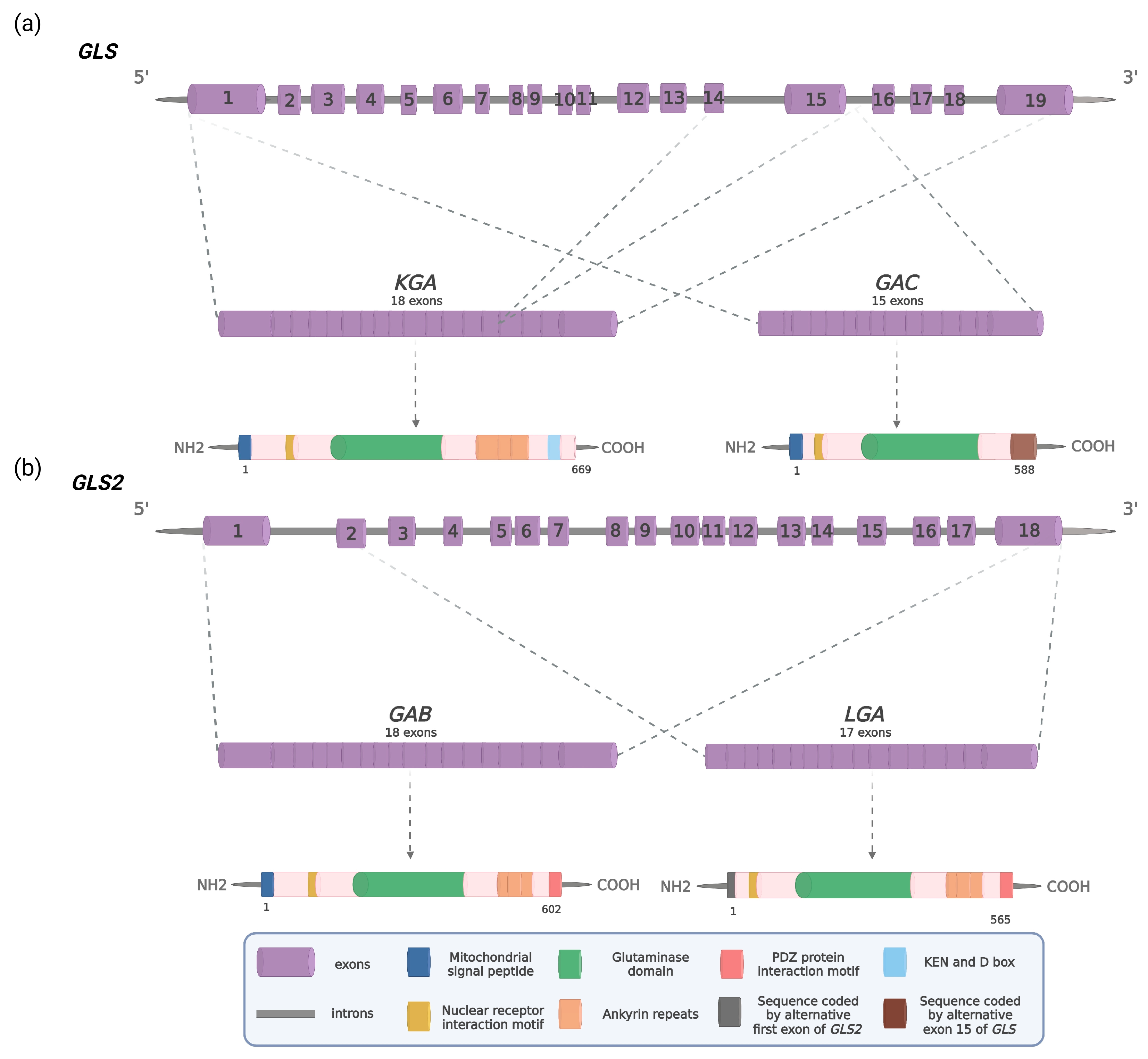
Figure 1. Schematic illustration of human glutaminases: (a) The GLS gene and two alternative transcripts and proteins KGA and GAC; (b) The GLS2 gene and two alternative transcripts and proteins GAB and LGA. Introns are depicted as a solid grey line and exons as numbered purple cylinders. Dashed gray lines indicate the exons forming KGA, GAC, GAB, and LGA mRNA transcripts, respectively. Dashed gray arrows indicate KGA, GAC, GAB, and LGA proteins with defined main signature sequences and motifs. Adapted with permission from [13]. 2016, SNCSC. Adapted with permission from [14]. 2015, Elsevier. Created with BioRender.com.
Cancers 15 05566 g001
Figure 2. Molecules which regulate GLS2 expression. Based on [39,41,42,43,44,45,46,47,48,49,50,51,52,53,54]. Created with BioRender.com.
Cancers 15 05566 g002
Table 1. Experimental studies indicating that GLS2 acts as a tumor promoter.
CancerModelOutcomeRef.
Effects of GLS2 Inhibition
Non-small cell lung cancer (NSCLC)A549 cells with GLS2 knockdownDecreased cell growth[75]
Hepatocellular carcinoma (HCC)HepG2 cells with GLS2 knockdown or AV-1 treatmentDecreased cell growth, inhibited anchorage-independent colony formation, autophagy induction, P13K/Akt inhibition, suppressed mTORC1 activity
Lung squamous-cell carcinoma (SCC)QG56 cells with GLS2 knockdownDiminished viability, decreased mTORC1 activity[73]
NeuroblastomaMYCN-amplified Kelly and BE-2C cells with GLS2 knockdownReduced proliferation and clonogenic potential,
attenuated tumors forming ability, decreased GSH, α-KG and ATP level,
increased ROS level, inhibited Gln-dependent anaplerosis of TCA cycle and aerobic glycolysis
[43]
Cervical cancerHeLa and HeLaR cells with GLS2 knockdownIncreased intracellular ROS levels, decreased production of NADH, NADPH, GSH, enhanced radiosensitivity[77]
mouse model injected with HeLa, HeLaR and HeLaR cells with GLS2 knockdownEnhanced radiosensitivity
Luminal-subtype breast cancerMDA-MB-453 cells with GLS2 knockdownReduced proliferation,
suppressed glutamine-mediated TCA cycle anaplerosis
[47]
mouse model injected with MDA-MB-453 cellsReduced tumorigenesis
Triple-negative (TN) breast cancerTN and non-TN cells with GLS2 knockdownDiminished cell proliferation, increased oxidative stress[79]
HMLE cells with GLS2 knockdownGln independence, reduced mitochondrial activity[81]
Effects of GLS2 Overexpression
Triple-negative (TN) breast cancerTN and non-TN breast cancer cells overexpressing GLS2Elevated invasion capacity[79]
mouse model injected with MDA-MB-231 cellsPro-tumorigenic effect, decreased overall, disease-free and distant metastasis-free survival
SUM159 cells overexpressing GLS2Enhanced mitochondrial activity, glutamine independence, inhibited mammosphere formation, higher intracellular GSH and ATP level, higher basal respiration, reduced cells viability[81]
Table 2. Small molecule allosteric inhibitors showing at least partial specificity for GLS2.
CompoundClassSelectivityAnticancer Activity
AV-1 (ardisianone)alkyl
benzoquinone
approximately tenfold selectivity against GLS2 over GLS [75]human hormone-refractory prostate cancer cells PC-3 and DU-145145 [83], acute myeloid leukemia cells HL-60 [84]
986benzophenanthridinonethreefold selectivity against GLS2 over GLS [47]breast cancer cell lines DU4475 [85], MDA-MB-453, MDA-MB-453
xenografts [47],
NSCLC cell lines A549, H23, H1299, and Spc-A1 [86],
ovarian cell lines HEY, SKOV3, IGROV-1 [87],
endometrial cancer cell lines Ishikawa and HEC-1B [88],
HCC cell lines LM3, 7402 and HepG2 [89]
derivative 6thiazoli-dine-
2,4-dione
twofold selectivity against GLS over GLS2 [90]human pancreas adenocarcinoma AsPC-1 cells
AsPC-1 mice xenografts [90]
Table 3. Experimental studies indicating that GLS2 acts as a tumor suppressor.
CancerModelOutcomeRef.
Effects of GLS2 Inhibition
Hepatocellular carcinoma (HCC)HepG2 cell lineIncreased ROS production, sensitization to H2O2-induced apoptosis, decreased oxygen consumption, α-ketoglutarate and ATP levels[39]
PLC/PRF/5 cell line with GLS2 knockdownEnhanced anchorage-independent cell growth and formation of tumors[45]
HepG2, HCC36 and Mahlavu cell lines with GLS2 knockdownEnhanced cell migration and invasion, diminished level of Snail, reduction of miR-34a expression[93]
Mice with GLS2 knockoutEarlier development of larger tumors, resistance to ferroptosis, increased level of MDA and oxidative stress[103]
Effects of GLS2 Overexpression
Hepatocellular carcinoma (HCC)HepG2 cells overexpressing GLS2Reduced cell colony formation[39]
Huh1 and Huh7 cells overexpressing GLS2Reduced xenograft tumor growth, downregulation of PI3K/AKT signaling pathway[45]
PLC/PRF/5 cells with GLS2 knockdownDownregulation of PI3K/AKT signaling pathway
Mahlavu and Huh7
cells overexpressing GLS2
Decreased cell migration and invasion, stabilization of Dicer protein, induction of Dicer-dependent miR-34a maturation, repressed EMT, motility and metastasis[93]
Huh-1 cells co-transduced with GLS2 and RAC1Inhibited cell migration, invasion, metastasis and Rac1 activity[98]
HepG2, HepG3, SKHep1 cells overexpressing GLS2Enhanced ferroptosis through αKG-dependent increase of lipid ROS[99]
Mice model injected with SKHep1 cells overexpressing GLS2Reduced tumor formation, increased expression of ferroptosis markers
Colon cancerHCT116 cells overexpressing GLS2Reduced viability and number of cell colonies, increased number of cells in G2/M phase, reduced levels of p21 and cyclin D1[44]
Gastric cancerMGC-803 cells overexpressing GLS2Suppressed cell viability and ability to migration, induced apoptosis[103]
GlioblastomaT98G, U87MG and LN229 cells overexpressing GLS2Decreased cells viability, proliferation, ability to form colonies and migration,
increased TMZ and H2O2-mediated oxidative stress sensitivity, downregulation of the PI3K/AKT pathway
[106,107]
T98G, SFxL and LN229 cells overexpressing GLS2Inhibition of cell migration, decreased apoptosis, antioxidant status and cellular motility, H2O2 and ATO sensitivity[29]
T98G and LN229 cells overexpressing GLS2Reduced CAT activity, increased total antioxidant capacity, decreased lipid peroxidation, miRNA-140-3p, miRNA-1246, miRNA-1260a, miRNA-21-5p and miRNA-146a-5p overexpression[112]
LN229 cells overexpressing GLS2Diminished SOD activity, miRNA-92a-3p upregulation
U251 and U251TR cells overexpressing GLS2; intracranial U251TR modelDiminished cell proliferation and migration, increased lipid ROS and E-cadherin level, decreased vimentin level, repressed EMT[53]
T98G cells overexpressing GLS2Decreased cell proliferation, increased level of p53 in nuclei, decreased level of c-Myc in cytoplasm[58]
NeuroblastomaSH-SY5Y cells treated with PMAInhibited proliferation, increased p53 and p21 level, cell cycle arrested at G2/M stage
Disclaimer/Publisher’s Note: The statements, opinions and data contained in all publications are solely those of the individual author(s) and contributor(s) and not of MDPI and/or the editor(s). MDPI and/or the editor(s) disclaim responsibility for any injury to people or property resulting from any ideas, methods, instructions or products referred to in the content.