Article

YAP1 Expression in HR+HER2− Breast Cancer: 21-Gene Recurrence Score Analysis and Public Dataset Validation

1
Department of Pathology, Gangnam Severance Hospital, Yonsei University College of Medicine, Seoul 06273, Republic of Korea
2
Center for Precision Medicine, Gangnam Severance Hospital, Yonsei University College of Medicine, Seoul 06273, Republic of Korea
3
Institute of Breast Cancer Precision Medicine, Yonsei University College of Medicine, Seoul 06273, Republic of Korea
4
Division of Medical Oncology, Department of Internal Medicine, Yonsei University College of Medicine, Seoul 06273, Republic of Korea
5
Department of Surgery, Gangnam Severance Hospital, Yonsei University College of Medicine, Seoul 06273, Republic of Korea
*
Author to whom correspondence should be addressed.
Cancers 2023, 15(20), 5034; https://doi.org/10.3390/cancers15205034
Submission received: 26 September 2023 / Revised: 13 October 2023 / Accepted: 16 October 2023 / Published: 18 October 2023
(This article belongs to the Special Issue Advances in Invasive Breast Cancer: Treatment and Prognosis)

Abstract

:

Simple Summary

YAP1, a downstream transcription factor of the Hippo pathway, is regarded as an oncogene in various solid tumors. This study explores the relationship between YAP1 expression and the risk score from the Oncotype Dx test in patients with hormone-receptor-positive, HER2-negative (HR+HER2−) breast cancer. In a retrospective review of 401 patients using YAP1 nuclear localization via immunohistochemical staining and clinicopathologic analysis, high-YAP1 expression significantly correlated with a lower risk score. In a public dataset analysis, elevated YAP1 mRNA expression was associated with better clinical outcomes, particularly in ER-positive patients. In summary, YAP1 could serve as a prognostic marker as well as potential therapeutic target in HR+HER2− breast cancer patients.

Abstract

Background: YAP1, an oncogene in numerous cancers, is a downstream transcription factor of the Hippo pathway. This study focuses on its relationship with the Oncotype Dx (ODX) test risk score (RS) in patients with hormone-receptor-positive, HER2-negative (HR+HER2−) breast cancer. Methods: We retrospectively analyzed 401 HR+HER2− breast cancer patients from Gangnam Severance Hospital who underwent ODX tests (May 2014–April 2020). YAP1 nuclear localization was evaluated via immunohistochemical staining and its clinical correlation with clinicopathological parameters, including RS, was analyzed. Public datasets TCGA-BRCA and METABRIC validated clinical outcomes. Results: YAP1 expression negatively correlated with ODX RS (OR 0.373, p = 0.002). Elevated YAP1 mRNA levels corresponded to better clinical outcomes, specifically in ER-positive patients, with significant results in METABRIC and TCGA-BRCA datasets (p < 0.0001 OS in METABRIC, p = 0.00085 RFS in METABRIC, p = 0.040 DFS in TCGA-BRCA). In subsets with varying ESR1 mRNA expression and pronounced YAP1 expression, superior survival outcomes were consistently observed. Conclusion: YAP1 may be a valuable prognostic marker and potential therapeutic target in HR+HER2− breast cancer patients.

1. Introduction

Breast cancer is the most commonly diagnosed cancer in women worldwide [1,2]. Clinically, breast cancer is subdivided into three subtypes based on the immunohistochemistry (IHC) status of hormone receptors’ (HRs) estrogen receptor (ER) and progesterone receptor (PR) and human epidermal growth factor receptor 2 (HER2). Among them, HR-positive-HER2-negative (HR+HER2−) breast cancer is the most predominant subtype, accounting for nearly 70% of breast cancer [2]. HR+HER2− breast cancer is generally less aggressive and has anti-hormonal therapeutic options such as tamoxifen, with a relatively superior prognosis compared to other subtypes [2]. Oncotype DX 21-gene Breast Cancer Recurrence Score ® assay (ODX RS) (Genomic Health; Redwood City, CA, USA) is a precise genomic test based on the gene expression level of 21 genes in breast cancer tumor tissue [3], provided the recurrence score (RS) is (0–100). A high RS (usually >26) indicates that the patient belongs to the high-risk group and might benefit from chemotherapy [3].
Yes-associated protein 1 (YAP1) and transcriptional coactivator with PDZ-binding motif (TAZ) are downstream transcription factors of the Hippo pathway. When the Hippo pathway is disrupted, unphosphorylated YAP1/TAZ moves into the nucleus of the cells and, together with the TEA domain transcription factor (TEAD), upregulate the transcription of multiple genes involved in cell proliferation, apoptosis, and survival [4]. YAP1, together with TAZ, has been considered an oncogene as overexpression or activation of YAP1 correlated with poor prognosis with cancer of variable organs, including ovary, [5,6] lung, [7] esophagus, [8] colon, [9], and pancreas [10]. In breast cancer, there have been controversies on the role of YAP1 in tumor biology. YAP1 was reported as tumor suppressor in some studies [11,12], whereas it was correlated with poor prognosis in others [13,14,15].
In a previous study, we showed that YAP1 nuclear expression was increased along with tumor stiffness measured by shear-wave elastography in HR+HER2− breast cancer [16]. Tumor stiffness is derived from tumor cell invasion beyond the basement membrane and the production of collagen by cancer-associated fibroblasts in the extracellular matrix of the tumor microenvironment, which is also known as desmoplastic reaction [17]. High tumor stiffness in breast cancer is an indicator of aggressive histologic features and differentiates high-risk patient groups [18,19,20,21]. Based on the positive correlation between YAP1 expression and tumor stiffness in HR+HER2− breast cancer, we further aimed to evaluate whether YAP1 expression correlates with ODX RS. As most of the patients with HR+HER2− breast cancer have excellent 5-year survival with very rare events to analyze in the limited period, we expected that ODX RS could serve as a surrogate marker for a prognosis for patients with HR+HER2− breast cancer.

2. Materials and Methods

This study was approved by the Institutional Review Board of Gangnam Severance Hospital (3-2022-0119) and adhered to the clinical practice guidelines of the Declaration of Helsinki (2013 amendment). Informed consent was waived for all patients due to the retrospective design of this study.

2.1. Patients

We retrospectively selected patients who underwent upfront curative surgery followed by adjuvant treatments for breast cancer at Gangnam Severance Hospital in Seoul, Korea, from May 2014 to April 2020. The clinical and pathological data of the patients were obtained by reviewing the electronic medical records (EMRs). Stages were determined according to the 8th edition of the American Joint Committee on Cancer staging system. Clinicopathologic parameters evaluated in each case from EMR included patient age at initial diagnosis, menopausal status, tumor size, histologic grade (HG) based on the Nottingham grading system [22], lymphovascular invasion (LVI), lymph node metastasis, tumor recurrence, distant metastasis, patient survival, and ODX RS.
The inclusion and exclusion criteria were as follows:
  • Inclusion criteria:
    • patients aged ≥20 years;
    • invasive breast cancer confirmed by pathological diagnosis;
    • available ODX RS;
    • ER- and/or PR-positive and HER2-negative cancer.
  • Exclusion criteria:
    • any other carcinoma in situ;
    • other cancer histories (except for thyroid cancer);
    • inaccessible electronic medical records;
    • received neoadjuvant chemotherapy (NAC).

2.2. Oncotype Dx® Assays

The ODX assay was performed using RNA extracted from formalin-fixed, paraffin-embedded (FFPE) tissue and supplied by Genomic Health (Redwood City, CA, USA). RNA was extracted from unstained sections containing sufficient invasive breast cancer tissue of appropriate quality. Patients with an RS of 26 or higher were assigned to a high-risk group based on the TAILORX trial [23].

2.3. Pathologic Review of Breast Cancer Slides

2.3.1. Histologic Evaluation of the Tumor–Stroma Ratio (TSR) and Tumor-Infiltrating Lymphocytes (TILs)

Histology slides of patients were reviewed by two breast pathologists (Y.K. and Y.J.C.). The tumor–stroma ratio (TSR) is defined as tumor cellularity relative to the surrounding stroma in the overall tumor bed [16,24]. The TSR assessment was conducted using scoring percentages in 10% increments. For statistical analysis, cases with ≤50% TSR were assigned to the stroma-high group, and those with >50% TSR were assigned to the stroma-low group. The tumor-infiltrating lymphocyte (TIL) level was concurrently evaluated according to the guidelines suggested by the International TIL Working Group [25]. Except for polymorphonuclear leukocytes, other mononuclear cells, including lymphocytes and plasma cells, were counted. For statistical analysis, a 10% cutoff was applied to separate patients into low-TIL (<10%) and high-TIL (≥10%) groups.

2.3.2. IHC for Clinical Subtype

Nuclear staining values of 1% or higher were considered positive for ER (clone 6F11; dilution 1:200; Leica Biosystems, Wetzlar, Germany) and PR (clone 16; dilution 1:500; Leica Biosystems, Wetzlar, Germany) [26]. HER2 (clone 4B5; dilution 1:5; Ventana Medical System, Oro Valley, AZ, USA) staining was performed according to the 2018 American Society of Clinical Oncology/College of American Pathologists [27]. Only samples with strong and circumferential membranous HER2 immunoreactivity (3+) were considered positive, whereas those with 0 or 1+ HER2 staining were considered negative. Cases with equivocal HER2 expression (2+) were further evaluated for HER2 gene amplification via silver in situ hybridization (SISH). Positive nuclear Ki67 (clone MIB; dilution 1:1000; Abcam, Cambridge, UK) staining was assessed based on the percentage of positive tumor cells, defined as the Ki67 labelling index (LI). ER- and/or PR-positive and HER2-negative cases were selected for this study.

2.4. Tissue Microarray (TMA) Construction

Hematoxylin and eosin (H&E)-stained slides from the resected breast cancer specimens were examined and representative areas marked. The matched tissue cores (2 mm) were extracted from FFPE tumor blocks and placed into 5 × 10 recipient TMA blocks. Each tissue core was assigned a unique TMA location number that was linked to a database containing other clinicopathologic data.

2.5. YAP1 IHC and Interpretation

Briefly, 3 µm thick tissue sections were cut from the FFPE tissue block of the TMA blocks. After deparaffinization and rehydration with graded xylene and alcohol solutions, IHC was performed using a Ventana Discovery XT Automated Slide Stainer (Ventana Medical System, Tucson, AZ, USA). Cell conditioning 1 (CC1) buffer (citrate buffer, pH 6.0; Ventana Medical System) was used for antigen retrieval. Whole tissue slides were stained with an anti-YAP1 antibody (clone 63.7; dilution 1:200; Santa Cruz Biotechnology, Dallas, TX, USA). After staining, nuclear YAP1 expression was assessed by two breast pathologists (YL and YJC; 400× magnification). Nuclear staining was evaluated using the H-score, which was obtained by multiplying the staining intensity (0, 1, 2, or 3) by the percentage of stained area (%). The myoepithelial cells’ nuclear staining intensity was assigned a value of moderate intensity and used as an internal control. Weaker and stronger signals were assigned a value of weak and strong intensities, respectively. Negative or weak nuclear staining were categorized as low expression, while moderate or strong nuclear staining were grouped as high expression (Figure 1). The IHC results were interpreted blindly, without any information regarding clinical parameters or outcomes.

2.6. Public Dataset Analysis

We investigated the relationship between YAP1 expression and survival outcomes, specifically overall survival (OS) and event free survival (EFS), across various subgroups within the two publicly available datasets: The Cancer Genome Atlas Breast Invasive Carcinoma (TCGA-BRCA) and The Molecular Taxonomy of Breast Cancer International Consortium (METABRIC). The clinical and gene expression data were obtained from the cBioPortal (https://www.cbioportal.org, accessed on 9 May 2023). In our analysis, we considered disease-free survival (DFS) from the TCGA-BRCA dataset and relapse-free survival (RFS) from METABRIC as EFS. Subgroups analyzed included IHC based ER-positive (ER+)/negative (ER−) and PAM50-based molecular subtypes. We utilized gene expression data in the form of z-scores relative to normal samples, which were obtained from the cBioPortal.
Within each subgroup, we evaluated the differences in survival outcomes between the dichotomized samples for every potential YAP1 expression threshold, covering the range from the 10th to the 90th percentile of YAP1 expression within the tumor samples. The optimal dichotomization was determined based on the p-value obtained from the log-rank survival difference test. Survival outcome analysis based on YAP1 expression was conducted in patients with varying levels of ESR1 expression, particularly those with ESR1 levels in the upper 20-, 40-, 60-, and 80 percentiles.

2.7. Statistical Analysis

The continuous variables between the two groups were compared using the Student’s t-test or the Mann–Whitney test. The categorical variables were compared by using the Chi-square test or Fisher’s exact test. Survival curves were obtained using the Kaplan–Meier method and two-group comparisons were made using the log-rank test. Univariate and multivariate regression analyses were conducted to identify the significant parameters. Statistical analyses were performed using SPSS version 24 (IBM co Chicago, IL, USA) and R software (https://www.r-projet.org; version 4.3.0, accessed on 9 May 2023). The threshold for statistical significance was set at p < 0.05, with a 95% confidence interval (CI) not including 1.

3. Results

3.1. Basal Characteristics of the Study Population

Data from a total of 401 female patients were evaluated and summarized in Supplementary Table S1. The median age of patients was 49 years (range 25–81 years). Patients with premenopausal status were larger in number (n = 235, 58.6%) than those with menopausal status (n = 155, 38.7%). Most patients were in the early stage of the tumor (pT1, n = 246, 61.3%; pT2, n = 154, 38.4%; pT3, n = 1, 0.2%) with a mean tumor size of 1.9 cm, and invasive ductal carcinoma was the most predominant histologic subtype (91.0%). The mean RS of the ODX assay was 18.1, and applying the RS 26 cutoff, 340 (84.8%) and 61 (15.2%) patients were assigned to low- and high-risk groups, respectively. Regarding pathologic parameters, histologic grade (HG) II was the most common (n = 311, 77.6%), followed by HG I (n = 63, 15.7%) and HG III (n = 27, 6.7%). LVI was detected in 122 cases (30.4%), and lymph node metastasis was present in 96 (23.9%) cases. Overall, the mean TIL level was 11.2%, and using a 10% cutoff, 302 (75.3%) and 99 (24.7%) cases were subgrouped as low- and high-TIL groups, respectively. The mean TSR was 71.0%, and using a 50% cutoff, 344 (86.0%) and 56 (14.0%) cases were classified into stroma-low and stroma-high group, respectively. With dichotomized YAP1 scoring, 139 (34.7%) and 262 (65.3%) cases corresponded to low- and high-YAP1 groups, respectively.

3.2. Comparison of Clinicopathologic Factors Based on YAP1 Expression and ODX RS

We investigated the characteristics of each group based on the YAP1 expression and ODX RS. First, we compared the clinicopathologic parameters, including ODX RS, between low- and high-YAP1 groups (Table 1). The low-YAP1 group showed a significantly higher ODX RS (20.4 ± 11.6 vs. 16.8 ± 7.2, p = 0.001), and a had significantly larger proportion of patients in the high-risk group (25.9% vs. 9.5%, p < 0.001) than those in high-YAP1 group. The distribution of HG differed; specifically, HG III was significantly frequent, four times higher, in the low-YAP1 group than in the high-YAP1 group (12.9% vs. 3.4%, p = 0.001). The proliferation index was significantly higher in the low-YAP1 group (13.8 ± 9.5 vs. 10.9 ± 10.9, p = 0.007). Conversely, there was no difference in menopausal status, tumor size, lymph node metastasis, LVI, TIL, or TSR based on the YAP1 expression.
Second, we performed the t-test using the ODX risk group (Table 2). The high-risk group had a considerably larger number of patients with menopause than the low-risk group (60.7% vs. 34.7%, p = 0.001), as well as aggressive histologic features, including HG III (p < 0.001) and higher Ki67 LI (p < 0.001). However, lymph node metastasis (6.6% in high-risk group vs. 27.1% in low-risk group, p = 0.001) and LVI (19.7% in high-risk group vs. 32.4% in the low-risk group, p = 0.047) were significantly more frequent in the low-risk group. No significant difference was found in tumor size, TIL, or TSR in the RS group.

3.3. Correlation of YAP1 Expression and ODX RS Using Regression Analysis

In univariate linear regression analysis with ODX RS as a continuous variable, significantly related parameters with higher ODX RS were menopause status (p = 0.021), higher TIL level (p = 0.006), higher Ki67 LI (p < 0.001), higher HG (p < 0.001), absence of lymph node metastasis (p < 0.001), and low YAP1 expression (p < 0.001) (Table 3). Multivariate linear regression analysis revealed menopause status (p = 0.047) and higher Ki67 LI (p < 0.001) as positively correlated factors with ODX RS, whereas lymph node metastasis (p = 0.003) and YAP1 expression (p = 0.002) showed a significant negative correlation with ODX RS (Table 4).
Additionally, logistic regression analysis was performed to find the significant parameters of the ODX high-risk group (Table 4). In univariate analysis, menopause and HG III were significantly associated with RS ≥ 26 (menopause: odds ratio [OR] 2.890, 95% CI 1.639–5.096, p < 0.001; HG III: OR 10.14, 95% CI 2.846–36.136, p < 0.001). High-TIL and high Ki67 LI also showed significant association with ODX high-risk group, with an OR of 1.017 and 1.078, respectively. Lymph node metastasis and high YAP1 expression showed significant association with the ODX low-risk group of RS < 26 (lymph node metastasis: OR 0.189, 95%CI 0.067–0.536, p = 0.002; high YAP1: OR 0.302, 95% CI 0.172–0.528, p < 0.001). In multivariate analysis, menopause (OR 2.897, 95% CI 1.538–5.454, p < 0.001), HG III (OR 4.625, 95% CI 1.070–19.990, p = 0.040), and high Ki67 LI (OR 1.062, 95% CI 1.035–1.090, p < 0.001) showed significant association with RS ≥ 26. With lymph node metastasis (OR 0.270, 95% CI 0.091–0.801, p = 0.018), high YAP1 expression remained an independently significant parameter with ODX low risk, RS ≤26 (OR 0.373, 95% CI 0.198–0.703, p = 0.002).

3.4. Validation of the Prognostic Effect of YAP1 Expression in Public Datasets

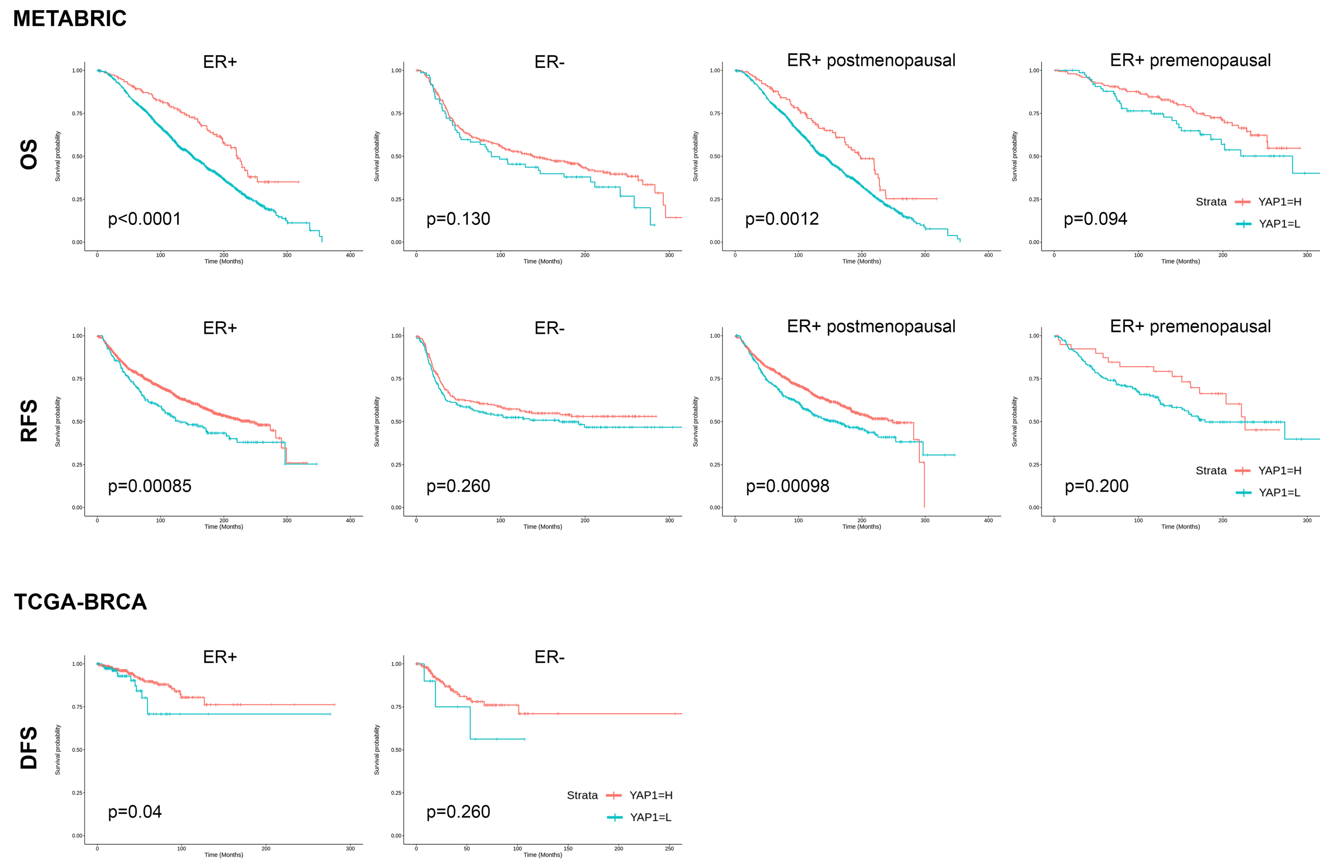
In METABRIC and TCGA-BRCA datasets, high YAP1 expression showed superior clinical outcomes in ER+ patients, whereas no significant difference was found in ER-negative patients (ER+ vs. ER−: p < 0.0001 vs. p = 0.130, OS in METABRIC; p = 0.00085 vs. p = 0.260, RFS in METABRIC; p = 0.040 vs. p = 0.260, DFS in TCGA-BRCA) (Figure 2). In the METABRIC dataset, the prognostic effect of YAP1 expression level on ER+ patients was only significant in patients of postmenopausal status (OS, p = 0.012; RFS, p = 0.00098).
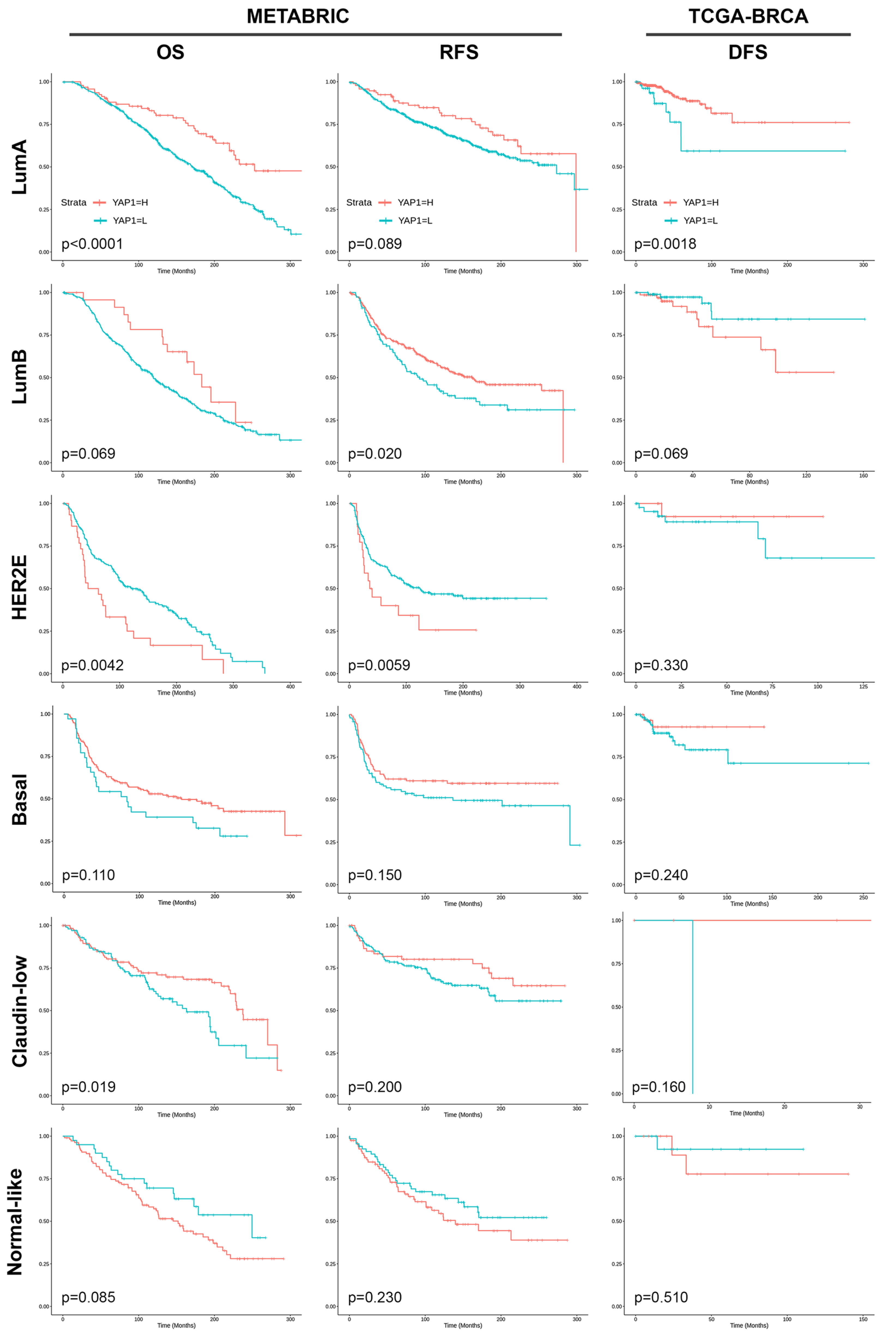
Based on the molecular subtype, high YAP1 expression demonstrated superior OS in the luminal A subtype (p < 0.0001), while exhibiting significantly improved RFS specifically in the luminal B subtype (p = 0.020) and a tendency towards better RFS in the luminal A subtype (p = 0.089) within the METABRIC dataset. Conversely, high YAP1 expression in HER2 molecular subtype showed significantly worse OS (p = 0.0042) and a tendency of worse RFS (p = 0.059). In the TCGA-BRCA dataset, high YAP1 expression was significantly associated exclusively with the luminal A subtype (p = 0.0018). Patients with high YAP1 expression tended to have worse DFS in the luminal B subtype (p = 0.069) (Figure 3).
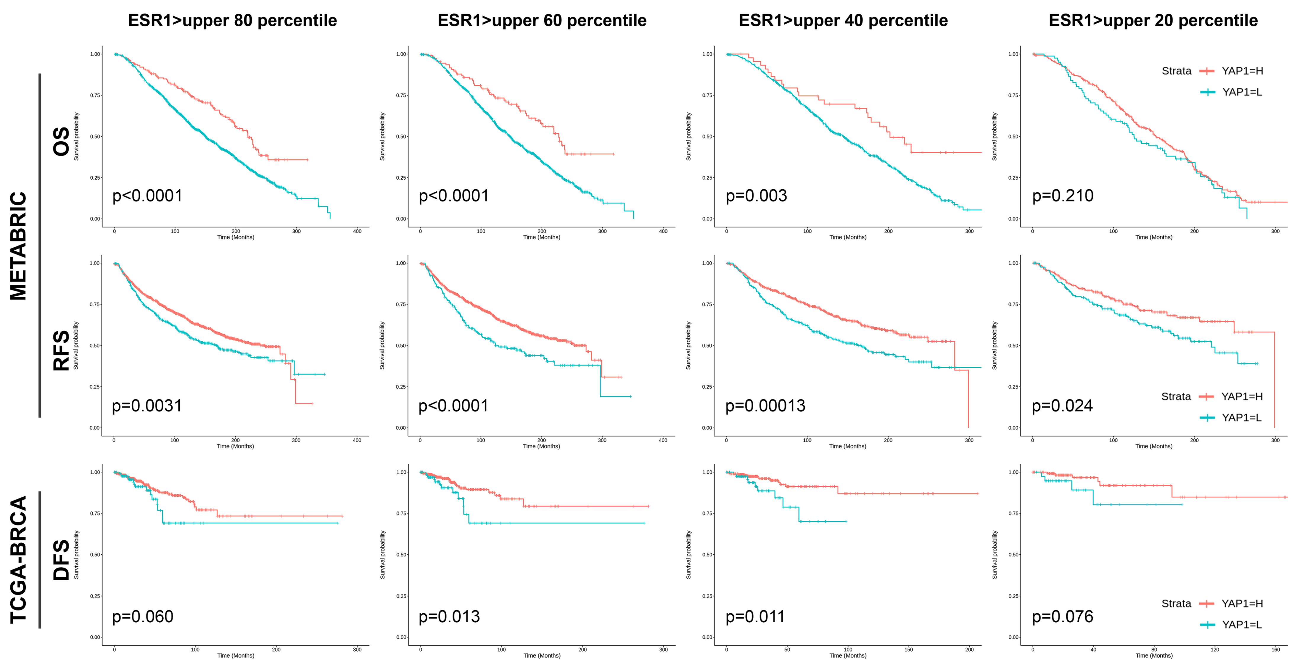
With different ESR1 expression levels in the METABRIC dataset, patients having ESR1 level in the upper 80, 60, and 40 percentiles, high YAP1 expression consistently demonstrated superior OS (p < 0.0001, p < 0.0001, and p = 0.003, respectively) and RFS (p = 0.0031, p < 0.0001, and p = 0.00013, respectively). Conversely, for patients in the upper 20 percentiles of ESR1 expression level, high YAP1 expression still showed better RFS (p = 0.024) but not significant OS (p = 0.210). In the TCGA-BRCA dataset, patients within upper 60 and 40 ESR1 expression percentiles exhibited superior DFS with high YAP1 expression (p = 0.013 and p = 0.011, respectively), whereas those in the upper 80 and 20 percentiles of ESR1 levels only showed a tendency towards better DFS (p = 0.060 and p = 0.076, respectively) (Figure 4).

4. Discussion

In this study, YAP1 activation was clearly correlated with lower RS and independently predicted the lower RS. This result implies that YAP1 functions like a tumor-suppressor in HR+HER− breast cancer, which was further supported by the analytic result of public datasets. Patients with HR+HER2− breast cancer have the most favorable prognosis, with 94.8% of 5-year relative survival rate [28]. However, drug resistance to hormone therapy develops over time, and at the advanced stage, the relative survival rate of patients with HR+HER2 breast cancer is decreased, even worse than that of patients with HER2+ breast cancer [28].
ODX is a multigene test that predicts the benefit of chemotherapy in patients with HR+HER2− breast cancer. In low-grade breast cancer cases, chemotherapy administration has no therapeutic benefit that overcomes the side effects of treatment. ODX RS ≥ 26 is the general cutoff for patients at high risk that might benefit from additional chemotherapy. Since surgically treated HR+HER2− breast cancer has nearly a 100% 5-year survival, we used ODX RS as a surrogate for tumor aggressiveness in this study.
YAP1 has been described as an oncogene in diverse organs [5,6,8,9,10]. However, in breast cancer, controversies regarding the role of YAP1 in tumor biology exist [11,15,29,30,31]. In our previous study with triple-negative breast cancer (TNBC), YAP1 activation was correlated with a poor clinical outcome [13]. As multiple previous studies did not specifically examine the subcellular localization of YAP1 expression (nucleus or cytoplasm) [11,29,32], we conducted a comprehensive YAP1 immunohistochemistry (IHC) interpretation to confirm its nuclear localization and determine YAP1 activation. Another previous study of our group showed a correlation between YAP1 expression and tumor stiffness in HR+HER2− breast cancer tissue [16]. As tumor stiffness in breast cancer is associated with aggressive features affecting prognosis [18,19,20], we expected a correlation between high YAP1 expression and high ODX RS in this study.
Regarding ODX RS, high RS showed significant correlation with high HG and high Ki67 LI, which are classical parameters of tumor aggressiveness. What was unexpected was the inverse correlation between RS and LVI or lymph node metastasis; however, this appeared to be an intrinsic limitation of ODX assay, as it focused on the proliferation and invasion of primary breast cancer cells [3]. Several studies have highlighted the limited reliability of ODX assay in nodal burden prediction [33,34,35].
In this study, YAP1 expression showed a different pattern compared to our previous study with TNBC [13]. In HR+HER2− breast cancer, high YAP1 expression was associated with low HG, low Ki67 LI, and low ODX RS. This favorable prognostic impact of YAP1 expression was also seen in the public datasets analyses, which showed significant superior survival in high-YAP1 group of IHC-defined ER+ breast cancer. Recently, several studies regarding the Hippo signaling pathway and ERα regulation suggested the inhibitory role of YAP1 on ER+ breast cancer growth [36,37,38]. In ER+ breast cancer cell lines, LATS1/2, upstream inhibitors of YAP1, are required to maintain ER+ cancer cell growth while little effect was observed in ER− cancer cells [38]. Further study showed that YAP1, together with TEAD, targets VGLL3, which recruits NCOR2 and represses ESR1 transcription [37]. YAP1 physically interrupts the ERα/TEAD interaction by competing with ERα in ER+ breast cancer cells [36]. In in vitro analysis, ERα is dissociated from its target promoters/enhancers by YAP1, which results in ERα degradation and subsequent ESR1 gene downregulation [36]. Collectively, YAP1 acts differently in ER+ breast cancer cells by inhibiting the downstream signaling pathway of the ESR1 gene, which is crucial for ER+ tumor growth. Specifically, the favorable prognosis of high-YAP1 tumor in incremental ESR1 expression supports the inhibitory role of YAP1 on ESR1 signaling. However, in tumors of ESR1-level upper 20 percentiles, YAP1 seemed to have less effect on the favorable clinical outcome. Those high-ESR1 expression tumors are exclusively composed of a comparable number of luminal A and luminal B tumors (Supplementary Figure S1). A luminal B tumor is an aggressive and proliferative subtype, which might not be solely dependent on ESR1 signaling. In addition, YAP1 might not be sufficient to inhibit the overwhelmingly expressed ESR1.
So far, there have been few studies reporting associations between YAP1 expression and favorable outcome in patients with luminal breast cancer [30,32]. Additionally, these studies had limitations in interpreting YAP1 subcellular localization or were conducted on small luminal cohorts. Therefore, the present study might be the first large-cohort study specifically comprised of HR+HER2− breast cancer with precise YAP1 IHC interpretation and validation of its clinical impact using public datasets. Also, we used human breast tissues and observed significant correlations between YAP1 expression and ODX RS, which confirmed previous in vitro studies [36,37,38], and clarified the role of YAP1 in breast cancer.
In the disease course of HR+HER2− breast cancer, the development of tamoxifen-resistance is an important clinical issue, which cannot be predicted with an ODX assay. The regulation of YAP1 expression could help in overcoming tamoxifen resistance [36,37,39,40]. The mechanism of endocrine resistance in ER+ breast cancer includes the loss of ERα expression, the mutation of the ESR1 gene, and the activation of alternative signaling pathways, such as HER2 [41]. In cases with lost ERα expression, YAP1 inhibition could restore the ERα expression that sensitizes the tamoxifen-resistant luminal cancer cell line [40]. Conversely, if mutant ERα causes tamoxifen resistance, the inhibition of the upstream molecules of YAP1, MST1/2, or LATS1/2 induce YAP1 activation, which could repress the downstream signaling of the ESR1 gene and overcome the resistance [36,37,39]. Notably, high YAP1 expression appeared to be more beneficial to postmenopausal than to premenopausal patients in this study. This could be explained by the lower estrogen levels in postmenopausal patients, which may have a similar effect to tamoxifen or facilitate YAP1 interference with ESR1 signaling more easily. The modulation of YAP1 activity could help to manage the long-term treatment plan in HR+HER2− breast cancer.
There are several limitations and future tasks to address. First, our cohort had a relatively short follow-up period with all alive patients and with only four recurrent cases. This was because we aimed to focus on YAP1 expression and ODX as sufficient follow-up data were unavailable. Although ODX RS showed a significant correlation with YAP1 expression, it only could provide the likelihood of risk. Second, our YAP1 activation was defined based on the IHC results of TMA. As YAP1 is heterogeneously expressed across the tumor, we might have missed the YAP1-positive area in low-YAP1 tumors. To compensate for these limitations, we validated our results with public datasets with larger numbers of patients with mRNA expression data; however, a more refined large dataset should be further validated combined with cancer stage, menopausal status, and tamoxifen treatment and resistance. Our study primarily concentrated on HR+HER2− breast cancer; however, the relevance of HER2, especially in contexts like ductal carcinoma in situ (DCIS), cannot be understated [42]. Furthermore, although our investigations have ascertained the detrimental prognostic implications of YAP1 in TNBC, the precise association between YAP1 expression and HER2 overexpression remains to be comprehensively elucidated. This represents a significant gap in the current body of knowledge and warrants rigorous examination in future studies. Lastly, the different role of YAP1 as a mechanotransducer in HR+HER2− breast cancer should be further elucidated in the context of clinical implication. As we observed in our previous study, YAP1 expression increased along with tumor stiffness in HR+HER2− breast cancer [16]. We could not analyze the association between YAP1, tumor stiffness, and clinical implication because of different patient cohorts. However, retrospective clinicopathological validation with the genomic dataset of advanced-stage HR+HER2− breast cancer cases might clarify the conundrum.

5. Conclusions

In conclusion, YAP1 acts as a tumor suppressor in HR+HER2− breast cancer. Interaction of YAP1 and ESR1 in nuclei is a unique feature of HR+HER2− breast cancer, and thus YAP1 could serve as a prognostic marker as well as a therapeutic target in patients with HR+HER2− breast cancer.

Supplementary Materials

The following supporting information can be downloaded at: https://www.mdpi.com/article/10.3390/cancers15205034/s1, Figure S1: Distribution of molecular subtype of breast cancer based on ESR1 expression level; Table S1: Basal characteristics of study cohort.

Author Contributions

I.P.: Data curation, formal analysis, methodology, software, validation, visualization, writing—original draft, writing—review and editing. Y.L.: Data curation, investigation, resources, writing—review and editing. J.H.K.: Data curation, investigation, resources, writing—review and editing. S.J.B.: Data curation, investigation, resources, writing—review and editing. S.G.A.: Data curation, investigation, resources, writing—review and editing. J.J.: Data curation, investigation, resources, writing—review and editing. Y.J.C.: Conceptualization, data curation, formal analysis, funding acquisition, investigation, methodology, project administration, resources, supervision, writing—original draft, writing—review and editing. All authors have read and agreed to the published version of the manuscript.

Funding

This research was supported by faculty research grants from Yonsei University College of Medicine [grant numbers: 6-2021-0159 and 6-2022-0123]. This research was supported by a National Research Foundation of Korea (NRF) grant funded by the Korean government [grant number: NRF-2021R1G1A109359612]. This research was supported by a grant of the Korea Health Technology R&D Project through the Korea Health Industry Development Institute (KHIDI), funded by the Ministry of Health & Welfare, Republic of Korea (grant number: HI22C2194).

Institutional Review Board Statement

This study was conducted according to the guidelines of the Declaration of Helsinki, and approved by the Institutional Review Board Gangnam Severance Hospital (IRB number: 3-2022-0119; date of approval: 03 May, 2022) and adhered to the clinical practice guidelines of the Declaration of Helsinki (2013 amendment).

Informed Consent Statement

Patient consent was waived due to the retrospective design of this study.

Data Availability Statement

The datasets used and/or analyzed during the current study are available from the corresponding author on reasonable request.

Acknowledgments

The authors would like to thank Yeon Soo Lee for providing excellent support with immunohistochemical staining.

Conflicts of Interest

The authors declare no conflict of interest.

References

  1. Sung, H.; Ferlay, J.; Siegel, R.L.; Laversanne, M.; Soerjomataram, I.; Jemal, A.; Bray, F. Global cancer statistics 2020: Globocan estimates of incidence and mortality worldwide for 36 cancers in 185 countries. CA Cancer J. Clin. 2021, 71, 209–249. [Google Scholar] [CrossRef] [PubMed]
  2. Surveillance Research Program, N.C.I. Seer*explorer: An Interactive Website for Seer Cancer Statistics. Data Source(s): Seer Incidence Data, November 2022 Submission (1975–2020). Available online: https://seer.cancer.gov/statistics-network/explorer/ (accessed on 22 May 2023).
  3. Paik, S.; Shak, S.; Tang, G.; Kim, C.; Baker, J.; Cronin, M.; Baehner, F.L.; Walker, M.G.; Watson, D.; Park, T.; et al. A multigene assay to predict recurrence of tamoxifen-treated, node-negative breast cancer. N. Engl. J. Med. 2004, 351, 2817–2826. [Google Scholar] [CrossRef] [PubMed]
  4. Huang, J.; Wu, S.; Barrera, J.; Matthews, K.; Pan, D. The hippo signaling pathway coordinately regulates cell proliferation and apoptosis by inactivating yorkie, the drosophila homolog of yap. Cell 2005, 122, 421–434. [Google Scholar] [CrossRef]
  5. Hall, C.A.; Wang, R.; Miao, J.; Oliva, E.; Shen, X.; Wheeler, T.; Hilsenbeck, S.G.; Orsulic, S.; Goode, S. Hippo pathway effector yap is an ovarian cancer oncogeneyap is an ovarian cancer oncogene. Cancer Res. 2010, 70, 8517–8525. [Google Scholar] [CrossRef]
  6. Zhang, X.; George, J.; Deb, S.; Degoutin, J.L.; Takano, E.A.; Fox, S.B.; Bowtell, D.D.; Harvey, K.F. The hippo pathway transcriptional co-activator, yap, is an ovarian cancer oncogene. Oncogene 2011, 30, 2810–2822. [Google Scholar] [CrossRef]
  7. Wang, Y.; Dong, Q.; Zhang, Q.; Li, Z.; Wang, E.; Qiu, X. Overexpression of yes-associated protein contributes to progression and poor prognosis of non-small-cell lung cancer. Cancer Sci. 2010, 101, 1279–1285. [Google Scholar] [CrossRef]
  8. Muramatsu, T.; Imoto, I.; Matsui, T.; Kozaki, K.-I.; Haruki, S.; Sudol, M.; Shimada, Y.; Tsuda, H.; Kawano, T.; Inazawa, J. Yap is a candidate oncogene for esophageal squamous cell carcinoma. Carcinogenesis 2011, 32, 389–398. [Google Scholar] [CrossRef]
  9. Cho, S.Y.; Gwak, J.W.; Shin, Y.C.; Moon, D.; Ahn, J.; Sol, H.W.; Kim, S.; Kim, G.; Shin, H.M.; Lee, K.H.; et al. Expression of hippo pathway genes and their clinical significance in colon adenocarcinoma. Oncol. Lett. 2018, 15, 4926–4936. [Google Scholar] [CrossRef]
  10. Zhou, Q.; Bauden, M.; Andersson, R.; Hu, D.; Marko-Varga, G.; Xu, J.; Sasor, A.; Dai, H.; Pawłowski, K.; Said Hilmersson, K.; et al. Yap1 is an independent prognostic marker in pancreatic cancer and associated with extracellular matrix remodeling. J. Transl. Med. 2020, 18, 77. [Google Scholar] [CrossRef]
  11. Yuan, M.; Tomlinson, V.; Lara, R.; Holliday, D.; Chelala, C.; Harada, T.; Gangeswaran, R.; Manson-Bishop, C.; Smith, P.; Danovi, S.A.; et al. Yes-associated protein (yap) functions as a tumor suppressor in breast. Cell Death Differ. 2008, 15, 1752–1759. [Google Scholar] [CrossRef]
  12. Jaramillo-Rodríguez, Y.; Cerda-Flores, R.M.; Ruiz-Ramos, R.; López-Márquez, F.C.; Calderón-Garcidueñas, A.L. Yap expression in normal and neoplastic breast tissue: An immunohistochemical study. Arch. Med. Res. 2014, 45, 223–228. [Google Scholar] [CrossRef] [PubMed]
  13. Liu, C.; Zhou, J.; Chang, C.; Zhi, W. Feasibility of shear wave elastography imaging for evaluating the biological behavior of breast cancer. Front. Oncol. 2021, 11, 820102. [Google Scholar] [CrossRef] [PubMed]
  14. Kim, S.K.; Jung, W.H.; Koo, J.S. Yes-associated protein (yap) is differentially expressed in tumor and stroma according to the molecular subtype of breast cancer. Int. J. Clin. Exp. Pathol. 2014, 7, 3224–3234. [Google Scholar] [PubMed]
  15. Guo, L.; Chen, Y.; Luo, J.; Zheng, J.; Shao, G. Yap1 overexpression is associated with poor prognosis of breast cancer patients and induces breast cancer cell growth by inhibiting pten. FEBS Open Bio 2019, 9, 437–445. [Google Scholar] [CrossRef]
  16. Lee, Y.; Bae, S.J.; Eun, N.L.; Ahn, S.G.; Jeong, J.; Cha, Y.J. Correlation of yes-associated protein 1 with stroma type and tumor stiffness in hormone-receptor positive breast cancer. Cancers 2022, 14, 4971. [Google Scholar] [CrossRef]
  17. Valkenburg, K.C.; de Groot, A.E.; Pienta, K.J. Targeting the tumour stroma to improve cancer therapy. Nat. Rev. Clin. Oncol. 2018, 15, 366–381. [Google Scholar] [CrossRef]
  18. Choi, W.J.; Kim, H.H.; Cha, J.H.; Shin, H.J.; Kim, H.; Chae, E.Y.; Hong, M.J. Predicting prognostic factors of breast cancer using shear wave elastography. Ultrasound Med. Biol. 2014, 40, 269–274. [Google Scholar] [CrossRef]
  19. Patel, B.K.; Pepin, K.; Brandt, K.R.; Mazza, G.L.; Pockaj, B.A.; Chen, J.; Zhou, Y.; Northfelt, D.W.; Anderson, K.; Kling, J.M.; et al. Association of breast cancer risk, density, and stiffness: Global tissue stiffness on breast mr elastography (mre). Breast Cancer Res. Treat. 2022, 194, 79–89. [Google Scholar] [CrossRef]
  20. Gemici, A.A.; Ozal, S.T.; Hocaoglu, E.; Inci, E. Relationship between shear wave elastography findings and histologic prognostic factors of invasive breast cancer. Ultrasound Q. 2020, 36, 79–83. [Google Scholar] [CrossRef]
  21. Youk, J.H.; Son, E.J.; Jeong, J.; Gweon, H.M.; Eun, N.L.; Kim, J.A. Shear-wave elastography-based nomograms predicting 21-gene recurrence score for adjuvant chemotherapy decisions in patients with breast cancer. Eur. J. Radiol. 2023, 158, 110638. [Google Scholar] [CrossRef]
  22. Elston, C.W.; Ellis, I.O. Pathological prognostic factors in breast cancer. I. The value of histological grade in breast cancer: Experience from a large study with long-term follow-up. Histopathology 1991, 19, 403–410. [Google Scholar] [CrossRef] [PubMed]
  23. Sparano, J.A.; Gray, R.J.; Makower, D.F.; Pritchard, K.I.; Albain, K.S.; Hayes, D.F.; Geyer, C.E., Jr.; Dees, E.C.; Goetz, M.P.; Olson, J.A., Jr.; et al. Adjuvant chemotherapy guided by a 21-gene expression assay in breast cancer. N. Engl. J. Med. 2018, 379, 111–121. [Google Scholar] [CrossRef]
  24. Kemi, N.; Eskuri, M.; Herva, A.; Leppänen, J.; Huhta, H.; Helminen, O.; Saarnio, J.; Karttunen, T.J.; Kauppila, J.H. Tumour-stroma ratio and prognosis in gastric adenocarcinoma. Br. J. Cancer 2018, 119, 435–439. [Google Scholar] [CrossRef] [PubMed]
  25. Salgado, R.; Denkert, C.; Demaria, S.; Sirtaine, N.; Klauschen, F.; Pruneri, G.; Wienert, S.; Van den Eynden, G.; Baehner, F.L.; Pénault-Llorca, F. The evaluation of tumor-infiltrating lymphocytes (tils) in breast cancer: Recommendations by an international tils working group 2014. Ann. Oncol. 2015, 26, 259–271. [Google Scholar] [CrossRef] [PubMed]
  26. Hammond, M.; Hayes, D.F.; Dowsett, M.; Allred, D.C.; Hagerty, K.L.; Badve, S.; Fitzgibbons, P.L.; Francis, G.; Goldstein, N.S.; Hayes, M. College of american pathologists guideline recommendations for immunohistochemical testing of estrogen and progesterone receptors in breast cancer. J. Clin. Oncol. 2010, 28, 2784–2795. [Google Scholar] [CrossRef]
  27. Wolff, A.C.; Hammond, M.E.H.; Allison, K.H.; Harvey, B.E.; Mangu, P.B.; Bartlett, J.M.S.; Bilous, M.; Ellis, I.O.; Fitzgibbons, P.; Hanna, W.; et al. Human epidermal growth factor receptor 2 testing in breast cancer: American society of clinical oncology/college of american pathologists clinical practice guideline focused update. J. Clin. Oncol. 2018, 36, 2105–2122. [Google Scholar] [CrossRef]
  28. Surveillance Research Program, N.C.I. Cancer Stat Facts: Female Breast Cancer Subtypes. Available online: https://seer.cancer.gov/statfacts/html/breast-subtypes.html (accessed on 21 May 2023).
  29. Sheen-Chen, S.M.; Huang, C.Y.; Tsai, C.H.; Liu, Y.W.; Wu, S.C.; Huang, C.C.; Eng, H.L.; Chan, Y.C.; Ko, S.F.; Tang, R.P. Yes-associated protein is not an independent prognostic marker in breast cancer. Anticancer Res. 2012, 32, 3321–3325. [Google Scholar]
  30. Lehn, S.; Tobin, N.P.; Sims, A.H.; Stål, O.; Jirström, K.; Axelson, H.; Landberg, G. Decreased expression of yes-associated protein is associated with outcome in the luminal a breast cancer subgroup and with an impaired tamoxifen response. BMC Cancer 2014, 14, 119. [Google Scholar] [CrossRef]
  31. Vlug, E.J.; van de Ven, R.A.; Vermeulen, J.F.; Bult, P.; van Diest, P.J.; Derksen, P.W. Nuclear localization of the transcriptional coactivator yap is associated with invasive lobular breast cancer. Cell. Oncol. 2013, 36, 375–384. [Google Scholar] [CrossRef]
  32. Cao, L.; Sun, P.L.; Yao, M.; Jia, M.; Gao, H. Expression of yes-associated protein (yap) and its clinical significance in breast cancer tissues. Hum. Pathol. 2017, 68, 166–174. [Google Scholar] [CrossRef]
  33. Bello, D.M.; Russell, C.; McCullough, D.; Tierno, M.; Morrow, M. Lymph node status in breast cancer does not predict tumor biology. Ann. Surg. Oncol. 2018, 25, 2884–2889. [Google Scholar] [CrossRef]
  34. Tevis, S.E.; Bassett, R.; Bedrosian, I.; Barcenas, C.H.; Black, D.M.; Caudle, A.S.; DeSnyder, S.M.; Fitzsullivan, E.; Hunt, K.K.; Kuerer, H.M.; et al. Oncotypedx recurrence score does not predict nodal burden in clinically node negative breast cancer patients. Ann. Surg. Oncol. 2019, 26, 815–820. [Google Scholar] [CrossRef]
  35. Durrani, S.; Al-Mushawa, F.; Heena, H.; Wani, T.; Al-Qahtani, A. Relationship of oncotype dx score with tumor grade, size, nodal status, proliferative marker ki67 and nottingham prognostic index in early breast cancer tumors in saudi population. Ann. Diagn. Pathol. 2021, 51, 151674. [Google Scholar] [CrossRef] [PubMed]
  36. Luo, C.; Lu, L.; Zhang, W.; Li, X.; Zhou, P.; Ran, Z. The value of shear wave elastography in the diagnosis of breast cancer axillary lymph node metastasis and its correlation with molecular classification of breast masses. Front. Oncol. 2022, 12, 846568. [Google Scholar] [CrossRef] [PubMed]
  37. Ma, S.; Tang, T.; Probst, G.; Konradi, A.; Jin, C.; Li, F.; Gutkind, J.S.; Fu, X.D.; Guan, K.L. Transcriptional repression of estrogen receptor alpha by yap reveals the hippo pathway as therapeutic target for er(+) breast cancer. Nat. Commun. 2022, 13, 1061. [Google Scholar] [CrossRef]
  38. Ma, S.; Wu, Z.; Yang, F.; Zhang, J.; Johnson, R.L.; Rosenfeld, M.G.; Guan, K.L. Hippo signalling maintains er expression and ER+ breast cancer growth. Nature 2021, 591, E1–E10. [Google Scholar] [CrossRef]
  39. Kim, H.; Son, S.; Ko, Y.; Lee, J.E.; Kim, S.; Shin, I. Yap, ctgf and cyr61 are overexpressed in tamoxifen-resistant breast cancer and induce transcriptional repression of erα. J. Cell Sci. 2021, 134, jcs256503. [Google Scholar] [CrossRef] [PubMed]
  40. Kim, Y.J.; Jang, S.K.; Hong, S.E.; Park, C.S.; Seong, M.K.; Kim, H.A.; Park, K.S.; Kim, C.H.; Park, I.C.; Jin, H.O. Knockdown of yap/taz sensitizes tamoxifen-resistant mcf7 breast cancer cells. Biochem. Biophys. Res. Commun. 2022, 601, 73–78. [Google Scholar] [CrossRef] [PubMed]
  41. Musgrove, E.A.; Sutherland, R.L. Biological determinants of endocrine resistance in breast cancer. Nat. Rev. Cancer 2009, 9, 631–643. [Google Scholar] [CrossRef]
  42. Akrida, I.; Mulita, F. The clinical significance of her2 expression in dcis. Med. Oncol. 2022, 40, 16. [Google Scholar] [CrossRef]
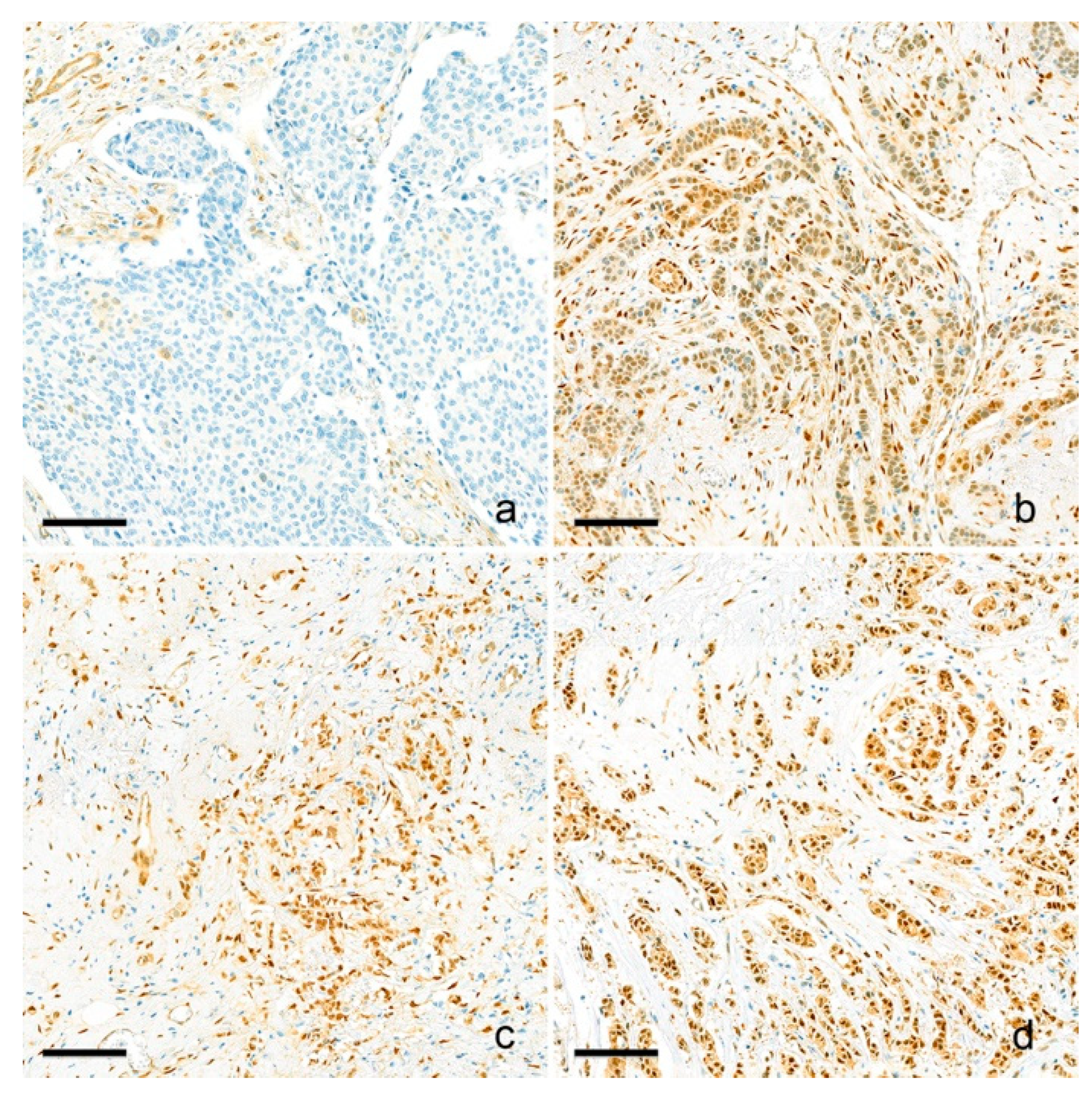
Figure 1. Representative pictures of YAP1 immunohistochemistry. Nuclear YAP1 expression is evaluated and scored as negative (no expression) (a), weak (b), moderate (c), and strong (d) nuclear expression. Negative/weak expressions are considered as low-YAP1 expression, and moderate/strong expressions are considered high YAP1 expression. Scale bar: 100 μm.
Cancers 15 05034 g001
Figure 2. Clinical outcome of ER-positive and ER-negative breast cancer based on YAP1 expression.
Cancers 15 05034 g002
Figure 3. Clinical outcome of different molecular subtypes of breast cancer based on YAP1 expression.
Cancers 15 05034 g003
Figure 4. Clinical outcomes of patients with different ESR1 expression levels based on YAP1 expression.
Cancers 15 05034 g004
Table 1. Comparison of clinicopathologic parameters based on the YAP1 expression.
ParametersYAP1-Low (n = 139)YAP1-High (n = 262)p-Value
Menopausal Status, n (%) 0.343
Premenopausal75 (54.0)160 (61.1)
Menopause59 (42.4)96 (36.6)
Not assessable5 (3.6)6 (2.3)
Oncotype Dx RS (mean ± SD)20.4 ± 11.616.8 ± 7.20.001
ODX risk group, n (%) <0.001
Low-risk (<26)103 (74.1)237 (90.5)
High-risk (≥26)36 (25.9)25 (9.5)
Histologic grade, n (%) 0.001
I17 (12.2)46 (17.6)
II104 (74.8)207 (79.0)
III18 (12.9)9 (3.4)
Tumor size, cm (mean ± SD)2.0 ± 0.82.0 ± 0.80.405
Lymph node metastasis, n (%) 0.624
Absent108 (77.7)197 (75.2)
Present31 (22.3)65 (24.8)
Lympovascular invasion, n (%) 1.000
Absent97 (69.8)182 (69.5)
Present42 (30.2)80 (30.5)
TIL level, % (mean ± SD)11.6 ± 17.311.0 ± 11.40.693
TIL group, n (%) 0.903
Low-TIL (≤10%)104 (74.8)198 (75.6)
High-TIL (>10%)35 (25.2)64 (24.4)
TSR, % (mean ± SD)71.9 ± 15.770.5 ± 1.20.454
TSR group, n (%) 0.226
Stroma-low (TSR >50%)124 (89.2)221 (84.4)
Stroma-high (TSR ≤50%)15 (10.8)41 (15.6)
Ki67 LI, % (mean ± SD)13.8 ± 9.510.9 ± 10.90.007
YAP1, Yes-associated protein 1; RS, risk score; SD, standard deviation; ODX, oncotype Dx; TIL, tumor-infiltrating lymphocyte; TSR, tumor–stroma ratio; LI, labelling index.
Table 2. Comparison of clinicopathologic parameters based on the risk score.
ParametersHigh-Risk (RS ≥ 26) (n = 61)Low-Risk (RS < 26) (n = 340)p-Value
Menopausal Status, n (%) 0.001
Premenopausal23 (37.7)212 (62.4)
Menopause37 (60.7)118 (34.7)
Not assessable1 (1.6)10 (2.9)
YAP1 expression, n (%) <0.001
Low-YAP136 (59.0)103 (30.3)
High-YAP1 25 (41.0)237 (69.7)
Histologic grade, n (%) <0.001
I4 (6.6)59 (17.4)
II46 (75.4)265 (77.9)
III11 (18.0)16 (4.7)
Tumor size, cm (mean ± SD)2.0 ± 0.71.9 ± 0.80.919
Lymph node metastasis, n (%) 0.001
Absent57 (93.4)248 (72.9)
Present4 (6.6)92 (27.1)
Lymphovascular invasion, n (%) 0.047
Absent49 (80.3)230 (67.6)
Present12 (19.7)110 (32.4)
TIL, % (mean ± SD)14.5 ± 20.710.6 ± 12.00.157
TIL group, n (%) 0.762
Low-TIL (≤10%)45 (73.8)257 (75.6)
High-TIL (>10%)16 (26.2)83 (24.4)
TSR, % (mean ± SD)70.2 ± 18.171.2 ± 18.20.695
TSR group, n (%) 0.847
Stroma-low (TSR >50%)52 (85.2)293 (86.2)
Stroma-high (TSR ≤50%)9 (14.8)47 (13.8)
Ki67 LI, % (mean ± SD)20.3 ± 12.210.4 ± 9.4<0.001
RS, risk score; YAP1, Yes-associated protein 1; SD, standard deviation; TIL, tumor-infiltrating lymphocyte; TSR, tumor–stroma ratio; LI, labelling index.
Table 3. Logistic regression analysis for parameters associated with ODX high risk (RS ≥ 26).
UnivariateMultivariate
ParametersOR95% CIp-ValueOR95% CIp-Value
LowerUpperLowerUpper
Menopausal status <0.001
PremenopausalRef Ref
Menopause2.8901.6395.095 2.8971.5385.4540.001
YAP1 expression <0.001
LowRef Ref
High0.3020.1720.528 0.3730.1980.7030.002
Tumor size1.0180.7201.4410.918
Histologic grade
IRef
II2.5600.8877.3900.082
III10.1402.84636.136<0.0014.6251.07019.990.040
Lymph node metastasis 0.002
AbsentRef Ref
Present0.1890.0670.536 0.2700.0910.8010.018
Lymphovascular invasion 0.051
AbsentRef
Present0.5120.2621.002
TIL
Low-TILRef
High-TIL1.0171.0001.0350.045
TSR
Stroma-lowRef
Stroma-high0.9970.9821.0120.695
Ki67 LI1.0781.051.107<0.0011.0621.0351.09<0.001
OR, odds ratio; CI, confidence interval; Ref, reference; YAP1, Yes-associated protein 1; TIL, tumor-infiltrating lymphocyte; TSR, tumor–stroma ratio; LI, labelling index.
Table 4. Parameters correlated with ODX RS.
Univariate AnalysisBetaSE95% CIp-Value
LowerUpper
Menopausal Status2.1670.9380.3244.0110.021
Histologic grade4.1280.9542.2516.004<0.001
Tumor size0.5920.581−0.5511.7340.309
Lymph node metastasis−4.0641.045−6.118−2.010<0.001
Lymphovascular invasion−0.8520.986−2.7911.0870.388
TIL0.0910.0330.0270.1560.006
TSR−0.0090.025−0.0580.0400.718
Ki67 LI0.2990.0410.2190.379<0.001
YAP1−3.6190.970−5.461−1.777<0.001
Multivariate AnalysisBetaSE95% CIp-Value
LowerUpper
Menopausal Status1.7130.8620.0193.4070.047
Histologic grade
Tumor size
Lymph node metastasis−2.9730.997−4.933−1.0140.003
Lymphovascular invasion
TIL
TSR
Ki67 LI0.2700.0410.1890.350<0.001
YAP1−2.8160.894−4.575−1.0140.002
SE, standard error; CI, confidence interval; TIL, tumor-infiltrating lymphocyte; TSR, tumor–stroma ratio; LI, labelling index; YAP1, Yes-associated protein 1.
Disclaimer/Publisher’s Note: The statements, opinions and data contained in all publications are solely those of the individual author(s) and contributor(s) and not of MDPI and/or the editor(s). MDPI and/or the editor(s) disclaim responsibility for any injury to people or property resulting from any ideas, methods, instructions or products referred to in the content.