Article

Diabetes Mellitus Is a Strong Independent Negative Prognostic Factor in Patients with Brain Metastases Treated with Radiotherapy

1
Department of Radiation Oncology, University of Leipzig Medical Center, 04103 Leipzig, Germany
2
Comprehensive Cancer Center Central Germany, Partner Site Leipzig, 04103 Leipzig, Germany
3
Clinical Cancer Registry, 04103 Leipzig, Germany
4
Department of Neurosurgery, University of Leipzig Medical Center, 04103 Leipzig, Germany
*
Author to whom correspondence should be addressed.
These authors contributed equally to this work.
Cancers 2023, 15(19), 4845; https://doi.org/10.3390/cancers15194845
Submission received: 3 July 2023 / Revised: 7 September 2023 / Accepted: 29 September 2023 / Published: 4 October 2023
(This article belongs to the Special Issue Advances in Modern Radiation Oncology)

Abstract

:

Simple Summary

Cerebrovascular disorders are common among cancer patients. They might influence tumor growth, treatment sensitivity and, ultimately, the prognoses of patients with brain metastases (BM). In a retrospective exploratory study, we examined if the presence of arterial hypertension, smoking, diabetes mellitus (DM), hypercholesterolemia or peripheral arterial occlusive disease has a prognostic impact in patients with BM. In uni- and multivariate analysis, the presence of DM was associated with a worse prognosis across several tumor types, while for the other cerebrovascular risk factors, significant differences in survival were not found. From molecular data, it can be hypothesized that RAGE activation plays an important role in the interaction between DM and BM. In future studies, it remains to be determined to what extent serum glucose levels and antidiabetic treatments may influence survival and if optimized antidiabetic treatment or RAGE targeted treatments are able to improve prognoses of patients with BM.

Abstract

Background: Brain metastases (BM) cause relevant morbidity and mortality in cancer patients. The presence of cerebrovascular diseases can alter the tumor microenvironment, cellular proliferation and treatment resistance. However, it is largely unknown if the presence of distinct cerebrovascular risk factors may alter the prognosis of patients with BM. Methods: Patients admitted for the radiotherapy of BM at a large tertiary cancer center were included. Patient and survival data, including cerebrovascular risk factors (diabetes mellitus (DM), smoking, arterial hypertension, peripheral arterial occlusive disease, hypercholesterolemia and smoking) were recorded. Results: 203 patients were included. Patients with DM (n = 39) had significantly shorter overall survival (OS) (HR 1.75 (1.20–2.56), p = 0.003, log-rank). Other vascular comorbidities were not associated with differences in OS. DM remained prognostically significant in the multivariate Cox regression including established prognostic factors (HR 1.92 (1.20–3.06), p = 0.006). Furthermore, subgroup analyses revealed a prognostic role of DM in patients with non-small cell lung cancer, both in univariate (HR 1.68 (0.97–2.93), p = 0.066) and multivariate analysis (HR 2.73 (1.33–5.63), p = 0.006), and a trend in melanoma patients. Conclusion: DM is associated with reduced survival in patients with BM. Further research is necessary to better understand the molecular mechanisms and therapeutic implications of this important interaction.

1. Introduction

Brain metastases (BM) are frequent complications in patients with cancer. According to a SEER database analysis among patients with metastatic disease, patients with melanoma (28.2%), lung adenocarcinoma (26.8%), small cell lung cancer (23.5%), squamous cell carcinoma of the lung (15.9%), renal cancer (10.8%) and breast cancer (7.6%) had brain metastases [1]. Importantly, the incidence of brain metastases (BM) is increasing, partially due to longer life expectancy and better means of detection [2,3].
Despite more available treatments for BM, including surgery, stereotactic radiotherapy (SRT), whole-brain radiation therapy (WBRT), molecularly targeted therapeutics and immunotherapies, patients with BM mostly still have a poor prognosis [1,3,4,5,6].
However, in recent years, several radiotherapeutic developments have improved the treatment of brain metastases. One main focus of research was the prevention of cognitive decline that is frequent after conventional whole-brain radiotherapy (WBRT) [7,8]. Firstly, it has been shown in a multicentric observational trial that stereotactic radiotherapy can be applied to patients with 5–10 brain metastases without worse overall survival compared to patients with 2–4 BM [9,10]. In another approach to maintaining cognitive functioning, hippocampal sparing WBRT has shown significant benefits compared to conventional WBRT in a phase 2 and comparative phase 3 trial [11,12]. Within these trials, in combination with WBRT, the partial N-Methyl-D-Aspartate (NMDA)-receptor antagonist memantine was applied in order to further preserve cognitive function after WBRT [12,13].
Prognostic scores for patients with BM developed from disease-unspecific scores, like recursive partitioning analysis (RPA), to more disease specific approaches, like the disease-specific graded prognostic assessment (ds-GPA) score [14,15]. The ds-GPA encompasses tumor specific variables, like mutation status, extracranial tumor control and number of brain metastases, as well as some patient variables like patient age and Karnofsky performance score (KPS) [16]. Except for KPS, patient frailty and comorbidities are not reflected in the ds-GPA.
The concept of patient frailty comprises different parameters that better describe patient’s well-being, physical independency and comorbidities. Measures of frailty include G8 and Hurria Score [17,18]. Within general oncology, considerable knowledge has been gained regarding the relevance of comorbidities in prognoses and treatment of colorectal cancer, breast cancer and lung cancer [19,20,21]. However, comorbidities, as a part of the frailty concept, have not been a scientific focus in patients with BM so far.
Among co-morbidities, vascular conditions or risk factors might be particularly relevant in a specific and non-specific fashion. Firstly, and specifically, BM heavily rely on and interact with cerebral blood vessels as part of their microenvironment. Alterations in cerebral vasculature might modulate the number and growth of BM, as well as the sensitivity to therapeutic interventions like radiotherapy, which relies on appropriate perfusion and oxygenation [22,23,24].
Non-specifically, cerebrovascular comorbidities (diabetes mellitus (DM), smoking, arterial hypertension, peripheral arterial occlusive disease (PAOD), hypercholesterolemia) are frequent in cancer patients and there are ample indications that they may negatively affect the course of malignant disease.
The prevalence of hypertension is greater in cancer patients and survivors compared with the general population, and arterial hypertension carries a risk of multiple cardiovascular complications during cancer treatment and potentially increased mortality [25,26].
Smoking is associated with a poorer prognosis in patients with small-/non-small cell lung cancer (SCLC/NSCLC) and breast cancer [27,28,29]. Regarding hypercholesterolemia, cholesterol-lowering medication was associated with a decrease in cancer mortality in in a large meta-analysis of breast cancer patients [30]. Finally, diabetes mellitus (DM) appears to be associated with increased cancer mortality across several primary tumors [31,32].
The aim of this exploratory retrospective study was to examine the prognostic role of frequent cerebrovascular comorbidities, specifically in patients with BM undergoing radiotherapy.

2. Materials and Methods

This study was performed according to the Declaration of Helsinki in its current form. Approval was granted by the Ethics Committee of University of Leipzig (Date 3rd of August.2021/No332/21-ek). All patients consented to the anonymized scientific use of their clinical data. Patients treated with radiotherapy at the Department of Radiation Oncology, University of Leipzig Medical Center, Leipzig, Germany, between 2004 and 2016 were identified from clinical records. All patients (age ≥ 18 years) diagnosed with BM from a solid primary tumor were principally eligible for the study; consecutive patients were chosen with available pre-therapeutic cerebral MRI and detailed clinical patient charts. Treatment was either performed with SRT or WBRT, depending on number of metastases and patient conditions. Patient data were analyzed from existing patient charts. Various characteristics including age (at primary BM diagnosis), primary tumor type, KPS, number of metastases, systemic tumor control and the presence or absence of DM, arterial hypertension, smoking status, hypercholesterolemia, and PAOD were recorded prior to treatment of BM in a standardized fashion. Survival data were obtained from the local cancer registry. Data analysis was performed using Microsoft Excel 2016 (Microsoft Corporation, Albuquerque, NM, USA) and SPSS Version 28.0.1.1 (IBM Software Inc., Armonk, New York, USA). Overall survival (OS) was calculated from the date of diagnosis of brain metastasis until death; patients being lost to follow-up were censored at the last date known to be alive. OS was examined using Kaplan–Meier analyses with log-rank tests. Univariate and multivariate Cox proportional hazard regression analyses were conducted to reveal prognostic parameters associated with OS. For Cox proportional hazard regression analyses of established prognostic factors age was stratified to ≥70 years vs. <70 years, KPS <70 vs. ≥ 70 and number of BM >3 vs. ≤3 BM.

3. Results

3.1. Patient Characteristics

Data from 203 patients were included in the analysis. Patient age at diagnosis of BM ranged from 30 to 83 years with a median/mean age of 62.6/61.3 years. At diagnosis of BM, patients presented with a median Karnofsky performance status (KPS) of 70 (range: 20–100). The most common primary tumor was non-small cell lung cancer (NSCLC) in 84/203 patients (41.4%), followed by melanoma (28/203, 13.8%), breast cancer (21/203, 10.3%), small cell lung cancer (SCLC) and renal cell carcinoma (RCC) (each 20/203, 9.9%), colon cancer (7/203, 3.4%), and other cancers (23/203, 11.3%). Concomitant vascular co-morbidities were arterial hypertension (100/203 patients, 49.3%), DM (39/203, 19.2%), peripheral arterial occlusive disease (PAOD) (23/203, 11.3%), hypercholesterolemia (7/203, 3.5%). In total, 33.5% of patients were smokers (68/203 patients, Table 1), 39.4% (80/203) of patients were diagnosed with 1–3 BM, and 90 patients (44.3%) had >3 BM. The majority of patients had unstable systemic disease or synchronous brain metastases (137/203, 67.4%). Treatment of patients BM comprised stereotactic radiotherapy (SRT) (40/192, 19.7%) and whole-brain radiotherapy (WBRT) ± SRS (152/191, 74.8%).

3.2. Age, KPS and Tumor Histology Influenced Survival Outcomes

The median survival time of the entire cohort was 6 months (95% CI: 4.49–7.40), and the median follow up was 6.01 months (95% CI: 0.93–42.54). Median survival times were different between patients with age ≥70 years/<70 years (4.14/7.29 months, p = 0.001) and with KPS <70, ≥70 (3.06/6.70 months, p = 0.03) but not between patients with >3, ≤3 BM: 5.55/6.74 months (p = 0.443) respectively. Concerning tumor histology, survival times for patients were as follows: NSCLC, 6.01 months; SCLC, 5.52 months; melanoma, 7.46 months; breast cancer, 9.53 months; RCC, 2.96 months; and other cancers, 4.17 months.
In the univariate Cox regression analysis, patients age ≥70 years showed significant detrimental effects regarding survival (HR 1.75 [1.24–2.48], p = 0.002). Furthermore, KPS <70 and tumor histology (RCC) were associated with reduced survival (KPS <70: HR 1.47 [1.03–2.1], p = 0.033, RCC: HR 2.035 [1.26–3.28], p = 0.015). The number of BM was not associated with differences in survival time (>3 BM: HR 1.13 [0.83–1.54], p = 0.444; Table 2).

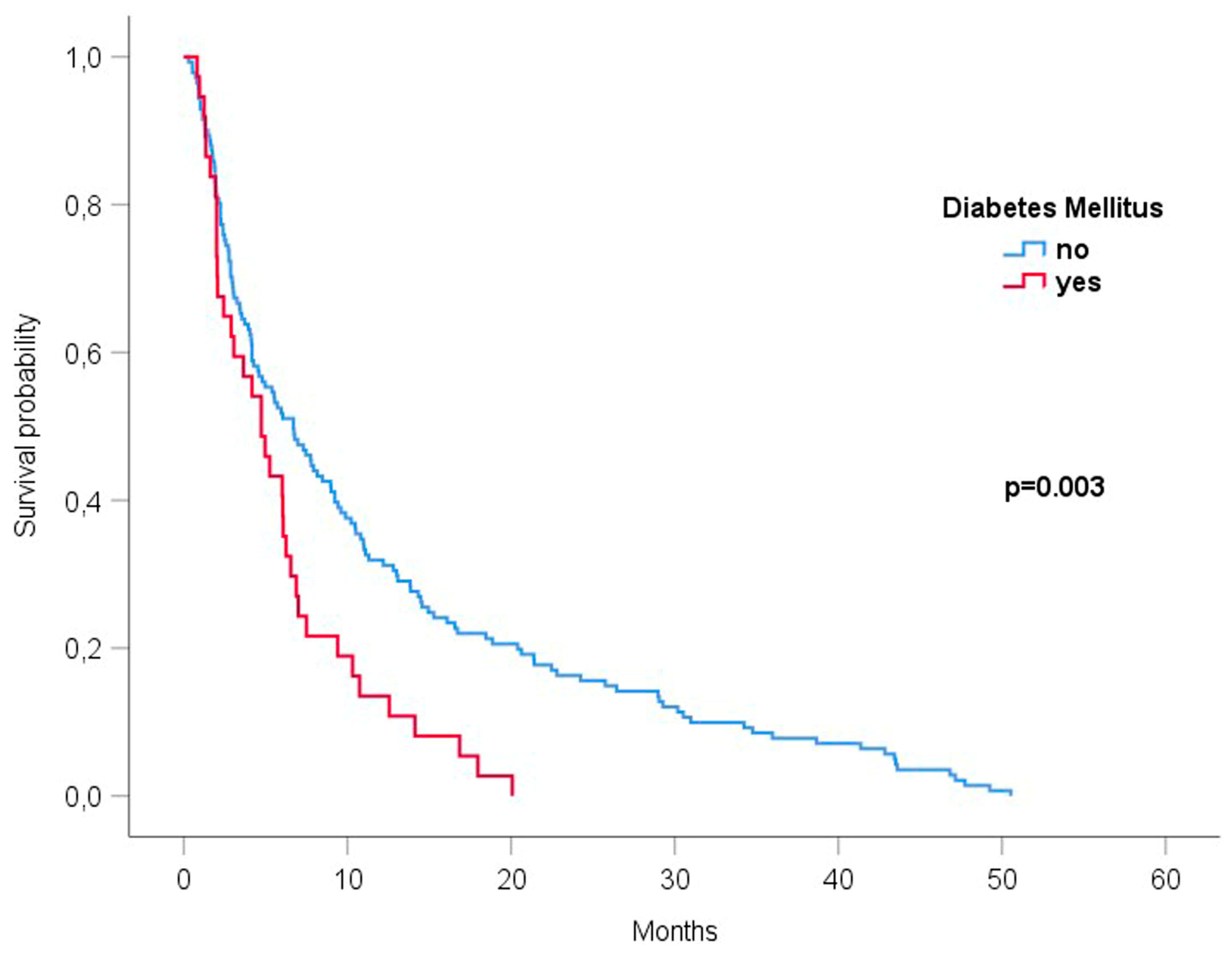
3.3. Diabetes Mellitus Was Associated with the Reduced Survival of Patients with Brain Metastases Undergoing Radiotherapy

Median survival times for patients with/without DM, with/without arterial hypertension, with/without smoking, with/without PAOD, and with/without hypercholesterolemia were 4.73/6.7 months (p = 0.003), 5.95/6.05 months (p = 0.443), 6.08/5.72 months (p = 0.307), 4.17/5.95 months (p = 0.266), and 4.11/5.71 months (p = 0.157), respectively.
In the Kaplan–Meier analysis, patients with DM had significantly shorter survival compared to patients without DM (HR 1.75 [1.20–2.56], p = 0.003, Figure 1).
The presence or absence of other vascular comorbidities was not associated with differences in survival (Table 2, Figure 2A–D).
Patients with and without DM were compared to investigate potential confounders. Patients without DM were younger (p = 0.027), systemic tumor progression at time of diagnosis of BM appeared somewhat but was insignificantly more frequent in patients with DM (19.4% vs. 5.7%, p = 0.154). Otherwise, the distribution of KPS (p = 0.841), histology (p = 0.606), number of BM (p = 0.792) and radiotherapy concept (p = 0.441) were not different, a shown in Table 3.

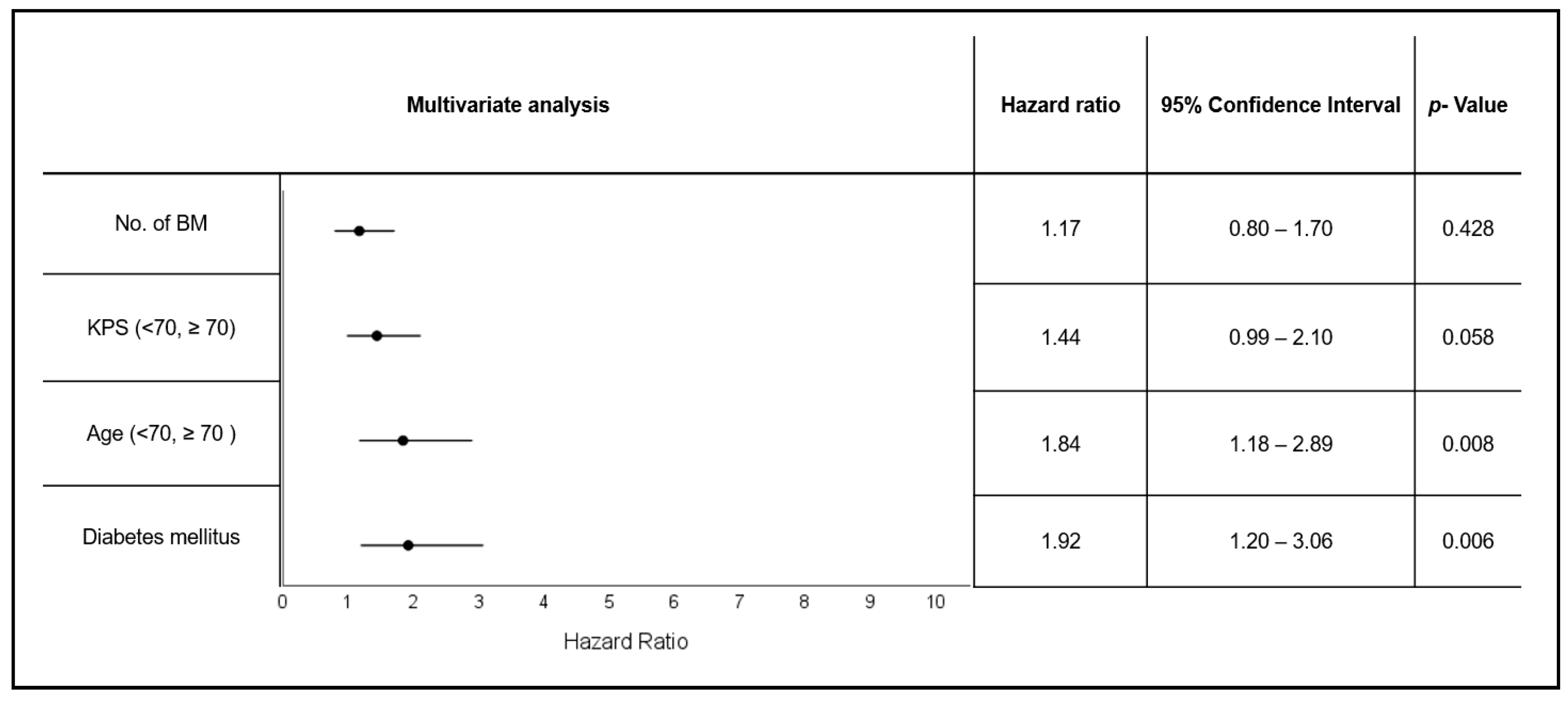
3.4. Diabetes and Patient Age Are Independent Negative Prognostic Factors in Patients with Brain Metastases Undergoing Radiotherapy

Multivariate Cox regression analysis was performed, including the following factors: DM, age (<70, ≥70), KPS score (<70, ≥70), and number of BM (≤3; >3).
DM (HR 1.92 [1.20–3.06], p = 0.006, Figure 3) and age ≥70 (HR 1.84 [1.18–2 89], p = 0.008) remained independently associated with worse OS. KPS <70 showed a trend (HR 1.44 [0.99–2.1], p = 0.058) towards deteriorated OS. No effect was seen for the number of BM (HR 1.17 [0.8–1.70], p = 0.428), Figure 3.

3.5. Diabetes Mellitus Is a Negative Prognostic Factor for Brain Metastases of Distinct Histologies

In order to further validate the negative prognostic impact of DM, univariate and multivariate analyses were performed separately for the two most common primary cancer types in our cohort, i.e., NSCLC (84 patients) and melanoma (28 patients). In patients with NSCLC, the presence of DM was associated with poorer survival in the univariate (HR 1.68 [0.97–2.93], p = 0.066) and multivariate analysis (HR 2.73 [1.33–5.63], p = 0.006, Supplementary Figure S1). Age as an established important factor lost significance in the analysis restricted to NSCLC (univariate: HR 1.54 [0.85–2.72], p = 0.151; multivariate: HR 1.31 [0.63–2.73], p = 0.475). The negative impact of lower KPS persisted (univariate: HR 2.47 [1.40–4.38], p = 0.002; multivariate: HR 2.85 [1.52–5.33], p = 0 001, Supplementary Table S1).
In the much smaller cohort of melanoma patients, a worse survival with DM appeared from survival curves; however, in uni- and multivariate Cox regression, this was not significant (multivariate: HR 4.62 [0.49–44.04], p = 0.183), and other factors were not associated with differences in survival (Supplementary Figure S2 and Supplementary Table S2).

4. Discussion

BM are a “special” complication of cancer in a special organ, often requiring specific treatments. Accordingly, aspects like metastasis formation, metastasis growth, treatment, resistance to treatment and prognosis need to be specifically addressed.
The formation of BM depends on the extravasation of tumor cells, perivascular tumor cell growth, and the co-option of pre-existing vessels in a complex multistep process [33,34,35]. In addition, cerebral perfusion and oxygenation are crucial for the effect of local radiotherapy on BM [36,37], and alterations in cerebral perfusion could also modulate the efficacy of systemic treatment [38,39]. Taking into account this relevant interplay of BM formation and treatment with vascular architecture, there is a strong need to more precisely understand the effects of vascular risk factors that could influence development and prognosis of BM. Cerebrovascular risk factors are frequent and often lead to chronic cerebrovascular diseases [40,41]. Macro- and microvascular changes predispose to multiple complications, including vascular stenosis, ischemic strokes, and small vessel disease of the brain [42,43].
Within this exploratory study, we examined if several frequent cerebrovascular risk factors may have a prognostic effect in patients with BM treated with radiotherapy. Arterial hypertension, smoking, PAOD, and hypercholesterolemia had no prognostic value both in monovariate and multivariate analysis. Based on chart-based diagnoses in this moderately sized cohort, this does not exclude minor individual effects, but a larger effect of these frequent cerebrovascular risk factors in patients with BM seems unlikely. Coherent with our results, a large retrospective study in 390 patients with lung cancer and BM, concerning the effect of smoking, found that smoking status and pack-year history of smoking had no effect on overall survival; a trend for an increased risk of neurologic death in non-adenocarcinoma patients who continued to smoke was postulated [44].
Beyond the functional effects of risk factors, existing structural vascular alterations that have not been measured in this patient cohort might be more important.
It is known that small vessel disease of the brain can influence the number of BM, potentially by reducing the accessibility of the less perfused brain areas [45,46,47].
The relevance of small vessel disease of the brain in patient prognosis should be examined in future studies. The same is true for the presence of angiopathic alterations in large cerebral vessels and history of ischemic vascular events/strokes.
In contrast to the other mentioned vascular risk factors, DM showed a strong independent negative prognostic effect.
From clinical evidence, little is known about the prognostic effects of DM specifically in patients with BM treated with radiotherapy thus far. In a smaller single-center retrospective study, 81 patients with BM from breast cancer who were treated with stereotactic radiation therapy were retrospectively analyzed regarding a prognostic effect of obesity and DM. Patients with DM (n = 17) had decreased median OS (11.8 vs. 26.2 months; p < 0.001) and median intracranial PFS (4.5 vs. 10.3 months; p = 0.001) compared to non-diabetic patients (n = 67). On multivariate analysis, both BMI ≥ 25 kg/m2 [HR 2.35 (1.39–3.98); p = 0.002] and diabetes (HR 2.77 [1.454–5.274]; p = 0.002) were associated with increased mortality [48].
A second retrospective single-center analysis of a larger cohort of patients with various primary tumors (498 patients/48 patients with DM) reported results for stereotactic radiotherapy [49]. DM was found to be a significant negative predictor of OS on multivariate analysis (HR: 1.41, CI: 1.02–1.95, p = 0.04).
Concurring with these two studies, our data support DM as an independent factor associated with mortality in patients with BM. In the series by McCall and colleagues, only patients with breast cancer were included, and in the work of LeCompte, several tumor histologies with a focus on NSCLC were included [48,49]. Together with our results, a tumor-independent effect concerning patients with disseminated malignancies is most likely. With a HR of 1.92, our results appear to be within the range of those from the cited studies: 1.4–2.77 [48,49]. The negative effect appears to persist in different age groups and treatment scenarios (McCall: younger patients with 80% WBRT [48], LeCompte: SRS only [49]).
Concerning the reasons for a negative prognostic impact, both series contain valuable information. While in the study by LeCompte, the major detriment appeared to arise from systemic effects (death prior to distant brain failure earlier in diabetics vs. non-diabetics (p = 0.04), in the series by McCall, median intracranial PFS was significantly reduced in patients with DM [48,49]. Our study did not plan to analyze PFS but was restricted to OS without providing reasons for death. Notably, we aimed to examine a patient cohort treated with radiotherapy to BM prior to the widespread use of systemic treatments effective in BM. With the more frequent use of immune checkpoint inhibitors or targeted treatments, the prognostic relevance of DM in patients with BM might be less pronounced and should also be examined in these patient cohorts.
We did not examine potential associations between DM and treatment-related toxicities. However, the study of LeCompte et al. did not observe significant differences in the incidence of radiation necrosis, radiation-induced edema, cerebrospinal fluid leak or postoperative infection in patients with DM [49].
While clinical evidence of the negative effects of DM in BM is limited, it is better known for its negative effects on cancer patients with regard to different primary tumors. DM contributes to increased mortality from colorectal cancer, liver cancer, pancreatic cancer, breast cancer, or lung cancer [22,50,51].
Several pathophysiologic aspects of diabetes mellitus type 2 interact with tumor metabolism. According to clinical and preclinical evidence, hyperinsulinemia, hyperglycemia and a diabetes-associated chronic inflammatory state appear to be associated with elevated cancer risk and mortality [50,52]. In a large prospective cohort study with around 10,000 participants, hyperinsulinemia, even without manifest diabetes mellitus, was associated with increased cancer mortality [53]. In a preclinical model, hyperinsulinemia promoted metastasis in the lungs in a mouse model of Her2-mediated breast cancer [54]. Hyperglycemia itself leads to growth promotion and increased proliferation as tumors mostly rely on anerobic glycolysis [55]. Glycolysis is facilitated by upregulated glucose transporters (GLUTs) in tumor cells, enabling the facilitative entry of glucose into a cell. A high rate of glucose can be consumed by malignant cells beyond that necessary for ATP synthesis [56,57].
Hyperglycemia is thought to play a key role in tumor progression by reprogramming glucose metabolism, stimulating cancer-associated inflammation, molecular alterations and hypoxia [58,59,60,61]. It can also lead to therapeutic resistance through immunosuppression [62,63], which contributes to poor outcomes in tumor patients [64].
An important mechanism in the interplay of diabetes mellitus and malignant disease is the overactivation of the receptor for advanced glycation end-products (RAGE). RAGE is activated as a consequence of the increase in glycolysis, which enhances the non-enzymatic glycation of proteins, leading to the formation of advanced glycation end-products (AGEs) [55]. AGEs, particularly N-carboxymethyllysine [CML]-modified proteins, were the first RAGE ligands to be identified [65]. The overexpression and activation of RAGE is able to continuously fuel an inflammatory milieu in the tumor microenvironment [66,67]. Interestingly, the overactivation of RAGE as part of the S100A9-RAGE-NF-κB-JunB pathway was discovered as a mechanism for the radioresistance of brain metastasis [68]. Brain metastatic cancer cells from different primary tumors were found to highly express S100A9 in the brain microenvironment, mediating resistance to radiotherapy via the downstream activation of RAGE and NF-κB. S100A9 expression in human brain metastasis from patients with lung cancer, breast cancer or melanoma negatively correlated with the benefits of radiotherapy. The genetic or pharmacological targeting of S100A9 via a blood–brain-barrier-permeable inhibitor of its receptor (RAGE) sensitized brain metastasis to irradiation in experimental models of brain metastasis as well as in patient-derived organotypic cultures [68]. Together with other data concerning DM, it can be suggested that DM/hyperglycemia might confer radioresistance via RAGE activation—potentially independently from S100A9—but might be reversed by RAGE inhibitors too (Figure 4). A clinical trial exploiting the mechanism of RAGE inhibition as a means of radiosensitisation is currently planned [69].
As a further molecular crosslink of altered glucose metabolism and treatment of BM, a recent multiplex immunofluorescence study of resected BM samples from 33 patients treated with radiotherapy and ipilimumab for BM of melanoma found a strong upregulation of the glucose transporter GLUT 1 in BM associated with a prognostic detriment in these patients [70].
It can further be speculated that DM or hyperglycemia is particularly relevant in BM due to the constantly high perfusion of the brain and frequent use of corticosteroids for the alleviation of neurologic symptoms. In other brain tumors like malignant glioma, a significant prognostic deterioration through the use of dexamethasone was shown in large pooled analysis [71].
From the existing data, both a systemic and local tumor-growth-promoting effect appear likely, while alterations of cerebral blood vessels seem less plausible as a cause of shortened survival.
Our analysis carries the limitations of a monocentric retrospective series with potential confounders. We tried to control many factors using a multivariate analysis, and the frequency of several other factors like treatment type and the state of systemic tumor control was not significantly different between patients with and without DM. However due to the limited sample size, not all effects may have been detected.
Several important questions remain from ours and other studies. Firstly, a potential direct negative correlation between survival time and serum glucose levels needs to be analyzed further, as well as the potential effect of antidiabetic treatments, most preferably in a prospective multicentric fashion. In our cohort, repeated serum glucose levels were not consistently available. Secondly, dexamethasone use in patients with BM should be monitored and correlated to blood glucose levels and outcomes. Furthermore, much more specific research is needed to characterize diabetes-specific changes in brain metastasis that might be prognostically and therapeutically relevant. In particular, a large-scale comparative analysis between tumors/brain metastasis in patients with and without DM should be performed to better understand biologic and prognostic differences. In the future, prognostic scores for BM should most likely be expanded to include DM and potential other factors.

5. Conclusions

DM seems to negatively affect survival time, but we did not find any general effect of smoking, arterial hypertension, PAOD, and hypercholesterolemia on overall survival in BM (multi-center validation pending). DM appears as a strong independent risk factor for growth, microenvironment, and therapy resistance, probably not related to vascular effects but more likely to pleiotropic effects. The correlation of blood serum levels to outcomes and potential therapeutic implications (e.g., of RAGE inhibition) need to be evaluated in patients with BM in future.
For now, the presence of DM appears potentially more relevant for patients’ survival times than the choice of radiotherapy concept [72,73]. The focus of treating radiation oncologists needs to shift more closely to this potentially influenceable patient condition.

Supplementary Materials

The following supporting information can be downloaded at: https://www.mdpi.com/article/10.3390/cancers15194845/s1, Figure S1: Univariate and Multivariate analysis of survival (Cox-Regression) of patients with/without DM in patients with NSCLC, Table S1: Univariate and Multivariate analysis of survival (Cox-Regression) of prognostic factors in patients with NSCLC, Figure S2: Univariate and Multivariate analysis of survival (Cox-Regression) of patients with/without DM in patients with melanoma, Table S2: Univariate and Multivariate analysis of survival (Cox-Regression) of prognostic factors in patients with melanoma.

Author Contributions

Conceptualization, N.H.N. and C.S.; Data curation, S.J., S.P. and C.S.; Formal analysis, S.J., S.P., S.K. and A.R.; Investigation, S.J. and S.P.; Methodology, S.J., S.P. and C.S.; Resources, S.K., T.K., K.P., E.G., F.N. and N.H.N.; Supervision, N.H.N. and C.S.; Validation, A.R. and C.S.; Writing—original draft, S.J., S.P. and C.S.; Writing—review & editing, S.J., S.P., T.K., K.P., P.H., J.W., E.G., F.N., A.R., N.H.N. and C.S. All authors have read and agreed to the published version of the manuscript.

Funding

The authors declare that no funds, grants, or other support were received during the preparation of this manuscript.

Institutional Review Board Statement

This study was performed in line with the principles of the Declaration of Helsinki. Approval was granted by the Ethics Committee of University of Leipzig (Date 03.08.2021/No332/21-ek).

Informed Consent Statement

All patients consented to an anonymized scientific use of their clinical data.

Data Availability Statement

The datasets generated during and/or analyzed during the current study are available from the corresponding author on reasonable request.

Conflicts of Interest

The authors declare no conflict of interest.

References

  1. Cagney, D.N.; Martin, A.M.; Catalano, P.J.; Redig, A.J.; Lin, N.U.; Lee, E.Q.; Wen, P.Y.; Dunn, I.F.; Bi, W.L.; Weiss, S.E.; et al. Incidence and prognosis of patients with brain metastases at diagnosis of systemic malignancy: A population-based study. Neuro-Oncol. 2017, 19, 1511–1521. [Google Scholar] [CrossRef]
  2. Li, A.Y.; Gaebe, K.; Jerzak, K.J.; Cheema, P.K.; Sahgal, A.; Das, S. Intracranial Metastatic Disease: Present Challenges, Future Opportunities. Front. Oncol. 2022, 12, 855182. [Google Scholar] [CrossRef] [PubMed]
  3. Habbous, S.; Forster, K.; Darling, G.; Jerzak, K.; Holloway, C.M.B.; Sahgal, A.; Das, S. Incidence and real-world burden of brain metastases from solid tumors and hematologic malignancies in Ontario: A population-based study. Neuro-Oncol. Adv. 2021, 3, vdaa178. [Google Scholar] [CrossRef] [PubMed]
  4. Ye, C.; Handa, P.; Sahgal, A.; Lo, S.; Vellayappan, B. Risk-reduction strategies for late complications arising from brain metastases treated with radiotherapy: A narrative review. Chin. Clin. Oncol. 2022, 11, 13. [Google Scholar] [CrossRef] [PubMed]
  5. Nieder, C.; Spanne, O.; Mehta, M.P.; Grosu, A.L.; Geinitz, H. Presentation, patterns of care, and survival in patients with brain metastases: What has changed in the last 20 years? Cancer 2011, 117, 2505–2512. [Google Scholar] [CrossRef]
  6. Soffietti, R.; Rudā, R.; Mutani, R. Management of brain metastases. J. Neurol. 2002, 249, 1357–1369. [Google Scholar] [CrossRef]
  7. Brown, P.D.; Jaeckle, K.; Ballman, K.V.; Farace, E.; Cerhan, J.H.; Anderson, S.K.; Carrero, X.W.; Barker, F.G.; Deming, R.; Burri, S.H.; et al. Effect of Radiosurgery Alone vs Radiosurgery with Whole Brain Radiation Therapy on Cognitive Function in Patients With 1 to 3 Brain Metastases: A Randomized Clinical Trial. JAMA 2016, 316, 401–409. [Google Scholar] [CrossRef]
  8. Chang, E.L.; Wefel, J.S.; Hess, K.R.; Allen, P.K.; Lang, F.F.; Kornguth, D.G.; Arbuckle, R.B.; Swint, J.M.; Shiu, A.S.; Maor, M.H.; et al. Neurocognition in patients with brain metastases treated with radiosurgery or radiosurgery plus whole-brain irradiation: A randomised controlled trial. Lancet Oncol. 2009, 10, 1037–1044. [Google Scholar] [CrossRef]
  9. Yamamoto, M.; Serizawa, T.; Shuto, T.; Akabane, A.; Higuchi, Y.; Kawagishi, J.; Yamanaka, K.; Sato, Y.; Jokura, H.; Yomo, S.; et al. Stereotactic radiosurgery for patients with multiple brain metastases (JLGK0901): A multi-institutional prospective observational study. Lancet Oncol. 2014, 15, 387–395. [Google Scholar] [CrossRef]
  10. Yamamoto, M.; Serizawa, T.; Higuchi, Y.; Sato, Y.; Kawagishi, J.; Yamanaka, K.; Shuto, T.; Akabane, A.; Jokura, H.; Yomo, S.; et al. A Multi-institutional Prospective Observational Study of Stereotactic Radiosurgery for Patients with Multiple Brain Metastases (JLGK0901 Study Update): Irradiation-related Complications and Long-term Maintenance of Mini-Mental State Examination Scores. Int. J. Radiat. Oncol. 2017, 99, 31–40. [Google Scholar] [CrossRef]
  11. Gondi, V.; Pugh, S.L.; Tome, W.A.; Caine, C.; Corn, B.; Kanner, A.; Rowley, H.; Kundapur, V.; DeNittis, A.; Greenspoon, J.N.; et al. Preservation of memory with conformal avoidance of the hippocampal neural stem-cell compartment during whole-brain radiotherapy for brain metastases (RTOG 0933): A phase II multi-institutional trial. J. Clin. Oncol. 2014, 32, 3810–3816. [Google Scholar] [CrossRef] [PubMed]
  12. Brown, P.D.; Gondi, V.; Pugh, S.; Tome, W.A.; Wefel, J.S.; Armstrong, T.S.; Bovi, J.A.; Robinson, C.; Konski, A.; Khuntia, D.; et al. Hippocampal Avoidance During Whole-Brain Radiotherapy Plus Memantine for Patients with Brain Metastases: Phase III Trial NRG Oncology CC001. J. Clin. Oncol. 2020, 38, 1019–1029. [Google Scholar] [CrossRef]
  13. Brown, P.D.; Pugh, S.; Laack, N.N.; Wefel, J.S.; Khuntia, D.; Meyers, C.; Choucair, A.; Fox, S.; Suh, J.H.; Roberge, D.; et al. Memantine for the prevention of cognitive dysfunction in patients receiving whole-brain radiotherapy: A randomized, double-blind, placebo-controlled trial. Neuro-Oncology 2013, 15, 1429–1437. [Google Scholar] [CrossRef]
  14. Gaspar, L.; Scott, C.; Rotman, M.; Asbell, S.; Phillips, T.; Wasserman, T.; McKenna, W.G.; Byhardt, R. Recursive partitioning analysis (RPA) of prognostic factors in three radiation therapy oncology group (RTOG) brain metastases trials. Int. J. Radiat. Oncol. Biol. Phys. 1997, 37, 745–751. [Google Scholar] [CrossRef] [PubMed]
  15. Sperduto, P.W.; Kased, N.; Roberge, D.; Xu, Z.; Shanley, R.; Luo, X.; Sneed, P.K.; Chao, S.T.; Weil, R.J.; Suh, J.; et al. Summary Report on the Graded Prognostic Assessment: An Accurate and Facile Diagnosis-Specific Tool to Estimate Survival for Patients with Brain Metastases. J. Clin. Oncol. 2012, 30, 419–425. [Google Scholar] [CrossRef] [PubMed]
  16. Sperduto, P.W.; De, B.; Li, J.; Carpenter, D.; Kwithatrick, J.; Milligan, M.; Shih, H.A.; Kutuk, T.; Kotecha, R.; Higaki, H.; et al. Graded Prognostic Assessment (GPA) for Patients with Lung Cancer and Brain Metastases: Initial Report of the Small Cell Lung Cancer GPA and Update of the Non-Small Cell Lung Cancer GPA Including the Effect of Programmed Death Ligand 1 and Other Prognostic Factors. Int. J. Radiat. Oncol. 2022, 114, 60–74. [Google Scholar] [CrossRef]
  17. Bellera, C.A.; Rainfray, M.; Mathoulin-Pélissier, S.; Mertens, C.; Delva, F.; Fonck, M.; Soubeyran, P.L. Screening older cancer patients: First evaluation of the G-8 geriatric screening tool. Ann. Oncol. 2012, 23, 2166–2172. [Google Scholar] [CrossRef]
  18. Extermann, M.; Hurria, A. Comprehensive Geriatric Assessment for Older Patients with Cancer. J. Clin. Oncol. 2007, 25, 1824–1831. [Google Scholar] [CrossRef]
  19. Boakye, D.; Rillmann, B.; Walter, V.; Jansen, L.; Hoffmeister, M.; Brenner, H. Impact of comorbidity and frailty on prognosis in colorectal cancer patients: A systematic review and meta-analysis. Cancer Treat. Rev. 2018, 64, 30–39. [Google Scholar] [CrossRef]
  20. Land, L.H.; Dalton, S.O.; Jørgensen, T.L.; Ewertz, M. Comorbidity and survival after early breast cancer. A review. Crit. Rev. Oncol. 2011, 81, 196–205. [Google Scholar] [CrossRef]
  21. Firat, S.; Bousamra, M.; Gore, E.; Byhardt, R.W. Comorbidity and KPS are independent prognostic factors in stage I non-small-cell lung cancer. Int. J. Radiat. Oncol. 2002, 52, 1047–1057. [Google Scholar] [CrossRef] [PubMed]
  22. Chen, Y.; Wu, F.; Saito, E.; Lin, Y.; Song, M.; Luu, H.N.; Gupta, P.C.; Sawada, N.; Tamakoshi, A.; Shu, X.-O.; et al. Association between type 2 diabetes and risk of cancer mortality: A pooled analysis of over 771,000 individuals in the Asia Cohort Consortium. Diabetologia 2017, 60, 1022–1032. [Google Scholar] [CrossRef] [PubMed]
  23. Sørensen, B.S.; Horsman, M.R. Tumor Hypoxia: Impact on Radiation Therapy and Molecular Pathways. Front. Oncol. 2020, 10, 562. [Google Scholar] [CrossRef] [PubMed]
  24. Corroyer-Dulmont, A.; Valable, S.; Fantin, J.; Chatre, L.; Toutain, J.; Teulier, S.; Bazille, C.; Letissier, E.; Levallet, J.; Divoux, D.; et al. Multimodal evaluation of hypoxia in brain metastases of lung cancer and interest of hypoxia image-guided radiotherapy. Sci. Rep. 2021, 11, 11239. [Google Scholar] [CrossRef] [PubMed]
  25. Armstrong, G.T.; Oeffinger, K.C.; Chen, Y.; Kawashima, T.; Yasui, Y.; Leisenring, W.; Stovall, M.; Chow, E.J.; Sklar, C.A.; Mulrooney, D.A.; et al. Modifiable Risk Factors and Major Cardiac Events Among Adult Survivors of Childhood Cancer. J. Clin. Oncol. 2013, 31, 3673–3680. [Google Scholar] [CrossRef]
  26. Cohen, J.B.; Geara, A.S.; Hogan, J.J.; Townsend, R.R. Hypertension in Cancer Patients and Survivors. JACC CardioOncol. 2019, 1, 238–251. [Google Scholar] [CrossRef]
  27. Huang, L.; Shi, Y. Prognostic value of pretreatment smoking status for small cell lung cancer: A meta-analysis. Thorac. Cancer 2020, 11, 3252–3259. [Google Scholar] [CrossRef]
  28. Ban, W.H.; Yeo, C.D.; Han, S.; Kang, H.S.; Park, C.K.; Kim, J.S.; Kim, J.W.; Kim, S.J.; Lee, S.H.; Kim, S.K. Impact of smoking amount on clinicopathological features and survival in non-small cell lung cancer. BMC Cancer 2020, 20, 848. [Google Scholar] [CrossRef]
  29. Passarelli, M.N.; Newcomb, P.A.; Hampton, J.M.; Trentham-Dietz, A.; Titus, L.J.; Egan, K.M.; Baron, J.A.; Willett, W.C. Cigarette Smoking Before and After Breast Cancer Diagnosis: Mortality from Breast Cancer and Smoking-Related Diseases. J. Clin. Oncol. 2016, 34, 1315–1322. [Google Scholar] [CrossRef]
  30. Jaiswal, V.; Agrawal, V.; Ang, S.P.; Saleeb, M.; Ishak, A.; Hameed, M.; Rajak, K.; Kalra, K.; Jaiswal, A. Post-diagnostic statin use and its association with cancer recurrence and mortality in breast cancer patients: A systematic review and meta-analysis. Eur. Hear. J. Cardiovasc. Pharmacother. 2023. Online ahead of print. [Google Scholar] [CrossRef]
  31. Barone, B.B.; Yeh, H.-C.; Snyder, C.F.; Peairs, K.S.; Stein, K.B.; Derr, R.L.; Wolff, A.C.; Brancati, F.L. Long-term all-cause mortality in cancer patients with preexisting diabetes mellitus: A systematic review and meta-analysis. JAMA 2008, 300, 2754–2764. [Google Scholar] [CrossRef] [PubMed]
  32. Lipscombe, L.L.; Goodwin, P.J.; Zinman, B.; McLaughlin, J.R.; Hux, J.E. The impact of diabetes on survival following breast cancer. Breast Cancer Res. Treat. 2008, 109, 389–395. [Google Scholar] [CrossRef]
  33. Lorger, M.; Felding-Habermann, B. Capturing changes in the brain microenvironment during initial steps of breast cancer brain metastasis. Am. J. Pathol. 2010, 176, 2958–2971. [Google Scholar] [CrossRef]
  34. Kienast, Y.; Baumgarten, L.v.; Fuhrmann, M.; Klinkert, W.E.F.; Goldbrunner, R.; Herms, J.; Winkler, F. Real-time imaging reveals the single steps of brain metastasis formation. Nat. Med. 2010, 16, 116–122. [Google Scholar] [CrossRef] [PubMed]
  35. Łazarczyk, M.; Mickael, M.E.; Skiba, D.; Kurzejamska, E.; Ławiński, M.; Horbańczuk, J.O.; Radziszewski, J.; Fraczek, K.; Wolinska, R.; Paszkiewicz, J.; et al. The Journey of Cancer Cells to the Brain: Challenges and Opportunities. Int. J. Mol. Sci. 2023, 24, 3854. [Google Scholar] [CrossRef] [PubMed]
  36. Hartford, A.C.; Gill, G.S.; Ravi, D.; Tosteson, T.D.; Li, Z.; Russo, G.; Eskey, C.J.; Jarvis, L.A.; Simmons, N.E.; Evans, L.T.; et al. Sensitizing brain metastases to stereotactic radiosurgery using hyperbaric oxygen: A proof-of-principle study. Radiother. Oncol. 2022, 177, 179–184. [Google Scholar] [CrossRef] [PubMed]
  37. Peng, L.; Wang, Y.; Fei, S.; Wei, C.; Tong, F.; Wu, G.; Ma, H.; Dong, X. The effect of combining Endostar with radiotherapy on blood vessels, tumor-associated macrophages, and T cells in brain metastases of Lewis lung cancer. Transl. Lung Cancer Res. 2020, 9, 745–760. [Google Scholar] [CrossRef]
  38. Corroyer-Dulmont, A.; Valable, S.; Falzone, N.; Frelin-Labalme, A.-M.; Tietz, O.; Toutain, J.; Soto, M.S.; Divoux, D.; Chazalviel, L.; Pérès, E.A.; et al. VCAM-1 targeted alpha-particle therapy for early brain metastases. Neuro-Oncol. 2020, 22, 357–368. [Google Scholar] [CrossRef]
  39. Berghoff, A.S.; Preusser, M. Anti-angiogenic therapies in brain metastases. Memo 2018, 11, 14–17. [Google Scholar] [CrossRef]
  40. Sigurdsson, S.; Aspelund, T.; Kjartansson, O.; Gudmundsson, E.; Jonsson, P.V.; van Buchem, M.A.; Gudnason, V.; Launer, L.J. Cerebrovascular Risk-Factors of Prevalent and Incident Brain Infarcts in the General Population: The AGES-Reykjavik Study. Stroke 2022, 53, 1199–1206. [Google Scholar] [CrossRef]
  41. Knopman, D.S.; Penman, A.D.; Catellier, D.J.; Coker, L.H.; Shibata, D.K.; Sharrett, A.R.; Mosley, T.H. Vascular risk factors and longitudinal changes on brain MRI: The ARIC study. Neurology 2011, 76, 1879–1885. [Google Scholar] [CrossRef] [PubMed]
  42. Pantoni, L. Cerebral small vessel disease: From pathogenesis and clinical characteristics to therapeutic challenges. The Lancet. Neurology 2010, 9, 689–701. [Google Scholar] [CrossRef]
  43. Chen, R.; Ovbiagele, B.; Feng, W. Diabetes and Stroke: Epidemiology, Pathophysiology, Pharmaceuticals and Outcomes. Am. J. Med. Sci. 2016, 351, 380–386. [Google Scholar] [CrossRef] [PubMed]
  44. Shenker, R.F.; McTyre, E.R.; Ruiz, J.; Weaver, K.E.; Cramer, C.; Alphonse-Sullivan, N.K.; Farris, M.; Petty, W.J.; Bonomi, M.R.; Watabe, K.; et al. The Effects of smoking status and smoking history on patients with brain metastases from lung cancer. Cancer Med. 2017, 6, 944–952. [Google Scholar] [CrossRef] [PubMed]
  45. Berk, B.-A.; Nagel, S.; Hering, K.; Paschke, S.; Hoffmann, K.-T.; Kortmann, R.-D.; Gaudino, C.; Seidel, C. White matter lesions reduce number of brain metastases in different cancers: A high-resolution MRI study. J. Neuro-Oncol. 2016, 130, 203–209. [Google Scholar] [CrossRef]
  46. Mazzone, P.J.; Marchi, N.; Fazio, V.; Taylor, J.M.; Masaryk, T.; Bury, L.; Mekhail, T.; Janigro, D. Small vessel ischemic disease of the brain and brain metastases in lung cancer patients. PLoS ONE 2009, 4, e7242. [Google Scholar] [CrossRef]
  47. Berk, B.-A.; Hering, K.; Kortmann, R.-D.; Hoffmann, K.-T.; Ziemer, M.; Seidel, C. Vascular white matter lesions negatively correlate with brain metastases in malignant melanoma-Results from a retrospective comparative analysis. Clin. Neurol. Neurosurg. 2019, 180, 117–121. [Google Scholar] [CrossRef] [PubMed]
  48. McCall, N.S.; Simone, B.A.; Mehta, M.; Zhan, T.; Ko, K.; Nowak-Choi, K.; Rese, A.; Venkataraman, C.; Andrews, D.W.; Anne’, P.R.; et al. Onco-metabolism: Defining the prognostic significance of obesity and diabetes in women with brain metastases from breast cancer. Breast Cancer Res. Treat. 2018, 172, 221–230. [Google Scholar] [CrossRef]
  49. LeCompte, M.C.; McTyre, E.R.; Strowd, R.E.; Lanier, C.; Soike, M.H.; Hughes, R.T.; Masters, A.H.; Cramer, C.K.; Farris, M.; Ruiz, J.; et al. Impact of diabetes mellitus on outcomes in patients with brain metastasis treated with stereotactic radiosurgery. J. Radiosurgery SBRT 2018, 5, 285–291. [Google Scholar] [CrossRef]
  50. Shahid, R.K.; Ahmed, S.; Le, D.; Yadav, S. Diabetes and Cancer: Risk, Challenges, Management and Outcomes. Cancers 2021, 13, 5735. [Google Scholar] [CrossRef]
  51. Liu, X.; Zheng, K.; Ji, W.; Zhang, W.; Li, Y.; Liu, M.; Cui, J.; Li, W. Effects of Diabetes on Inflammatory Status and Prognosis in Cancer Patients. Front. Nutr. 2022, 9, 792577. [Google Scholar] [CrossRef]
  52. Wang, M.; Yang, Y.; Liao, Z. Diabetes and cancer: Epidemiological and biological links. World J. Diabetes 2020, 11, 227–238. [Google Scholar] [CrossRef]
  53. Tsujimoto, T.; Kajio, H.; Sugiyama, T. Association between hyperinsulinemia and increased risk of cancer death in nonobese and obese people: A population-based observational study. Int. J. Cancer 2017, 141, 102–111. [Google Scholar] [CrossRef] [PubMed]
  54. Ferguson, R.D.; Gallagher, E.J.; Cohen, D.; Tobin-Hess, A.; Alikhani, N.; Novosyadlyy, R.; Haddad, N.; Yakar, S.; LeRoith, D. Hyperinsulinemia promotes metastasis to the lung in a mouse model of Her2-mediated breast cancer. Endocr. Relat. Cancer 2013, 20, 391–401. [Google Scholar] [CrossRef] [PubMed]
  55. Vander Heiden, M.G.; Cantley, L.C.; Thompson, C.B. Understanding the Warburg effect: The metabolic requirements of cell proliferation. Science 2009, 324, 1029–1033. [Google Scholar] [CrossRef]
  56. Gallo, M.; Muscogiuri, G.; Felicetti, F.; Faggiano, A.; Trimarchi, F.; Arvat, E.; Vigneri, R.; Colao, A. Adverse glycaemic effects of cancer therapy: Indications for a rational approach to cancer patients with diabetes. Metab. Clin. Exp. 2018, 78, 141–154. [Google Scholar] [CrossRef]
  57. Adekola, K.; Rosen, S.T.; Shanmugam, M. Glucose transporters in cancer metabolism. Curr. Opin. Oncol. 2012, 24, 650–654. [Google Scholar] [CrossRef]
  58. Ramteke, P.; Deb, A.; Shepal, V.; Bhat, M.K. Hyperglycemia Associated Metabolic and Molecular Alterations in Cancer Risk, Progression, Treatment, and Mortality. Cancers 2019, 11, 1402. [Google Scholar] [CrossRef] [PubMed]
  59. Li, W.; Liu, H.; Qian, W.; Cheng, L.; Yan, B.; Han, L.; Xu, Q.; Ma, Q.; Ma, J. Hyperglycemia aggravates microenvironment hypoxia and promotes the metastatic ability of pancreatic cancer. Comput. Struct. Biotechnol. J. 2018, 16, 479–487. [Google Scholar] [CrossRef] [PubMed]
  60. Masur, K.; Vetter, C.; Hinz, A.; Tomas, N.; Henrich, H.; Niggemann, B.; Zänker, K.S. Diabetogenic glucose and insulin concentrations modulate transcriptome and protein levels involved in tumour cell migration, adhesion and proliferation. Br. J. Cancer 2011, 104, 345–352. [Google Scholar] [CrossRef] [PubMed]
  61. García-Jiménez, C.; García-Martínez, J.M.; Chocarro-Calvo, A.; La Vieja, A.d. A new link between diabetes and cancer: Enhanced WNT/β-catenin signaling by high glucose. J. Mol. Endocrinol. 2014, 52, R51-66. [Google Scholar] [CrossRef]
  62. Berbudi, A.; Rahmadika, N.; Tjahjadi, A.I.; Ruslami, R. Type 2 Diabetes and its Impact on the Immune System. Curr. Diabetes Rev. 2020, 16, 442–449. [Google Scholar] [CrossRef]
  63. Leshem, Y.; Dolev, Y.; Siegelmann-Danieli, N.; Sharman Moser, S.; Apter, L.; Chodick, G.; Nikolaevski-Berlin, A.; Shamai, S.; Merimsky, O.; Wolf, I. Association between diabetes mellitus and reduced efficacy of pembrolizumab in non-small cell lung cancer. Cancer 2023, 129, 2789–2797. [Google Scholar] [CrossRef]
  64. Duan, Q.; Li, H.; Gao, C.; Zhao, H.; Wu, S.; Wu, H.; Wang, C.; Shen, Q.; Yin, T. High glucose promotes pancreatic cancer cells to escape from immune surveillance via AMPK-Bmi1-GATA2-MICA/B pathway. J. Exp. Clin. Cancer Res. CR 2019, 38, 192. [Google Scholar] [CrossRef]
  65. Kislinger, T.; Fu, C.; Huber, B.; Qu, W.; Taguchi, A.; Du Yan, S.; Hofmann, M.; Yan, S.F.; Pischetsrieder, M.; Stern, D.; et al. N(epsilon)-(carboxymethyl)lysine adducts of proteins are ligands for receptor for advanced glycation end products that activate cell signaling pathways and modulate gene expression. J. Biol. Chem. 1999, 274, 31740–31749. [Google Scholar] [CrossRef]
  66. Rojas, A.; González, I.; Morales, E.; Pérez-Castro, R.; Romero, J.; Figueroa, H. Diabetes and cancer: Looking at the multiligand/RAGE axis. World J. Diabetes 2011, 2, 108–113. [Google Scholar] [CrossRef]
  67. Rojas, A.; Figueroa, H.; Morales, E. Fueling inflammation at tumor microenvironment: The role of multiligand/RAGE axis. Carcinogenesis 2010, 31, 334–341. [Google Scholar] [CrossRef]
  68. Monteiro, C.; Miarka, L.; Perea-García, M.; Priego, N.; García-Gómez, P.; Álvaro-Espinosa, L.; Pablos-Aragoneses, A.d.; Yebra, N.; Retana, D.; Baena, P.; et al. Stratification of radiosensitive brain metastases based on an actionable S100A9/RAGE resistance mechanism. Nat. Med. 2022, 28, 752–765. [Google Scholar] [CrossRef]
  69. Valiente, M.; Sepúlveda, J.M.; Pérez, A. Emerging targets for cancer treatment: S100A9/RAGE. ESMO Open 2023, 8, 100751. [Google Scholar] [CrossRef]
  70. Mayer, A.; Haist, M.; Loquai, C.; Grabbe, S.; Rapp, M.; Roth, W.; Vaupel, P.; Schmidberger, H. Role of Hypoxia and the Adenosine System in Immune Evasion and Prognosis of Patients with Brain Metastases of Melanoma: A Multiplex Whole Slide Immunofluorescence Study. Cancers 2020, 12, 3753. [Google Scholar] [CrossRef]
  71. Pitter, K.L.; Tamagno, I.; Alikhanyan, K.; Hosni-Ahmed, A.; Pattwell, S.S.; Donnola, S.; Dai, C.; Ozawa, T.; Chang, M.; Chan, T.A.; et al. Corticosteroids compromise survival in glioblastoma. Brain 2016, 139 Pt 5, 1458–1471. [Google Scholar] [CrossRef] [PubMed]
  72. Churilla, T.M.; Handorf, E.; Collette, S.; Collette, L.; Dong, Y.; Aizer, A.A.; Kocher, M.; Soffietti, R.; Alexander, B.M.; Weiss, S.E. Whole brain radiotherapy after stereotactic radiosurgery or surgical resection among patients with one to three brain metastases and favorable prognoses: A secondary analysis of EORTC 22952–26001. Ann. Oncol. 2017, 28, 2588–2594. [Google Scholar] [CrossRef] [PubMed]
  73. Chen, Z.; Zhou, L.; Zhao, M.; Cao, K.; Li, Y.; Liu, X.; Hou, Y.; Li, L.; Wang, L.; Chang, L.; et al. Real-world analysis of different intracranial radiation therapies in non-small cell lung cancer patients with 1–4 brain metastases. BMC Cancer 2022, 22, 1010. [Google Scholar] [CrossRef] [PubMed]
Figure 1. Survival after diagnosis of BM (Kaplan–Meier analysis and log-rank test).
Cancers 15 04845 g001
Figure 2. Survival according to vascular comorbidity (Kaplan–Meier analysis and log-rank test). (A). Survival propability of patients with (red)/without (blue) arterial hypertension, (B). Survival propability of smokers (red)/non-smokers (blue), (C). Survival propability of patients with (red)/without (blue) peripheral arterial occlusive disease, (D). Survival propability of patients with (red) /without (blue) hypercholesterolemia.
Cancers 15 04845 g002
Figure 3. Multivariate analysis of survival, including relevant prognostic factors.
Cancers 15 04845 g003
Figure 4. Role of RAGE activation in BM and DM; RAGE can be activated by several mechanisms including hyperglycemia and, via NF-ҝB, contributes to immunosuppression as well as local tumor microenvironment and, eventually, the radioresistance of BM. RAGE activation can be reversed by specific receptor inhibitors.
Cancers 15 04845 g004
Table 1. Patients’ characteristics.
CharacteristicGroupPatients n = 203 (%)
Age<70 years 154 (76)
≥70 years49 (24)
KPS≥70104 (51.2)
<7052 (25.6)
Missing data47 (23.2)
No. of brain metastases>390 (44.3)
≤380 (39.4)
Missing data33 (16.3)
Tumor typeNSCLC84 (41.4)
SCLC20 (9.9)
Breast cancer21 (10.3)
Melanoma28 (13.8)
RCC20 (9.9)
Other30 (14.7)
Stable systemic diseaseYes27 (13.3)
No71 (34.9)
Synchronous BM66 (32.5)
Missing data39 (19.2)
Radiotherapy techniqueSRT40 (19.7)
WBRT ± SRT152 (74.8)
Missing data11 (5.5)
Diabetes mellitusYes39 (19.2)
No164 (80.3)
Missing data0 (0)
Arterial hypertensionYes100 (49.3)
No94 (46.3)
Missing data9 (4.4)
Smoking Yes68 (33.5)
No128 (63.1)
Missing data7 (3.4)
Peripheral arterial occlusive diseaseYes23 (11.3)
No 176 (86.7)
Missing data4 (2)
HypercholesterolemiaYes7 (3.5)
No189 (93)
Missing data7 (3.5)
Table 2. Univariate analysis of survival in all patients.
CharacteristicMedian OSp-Value (Log Rank)
Age 0.001
 <70 years7.29 (5.34–9.24)
 ≥70 years4.14 (2.43–5.85)
KPS 0.03
 ≥706.70 (2.99–10.42)
 <703.06 (1.16–4.95)
No. of brain metastases 0.443
 >35.55 (4.03–7.07)
 ≤36.74 (4.63–8.84)
Tumor type 0.02
 NSCLC6.01 (3.93–8.09)
 SCLC5.52 (3.2–7.84)
 Breast cancer7.46 (1.89–13.03)
 Melanoma9.53 (3.73–15.32)
 RCC2.96 (1.73–4.18)
 other4.17 (2.84–5.5)
Diabetes mellitus 0.003
 Yes4.73 (2.81–6.65)
 No6.7 (4.5–8.89)
Arterial hypertension 0.443
 Yes5.95 (4.29–7.61)
 No6.05 (3.51–8.58)
Smoking history 0.307
 Yes6.08 (3.96–8.20)
 No5.72 (3.67–7.77)
PAOD 0.266
 Yes4.17 (0–9.3)
 No5.95 (4.54–7.36)
Hypercholesterolemia 0.157
 Yes4.11 (0–8.62)
 No0.71 (4.65–7.45)
Table 3. Patient characteristics in patients with/without diabetes mellitus.
CharacteristicnWith DMWithout DMp-Value
Age (years) 0.027
 <7014924 (61.5%)125 (78.6%)
 ≥704915 (38.5%)34 (21.4%)
KPS 0.841
 <705110 (34.5%)41 (32.5%)
 ≥7010419 (65.5%)85 (67.5%)
Histology 0.606
 NSCLC8420 (51,3%)64 (40.3%)
 SCLC204 (10%)16 (10.1%)
 Melanoma282 (5.1%)26 (16.4%)
 Breast cancer203 (7.7%)17 (10.7%)
 Colorectal cancer5 1 (2.6%)4 (2.5%)
 RCC19 5 (12.8%)14 (8.8%)
 Other22 4 (10.3%)18 (11.3%)
No. BM 0.792
 ≤377 16 (44.4%)61 (46.9%)
 >369 20 (55.6%)69 (53.1%)
Stable systemic disease 0.154
 Yes272 (5.7%)25 (19.4%)
 No7117 (48.6%)54 (41.9%)
 Synchronous BM6616 (45.7%)50 (38.8%)
Radiation modality 0.441
 SRT alone40 6 (16.2%)34 (21.9%)
 WBRT ± SRT152 31 (83.8%)121 (78.1%)
Disclaimer/Publisher’s Note: The statements, opinions and data contained in all publications are solely those of the individual author(s) and contributor(s) and not of MDPI and/or the editor(s). MDPI and/or the editor(s) disclaim responsibility for any injury to people or property resulting from any ideas, methods, instructions or products referred to in the content.

