Dynamic Optical Coherence Tomography of Blood Vessels in Cutaneous Melanoma—Correlation with Histology, Immunohistochemistry and Dermoscopy
by
 , , , ,
Sandra Schuh
1,*,† ,
,
Elke Christina Sattler
2,†,
Anna Rubeck
3,
Stefan Schiele
3,
Nathalie De Carvalho
4,
Lotte Themstrup
5,
Martina Ulrich
6,
Gregor B. E. Jemec
5,
Jon Holmes
7,
Giovanni Pellacani
4 and
Julia Welzel
1 1
Department of Dermatology, University Hospital Augsburg, 86179 Augsburg, Germany
2
Department of Dermatology and Allergy, University Hospital, Ludwig Maximilian University of Munich, 80337 Munich, Germany
3
Institute of Mathematics, University of Augsburg, 86159 Augsburg, Germany
4
Department of Dermatology, University of Modena and Reggio Emilia, 41124 Modena, Italy
5
Department of Dermatology, Roskilde Hospital, Health Science Faculty, University of Copenhagen, 4000 Roskilde, Denmark
6
CMB Collegium Medicum Berlin, 10117 Berlin, Germany
7
Michelson Diagnostics, Maidstone ME14 5FY, UK
*
Author to whom correspondence should be addressed.
†
These authors contributed equally to this work.
Cancers 2023, 15(17), 4222; https://doi.org/10.3390/cancers15174222
Submission received: 5 July 2023
/
Revised: 28 July 2023
/
Accepted: 15 August 2023
/
Published: 23 August 2023
(This article belongs to the Section Cancer Causes, Screening and Diagnosis)
Simple Summary
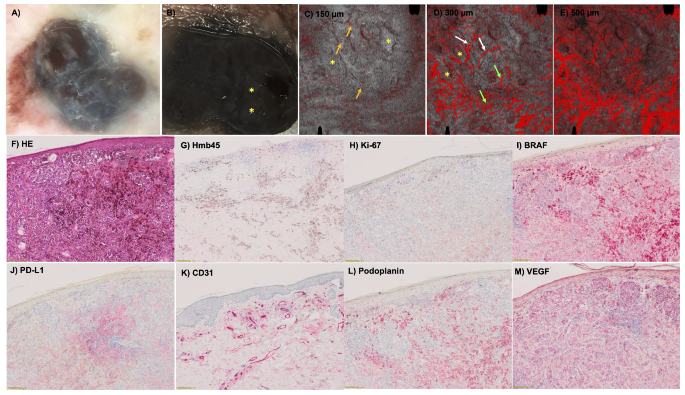
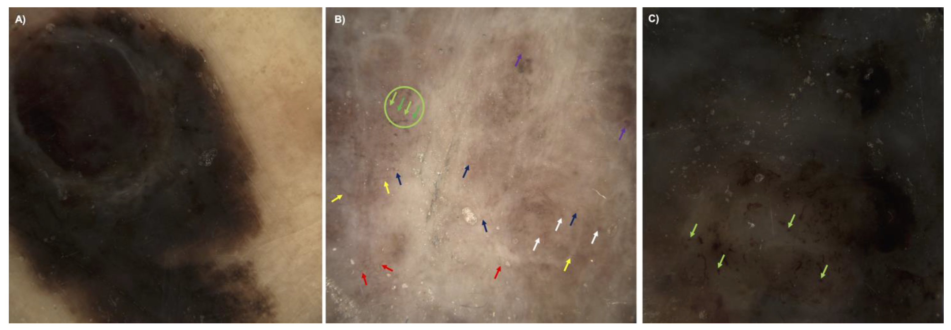
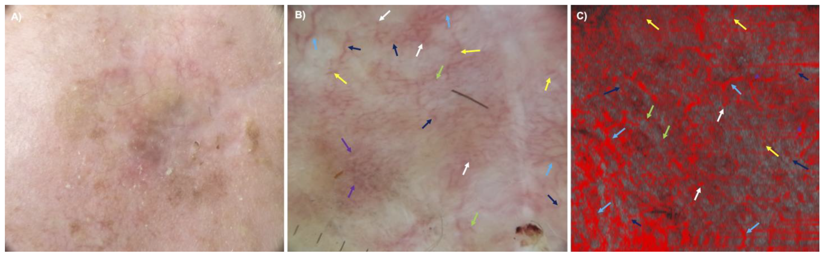
The formation of new blood vessels is crucial for tumor progression and worsens the prognosis of melanoma patients. Now the prediction of risk progression depends on histologic parameters assessed after surgery. Dermoscopy also assesses pieces of information such as the vascularization of melanomas. Due to the possible in vivo evaluation of blood vessels in melanomas with dynamic optical coherence tomography (D-OCT), we wanted to examine atypical vessel patterns, density, shape and distribution as well as the presence/type of vessel branching in melanomas in three depths and to correlate the data with the same patterns with dermoscopic and histologic malignancy parameters (like ulceration, regression and inflammation) for the evaluation of risk for metastases. The aim is to show that tumor vasculature can be noninvasively assessed using D-OCT before surgery.
Abstract
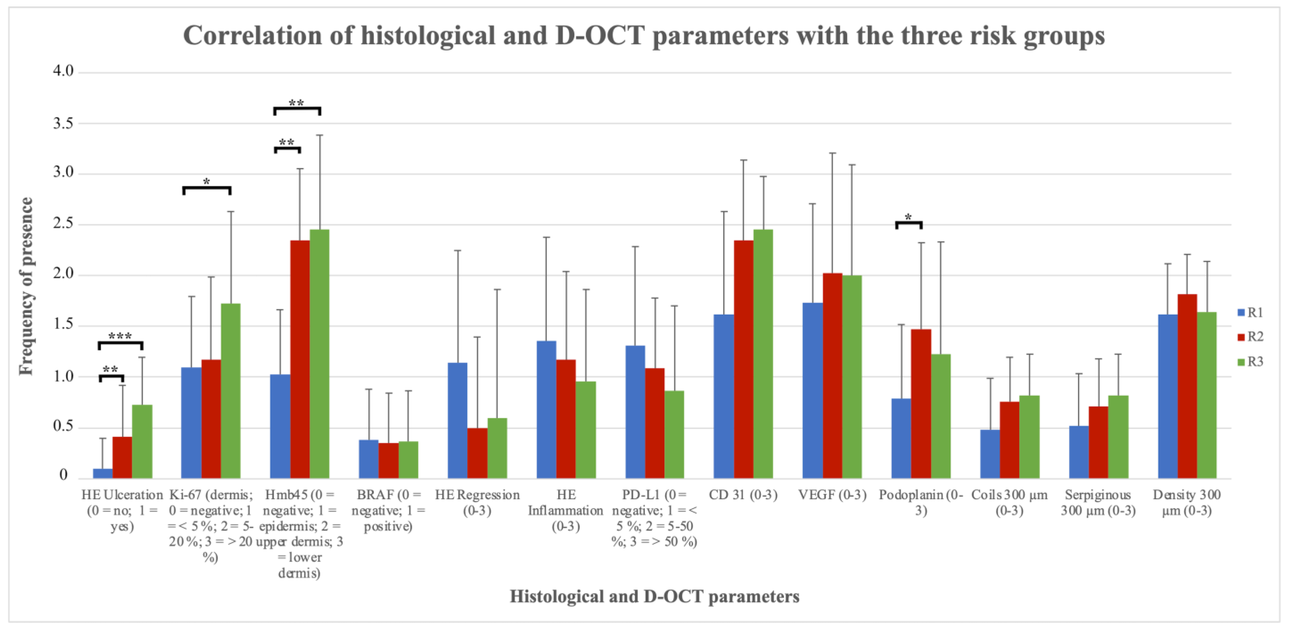
Dermoscopy adds important information to the assessment of cutaneous melanoma, but the risk of progression is predicted by histologic parameters and therefore requires surgery and histopathologic preparation. Neo-vascularization is crucial for tumor progression and worsens prognosis. The aim of this study was the in vivo evaluation of blood vessel patterns in melanoma with dynamic optical coherence tomography (D-OCT) and the correlation with dermoscopic and histologic malignancy parameters for the risk assessment of melanoma. In D-OCT vessel patterns, shape, distribution and presence/type of branching of 49 melanomas were evaluated in vivo at three depths and correlated with the same patterns in dermoscopy and with histologic parameters after excision. In D-OCT, blood vessel density and atypical shapes (coils and serpiginous vessels) increased with higher tumor stage. The histologic parameters ulceration and Hmb45- and Ki67-positivity increased, whereas regression, inflammation and PD-L1-positivity decreased with risk. CD31, VEGF and Podoplanin correlated with D-OCT vasculature findings. B-RAF mutation status had no influence. Due to pigment overlay and the summation effect, the vessel evaluation in dermoscopy and D-OCT did not correlate well. In summary, atypical vessel patterns in melanoma correlate with histologic parameters for risk for metastases. Tumor vasculature can be noninvasively assessed using D-OCT before surgery.