Article

Incidence and Predictors for Oncologic Etiologies in Chinese Children with Pituitary Stalk Thickening

1
Department of Paediatrics and Adolescent Medicine, The Hong Kong Children’s Hospital, Hong Kong, China
2
Department of Paediatrics and Adolescent Medicine, Queen Mary Hospital, Hong Kong, China
3
Department of Radiology, The Hong Kong Children’s Hospital, Hong Kong, China
4
Department of Paediatrics, Prince of Wales Hospital, Hong Kong, China
5
Department of Paediatrics and Adolescent Medicine, School of Clinical Medicine, The University of Hong Kong, Hong Kong, China
*
Author to whom correspondence should be addressed.
These authors contributed equally to this work.
Cancers 2023, 15(15), 3935; https://doi.org/10.3390/cancers15153935
Submission received: 22 June 2023 / Revised: 24 July 2023 / Accepted: 31 July 2023 / Published: 2 August 2023

Abstract

:

Simple Summary

This is a review of the clinical course in 28 Chinese children with pituitary stalk thickening (PST) and endocrinopathies revealed a higher rate of germ cell tumors (GCT) compared to the literature based largely on non-Asian cohorts. Predictors for oncologic etiologies included male sex, severe stalk thickening ≥6.5 mm, central diabetes insipidus or ≥2 pituitary hormone deficiencies as presenting features. Management guidelines for PST should take into account patient ethnicity with a lower threshold of biopsy for Asian children. Multidisciplinary approach with serial monitoring of clinical, tumour markers, endocrinopathies and imaging are essential for diagnosis.

Abstract

Background: With the increasing use of magnetic resonance imaging (MRI) in the evaluation of children with endocrine disorders, pituitary stalk thickening (PST) poses a clinical conundrum due to the potential for underlying neoplasms and challenges in obtaining a tissue biopsy. The existing literature suggests Langerhans cell histiocytosis (LCH) to be the commonest (16%) oncologic cause for PST, followed by germ cell tumors (GCTs, 13%) (CCLG 2021). As the cancer epidemiology varies according to ethnicity, we present herein the incidence and predictors for oncologic etiologies in Hong Kong Chinese children with PST. Methods: Based on a territory-wide electronic database, we reviewed patients aged < 19 years who presented to three referral centers with endocrinopathies between 2010 and 2022. Records for patients who underwent at least one MRI brain/pituitary were examined (n = 1670): those with PST (stalk thickness ≥ 3 mm) were included, while patients with pre-existing cancer, other CNS and extra-CNS disease foci that were diagnostic of the underlying condition were excluded. Results: Twenty-eight patients (M:F = 10:18) were identified. The median age at diagnosis of PST was 10.9 years (range: 3.8–16.5), with central diabetes insipidus (CDI) and growth hormone deficiency (GHD) being the most frequent presenting endocrine disorders. At a median follow-up of 4.8 years, oncologic diagnoses were made in 14 patients (50%), including 13 GCTs (46%; germinoma = 11, non-germinoma = 2) and one LCH (4%). Among patients with GCTs, 10 were diagnosed based on histology, two by abnormal tumor markers and one by a combination of histology and tumor markers. Three patients with germinoma were initially misdiagnosed as hypophysitis/LCH. The cumulative incidence of oncologic diagnoses was significantly higher in boys and patients with PST at presentation ≥6.5 mm, CDI or ≥2 pituitary hormone deficiencies at presentation and evolving hypopituitarism (all p < 0.05 by log-rank). Conclusions: A higher rate of GCTs was observed in Chinese children with endocrinopathy and isolated PST. The predictors identified in this study may guide healthcare providers in Asia in clinical decision making. Serial measurement of tumor markers is essential in management.

1. Introduction

The pituitary stalk connects the median eminence of the hypothalamus to the pituitary gland [1]. Pathologies involving the pituitary stalk may present with hormonal deficiencies, precocity and non-specific symptoms such as headache and visual disturbance [2,3,4,5,6]. Isolated pituitary stalk thickening (PST) may be a result of neoplasms, such as germ cell tumors (GCTs) and Langerhans cell histiocytosis (LCH), inflammation, infection or congenital variants or can be idiopathic [6,7,8,9,10,11,12,13,14,15]. With the increasing use of magnetic resonance imaging (MRI) in the evaluation of probable growth and hormonal disturbances, PST is increasingly being diagnosed in children and adolescents [6,16,17,18,19]. Given the potential morbidities and concerns regarding sampling errors in surgical biopsies, the management of children with PST represents a clinical conundrum for physicians [20,21,22,23,24,25,26]. Risk assessment by clinical, biochemical and radiographic features is essential in making an informed decision in order to avoid unnecessary surgical procedures and delays in tumor diagnosis.
The National Institute for Health and Care Excellence (NICE) has formulated guidelines on the management of children and adolescents with PST and/or central diabetes insipidus (CDI) with no apparent diagnosis [27]. A review of the literature indicates that 45.5% of stalk thickenings are due to neoplastic causes, while 29.1% are idiopathic. Specifically, LCH is the most common oncologic diagnosis (16.1%), followed by GCT (13.1%) and craniopharyngioma (12.3%). Congenital lesions account for 19.1%, with the commonest being septo-optic dysplasia (5.8%). Only a minority are found to have infectious, inflammatory, autoimmune or post-traumatic etiologies.
Nonetheless, it is well established that the epidemiology of CNS tumors varies according to ethnic groups. In particular, CNS GCTs have a significantly higher incidence in Asians when compared to the Western population, which the current literature is largely based on [28,29]. In addition, report cohorts do not precisely address the question of isolated PST, and pathologies involving the hypothalamus (such as craniopharyngioma) and pituitary gland (such as pituitary adenoma) are frequently included. Herein, we report the incidence rates, risk factors and predictors for neoplasms in a multi-institutional cohort of Chinese children with PST presenting with endocrinopathies in Hong Kong. Our findings are discussed in relation to the applicability of the NICE guidelines for our local population. As an exploratory study, the feasibility of incorporating 11C-methionine PET-MRI in the context of PST is also evaluated.

2. Materials and Methods

2.1. Study Cohort

This was a retrospective cohort study of Chinese children presenting with endocrinopathies and the MRI findings of PST, managed at two university-affiliated pediatric units (Queen Mary Hospital, Prince of Wales Hospital) and Hong Kong Children’s Hospital between 2010 and 2020. Ethics approval was granted by the Hospital Authority Central Institutional Review Board (reference number: PAED-2023-009).
Study subjects were identified using the territory-wide electronic patient database Clinical Data Analysis and Reporting System (CDARS). The database was first inquired for pediatric patients (<19 years of age) diagnosed with one or more endocrinopathies, including growth hormone deficiency (GHD), hypogonadotropic hypogonadism (HH), central hypothyroidism (CH), adrenal insufficiency (AI), central precocious puberty (CPP) and central diabetes insipidus (CDI) (ICD 9CM codes 253.2, 253.3, 253.4, 253.5, 255.4, 259.1). The patient list was further filtered for patients who had at least one MRI of the brain and/or pituitary region performed in the three study institutions. Individual clinical records (n = 1670) were then reviewed to identify patients according to the following criteria. Inclusion criteria included (1) Chinese patients <19 years of age at first imaging, (2) neuro-imaging performed as part of evaluation for the presenting endocrinopathies, (3) PST according to criteria specified in the NICE guidelines [27] and (4) follow-up (FU) duration of 2 years or more in patients without oncologic diagnosis. Exclusion criteria included (1) patients with radiographic abnormalities beyond PST, such as mass lesions within the pituitary gland or the hypothalamic region, (2) patients with known oncologic diagnosis prior to the index neuroimaging (3) and patients with extra-CNS features that were diagnostic of the underlying condition—for example, skeletal or cutaneous features in LCH.

2.2. Data Collection

We reviewed individual patient records for demographic, clinical, biochemical, histological and outcome data. These included sex, age at presentation, age at diagnosis of PST, symptoms and hormonal profiles at diagnosis of PST, before treatment and during FU, available tumor marker levels (serum and cerebrospinal fluid (CSF) alpha-fetoprotein (AFP) and human chorionic gonadotropin (hCG)), extent of PST, morphologic characteristics of pituitary stalks and histologic features on sampled tissue. In patients with subsequent oncologic diagnoses, details of the anti-tumor therapy and treatment outcomes were captured.

2.3. Definition of PST

As the designation of PST varies in the literature [28,30,31,32,33,34], we adopted definitions from the NICE guidelines in this study [27]. MRI images for the included subjects, where available (n = 24), were reviewed by two radiologists (C.C. and C.K.C.W.). For patients presenting with clinical features (as in our cohort), the cut-off for PST was 2 mm at the pituitary insertion or 3 mm at the level of the optic chiasm. The degree of PST was classified as mild (<4 mm), moderate (4–6.5 mm) and severe (≥6.5 mm) thickening [18].

2.4. Primary and Secondary Outcomes

Our primary outcome was the cumulative incidence rate of oncologic diagnosis in the study cohort. The secondary outcomes were predictors for underlying oncologic etiologies and the natural course of endocrinopathy in patients with or without a defined neoplastic diagnosis.

2.5. 11C-Methionine PET-MRI

The 11C-methionine (MET) PET-MRI studies were performed in a PET-MR scanner with simultaneous MR DIXON HiRes sequences for attenuation correction and T1 3D MPRAGE for localization. Amino acid PET brain studies were performed at 25 min after the administration of 0.2 mCi/kg of 11C-methionine. The PET uptake images were presented in axial, coronal and sagittal planes. The lesion-to-normal region SUVmean and SUVmax ratios (LNRmean and LNRmax) were determined with reference to the normal contralateral corresponding structure. For the MR study, the following sequences were obtained: sagittal GRE T1-weighted with and without fat suppression; axial SE T1-weighted; axial Turbo SE T2-weighted; axial diffusion-weighted with ADC map; axial SWI; axial FLAIR; coronal FLAIR; sagittal turbo SE T2-weighted pituitary gland; post-contrast sagittal 3D vibe; and space T1-weighted with fat suppression.

2.6. Statistical Analysis

Continuous data were presented as medians with ranges, whereas categorical data were presented as counts with percentages. Numerical variables were compared between groups by the Mann–Whitney U test and categorical variables were compared between groups by the Chi-square or Fisher’s exact test. The cumulative incidence of oncologic events was derived from Kaplan–Meier estimates, with the date of diagnosis of PST defined as date of the first MRI revealing such a finding, the date of the event defined as the date of biopsy in those who underwent tissue sampling supporting the final cancer diagnosis or the date of first tumor marker elevation. Patients without an oncologic diagnosis were censored on the date of their last FU. Comparison of the cumulative incidence between groups was performed using a log-rank test, as no competing outcome was identified. Hazard ratios (HR) were obtained with Cox regression. A p value < 0.05 was considered statistically significant.

3. Results

3.1. Demographics and Baseline Characteristics

Twenty-eight patients were identified for our study, including 18 females (64%) and with a median age of diagnosis of PST of 10.9 years (range: 3.8–16.5) (Supplementary Table S1). The initial symptoms were polyuria and polydipsia (n = 17, symptom duration median = 3 months, range 1–35 months), growth retardation (n = 9), precocious puberty (n = 8) and delayed puberty (n = 5). These were attributed to biochemically confirmed endocrinopathies, including CDI (n = 18), GHD (n = 14), CPP (n = 7), AI (n = 7), HH (n = 7) and CH (n = 6). Fifteen patients (54%) had ≥2 hormonal deficiencies at diagnosis of PST. Visual problems represented the second most common class of presenting symptoms in these patients. Among the nine patients (32%) with visual issues, four had strabismus, two had visual field defects, one had papilloedema and one had amblyopia.

3.2. Etiology for PST and Patient Outcome

With a median FU of 4.8 years (range: 0.5–19.3), 14 patients (50%) were diagnosed with oncologic conditions, namely GCT in 13 (46%; germinoma = 11; non-germinomatous GCT (NGGCT) = 2) and LCH in one (4%) (Figure 1). The diagnoses were made after tissue biopsy in 10, the detection of elevated tumor markers in two (both in serum and CSF) and a combination in one. The patient with LCH was diagnosed clinically based on normal tumor markers and the response to the LCH treatment regimen (FU for 9.4 years).
Three patients with GCTs were previously misdiagnosed as having hypophysitis (n = 2) or LCH (n = 1). Patient No. 8 had an initial diagnosis of hypophysitis and underwent three biopsies over the course of 22 months before the diagnosis of an embryonal carcinoma was made. The first biopsy contained normal pituitary tissue, while the second biopsy contained tissue with inflammatory infiltrates that was considered to be indicative of hypophysitis. His blood and CSF AFP and HCG were normal initially but the serum HCG level was raised to 7 IU/L (normal < 5 IU/L) shortly after the third biopsy. Patient No. 13 was initially managed as LCH with steroid and vinblastine. He was eventually diagnosed with germinoma 3 years later, when he progressed with ventricular metastasis. His serum and CSF AFP and HCG were normal initially but the serum and CSF HCG became raised 3 years later when he was diagnosed with ventricular metastasis. Patient No. 1 presented with decreased growth velocity and CDI, which later evolved into panhypopituitarism with PST of 5 mm. Her serum and CSF tumor markers were again all normal. A biopsy was performed, and she was initially misdiagnosed as having lymphocytic hypophysitis. However, with a further pathology review, germinoma cells were identified, once again highlighting the caveats for diagnosis with the availability of tumor tissue. The median time from the appearance of the first symptom to the diagnosis of GCT/LCH was 1.3 years (range: 0.5–5.4). The cause of PST in the rest of the patients (n = 14, 50%; median FU 3 years, range: 1.3–12.2) remained idiopathic. Among the study cohort, one patient with NGGCT died due to disease progression.

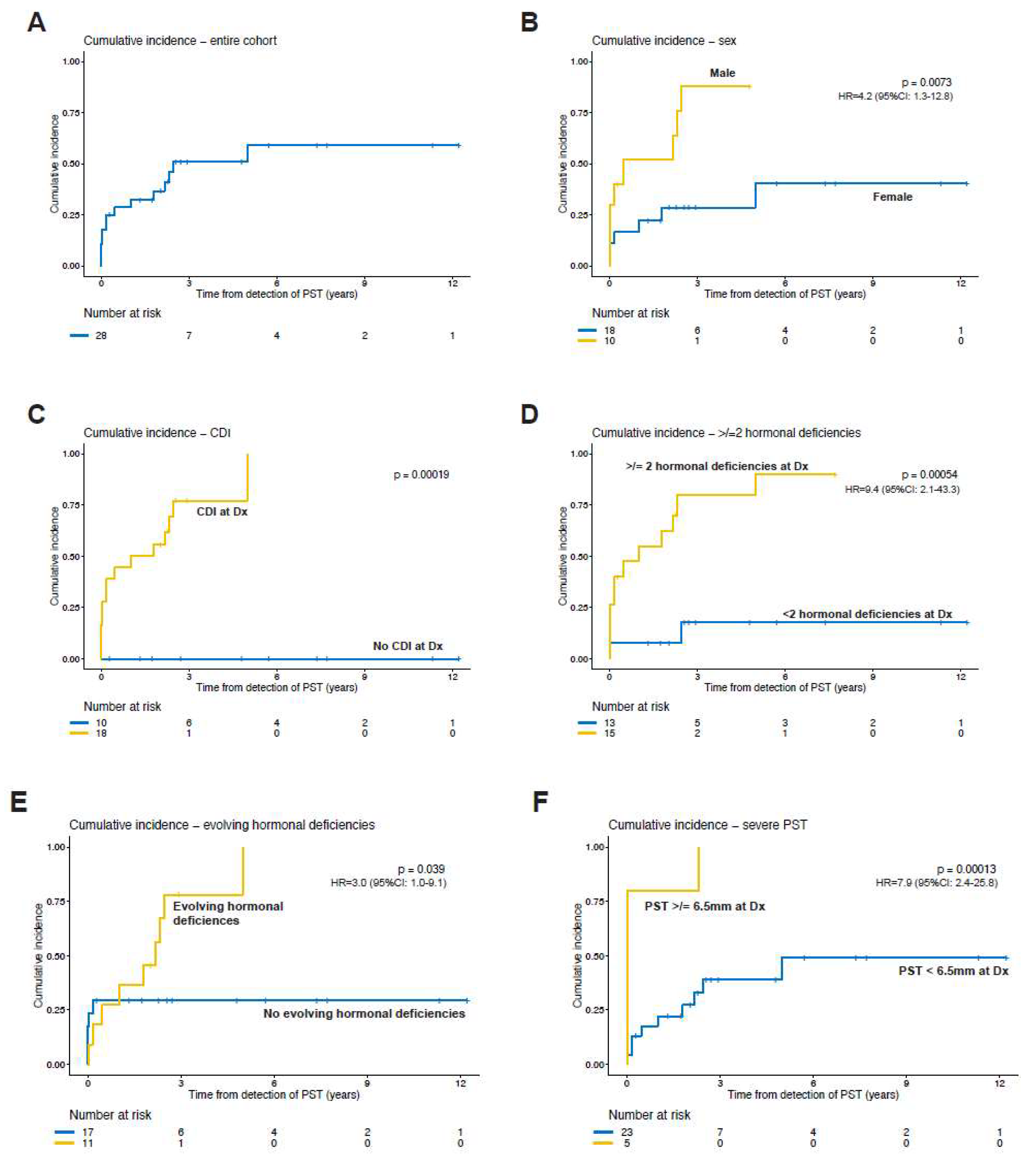
3.3. Risk Factors for Underlying Neoplasm

Figure 1 shows the cumulative incidence of neoplasms based on clinical, biochemical and radiographic risk factors. Male gender, central diabetes insipidus, ≥2 hormonal deficiencies at diagnosis of PST and PST ≥ 6.5 mm at presentation were predictors of oncologic etiologies. In addition, the evolution of the endocrinopathies was significantly associated with underlying neoplasms.

3.4. Clinical Course and Endocrine Outcome

Table 1 summarizes the evolution of the endocrinopathies during the study period. At the last FU, CDI remained as the most common endocrinopathy (n = 18), followed by GHD (n = 14), AI (n = 12) and CH (n = 12).
For the patients with an oncologic diagnosis, the evolution of a pituitary hormone deficiency was more common (n = 9/14; 64%) than in the idiopathic group (n = 2/14, 14%) (evolution of pituitary hormone deficiencies was more common in patients with oncologic diagnosis compared to those with idiopathic causes (64% vs. 14%)). Panhypopituitarism (defined as deficiencies in ≥3 pituitary hormones) was observed in most patients with an oncologic diagnosis before the start of anti-tumor therapy (n = 13). Of interest, patient No. 4, who initially presented with CDI, had an apparent ‘resolution’ of CDI and was able to stop minirin due to the emergence of AI during FU. After the completion of the anti-tumor therapy (chemo-irradiation in GCT, chemotherapy in LCH), the hormonal profile remained stable until the last FU in eight patients; pituitary hormone deficiencies improved in five patients and they worsened in one patient. Among patients with idiopathic PST, the majority of them had stable hormonal profiles during the course of their FU (n = 11), two had progressive endocrinopathies, and one showed the regression of the endocrinopathy.
For those presenting with visual problems, we observed a slightly greater number of patients in the neoplastic group (n = 5) when compared with the idiopathic group (n = 4), but the difference was not clinically significant (p > 0.05).

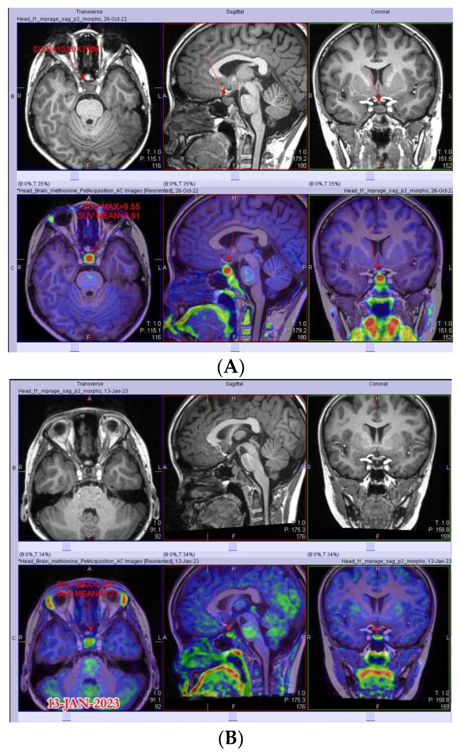
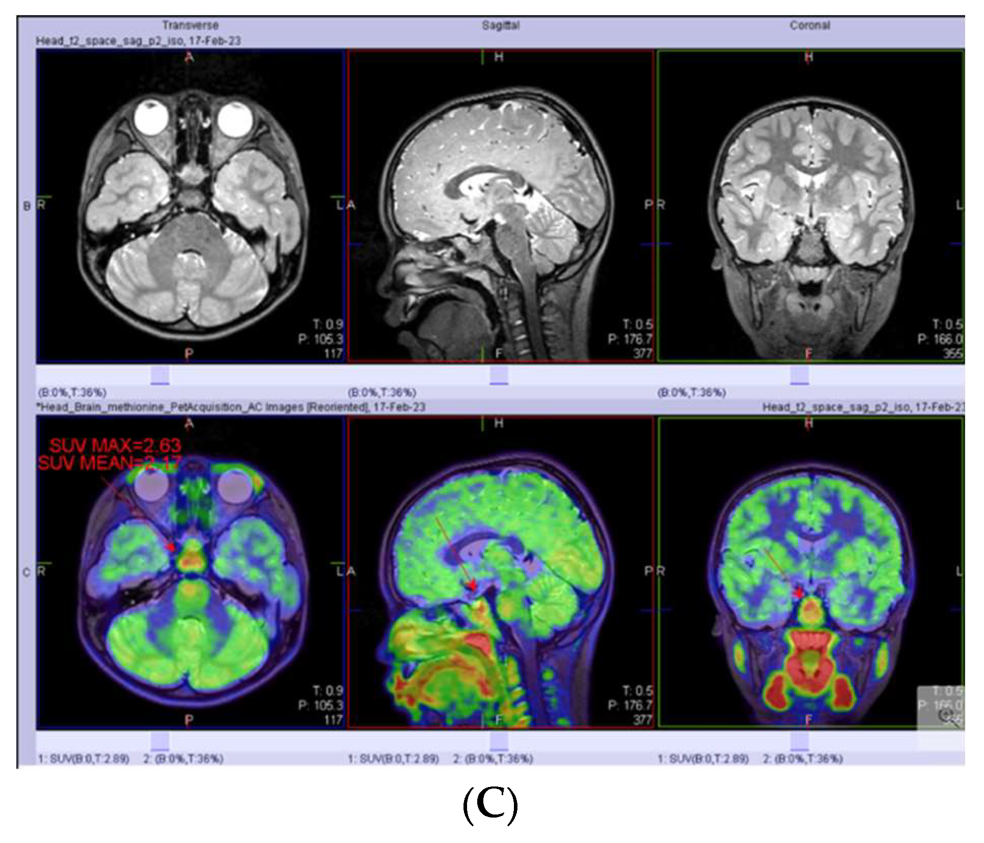
3.5. PET-MRI as a Complementary Tool in PST Diagnostics

As an exploratory, proof-of-principle analysis, we incorporated PET-MRI in the evaluation of two patients with PST (Figure 2). Patient No. 28 was a 14-year-old boy who presented with CDI, GHD, HH and AI, with MRI showing PST of 6.6 mm and normal tumor markers. The PET uptake of pituitary tissue at diagnosis showed SUVmax of 9.6, and a subsequent biopsy confirmed the diagnosis of germinoma. Upon completion of chemotherapy, an interval PET scan showed a reduction in SUVmax to 2.8. Patient No. 12 was a 7-year-old girl who presented with CDI and GHD. The growth velocity was decreased to 2.5 cm/year at presentation and her MRI revealed PST of 4 mm. Despite FU for over 2 years, the family refused a CSF examination and biopsy, thus representing a dilemma between the need for GH replacement and the concern about a neoplasm. She eventually underwent a PET-MRI scan, which revealed an SUVmax of 2.6, which was within normal limits. The patient was then counselled on the likelihood of a non-neoplastic cause and GH replacement was commenced.

4. Discussion

One of the strengths of our study is its basis on a sizable cohort of patients with endocrine dysfunction and with MRI performed in three pediatric units, which allowed for unbiased subject inclusion with an adequate duration of FU. Additionally, our cohort was uniquely of Chinese ethnicity, adding to the literature data based on a diversity of populations. Neoplastic causes accounted for 50% of the PST in our cohort, which is similar to that reported in the literature (45.5%) based on the summary of a largely Western population (n = 684 from 11 studies, 1 study with 48 patients from Asian region) [27]. Compared with the reported pooled experience (13.1% and 16.1% respectively), we saw a higher rate of GCT (46.4%) and lower rate of LCH (3.6%). The only Asian study from this combined analysis was by Liu et al., based on a single-institution experience from Taiwan—among children with DI with/without PST, 32.2% and 19.4% had GCT and LCH, respectively [35]. Nevertheless, only half of these patients with GCT/LCH had PST and the criteria for diagnosis were not specified, rendering such evidence difficult to apply for decision making in the context of children with pituitary stalk anomalies and endocrinopathies. Overall, the observed larger proportion of patients with CNS GCT in our cohort is consistent with the higher incidence of this condition in Chinese and Asians [36,37]. With almost half of our patients suffering from GCTs, the upfront testing of tumor markers and subsequent surveillance in patients with presumed idiopathic disease are essential.
Previous studies did not evaluate predictors of oncologic etiologies in patients with PST. Based on our findings, males with PST were more likely have neoplastic causes, and this is consistent with another study from Korea by Yoon et al. [14]. Additional predictors included severe stalk thickening and CDI with/without additional pituitary hormonal deficiencies. The proposed NICE algorithm takes into account the degree of PST, hormonal disturbances, the evolution of the hormonal profile, radiological features, tumor marker levels and vision for the formulation of the management plan. However, the NICE guidelines do not account for the higher incidence of GCTs in Asians and thus the discordance in risk by sex. In addition, we opine that the guidelines remain relatively conservative when applied to our local context, as surveillance is only recommended after unrevealing second-line investigations (i.e., CSF examination ± whole-body PET for LCH), even in patients with severe PST or a combination of PST and CDI/evolving hormonal deficiencies. Based on our results, we suggest that in ethnic groups where the incidence of CNS GCT is higher, biopsy should be offered after non-diagnostic second-line investigations for patients with PST who (1) are male, (2) have severe stalk thickening (≥6.5 mm) or (3) present with CDI. However, it should be highlighted that even with the histological diagnosis of ‘lymphocytic hypophysitis’, the ultimate underlying diagnosis could still be GCT, as illustrated by three of our patients in this cohort. Ongoing monitoring and appropriate counselling is needed in this subset of patients. In a situation where a decision remains difficult, additional anterior hormonal deficiencies and/or the evolution of hormonal deficiencies should be considered as pressing indicators for tissue sampling. On the other hand, a more conservative approach with serial biochemical and radiological monitoring can be advised for female patients or those presenting with CPP, where PST is more likely due to non-oncologic causes. In such a low-risk group, an annual MRI for three years may be considered before discharging the patient from routine neuroimaging, in the absence of evolving endocrinologic, neurological or visual symptoms; meanwhile, a CSF examination for tumor markers may be considered at the diagnosis of stalk thickening after thorough counseling. The timely discussion of appropriate diagnostic procedures will mitigate the delay in diagnosis and prevent further tumor-related complications, such as hydrocephalus, metastasis and worsening hypopituitarism. As demonstrated in our experience, the loss of pituitary function can be partially reversed in a subset of patients with CNS GCT after treatment.
On the whole, the diagnosis of childhood brain tumors is more often delayed than in other pediatric cancers. This, as demonstrated in our study, is particularly important for sellar–suprasellar tumors, including GCT, low-grade glioma and craniopharyngioma. For such tumors, symptoms can be insidious, atypical and may even wax and wane, adding to the diagnostic challenges [38]. To shorten the lead time to diagnosis, education starting with the medical curriculum should be undertaken [39].
It may be appreciated that even with the availability of tumor tissue, the interpretation of histology may be hampered by sampling bias, tissue heterogeneity and the prominent inflammatory infiltrates frequently present in CNS GCTs. Recently, there has been increasing interest in the application of 11C-MET PET-MRI as a diagnostic tool in patients with intracranial neoplasms [40,41], including initial experience in patients with CNS GCTs [42,43,44,45,46,47]. Such a methodology offers functional data that complement the anatomical data captured by MRI alone. The potential utility of 11C-MET PET includes differentiation between malignant and non-malignant brain lesions, the identification of hypermetabolic regions within neoplasms to guide sampling and the tracking of the response to therapy, including the delineation between disease progression and pseudoprogression [41]. Its application in two of our patients with PST demonstrates its feasibility and supports further evaluation for its utility in this setting.
The limitations of the study include its retrospective nature, the relatively small cohort size and the possibility of the theoretically delayed presentation of neoplasms in those who were labelled thus far as having idiopathic PST. The patient number did not allow a meaningful multivariate analysis of cancer predictors to be performed. Nonetheless, our study represents the only study on Asian children to date focusing on patients presenting with isolated PST. Within the scope of this study, we have not been able to incorporate novel biomarkers for CNS GCTs, such as CSF and serum microRNAs. Specific microRNA species allow differentiation between patients with and without CNS GCTs [48]. MicroRNA profiling may represent a powerful platform as serial monitoring allows the tracking of treatment responses.

5. Conclusions

The risk of oncologic etiologies in children with endocrinopathies as well as PST differs according to ethnicity, and our study offers invaluable data on the rates and predictors of oncologic outcomes in a carefully curated Chinese cohort. CNS GCTs are more prevalent in Asian patients, calling for an ethnicity-specific diagnostic strategy, with a lower threshold for biopsy in patients of male sex, with severe stalk thickening and CDI. Careful scrutinization of sampled tissue is essential, while PET-MRI represents a potentially useful tool to complement existing modalities of investigation. Early diagnosis will allow the better preservation of function.

Supplementary Materials

The following supporting information can be downloaded at: https://www.mdpi.com/article/10.3390/cancers15153935/s1, Table S1: Baseline Demographics of non-neoplastic versus neoplastic groups.

Author Contributions

Conceptualization: M.W.T.L., S.W.Y.P., M.M.K.S., T.T.W.C., S.L.K.L., G.S.W.P., G.W.K.P., H.-C.Y., J.Y.L.T. and A.P.Y.L.; methodology: M.W.T.L., S.W.Y.P., J.Y.L.T. and A.P.Y.L.; formal analysis: M.W.T.L.; investigation: M.W.T.L., C.C. and C.K.C.W.; data curation: M.W.T.L., S.W.Y.P., J.Y.L.T. and A.P.Y.L.; writing—original draft preparation: M.W.T.L.; writing—review and editing: S.W.Y.P., E.Y.W.K., J.Y.L.T. and A.P.Y.L.; supervision: J.Y.L.T. and A.P.Y.L.; project administration: M.W.T.L., S.W.Y.P., J.Y.L.T. and A.P.Y.L. All authors have read and agreed to the published version of the manuscript.

Funding

This research received no external funding.

Institutional Review Board Statement

The study was conducted in accordance with the Declaration of Helsinki. Ethics approval was granted by the Hospital Authority Central Institutional Review Board (reference number: PAED-2023-009).

Informed Consent Statement

Patient consent was waived because this was a retrospective study.

Data Availability Statement

The data presented in this study are available in this article.

Acknowledgments

The authors would like to acknowledge Wilfred Wong of the Department of Paediatrics and Adolescent Medicine, the University of Hong Kong, for his assistance with the database search. We appreciate all healthcare providers from the participating units for offering expert care to the patients included.

Conflicts of Interest

The authors have no conflict of interest to declare that are relevant to the content of this article.

References

  1. Sbardella, E.; Joseph, R.N.; Jafar-Mohammadi, B.; Isidori, A.M.; Cudlip, S.; Grossman, A.B. Pituitary stalk thickening: The role of an innovative MRI imaging analysis which may assist in determining clinical management. Eur. J. Endocrinol. 2016, 175, 255–263. [Google Scholar] [CrossRef] [PubMed] [Green Version]
  2. Chen, H.; Ni, M.; Xu, Y.; Zhong, L.Y. Pituitary Stalk Germ Cell Tumors: Retrospective Case Series and Literature Review. Int. J. Endocrinol. 2022, 2022, 9213220. [Google Scholar] [CrossRef] [PubMed]
  3. Liu, W.; Hou, J.; Liu, X.; Wang, L.; Li, G. Causes and Follow-Up of Central Diabetes Insipidus in Children. Int. J. Endocrinol. 2019, 2019, 5303765. [Google Scholar] [CrossRef] [Green Version]
  4. Redhu, R.; Nadkarni, T.; Mahesh, R. Diabetes insipidus associated with a thickened pituitary stalk in a case of Langerhans Cell Histiocytosis. J. Pediatr. Neurosci. 2011, 6, 62–64. [Google Scholar] [PubMed]
  5. Kluczyński, Ł.; Gilis-Januszewska, A.; Godlewska, M.; Wójcik, M.; Zygmunt-Górska, A.; Starzyk, J.; Hubalewska-Dydejczyk, A. Diversity of Pathological Conditions Affecting Pituitary Stalk. J. Clin. Med. 2021, 10, 1692. [Google Scholar] [CrossRef]
  6. Alter, C.A.; Bilaniuk, L.T. Utility of magnetic resonance imaging in the evaluation of the child with central diabetes insipidus. J. Pediatr. Endocrinol. Metab. 2002, 15 (Suppl. 2), 681–687. [Google Scholar] [CrossRef]
  7. Prosch, H.; Grois, N.; Bökkerink, J.; Prayer, D.; Leuschner, I.; Minkov, M.; Gadner, H. Central diabetes insipidus: Is it Langerhans cell histiocytosis of the pituitary stalk? A diagnostic pitfall. Pediatr. Blood Cancer 2006, 46, 363–366. [Google Scholar] [CrossRef]
  8. Robison, N.J.; Prabhu, S.P.; Sun, P.; Chi, S.N.; Kieran, M.W.; Manley, P.E.; Cohen, L.E.; Goumnerova, L.; Smith, E.R.; Scott, R.M.; et al. Predictors of neoplastic disease in children with isolated pituitary stalk thickening. Pediatr. Blood Cancer 2013, 60, 1630–1635. [Google Scholar] [CrossRef]
  9. Maghnie, M. Diabetes insipidus. Horm. Res. 2003, 59 (Suppl. 1), 42–54. [Google Scholar] [CrossRef]
  10. Mootha, S.L.; Barkovich, A.J.; Grumbach, M.M.; Edwards, M.S.; Gitelman, S.E.; Kaplan, S.L.; Conte, F.A. Idiopathic hypothalamic diabetes insipidus, pituitary stalk thickening, and the occult intracranial germinoma in children and adolescents. J. Clin. Endocrinol. Metab. 1997, 82, 1362–1367. [Google Scholar]
  11. Turcu, A.F.; Erickson, B.J.; Lin, E.; Guadalix, S.; Schwartz, K.; Scheithauer, B.W.; Atkinson, J.L.D.; Young, W.F. Pituitary stalk lesions: The Mayo Clinic experience. J. Clin. Endocrinol. Metab. 2013, 98, 1812–1818. [Google Scholar] [CrossRef] [Green Version]
  12. Czernichow, P.; Garel, C.; Léger, J. Thickened pituitary stalk on magnetic resonance imaging in children with central diabetes insipidus. Horm. Res. 2000, 53 (Suppl. 3), 61–64. [Google Scholar] [CrossRef]
  13. Leger, J.; Velasquez, A.; Garel, C.; Hassan, M.; Czernichow, P. Thickened pituitary stalk on magnetic resonance imaging in children with central diabetes insipidus. J. Clin. Endocrinol. Metab. 1999, 84, 1954–1960. [Google Scholar] [CrossRef]
  14. Yoon, S.C.; Shin, C.H.; Yang, S.W.; Lee, S.Y. Clinical and radiological features of pituitary stalk lesions in children and adolescents. Ann. Pediatr. Endocrinol. Metab. 2014, 19, 202–207. [Google Scholar] [CrossRef] [Green Version]
  15. Halefoglu, A.M. Magnetic resonance imaging of thickened pituitary stalk proceeding to Langerhans cell histiocytosis in a child. Australas. Radiol. 2006, 50, 175–178. [Google Scholar] [CrossRef]
  16. Moszczyńska, E.; Baszyńska-Wilk, M.; Zasada, K.; Majak, D.; Szaniawska, M.; Szalecki, M. Pituitary stalk thickening in patients under 18 years of age—The most common causes and diagnostic procedures. Pediatr. Endocrinol. Diabetes Metab. 2022, 28, 213–227. [Google Scholar] [CrossRef]
  17. Langlois, F.; Varlamov, E.V.; Fleseriu, M. Hypophysitis, the Growing Spectrum of a Rare Pituitary Disease. J. Clin. Endocrinol. Metab. 2022, 107, 10–28. [Google Scholar] [CrossRef]
  18. Di Iorgi, N.; Morana, G.; Maghnie, M. Pituitary stalk thickening on MRI: When is the best time to re-scan and how long should we continue re-scanning for? Clin. Endocrinol. 2015, 83, 449–455. [Google Scholar] [CrossRef]
  19. Uddin, A.; Kalladi Puthanpurayil, S. Central Diabetes Insipidus with Pituitary Stalk Thickening. In Diabetes Insipidus in Children: A Pocket Guide; Springer: Berlin/Heidelberg, Germany, 2021; pp. 41–53. [Google Scholar]
  20. Devuyst, F.; Kazakou, P.; Balériaux, D.; Alexopoulou, O.; Burniat, A.; Salenave, S.; Chanson, P.; Corvilain, B.; Maiter, D. Central diabetes insipidus and pituitary stalk thickening in adults: Distinction of neoplastic from non-neoplastic lesions. Eur. J. Endocrinol. 2020, 183, 95–105. [Google Scholar] [CrossRef]
  21. Moszczyńska, E.; Kunecka, K.; Baszyńska-Wilk, M.; Perek-Polnik, M.; Majak, D.; Grajkowska, W. Pituitary Stalk Thickening: Causes and Consequences. The Children’s Memorial Health Institute Experience and Literature Review. Front. Endocrinol. 2022, 13, 868558. [Google Scholar] [CrossRef]
  22. Chaudhary, V.; Bano, S. Imaging of pediatric pituitary endocrinopathies. Indian J. Endocrinol. Metab. 2012, 16, 682–691. [Google Scholar] [CrossRef] [PubMed]
  23. Capra, M.; Wherrett, D.; Weitzman, S.; Dirks, P.; Hawkins, C.; Bouffet, E. Pituitary stalk thickening and primary central nervous system lymphoma. J. Neurooncol. 2004, 67, 227–231. [Google Scholar] [CrossRef] [PubMed]
  24. Jian, F.; Bian, L.; Sun, S.; Yang, J.; Chen, X.; Chen, Y.; Ma, Q.; Miao, F.; Wang, W.; Ning, G.; et al. Surgical biopsies in patients with central diabetes insipidus and thickened pituitary stalks. Endocrine 2014, 47, 325–335. [Google Scholar] [CrossRef] [PubMed]
  25. Beni-Adani, L.; Sainte-Rose, C.; Zerah, M.; Brunelle, F.; Constantini, S.; Renier, D.; Lellouch-Tubiana, A.; Leger, J.; Pierre-Kahn, A. Surgical implications of the thickened pituitary stalk accompanied by central diabetes insipidus. J. Neurosurg. 2005, 103 (Suppl. 2), 142–147. [Google Scholar] [CrossRef] [PubMed]
  26. Day, E.L.; Smith, E.R.; Fehnel, K.P. Single-institution case series of pituitary biopsy for suspected germinoma in the pediatric population: Diagnostic utility, operative risks, and biopsy approaches. Sci. Rep. 2020, 10, 15257. [Google Scholar] [CrossRef]
  27. Cerbone, M.; Visser, J.; Bulwer, C.; Ederies, A.; Vallabhaneni, K.; Ball, S.; Kamaly-Asl, I.; Grossman, A.; Gleeson, H.; Korbonits, M.; et al. Management of children and young people with idiopathic pituitary stalk thickening, central diabetes insipidus, or both: A national clinical practice consensus guideline. Lancet Child Adolesc. Health 2021, 5, 662–676. [Google Scholar] [CrossRef]
  28. Zhou, X.; Zhu, H.; Yao, Y.; Lian, X.; Feng, F.; Wang, L.; Liu, S.; Deng, K.; You, H.; Yang, H.; et al. Etiological Spectrum and Pattern of Change in Pituitary Stalk Thickening: Experience in 321 Patients. J. Clin. Endocrinol. Metab. 2019, 104, 3419–3427. [Google Scholar] [CrossRef]
  29. Ji, X.; Wang, Z.; Wang, W.; Gao, L.; Guo, X.; Feng, C.; Lian, W.; Deng, K.; Xing, B. Clinical Characteristics of Pediatric Patients With Sellar and Suprasellar Lesions Who Initially Present With Central Diabetes Insipidus: A Retrospective Study of 55 Cases From a Large Pituitary Center in China. Front. Endocrinol. 2020, 11, 76. [Google Scholar] [CrossRef] [Green Version]
  30. Satogami, N.; Miki, Y.; Koyama, T.; Kataoka, M.; Togashi, K. Normal pituitary stalk: High-resolution MR imaging at 3T. Am. J. Neuroradiol. 2010, 31, 355–359. [Google Scholar] [CrossRef] [Green Version]
  31. Di Iorgi, N.; Allegri, A.E.M.; Napoli, F.; Calcagno, A.; Calandra, E.; Fratangeli, N.; Vannati, M.; Rossi, A.; Bagnasco, F.; Haupt, R.; et al. Central diabetes insipidus in children and young adults: Etiological diagnosis and long-term outcome of idiopathic cases. J. Clin. Endocrinol. Metab. 2014, 99, 1264–1272. [Google Scholar] [CrossRef]
  32. Marchand, I.; Barkaoui, M.A.; Garel, C.; Polak, M.; Donadieu, J.; Committee, W. Central diabetes insipidus as the inaugural manifestation of Langerhans cell histiocytosis: Natural history and medical evaluation of 26 children and adolescents. J. Clin. Endocrinol. Metab. 2011, 96, E1352–E1360. [Google Scholar] [CrossRef] [Green Version]
  33. Skrypek, M.; Brown, A.; Pape-Blabolil, J.; Bendelsmith, C.; Patel, S.; Patterson, R.; Abuzzahab, M.J.; Ingram, K.; Linabery, A.; Bendel, A. EPID-13. Isolated Pituitary Stalk Thickening (IPST) in Children: Natural History and Predictors of Neoplastic Disease. Neuro-Oncology 2018, 20 (Suppl. 2), i83. [Google Scholar] [CrossRef] [Green Version]
  34. Simmons, G.E.; Suchnicki, J.E.; Rak, K.M.; Damiano, T.R. MR imaging of the pituitary stalk: Size, shape, and enhancement pattern. AJR Am. J. Roentgenol. 1992, 159, 375–377. [Google Scholar] [CrossRef] [Green Version]
  35. Liu, S.Y.; Tung, Y.C.; Lee, C.T.; Liu, H.M.; Peng, S.F.; Wu, M.Z.; Kuo, M.-F.; Tsai, W.-Y. Clinical characteristics of central diabetes insipidus in Taiwanese children. J. Formos. Med. Assoc. 2013, 112, 616–620. [Google Scholar] [CrossRef] [Green Version]
  36. Liu, A.P.Y.; Liu, Q.; Shing, M.M.K.; Ku, D.T.L.; Fu, E.; Luk, C.W.; Ling, S.-C.; Cheng, K.K.F.; Kwong, D.L.W.; Ho, W.W.S.; et al. Incidence and Outcomes of CNS Tumors in Chinese Children: Comparative Analysis With the Surveillance, Epidemiology, and End Results Program. JCO Glob. Oncol. 2020, 6, 704–721. [Google Scholar] [CrossRef]
  37. Gao, Y.; Jiang, J.; Liu, Q. Clinicopathological and immunohistochemical features of primary central nervous system germ cell tumors: A 24-years experience. Int. J. Clin. Exp. Pathol. 2014, 7, 6965–6972. [Google Scholar]
  38. Qaddoumi, I.; Merchant, T.E.; Boop, F.A.; Gajjar, A. Diagnostic delay in children with central nervous system tumors and the need to improve education. J. Neurooncol. 2019, 145, 591–592. [Google Scholar] [CrossRef]
  39. Arnautovic, A.; Billups, C.; Broniscer, A.; Gajjar, A.; Boop, F.; Qaddoumi, I. Delayed diagnosis of childhood low-grade glioma: Causes, consequences, and potential solutions. Childs Nerv. Syst. 2015, 31, 1067–1077. [Google Scholar] [CrossRef]
  40. Park, Y.J.; Lee, J.W.; Cho, H.W.; Choe, Y.S.; Lee, K.H.; Choi, J.Y.; Sung, K.W.; Moon, S.H. Value of C-11 methionine PET/CT in patients with intracranial germinoma. PLoS ONE 2022, 17, e0263690. [Google Scholar] [CrossRef]
  41. Treglia, G.; Muoio, B.; Trevisi, G.; Mattoli, M.V.; Albano, D.; Bertagna, F.; Giovanella, L. Diagnostic Performance and Prognostic Value of PET/CT with Different Tracers for Brain Tumors: A Systematic Review of Published Meta-Analyses. Int. J. Mol. Sci. 2019, 20, 4669. [Google Scholar] [CrossRef] [Green Version]
  42. Kawai, N.; Miyake, K.; Yamamoto, Y.; Nishiyama, Y.; Maeda, Y.; Kageji, T.; Tamiya, T. Use of 11C-methionine positron emission tomography in basal germinoma: Assessment of treatment response and residual tumor. Childs Nerv. Syst. 2009, 25, 845–853. [Google Scholar] [CrossRef] [PubMed]
  43. Fujii, Y.; Saito, Y.; Ogawa, T.; Fujii, S.; Kamitani, H.; Kondo, S.; Horie, Y.; Togawa, M.; Senda, M.; Maegaki, Y.; et al. Basal ganglia germinoma: Diagnostic value of MR spectroscopy and (11)C-methionine positron emission tomography. J. Neurol. Sci. 2008, 270, 189–193. [Google Scholar] [CrossRef] [PubMed]
  44. Okochi, Y.; Nihashi, T.; Fujii, M.; Kato, K.; Okada, Y.; Ando, Y.; Maesawa, S.; Takebayashi, S.; Wakabayashi, T.; Naganawa, S. Clinical use of (11)C-methionine and (18)F-FDG-PET for germinoma in central nervous system. Ann. Nucl. Med. 2014, 28, 94–102. [Google Scholar] [CrossRef] [PubMed] [Green Version]
  45. Koulouri, O.; Kandasamy, N.; Hoole, A.C.; Gillett, D.; Heard, S.; Powlson, A.S.; O’donovan, D.G.; Annamalai, A.K.; Simpson, H.L.; A Akker, S.; et al. Successful treatment of residual pituitary adenoma in persistent acromegaly following localisation by 11C-methionine PET co-registered with MRI. Eur. J. Endocrinol. 2016, 175, 485–498. [Google Scholar] [CrossRef] [Green Version]
  46. Kawai, N.; Miyake, K.; Nishiyama, Y.; Yamamoto, Y.; Miki, A.; Haba, R.; Imai, T.; Tamiya, T.; Nagao, S. Targeting optimal biopsy location in basal ganglia germinoma using (11)C-methionine positron emission tomography. Surg. Neurol. 2008, 70, 408–413; discussion 13. [Google Scholar] [CrossRef]
  47. Sonoda, Y.; Kumabe, T.; Sugiyama, S.-I.; Kanamori, M.; Yamashita, Y.; Saito, R.; Ariga, H.; Takai, Y.; Tominaga, T. Germ cell tumors in the basal ganglia: Problems of early diagnosis and treatment. J. Neurosurg. Pediatr. 2008, 2, 118–124. [Google Scholar] [CrossRef]
  48. Schönberger, S.; Mohseni, M.M.; Ellinger, J.; Tran, G.V.Q.; Becker, M.; Claviez, A.; Classen, C.-F.; Hermes, B.; Driever, P.H.; Jorch, N.; et al. MicroRNA-profiling of miR-371~373- and miR-302/367-clusters in serum and cerebrospinal fluid identify patients with intracranial germ cell tumors. J. Cancer Res. Clin. Oncol. 2023, 149, 791–802. [Google Scholar] [CrossRef]
Figure 1. Cumulative incidence of oncologic diagnosis for (A) the entire cohort, and by significant predictors, including (B) sex, (C) central diabetes insipidus (CDI) at diagnosis (Dx), (D) 2 or more pituitary hormonal deficiencies at Dx, (E) evolving hormonal deficiencies and (F) severe pituitary stalk thickening (PST) at Dx. p-values calculated by log-rank analysis. Hazard ratios (HR) and 95% confidence intervals (CI) derived from Cox regression (not available for panel (C) as no event occurred in the subset with no CDI).
Cancers 15 03935 g001
Figure 2. Representative images from C11-methionine PET-MRI on two patients with PST—location of pituitary stock indicated by white circles on composite axial images. Patient No. 28, with biopsy-proven germinoma, with hypermetabolic pituitary stalk (SUVmax 9.6) (A), which responded after treatment (SUVmax 2.83) (B). Patient No. 12 (C), with normal metabolic activity at the stalk region (SUVmax 2.6), who was then followed clinically and radiographically for presumed idiopathic stalk thickening.
Cancers 15 03935 g002aCancers 15 03935 g002b
Table 1. Summary and evolution of endocrinopathies at diagnosis, pre-treatment and latest follow-up among patients with and without known oncologic etiologies.
Patient NumberFinal DiagnosisSexAge of Dx of PST (Years)Growth HormoneGonadal AxisThyroid AxisAdrenal AxisCentral Diabetes InsipidusCPP
Patient with Oncological Diagnosis At DxPre TxLast FUAt DxPre TxLast FUAt DxPre TxLast FUAt DxPre TxLast FUAt DxPre TxLast FUAt Dx
1GerminomaF10.3UnknownNormalNormalNormalNormalNormalYesYesYesNo
3GerminomaF5.9UnknownUnknownNormalNormalNormalYesYesYesNo
4GerminomaM16.5UnknownNormalNormalNormalYesYesYesNo
8NGGCTM8.2NormalYesYesYesNo
9LCHF5.2NormalNormalNormalNormalNormalNormalNormalNormalNormalNormalYesYesYesNo
10GerminomaF9.4UnknownUnknownUnknownNormalYesYesYesNo
13GerminomaM14.6NormalUnknownUnknownNormalNormalNormalYesYesYesNo
14NGGCTM9.5NormalNormalUnknownUnknownYesYesYesNo
16GerminomaM13.4NormalYesYesYesNo
20GerminomaF12UnknownNormalNormalYesYesYesNo
24GerminomaM10.9NormalNormalNormalNormalYesYesYesNo
25GerminomaF12.2UnknownYesYesYesNo
26GerminomaM15.4NormalNormalNormalNormalYesYesYesNo
28GerminomaM14.1NormalNormalNormalNormalYesYesYesNo
Patient NumberFinal DiagnosisSexAge of Dx of PST (Years)Growth HormoneGonadal AxisThyroid AxisAdrenal AxisCentral Diabetes InsipidusCPP
Patient with Non-Oncological Diagnosis At Dx Last FUAt Dx Last FUAt Dx Last FUAt Dx Last FUAt Dx Last FUAt Dx
2IdiopathicF10.9Normal NormalNormal NormalNormal NormalNormal NormalYes YesNo
5IdiopathicM15.6 Normal NormalNormal NormalNo NoNo
6IdiopathicF10.4Normal NormalNormal NormalNormal NormalNormal NormalNo NoYes
7IdiopathicF15 Normal NormalNormal NormalYes YesNo
11IdiopathicF3.8Normal Normal NormalNormal NormalNormal NormalYes YesNo
12IdiopathicF5.6Unknown Normal NormalNormal NormalNormal NormalYes YesNo
15IdiopathicF13.8Normal NormalNormal NormalNormal Normal NormalNo NoNo
17IdiopathicF8.8Normal NormalNormal NormalNormal NormalNormal NormalNo NoYes
18IdiopathicF11.6Normal NormalNormal NormalNormal NormalNormal NormalNo NoYes
19IdiopathicF9.7Normal NormalNormal NormalNormal NormalNormal NormalNo NoYes
21IdiopathicF9.7Normal NormalNormal NormalNormal NormalNormal NormalNo NoYes
22IdiopathicF7.3Normal NormalNormal NormalNormal NormalNormal NormalNo NoYes
23IdiopathicF16.2 Normal NormalNormal NormalNo NoNo
27IdiopathicM13.5Normal NormalNormal NormalNormal NormalNormal NormalNo NoYes
Disclaimer/Publisher’s Note: The statements, opinions and data contained in all publications are solely those of the individual author(s) and contributor(s) and not of MDPI and/or the editor(s). MDPI and/or the editor(s) disclaim responsibility for any injury to people or property resulting from any ideas, methods, instructions or products referred to in the content.

