Article

Predicting Hearing Loss in Testicular Cancer Patients after Cisplatin-Based Chemotherapy

1
Department of Health Technology, Technical University of Denmark, 2800 Kongens Lyngby, Denmark
2
Novo Nordisk Foundation Center for Protein Research, Faculty of Health and Medical Sciences, University of Copenhagen, 1958 Copenhagen, Denmark
3
Department of Oncology, Copenhagen University Hospital, 2730 Copenhagen, Denmark
4
Department of Computational Biology, Novo Nordisk Research Centre Oxford, Oxford OX3 7FZ, UK
*
Author to whom correspondence should be addressed.
These authors contributed equally to this work.
Cancers 2023, 15(15), 3923; https://doi.org/10.3390/cancers15153923
Submission received: 19 June 2023 / Revised: 25 July 2023 / Accepted: 30 July 2023 / Published: 1 August 2023
(This article belongs to the Section Cancer Therapy)

Abstract

:

Simple Summary

To our knowledge, this is the first study that presents a machine learning setup incorporating genetics and clinical factors to predict hearing loss in a large and fairly unique cohort of testicular cancer patients, with follow-up data examining long-range side effects of chemotherapy. Genetic variants in SOD2 and MGST3 are proposed as mechanistically associated with cisplatin-induced hearing loss. Further, the models in this study focus on individual patient benefit and incorporation of quality of life measures to identify hearing loss impact. To study short- and long-term effects of chemotherapy, testicular cancer is ideal as a model disease for other cancers, as patients are young with long life-expectancy and without significant comorbidity. With small adjustments, the model can likely be applied in the treatment of other cancers where cisplatin is used, thus helping with choice of treatment without risking a trade-off in efficacy, standing to influence clinical practice.

Abstract

Testicular cancer is predominantly curable, but the long-term side effects of chemotherapy have a severe impact on life quality. In this research study, we focus on hearing loss as a part of overall chemotherapy-induced ototoxicity. This is a unique approach where we combine clinical data from the acclaimed nationwide Danish Testicular Cancer (DaTeCa)-Late database. Clinical and genetic data on 433 patients were collected from hospital files in October 2014. Hearing loss was classified according to the FACT/GOG-Ntx-11 version 4 self-reported Ntx6. Machine learning models combining a genome-wide association study within a nested cross-validated logistic regression were applied to identify patients at high risk of hearing loss. The model comprising clinical and genetic data identified 67% of the patients with hearing loss; however, this was with a false discovery rate of 49%. For the non-affected patients, the model identified 66% of the patients with a false omission rate of 19%. An area under the receiver operating characteristic (ROC-AUC) curve of 0.73 (95% CI, 0.71–0.74) was obtained, and the model suggests genes SOD2 and MGST3 as important in improving prediction over the clinical-only model with a ROC-AUC of 0.66 (95% CI, 0.65–0.66). Such prediction models may be used to allow earlier detection and prevention of hearing loss. We suggest a possible biological mechanism for cisplatin-induced hearing loss development. On confirmation in larger studies, such models can help balance treatment in clinical practice.

1. Introduction

Testicular cancer is the most common cancer in men below 40 years of age in developed countries with a continuously rising incidence in many countries [1]. It is a highly curable disease with a 5-year survival of more than 90% disregarding initial stage, which results in an increasing population of long-term testicular cancer survivors (TCS) [2,3]. Treatment for patients with disseminated disease includes a multi-modality approach with initial orchiectomy and either radiotherapy or, most frequently, chemotherapy, followed by possible secondary surgery. Standard chemotherapy is bleomycin–etoposide–cisplatin, which has been the standard of care since the early 1980s [4] in generally unchanged regimens. While treatment is associated with high cure rates, it is hampered by late effects such as ototoxicity, neurotoxicity, nephrotoxicity, and cardiovascular disease [5,6].
Prevalence of ototoxicity has been associated with cumulative cisplatin doses and age at diagnosis. For example, with every 100 mg/m2 increase in cisplatin dose, a 3.2 dB decrease in age-adjusted hearing threshold has been identified [7]. In a large cross-sectional study, hearing impairment was reported in 21% of patients who received chemotherapy compared to 12% in those who did not [8]. In another cross-sectional study, 18.3 years after treatment, treatment with BEP was associated with 2.4–2.8 times increased risk of self-reported ototoxicity [9]. There is, however, considerable interindividual variability, which may be caused by genetic factors [9,10,11].
Platinum ototoxicity has been associated with death of hair cells in the organ of Corti, which reside within a blood–labyrinth barrier [10,12], which may hold a key to understanding the variability of toxicity. There is a need for further identification of risk factors to more accurately ascertain which patients are at risk of ototoxicity and possibly initiate preventive measures. In this study, we aimed to identify risk factors for hearing loss after cisplatin-based chemotherapy, as part of ototoxicity, in TCS via the usage of a prediction logistic regression model integrating clinical and genetic data to address the burden of cisplatin-induced hearing loss.

2. Materials and Methods

2.1. Source of the Data

Long-term TCS were identified in the DaTeCa-Late cohort [13] with patients initially treated for testicular cancer in Denmark from 1984 to 2007. All patients in this cohort filled in a range of questionnaires related to late toxicity from January 2014 to December 2016 (n = 2572). Clinical features were identified in hospital files [14]. In October 2014, 433 of these TCS who had received one line of treatment were asked to deliver a saliva sample for genotyping, as previously described [15].
Patients gave informed consent to participate in this study, and the study was approved by the regional ethical committee (File number, H-2-2012-044) and the National Board of Data Protection (File number, 2012-41-0751).

2.2. Treatment and Clinical Information

All patients received bleomycin–etoposide–cisplatin (BEP) for disseminated testicular cancer, for three cycles or more, as previously described [15]. The majority received cisplatin 20 mg/m2 and etoposide 100 mg/m2 for five days each cycle, while 43 (10%) received double-dose cisplatin (40 mg/m2) and etoposide (200 mg/m2) as part of a research protocol. Bleomycin was administered at a dose of 15.000 IE/m2 with a cumulative maximum dose of 150.000/m2.
Clinical information consisted of age at diagnosis and at time of completion of the questionnaire, body mass index (BMI), glomerular filtration rate before treatment, cumulative cisplatin dose per square meter of body surface area (BSA), number of BEP cycles, histology (seminoma vs. non-seminoma), prognostic classification as per International Germ Cell Cancer Collaborative Group (IGCCCG) [16], alcohol consumption (units/week), and smoking habits (never; former; or current). BMI, alcohol, and smoking information were collected at the time of the questionnaire. Age at time of completion of the questionnaire was correlated with age at diagnosis (Pearson correlation 0.76) and omitted for further analysis.

2.3. Assessment of Hearing Loss

Self-perceived hearing loss was assessed with the Ntx subscale of the FACT/GOG-Ntx-11, version 4, which evaluates the severity and impact of neuropathy [17]. The questionnaire consists of 11 items rated from 0 (not at all) to 4 (very much). The scale can be divided into four subscales: sensory neuropathy, motor neuropathy, auditory neuropathy, and dysfunctional problems [17]. Auditory neuropathy comprises two different questions, where FACT/GOG-Ntx6 measures difficulty hearing, and FACT/GOG-Ntx7 measures tinnitus (Supplementary Note S1).
Here, we aim at predicting hearing loss; thus, only FACT/GOG-Ntx6 is further explored. FACT/GOG-Ntx6 and FACT/GOG-Ntx7 were not strongly correlated, which may indicate different biological etiologies. For FACT/GOG-Ntx6, to ensure clinical relevance, the outcome was dichotomized. Low-risk (score from 0 to 1) and high-risk groups (score from 2 to 4) were considered.
It is important to point out that the FACT/GOG-Ntx questionnaire was completed in 2014, and patients had answered FACT/GOG-Ntx6 according to their current experience of hearing levels. However, at that time, the patients were also asked if they recalled experiencing worse hearing during treatment (hearing change question 1, HC Q1), and whether it returned to normal afterwards (hearing change question 2, HC Q2). Even though HC Q1 and HC Q2 are not validated at the same level as FACT/GOG-Ntx [17], we used Spearman’s rank correlation between FACT/GOG-Ntx6 and HC Q1 and HC Q2 to understand if the reported patients’ hearing loss at the time of the FACT/GOG-Ntx questionnaire was due to cisplatin treatment.

2.4. DNA Preparation and Quality Control

DNA samples were prepared at DTU Multi-Assay Core (Lyngby, Denmark) and genotyped at AROS Applied Biotechnology A/S company (Aarhus, Denmark) using Illumina® HumanOmniExpressExome-8-v1-2-B-b37 chip (approximately 1 million markers).
Genotyping data were converted into pedigree format using GenomeStudio® (v2011.1) with PLINK Input Report Plug-in (v2.1.3). Variants with genotyping call rate < 0.98, not in Hardy–Weinberg equilibrium (p value < 5 × 10⁻⁶), or with a minor allele frequency < 0.01 were excluded. Quality control for both single nucleotide polymorphisms (SNPs) and patient samples is described in detail in Supplementary Figure S1.

2.5. Genetic Data Feature Selection

SNPs were selected via a genome-wide association study (GWAS) within a nested cross-validated logistic regression (described in “Statistical Analysis”) and a systematic review search. Genes linked with cisplatin metabolism or ototoxicity were obtained (Table 1 and Supplementary Note S2) from databases Uniprot [18], DrugBank [19], KEGG [20,21,22], and BioCyc [23]. SNPs from our dataset were gene-annotated with Ensembl Variant Effect Predictor (VEP) [24], and SNPs located in the database-derived prior genes were extracted. SNPs were further filtered using Ensembl VEP for high functional impact or drug response, thus prioritizing 19 SNPs representing prior knowledge: CYP2J2 rs11572279, MGST3 rs9333378, ABCA12 rs10498027, ABCC5 rs939336, WFS1 rs1801206, SLC44A4 rs494620, NOX3 rs12195525, CEP78 rs17787781, CYP2C9 rs4917639, CYP2C8 rs2071426, SYCE1 rs2149616, ABCC8 rs2074308, DUSP6 rs808820, DMXL2 rs2414105, ABCA10 rs10491178, ABCA7 rs3752229, CYP2B6 rs2279345, ERCC1 rs3212986, MCM8 rs3761873. Additionally, seven SNPs reported to be associated with cisplatin-induced hearing loss in a recent systematic review [25] were included: LRP2 rs2075252, LRP2 rs4668123, TPMT rs1800460, SOD2 rs4880, GSTP1 rs1695, COMT rs4646316, COMT rs9332377.

2.6. Statistical Analysis

2.6.1. Missing Data

All patients included after quality control had complete hearing loss outcome data. In patients with missing values in predictors (for BMI and smoking), a multiple imputation method [26,27] with ten iterations was used.

2.6.2. Logistic Regression with Cross-Validated GWAS

A nested five-outer, five-inner cross-validation logistic regression was implemented using SciKit-learn [28] (v0.23.2) in Python (v3.6.10). As performance was similar across other machine learning models (random forests and artificial neural networks), the more simplistic logistic regression was chosen to be closest to interpretability and eventual implementation.
Forward feature selection and parameter optimization were performed in the inner training-validation sets, and the model was deployed on the outer test sets. Initially, only clinical data were included in the model. The area under the ROC-AUC was used to evaluate the model’s prediction ability. An increasing number of clinical features was evaluated in exhaustive combinations until the ROC-AUC reached a plateau.
The genetic data were then added to the model. A cross-validated GWAS was performed on the inner training sets to select SNPs for model training. Genetic variants were tested for association with hearing loss using logistic regression (PLINK [29] (v1.9)) adjusting for potential confounding effects: age at time of questionnaire and cisplatin dose. A suggestive p value threshold of 1 × 10⁻⁴ was used to select SNPs for model training (Supplementary Figure S2). Then, forward feature selection was performed on the combined dataset comprising both SNPs identified through GWAS and a systematic review search, along with the clinical data. SHapley Additive exPlanations (SHAP) values [30] helped interpret the impact of individual features contributing to the model’s performance.
The dataset was randomly split 30 different times in training, validation, and test sets to ensure model reproducibility and robustness. More information on model hyperparameters, encoding of variables, and feature normalization is included in Supplementary Note S3.
For the model with clinical data only, permutation tests were applied to ensure the model was not fitting random noise. For the model with clinical and genetic data, this was achieved by adding randomly selected SNPs.

3. Results

Out of 478 patients from the Danish Testicular Cancer (DaTeCa)-Late cohort [13], 45 patients who received more than one line of treatment were excluded in the present study; therefore, 433 patients were available. Out of these 433 patients, 424 filled in the FACT/GOG-Ntx6 question on self-perceived hearing loss. Of those, 146 (34.4%) patients scored 2 to 4, phenotypical hearing loss. These affected patients had a median age at diagnosis (interquartile range (IQR)) of 34 (27–41) years, while non-affected (n = 278) patients had a median age (IQR) of 29 (26–36) years. Demographic features are presented in Table 2.
After genotype quality control, 393 patients with data on 611,129 SNPs were available for analysis (Supplementary Figure S1).
In this study, we used the FACT/GOG-Ntx-11 version 4, which provides a targeted assessment of peripheral neuropathy such as auditory neuropathy. Auditory neuropathy comprises two different questions, where FACT/GOG-Ntx6 measures difficulty hearing, and FACT/GOG-Ntx7 measures tinnitus (Supplementary Note S1). A moderate correlation was observed between FACT/GOG-Ntx6 and FACT/GOG-Ntx7 (Spearman’s rank correlation coefficient 0.55). Additionally, the patients were asked if they recalled experiencing worse hearing during treatment (hearing change question 1, HC Q1) and whether it returned to normal afterwards (hearing change question 2, HC Q2). In order to understand if the patient’s hearing loss at the time of the FACT/GOG-Ntx questionnaire (2014) was due to cisplatin treatment (between 1984 and 2007), we investigated the correlation between these questions as well. FACT/GOG-Ntx6 from the validated FACT/GOG-Ntx questionnaire showed a high correlation with HC Q2 concerning self-perceived long-lasting changes after treatment (Spearman’s rank correlation coefficient 0.56 for HC Q1 and 0.76 for HC Q2).
First, the prediction ability of the routinely available clinical information was assessed. Nine features were incrementally included in the model through exploring exhaustive permutations with each single feature addition. The two most informative clinical features (receiver operating characteristic curve ROC-AUC of 0.66 (95% CI, 0.65–0.66), Figure 1A,C)—age at diagnosis and number of treatment cycles—were prioritized for further modeling and combined with genetic data from the SNP array chip.
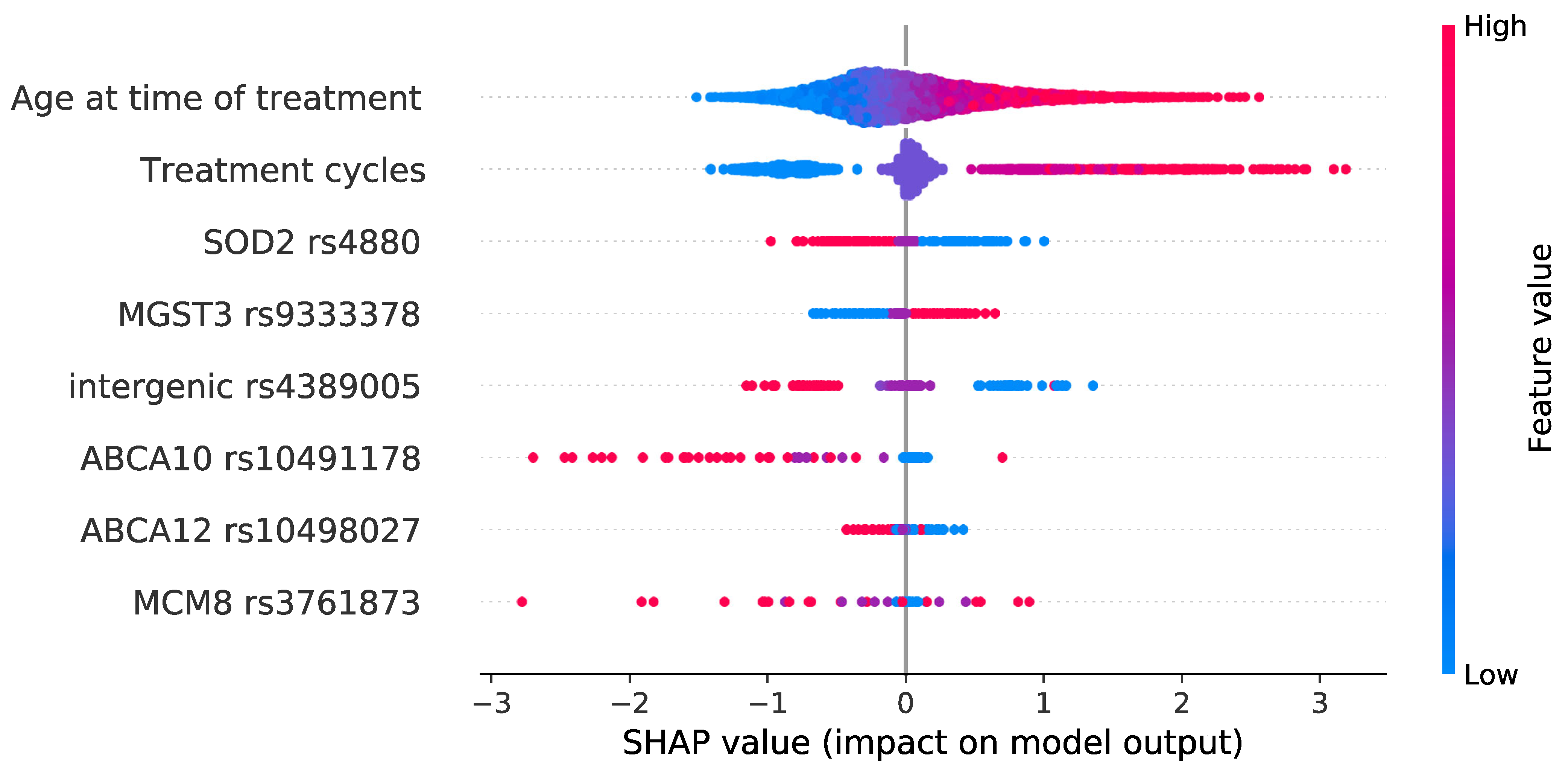
Prediction performance, assessed as ROC-AUC, reached a plateau when six genetic features were added to the model (in addition to the two most informative clinical parameters), with a mean ROC-AUC of 0.73 (95% CI, 0.71–0.74) (Figure 1B,D and Figure 2A). The most informative SNPs were: SOD2 rs4880, MGST3 rs9333378, intergenic rs4389005, ABCA10 rs10491178, ABCA12 rs10498027, MCM8 rs3761873 (Table 3). Out of 30 models, these SNPs were selected 15, 9, 7, 6, 5, and 4 times, respectively (Supplementary Table S1). Only the intergenic rs4389005 has been pre-selected from the cross-validated GWAS. All other SNPs had a p value > 1 × 10⁻⁴ and were pre-selected from a systematic review of genes shown to be related with cisplatin metabolism or ototoxicity. The two most influential SNPs according to SHAP metrics [30] were SOD2 rs4880 and MGST3 rs9333378 (Figure 3). Homozygous genotypes for the risk alleles SOD2 rs4880:AA and MGST3 rs9333378:AA were found in 47% of patients who replied FACT/GOG-Ntx6 = 0 or 1, 63% of patients who replied FACT/GOG-Ntx6 = 2, and 76% of patients who replied FACT/GOG-Ntx6 = 3 or 4 (chi-squared p value 1 × 10⁻⁴).
For each sample, prediction scores ranged between 0 and 1, where a value closer to 1 indicated a higher probability of hearing loss. Using a default cut-off of 0.50, a sensitivity of 67% was reached and a positive predictive value of 51%. Correspondingly, this resulted in a specificity of 66% and a negative predictive value of 80% (Figure 2B). The model performed best on patients with the highest toxicity (Figure 2C).
For most patients (320 out of 393), adding genetic data improved hearing loss prediction; however, for 42 out of 320, this was still not enough to correctly classify these patients. In 7 out of 393 patients, the addition of genetic data led to misclassification. For 55 out of 393 patients, neither clinical nor genetic data helped on prediction and/or classification (Supplementary Figure S3).
Overall, we were able to improve prediction performance when adding genetic features to clinical data (ROC-AUC 0.73) compared to the models with only clinical data (ROC-AUC 0.66).
To test robustness and non-randomness of the selected models, in the models with only clinical data, all variables were permutated, which led to a mean ROC-AUC close to 0.50 throughout the forward feature selection (Supplementary Figure S4A). The mean ROC-AUC for the random model with two features was 0.50 (95% CI, 0.49–0.51) (Supplementary Figure S4B).
In an additional test, random genetic variants were added to the model with the informative clinical features (age at diagnosis and number of treatment cycles). Mean ROC-AUC was 0.67 (95% CI, 0.66–0.68) for the model with six random genetic variants and two informative clinical features (Supplementary Figure S4D), which was not so different from the ROC-AUC with two clinical features only (ROC-AUC of 0.66 (95% CI, 0.65–0.66)), indicating that the random genetic variants were indeed not adding any relevant information for the prediction. From this point on, ROC-AUC started to steadily decrease as more randomly selected SNPs were added to the models (Supplementary Figure S4C).

4. Discussion

In this study, we present a model for the prediction of hearing loss after cisplatin-containing chemotherapy based on a combination of clinical and genetic features, achieving a classification performance of ROC-AUC 0.73. We observed an improved prediction after the inclusion of genetic data compared to clinical data only. Age at diagnosis and number of treatment cycles were the most important clinical predictors, matching what has previously been reported [7,9,31].
We have focused on hearing loss as part of ototoxicity, as we did not observe a strong correlation between hearing loss (FACT/GOG-Ntx6), and tinnitus (FACT/GOG-Ntx7), which may indicate independent biological mechanisms. Indeed, not all people who suffer from hearing loss have tinnitus, and vice versa, and studies on the genetics behind tinnitus are still at an early stage [32,33].
The first SNP selected in the model, the functional rs4880 SNP, is located on codon 16 exon 2 of SOD2 that codes for the superoxide dismutase 2 (SOD2), a mitochondrial protein [34]. SNP rs4880 is the most studied SOD2 SNP [35]; however, there is no agreement regarding how it influences SOD2 enzymatic activity. SNP rs4880 (A > G, Val16Ala) is predicted to change the structure of the SOD2 mitochondrial targeting sequence, converting a beta-sheet secondary structural motif to a partial alpha-helix [36]. Some state that due to partial arrest of the beta-sheet structure during transport across the inner mitochondrial membrane, this will likely inhibit efficient mitochondrial import of Val-SOD2 precursors and, thus, reduce enzyme activity [37]. A follow-up study has reported that the Ala variant, associated with increased SOD2 activity according to the previously mentioned study, was associated with hearing damage in cisplatin-treated pediatric medulloblastoma [38]. However, others have measured SOD2 activity and observed that it was lower in SOD2 rs4880:GG individuals compared with SOD2 rs4880:AA, or SOD2 rs4880:GA [39].
The second SNP selected in the model, SNP rs9333378, is located in MGST3, that codes for the microsomal glutathione S-transferase 3 (MGST3) [34]. Among the microsomal glutathione S-transferases, MGST1, MGST2, and MGST3 have been reported to be important in the detoxification process [40].
Here, we hypothesized a combined effect of SOD2 rs4880 and MGST3 rs9333378 on cisplatin-induced hearing loss.
When platinum enters the cells, it is metabolized by the mitochondria, which will lead to the production of reactive oxygen species (ROS) such as superoxide. SOD2 will then degrade superoxide into hydrogen peroxide until complete superoxide anion degradation. If SOD2 is prevented from entering the mitochondria due to partial arrest of beta-helix, this may lead to an accumulation of ROS. ROS cause lipid peroxidation, activation of pro-inflammatory factors, and cell death by apoptosis, including hair cells [41,42]. Indeed, we observed the A-allele with a higher frequency in patients who reported hearing loss (odds ratio = 1.55, 95% CI: 1.13–2.13), contrary to what has been reported previously [38]. Furthermore, glutathiones, including glutathione S-transferase, are known to help with complete superoxide anion degradation [38]. The Genotype-Tissue Expression (GTEx) database [43] reports lower MGST3 expression levels for rs9333378:AA compared to the rs9333378:GG genotype in the brain. It is hypothesized that the rs9333378 variant leads to accumulation of cisplatin in the hair cells through decreased MGST3 activity.
Additionally, potential novel variants associated with cisplatin-induced hearing loss were selected on the logistic regression model. SNP rs4389005 located in an intergenic region was found in the cross-validated GWAS. The closest gene is GPR12 (64 kilo base pairs 5′ to canonical transcription start site), a G protein-coupled receptor (GPCR). GPCRs have been seen to be involved in several physiological and pathological functions [44]. The subsequent SNPs, found via systematic review search, and with contribution to model performance, were SNPs ABCA10 rs10491178 and ABCA12 rs10498027, both leading to stop-gains within the ABCA genes which encode ATP-binding cassette (ABC) transporters. Overexpression of ABC transporters have been associated with multidrug resistance, including cisplatin, in multiple tumors [45]. ABCA10 rs10491178:GG has been associated with lower expression of ABCA6 [43], which can lead to higher sensitivity to cisplatin and higher toxicity [46]. MCM8 rs3761873 was the last SNP selected by the model, which leads to a stop-gain. MCM8 encodes the mini-chromosome maintenance 8 homologous recombinant repair factor protein (MCM8), and in a recent mouse study, inhibition of MCM8 (and MCM9) hypersensitized cells to cisplatin [47].
While we observed a false discovery rate of 49% using a 0.50 cut-off, it is promising to see that only four of the twenty-three patients with the highest score (FACT/GOG-Ntx6 = 4) were misclassified. Three of them had a prediction score very close to 0.50 (two patients with 0.48 and one with 0.49 prediction scores). The last misclassified patient had a prediction score of 0.31 and was also the youngest of the 23. Furthermore, he received one of the lowest amounts of cisplatin (300 mg/m2 and three treatment cycles). This points to other relevant features that led this patient to develop hearing loss, either clinical or genetic predispositions that might be underrepresented in this dataset and, hence, may not have been detected.
The diagnosis of hearing loss is challenging to perform, and its definition is still far from being robustly defined [48]. Here, several potential factors for hearing loss were not explored, such as noise, infection, or vascular problems, and the toxicity was assessed several years after exposure. However, long-term toxicity also has a high impact on quality of life [9] and may be important to predict for balancing treatment intensity.
The models were trained on labels that derive from the FACT/GOG-Ntx questionnaire, which are not objectively quantified. Other measurements, such as pure-tone audiometry, which are not yet implemented routinely in clinical practice, could have been undertaken to improve precision [48]. On the other hand, using quality of life measures ensures that the focus is on the patient [49]. For instance, objective measurements might detect a similar level of toxicity between two individuals; however, only one may be affected by the symptoms and, thus, objective measurements may not be a true assessment of quality of life.
Further, BMI, as well as information about alcohol consumption and smoking habits, were retrieved in 2014 when the questionnaire was completed. These clinical features were used as a proxy at the time of treatment, but they may not represent the true values. While those features were not selected in the final model, we are unaware if the real values at the time of treatment could have added relevant information to the model. Incorporating longitudinal data, such as information collected one year after treatment, could also have been advantageous in further improving the model’s performance.
Finally, models in this study were trained on 393 patients adhering to most of the best practices of healthcare-related prediction models [50] using a logistic regression with cross-validated GWAS; nonetheless a future replication on a larger and independent patient cohort would be warranted.
Cisplatin is essential in treatment of several neoplasms; however, the inability to predict how patients will react to chemotherapy represents a major challenge, and hearing loss is one of the most common late side effects of cisplatin-based chemotherapy. In this study, we present a logistic regression with cross-validated GWAS prediction model based on a combination of genetic and clinical features able to classify patients at high (67% sensitivity), or low risk (66% specificity) of hearing loss after cisplatin-based treatment.
We also propose a combined effect involving SOD2 rs4880 and MGST3 rs9333378 on cisplatin-induced hearing loss development. In our study, these SNPs have not yielded significant results when single associations between SNPs and outcome have been performed; thus, a combination of cross-validated GWAS and systemic review search is suggested as a feature selection approach for machine learning.
Following confirmation in a prospective clinical setting and replication in larger independent studies, such a model could be used as a complement to support clinical decision-making and help in reducing hearing loss cases by adjusting treatment for patients in the high-risk group, especially with treatment of other cancers where cisplatin is used.

Supplementary Materials

The following supporting information can be downloaded at: https://www.mdpi.com/article/10.3390/cancers15153923/s1. Supplementary Table S1: Number of times each feature is selected in all 30 models. Each “Feature no. X” column theoretically sums up to 30, however, for illustration purposes, only eight features (best ROC-AUC performance) are shown in the table. Supplementary Figure S1: Step-by-step demonstration of genetic data quality control and information on patients where questionnaire information was missing. Single-nucleotide polymorphism (SNP) quality filtering included removal of duplicated SNPs and those with ambiguous genome position, strand, and alleles; call rate (<98%); extreme deviation from Hardy–Weinberg equilibrium (p value < 5 × 10⁻⁶); and MAF (<1%). Quality controls applied on the patient samples were based on genotype (chromosome X homozygosity rate <20% for females and >80% for males) and phenotype sex discordance; extreme heterozygosity or homozygosity (±4 SD from sample’s hetero-/homozygosity rate mean); outliers from the European descent using 1000 Genomes(49) as reference samples; cryptic relatedness (IBD > 18.75%); and population outliers (±4 SD from cluster centroid mean). European outliers were detected by (1) doing principal component analysis (PCA) to find the center of the European reference samples, and (2) remove samples whose Euclidean distance from the center > 1.5 * maximum Euclidean distance of the European reference samples (50). Patients with missing questionnaire information consisted of 45 patients who received more than one line of treatment and therefore were not relevant for the present study and were not invited for the questionnaire in 2014. These were still included for the purpose of quality control only. Supplementary Figure S2: Illustration of logistic regression model used in this study. Model was run at Computerome 2.0 (https://www.computerome.dk, accessed on 12 November 2022 (developed for a range of time)). The 30 random data splits were run in parallel to reduce running time, thus 32 nodes were used (30 allocated for each random split and 2 for other initializations). Each node contains 2 CPUs with 20 cores/CPU. 192 GB is the memory distributed through all cores. Supplementary Figure S3: Misclassified patients and/or patients where genetic “pushed” final classification in the wrong direction. Arrow starts at prediction score of model with only clinical data (model 1) and ends at prediction score of model with clinical and genetic data (model 2). A: Inclusion of genetic data helped but not enough to correctly classify these patients; B: Patients where genetic data “pushed” the classification in the wrong direction, even though some of them were correctly classified; C, D: Neither clinical nor genetic data helped on these patients classification (in D, score difference between model 2 and 1 was below 0.05). All other patients not represented here were correctly classified and genetic data “pushed” the classification in the right direction (or if in the wrong direction, score difference between model 2 and model 1 was below 0.05). Supplementary Figure S4: ROC-AUC mean (30 random splits) performances for the random and real models. A: Model with permuted clinical data with forward feature selection up until nine features. Shaded blue area indicates 95% CI. B: Comparison between real model (mean ROC-AUC 0.66 (95% CI, 0.65–0.66, blue histogram) and permuted models (mean ROC-AUC 0.50 (95% CI, 0.49–0.51, orange histogram); C: Model with clinical and randomly selected markers with forward feature selection up until 32 features. Shaded areas indicate 95% CI. D: Comparison between real model (mean ROC-AUC of 0.73 (95% CI, 0.71–0.74) and random models (mean ROC-AUC was 0.67 (95% CI, 0.66–0.68). In B and D, count (y-axis) sums up to 150 as the model consists of 5 outer folds and 30 data splits were done (5 × 30). ROC-AUC = area under the receiver operating characteristic curve; No. = number; CI = confidence interval. Supplementary Note S1: FACT/GOG-Ntx (Version 4). Hearing loss as part of ototoxicity. Supplementary Note S2: Genes obtained from database search. Supplementary Note S3: Model hyperparameters, encoding, and normalization.

Author Contributions

J.L., G.D. and R.G. contributed for the concept and study design. S.L.G., J.L., B.K.C., I.F.H., R.G. and M.B. contributed for the acquisition, analysis, or interpretation of data. S.L.G., B.K.C. and I.F.H. performed statistical analysis. S.L.G. and J.L. wrote the paper. M.D.D. and R.G. supervised the study. J.L. and S.L.G. contributed equally to this work. The work reported in the paper has been performed by the authors, unless clearly specified in the text. All authors have read and agreed to the published version of the manuscript.

Funding

This research was funded by Danish Cancer Society grant number R40-A2119. S.L.G. was supported by Idella Foundation.

Institutional Review Board Statement

The study was approved by the regional ethical committee (File number, H-2-2012-044) and the National Board of Data Protection (File number, 2012-41-0751).

Informed Consent Statement

All patients gave informed consent to participate in this study.

Data Availability Statement

For data transparency, the GWAS summary statistics, on the entire cohort are available through the NHGRI-EBI GWAS Catalog under study accession number GCST90133383. Note that performance in the machine learning models is higher than the single locus association tests represented in GWAS summary statistics, as machine learning capitalizes on non-linear correlations between individual SNPs as well as with other patient backgrounds and clinical information. In order to train and evaluate the model described in this work, SciKit-learn [28] (v0.23.2) in Python (v3.6.10) was used. PLINK [29] (v1.9) was used to perform the logistic regression association on the training set of the inner fold. The code used in this study is available at https://github.com/sblg/LR_cross_validated_GWAS, accessed on 12 November 2022. Further details and other data that support the findings of this study are available from the corresponding author upon request.

Acknowledgments

This study was supported by the Danish cancer society. S.L.G. was supported by the Idella foundation. The funding source had no role in the design and conduct of the study; collection, management, analysis, and interpretation of the data; preparation, review, or approval of the manuscript; and decision to submit.

Conflicts of Interest

R.G. is employed with Novo Nordisk Research Centre Oxford. The other authors have no conflicts of interest to disclose.

Abbreviations

ABC: ATP-binding cassette; BEP: bleomycin–etoposide–cisplatin; BMI: body mass index; BSA: body surface area; DaTeCa: Danish Testicular Cancer; FACT/GOG-Ntx: Functional Assessment of Cancer Therapy/Gynecologic Oncology Group-Neurotoxicity; GPCR: G protein-coupled receptor; GTEx: Genotype-Tissue Expression; GWAS: genome-wide association study; HC: hearing change; IGCCCG: International Germ Cell Cancer Collaborative Group; IQR: interquartile range; MCM: mini-chromosome maintenance; MGST: microsomal glutathione S-transferase; ROC-AUC: area under the receiver operating characteristic; ROS: reactive oxygen species; SHAP: SHapley Additive exPlanations; SOD2: superoxide dismutase 2; SNP: single nucleotide polymorphisms; TCS: testicular cancer survivors; VEP: Variant Effect Predictor.

References

  1. Chia, V.M.; Quraishi, S.M.; Devesa, S.S.; Purdue, M.P.; Cook, M.B.; McGlynn, K.A. International trends in the incidence of testicular cancer, 1973–2002. Cancer Epidemiol. Biomark. Prev. 2010, 19, 1151–1159. [Google Scholar] [CrossRef] [PubMed] [Green Version]
  2. Shanmugalingam, T.; Soultati, A.; Chowdhury, S.; Rudman, S.; Van Hemelrijck, M. Global incidence and outcome of testicular cancer. Clin. Epidemiol. 2013, 5, 417–427. [Google Scholar] [CrossRef] [PubMed] [Green Version]
  3. Kier, M.G.; Lauritsen, J.; Mortensen, M.S.; Bandak, M.; Andersen, K.K.; Hansen, M.K.; Agerbaek, M.; Holm, N.V.; Dalton, S.O.; Johansen, C.; et al. Prognostic Factors and Treatment Results after Bleomycin, Etoposide, and Cisplatin in Germ Cell Cancer: A Population-based Study. Eur. Urol. 2017, 71, 290–298. [Google Scholar] [CrossRef] [PubMed]
  4. Einhorn, L.H.; Donohue, J. Cis-diamminedichloroplatinum, vinblastine, and bleomycin combination chemotherapy in disseminated testicular cancer. Ann. Intern. Med. 1977, 87, 293–298. [Google Scholar] [CrossRef]
  5. Chovanec, M.; Lauritsen, J.; Bandak, M.; Oing, C.; Kier, G.G.; Kreiberg, M.; Rosenvilde, J.; Wagner, T.; Bokemeyer, C.; Daugaard, G. Late adverse effects and quality of life in survivors of testicular germ cell tumour. Nat. Rev. Urol. 2021, 18, 227–245. [Google Scholar] [CrossRef]
  6. Lauritsen, J.; Mortensen, M.S.; Kier, M.G.G.; Christensen, I.J.; Agerbaek, M.; Gupta, R.; Daugaard, G. Renal impairment and late toxicity in germ-cell cancer survivors. Ann. Oncol. 2015, 26, 173–178. [Google Scholar] [CrossRef]
  7. Frisina, R.D.; Wheeler, H.E.; Fossa, S.D.; Kerns, S.L.; Fung, C.; Sesso, H.D.; Monahan, P.O.; Feldman, D.R.; Hamilton, R.; Vaughn, D.J.; et al. Comprehensive audiometric analysis of hearing impairment and tinnitus after cisplatin-based chemotherapy in survivors of adult-onset cancer. J. Clin. Oncol. 2016, 34, 2712–2720. [Google Scholar] [CrossRef]
  8. Brydøy, M.; Oldenburg, J.; Klepp, O.; Bremnes, R.M.; Wentzel-Larsen, T.; Hauge, E.R.; Dahl, O.; Foss, S.D. Observational study of prevalence of long-term Raynaud-like phenomena and neurological side effects in testicular cancer survivors. J. Natl. Cancer Inst. 2009, 101, 1682–1695. [Google Scholar] [CrossRef]
  9. Lauritsen, J.; Bandak, M.; Kreiberg, M.; Skøtt, J.W.; Wagner, T.; Rosenvilde, J.J.; Dysager, L.; Agerbæk, M.; Daugaard, G. Long-term neurotoxicity and quality of life in testicular cancer survivors—A nationwide cohort study. J. Cancer Surviv. 2021, 15, 509–517. [Google Scholar] [CrossRef]
  10. Brock, P.R.; Knight, K.R.; Freyer, D.R.; Campbell, K.C.M.; Steyger, P.S.; Blakley, B.W.; Rassekh, S.R.; Chang, K.W.; Fligor, B.J.; Rajput, K.; et al. Platinum-induced ototoxicity in children: A consensus review on mechanisms, predisposition, and protection, including a new International Society of Pediatric Oncology Boston ototoxicity scale. J. Clin. Oncol. 2012, 30, 2408–2417. [Google Scholar] [CrossRef] [Green Version]
  11. Wheeler, H.E.; Gamazon, E.R.; Frisina, R.D.; Perez-Cervantes, C.; El Charif, O.; Mapes, B.; Fossa, S.D.; Feldman, D.R.; Hamilton, R.J.; Vaughn, D.J.; et al. Variants in WFS1 and Other Mendelian Deafness Genes Are Associated with Cisplatin-Associated Ototoxicity. Clin. Cancer Res. 2017, 23, 3325–3333. [Google Scholar] [CrossRef] [Green Version]
  12. Rybak, L.P.; Whitworth, C.A.; Mukherjea, D.; Ramkumar, V. Mechanisms of cisplatin-induced ototoxicity and prevention. Hear. Res. 2007, 226, 157–167. [Google Scholar] [CrossRef] [PubMed]
  13. Kreiberg, M.; Bandak, M.; Lauritsen, J.; Skøtt, J.W.; Johansen, N.B.; Agerbaek, M.; Holm, N.V.; Johansen, C.; Daugaard, G. Cohort profile: The Danish Testicular Cancer late treatment effects cohort (DaTeCa-LATE). Front. Oncol. 2018, 8, 37. [Google Scholar] [CrossRef] [PubMed] [Green Version]
  14. Daugaard, G.; Kier, M.G.G.; Bandak, M.; Mortensen, M.S.; Larsson, H.; Søgaard, M.; Toft, B.G.; Engvad, B.; Agerbæk, M.; Holm, N.V.; et al. The Danish testicular cancer database. Clin. Epidemiol. 2016, 8, 703–707. [Google Scholar] [CrossRef] [Green Version]
  15. Garcia, S.L.; Lauritsen, J.; Zhang, Z.; Bandak, M.; Dalgaard, M.D.; Nielsen, R.L.; Daugaard, G.; Gupta, R. Prediction of Nephrotoxicity Associated With Cisplatin-Based Chemotherapy in Testicular Cancer Patients. JNCI Cancer Spectr. 2020, 4, pkaa032. [Google Scholar] [CrossRef]
  16. International Germ Cell Cancer Collaborative Group. Germ Cell Consensus Classification: A prognostic factor-based staging system for metastatic germ cell cancers. J. Clin. Oncol. 1997, 15, 594–603. [Google Scholar] [CrossRef]
  17. Huang, H.Q.; Brady, M.F.; Cella, D.; Fleming, G.; Mackey, D. Validation and reduction of FACT/GOG-Ntx subscale for platinum/paclitaxel- induced neurologic symptoms: A gynecologic oncology group study. Int. J. Gynecol. Cancer 2007, 17, 387–393. [Google Scholar] [CrossRef] [PubMed]
  18. Consortium, T.U. UniProt: A worldwide hub of protein knowledge. Nucleic Acids Res. 2018, 47, D506–D515. [Google Scholar] [CrossRef] [Green Version]
  19. Wishart, D.S.; Feunang, Y.D.; Guo, A.C.; Lo, E.J.; Marcu, A.; Grant, J.R.; Sajed, T.; Johnson, D.; Li, C.; Sayeeda, Z.; et al. DrugBank 5.0: A major update to the DrugBank database for 2018. Nucleic Acids Res. 2018, 46, D1074–D1082. [Google Scholar] [CrossRef]
  20. Kanehisa, M. Toward understanding the origin and evolution of cellular organisms. Protein Sci. 2019, 28, 1947–1951. [Google Scholar] [CrossRef]
  21. Kanehisa, M.; Sato, Y.; Furumichi, M.; Morishima, K.; Tanabe, M. New approach for understanding genome variations in KEGG. Nucleic Acids Res. 2019, 47, D590–D595. [Google Scholar] [CrossRef] [PubMed] [Green Version]
  22. Kanehisa, M. KEGG: Kyoto Encyclopedia of Genes and Genomes. Nucleic Acids Res. 2000, 28, 27–30. [Google Scholar] [CrossRef] [PubMed]
  23. Caspi, R.; Billington, R.; Ferrer, L.; Foerster, H.; Fulcher, C.A.; Keseler, I.M.; Kothari, A.; Krummenacker, M.; Latendresse, M.; Mueller, L.A.; et al. The MetaCyc database of metabolic pathways and enzymes and the BioCyc collection of pathway/genome databases. Nucleic Acids Res. 2016, 44, D471–D480. [Google Scholar] [CrossRef]
  24. McLaren, W.; Gil, L.; Hunt, S.E.; Riat, H.S.; Ritchie, G.R.S.; Thormann, A.; Flicek, P.; Cunningham, F. The Ensembl Variant Effect Predictor. Genome Biol. 2016, 17, 122. [Google Scholar] [CrossRef] [Green Version]
  25. Tserga, E.; Nandwani, T.; Edvall, N.K.; Bulla, J.; Patel, P.; Canlon, B.; Cederroth, C.R.; Baguley, D.M. The genetic vulnerability to cisplatin ototoxicity: A systematic review. Sci. Rep. 2019, 9, 3455. [Google Scholar] [CrossRef] [PubMed] [Green Version]
  26. Buck, S.F. A Method of Estimation of Missing Values in Multivariate Data Suitable for Use with an Electronic Computer. J. R. Stat. Soc. Ser. B 1960, 22, 302–306. [Google Scholar] [CrossRef]
  27. van Buuren, S.; Groothuis-Oudshoorn, K. mice: Multivariate Imputation by Chained Equations in R. J. Stat. Softw. 2011, 45, 1–67. [Google Scholar] [CrossRef] [Green Version]
  28. Pedregosa, F.; Varoquaux, G.; Gramfort, A.; Michel, V.; Thirion, B.; Grisel, O.; Blondel, M.; Müller, A.; Nothman, J.; Louppe, G.; et al. Scikit-learn: Machine Learning in Python. J. Mach. Learn. Res. 2012, 12, 2825–2830. [Google Scholar]
  29. Purcell, S.; Neale, B.; Todd-Brown, K.; Thomas, L.; Ferreira, M.A.R.; Bender, D.; Maller, J.; Sklar, P.; de Bakker, P.I.W.; Daly, M.J.; et al. PLINK: A Tool Set for Whole-Genome Association and Population-Based Linkage Analyses. Am. J. Hum. Genet. 2007, 81, 559–575. [Google Scholar] [CrossRef] [Green Version]
  30. Lundberg, S.; Lee, S.-I. A Unified Approach to Interpreting Model Predictions. In NIPS’17, Proceedings of the 31st International Conference on Neural Information Processing Systems, Long Beach, CA, USA, 4–9 December 2017; Curran Associates Inc.: Red Hook, NY, USA, 2017. [Google Scholar]
  31. Haugnes, H.S.; Stenklev, N.C.; Brydøy, M.; Dahl, O.; Wilsgaard, T.; Laukli, E.; Fosså, S.D. Hearing loss before and after cisplatin-based chemotherapy in testicular cancer survivors: A longitudinal study. Acta Oncol. (Madr.) 2018, 57, 1075–1083. [Google Scholar] [CrossRef] [Green Version]
  32. Lopez-Escamez, J.A.; Bibas, T.; Cima, R.F.F.; Van de Heyning, P.; Knipper, M.; Mazurek, B.; Szczepek, A.J.; Cederroth, C.R. Genetics of Tinnitus: An Emerging Area for Molecular Diagnosis and Drug Development. Front. Neurosci. 2016, 10, 377. [Google Scholar] [CrossRef] [PubMed] [Green Version]
  33. Vona, B.; Nanda, I.; Shehata-Dieler, W.; Haaf, T. Genetics of Tinnitus: Still in its Infancy. Front. Neurosci. 2017, 11, 236. [Google Scholar] [CrossRef] [PubMed]
  34. Stelzer, G.; Rosen, N.; Plaschkes, I.; Zimmerman, S.; Twik, M.; Fishilevich, S.; Stein, T.I.; Nudel, R.; Lieder, I.; Mazor, Y.; et al. The GeneCards Suite: From Gene Data Mining to Disease Genome Sequence Analyses. Curr. Protoc. Bioinform. 2016, 54, 1–30. [Google Scholar] [CrossRef]
  35. Pourvali, K.; Abbasi, M.; Mottaghi, A. Role of Superoxide Dismutase 2 Gene Ala16Val Polymorphism and Total Antioxidant Capacity in Diabetes and its Complications. Avicenna J. Med. Biotechnol. 2016, 8, 48–56. [Google Scholar] [PubMed]
  36. Sutton, A.; Khoury, H.; Prip-Buus, C.; Cepanec, C.; Pessayre, D.; Degoul, F. The Ala16Val genetic dimorphism modulates the import of human manganese superoxide dismutase into rat liver mitochondria. Pharmacogenetics 2003, 13, 145–157. [Google Scholar] [CrossRef] [PubMed]
  37. Sutton, A.; Imbert, A.; Igoudjil, A.; Descatoire, V.; Cazanave, S.; Pessayre, D.; Degoul, F. The manganese superoxide dismutase Ala16Val dimorphism modulates both mitochondrial import and mRNA stability. Pharmacogenet. Genom. 2005, 15, 311–319. [Google Scholar] [CrossRef]
  38. Brown, A.L.; Lupo, P.J.; Okcu, M.F.; Lau, C.C.; Rednam, S.; Scheurer, M.E. SOD2 genetic variant associated with treatment-related ototoxicity in cisplatin-treated pediatric medulloblastoma. Cancer Med. 2015, 4, 1679–1686. [Google Scholar] [CrossRef]
  39. Bastaki, M.; Huen, K.; Manzanillo, P.; Chande, N.; Chen, C.; Balmes, J.R.; Tager, I.B.; Holland, N. Genotype-activity relationship for Mn-superoxide dismutase, glutathione peroxidase 1 and catalase in humans. Pharmacogenet. Genom. 2006, 16, 279–286. [Google Scholar] [CrossRef] [Green Version]
  40. Uno, Y.; Murayama, N.; Kunori, M.; Yamazaki, H. Characterization of Microsomal Glutathione S -Transferases MGST1, MGST2, and MGST3 in Cynomolgus Macaque. Drug Metab. Dispos. 2013, 41, 1621–1625. [Google Scholar] [CrossRef] [Green Version]
  41. Romano, A.; Capozza, M.A.; Mastrangelo, S.; Maurizi, P.; Triarico, S.; Rolesi, R.; Attinà, G.; Fetoni, A.R.; Ruggiero, A. Assessment and Management of Platinum-Related Ototoxicity in Children Treated for Cancer. Cancers 2020, 12, 1266. [Google Scholar] [CrossRef]
  42. Paciello, F.; Fetoni, A.R.; Mezzogori, D.; Rolesi, R.; Di Pino, A.; Paludetti, G.; Grassi, C.; Troiani, D. The dual role of curcumin and ferulic acid in counteracting chemoresistance and cisplatin-induced ototoxicity. Sci. Rep. 2020, 10, 1063. [Google Scholar] [CrossRef] [PubMed] [Green Version]
  43. Lonsdale, J.; Thomas, J.; Salvatore, M.; Phillips, R.; Lo, E.; Shad, S.; Hasz, R.; Walters, G.; Garcia, F.; Young, N.; et al. The Genotype-Tissue Expression (GTEx) project. Nat. Genet. 2013, 45, 580–585. [Google Scholar] [CrossRef] [PubMed]
  44. Allende, G.; Chávez-Reyes, J.; Guerrero-Alba, R.; Vázquez-León, P.; Marichal-Cancino, B.A. Advances in Neurobiology and Pharmacology of GPR12. Front. Pharmacol. 2020, 11, 628. [Google Scholar] [CrossRef] [PubMed]
  45. Pasello, M.; Giudice, A.M.; Scotlandi, K. The ABC subfamily A transporters: Multifaceted players with incipient potentialities in cancer. Semin. Cancer Biol. 2020, 60, 57–71. [Google Scholar] [CrossRef]
  46. Robey, R.W.; Pluchino, K.M.; Hall, M.D.; Fojo, A.T.; Bates, S.E.; Gottesman, M.M. Revisiting the role of ABC transporters in multidrug-resistant cancer. Nat. Rev. Cancer 2018, 18, 452–464. [Google Scholar] [CrossRef]
  47. Morii, I.; Iwabuchi, Y.; Mori, S.; Suekuni, M.; Natsume, T.; Yoshida, K.; Sugimoto, N.; Kanemaki, M.T.; Fujita, M. Inhibiting the MCM8-9 complex selectively sensitizes cancer cells to cisplatin and olaparib. Cancer Sci. 2019, 110, 1044–1053. [Google Scholar] [CrossRef] [Green Version]
  48. Ganesan, P.; Schmiedge, J.; Manchaiah, V.; Swapna, S.; Dhandayutham, S.; Kothandaraman, P.P. Ototoxicity: A Challenge in Diagnosis and Treatment. J. Audiol. Otol. 2018, 22, 59–68. [Google Scholar] [CrossRef] [Green Version]
  49. Higginson, I.J. Measuring quality of life: Using quality of life measures in the clinical setting. BMJ 2001, 322, 1297–1300. [Google Scholar] [CrossRef]
  50. Kakarmath, S.; Esteva, A.; Arnaout, R.; Harvey, H.; Kumar, S.; Muse, E.; Dong, F.; Wedlund, L.; Kvedar, J. Best practices for authors of healthcare-related artificial intelligence manuscripts. NPJ Digit. Med. 2020, 3, 134. [Google Scholar] [CrossRef]
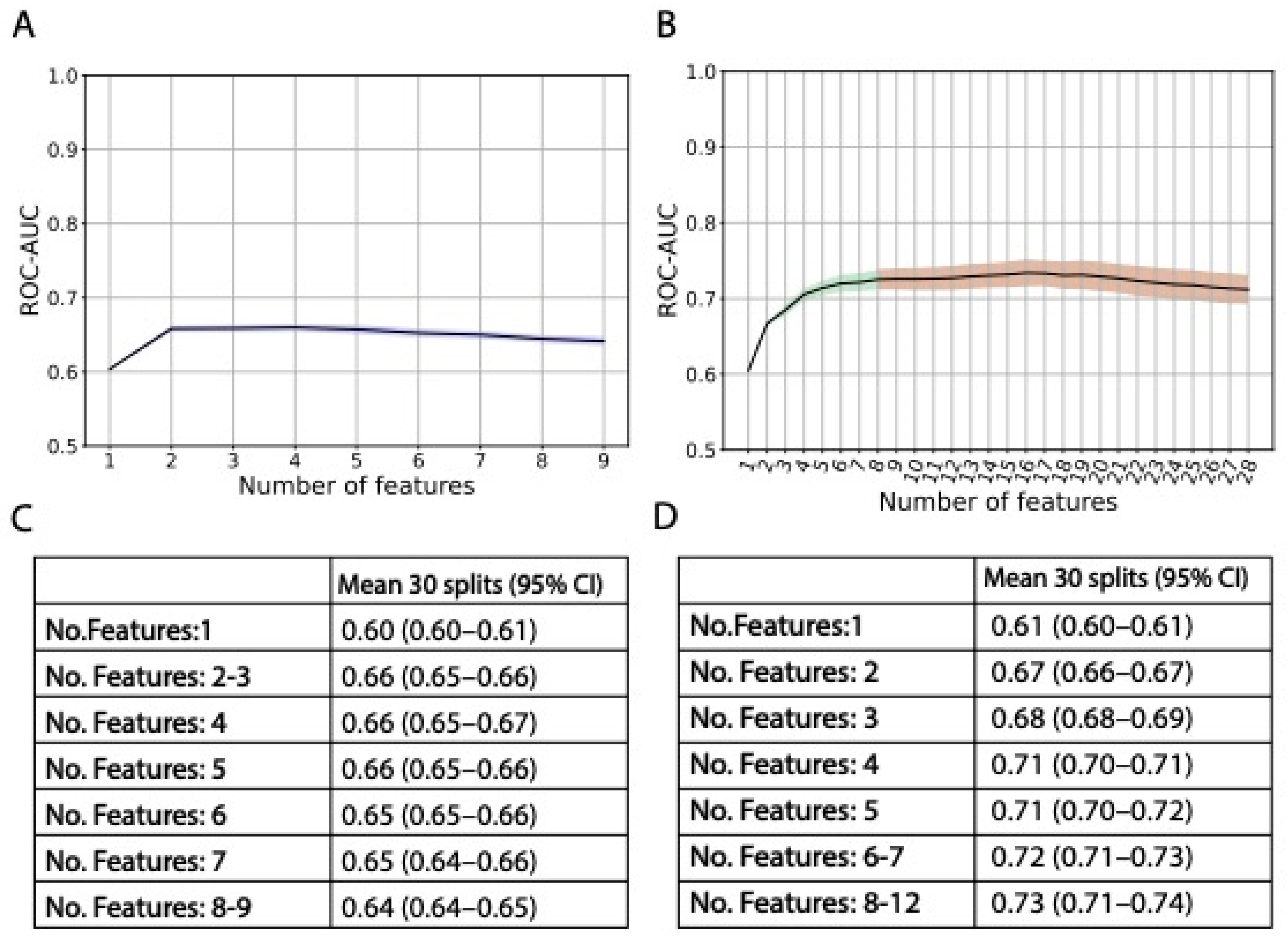
Figure 1. ROC-AUC mean (30 random data splits) performances in each step of the forward feature selection. (A): Model with clinical data with forward feature selection up until nine features. Shaded blue area indicates 95% CI. Exact ROC-AUC mean and 95% CI in (C). (B): Model with clinical and genetic data with forward feature selection up until 28 features. Shaded areas indicate 95% CI, blue color indicates that only clinical data were added, green color that clinical and genetic data were added, and red color that ROC-AUC reached a plateau. Exact ROC-AUC mean and 95% CI in (D). For illustration purposes, exact ROC-AUC mean and 95% CI are not indicated in (D) from 13 features. From 13 to 28 features, ROC-AUC mean (95% CI) was 0.73 (0.71–0.75) (13–15 features); 0.73 (0.71–0.75) (14–15 features); 0.73 (0.72–0.75) (16–17 features); 0.73 (0.71–0.75) (18–21 features); 0.72 (0.70–0.74) (22–25 features); 0.72 (0.70–0.73) (26 features); and 0.71 (0.69–0.73) (27–28 features). ROC-AUC = area under the receiver operating characteristic curve; No. = number; CI = confidence interval.
Cancers 15 03923 g001
Figure 2. Final model performance measures and prediction scores. (A): Model performance shown as ROC-AUC curve. Solid blue line and shaded area indicate the mean and standard deviation across 30 data splits. Dashed red line indicates a random classifier. (B): ROC-AUC and other performance measures, i.e., MCC, sensitivity, specificity, PPV, and NPV using a cut-off of 0.50. (C): Final prediction scores (x-axis) for each patient, represented by a dot. Orange dots represent controls or non-affected patients (FACT/GOG-Ntx6 score 0–1), while blue dots represent cases or affected patients (FACT/GOG-Ntx6 score 2–4). Dashed vertical line represents a cut-off of 0.50, where patients with a prediction score of 0.50 or higher are considered cases. ROC-AUC = area under the receiver operating characteristic curve; MCC = Matthews correlation coefficient; PPV = positive predictive value; NPV = negative predictive value.
Cancers 15 03923 g002
Figure 3. SHAP value feature importance. Individual features are ranked by importance, where age at diagnosis is the most important feature. The color represents the feature value (red: high; blue: low). Negative SHAP values (x-axis) contribute toward a negative model outcome (control or non-affected), while positive SHAP values contribute toward a positive model outcome (case or affected).
Cancers 15 03923 g003
Table 1. Overview of the gene database search identifying genetic markers associated with cisplatin metabolism and ototoxicity.
DescriptionNumber of Genes
Cisplatin metabolismResistance Pathway
(KEGG Pathways)
Overview of genes and interactions
resulting in platinum-based drugs
resistance.
46
Detoxification Pathway
(BioCyc Pathway)
Cisplatin is degraded via the
glutathione-mediated detoxification
pathway.
9
Glutathione Transferases
Cytochrome P450 Enzymes
ABC Transporters
(Uniprot)
The three protein groups may be
associated with cisplatin-introduced
neurotoxicity, since they affect the
uptake and disposition. Genes
associated with the groups were
identified with Uniprot.
26
61
49
Cisplatin (Uniprot)Systematic search identifying cisplatin-related genes conducted with Uniprot.22
Cisplatin (DrugBank)The DrugBank database contains
information on pharmaceutical drugs including cisplatin.
31
OtotoxicitySensorineural Hearing LossSensorineural hearing loss-related genes conducted with Uniprot.155
OtotoxicityOtotoxicity-related genes conducted with Uniprot.2
Table 2. Comparison of baseline characteristics between affected (FACT/GOG-Ntx6 score 2–4) and non-affected (FACT/GOG-Ntx6 score 0, 1) patients. Out of 433, 9 patients had not replied on FACT/GOG-Ntx6; thus, only 424 patients are represented in the table. Values show the median and interquartile range (IQR; 25–75%), or number of patients and percentages (%). IQR = interquartile range; BMI = body mass index; BEP = bleomycin–etoposide–cisplatin; GFR = glomerular filtration rate.
Affected, Number (%)Non-Affected, Number (%)p Values a
Number of patients146 (34.4)278 (65.6)-
Age at diagnosis, median (IQR)34 (27–41)29 (26–36)0.002
BMI, median (IQR)
Unknown: 8 Affected; 10 Non-affected
21 (19–27)22 (19–26)0.38
BEP regimenNormal dose113 (78.5)260 (95.6)6 × 10⁻²⁷
Double dose31 (21.5)12 (4.4)
Unknown26
GFR before treatment, median (IQR), mL/min/1.73 m2
Unknown: 2 Non-affected
122 (111–135)121 (110–133)0.68
Cisplatin, median (IQR), mg/m2400 (385–403)400 (300–400)p < 0.001
Treatment cycles330 (20.5)86 (30.9)1 × 10⁻⁵
485 (58.2)180 (64.7)
5 or more9 (6.2)10 (3.6)
High-dose22 (15.1)2 (0.7)
HistologySeminoma34 (23.3)54 (19.4)0.42
Non-Seminoma112 (76.7)224 (80.6)
Prognostic groupGood103 (70.5)239 (86)p < 0.001
Intermediate32 (21.9)30 (10.8)
Poor11 (7.5)9 (3.2)
Alcohol consumption in number of units per week5 (1–10)5 (2–10)0.30
smokingNever61 (41.8)128 (46.4)0.40
Former55 (37.7)88 (31.9)
Current30 (20.5)60 (21.7)
Unknown-2
a p values were calculated by 2-sided Mann–Whitney U test for continuous or ordinal characteristics. For “histology”, p value was calculated by χ2 test. All tests are appropriate for unpaired data, and in the case of continuous variables, non-normal distributed data. Distribution of continuous variables was assessed through Shapiro–Wilk normality test.
Table 3. Single nucleotide polymorphisms (SNPs) selected on the final prediction model. SNPs are ordered by genetic position, and not by the number of times selected in the model. Chr. = Chromosome; SNP = single nucleotide polymorphism; MAF = minor allele frequency; CEU = European; OR = odds ratio; CI = confidence interval.
Chr.SNPGenetic Position aGeneReference AlleleAlternative AlleleRisk AlleleMAF (CEU)EffectOR (95% CI) bp Values c
1rs9333378165601466MGST3GAAG: 0.39Splice acceptor1.37 (1–1.86)0.0441
2rs10498027215820013ABCA12GAGA: 0.40Stop gained1.11 (0.81–1.51)0.5158
6rs4880160113872SOD2AGAG: 0.47Missense1.55 (1.13–2.13)0.007183
13rs438900527399338GPRR12 (nearest gene)AGAA: 0.45Intergenic2.09 (1.56–2.89)7 × 10⁻⁶
17rs1049117867149973ABCA10GAGA: 0.07Stop gained1.84 (0.80–4.22)0.1525
20rs37618735939214MCM8ACAC: 0.06Stop gained1.35 (0.66–2.75)0.4148
a Genetic position based on NCBI Human Genome Build 37 coordinates. b Odds ratio with 95% confidence interval for the risk allele. c A logistic model was adjusted for cisplatin dosage and age at completion of the questionnaire, and p values represent how likely the variant association was by random chance.
Disclaimer/Publisher’s Note: The statements, opinions and data contained in all publications are solely those of the individual author(s) and contributor(s) and not of MDPI and/or the editor(s). MDPI and/or the editor(s) disclaim responsibility for any injury to people or property resulting from any ideas, methods, instructions or products referred to in the content.