Review

Genomic and Epigenetic Changes Drive Aberrant Skeletal Muscle Differentiation in Rhabdomyosarcoma

1
Department of Hematology/Oncology, Cell and Gene Therapy, Bambino Gesù Children’s Hospital, IRCCS Istituto Ospedale Pediatrico Bambino Gesu, Viale San Paolo 15, 00146 Rome, Italy
2
Department of Clinical Sciences and Translational Medicine, University of Rome Tor Vergata, Via Montpellier 1, 00133 Rome, Italy
3
Department of Oncology and Children’s Research Center, University Children’s Hospital of Zurich, 8032 Zürich, Switzerland
4
Department of Pathology, Cell and Gene Therapy, Bambino Gesù Children’s Hospital, IRCCS, Viale San Paolo 15, 00146 Rome, Italy
5
Division of Pediatric Hematology and Oncology, Department of Pediatric and Adolescent Medicine, University Medical Center Freiburg, University of Freiburg, 79106 Freiburg, Germany
6
Sarcoma Molecular Pathology Team, Divisions of Molecular Pathology and Cancer Therapeutics, The Institute of Cancer Research, London SM2 FNG, UK
7
Translational Epigenomics Team, Cancer Sciences, Faculty of Medicine, University of Southampton, Southampton SO17 1BJ, UK
8
Spemann Graduate School of Biology and Medicine (SGBM), 79104 Freiburg, Germany
9
Comprehensive Cancer Centre Freiburg (CCCF), University Medical Center Freiburg, 790106 Freiburg, Germany
*
Author to whom correspondence should be addressed.
These authors contributed equally to this work.
Cancers 2023, 15(10), 2823; https://doi.org/10.3390/cancers15102823
Submission received: 19 March 2023 / Revised: 14 May 2023 / Accepted: 16 May 2023 / Published: 18 May 2023

Abstract

:

Simple Summary

Rhabdomyosarcoma is the most common soft tissue cancer in children and adolescents. Its resemblance to skeletal muscle tissue distinguishes rhabdomyosarcomas from other types of soft tissue cancer. The development and integrity of healthy skeletal muscle depend on a strictly regulated, hierarchically organized machinery in cells. In rhabdomyosarcoma, this process goes awry, resulting in aberrant, malignant skeletal muscle states. These aberrant skeletal muscle states define subtypes of rhabdomyosarcomas. In this review, we describe normal muscle development and summarize recent insights into how changes in rhabdomyosarcoma cells disrupt normal skeletal muscle homeostasis, thereby defining the cancerous nature of this disease. We also describe differences in myogenic differentiation characteristics between different groups of cells within the tumors. Such differences appear to be dynamic and influence the behavior of the cells. We believe that interactions between cancer genes/proteins and basic muscle programs are key to understanding the cancerous identity of rhabdomyosarcoma and may provide windows of opportunity with regard to rhabdomyosarcoma treatment.

Abstract

Rhabdomyosarcoma (RMS), the most common soft-tissue sarcoma in children and adolescents, represents an aberrant form of skeletal muscle differentiation. Both skeletal muscle development, as well as regeneration of adult skeletal muscle are governed by members of the myogenic family of regulatory transcription factors (MRFs), which are deployed in a highly controlled, multi-step, bidirectional process. Many aspects of this complex process are deregulated in RMS and contribute to tumorigenesis. Interconnected loops of super-enhancers, called core regulatory circuitries (CRCs), define aberrant muscle differentiation in RMS cells. The transcriptional regulation of MRF expression/activity takes a central role in the CRCs active in skeletal muscle and RMS. In PAX3::FOXO1 fusion-positive (PF+) RMS, CRCs maintain expression of the disease-driving fusion oncogene. Recent single-cell studies have revealed hierarchically organized subsets of cells within the RMS cell pool, which recapitulate developmental myogenesis and appear to drive malignancy. There is a large interest in exploiting the causes of aberrant muscle development in RMS to allow for terminal differentiation as a therapeutic strategy, for example, by interrupting MEK/ERK signaling or by interfering with the epigenetic machinery controlling CRCs. In this review, we provide an overview of the genetic and epigenetic framework of abnormal muscle differentiation in RMS, as it provides insights into fundamental mechanisms of RMS malignancy, its remarkable phenotypic diversity and, ultimately, opportunities for therapeutic intervention.

1. Introduction

Rhabdomyosarcoma (RMS), the most common soft-tissue sarcoma in children and adolescents, comprises a diverse group of cancers that are distinguished from other types of soft-tissue sarcomas by the presence of skeletal muscle features [1,2].
Normal skeletal muscle development combines the sequential expression of muscle-specific transcription factors (TFs) and terminal withdrawal from the cell cycle to produce a rather large organ, which accounts for 38% of total body mass in men and 30% in women. Skeletal muscle is composed of bundles of terminally differentiated myofibers, which contain multiple post-mitotic nuclei alongside myofibrils consisting of sarcomeres in repeating series. Two main protein filaments, actin and myosin, slide past each other and thereby change sarcomere length. This produces the vital forces necessary to breathe, maintain posture and move. Movements influence the production of cytokines, small peptides, and proteoglycans, which regulate muscle growth, differentiation, and remodeling [3]. Throughout life, muscle integrity is maintained by a population of muscle-resident stem cells, called satellite cells, which are positioned between the basal lamina and the plasmalemma of myofibers. Upon activation, a group of highly homologous transcription factors—called myogenic regulatory factors (MRFs)—govern satellite cell activation, myoblast proliferation and fusion into new myofibers, all the while maintaining the pool of satellite cells required to maintain muscle integrity in the future [4].
RMS partially recapitulates skeletal muscle development, but terminal skeletal muscle differentiation is blocked in RMS cells [5,6,7,8]. In this review, we provide an overview of recent insights into the genetic and epigenetic landscape of RMS and how these advances may shape our understanding of disease-driving mechanisms in RMS. As skeletal muscle features define RMS identity and distinguish the disease from other types of soft-tissue sarcomas [1,2], understanding the cellular and molecular underpinnings of aberrant skeletal muscle differentiation and how RMS-relevant oncogenic alterations hijack the complex processes governing muscle differentiation provides fundamental insights into heterogeneity across the RMS spectrum and may provide opportunities for therapeutic intervention.

2. Normal Skeletal Muscle Development and Homeostasis

2.1. The Myogenic Regulatory Factor (MRF) Family

The myogenic regulatory factors (MRFs) comprise Myogenic Differentiation 1 (MYOD1) [9], Myogenin (MYOG/MYF4) [10], Myogenic factor 5 (MYF5) [11], and Myogenic factor 6 (MRF4/MYF6) [12]. MRFs likely evolved from a common ancestral gene and retained a certain degree of functional overlap. They are considered master regulators of myogenesis. Their exogenous expression induces lineage reprogramming and drives non-myogenic cells toward a skeletal muscle fate [13,14]. In healthy adult muscle, transcript levels of MYF6 are the highest of all MRFs, while MYOD1 and MYF4 transcript levels are generally low. MYF5 is expressed in quiescent satellite cells [15].
We note that skeletal muscle contains two types of muscle fibers: slow twitch (type I) and fast twitch (type II). Most muscles are composed of a mixture of slow-twitch and fast-twitch fibers. Slow twitch myofibers—making up most of the soleus muscle and the back muscles—contain Myosin heavy chain (MyHC) isoforms with lower ATPase activity and provide skeletal support/resistance to fatigue. Fast twitch myofibers—a large portion of the small muscles in hands and eyes—contain MyHC isoforms with higher ATPase activity and utilize glycolysis to rapidly produce ATP and support quick, high-power movements. MYOD1 is preferentially expressed in fast-twitch myofibers and MYF4 in slow-twitch myofibers [4].
During development and regeneration, MRFs collaborate in directing progenitor cells to establish the skeletal muscle lineage. MYOD1 and MYF5 are generally viewed as factors responsible for the myogenic determination of cells, while MYF4 and MYF6 are linked to terminal differentiation [13,16].

2.2. Embryonal Skeletal Muscle Development

During embryonal development, skeletal muscles of the head originate from unsegmented cranial mesoderm. Skeletal muscles of the trunk and limb derive from paraxial mesoderm, which segments into pairs of somites, whose dorsolateral parts form the dermomyotome. The hypaxial lip of the dermomyotome provides PAX3+/MRF− cells, which migrate to form a portion of the tongue, some neck/jaw muscles, and all limb muscles; they express MRFs post-migration. By contrast, the dermomyotome supplies MYF5+/MYF6+ cells to the early myotome; these cells are depleted later on and replaced by PAX3+/PAX7+ cells to form the segmented muscles associated with vertebrae and rips. Ultimately, MYF4 governs the myogenic differentiation program along with MYOD1 and MYF6 [4,17,18,19,20]. Gene inactivation studies in mice have shown that viable animals can be generated after the inactivation of MYF5, MYF6, or MYOD1, although the combined loss of MYF5, MYF6, and MYOD1 results in a complete failure of the myogenic specification [21]. However, MYF4 was shown to be indispensable for myogenic differentiation, and MYF4-null mice die at birth [22].

2.3. Post-Natal Regeneration of Skeletal Muscle

Throughout life, there is enormous regenerative potential in skeletal muscle. The growth and repair of skeletal muscle after birth depend on satellite cells expressing PAX7 and, in certain muscles, PAX3. In adult mice, in which the Pax-3 gene was targeted with nLacZ reporters, evidence for Pax-3 expression in satellite cells was found in the diaphragm, in 50% of forelimb muscles, in the gracilis muscle, in the ventral trunk and in body wall muscles, but not in intercostal muscles, in muscles of the head and in most hindlimb muscles [23]. Still, PAX7 is considered the master regulator of satellite cell function [24]. PAX3 cannot replace the anti-apoptotic functions of PAX7, resulting in progressive postnatal loss of satellite cells in Pax7-deficient, Pax3-expressing mouse muscles [23].
Developmentally, satellite cells derive from the same founder cells that form the muscles they reside in; PAX3 and PAX7 appear to be redundant during this process [23]. MYF5, MYOD1, and MYF6 were all shown to be active at some point during satellite cell specification, followed by the down-regulation of MYOD1 and MYF6 during further development [4]. During muscle regeneration post-injury (Figure 1), MRFs are redeployed to activate satellite cells and govern myoblast proliferation/fusion.
MYF5 is the only active MRF expressed alongside PAX7 in quiescent satellite cells in adult skeletal muscle. Injury rapidly induces MYOD1 expression. MYOD1-/PAX7-/MYF5-myoblasts continue to proliferate and down-regulate PAX7 while maintaining MYOD1, before they finally commit to myogenic differentiation via induction of MYF4. Other myoblasts maintain PAX7 but down-regulate MYOD1, and ultimately withdraw from the cell-cycle to regain quiescence and repopulate the satellite cell pool [25,26,27]. We note that the sequential activation of MRFs in satellite cells appears to be bidirectional both during developmental specification and muscle regeneration [4].

2.4. Epigenetic Regulation of Muscle Development/Differentiation

Generally, MRFs act as tissue-restricted transcription factors to remodel chromatin structure (Figure 1) and allow for the binding of the transcriptional machinery at muscle-specific genes. MRF function depends on heterodimerization with members of the E-protein family of basic helix-loop-helix (bHLH) proteins, which are ubiquitously expressed [13,28]. The MRF/E-protein heterodimeric complexes bind the E-box consensus sequence (CANNTG) in the regulatory regions of muscle-specific genes. This interaction is limited by chromatin accessibility. While MYOD1 binds many genes in muscle cells, it modifies gene expression only at a fraction of its targets. Instead, MYOD1 binding correlates with the opening of the chromatin structure at target genes through the acetylation of histones, thereby permitting MYF4 binding [13].
Mitogen-Activated Protein Kinase 14 (p38α/MAPK14, hereafter referred to as p38α) is a critical regulator of muscle differentiation. Chromatin binding of p38α to a large set of active promoters drives the expression of muscle differentiation-relevant transcripts. Forced activation of p38α in myoblasts results in terminal muscle differentiation [29]. Also, expression of muscle-specific TFs is controlled by specific actions of various chromatin modifiers such as mammalian SWItch/Sucrose Non-Fermentable (mSWI/SNF) and Polycomb repressive complex 2 (PRC2). When satellite cells are induced to differentiate, the PRC2 component Enhancer of Zeste 2 Polycomb Repressive Complex 2 Subunit (EZH2) is phosphorylated by p38α, which leads to its recruitment to the PAX7 locus and, subsequently, repression of PAX7 expression. In turn, when myoblasts initiate differentiation, EZH2 departs from promoters of muscle-specific genes to allow their expression [30].

3. Genetic and Epigenetic Landscape of RMS

3.1. RMS Classification

The World Health Organization recognizes four RMS histotypes, i.e., embryonal, alveolar, spindle-cell sclerosing and pleomorphic RMS [31], although true phenotypic heterogeneity across the RMS spectrum goes far beyond this classification system [1]. The two main histological subtypes diagnosed in the pediatric population are alveolar and embryonal RMS [1,2]. Tumors with pleomorphic and not otherwise specified (NOS) histology account for the majority of RMS diagnosed in individuals >18 years of age [31,32].
While RMS histological criteria have changed over time [2], the presence of the PAX3::FOXO1 (P3F) fusion gene has been associated with significant negative prognostic value [33,34,35,36] and distinct tumor biology [37,38,39]. These observations have led physicians and scientists to consider PAX::FOXO1 fusion-positive (PF+) and PAX::FOXO1 fusion-negative (PF−) RMS as profoundly different disease categories. The P3F fusion oncogene more or less exclusively occurs in alveolar histology RMS. Nevertheless, we strongly caution against equating PF+ and alveolar histology RMS. This simplified view fails not only alveolar histology tumors that do not express PF (their biology continues to be an area of active investigation), but also the distinct oncogenic impact of PAX7::FOXO1 (P7F, another area of active investigation). For the purpose of this overview of the genetic landscape of RMS, we distinguish alveolar RMS (including PF+ and PF− alveolar histology tumors) and PF−, non-alveolar RMS.

3.2. Genetic Landscape of Alveolar RMS

Alveolar RMS has long been associated with very poor survival [40,41]. In approximately 80% of alveolar histology cases [31,42,43], balanced t(2;13)(q35;q14) and t(1;13)(p36;q14) translocations [44,45] result in expression of fusion oncoproteins consisting of the N-terminal DNA-binding domains of PAX3 or PAX7 and the C-terminal transcription activation domain of FOXO1 [46,47]. Retrospective studies suggest that the FOXO1 translocation partner may hold prognostic significance with superior outcomes for P7F+ compared to P3F+ tumors [34,35,43,48,49,50]. Generally, PAX::FOXO1 fusion-positive (PF+) alveolar histology tumors are characterized by extremely low sequence variation rates [38,51], distinct DNA methylation patterns [39,52,53,54] and frequent amplification events [51,55,56]. Classic regions of chromosomal amplification include the 12q13-14 amplicon containing CDK4 [51,55] and the 2p24 amplicon containing the MYCN oncogene [55,56].
Among PAX3/7::FOXO1 fusion-negative (PF−) alveolar histology RMS, occasional alternative PAX gene translocations (e.g., PAX3::AFX (FOXO4), PAX3::NCOA1, PAX3::NCOA2 and PAX3::INO80D [48,57,58,59]) have been identified and may be predictive of aggressive behavior. For the remaining PAX::FOXO1 fusion-negative (PF−) alveolar histology tumors, gene expression profiling revealed profound overlap with embryonal histology tumors, which has brought forth recommendations to reduce the intensity of therapy for patients with PF−, localized alveolar histology tumors [34,60].

3.3. Genetic Landscape of PF−, Non-Alveolar RMS

In children and adolescents, PF− RMS typically exhibits embryonal histology and more favorable outcomes. Whole genome, whole exome and hybrid capture panel sequencing demonstrated that the frequency of mutations in PF− RMS was consistently higher than in PF+ RMS [38,51,53,61,62,63]. The most common finding in PF− tumors was a mutation of at least one member of the RAS pathway (detected in more than 50% of all cases; [38]).
Approximately 30% of PF− RMS exhibit non-satisfactory responses to treatment and poor survival. TP53 variants, detected in 12–13% of PF− cases [38,51], correlated with inferior outcomes in PF− RMS (51,53,61). Besides, somatic variants in exon 1 of the MYOD1 gene (c.365G > Tp.L122R) were associated with aggressive behavior and poor outcomes among PF− RMS. Specifically, MYOD1 mutations at the hotspot codon L122R were observed in 3% of all PF− RMS cases and often co-occurred with other gene mutations, most notably variants in PIK3C, RAS pathway genes or CDKN2A [51,64].
Of note, somatic L122R mutations in the MYOD1 gene were first detected in older patients diagnosed with RMS with spindle cell/sclerosing morphology and extremely aggressive clinical course [64,65,66]. Yet, spindle cell RMS may also be diagnosed in very young children under one year of age. These prognostically rather favorable RMS manifestations [67] have been associated with non-PAX gene translocations, where the genes encoding the Serum Response Factor (SRF) and the TF Vestigial Like Family Member 2 (VGLL2) fuse with the Nuclear Receptor Coactivator (NCOA2; [66,68]) or Cbp/P300 Interacting Transactivator With Glu/Asp Rich Carboxy-Terminal Domain 2 (CITED2; [66]).

3.4. Methylation Profiling of RMS

Methylation profiling appears to recapitulate some of the previously described histological and genetic heterogeneity of RMS. Clustering based on differences in DNA methylation distinguishes between embryonal and alveolar RMS [39,52,53,54] and identifies subsets of tumors corresponding to adult-type pleomorphic RMS or spindle-cell sclerosing histology RMS with MYOD1 p.L122R variants [52]. DNA methylation also correlated with the presence of DICER1 variants in tumor tissue [69] and appeared to predict outcomes among embryonal RMS [53]. Methylation profiling may thus aid in recognition of biologically and clinically distinct RMS manifestations.

3.5. Imprinting in RMS

Imprinting—an epigenetic mechanism whereby gene expression is dictated by germline expression of the maternal (or paternal) allele through differences in methylation at “imprinting control region” (ICRs) rich in cytidine-phosphate-guanosine (CpG) repeats [70]—appears to contribute to RMS malignancy. In many embryonal histology RMS tumors, the D11S988 locus on chromosome 11p15.5 was identified as a common region of loss of heterozygosity [71] and disruption of imprinted genes, including Insulin-like Growth Factor 2 (IGF2), H19 Imprinted Maternally Expressed Transcript (H19), and Cyclin-Dependent Kinase Inhibitor 1C (CDKN1C; [72]). The 11p15.5 locus contains two imprinting centers, IGF2/H19 and CDKN1C/KCNQ1OT1 and was found to be aberrantly methylated in embryonal and alveolar RMS [73].

3.6. Genetic RMS Susceptibility due to Pathogenic Germline Variants in Cancer Genes

It has long been established that germline variation confers genetic RMS susceptibility [74]. Pathogenic/likely pathogenic (P/LP) germline variants in cancer predisposition genes were detected in 7–17% of children, adolescents and young adults with RMS who were included in published germline sequencing cohorts [75,76,77,78,79]. Specific RMS phenotypes [80] were linked to P/LP variants in certain cancer predisposition genes, such as P/LP TP53 germline variants (anaplastic histology, [81]) and P/LP DICER1 germline variants (location of primary tumors in the female urogenital tract, [82]). The most frequent P/LP germline variants associated with young-onset RMS were TP53, NF1, and BRCA2 [75,76,77,78,79]. We note that childhood cancer risk in BRCA1/2 germline variant carriers has been deemed negligible. In-depth studies, including parallel tumor/germline sequencing, are necessary to further explore the possible causal relationship between the development of RMS (including associated phenotypes and disease risk) and BRCA2 and other germline variants [80].

4. Cell Cycle Progression in Skeletal Muscle and RMS

4.1. Cell Cycle Regulation in Skeletal Muscle

To maintain the regenerative potential of skeletal muscle, cell cycle regulation must allow for satellite cell quiescence, the rapid proliferation of myoblasts, and orderly exit from cell division of terminally differentiating myocytes. MYOD1/MYF5 and MYF4 have important and opposing roles in the regulation of this process.
Cell cycle progression results from consecutive activation and inhibition of phosphoproteins by cyclin-dependent kinases (CDKs), which are bound by activatory cyclins. These complexes are regulated by cyclin-dependent kinase inhibitors (CDKIs). MYOD1 and MYF5 drive the expression of transcripts with critical functions in the cell cycle to promote the expansion of the muscle satellite cell pool/myoblast proliferation [83,84]. The functions of MYOD1 and MYF5 are non-redundant. Importantly, MYOD1 (not MYF5) plays an important role in mediating DNA damage response. Phosphorylation of MYOD1 at Tyr30 is inhibited by c-Abl tyrosine kinase in response to genotoxic stress [85]. This prevents myotube formation in cells with DNA damage [13].
In order to complete myogenic differentiation, myoblasts must exit the cell cycle (Figure 1). MYOD1 and MYF4 act in a coordinated fashion to orchestrate cell cycle exit during muscle differentiation. While MYOD1 plays an important role and MYOD1−/− cells fail to exit the cell cycle, MYF4 assumes the key role in this process. Ectopic expression of MYF4 mediates cell cycle exit by activating key genes (e.g., retinoblastoma susceptibility protein (RB), CDKN1A, miR17-92 cluster of microRNAs) involved in suppressing translation and transactivation of E2F transcription factors (E2F) [13]. When RB and Cyclin-Dependent Kinase Inhibitor 2A (CDKN2A) are downregulated, MYF4-expressing myocytes may re-enter the cell cycle [86].
The transcriptional activity and protein availability of MRFs during cell cycle progression are modulated through phosphorylation-dependent degradation by the ubiquitin-proteasome system or inhibition of their DNA binding/transcriptional activation ability [13]. Myf5 protein levels peak in G0, decline during G1 and rise again at the end of G1. MYOD1 protein levels peak in mid-G1, are downregulated during G1/S transition (important as MYOD1 blocks G1/S transition), and rise again at G2/M. MYF5 and MYOD1 levels are similar at the transition from G2 to M (Figure 1) [87,88].

4.2. Cell Cycle Regulation in RMS

In RMS, Stewart et al. employed integrated genomic, epigenomic and proteomic analyses to pinpoint activation of the cyclin D/CDK4/6-RB/E2F pathway and alteration of the G2/M mitotic spindle checkpoint pathway in RMS cells [89].
Further evidence indicates that PF might be involved in cell cycle regulation. In cells derived from a conditional genetic mouse model of P3F+ RMS, dynamic expression of P3F at the single-cell level was observed [90]. Interestingly, the P3Fhigh subset of mouse RMS cells contained a higher proportion of cells in G2/M than P3Flow expressing cells, which were mostly in G0/G1 [90,91]. High levels of P3F in G2 appeared to facilitate the transition to M, thereby mediating checkpoint adaptation under stress conditions and tolerizing RMS cells to irradiation/chemotherapy [90].

4.3. Aneuploidy in RMS

Activation of two P3F alleles drastically enhanced tumor development in the mouse model of P3F+ RMS [92]. Interestingly, in human alveolar RMS, PF expression is upregulated by different mechanisms. Duplication is the second most frequent event after PF translocation. P7F is often amplified. This has been recognized early on with relatively simple studies of DNA content establishing that alveolar histology was strongly associated with near tetraploidy (1.80 to 2.60 times the DNA content of normal cells; [93]). Sequencing studies recently confirmed this observation [38].

5. Developmental Myogenic Heterogeneity of RMS

5.1. Cell-to-Cell Heterogeneity within the Cancer Cell Pool

Tumors are complex systems with single cells as their fundamental unit. The characterization and classification of tumor cells have puzzled cancer biologists for a long time, but traditionally relied on microscopic observations and protein staining techniques, which are low-throughput, and therefore offer a limited and biased understanding of intratumoral heterogeneity. Recently, single-cell RNA sequencing (scRNAseq) has emerged as a powerful tool to measure gene expression levels from thousands of individual single cells. ScRNAseq offers an unbiased picture of cancer cell subpopulations and thereby has the potential to revolutionize our understanding of cancer [94].

5.2. Aberrant MRF Expression in RMS Tissue

Immunohistochemical staining for myofilaments and MRFs has been used to facilitate diagnosis and distinguish RMS from its histological mimics and pinpoint aberrant myogenic differentiation states across the diverse spectrum of this cancer. While MYOD1 and MYF4 expression is very low in adult skeletal muscle [4], most RMS tumors express easily detectable amounts of MRFs [95]. In embryonal histology RMS, MYF4-positive tumor cells are typically interspersed with many MYF4-negative cells. Alveolar histology RMS has been associated with strong expression of MYF4 in virtually all nuclei [96], which may indicate a late block in myogenesis (after expression of MYF4) or oncogenic processes resulting in strong expression of MYF4. Spindle cell/sclerosing RMS typically exhibits weak to absent MYF4 but strong nuclear MYOD1 staining [2].

5.3. Developmental Heterogeneity at the Single Cell Level within the RMS Cell Pool

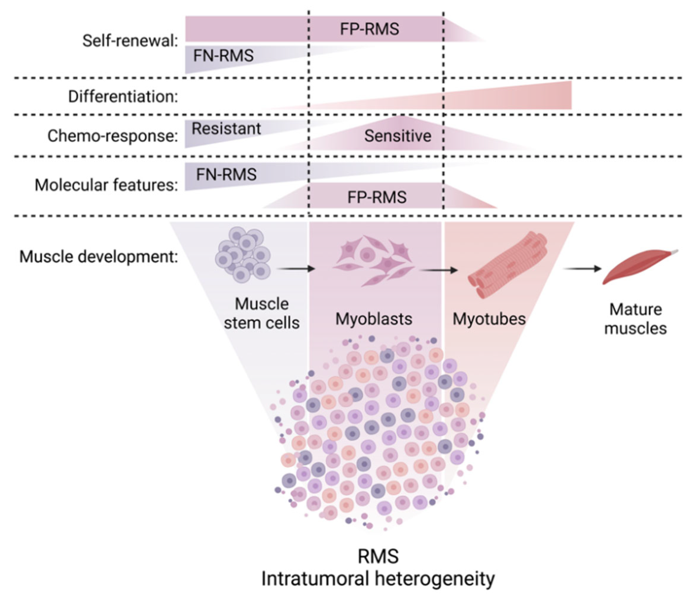
Elegant studies employing scRNAseq to investigate the transcriptional heterogeneity within the RMS cell pool revealed that both alveolar and embryonal RMS harbor cells with myogenic potential, which are stalled at distinct developmental states. These include a transcriptionally immature stem cell-like/mesoderm/mesenchymal state (expressing PAX3/PAX7/CD44), a myoblast state (expressing MYF5/Cell Division Cycle 20 (CDC20)/Cyclin B1 (CCNB1)) containing highly proliferative/cycling cells, and a more differentiated myocyte state (expressing MYF4/MYH3/MYH8) associated with better patient outcomes (Figure 2) [97,98,99]. It is important to note that the tumor cell pool spanned a wide range of developmental indices in embryonal RMS, while it was more skewed towards later stages of myogenesis in alveolar RMS [97,98,99], which is consistent with previously published immunohistochemical findings described above [82].
PF+ and PF− RMS appear to be arrested at specific stages of skeletal muscle development. The core transcriptional signature of PF+ RMS correlates with the signature found in developing muscle at 7–7.75 weeks of age, at which time embryonic muscle transitions to fetal muscle [99]. By contrast, the PF− RMS core signature was expressed in both embryonic and fetal muscle cells, but not in adult muscle [99].

5.4. Cellular Hierarchies in RMS

RNA velocity analysis and/or lentiviral barcoding of cells within patient-derived xenografts (PDX) have been applied to show that RMS tumors are hierarchically organized and recapitulate normal myogenesis, with cells transitioning from the undifferentiated stem-like/mesoderm state through the myoblast to the differentiated myocyte state [97,98]. However, recent observations suggest substantial differences in the organization of cellular hierarchies across the RMS spectrum, with PF− RMS following a possible, stem-like cell-driven model and prominent plasticity of PF+ RMS.
Wei et al. used lineage barcoding and functional assays to show that prospectively isolated CD44+/CD90+ or CD90+/CHODL+ cells of the mesenchymal subpopulation establish more, larger tumorspheres and exhibit more efficient, faster tumor growth in mice [99]. Previous studies using fluorescent transgenic zebrafish bearing KRAS(G12D)-induced RMS yielded similar insights [100]: GFP-labeled MYF5high zebrafish tumor cells (expressing markers of satellite and early muscle progenitor cells) and mCherry-labeled MYF4high zebrafish tumor cells (expressing high levels of mature muscle markers) were isolated by fluorescence-activated cell sorting and transplanted. Tumor-propagating capacity was enriched within the MYF5high cell population, while MYF4high cells were prone to migrate, invade through a normally impenetrable collagen matrix and enter the vasculature. In zebrafish, slow-moving MYF5high cells were found in newly colonized regions after initial invasion by MYF4high cells [100]. These studies indicate that the mesenchymal state characterizes the only tumor-propagating subpopulation—i.e., a stem-like population of cells capable of re-creating original tumor heterogeneity—in PF− RMS [99].
By contrast, PF+ RMS displays more prominent intrinsic plasticity, with both stem-like and cycling-myoblast states having the capacity to re-create the original composition of the tumor. These observations are consistent with in vivo tumor-propagating capacity of all single-cell clone-derived clones of P3F+ RMS cells examined by Generali et al. [101]. Of note, fluctuating levels of P3F expression in single, mouse P3F+ RMS cells were linked to differences in in vitro clonal activity and in vivo tumor-propagating capacity, indicating plasticity in P3F expression at the single-cell level [91]. Similar to what has been proposed for certain other cancers [102], cellular plasticity may be the mechanism at the root of PF+ RMS aggressiveness [97].

5.5. Differential Drug Responsiveness of RMS Cell Subsets

The functional consequences of intratumoral heterogeneity on drug response are an emerging topic of interest. Lentiviral barcoding of cells within PDX tumors followed by scRNAseq revealed that the highly proliferative, myoblast-like population of PF− RMS cells was more sensitive to conventional chemotherapy, while the more quiescent, mesoderm-like population was more resistant (Figure 2). Following chemotherapy, there was a decrease in clonal diversity, and the quiescent population of immature mesoderm-like cells expanded to reconstitute the developmental hierarchy of the tumor [98].
A similar phenomenon was observed in PF+ RMS, where—following in vitro treatment with etoposide or vincristine—the cycling/myoblast state was depleted, and stem-like cells were enriched [97]. This shift towards stem-like cell states could be explained by two scenarios: (i) selection of more resistant cells similar to what has been observed in PF− RMS, (ii) cells in the cycling/myoblast state and/or more differentiated cells may transition to stem-like states in response to treatment, or (iii) stem-like cells may increase their proliferation under therapy pressure, as it was recently indicated in glioblastomas [102]. Taken together, it is conceivable that chemotherapy drives a shift in RMS cellular states toward stem-like states.
These findings suggest that subpopulations of cells within the RMS cell pool are locked in distinct cell states, including an immature stem-cell state and more mature muscle states. Differentiated cells represent a minority of the tumor, do not cycle, have lower malignant potential and correlate with better patient outcomes (Figure 2). Similar developmental hierarchies (with subpopulations of stem-like cells capable of self-renewal and more differentiated cells with lower capacity to initiate tumors) have been identified in other pediatric tumor types, such as glioma [103], neuroblastoma [104], medulloblastoma [105], and rhabdoid tumors [106]. Shifting undesired stem-like states into “desired” differentiated states could provide therapeutic benefits in such cancers. This will require an in-depth understanding of how transitions between RMS cell states are regulated and how these processes can be manipulated therapeutically.

6. Aberrant Myogenic Differentiation due to Miswiring of Core Regulatory Circuits (CRCs) in RMS

6.1. Regulation of Cell Fate by CRCs

The molecular mechanisms involved in blocking differentiation in RMS are an area of active research. In general, cell type-specific transcriptional programs that govern cell fate specificity are tightly controlled by a highly coordinated array of master TFs (MTFs). These MTFs are organized in interconnected transcriptional loops, called core regulatory circuitries (CRCs).
Within CRCs, individual MTFs control their own expression as well as the expression of the other involved TFs and exert feed-forward transcriptional control [107]. These transcriptional networks ultimately control key transcriptional programs and drive processes such as myogenic differentiation in the muscle lineage. At the chromatin level, MTFs are accumulated at super-enhancers (SEs) and function under the control of SEs (Figure 3). Hence, insights into the SE landscape and associated TF binding motifs can be exploited bioinformatically to predict the CRCs that are active in individual cell types (Figure 3).
Cell-type specific CRCs have been identified for a number of different tissues, including normal skeletal muscle. Two families of TFs are at the center of the CRC active in skeletal muscle cells: the MRF and the myocyte enhancer factor 2 (MEF2) family [108,109].

6.2. Reshaping of Skeletal Muscle CRCs in RMS

The normal muscle CRC is active in both PF+ and PF− RMS. However, in both cases, the muscle CRC is further complemented with additional modules [5], including a pan-RMS module composed of MYOD1 and MYF4 and additional subtype-specific modules (Figure 3).
The P3F+ RMS-specific module relies on P3F and MYCN [5,110,111,112]. P3F was shown to have pioneer factor activity and shape the SE landscape in PF+ RMS cells, thereby demonstrating its importance for cell fate regulation [5,113]. Importantly, the expression of P3F is under the control of a FOXO1 enhancer that is normally active in late-stage myogenesis (Figure 3A) [6]. This P3F+ RMS-specific module replaces the originally sequentially organized CRC active in muscle precursor cells by an infinite loop that blocks the cells in an undifferentiated state.
By contrast, the PF− RMS-specific module involves PAX7 and Activator Protein-1 (AP1)-family TFs (Figure 3B) [5]. Additional TFs were found to be involved in CRCs in specific RMS cell lines (dbCoRC database http://dbcorc.cam-su.org/ accessed on 8 December 2022).

6.3. Differential Use of CRCs at the Single Cell Level

We note that the studies defining the CRC landscape in RMS were performed with bulk cell populations. As described above, recent scRNAseq and ATACseq analyses identified different subpopulations of cells to be present in RMS tumors [97,98,99]. These subpopulations of cells are aligned along the myogenic lineage, from muscle stem cell-like cell states to myosin-heavy chain positive, differentiated cell states. This has been linked to the differential use of CRC SEs [98], but the exact CRCs involved in the different cell states are yet to be determined.
It remains to be seen if some cells may be able to escape, potentially due to the influence of the microenvironment. Strong differentiation tendencies have been observed in RMS cells when they were removed from their regular in vivo niche and transferred into culture dishes [114]. Changes in nutrient availability, surface matrices, proximity to other tumor cells, etc., may also trigger the transition between different RMS cell states [91].

6.4. Evolving Understanding of Tumor Dependency Concepts in RMS

Overall, these data fit into important general tumor dependency concepts. It has been recognized for a long time that tumor cells often not only depend on the driver oncogenes (oncogene addiction) but also on survival mechanisms pre-programmed in the cells of origin and involving master regulatory genes (lineage addiction) [115]. Acquired genetic alterations might affect and cooperate with these mechanisms, providing an explanation for lineage-restricted patterns of genetic alterations in cancer, which are prime characteristics for many sarcomas, including RMS. The concept of CRCs forms a framework that links oncogene- and lineage-addiction in both TF-driven PF+ and also PF− tumors. We argue that a deeper understanding of how acquired, RMS-relevant oncogenic alterations interact/cooperate with the cellular mechanisms that are pre-programmed in the cell-of-origins and/or define skeletal muscle identity will be key to breaking the code of RMS malignancy.

7. Epigenetic Regulators of Aberrant Myogenic Differentiation in RMS

During muscle differentiation, chromatin-modifying enzymes/remodeling complexes reprogram muscle promoters, alter chromatin structure through post-translational modifications and, ultimately, impact the activity of MRFs. A growing body of evidence has illuminated our understanding of the epigenetic mechanisms by which skeletal muscle differentiation is dysregulated in RMS.

7.1. Aberrant Epigenetic Control of MYOD1 Expression in RMS

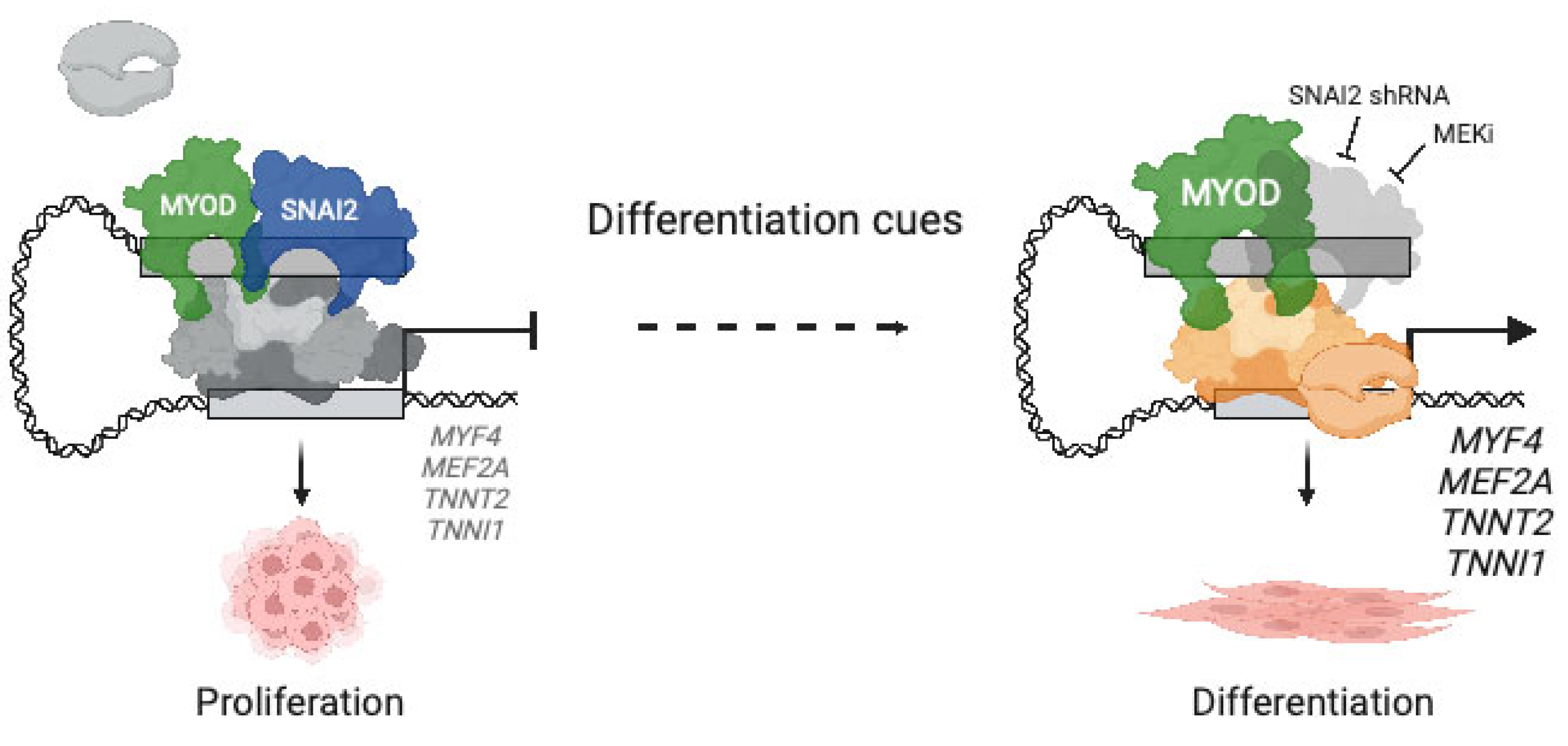
Changes in gene expression during differentiation processes are orchestrated chiefly by changes in histone methylation and acetylation. Epigenetic regulation of MYOD1 activity by the SUV39H1 histone lysine methyltransferase (KMT1A/SUV39H1; hereafter KMT1A), histone deacetylase 3 (HDAC3), Lysine Acetyltransferase 2B (PCAF/KAT2B; hereafter PCAF) and Snail Family Transcriptional Repressor 2 (SNAI2) are examples of how different players coordinate to block MYOD1-dependent myogenic differentiation in RMS (refer to Figure 4 for a schematic representation of epigenetic enzymes involved in aberrant myogenic differentiation in RMS).
Firstly, KMT1A associates with MYOD1 and represses gene expression through trimethylation of lysine 9 on histone H3 (H3K9) [116,117]. In normal myoblasts, the MYOD1-dependent histone methyltransferase activity of KMT1A diminishes as differentiation proceeds due to the activation of p38α, which phosphorylates KMT1A and disrupts its association with MYOD1, thus enabling transactivation of downstream myogenic genes [118]. However, in P3F+ RMS cells, p38α signaling is defective [85]. In response to differentiation cues, histone methyltransferase activity of KMT1A and H3K9me3 levels increases and disrupts the transactivation of downstream myogenic genes by MYOD1 [85,117,118].
Secondly, HDAC3 is overexpressed in RMS tissue compared to normal muscle and emerged as the main HDAC restricting RMS differentiation from a high-efficiency clustered regularly interspaced short palindromic repeats (CRISPR)-based phenotypic screen of class I and II HDAC genes [119]. HDAC3 associates with nuclear receptor corepressor 1 (NCOR1) and nuclear receptor corepressor 2 (NCOR2) to form the NCOR/HDAC3 complex, which interacts with MYOD1 to suppress transcriptional activation of myogenic genes. Silencing of HDAC3 enhances H3K9 acetylation in regulatory regions of MYOD1 target genes to induce differentiation [119].
Thirdly, recruitment of PCAF to the MYF4 promoter in differentiating myoblasts acetylates MYOD1 and thereby enhances its binding to DNA and ability to transactivate muscle genes [120,121]. Coactivator Associated Arginine Methyltransferase 1 (hereafter CARM1/PRMT4) associates with PCAF and facilitates its recruitment to the MYF4 promoter [122]. In PF− RMS cells, PCAF is recruited to the MYF4 promoter upon exposure to differentiation cues [123].
Finally, the MYOD1 differentiation axis is further regulated by the zinc finger TF SNAI2, which acts as a repressor of gene transcription and restricts differentiation in RMS. Downregulation of SNAI2 in skeletal muscle precursor cells regulates the transition between proliferative cell states and differentiation [124]. In PF− RMS cells, overexpression of SNAI2 is induced by MYOD1 through SE regulation [125]. SNAI2 competes with MYOD1 for binding to enhancer elements of muscle genes, and suppression of SNAI2 releases these enhancer regions and allows MYOD1 to activate the myogenic differentiation program and prevent tumor growth (refer to Figure 5 for the mechanism of action by which SNAI2 inhibits myogenic differentiation).

7.2. Chromatin Regulatory Complex PRC2

PRC2 works in concert with Polycomb repressive complex 1 (PRC1) to bind H3K27me3 marks and promote gene repression [126]. EZH2 is the catalytic subunit of PRC2 and trimethylates histone H3 at lysine 27 (H3K27me3). EZH2 plays a key role among epigenetic enzymes regulating tissue development. EZH2 is downregulated during cellular differentiation and undetectable in normal adult tissues, but its expression and activity are deregulated in a variety of cancers [127]. In skeletal muscle, EZH2 repressive functions are required to delay differentiation and allow myogenic precursors to proliferate and fill the anatomic space in specific regions of the embryo.
In sarcomas, upregulation of PRC2 components—in particular EZH2—has been correlated with metastases and lower survival [128,129,130]. In PF− RMS, aberrant PRC2 activity—including aberrant EZH2 activity—has been shown to block skeletal muscle differentiation by repressing the transcription of myogenic genes (refer to Figure 4 for a schematic representation of epigenetic enzymes involved in aberrant myogenic differentiation in RMS) [131,132]. EZH2 is also recruited by YIN-YANG-1 (YY1) [133] and by GATA Binding Protein 4 (GATA-4) [134] to repress the expression of selected myogenic microRNAs in proliferating myoblasts and RMS cells.
Jumonji And AT-Rich Interaction Domain Containing 2 (JARID2), a Jumonji domain-containing interacting protein, use the PRC2 component Embryonic Ectoderm Development (EED) to regulate H3K27me3 in the promoter region of MYF4 and Myosin Light Chain (MYL1). JARID2 is a direct transcriptional target of the fusion oncogene and uses EED to maintain an aberrant myogenic phenotype in P3F+ RMS [135]. Silencing of JARID2 resulted in myogenic differentiation of P3F+ RMS [135].

7.3. Other Epigenetic Regulators of Aberrant Myogenic Differentiation in RMS

Euchromatic Histone Lysine Methyltransferase 2 (G9a/EHMT2; hereafter G9a) has been associated with tumor-promoting activity in several tumor types, including RMS [136,137,138,139,140,141]. G9a and EHMT1 are paralogs that perform the same enzymatic functions: catalyzing mono- or di-methylation of histone H3 at lysine 27 (H3K9me1/H3K9me2). G9a and EHMT1 cooperate by forming a heteromeric complex [142], but they are not completely functionally redundant and maintain their own unique roles [143]. In PF− RMS, G9a overexpression indirectly suppresses the canonical WNT signaling pathway to prevent myogenic differentiation by activating, rather than repressing, the expression of the WNT antagonist Dickkopf WNT Signaling Pathway Inhibitor 1 (DKK1) [141] through recruitment of Sp1 TF (SP1) and E1A Binding Protein P300 (P300) to the DKK1 promoter. In PF+ RMS, G9a is regulated by the orphan receptor Nuclear Receptor Subfamily 4 Group A Member 1 (NR4A1, [109]), which drives the expression of the P3F fusion gene itself [144]. G9a directly represses Phosphatase and Tensin Homolog (PTEN) expression, thus increasing AKT Serine and Threonine Kinase (AKT) and RAC Family Small GTPase 1 (RAC1) activity and impeding Phosphoinositide-3-Kinase (PI3K) pathway activity [139].
Less is known about the oncogenic role of Euchromatic Histone Lysine Methyltransferase 1 (GLP/EHMT1; hereafter, EHMT1) in RMS. There is some evidence that silencing of EHMT1 in P3F+ RMS cell lines results in decreased motility and induction of differentiation, along with reduced tumor progression in xenograft models in vivo [145]. Indeed, EHMT1 stabilizes the CCAAT-enhancer-binding protein (C/EBPb) to transactivate Aldehyde Dehydrogenase 1 Family Member A1 (ALDH1A1) and thereby maintain a stem-like state in RMS [146].
Finally, the TF SNAIL Family Transcription SNAI1 (SNAIL) [147] and the SWI/SNF Related, Matrix Associated, Actin Dependent Regulator Of Chromatin, Subfamily A, Member 4 (SMARCA4/BRG1) subunit of the mSWI/SNF chromatin remodeling complex [148] were identified as key suppressors of myogenic differentiation and promoters of oncogenesis in PF+ RMS.

8. MicroRNA (miRNA)-Dependent Post-Transcriptional Dysregulation of Myogenic Differentiation in RMS

8.1. MiRNAs in Cancer

Post-transcriptional regulation of gene expression by miRNAs, a class of small non-coding (nc) RNAs, is one of the key mechanisms allowing for a rapid adaptive response to environmental cues during development. MiRNAs are short ncRNAs of about 22 nucleotides, which are produced in the nucleus as pri-miRNAs and then processed by the Drosha complex into shorter precursors of about 70 nucleotides called pre-miRNAs. Pre-miRNAs translocate to the cytoplasm, where they are processed again by the DICER1 enzyme to give rise to mature miRNAs [149,150]. MiRNAs function by binding to complementary sequences on target mRNAs, resulting in translational repression or mRNA degradation, depending on the grade of complementarity between the seed region of the miRNA and the 3′ untranslated region (UTR) of the target mRNA [151]. MiRNAs always target multiple mRNAs, thereby mediating post-transcriptional control of several processes concurrently, including differentiation, proliferation and survival [149,152]. MiRNAs are crucial players in cancer malignancy and may behave as tumor suppressors or oncogenes depending on tumor context. Notably, germline variants in DICER1 have long been recognized to confer genetic susceptibility to develop cancers, including RMS [80,82].

8.2. MYOmiR Family of miRNAs

MyomiRs are a group of miRNAs that are expressed in a tissue-specific manner in skeletal and/or cardiac muscle. During skeletal muscle differentiation, myomiRs are induced by MYF5, MYOD1 and MYF4 with the help of MEF2 factors through binding to muscle-specific intronic enhancers [153,154]. Specifically, the miR-1 family includes miR-1-1/miR-133a-2, miR-1-2/miR-133a-1 and miR-206/miR-133b bicistronic pairs, which are located on three different chromosomes [155]. MiR-1-1 and miR-1-2 are identical; only three nucleotides differ in miR-206. All carry the same seed sequence. Similarly, miR-133a-1 and miR-133a-2 are identical; only one nucleotide differs in miR-133b. Again, all three contain the same seed sequence [156]. Consequently, they share the same mRNA targets. MiR-206 is the only miRNA exclusively expressed in skeletal muscle [150]. MyomiR targets include Histone deacetylase 4 (HDAC4) [157] and the SWI/SNF Related, Matrix Associated, Actin Dependent Regulator Of Chromatin, Subfamily D, Member 1 (SMARCD1/BAF60a) subunit of the mSWI/SNF chromatin remodeling complex [158].

8.3. Deregulation of MYOmiRs in RMS

MiRNA deregulation contributes to RMS pathogenesis [150,159,160]. MiR-1 and miR-206 levels are lower in RMS compared to muscle [161,162,163], where low miR-206 levels correlated with poor survival, higher stage and presence of metastases in patients with RMS [161]. Low miR-206 levels in RMS appear to be under the control of SNAI1/2, and SNAI1 silencing results in miR-206 up-regulation followed by cell differentiation [124,147]. Low miR-206 expression was linked to the expression of genes associated with muscle differentiation [161], and forced expression of pre-miR-206 in RMS cells halted proliferation, induced myogenic differentiation and inhibited tumor growth in vitro and in vivo.
The differentiating effects of MiR-206 in RMS were in part mediated by MET Proto-Oncogene, Receptor Tyrosine Kinase (MET) [163,164]. MiR-206 oligo miRNA mimics induced G0/G1 cell cycle blockade and inhibited cell migration in several RMS cell lines through suppression of MET expression [161]. We note that the proto-oncogene MET—a downstream target of both PAX3 and PAX3:FOXO1 [165,166]—plays an important role during skeletal muscle formation by favoring the delamination of myoblasts precursors and their migration to the limb bud [167,168]. MET also promotes migration and cell survival of RMS cells, and MET sequence variation, amplification and overexpression were observed in human RMS tumors [38].
Other MiR-206 targets in RMS include PAX7 [147,169,170], NOTCH Receptor 3 (NOTCH3), and Cyclin D2 (CCND2) [163,164,170,171].

8.4. Deregulation of Other miRNAs in RMS

Certain miRNAs are not exclusively expressed in skeletal muscle but have pro-myogenic effects. In fact, the very first report on the involvement of miRNAs in the reluctance of RMS cells to differentiate pointed to the miR-29 family of miRNAs, which plays an important role in the NF-κB-YY1-miR-29 regulatory circuitry of myogenic differentiation. In brief, NF-κB activation sustains YY1 levels and polycomb activity in myoblasts. YY1 and EZH2 bind to a regulatory element upstream of miR-29b-2 and miR-29c on chromosome 1 to form a YY1/PRC2 repressor complex, which prevents the expression of miR-29a/b/c. Through a negative feedback loop, miR-29a/b/c precursors target the YY1 3′UTR mRNA region, reducing YY1 protein levels and inducing differentiation [133]. Forced expression of miR-29 in RMS cell lines slowed down cell proliferation and inhibited tumor growth, increased Cyclin D1 (CCND1) and reduced CDKN1A expression, and induced cell cycle arrest and terminal muscle differentiation. These effects were mirrored by the silencing of YY1, which led to miR-29 induction, further supporting the important role of the YY1/miR-29 feedback loop in the governance of muscle differentiation in RMS [133,172].
A number of additional miRNAs have also been implicated in aberrant myogenic differentiation in RMS, including miR-214 (targeting NRAS) [173], miR-203 (targeting TP63 and leukemia inhibitor factor receptor (LIFR)) [174], miR-378a-3p (targeting IGF1R) [175], and others. For example, miR-28-3p and miR-193a-5p are strongly upregulated in differentiating myoblasts, and their forced expression in RMS cells prevented invasion/migration, reduced proliferation, induced differentiation and inhibited tumor growth in vivo. Interestingly, overexpression of miR-193a-5p led to miR-206 upregulation, thereby supporting an intricate network of epigenetic miRNAs governing myogenesis [176,177].

8.5. Potential Avenues towards Therapeutic Targeting of miRNAs

Several RNA-based treatments (i.e., anti-sense nucleotides and small interfering RNAs) have gained FDA/EMA approval (e.g., Nusinersen, targeting Survival of Motor Neuron 2 (SMN2) pre-mRNA splicing in spinal muscular atrophy) or entered early phase clinical trials (e.g., for treatment of myeloid leukemias or carcinomas) [178]. In light of the clear role of miRNAs in dysregulated myogenic differentiation in RMS, a role for RNA-based treatments in RMS therapy may be possible.

9. Myogenic Differentiation as a Target for RMS Therapy

9.1. Differentiation Therapy in Cancer

Given the developmental nature of pediatric cancers, the idea of chemically inducing the differentiation of cells for therapeutic purposes—i.e., steering cancer cells into a benign direction—has long been entertained and pursued by pediatric oncologists [179]. Generally, terminal differentiation has been associated with growth arrest/blocking hyperproliferation and loss of migratory/invasive capacities of cancer cells. As a bonus, differentiating treatments often cause less therapy-related toxicity than conventional chemotherapy. In the clinical setting, two successful examples of differentiation therapies come to mind: The prime example is the treatment of acute promyelocytic leukemia (APL) with retinoic acid. APL used to be fatal, but it has become curable since two clinically effective therapies were identified—retinoic acid and arsenic—which trigger terminal differentiation of leukemic cells into granulocytes. A second example is the treatment of neuroblastoma, a neural crest-derived cancer. Because retinoic acid signaling partially controls differentiation of the neural crest into peripheral neural cells, cis-retinoic acid may be used to induce differentiation of residual neuroblastoma cells.

9.2. Overcoming the Differentiation Block in RMS as a Therapeutic Principle

As RMS cells are characterized by their reluctance to undergo terminal myogenic differentiation [180], there is significant interest in exploiting the causes of developmental stalling in RMS to restore differentiation as a therapeutic strategy. A notable early finding in RMS was that conventional chemotherapy increased the proportion of rhabdomyoblasts—variably differentiated, elongated, eosinophilic cells—which appeared to be further along the spectrum of maturation morphologically. Such cytodifferentiation, sometimes extensive, was more frequently observed in embryonal histology RMS, and it appeared to identify tumors that were responsive to therapy [181,182]. Extending beyond conventional therapy, pharmacological interventions that induce differentiation in RMS cells may provide means to overcome arrested myogenic development and/or render tumor cells more sensitive to previously established cytostatic agents.

9.3. Targeting Cell Cycle Progression in RMS

Integrated genomic, epigenomic and proteomic analyses of RMS cells derived from orthotopic PDXs highlighted activation of the cyclin D/CDK4/6-RB/E2F pathway and alteration of the G2/M mitotic spindle checkpoint pathway in RMS [89]. In fact, CDK4/6 inhibitor treatment of RMS cells caused G1 arrest accompanied by myogenic differentiation. This correlated with a reduction of tumor growth in vitro and in vivo [183]. However, palbociclib and abemaciclib—administered in combination with trametinib—failed to produce relevant anti-tumor effects in a randomized, preclinical phase II trial. By contrast, the WEE1 G2 checkpoint kinase (WEE1) inhibitor AZD1775—used in combination with vincristine and irinotecan—was found to be highly efficacious with a 70% complete remission/partial remission rate in a blinded, randomized, and placebo-controlled preclinical phase 3 trial [89]. WEE1 kinase is expressed highly in RMS [184] and regulates the G2/M checkpoint and DNA replication during the S phase of the cell cycle [185] by phosphorylating CDK1. WEE1 kinase inhibitor effects on myogenic differentiation have not been reported as of yet.

9.4. Targeting MAPK Signaling in PF− und PF+ Cells

In PF− RMS cell lines driven by H/NRAS-Q61X mutations, activation of the RAS pathway inhibits myogenic differentiation through the RAF-MEK-ERK effector pathway by repressing the expression of MYF4 [186]. As a consequence, treatment with the Mitogen-Activated Protein Kinase Kinase 1/2 (MEK1/2) inhibitor trametinib releases transcriptional stalling at the MYF4 promoter, followed by a widespread MYF4-induced change in SE landscape, myogenic differentiation and reduced tumor growth in mouse models [186]. Unfortunately, Yohe et al. demonstrated in preclinical models of PF− RMS that tumor cells rapidly acquired resistance to trametinib when used as a single agent [186].
Treatment with trametinib and IGFR1 inhibitors in combination could be a promising combinatorial approach; trametinib and the IGFR1 inhibitor ganitumab in combination were shown to be efficacious and well tolerated in preclinical models of RAS-mutant PF− RMS [187]. In an attempt to further optimize the therapeutic potential of trametinib, vertical double targeting of the RAF-MEK-ERK cascade, using RAF1 Proto-Oncogene, Serine/Threonine Kinase (RAF1) or pan-RAF inhibitors combined with MEK/ERK inhibitors, synergistically triggered myogenic differentiation and suppressed embryonal RMS tumor growth [188]. Currently, there are no specific RAF1 inhibitors available for clinical use, but several pan-RAF inhibitors are being studied in phase I/II clinical trials. Of particular interest is the pan-RAF inhibitor DAY101, which is currently being tested in a phase II clinical trial in pediatric low-grade glioma patients (NCT04775485).

9.5. Targeting Cellular Hierarchies in RMS

Recent scRNA sequencing efforts identified three developmentally distinct cell states (stem-like, myoblast/progenitor, and differentiated) in RMS. In PF− RMS, the mesoderm-like subset of cells was shown to be rather resistant to conventional chemotherapy and dependent on Epidermal Growth Factor Receptor (EGFR) signaling. Consequently, EGFR inhibitors may have a role in overcoming drug resistance of mesoderm-like cells within the PF− RMS cell pool. Indeed, EGFR inhibitors combined with irinotecan and vincristine significantly prolonged the survival of mice with embryonal histology PDX tumors [98].
For PF+ RMS, a subsequent pharmacological screen focused on measuring chemically induced changes in the abundance of the three main cell states and identified the MEK inhibitor trametinib as a strategy to rewire the cellular composition of PF+ RMS towards differentiated cell states [97]. When trametinib was combined with the multi-kinase inhibitors regorafenib, sorafenib or dabrafenib, the differentiation effect was increased, further supporting the notion that vertical double targeting of the MAPK pathway may be a strategy to interfere with RMS cell state transitions and overcome arrested differentiation in RMS. Co-treatment with trametinib and regorafenib (currently in clinical trials, NCT02085148) not only triggered differentiation but also suppressed PF+ RMS PDX tumor growth effectively [97].

9.6. Indirect Targeting of MYOD1 to Trigger RMS Differentiation

In large-scale CRISPR-based essentiality screens, MYOD1 was among the most RMS-specific genetic vulnerabilities (DepMap; https://depmap.org/portal/ accessed 8 December 2022) [189]. Hence, interference with its activity by targeting key transcriptional regulators offers an RMS-specific therapeutic route. As described above and illustrated in Figure 4, SNAI2 is highly expressed and acts as a transcriptional repressor to inhibit MYOD1-driven differentiation and downregulate TFs necessary for terminal differentiation [125]. Incidentally, SNAI2 also represents an effector of the RAF-MEK-ERK signaling pathway. Trametinib blocks SNAI2, thereby allowing MYOD1 to bind to the enhancer elements of muscle-specific genes. Genetic removal of SNAI2 induces myogenic differentiation and phenocopies the effects of MEK inhibition [125].
Other repressors of embryonal RMS differentiation were described. The TF SIX1 maintains the cells in an undifferentiated state by reprogramming MYOD1 to occupy loci that drive tumor growth instead of muscle differentiation [190]. Loss of SIX1 restores the MYOD1/MYF4 gene regulatory network, induces tumor cell differentiation, and inhibits in vivo tumor growth [190]. Yet, TFs are challenging drug targets and indirect ways to interfere with their activity—e.g., by blocking the machinery associated with SE regulation—might be more straightforward.

9.7. HDAC and EZH2 Inhibitors

Interference with the activity of CRCs offers alternative therapeutic opportunities in RMS. High levels of histone modifications present at CRC-SEs make them vulnerable to inhibitors of epigenetic regulators [191], such as inhibitors of DNA demethylation and histone deacetylation.
In RMS, maintenance of the SE architecture appears to depend on appropriate histone acetylation [5,7]. Hyperacetylation due to HDAC inhibitor exposure spreads beyond the original SE borders and disrupts their 3D organization, thereby disrupting SE functionality [5]. HDAC inhibitors were shown to stimulate muscle differentiation and have been considered as a candidate drug in the treatment of muscular dystrophy [30]. Bharathy et al. demonstrated that the HDAC1/3 inhibitor Entinostat, administered in combination with vincristine, slowed PF− RMS tumor growth while inducing myo-differentiation in vivo [192], but pan-HDAC inhibitors have produced disappointing results. Phelps et al. suggested that the development of more selective HDAC inhibitors might improve anti-RMS efficacy [119].
The PRC2 component EZH2 has emerged as an important regulator of muscle differentiation and a candidate treatment target in RMS [128,129,130]. Various inhibitors of EZH2 have been used in preclinical studies. Treatment of RMS cells with MC1948 and MC1945—inhibitors of EZH2 catalytic activity—resulted in upregulation of MYF4, Creatine Kinase, M-type (CKM) and Myosin Heavy Chain (MHC) genes, and ultimately, skeletal muscle differentiation. These changes were reversed by enforced overexpression of EZH2, indicating an EZH2-specific effect [131,193,194]. The EZH2 inhibitor tazemetostat has been examined in a pediatric phase I trial (Table S1; [195]) and may be considered in anti-RMS therapeutic strategies in the future.
Finally, a first-in-class dual EZH2/HDAC inhibitor 5 with (sub-) micromolar activity against both targets was introduced recently, representing another promising anti-RMS candidate compound [196].

9.8. Indirect Targeting of the PF Fusion Protein

In PF+ RMS, the differentiation block is mainly mediated by direct and/or indirect effects of the disease-driving fusion oncoprotein. As targeting of P3F remains challenging and possible plasticity in P3F expression [90,91] may counteract pharmacological interventions, substantial efforts have been directed towards identifying alternative options to alter PAX3:FOXO1 function. Key suppressors of myogenic differentiation and promoters of oncogenesis in PF+ RMS include transcriptional regulators such as JARID2 [135] and SNAIL [147], as well as the chromatin modifier SMARCA4 [148]. Pharmacological interference with their functions could overcome the differentiation block in PF+ RMS cells, but it has proven almost as difficult as targeting the fusion oncogene itself. Proteolysis targeting chimera degraders (such as PROTACs) may allow for the drugging of previously intractable therapeutic targets in the future by ubiquitinating and degrading them via the proteasome [197].
For the time being, tractable P3F/P7F targets such as Fibroblast Growth Factor Receptor 4 (FGFR4, [111,198,199]), Insulin-like Growth Factor Receptor 1 (IGF1R, [200]), and the MET Proto-Oncogene, Receptor Tyrosine Kinase receptor (MET, [165,166]) are thought to inhibit alveolar RMS differentiation and might offer windows of opportunity for (combination) therapy. Pharmacological inhibition of the histone methyltransferase G9a or the histone acetyltransferase PCAF would also be relatively simple. G9a inhibition prompted differentiation and decreased P3F+ RMS tumor growth in vivo as a result of increased PTEN expression [139]. PCAF overexpression in PF+ RMS was linked to higher affinity for P3F than for its normal target MYOD1, thereby acetylating and stabilizing the fusion protein. Inhibition of PCAF resulted in impaired PF+ RMS tumorigenesis in vitro and in vivo [192].
Epigenetic co-factors of P3F such as Bromodomain Containing 4 (BRD4), Chromodomain Helicase DNA Binding Protein 4 (CHD4), and Lysine Demethylase 4B (KDM4B) represent attractive targets for interference with oncogenic SE organization in PF+ RMS cells [113,201,202,203,204]. For example, P3F recruits BET bromodomain protein BRD4 [113] to establish extensive miswiring of enhancers and set up autoregulatory loops that maintain P3F expression [113], disrupt normal myogenic differentiation and lock RMS in an undifferentiated stage. Interference with BRD4 was shown to destabilize the expression of the fusion oncogene [113]. As BRD4 interacts with Cyclin-dependent Kinase 9 (CDK9) in other fusion-driven sarcomas, combinatorial treatment with BRD4 and CDK9 inhibitors may exert synergistic anti-tumor effects in PF+ RMS [205]. The combinatorial effects of BRD4 and CDK9 inhibitors on PF+ RMS cell cultures and PDX tumors are currently evaluated in a randomized, confirmatory preclinical study (animalstudyregistry.org; 10.17590/asr.0000286 accessed 18 March 2023).

9.9. Clinical Testing of Differentiating Agents in RMS

There are a number of chemical compounds that were investigated preclinically as inducers of differentiation in RMS and used in early-phase clinical trials for patients with pediatric cancer. Table S1 provides an overview of completed, terminated, active/non-recruiting and active/recruiting trials involving such compounds. Many trials did not recruit patients with RMS (e.g., NCT02601937 investigating the EZH2 inhibitor tazemetostat [195]). For HDAC inhibitors, no objective responses were observed in four patients with RMS and one patient with STS [206,207,208]. NCT02095132 investigated the effects of AZD1775 and irinotecan in patients with RMS (no response data available yet) but did not include vincristine despite preclinical studies clearly indicating that vincristine enhanced the anti-RMS efficacy of AZD1775 and irinotecan significantly.
Despite convincing data supporting the important role of MAPK signaling in muscle differentiation and RMS, we caution heavily against off-label trametinib monotherapy, as trametinib rapidly produced resistance in preclinical models of RMS [186]. Finally, we advocate very strongly for enrolling patients in early-phase clinical trials to systematically investigate the anti-RMS effects of candidate drugs!

10. Summary and Conclusions

RMS derives its name from the skeletal muscle features, which were observed microscopically in tumor tissue early on. Yet, more recent insights into the molecular mechanisms underpinning the disease have highlighted aberrant myogenic differentiation as a driving force in RMS malignancy.
This review summarizes our current understanding of the genetic and epigenetic characteristics of the disease as they determine aberrant muscle differentiation in RMS tissue. During embryonal development and regeneration of adult muscle, MRFs collaborate in directing skeletal muscle progenitor cells to establish and maintain the muscle lineage [4]. There is strong evidence to support the notion that, during the development and maintenance of RMS, MRFs collaborate with disease-driving oncogenic aberrations to initiate and sustain RMS cell proliferation, migration and aberrant differentiation [6,107,115]. Core features of RMS malignancy include deregulation of the epigenetically strictly controlled, hierarchically organized transcriptional events that define skeletal muscle development/differentiation and miswiring of muscle-specific CRCs.
RMS is a diverse cancer that manifests itself on a wide clinical and biological spectrum that goes far beyond the distinction of the four RMS histotypes recognized by the WHO [1,2]. In the clinical setting, adequate recognition of distinct RMS manifestations is a critical prerequisite for the adequate stratification of risk of treatment failure/death versus short-term/long-term treatment toxicity in a very young population of patients. Specific features of aberrant muscle differentiation observed in RMS tissue may aid in adequately recognizing distinct RMS manifestations [2] and likely result from the interaction of RMS-relevant driver oncogenes and mechanisms of survival/proliferation/differentiation pre-programmed in RMS cells-of-origin [115].
Differentiation of cells, including myogenic differentiation, is one domain in which cellular plasticity—i.e., the ability of cells to change their identity/states—is demonstrated. Plasticity may also serve as an escape mechanism enabling cells to adapt to changing environments. There is increasing evidence to support prominent plasticity within the RMS cell pool [90,91,97,98,99], especially with respect to PF+ RMS [90,91,97]. We argue that plasticity may be the mechanism at the root of PF+ RMS aggressiveness. Pharmacological interventions to overcome arrested myogenic development in RMS have garnered attention as a much-needed new window of opportunity.
Chemical compounds targeting muscle-relevant epigenetic pathways [119], the RAF-MEK-ERK cascade [97,186,188] and the stability of the fusion oncogene [113] have been brought forth as candidate anti-RMS drugs. Yet, several attractive treatment targets are not druggable thus far, and in the case of chemically tractable alterations, only a few compounds have entered clinical investigation due to limitations in drug specificity and/or selectivity. Future efforts should focus on rational combinations of drugs [97], dual-targeting of chemical compounds [209], and PROTACs to tackle previously undruggable targets by ubiquitinating and degrading them via the proteasome [197]. Ultimately, hope for the identification of new superior differentiation approaches in RMS and their use in rational combination therapies rests on future research deepening our understanding of RMS biology, including aberrant myogenic differentiation as a hallmark of this cancer.

Supplementary Materials

The following supporting information can be downloaded at: https://www.mdpi.com/article/10.3390/cancers15102823/s1, Table S1. Drugs inducing myogenic differentiation in RMS cells in preclinical studies, which have been examined in early phase clinical trials in children and adolescents. References [210,211,212,213,214] are cited in the Supplementary Materials.

Author Contributions

Conceptualization, S.H., J.M.S. and R.R.; writing—original draft preparation, S.P., S.G.D., W.B.B., E.H., J.S., M.W., Z.S.W., R.R. and S.H.; writing—review and editing, S.P., S.G.D., R.A., W.B.B., E.H., J.S., M.W., Z.S.W., B.W.S., R.R., J.M.S. and S.H.; visualization, S.P., S.G.D., R.R. and S.H.; supervision, S.H. All authors have read and agreed to the published version of the manuscript.

Funding

This work was funded in part by the Italian Ministry of Health Ricerca Finalizzata 2021 (GR-2021-12374415, to S.P.), Hanne Liebermann Stiftung (to W.B.B.), Alice’s Arc Children’s Cancer Charity (to J.M.S.), Children with Cancer UK (grant 14-177, to Z.S.W.), Sarcoma UK (grant SUK206.2017, to Z.S.W.), Cancer League Switzerland (KLS-5143-08-2020, to B.W.S.), Sarcoma Foundation of America (to B.W.S.), Swiss Foundation for Childhood Cancer Research (to B.W.S.), Associazione Italiana per la Ricerca sul Cancro (AIRC, IG 27794, to R.R.), Förderverein für krebskranke Kinder Freiburg e.V. (to S.H.) and the Bundesministerium für Bildung und Forschung (01KC2012A, to S.H.). We also acknowledge support by the Open Access Publication Fund of the University of Freiburg. The authors declare no competing financial interests.

Data Availability Statement

Not applicable.

Conflicts of Interest

The authors declare no conflict of interest. The funders had no role in the design of the review; in the collection, analyses, or interpretation of data; in the writing of the manuscript, or in the decision to publish the review.

References

  1. Parham, D.M. Pathologic classification of rhabdomyosarcomas and correlations with molecular studies. Mod. Pathol. 2001, 14, 506–514. [Google Scholar] [CrossRef] [PubMed]
  2. Rudzinski, E.R.; Kelsey, A.; Vokuhl, C.; Linardic, C.M.; Shipley, J.; Hettmer, S.; Koscielniak, E.; Hawkins, D.S.; Bisogno, G. Pathology of childhood rhabdomyosarcoma: A consensus opinion document from the Children’s Oncology Group, European Paediatric Soft Tissue Sarcoma Study Group, and the Cooperative Weichteilsarkom Studiengruppe. Pediatr. Blood Cancer 2021, 68, e28798. [Google Scholar] [CrossRef] [PubMed]
  3. Knight, J.D.; Kothary, R. The myogenic kinome: Protein kinases critical to mammalian skeletal myogenesis. Skelet. Muscle 2011, 1, 29. [Google Scholar] [CrossRef] [PubMed]
  4. Zammit, P.S. Function of the myogenic regulatory factors Myf5, MyoD, Myogenin and MRF4 in skeletal muscle, satellite cells and regenerative myogenesis. Semin. Cell Dev. Biol. 2017, 72, 19–32. [Google Scholar] [CrossRef]
  5. Gryder, B.E.; Pomella, S.; Sayers, C.; Wu, X.S.; Song, Y.; Chiarella, A.M.; Bagchi, S.; Chou, H.C.; Sinniah, R.S.; Walton, A.; et al. Histone hyperacetylation disrupts core gene regulatory architecture in rhabdomyosarcoma. Nat. Genet. 2019, 51, 1714–1722. [Google Scholar] [CrossRef]
  6. Gryder, B.E.; Wachtel, M.; Chang, K.; El Demerdash, O.; Aboreden, N.G.; Mohammed, W.; Ewert, W.; Pomella, S.; Rota, R.; Wei, J.S.; et al. Miswired Enhancer Logic Drives a Cancer of the Muscle Lineage. iScience 2020, 23, 101103. [Google Scholar] [CrossRef]
  7. Gryder, B.E.; Wu, L.; Woldemichael, G.M.; Pomella, S.; Quinn, T.R.; Park, P.M.C.; Cleveland, A.; Stanton, B.Z.; Song, Y.; Rota, R.; et al. Chemical genomics reveals histone deacetylases are required for core regulatory transcription. Nat. Commun. 2019, 10, 3004. [Google Scholar] [CrossRef]
  8. Saab, R.; Spunt, S.L.; Skapek, S.X. Myogenesis and rhabdomyosarcoma the Jekyll and Hyde of skeletal muscle. Curr. Top. Dev. Biol. 2011, 94, 197–234. [Google Scholar] [CrossRef]
  9. Davis, R.L.; Weintraub, H.; Lassar, A.B. Expression of a single transfected cDNA converts fibroblasts to myoblasts. Cell 1987, 51, 987–1000. [Google Scholar] [CrossRef]
  10. Wright, W.E.; Sassoon, D.A.; Lin, V.K. Myogenin, a factor regulating myogenesis, has a domain homologous to MyoD. Cell 1989, 56, 607–617. [Google Scholar] [CrossRef]
  11. Braun, T.; Buschhausen-Denker, G.; Bober, E.; Tannich, E.; Arnold, H.H. A novel human muscle factor related to but distinct from MyoD1 induces myogenic conversion in 10T1/2 fibroblasts. EMBO J. 1989, 8, 701–709. [Google Scholar] [CrossRef]
  12. Yutzey, K.E.; Rhodes, S.J.; Konieczny, S.F. Differential trans activation associated with the muscle regulatory factors MyoD1, myogenin, and MRF4. Mol. Cell. Biol. 1990, 10, 3934–3944. [Google Scholar] [CrossRef]
  13. Singh, K.; Dilworth, F.J. Differential modulation of cell cycle progression distinguishes members of the myogenic regulatory factor family of transcription factors. FEBS J. 2013, 280, 3991–4003. [Google Scholar] [CrossRef]
  14. Weintraub, H.; Tapscott, S.J.; Davis, R.L.; Thayer, M.J.; Adam, M.A.; Lassar, A.B.; Miller, A.D. Activation of muscle-specific genes in pigment, nerve, fat, liver, and fibroblast cell lines by forced expression of MyoD. Proc. Natl. Acad. Sci. USA 1989, 86, 5434–5438. [Google Scholar] [CrossRef]
  15. Gayraud-Morel, B.; Chretien, F.; Flamant, P.; Gomes, D.; Zammit, P.S.; Tajbakhsh, S. A role for the myogenic determination gene Myf5 in adult regenerative myogenesis. Dev. Biol. 2007, 312, 13–28. [Google Scholar] [CrossRef]
  16. Tapscott, S.J. The circuitry of a master switch: Myod and the regulation of skeletal muscle gene transcription. Development 2005, 132, 2685–2695. [Google Scholar] [CrossRef]
  17. Kassar-Duchossoy, L.; Giacone, E.; Gayraud-Morel, B.; Jory, A.; Gomes, D.; Tajbakhsh, S. Pax3/Pax7 mark a novel population of primitive myogenic cells during development. Genes. Dev. 2005, 19, 1426–1431. [Google Scholar] [CrossRef]
  18. Relaix, F.; Rocancourt, D.; Mansouri, A.; Buckingham, M. A Pax3/Pax7-dependent population of skeletal muscle progenitor cells. Nature 2005, 435, 948–953. [Google Scholar] [CrossRef]
  19. Summerbell, D.; Halai, C.; Rigby, P.W. Expression of the myogenic regulatory factor Mrf4 precedes or is contemporaneous with that of Myf5 in the somitic bud. Mech. Dev. 2002, 117, 331–335. [Google Scholar] [CrossRef]
  20. Tajbakhsh, S.; Rocancourt, D.; Cossu, G.; Buckingham, M. Redefining the genetic hierarchies controlling skeletal myogenesis: Pax-3 and Myf-5 act upstream of MyoD. Cell 1997, 89, 127–138. [Google Scholar] [CrossRef]
  21. Rudnicki, M.A.; Schnegelsberg, P.N.; Stead, R.H.; Braun, T.; Arnold, H.H.; Jaenisch, R. MyoD or Myf-5 is required for the formation of skeletal muscle. Cell 1993, 75, 1351–1359. [Google Scholar] [CrossRef] [PubMed]
  22. Hasty, P.; Bradley, A.; Morris, J.H.; Edmondson, D.G.; Venuti, J.M.; Olson, E.N.; Klein, W.H. Muscle deficiency and neonatal death in mice with a targeted mutation in the myogenin gene. Nature 1993, 364, 501–506. [Google Scholar] [CrossRef] [PubMed]
  23. Relaix, F.; Montarras, D.; Zaffran, S.; Gayraud-Morel, B.; Rocancourt, D.; Tajbakhsh, S.; Mansouri, A.; Cumano, A.; Buckingham, M. Pax3 and Pax7 have distinct and overlapping functions in adult muscle progenitor cells. J. Cell Biol. 2006, 172, 91–102. [Google Scholar] [CrossRef] [PubMed]
  24. Seale, P.; Sabourin, L.A.; Girgis-Gabardo, A.; Mansouri, A.; Gruss, P.; Rudnicki, M.A. Pax7 is required for the specification of myogenic satellite cells. Cell 2000, 102, 777–786. [Google Scholar] [CrossRef]
  25. Cooper, R.N.; Tajbakhsh, S.; Mouly, V.; Cossu, G.; Buckingham, M.; Butler-Browne, G.S. In vivo satellite cell activation via Myf5 and MyoD in regenerating mouse skeletal muscle. J. Cell Sci. 1999, 112 Pt 17, 2895–2901. [Google Scholar] [CrossRef]
  26. Fuchtbauer, E.M.; Westphal, H. MyoD and myogenin are coexpressed in regenerating skeletal muscle of the mouse. Dev. Dyn. 1992, 193, 34–39. [Google Scholar] [CrossRef]
  27. Grounds, M.D.; Garrett, K.L.; Lai, M.C.; Wright, W.E.; Beilharz, M.W. Identification of skeletal muscle precursor cells in vivo by use of MyoD1 and myogenin probes. Cell Tissue Res. 1992, 267, 99–104. [Google Scholar] [CrossRef]
  28. Berkes, C.A.; Tapscott, S.J. MyoD and the transcriptional control of myogenesis. Semin. Cell Dev. Biol. 2005, 16, 585–595. [Google Scholar] [CrossRef]
  29. Segales, J.; Islam, A.B.; Kumar, R.; Liu, Q.C.; Sousa-Victor, P.; Dilworth, F.J.; Ballestar, E.; Perdiguero, E.; Munoz-Canoves, P. Chromatin-wide and transcriptome profiling integration uncovers p38alpha MAPK as a global regulator of skeletal muscle differentiation. Skelet. Muscle 2016, 6, 9. [Google Scholar] [CrossRef]
  30. Sincennes, M.C.; Brun, C.E.; Rudnicki, M.A. Concise Review: Epigenetic Regulation of Myogenesis in Health and Disease. Stem Cells Transl. Med. 2016, 5, 282–290. [Google Scholar] [CrossRef]
  31. Rudzinski, E.R.; Anderson, J.R.; Hawkins, D.S.; Skapek, S.X.; Parham, D.M.; Teot, L.A. The World Health Organization Classification of Skeletal Muscle Tumors in Pediatric Rhabdomyosarcoma: A Report From the Children’s Oncology Group. Arch. Pathol. Lab. Med. 2015, 139, 1281–1287. [Google Scholar] [CrossRef]
  32. Sultan, I.; Qaddoumi, I.; Yaser, S.; Rodriguez-Galindo, C.; Ferrari, A. Comparing adult and pediatric rhabdomyosarcoma in the surveillance, epidemiology and end results program, 1973 to 2005: An analysis of 2600 patients. J. Clin. Oncol. 2009, 27, 3391–3397. [Google Scholar] [CrossRef]
  33. Arnold, M.A.; Anderson, J.R.; Gastier-Foster, J.M.; Barr, F.G.; Skapek, S.X.; Hawkins, D.S.; Raney, R.B., Jr.; Parham, D.M.; Teot, L.A.; Rudzinski, E.R.; et al. Histology, Fusion Status, and Outcome in Alveolar Rhabdomyosarcoma With Low-Risk Clinical Features: A Report From the Children’s Oncology Group. Pediatr. Blood Cancer 2016, 63, 634–639. [Google Scholar] [CrossRef]
  34. Missiaglia, E.; Williamson, D.; Chisholm, J.; Wirapati, P.; Pierron, G.; Petel, F.; Concordet, J.P.; Thway, K.; Oberlin, O.; Pritchard-Jones, K.; et al. PAX3/FOXO1 fusion gene status is the key prognostic molecular marker in rhabdomyosarcoma and significantly improves current risk stratification. J. Clin. Oncol. 2012, 30, 1670–1677. [Google Scholar] [CrossRef]
  35. Skapek, S.X.; Anderson, J.; Barr, F.G.; Bridge, J.A.; Gastier-Foster, J.M.; Parham, D.M.; Rudzinski, E.R.; Triche, T.; Hawkins, D.S. PAX-FOXO1 fusion status drives unfavorable outcome for children with rhabdomyosarcoma: A children’s oncology group report. Pediatr. Blood Cancer 2013, 60, 1411–1417. [Google Scholar] [CrossRef]
  36. Williamson, D.; Missiaglia, E.; de Reynies, A.; Pierron, G.; Thuille, B.; Palenzuela, G.; Thway, K.; Orbach, D.; Lae, M.; Freneaux, P.; et al. Fusion gene-negative alveolar rhabdomyosarcoma is clinically and molecularly indistinguishable from embryonal rhabdomyosarcoma. J. Clin. Oncol. 2010, 28, 2151–2158. [Google Scholar] [CrossRef]
  37. Hettmer, S.; Linardic, C.M.; Kelsey, A.; Rudzinski, E.R.; Vokuhl, C.; Selfe, J.; Ruhen, O.; Shern, J.F.; Khan, J.; Kovach, A.R.; et al. Molecular testing of rhabdomyosarcoma in clinical trials to improve risk stratification and outcome: A consensus view from European paediatric Soft tissue sarcoma Study Group, Children’s Oncology Group and Cooperative Weichteilsarkom-Studiengruppe. Eur. J. Cancer 2022, 172, 367–386. [Google Scholar] [CrossRef]
  38. Shern, J.F.; Chen, L.; Chmielecki, J.; Wei, J.S.; Patidar, R.; Rosenberg, M.; Ambrogio, L.; Auclair, D.; Wang, J.; Song, Y.K.; et al. Comprehensive genomic analysis of rhabdomyosarcoma reveals a landscape of alterations affecting a common genetic axis in fusion-positive and fusion-negative tumors. Cancer Discov. 2014, 4, 216–231. [Google Scholar] [CrossRef]
  39. Sun, W.; Chatterjee, B.; Shern, J.F.; Patidar, R.; Song, Y.; Wang, Y.; Walker, R.L.; Pawel, B.R.; Linardic, C.M.; Houghton, P.; et al. Relationship of DNA methylation to mutational changes and transcriptional organization in fusion-positive and fusion-negative rhabdomyosarcoma. Int. J. Cancer 2019, 144, 2707–2717. [Google Scholar] [CrossRef]
  40. Hays, D.M.; Newton, W., Jr.; Soule, E.H.; Foulkes, M.A.; Raney, R.B.; Tefft, M.; Ragab, A.; Maurer, H.M. Mortality among children with rhabdomyosarcomas of the alveolar histologic subtype. J. Pediatr. Surg. 1983, 18, 412–417. [Google Scholar] [CrossRef]
  41. Newton, W.A., Jr.; Soule, E.H.; Hamoudi, A.B.; Reiman, H.M.; Shimada, H.; Beltangady, M.; Maurer, H. Histopathology of childhood sarcomas, Intergroup Rhabdomyosarcoma Studies I and II: Clinicopathologic correlation. J. Clin. Oncol. 1988, 6, 67–75. [Google Scholar] [CrossRef] [PubMed]
  42. Horn, C.; Jenewein, S.; Sohn-Bosser, L.; Bremer, E.; Schmitt, L. Biochemical and structural analysis of the Bacillus subtilis ABC transporter OpuA and its isolated subunits. J. Mol. Microbiol. Biotechnol. 2005, 10, 76–91. [Google Scholar] [CrossRef] [PubMed]
  43. Stegmaier, S.; Poremba, C.; Schaefer, K.L.; Leuschner, I.; Kazanowska, B.; Bekassy, A.N.; Bielack, S.S.; Klingebiel, T.; Koscielniak, E. Prognostic value of PAX-FKHR fusion status in alveolar rhabdomyosarcoma: A report from the cooperative soft tissue sarcoma study group (CWS). Pediatr. Blood Cancer 2011, 57, 406–414. [Google Scholar] [CrossRef] [PubMed]
  44. Davis, R.J.; D’Cruz, C.M.; Lovell, M.A.; Biegel, J.A.; Barr, F.G. Fusion of PAX7 to FKHR by the variant t(1;13)(p36;q14) translocation in alveolar rhabdomyosarcoma. Cancer Res. 1994, 54, 2869–2872. [Google Scholar] [PubMed]
  45. Douglass, E.C.; Valentine, M.; Etcubanas, E.; Parham, D.; Webber, B.L.; Houghton, P.J.; Houghton, J.A.; Green, A.A. A specific chromosomal abnormality in rhabdomyosarcoma. Cytogenet. Cell Genet. 1987, 45, 148–155. [Google Scholar] [CrossRef]
  46. Barr, F.G.; Galili, N.; Holick, J.; Biegel, J.A.; Rovera, G.; Emanuel, B.S. Rearrangement of the PAX3 paired box gene in the paediatric solid tumour alveolar rhabdomyosarcoma. Nat. Genet. 1993, 3, 113–117. [Google Scholar] [CrossRef]
  47. Galili, N.; Davis, R.J.; Fredericks, W.J.; Mukhopadhyay, S.; Rauscher, F.J., 3rd; Emanuel, B.S.; Rovera, G.; Barr, F.G. Fusion of a fork head domain gene to PAX3 in the solid tumour alveolar rhabdomyosarcoma. Nat. Genet. 1993, 5, 230–235. [Google Scholar] [CrossRef]
  48. Heske, C.M.; Chi, Y.Y.; Venkatramani, R.; Li, M.; Arnold, M.A.; Dasgupta, R.; Hiniker, S.M.; Hawkins, D.S.; Mascarenhas, L. Survival outcomes of patients with localized FOXO1 fusion-positive rhabdomyosarcoma treated on recent clinical trials: A report from the Soft Tissue Sarcoma Committee of the Children’s Oncology Group. Cancer 2021, 127, 946–956. [Google Scholar] [CrossRef]
  49. Kazanowska, B.; Reich, A.; Stegmaier, S.; Bekassy, A.N.; Leuschner, I.; Chybicka, A.; Koscielniak, E. Pax3-fkhr and pax7-fkhr fusion genes impact outcome of alveolar rhabdomyosarcoma in children. Fetal Pediatr. Pathol. 2007, 26, 17–31. [Google Scholar] [CrossRef]
  50. Sorensen, P.H.; Lynch, J.C.; Qualman, S.J.; Tirabosco, R.; Lim, J.F.; Maurer, H.M.; Bridge, J.A.; Crist, W.M.; Triche, T.J.; Barr, F.G. PAX3-FKHR and PAX7-FKHR gene fusions are prognostic indicators in alveolar rhabdomyosarcoma: A report from the children’s oncology group. J. Clin. Oncol. 2002, 20, 2672–2679. [Google Scholar] [CrossRef]
  51. Shern, J.F.; Selfe, J.; Izquierdo, E.; Patidar, R.; Chou, H.C.; Song, Y.K.; Yohe, M.E.; Sindiri, S.; Wei, J.; Wen, X.; et al. Genomic Classification and Clinical Outcome in Rhabdomyosarcoma: A Report From an International Consortium. J. Clin. Oncol. 2021, 39, 2859–2871. [Google Scholar] [CrossRef]
  52. Clay, M.R.; Patel, A.; Tran, Q.; Hedges, D.J.; Chang, T.C.; Stewart, E.; Charville, G.; Cline, C.; Dyer, M.A.; Orr, B.A. Methylation profiling reveals novel molecular classes of rhabdomyosarcoma. Sci. Rep. 2021, 11, 22213. [Google Scholar] [CrossRef]
  53. Seki, M.; Nishimura, R.; Yoshida, K.; Shimamura, T.; Shiraishi, Y.; Sato, Y.; Kato, M.; Chiba, K.; Tanaka, H.; Hoshino, N.; et al. Integrated genetic and epigenetic analysis defines novel molecular subgroups in rhabdomyosarcoma. Nat. Commun. 2015, 6, 7557. [Google Scholar] [CrossRef]
  54. Sun, W.; Chatterjee, B.; Wang, Y.; Stevenson, H.S.; Edelman, D.C.; Meltzer, P.S.; Barr, F.G. Distinct methylation profiles characterize fusion-positive and fusion-negative rhabdomyosarcoma. Mod. Pathol. 2015, 28, 1214–1224. [Google Scholar] [CrossRef]
  55. Barr, F.G.; Duan, F.; Smith, L.M.; Gustafson, D.; Pitts, M.; Hammond, S.; Gastier-Foster, J.M. Genomic and clinical analyses of 2p24 and 12q13-q14 amplification in alveolar rhabdomyosarcoma: A report from the Children’s Oncology Group. Genes. Chromosomes Cancer 2009, 48, 661–672. [Google Scholar] [CrossRef]
  56. Hachitanda, Y.; Toyoshima, S.; Akazawa, K.; Tsuneyoshi, M. N-myc gene amplification in rhabdomyosarcoma detected by fluorescence in situ hybridization: Its correlation with histologic features. Mod. Pathol. 1998, 11, 1222–1227. [Google Scholar]
  57. Barr, F.G.; Qualman, S.J.; Macris, M.H.; Melnyk, N.; Lawlor, E.R.; Strzelecki, D.M.; Triche, T.J.; Bridge, J.A.; Sorensen, P.H. Genetic heterogeneity in the alveolar rhabdomyosarcoma subset without typical gene fusions. Cancer Res. 2002, 62, 4704–4710. [Google Scholar]
  58. Sumegi, J.; Streblow, R.; Frayer, R.W.; Dal Cin, P.; Rosenberg, A.; Meloni-Ehrig, A.; Bridge, J.A. Recurrent t(2;2) and t(2;8) translocations in rhabdomyosarcoma without the canonical PAX-FOXO1 fuse PAX3 to members of the nuclear receptor transcriptional coactivator family. Genes. Chromosomes Cancer 2010, 49, 224–236. [Google Scholar] [CrossRef]
  59. Wachtel, M.; Dettling, M.; Koscielniak, E.; Stegmaier, S.; Treuner, J.; Simon-Klingenstein, K.; Buhlmann, P.; Niggli, F.K.; Schafer, B.W. Gene expression signatures identify rhabdomyosarcoma subtypes and detect a novel t(2;2)(q35;p23) translocation fusing PAX3 to NCOA1. Cancer Res. 2004, 64, 5539–5545. [Google Scholar] [CrossRef]
  60. Davicioni, E.; Anderson, M.J.; Finckenstein, F.G.; Lynch, J.C.; Qualman, S.J.; Shimada, H.; Schofield, D.E.; Buckley, J.D.; Meyer, W.H.; Sorensen, P.H.; et al. Molecular classification of rhabdomyosarcoma--genotypic and phenotypic determinants of diagnosis: A report from the Children’s Oncology Group. Am. J. Pathol. 2009, 174, 550–564. [Google Scholar] [CrossRef]
  61. Casey, D.L.; Wexler, L.H.; Pitter, K.L.; Samstein, R.M.; Slotkin, E.K.; Wolden, S.L. Genomic Determinants of Clinical Outcomes in Rhabdomyosarcoma. Clin. Cancer Res. 2020, 26, 1135–1140. [Google Scholar] [CrossRef] [PubMed]
  62. Chen, X.; Stewart, E.; Shelat, A.A.; Qu, C.; Bahrami, A.; Hatley, M.; Wu, G.; Bradley, C.; McEvoy, J.; Pappo, A.; et al. Targeting oxidative stress in embryonal rhabdomyosarcoma. Cancer Cell 2013, 24, 710–724. [Google Scholar] [CrossRef] [PubMed]
  63. Shukla, N.; Ameur, N.; Yilmaz, I.; Nafa, K.; Lau, C.Y.; Marchetti, A.; Borsu, L.; Barr, F.G.; Ladanyi, M. Oncogene mutation profiling of pediatric solid tumors reveals significant subsets of embryonal rhabdomyosarcoma and neuroblastoma with mutated genes in growth signaling pathways. Clin. Cancer Res. 2012, 18, 748–757. [Google Scholar] [CrossRef] [PubMed]
  64. Agaram, N.P.; LaQuaglia, M.P.; Alaggio, R.; Zhang, L.; Fujisawa, Y.; Ladanyi, M.; Wexler, L.H.; Antonescu, C.R. MYOD1-mutant spindle cell and sclerosing rhabdomyosarcoma: An aggressive subtype irrespective of age. A reappraisal for molecular classification and risk stratification. Mod. Pathol. 2019, 32, 27–36. [Google Scholar] [CrossRef] [PubMed]
  65. Rekhi, B.; Upadhyay, P.; Ramteke, M.P.; Dutt, A. MYOD1 (L122R) mutations are associated with spindle cell and sclerosing rhabdomyosarcomas with aggressive clinical outcomes. Mod. Pathol. 2016, 29, 1532–1540. [Google Scholar] [CrossRef]
  66. Alaggio, R.; Zhang, L.; Sung, Y.S.; Huang, S.C.; Chen, C.L.; Bisogno, G.; Zin, A.; Agaram, N.P.; LaQuaglia, M.P.; Wexler, L.H.; et al. A Molecular Study of Pediatric Spindle and Sclerosing Rhabdomyosarcoma: Identification of Novel and Recurrent VGLL2-related Fusions in Infantile Cases. Am. J. Surg. Pathol. 2016, 40, 224–235. [Google Scholar] [CrossRef]
  67. Whittle, S.; Venkatramani, R.; Schonstein, A.; Pack, S.D.; Alaggio, R.; Vokuhl, C.; Rudzinski, E.R.; Wulf, A.L.; Zin, A.; Gruver, J.R.; et al. Congenital spindle cell rhabdomyosarcoma: An international cooperative analysis. Eur. J. Cancer 2022, 168, 56–64. [Google Scholar] [CrossRef]
  68. Mosquera, J.M.; Sboner, A.; Zhang, L.; Kitabayashi, N.; Chen, C.L.; Sung, Y.S.; Wexler, L.H.; LaQuaglia, M.P.; Edelman, M.; Sreekantaiah, C.; et al. Recurrent NCOA2 gene rearrangements in congenital/infantile spindle cell rhabdomyosarcoma. Genes. Chromosomes Cancer 2013, 52, 538–550. [Google Scholar] [CrossRef]
  69. Kommoss, F.K.F.; Stichel, D.; Mora, J.; Esteller, M.; Jones, D.T.W.; Pfister, S.M.; Brack, E.; Wachtel, M.; Bode, P.K.; Sinn, H.P.; et al. Clinicopathologic and molecular analysis of embryonal rhabdomyosarcoma of the genitourinary tract: Evidence for a distinct DICER1-associated subgroup. Mod. Pathol. 2021, 34, 1558–1569. [Google Scholar] [CrossRef]
  70. Gardiner-Garden, M.; Frommer, M. CpG islands in vertebrate genomes. J. Mol. Biol. 1987, 196, 261–282. [Google Scholar] [CrossRef]
  71. Besnard-Guerin, C.; Newsham, I.; Winqvist, R.; Cavenee, W.K. A common region of loss of heterozygosity in Wilms’ tumor and embryonal rhabdomyosarcoma distal to the D11S988 locus on chromosome 11p15.5. Hum. Genet. 1996, 97, 163–170. [Google Scholar] [CrossRef]
  72. Anderson, J.; Gordon, A.; McManus, A.; Shipley, J.; Pritchard-Jones, K. Disruption of imprinted genes at chromosome region 11p15.5 in paediatric rhabdomyosarcoma. Neoplasia 1999, 1, 340–348. [Google Scholar] [CrossRef]
  73. Shern, J.F.; Yohe, M.E.; Khan, J. Pediatric Rhabdomyosarcoma. Crit. Rev. Oncog. 2015, 20, 227–243. [Google Scholar] [CrossRef]
  74. Li, F.P.; Fraumeni, J.F., Jr. Soft-tissue sarcomas, breast cancer, and other neoplasms. A familial syndrome? Ann. Intern. Med. 1969, 71, 747–752. [Google Scholar] [CrossRef]
  75. Akhavanfard, S.; Padmanabhan, R.; Yehia, L.; Cheng, F.; Eng, C. Comprehensive germline genomic profiles of children, adolescents and young adults with solid tumors. Nat. Commun. 2020, 11, 2206. [Google Scholar] [CrossRef]
  76. Grobner, S.N.; Worst, B.C.; Weischenfeldt, J.; Buchhalter, I.; Kleinheinz, K.; Rudneva, V.A.; Johann, P.D.; Balasubramanian, G.P.; Segura-Wang, M.; Brabetz, S.; et al. The landscape of genomic alterations across childhood cancers. Nature 2018, 555, 321–327. [Google Scholar] [CrossRef]
  77. Kim, J.; Light, N.; Subasri, V.; Young, E.L.; Wegman-Ostrosky, T.; Barkauskas, D.A.; Hall, D.; Lupo, P.J.; Patidar, R.; Maese, L.D.; et al. Pathogenic Germline Variants in Cancer Susceptibility Genes in Children and Young Adults With Rhabdomyosarcoma. JCO Precis. Oncol. 2021, 5, 75–87. [Google Scholar] [CrossRef]
  78. Li, H.; Sisoudiya, S.D.; Martin-Giacalone, B.A.; Khayat, M.M.; Dugan-Perez, S.; Marquez-Do, D.A.; Scheurer, M.E.; Muzny, D.; Boerwinkle, E.; Gibbs, R.A.; et al. Germline Cancer Predisposition Variants in Pediatric Rhabdomyosarcoma: A Report From the Children’s Oncology Group. J. Natl. Cancer Inst. 2021, 113, 875–883. [Google Scholar] [CrossRef]
  79. Zhang, J.; Walsh, M.F.; Wu, G.; Edmonson, M.N.; Gruber, T.A.; Easton, J.; Hedges, D.; Ma, X.; Zhou, X.; Yergeau, D.A.; et al. Germline Mutations in Predisposition Genes in Pediatric Cancer. N. Engl. J. Med. 2015, 373, 2336–2346. [Google Scholar] [CrossRef]
  80. Wurtemberger, J.; Ripperger, T.; Vokuhl, C.; Bauer, S.; Teichert-von Luttichau, I.; Wardelmann, E.; Niemeyer, C.; Kratz, C.P.; Schlegelberger, B.; Hettmer, S. Genetic susceptibility in children, adolescents, and young adults diagnosed with soft-tissue sarcomas. Eur. J. Med. Genet. 2023, 66, 104718. [Google Scholar] [CrossRef]
  81. Hettmer, S.; Archer, N.M.; Somers, G.R.; Novokmet, A.; Wagers, A.J.; Diller, L.; Rodriguez-Galindo, C.; Teot, L.A.; Malkin, D. Anaplastic rhabdomyosarcoma in TP53 germline mutation carriers. Cancer 2014, 120, 1068–1075. [Google Scholar] [CrossRef] [PubMed]
  82. Stewart, D.R.; Best, A.F.; Williams, G.M.; Harney, L.A.; Carr, A.G.; Harris, A.K.; Kratz, C.P.; Dehner, L.P.; Messinger, Y.H.; Rosenberg, P.S.; et al. Neoplasm Risk Among Individuals With a Pathogenic Germline Variant in DICER1. J. Clin. Oncol. 2019, 37, 668–676. [Google Scholar] [CrossRef] [PubMed]
  83. Megeney, L.A.; Kablar, B.; Garrett, K.; Anderson, J.E.; Rudnicki, M.A. MyoD is required for myogenic stem cell function in adult skeletal muscle. Genes. Dev. 1996, 10, 1173–1183. [Google Scholar] [CrossRef] [PubMed]
  84. Ustanina, S.; Carvajal, J.; Rigby, P.; Braun, T. The myogenic factor Myf5 supports efficient skeletal muscle regeneration by enabling transient myoblast amplification. Stem Cells 2007, 25, 2006–2016. [Google Scholar] [CrossRef] [PubMed]
  85. Puri, P.L.; Wu, Z.; Zhang, P.; Wood, L.D.; Bhakta, K.S.; Han, J.; Feramisco, J.R.; Karin, M.; Wang, J.Y. Induction of terminal differentiation by constitutive activation of p38 MAP kinase in human rhabdomyosarcoma cells. Genes. Dev. 2000, 14, 574–584. [Google Scholar] [CrossRef]
  86. Pajcini, K.V.; Corbel, S.Y.; Sage, J.; Pomerantz, J.H.; Blau, H.M. Transient inactivation of Rb and ARF yields regenerative cells from postmitotic mammalian muscle. Cell Stem Cell 2010, 7, 198–213. [Google Scholar] [CrossRef]
  87. Kitzmann, M.; Carnac, G.; Vandromme, M.; Primig, M.; Lamb, N.J.; Fernandez, A. The muscle regulatory factors MyoD and myf-5 undergo distinct cell cycle-specific expression in muscle cells. J. Cell Biol. 1998, 142, 1447–1459. [Google Scholar] [CrossRef]
  88. Lindon, C.; Montarras, D.; Pinset, C. Cell cycle-regulated expression of the muscle determination factor Myf5 in proliferating myoblasts. J. Cell Biol. 1998, 140, 111–118. [Google Scholar] [CrossRef]
  89. Stewart, E.; McEvoy, J.; Wang, H.; Chen, X.; Honnell, V.; Ocarz, M.; Gordon, B.; Dapper, J.; Blankenship, K.; Yang, Y.; et al. Identification of Therapeutic Targets in Rhabdomyosarcoma through Integrated Genomic, Epigenomic, and Proteomic Analyses. Cancer Cell 2018, 34, 411–426e419. [Google Scholar] [CrossRef]
  90. Kikuchi, K.; Hettmer, S.; Aslam, M.I.; Michalek, J.E.; Laub, W.; Wilky, B.A.; Loeb, D.M.; Rubin, B.P.; Wagers, A.J.; Keller, C. Cell-cycle dependent expression of a translocation-mediated fusion oncogene mediates checkpoint adaptation in rhabdomyosarcoma. PLoS Genet. 2014, 10, e1004107. [Google Scholar] [CrossRef]
  91. Regina, C.; Hamed, E.; Andrieux, G.; Angenendt, S.; Schneider, M.; Ku, M.; Follo, M.; Wachtel, M.; Ke, E.; Kikuchi, K.; et al. Negative correlation of single-cell PAX3:FOXO1 expression with tumorigenicity in rhabdomyosarcoma. Life Sci. Alliance 2021, 4, e202001002. [Google Scholar] [CrossRef]
  92. Keller, C.; Arenkiel, B.R.; Coffin, C.M.; El-Bardeesy, N.; DePinho, R.A.; Capecchi, M.R. Alveolar rhabdomyosarcomas in conditional Pax3:Fkhr mice: Cooperativity of Ink4a/ARF and Trp53 loss of function. Genes. Dev. 2004, 18, 2614–2626. [Google Scholar] [CrossRef]
  93. Shapiro, D.N.; Parham, D.M.; Douglass, E.C.; Ashmun, R.; Webber, B.L.; Newton, W.A., Jr.; Hancock, M.L.; Maurer, H.M.; Look, A.T. Relationship of tumor-cell ploidy to histologic subtype and treatment outcome in children and adolescents with unresectable rhabdomyosarcoma. J. Clin. Oncol. 1991, 9, 159–166. [Google Scholar] [CrossRef]
  94. Suva, M.L.; Tirosh, I. Single-Cell RNA Sequencing in Cancer: Lessons Learned and Emerging Challenges. Mol. Cell 2019, 75, 7–12. [Google Scholar] [CrossRef]
  95. Cessna, M.H.; Zhou, H.; Perkins, S.L.; Tripp, S.R.; Layfield, L.; Daines, C.; Coffin, C.M. Are myogenin and myoD1 expression specific for rhabdomyosarcoma? A study of 150 cases, with emphasis on spindle cell mimics. Am. J. Surg. Pathol. 2001, 25, 1150–1157. [Google Scholar] [CrossRef]
  96. Dias, P.; Chen, B.; Dilday, B.; Palmer, H.; Hosoi, H.; Singh, S.; Wu, C.; Li, X.; Thompson, J.; Parham, D.; et al. Strong immunostaining for myogenin in rhabdomyosarcoma is significantly associated with tumors of the alveolar subclass. Am. J. Pathol. 2000, 156, 399–408. [Google Scholar] [CrossRef]
  97. Danielli, S.G.; Porpiglia, E.; De Micheli, A.J.; Navarro, N.; Zellinger, M.J.; Bechtold, I.; Kisele, S.; Volken, L.; Marques, J.G.; Kasper, S.; et al. Single-cell profiling of alveolar rhabdomyosarcoma reveals RAS pathway inhibitors as cell-fate hijackers with therapeutic relevance. Sci. Adv. 2023, 9, eade9238. [Google Scholar] [CrossRef]
  98. Patel, A.G.; Chen, X.; Huang, X.; Clay, M.R.; Komorova, N.; Krasin, M.J.; Pappo, A.; Tillman, H.; Orr, B.A.; McEvoy, J.; et al. The myogenesis program drives clonal selection and drug resistance in rhabdomyosarcoma. Dev. Cell 2022, 57, 1226–1240. [Google Scholar] [CrossRef]
  99. Wei, Y.; Qin, Q.; Yan, C.; Hayes, M.N.; Garcia, S.P.; Xi, H.; Do, D.; Jin, A.H.; Eng, T.C.; McCarthy, K.M.; et al. Single-cell analysis and functional characterization uncover the stem cell hierarchies and developmental origins of rhabdomyosarcoma. Nat. Cancer 2022, 3, 961–975. [Google Scholar] [CrossRef]
  100. Ignatius, M.S.; Chen, E.; Elpek, N.M.; Fuller, A.Z.; Tenente, I.M.; Clagg, R.; Liu, S.; Blackburn, J.S.; Linardic, C.M.; Rosenberg, A.E.; et al. In vivo imaging of tumor-propagating cells, regional tumor heterogeneity, and dynamic cell movements in embryonal rhabdomyosarcoma. Cancer Cell 2012, 21, 680–693. [Google Scholar] [CrossRef]
  101. Generali, M.; Satheesha, S.; Bode, P.K.; Wanner, D.; Schafer, B.W.; Casanova, E.A. High Frequency of Tumor Propagating Cells in Fusion-Positive Rhabdomyosarcoma. Genes 2021, 12, 1373. [Google Scholar] [CrossRef] [PubMed]
  102. Wang, L.; Jung, J.; Babikir, H.; Shamardani, K.; Jain, S.; Feng, X.; Gupta, N.; Rosi, S.; Chang, S.; Raleigh, D.; et al. A single-cell atlas of glioblastoma evolution under therapy reveals cell-intrinsic and cell-extrinsic therapeutic targets. Nat. Cancer 2022, 3, 1534–1552. [Google Scholar] [CrossRef] [PubMed]
  103. Filbin, M.G.; Tirosh, I.; Hovestadt, V.; Shaw, M.L.; Escalante, L.E.; Mathewson, N.D.; Neftel, C.; Frank, N.; Pelton, K.; Hebert, C.M.; et al. Developmental and oncogenic programs in H3K27M gliomas dissected by single-cell RNA-seq. Science 2018, 360, 331–335. [Google Scholar] [CrossRef] [PubMed]
  104. Dong, R.; Yang, R.; Zhan, Y.; Lai, H.D.; Ye, C.J.; Yao, X.Y.; Luo, W.Q.; Cheng, X.M.; Miao, J.J.; Wang, J.F.; et al. Single-Cell Characterization of Malignant Phenotypes and Developmental Trajectories of Adrenal Neuroblastoma. Cancer Cell 2020, 38, 716–733e716. [Google Scholar] [CrossRef]
  105. Zhang, L.; He, X.; Liu, X.; Zhang, F.; Huang, L.F.; Potter, A.S.; Xu, L.; Zhou, W.; Zheng, T.; Luo, Z.; et al. Single-Cell Transcriptomics in Medulloblastoma Reveals Tumor-Initiating Progenitors and Oncogenic Cascades during Tumorigenesis and Relapse. Cancer Cell 2019, 36, 302–318.e307. [Google Scholar] [CrossRef]
  106. Custers, L.; Khabirova, E.; Coorens, T.H.H.; Oliver, T.R.W.; Calandrini, C.; Young, M.D.; Vieira Braga, F.A.; Ellis, P.; Mamanova, L.; Segers, H.; et al. Somatic mutations and single-cell transcriptomes reveal the root of malignant rhabdoid tumours. Nat. Commun. 2021, 12, 1407. [Google Scholar] [CrossRef]
  107. Chen, Y.; Xu, L.; Lin, R.Y.; Muschen, M.; Koeffler, H.P. Core transcriptional regulatory circuitries in cancer. Oncogene 2020, 39, 6633–6646. [Google Scholar] [CrossRef]
  108. Huang, M.; Chen, Y.; Yang, M.; Guo, A.; Xu, Y.; Xu, L.; Koeffler, H.P. dbCoRC: A database of core transcriptional regulatory circuitries modeled by H3K27ac ChIP-seq signals. Nucleic Acids Res. 2018, 46, D71–D77. [Google Scholar] [CrossRef]
  109. Saint-Andre, V.; Federation, A.J.; Lin, C.Y.; Abraham, B.J.; Reddy, J.; Lee, T.I.; Bradner, J.E.; Young, R.A. Models of human core transcriptional regulatory circuitries. Genome Res. 2016, 26, 385–396. [Google Scholar] [CrossRef]
  110. Ahn, E.H.; Mercado, G.E.; Lae, M.; Ladanyi, M. Identification of target genes of PAX3-FOXO1 in alveolar rhabdomyosarcoma. Oncol. Rep. 2013, 30, 968–978. [Google Scholar] [CrossRef]
  111. Cao, L.; Yu, Y.; Bilke, S.; Walker, R.L.; Mayeenuddin, L.H.; Azorsa, D.O.; Yang, F.; Pineda, M.; Helman, L.J.; Meltzer, P.S. Genome-wide identification of PAX3-FKHR binding sites in rhabdomyosarcoma reveals candidate target genes important for development and cancer. Cancer Res. 2010, 70, 6497–6508. [Google Scholar] [CrossRef]
  112. Zhang, L.; Wang, C. Identification of a new class of PAX3-FKHR target promoters: A role of the Pax3 paired box DNA binding domain. Oncogene 2007, 26, 1595–1605. [Google Scholar] [CrossRef]
  113. Gryder, B.E.; Yohe, M.E.; Chou, H.C.; Zhang, X.; Marques, J.; Wachtel, M.; Schaefer, B.; Sen, N.; Song, Y.; Gualtieri, A.; et al. PAX3-FOXO1 Establishes Myogenic Super Enhancers and Confers BET Bromodomain Vulnerability. Cancer Discov. 2017, 7, 884–899. [Google Scholar] [CrossRef]
  114. Manzella, G.; Schreck, L.D.; Breunis, W.B.; Molenaar, J.; Merks, H.; Barr, F.G.; Sun, W.; Rommele, M.; Zhang, L.; Tchinda, J.; et al. Phenotypic profiling with a living biobank of primary rhabdomyosarcoma unravels disease heterogeneity and AKT sensitivity. Nat. Commun. 2020, 11, 4629. [Google Scholar] [CrossRef]
  115. Garraway, L.A.; Sellers, W.R. Lineage dependency and lineage-survival oncogenes in human cancer. Nat. Rev. Cancer 2006, 6, 593–602. [Google Scholar] [CrossRef]
  116. Mal, A.K. Histone methyltransferase Suv39h1 represses MyoD-stimulated myogenic differentiation. EMBO J. 2006, 25, 3323–3334. [Google Scholar] [CrossRef]
  117. Lee, M.H.; Jothi, M.; Gudkov, A.V.; Mal, A.K. Histone methyltransferase KMT1A restrains entry of alveolar rhabdomyosarcoma cells into a myogenic differentiated state. Cancer Res. 2011, 71, 3921–3931. [Google Scholar] [CrossRef]
  118. Chatterjee, B.; Wolff, D.W.; Jothi, M.; Mal, M.; Mal, A.K. p38alpha MAPK disables KMT1A-mediated repression of myogenic differentiation program. Skelet. Muscle 2016, 6, 28. [Google Scholar] [CrossRef]
  119. Phelps, M.P.; Bailey, J.N.; Vleeshouwer-Neumann, T.; Chen, E.Y. CRISPR screen identifies the NCOR/HDAC3 complex as a major suppressor of differentiation in rhabdomyosarcoma. Proc. Natl. Acad. Sci. USA 2016, 113, 15090–15095. [Google Scholar] [CrossRef]
  120. Sartorelli, V.; Puri, P.L.; Hamamori, Y.; Ogryzko, V.; Chung, G.; Nakatani, Y.; Wang, J.Y.; Kedes, L. Acetylation of MyoD directed by PCAF is necessary for the execution of the muscle program. Mol. Cell 1999, 4, 725–734. [Google Scholar] [CrossRef]
  121. Dilworth, F.J.; Seaver, K.J.; Fishburn, A.L.; Htet, S.L.; Tapscott, S.J. In vitro transcription system delineates the distinct roles of the coactivators pCAF and p300 during MyoD/E47-dependent transactivation. Proc. Natl. Acad. Sci. USA 2004, 101, 11593–11598. [Google Scholar] [CrossRef] [PubMed]
  122. Gao, X.; Pan, W.S.; Dai, H.; Zhang, Y.; Wu, N.H.; Shen, Y.F. CARM1 activates myogenin gene via PCAF in the early differentiation of TPA-induced rhabdomyosarcoma-derived cells. J. Cell. Biochem. 2010, 110, 162–170. [Google Scholar] [CrossRef] [PubMed]
  123. Li, Z.Y.; Yang, J.; Gao, X.; Lu, J.Y.; Zhang, Y.; Wang, K.; Cheng, M.B.; Wu, N.H.; Zhang, Y.; Wu, Z.; et al. Sequential recruitment of PCAF and BRG1 contributes to myogenin activation in 12-O-tetradecanoylphorbol-13-acetate-induced early differentiation of rhabdomyosarcoma-derived cells. J. Biol. Chem. 2007, 282, 18872–18878. [Google Scholar] [CrossRef] [PubMed]
  124. Soleimani, V.D.; Yin, H.; Jahani-Asl, A.; Ming, H.; Kockx, C.E.; van Ijcken, W.F.; Grosveld, F.; Rudnicki, M.A. Snail regulates MyoD binding-site occupancy to direct enhancer switching and differentiation-specific transcription in myogenesis. Mol. Cell 2012, 47, 457–468. [Google Scholar] [CrossRef]
  125. Pomella, S.; Sreenivas, P.; Gryder, B.E.; Wang, L.; Milewski, D.; Cassandri, M.; Baxi, K.; Hensch, N.R.; Carcarino, E.; Song, Y.; et al. Interaction between SNAI2 and MYOD enhances oncogenesis and suppresses differentiation in Fusion Negative Rhabdomyosarcoma. Nat. Commun. 2021, 12, 192. [Google Scholar] [CrossRef]
  126. German, B.; Ellis, L. Polycomb Directed Cell Fate Decisions in Development and Cancer. Epigenomes 2022, 6, 28. [Google Scholar] [CrossRef]
  127. An, R.; Li, Y.Q.; Lin, Y.L.; Xu, F.; Li, M.M.; Liu, Z. EZH1/2 as targets for cancer therapy. Cancer Gene Ther. 2022, 30, 221–235. [Google Scholar] [CrossRef]
  128. Cho, Y.J.; Kim, S.H.; Kim, E.K.; Han, J.W.; Shin, K.H.; Hu, H.; Kim, K.S.; Choi, Y.D.; Kim, S.; Lee, Y.H.; et al. Prognostic implications of polycomb proteins ezh2, suz12, and eed1 and histone modification by H3K27me3 in sarcoma. BMC Cancer 2018, 18, 158. [Google Scholar] [CrossRef]
  129. Ramaglia, M.; D’Angelo, V.; Iannotta, A.; Di Pinto, D.; Pota, E.; Affinita, M.C.; Donofrio, V.; Errico, M.E.; Lombardi, A.; Indolfi, C.; et al. High EZH2 expression is correlated to metastatic disease in pediatric soft tissue sarcomas. Cancer Cell Int. 2016, 16, 59. [Google Scholar] [CrossRef]
  130. Karolak, M.; Tracy, I.; Shipley, J.; Walters, Z.S. Targeting EZH2 for the treatment of soft tissue sarcomas. J. Cancer Metastasis Treat. 2021, 7, 15. [Google Scholar] [CrossRef]
  131. Ciarapica, R.; Carcarino, E.; Adesso, L.; De Salvo, M.; Bracaglia, G.; Leoncini, P.P.; Dall’agnese, A.; Verginelli, F.; Milano, G.M.; Boldrini, R.; et al. Pharmacological inhibition of EZH2 as a promising differentiation therapy in embryonal RMS. BMC Cancer 2014, 14, 139. [Google Scholar] [CrossRef]
  132. Vella, S.; Pomella, S.; Leoncini, P.P.; Colletti, M.; Conti, B.; Marquez, V.E.; Strillacci, A.; Roma, J.; Gallego, S.; Milano, G.M.; et al. MicroRNA-101 is repressed by EZH2 and its restoration inhibits tumorigenic features in embryonal rhabdomyosarcoma. Clin. Epigenetics 2015, 7, 82. [Google Scholar] [CrossRef]
  133. Wang, H.; Garzon, R.; Sun, H.; Ladner, K.J.; Singh, R.; Dahlman, J.; Cheng, A.; Hall, B.M.; Qualman, S.J.; Chandler, D.S.; et al. NF-kappaB-YY1-miR-29 regulatory circuitry in skeletal myogenesis and rhabdomyosarcoma. Cancer Cell 2008, 14, 369–381. [Google Scholar] [CrossRef]
  134. Song, Y.L.; Yang, M.H.; Zhang, S.; Wang, H.; Kai, K.L.; Yao, C.X.; Dai, F.F.; Zhou, M.J.; Li, J.B.; Wei, Z.R.; et al. A GRIP-1-EZH2 switch binding to GATA-4 is linked to the genesis of rhabdomyosarcoma through miR-29a. Oncogene 2022, 41, 5223–5237. [Google Scholar] [CrossRef]
  135. Walters, Z.S.; Villarejo-Balcells, B.; Olmos, D.; Buist, T.W.; Missiaglia, E.; Allen, R.; Al-Lazikani, B.; Garrett, M.D.; Blagg, J.; Shipley, J. JARID2 is a direct target of the PAX3-FOXO1 fusion protein and inhibits myogenic differentiation of rhabdomyosarcoma cells. Oncogene 2014, 33, 1148–1157. [Google Scholar] [CrossRef]
  136. Wei, L.; Chiu, D.K.; Tsang, F.H.; Law, C.T.; Cheng, C.L.; Au, S.L.; Lee, J.M.; Wong, C.C.; Ng, I.O.; Wong, C.M. Histone methyltransferase G9a promotes liver cancer development by epigenetic silencing of tumor suppressor gene RARRES3. J. Hepatol. 2017, 67, 758–769. [Google Scholar] [CrossRef]
  137. Hua, K.T.; Wang, M.Y.; Chen, M.W.; Wei, L.H.; Chen, C.K.; Ko, C.H.; Jeng, Y.M.; Sung, P.L.; Jan, Y.H.; Hsiao, M.; et al. The H3K9 methyltransferase G9a is a marker of aggressive ovarian cancer that promotes peritoneal metastasis. Mol. Cancer 2014, 13, 189. [Google Scholar] [CrossRef]
  138. Casciello, F.; Al-Ejeh, F.; Kelly, G.; Brennan, D.J.; Ngiow, S.F.; Young, A.; Stoll, T.; Windloch, K.; Hill, M.M.; Smyth, M.J.; et al. G9a drives hypoxia-mediated gene repression for breast cancer cell survival and tumorigenesis. Proc. Natl. Acad. Sci. USA 2017, 114, 7077–7082. [Google Scholar] [CrossRef]
  139. Bhat, A.V.; Palanichamy Kala, M.; Rao, V.K.; Pignata, L.; Lim, H.J.; Suriyamurthy, S.; Chang, K.T.; Lee, V.K.; Guccione, E.; Taneja, R. Epigenetic Regulation of the PTEN-AKT-RAC1 Axis by G9a Is Critical for Tumor Growth in Alveolar Rhabdomyosarcoma. Cancer Res. 2019, 79, 2232–2243. [Google Scholar] [CrossRef]
  140. Shrestha, R.; Mohankumar, K.; Jin, U.H.; Martin, G.; Safe, S. The Histone Methyltransferase Gene G9A Is Regulated by Nuclear Receptor 4A1 in Alveolar Rhabdomyosarcoma Cells. Mol. Cancer Ther. 2021, 20, 612–622. [Google Scholar] [CrossRef]
  141. Pal, A.; Leung, J.Y.; Ang, G.C.K.; Rao, V.K.; Pignata, L.; Lim, H.J.; Hebrard, M.; Chang, K.T.; Lee, V.K.; Guccione, E.; et al. EHMT2 epigenetically suppresses Wnt signaling and is a potential target in embryonal rhabdomyosarcoma. eLife 2020, 9, e57683. [Google Scholar] [CrossRef] [PubMed]
  142. Tachibana, M.; Ueda, J.; Fukuda, M.; Takeda, N.; Ohta, T.; Iwanari, H.; Sakihama, T.; Kodama, T.; Hamakubo, T.; Shinkai, Y. Histone methyltransferases G9a and GLP form heteromeric complexes and are both crucial for methylation of euchromatin at H3-K9. Genes. Dev. 2005, 19, 815–826. [Google Scholar] [CrossRef] [PubMed]
  143. Liu, N.; Zhang, Z.; Wu, H.; Jiang, Y.; Meng, L.; Xiong, J.; Zhao, Z.; Zhou, X.; Li, J.; Li, H.; et al. Recognition of H3K9 methylation by GLP is required for efficient establishment of H3K9 methylation, rapid target gene repression, and mouse viability. Genes. Dev. 2015, 29, 379–393. [Google Scholar] [CrossRef] [PubMed]
  144. Lacey, A.; Rodrigues-Hoffman, A.; Safe, S. PAX3-FOXO1A Expression in Rhabdomyosarcoma Is Driven by the Targetable Nuclear Receptor NR4A1. Cancer Res. 2017, 77, 732–741. [Google Scholar] [CrossRef]
  145. Nachiyappan, A.; Soon, J.L.J.; Lim, H.J.; Lee, V.K.; Taneja, R. EHMT1 promotes tumor progression and maintains stemness by regulating ALDH1A1 expression in alveolar rhabdomyosarcoma. J. Pathol. 2022, 256, 349–362. [Google Scholar] [CrossRef]
  146. Pless, O.; Kowenz-Leutz, E.; Knoblich, M.; Lausen, J.; Beyermann, M.; Walsh, M.J.; Leutz, A. G9a-mediated lysine methylation alters the function of CCAAT/enhancer-binding protein-beta. J. Biol. Chem. 2008, 283, 26357–26363. [Google Scholar] [CrossRef]
  147. Skrzypek, K.; Kusienicka, A.; Trzyna, E.; Szewczyk, B.; Ulman, A.; Konieczny, P.; Adamus, T.; Badyra, B.; Kortylewski, M.; Majka, M. SNAIL is a key regulator of alveolar rhabdomyosarcoma tumor growth and differentiation through repression of MYF5 and MYOD function. Cell Death Dis. 2018, 9, 643. [Google Scholar] [CrossRef]
  148. Laubscher, D.; Gryder, B.E.; Sunkel, B.D.; Andresson, T.; Wachtel, M.; Das, S.; Roschitzki, B.; Wolski, W.; Wu, X.S.; Chou, H.C.; et al. BAF complexes drive proliferation and block myogenic differentiation in fusion-positive rhabdomyosarcoma. Nat. Commun. 2021, 12, 6924. [Google Scholar] [CrossRef]
  149. Bartel, D.P. MicroRNAs: Genomics, biogenesis, mechanism, and function. Cell 2004, 116, 281–297. [Google Scholar] [CrossRef]
  150. Ramadan, F.; Saab, R.; Hussein, N.; Clezardin, P.; Cohen, P.A.; Ghayad, S.E. Non-coding RNA in rhabdomyosarcoma progression and metastasis. Front. Oncol. 2022, 12, 971174. [Google Scholar] [CrossRef]
  151. Guo, H.; Ingolia, N.T.; Weissman, J.S.; Bartel, D.P. Mammalian microRNAs predominantly act to decrease target mRNA levels. Nature 2010, 466, 835–840. [Google Scholar] [CrossRef]
  152. Gebert, L.F.R.; MacRae, I.J. Regulation of microRNA function in animals. Nat. Rev. Mol. Cell Biol. 2019, 20, 21–37. [Google Scholar] [CrossRef]
  153. Liu, N.; Williams, A.H.; Kim, Y.; McAnally, J.; Bezprozvannaya, S.; Sutherland, L.B.; Richardson, J.A.; Bassel-Duby, R.; Olson, E.N. An intragenic MEF2-dependent enhancer directs muscle-specific expression of microRNAs 1 and 133. Proc. Natl. Acad. Sci. USA 2007, 104, 20844–20849. [Google Scholar] [CrossRef]
  154. Sweetman, D.; Goljanek, K.; Rathjen, T.; Oustanina, S.; Braun, T.; Dalmay, T.; Munsterberg, A. Specific requirements of MRFs for the expression of muscle specific microRNAs, miR-1, miR-206 and miR-133. Dev. Biol. 2008, 321, 491–499. [Google Scholar] [CrossRef]
  155. McCarthy, J.J. MicroRNA-206: The skeletal muscle-specific myomiR. Biochim. Biophys. Acta 2008, 1779, 682–691. [Google Scholar] [CrossRef]
  156. Horak, M.; Novak, J.; Bienertova-Vasku, J. Muscle-specific microRNAs in skeletal muscle development. Dev. Biol. 2016, 410, 1–13. [Google Scholar] [CrossRef]
  157. Chen, J.F.; Mandel, E.M.; Thomson, J.M.; Wu, Q.; Callis, T.E.; Hammond, S.M.; Conlon, F.L.; Wang, D.Z. The role of microRNA-1 and microRNA-133 in skeletal muscle proliferation and differentiation. Nat. Genet. 2006, 38, 228–233. [Google Scholar] [CrossRef]
  158. Goljanek-Whysall, K.; Mok, G.F.; Fahad Alrefaei, A.; Kennerley, N.; Wheeler, G.N.; Munsterberg, A. myomiR-dependent switching of BAF60 variant incorporation into Brg1 chromatin remodeling complexes during embryo myogenesis. Development 2014, 141, 3378–3387. [Google Scholar] [CrossRef]
  159. Gasparini, P.; Ferrari, A.; Casanova, M.; Limido, F.; Massimino, M.; Sozzi, G.; Fortunato, O. MiRNAs as Players in Rhabdomyosarcoma Development. Int. J. Mol. Sci. 2019, 20, 5818. [Google Scholar] [CrossRef]
  160. Rota, R.; Ciarapica, R.; Giordano, A.; Miele, L.; Locatelli, F. MicroRNAs in rhabdomyosarcoma: Pathogenetic implications and translational potentiality. Mol. Cancer 2011, 10, 120. [Google Scholar] [CrossRef]
  161. Missiaglia, E.; Shepherd, C.J.; Patel, S.; Thway, K.; Pierron, G.; Pritchard-Jones, K.; Renard, M.; Sciot, R.; Rao, P.; Oberlin, O.; et al. MicroRNA-206 expression levels correlate with clinical behaviour of rhabdomyosarcomas. Br. J. Cancer 2010, 102, 1769–1777. [Google Scholar] [CrossRef] [PubMed]
  162. Rao, P.K.; Missiaglia, E.; Shields, L.; Hyde, G.; Yuan, B.; Shepherd, C.J.; Shipley, J.; Lodish, H.F. Distinct roles for miR-1 and miR-133a in the proliferation and differentiation of rhabdomyosarcoma cells. FASEB J. 2010, 24, 3427–3437. [Google Scholar] [CrossRef] [PubMed]
  163. Taulli, R.; Bersani, F.; Foglizzo, V.; Linari, A.; Vigna, E.; Ladanyi, M.; Tuschl, T.; Ponzetto, C. The muscle-specific microRNA miR-206 blocks human rhabdomyosarcoma growth in xenotransplanted mice by promoting myogenic differentiation. J. Clin. Investig. 2009, 119, 2366–2378. [Google Scholar] [CrossRef] [PubMed]
  164. Yan, D.; Dong Xda, E.; Chen, X.; Wang, L.; Lu, C.; Wang, J.; Qu, J.; Tu, L. MicroRNA-1/206 targets c-Met and inhibits rhabdomyosarcoma development. J. Biol. Chem. 2009, 284, 29596–29604. [Google Scholar] [CrossRef] [PubMed]
  165. Epstein, J.A.; Shapiro, D.N.; Cheng, J.; Lam, P.Y.; Maas, R.L. Pax3 modulates expression of the c-Met receptor during limb muscle development. Proc. Natl. Acad. Sci. USA 1996, 93, 4213–4218. [Google Scholar] [CrossRef]
  166. Ginsberg, J.P.; Davis, R.J.; Bennicelli, J.L.; Nauta, L.E.; Barr, F.G. Up-regulation of MET but not neural cell adhesion molecule expression by the PAX3-FKHR fusion protein in alveolar rhabdomyosarcoma. Cancer Res. 1998, 58, 3542–3546. [Google Scholar]
  167. Bladt, F.; Riethmacher, D.; Isenmann, S.; Aguzzi, A.; Birchmeier, C. Essential role for the c-met receptor in the migration of myogenic precursor cells into the limb bud. Nature 1995, 376, 768–771. [Google Scholar] [CrossRef]
  168. Maina, F.; Casagranda, F.; Audero, E.; Simeone, A.; Comoglio, P.M.; Klein, R.; Ponzetto, C. Uncoupling of Grb2 from the Met receptor in vivo reveals complex roles in muscle development. Cell 1996, 87, 531–542. [Google Scholar] [CrossRef]
  169. Chen, J.F.; Tao, Y.; Li, J.; Deng, Z.; Yan, Z.; Xiao, X.; Wang, D.Z. microRNA-1 and microRNA-206 regulate skeletal muscle satellite cell proliferation and differentiation by repressing Pax7. J. Cell Biol. 2010, 190, 867–879. [Google Scholar] [CrossRef]
  170. Hanna, J.A.; Garcia, M.R.; Go, J.C.; Finkelstein, D.; Kodali, K.; Pagala, V.; Wang, X.; Peng, J.; Hatley, M.E. PAX7 is a required target for microRNA-206-induced differentiation of fusion-negative rhabdomyosarcoma. Cell Death Dis. 2016, 7, e2256. [Google Scholar] [CrossRef]
  171. Sun, Y.; Ge, Y.; Drnevich, J.; Zhao, Y.; Band, M.; Chen, J. Mammalian target of rapamycin regulates miRNA-1 and follistatin in skeletal myogenesis. J. Cell Biol. 2010, 189, 1157–1169. [Google Scholar] [CrossRef]
  172. Wang, Y.; Zhang, L.; Pang, Y.; Song, L.; Shang, H.; Li, Z.; Liu, Q.; Zhang, Y.; Wang, X.; Li, Q.; et al. MicroRNA-29 family inhibits rhabdomyosarcoma formation and progression by regulating GEFT function. Am. J. Transl. Res. 2020, 12, 1136–1154. [Google Scholar]
  173. Huang, H.J.; Liu, J.; Hua, H.; Li, S.E.; Zhao, J.; Yue, S.; Yu, T.T.; Jin, Y.C.; Cheng, S.Y. MiR-214 and N-ras regulatory loop suppresses rhabdomyosarcoma cell growth and xenograft tumorigenesis. Oncotarget 2014, 5, 2161–2175. [Google Scholar] [CrossRef]
  174. Diao, Y.; Guo, X.; Jiang, L.; Wang, G.; Zhang, C.; Wan, J.; Jin, Y.; Wu, Z. miR-203, a tumor suppressor frequently down-regulated by promoter hypermethylation in rhabdomyosarcoma. J. Biol. Chem. 2014, 289, 529–539. [Google Scholar] [CrossRef]
  175. Megiorni, F.; Cialfi, S.; McDowell, H.P.; Felsani, A.; Camero, S.; Guffanti, A.; Pizer, B.; Clerico, A.; De Grazia, A.; Pizzuti, A.; et al. Deep Sequencing the microRNA profile in rhabdomyosarcoma reveals down-regulation of miR-378 family members. BMC Cancer 2014, 14, 880. [Google Scholar] [CrossRef]
  176. Skrzypek, K.; Kot, M.; Konieczny, P.; Nieszporek, A.; Kusienicka, A.; Lasota, M.; Bobela, W.; Jankowska, U.; Kedracka-Krok, S.; Majka, M. SNAIL Promotes Metastatic Behavior of Rhabdomyosarcoma by Increasing EZRIN and AKT Expression and Regulating MicroRNA Networks. Cancers 2020, 12, 1870. [Google Scholar] [CrossRef]
  177. Dmitriev, P.; Barat, A.; Polesskaya, A.; O’Connell, M.J.; Robert, T.; Dessen, P.; Walsh, T.A.; Lazar, V.; Turki, A.; Carnac, G.; et al. Simultaneous miRNA and mRNA transcriptome profiling of human myoblasts reveals a novel set of myogenic differentiation-associated miRNAs and their target genes. BMC Genomics 2013, 14, 265. [Google Scholar] [CrossRef]
  178. Winkle, M.; El-Daly, S.M.; Fabbri, M.; Calin, G.A. Noncoding RNA therapeutics-challenges and potential solutions. Nat. Rev. Drug. Discov. 2021, 20, 629–651. [Google Scholar] [CrossRef]
  179. de The, H. Differentiation therapy revisited. Nat. Rev. Cancer 2018, 18, 117–127. [Google Scholar] [CrossRef]
  180. Anderson, J.; Gordon, A.; Pritchard-Jones, K.; Shipley, J. Genes, chromosomes, and rhabdomyosarcoma. Genes. Chromosomes Cancer 1999, 26, 275–285. [Google Scholar] [CrossRef]
  181. Coffin, C.M.; Rulon, J.; Smith, L.; Bruggers, C.; White, F.V. Pathologic features of rhabdomyosarcoma before and after treatment: A clinicopathologic and immunohistochemical analysis. Mod. Pathol. 1997, 10, 1175–1187. [Google Scholar] [PubMed]
  182. Jeyaraju, M.; Macatangay, R.A.; Munchel, A.T.; York, T.A.; Montgomery, E.A.; Kallen, M.E. Embryonal Rhabdomyosarcoma with Posttherapy Cytodifferentiation and Aggressive Clinical Course. Case Rep. Pathol. 2021, 2021, 1800854. [Google Scholar] [CrossRef] [PubMed]
  183. Saab, R.; Bills, J.L.; Miceli, A.P.; Anderson, C.M.; Khoury, J.D.; Fry, D.W.; Navid, F.; Houghton, P.J.; Skapek, S.X. Pharmacologic inhibition of cyclin-dependent kinase 4/6 activity arrests proliferation in myoblasts and rhabdomyosarcoma-derived cells. Mol. Cancer Ther. 2006, 5, 1299–1308. [Google Scholar] [CrossRef] [PubMed]
  184. Zhou, X.; Edmonson, M.N.; Wilkinson, M.R.; Patel, A.; Wu, G.; Liu, Y.; Li, Y.; Zhang, Z.; Rusch, M.C.; Parker, M.; et al. Exploring genomic alteration in pediatric cancer using ProteinPaint. Nat. Genet. 2016, 48, 4–6. [Google Scholar] [CrossRef]
  185. Mueller, S.; Haas-Kogan, D.A. WEE1 Kinase As a Target for Cancer Therapy. J. Clin. Oncol. 2015, 33, 3485–3487. [Google Scholar] [CrossRef]
  186. Yohe, M.E.; Gryder, B.E.; Shern, J.F.; Song, Y.K.; Chou, H.C.; Sindiri, S.; Mendoza, A.; Patidar, R.; Zhang, X.; Guha, R.; et al. MEK inhibition induces MYOG and remodels super-enhancers in RAS-driven rhabdomyosarcoma. Sci. Transl. Med. 2018, 10, eaan4470. [Google Scholar] [CrossRef]
  187. Hebron, K.E.; Wan, X.; Roth, J.S.; Liewehr, D.J.; Sealover, N.E.; Frye, W.J.E.; Kim, A.; Stauffer, S.; Perkins, O.L.; Sun, W.; et al. The combination of trametinib and ganitumab is effective in RAS-mutated PAX-fusion negative rhabdomyosarcoma models. Clin. Cancer Res. 2022, 29, 472–487. [Google Scholar] [CrossRef]
  188. Garcia, N.; Del Pozo, V.; Yohe, M.E.; Goodwin, C.M.; Shackleford, T.J.; Wang, L.; Baxi, K.; Chen, Y.; Rogojina, A.T.; Zimmerman, S.M.; et al. Vertical Inhibition of the RAF-MEK-ERK Cascade Induces Myogenic Differentiation, Apoptosis, and Tumor Regression in H/NRAS(Q61X) Mutant Rhabdomyosarcoma. Mol. Cancer Ther. 2022, 21, 170–183. [Google Scholar] [CrossRef]
  189. Tenente, I.M.; Hayes, M.N.; Ignatius, M.S.; McCarthy, K.; Yohe, M.; Sindiri, S.; Gryder, B.; Oliveira, M.L.; Ramakrishnan, A.; Tang, Q.; et al. Myogenic regulatory transcription factors regulate growth in rhabdomyosarcoma. eLife 2017, 6, e19214. [Google Scholar] [CrossRef]
  190. Hsu, J.Y.; Danis, E.P.; Nance, S.; O’Brien, J.H.; Gustafson, A.L.; Wessells, V.M.; Goodspeed, A.E.; Talbot, J.C.; Amacher, S.L.; Jedlicka, P.; et al. SIX1 reprograms myogenic transcription factors to maintain the rhabdomyosarcoma undifferentiated state. Cell Rep. 2022, 38, 110323. [Google Scholar] [CrossRef]
  191. Zheng, C.; Liu, M.; Fan, H. Targeting complexes of super-enhancers is a promising strategy for cancer therapy. Oncol. Lett. 2020, 20, 2557–2566. [Google Scholar] [CrossRef]
  192. Bharathy, N.; Suriyamurthy, S.; Rao, V.K.; Ow, J.R.; Lim, H.J.; Chakraborty, P.; Vasudevan, M.; Dhamne, C.A.; Chang, K.T.; Min, V.L.; et al. P/CAF mediates PAX3-FOXO1-dependent oncogenesis in alveolar rhabdomyosarcoma. J. Pathol. 2016, 240, 269–281. [Google Scholar] [CrossRef]
  193. Ciarapica, R.; De Salvo, M.; Carcarino, E.; Bracaglia, G.; Adesso, L.; Leoncini, P.P.; Dall’Agnese, A.; Walters, Z.S.; Verginelli, F.; De Sio, L.; et al. The Polycomb group (PcG) protein EZH2 supports the survival of PAX3-FOXO1 alveolar rhabdomyosarcoma by repressing FBXO32 (Atrogin1/MAFbx). Oncogene 2014, 33, 4173–4184. [Google Scholar] [CrossRef]
  194. Zoroddu, S.; Marchesi, I.; Bagella, L. PRC2: An epigenetic multiprotein complex with a key role in the development of rhabdomyosarcoma carcinogenesis. Clin. Epigenetics 2021, 13, 156. [Google Scholar] [CrossRef]
  195. Chi, S.N.; Bourdeaut, F.; Laetsch, T.W.; Fouladi, M.; Macy, M.E.; Makin, G.; Shukla, N.N.; Wetmore, C.; Margol, A.S.; Casanova, M.; et al. Phase I study of tazemetostat, an enhancer of zeste homolog-2 inhibitor, in pediatric pts with relapsed/refractory integrase interactor 1-negative tumors. JCO 2020, 38, 10525. [Google Scholar] [CrossRef]
  196. Romanelli, A.; Stazi, G.; Fioravanti, R.; Zwergel, C.; Di Bello, E.; Pomella, S.; Perrone, C.; Battistelli, C.; Strippoli, R.; Tripodi, M.; et al. Design of First-in-Class Dual EZH2/HDAC Inhibitor: Biochemical Activity and Biological Evaluation in Cancer Cells. ACS Med. Chem. Lett. 2020, 11, 977–983. [Google Scholar] [CrossRef]
  197. Qi, S.M.; Dong, J.; Xu, Z.Y.; Cheng, X.D.; Zhang, W.D.; Qin, J.J. PROTAC: An Effective Targeted Protein Degradation Strategy for Cancer Therapy. Front. Pharmacol. 2021, 12, 692574. [Google Scholar] [CrossRef]
  198. Crose, L.E.; Etheridge, K.T.; Chen, C.; Belyea, B.; Talbot, L.J.; Bentley, R.C.; Linardic, C.M. FGFR4 blockade exerts distinct antitumorigenic effects in human embryonal versus alveolar rhabdomyosarcoma. Clin. Cancer Res. 2012, 18, 3780–3790. [Google Scholar] [CrossRef]
  199. Lagha, M.; Kormish, J.D.; Rocancourt, D.; Manceau, M.; Epstein, J.A.; Zaret, K.S.; Relaix, F.; Buckingham, M.E. Pax3 regulation of FGF signaling affects the progression of embryonic progenitor cells into the myogenic program. Genes. Dev. 2008, 22, 1828–1837. [Google Scholar] [CrossRef]
  200. Engert, J.C.; Berglund, E.B.; Rosenthal, N. Proliferation precedes differentiation in IGF-I-stimulated myogenesis. J. Cell Biol. 1996, 135, 431–440. [Google Scholar] [CrossRef]
  201. Bohm, M.; Wachtel, M.; Marques, J.G.; Streiff, N.; Laubscher, D.; Nanni, P.; Mamchaoui, K.; Santoro, R.; Schafer, B.W. Helicase CHD4 is an epigenetic coregulator of PAX3-FOXO1 in alveolar rhabdomyosarcoma. J. Clin. Investig. 2016, 126, 4237–4249. [Google Scholar] [CrossRef] [PubMed]
  202. Marques, J.G.; Gryder, B.E.; Pavlovic, B.; Chung, Y.; Ngo, Q.A.; Frommelt, F.; Gstaiger, M.; Song, Y.; Benischke, K.; Laubscher, D.; et al. NuRD subunit CHD4 regulates super-enhancer accessibility in rhabdomyosarcoma and represents a general tumor dependency. eLife 2020, 9, e54993. [Google Scholar] [CrossRef] [PubMed]
  203. Singh, S.; Abu-Zaid, A.; Jin, H.; Fang, J.; Wu, Q.; Wang, T.; Feng, H.; Quarni, W.; Shao, Y.; Maxham, L.; et al. Targeting KDM4 for treating PAX3-FOXO1-driven alveolar rhabdomyosarcoma. Sci. Transl. Med. 2022, 14, eabq2096. [Google Scholar] [CrossRef] [PubMed]
  204. Walters, Z.S.; Aladowicz, E.; Villarejo-Balcells, B.; Nugent, G.; Selfe, J.L.; Eve, P.; Blagg, J.; Rossanese, O.; Shipley, J. Role for the Histone Demethylase KDM4B in Rhabdomyosarcoma via CDK6 and CCNA2: Compensation by KDM4A and Apoptotic Response of Targeting Both KDM4B and KDM4A. Cancers 2021, 13, 1734. [Google Scholar] [CrossRef] [PubMed]
  205. Richter, G.H.S.; Hensel, T.; Schmidt, O.; Saratov, V.; von Heyking, K.; Becker-Dettling, F.; Prexler, C.; Yen, H.Y.; Steiger, K.; Fulda, S.; et al. Combined Inhibition of Epigenetic Readers and Transcription Initiation Targets the EWS-ETS Transcriptional Program in Ewing Sarcoma. Cancers 2020, 12, 304. [Google Scholar] [CrossRef]
  206. Bukowinski, A.; Chang, B.; Reid, J.M.; Liu, X.; Minard, C.G.; Trepel, J.B.; Lee, M.J.; Fox, E.; Weigel, B.J. A phase 1 study of entinostat in children and adolescents with recurrent or refractory solid tumors, including CNS tumors: Trial ADVL1513, Pediatric Early Phase-Clinical Trial Network (PEP-CTN). Pediatr. Blood Cancer 2021, 68, e28892. [Google Scholar] [CrossRef]
  207. Fouladi, M.; Park, J.R.; Stewart, C.F.; Gilbertson, R.J.; Schaiquevich, P.; Sun, J.; Reid, J.M.; Ames, M.M.; Speights, R.; Ingle, A.M.; et al. Pediatric phase I trial and pharmacokinetic study of vorinostat: A Children’s Oncology Group phase I consortium report. J. Clin. Oncol. 2010, 28, 3623–3629. [Google Scholar] [CrossRef]
  208. van Tilburg, C.M.; Milde, T.; Witt, R.; Ecker, J.; Hielscher, T.; Seitz, A.; Schenk, J.P.; Buhl, J.L.; Riehl, D.; Fruhwald, M.C.; et al. Phase I/II intra-patient dose escalation study of vorinostat in children with relapsed solid tumor, lymphoma, or leukemia. Clin. Epigenetics 2019, 11, 188. [Google Scholar] [CrossRef]
  209. Stankovic, T.; Dinic, J.; Podolski-Renic, A.; Musso, L.; Buric, S.S.; Dallavalle, S.; Pesic, M. Dual Inhibitors as a New Challenge for Cancer Multidrug Resistance Treatment. Curr. Med. Chem. 2019, 26, 6074–6106. [Google Scholar] [CrossRef]
  210. Bouffet, E.; Geoerger, B.; Moertel, C.; Whitlock, J.A.; Aerts, I.; Hargrave, D.; Osterloh, L.; Tan, E.; Choi, J.; Russo, M.; et al. Efficacy and Safety of Trametinib Monotherapy or in Combination with Dabrafenib in Pediatric BRAF V600–Mutant Low-Grade Glioma. J. Clin. Oncol. 2023, 41, 664–674. [Google Scholar] [CrossRef]
  211. Geoerger, B.; Hargrave, D.; Thomas, F.; Ndiaye, A.; Frappaz, D.; Andreiuolo, F.; Varlet, P.; Aerts, I.; Riccardi, R.; Jaspan, T.; et al. Innovative Therapies for Children with Cancer pediatric phase I study of erlotinib in brainstem glioma and relapsing/refractory brain tumors. Neuro-Oncology 2011, 13, 109–118. [Google Scholar] [CrossRef]
  212. Weigel, B.; Malempati, S.; Reid, J.M.; Voss, S.D.; Cho, S.Y.; Chen, H.X.; Krailo, M.; Villaluna, D.; Adamson, P.C.; Blaney, S.M. Phase 2 trial of cixutumumab in children, adolescents, and young adults with refractory solid tumors: A report from the Children’s Oncology Group. Pediatr. Blood Cancer 2014, 61, 452–456. [Google Scholar] [CrossRef]
  213. Akshintala, S.; Bernstein, D.; Glod, J.; Kaplan, R.N.; Shern, J.F.; Yohe, M.E.; Gross, A.M.; Derdak, J.; Dombi, E.; Palacio-Yance, I.; et al. Results of a phase I trial of ganitumab plus dasatinib in patients with rhabdomyosarcoma (RMS). J. Clin. Oncol. 2022, 40, 11561. [Google Scholar] [CrossRef]
  214. Akshintala, S.; Widemann, B.C.; Barkauskas, D.A.; Hall, D.; Reid, J.M.; Voss, S.D.; Kim, A.; Fox, E.; Weigel, B. Phase 2 trial of cabozantinib in children and young adults with refractory sarcomas, Wilms tumor, and rare tumors: Children’s Oncology Group Study (ADVL1622). J. Clin. Oncol. 2021, 39, 10010. [Google Scholar] [CrossRef]
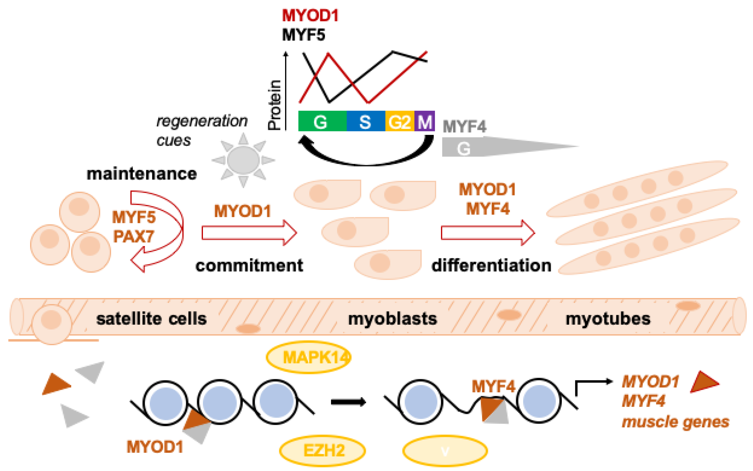
Figure 1. Normal skeletal muscle regeneration. Skeletal muscle is composed of bundles of terminally differentiated myofibers containing multiple post-mitotic nuclei. Muscle-resident stem cells, called satellite cells, are positioned beneath the basal lamina of mature myofibers. MYF5 is the only active MRF expressed alongside PAX7 in quiescent satellite cells in adult skeletal muscle. Upon muscle injury, MRFs are deployed to activate satellite cells, govern myoblast proliferation/fusion and, ultimately, withdrawal from the cell cycle and terminal differentiation. The transcriptional activity and protein availability of MRFs during myoblast proliferation/differentiation is tightly regulated. Regeneration cues rapidly induce MYOD1 expression. Activated MYOD1-/PAX7-/MYF5-expressing satellite cells (myogenic precursors or myoblasts) continue to proliferate (with MYOD1 and MYF5 expression depending on cell cycle phase) and down-regulate PAX7 while maintaining MYOD1, before they finally commit to myogenic differentiation via induction of MYF4. Other myoblasts maintain PAX7 but down-regulate MYOD1 and ultimately withdraw from the cell-cycle to regain quiescence and repopulate the satellite cell pool. This step-wise, hierarchical process is governed by complex epigenetic machinery. MRFs act as tissue-restricted transcription factors, which heterodimerize with bHLH proteins and bind to the regulatory regions of muscle-specific genes to activate transcription. MYOD1 binding also correlates with the opening of the chromatin structure at target genes through histone acetylation. Critical regulators of this process include p38α and a number of chromatin modifiers such as EZH2 and others.
Cancers 15 02823 g001
Figure 2. RMS cell-to-cell heterogeneity recapitulating distinct states of muscle development. The PAX::FOXO1 fusion-positive (FP) and PAX::FOXO1 fusion-negative (FN) RMS cell pool harbors cells stalled at distinct developmental states: a transcriptionally immature stem cell-like/mesoderm/mesenchymal state (expressing PAX3, PAX7 CD44), a myoblast state (expressing MYF5) containing highly proliferative/cycling cells, and a more differentiated myocyte state (expressing MYF4/MYH3/MYH8) associated with better patient outcomes. Conventional chemotherapy depletes the myoblast state and enriches cells in the mesenchymal state, which characterizes a tumor-propagating subpopulation capable of re-creating original tumor heterogeneity. The tumor cell pool in alveolar RMS is more skewed towards later stages of myogenesis prominent compared to embryonal RMS. FP RMS also displays more prominent intrinsic plasticity, which may be the mechanism at the root of its aggressive behavior.
Cancers 15 02823 g002
Figure 3. Core regulatory circuitries in RMS. Cell type-specific transcriptional programs are tightly controlled by master TFs (MTFs). CRCs are interconnected transcriptional loops in which individual MTFs control their own expression as well as the expression of the other involved TFs and exert feed-forward transcriptional control. These transcriptional networks control key transcriptional programs, which drive cell fate specification and differentiation. At the chromatin level, MTFs are accumulated at super-enhancers (SEs) and function under the control of SEs. CRCs have been identified for a number of different tissues, including normal skeletal muscle and RMS. Members of the MRF and MEF2 families are at the core of the normal muscle CRC, and MRFs appear to be the most common elements shared by CRCs in RMS. Generally speaking, the normal muscle CRC is active in RMS and complemented with additional modules: (A) The P3F+ RMS module relies on MYCN and P3F; the latter has pioneer factor activity and shapes the SE landscape in PF+ RMS cells. (B) The PF− RMS module involves PAX7 and AP1 family TFs.
Cancers 15 02823 g003
Figure 4. Epigenetic enzymes involved in aberrant myogenic differentiation in RMS. In the nucleus, human chromosomal DNA is organized in chromatin fiber loops, which are coiled around nucleosomes. The latter contain histones whose tails are subjected to post-translational modifications, including acetylation (Ac) and methylation (Me). This schematic representation summarizes the epigenetic enzymes, which have been reported to be responsible for histone modifications identified in RMS (green arrows indicate stimulation; red signs indicate blockade).
Cancers 15 02823 g004
Figure 5. Inhibition of myogenic differentiation by SNAI2. In proliferating myoblasts and PF− RMS cells, SNAI2 binding at super-enhancers dampens MYOD1 activity at myogenic genes, supporting proliferation at the expense of differentiation (left panel). Upon receipt of differentiation cues, SNAI2 is downregulated in differentiating myoblasts, thus permitting MYOD1 transcriptional activity at myogenic differentiation genes (MYF4, MEF2A, TNNT2, TNNI1) and inducing muscle differentiation (right panel). In PF− RMS, the myogenic differentiation process can be achieved through SNAI2 knockdown with short hairpin RNA (shRNA) alone or through treatment with the MEK inhibitor (MEKi) trametinib (right panel).
Cancers 15 02823 g005
Disclaimer/Publisher’s Note: The statements, opinions and data contained in all publications are solely those of the individual author(s) and contributor(s) and not of MDPI and/or the editor(s). MDPI and/or the editor(s) disclaim responsibility for any injury to people or property resulting from any ideas, methods, instructions or products referred to in the content.