Review

The Sweet Side of HIPK2

1
Unit of Cellular Networks, Department of Research and Advanced Technologies, IRCCS Regina Elena National Cancer Institute, 00144 Rome, Italy
2
Department of Surgery, Sapienza University, 00185 Rome, Italy
3
Centralized Procedures Office, Italian Medicines Agency (AIFA), 00187 Rome, Italy
4
Laboratory Affiliated to Pasteur Institute Italy Foundation Cenci Bolognetti, Department of Experimental Medicine, Sapienza University of Rome, 00161 Rome, Italy
5
Department of Neurosciences, Imaging and Clinical Sciences, University “G. D’Annunzio”, 66013 Chieti, Italy
*
Author to whom correspondence should be addressed.
Cancers 2023, 15(10), 2678; https://doi.org/10.3390/cancers15102678
Submission received: 27 March 2023 / Revised: 4 May 2023 / Accepted: 8 May 2023 / Published: 9 May 2023
(This article belongs to the Special Issue Apoptosis in Cancer 2.0)

Abstract

:

Simple Summary

Hyperglycemia is a hallmark of diabetes mellitus and contributes to several complications of this disease, such as diabetic retinopathy, diabetic nephropathy, and diabetic foot ulcer. Epidemiological evidence showed that patients suffering from diabetes are at a significantly higher risk for the development of cancer; in addition, the high glucose condition can reduce the efficacy of anticancer therapies. Therefore, uncovering novel molecular mechanisms deregulated by high glucose could give insights into the progression of high glucose-related pathologies other than the development of more efficient anticancer therapies. In this regard, homeodomain-interacting protein kinase 2 (HIPK2) has recently been disclosed in both high glucose-induced cancer resistance to chemotherapies and in diabetic complications, which will be summarized in the present review.

Abstract

HIPK2 is an evolutionary conserved protein kinase which modulates many molecular pathways involved in cellular functions such as apoptosis, DNA damage response, protein stability, and protein transcription. HIPK2 plays a key role in the cancer cell response to cytotoxic drugs as its deregulation impairs drug-induced cancer cell death. HIPK2 has also been involved in regulating fibrosis, angiogenesis, and neurological diseases. Recently, hyperglycemia was found to positively and/or negatively regulate HIPK2 activity, affecting not only cancer cell response to chemotherapy but also the progression of some diabetes complications. The present review will discuss how HIPK2 may be influenced by the high glucose (HG) metabolic condition and the consequences of such regulation in medical conditions.

1. Introduction

Hyperglycemia is an excess of glucose concentration in the blood which occurs when the pancreas produces too little insulin or when the body cannot use the insulin properly. The first condition is referred to as type 1 diabetes (T1D), while the second condition is referred to as type 2 diabetes (T2D) [1]. Hyperglycemia is a hallmark of diabetes mellitus and contributes to the development of several complications of this disease, such as diabetic retinopathy (DR), diabetic nephropathy (DN), and impairment of the angiogenesis/wound healing process leading to diabetic foot ulcer [2,3,4]. Medical conditions such as obesity, pancreatitis, chronic stress, and cancer can also be accompanied by hyperglycemia [5,6,7,8]. In addition, hyperglycemia can predispose individuals to cancer development and progression, as evidenced by epidemiologic evidence showing that patients affected by diabetes mellitus are at a higher risk of developing many types of cancers; in addition, the high glucose (HG) condition may promote cancer cell resistance to anticancer chemotherapies [7,8,9]. The link between obesity, T2D, and cancer is quite complex. It involves the production of pro-inflammatory cytokines leading to insulin resistance and to a chronic inflammation status which favor DNA damage and, therefore, cancer development [10]. Hyperglycemia can also reduce dendritic cell (DC) differentiation with impairment of the immune response, reducing the pressure of the immune system against carcinogenesis and infection [11]. The HG condition can induce chemoresistance through the modulation of several molecular pathways. In this regard, a link between HG and homeodomain-interacting protein kinase 2 (HIPK2) has recently been disclosed in both cancer resistance to chemotherapies and in diabetic complications, which will be summarized in the present review.

2. HIPK2 Function and Dysfunction

HIPK2 is a highly conserved nuclear serine/threonine kinase member of the corepressors for homeodomain transcription factors—HIPK—family (numbered 1–4) [12], related to the dual-specificity tyrosine phosphorylation regulated kinases (DYRK) [13]. HIPK1, 2, and 3 share 90% of their homology, suggesting, at least in part, functional redundancy [14]; HIPK4 is, until now, the least characterized member of the family [15]. HIPK2 is the best characterized member of the family: its structure comprises an N-terminal region containing a sumoylation site and a kinase domain, a homeobox-interacting domain and a C-terminal region which includes a speckle-retention signal, a putative autoinhibitory domain, and an ubiquitylation site [16]. HIPK2 acts as transcriptional regulator of critical signal transduction pathways, including Wnt/β-catenin, transforming growth factor (TGF)-β, MAPK, and Notch [17], and can negatively or positively regulate gene transcription [18]. Finally, HIPK2 may also have a role in chromatin compaction [19]. As a consequence of the interplay with multiple molecular pathways, HIPK2 regulates basic cellular functions, such as cell proliferation, apoptosis, DNA damage response, cytokinesis, protein stability, and transcription [18,20,21,22,23]. These basic functions of HIPK2 may explain its key role in several biological processes, including development, fibrosis, angiogenesis, and neurodegeneration [24,25,26,27,28], as well as its function as a “bona fide” tumor suppressor [16].
In the cancer field, HIPK2 plays a key role in inducing cancer cell apoptosis by modulating several different molecules. HIPK2 is activated in response to chemotherapeutic drugs (including cisplatin, doxorubicin, oxaliplatin, roscovitin, 5-FU, temozolomide) or by ultraviolet (UV) and ionizing radiations (IR). Once activated, HIPK2 phosphorylates the tumor suppressor p53 at serine 46 (Ser46) [20,29], the anti-apoptotic co-repressor CtBP [30], the p53 inhibitor MDM2 [31], or the pro-survival dominant negative isoform of the p53 family member p63 (ΔNp63α) [32], leading to cancer cell apoptosis (Figure 1). Tumor suppressor p53 is the most important target of HIPK2; thus, the inhibition of HIPK2 (e.g., by small interfering (si) RNA) impairs p53 function [33] and cancer cell response to anticancer therapies [34].
HIPK2 expression, stability, subcellular localization (nuclear and/or cytoplasmic), and activity are tightly regulated by post-translational modifications such as polyubiquitination, SUMOylation, phosphorylation, and acetylation [35,36,37]. The HIPK2 post-translational modifications, in response to different types and intensity of stress, may modify its function. For instance, ROS-induced HIPK2 acetylation has been shown to control the survival threshold to oxidative stress; thus, an acetylation-mimicking HIPK2 variant can promote cell survival even under conditions of high oxidative stress [38]. In addition, it has been shown using an in vivo Drosophila model that, in response to a high sugar diet, O-GlcNAc transferase (OGT) induces Hipk O-GlcNAcylation, which prevents Hipk from proteasomal degradation leading to Hipk-induced tissue growth abnormalities [39]. This is in line with a previous study showing that overexpression of Hipk in Drosophila promotes tissue growth abnormalities and several tumor-like features [40]. The authors showed that the regulation of Hipk protein stability by OGT is conserved in human HIPK2, in an in vitro cell system, underscoring how HIPK2 stability and function might be affected by metabolic cues through the nutrient sensor OGT [39]. Intriguingly, it has been shown that the high protein levels of HIPK2 in a mouse model of diabetes [41] depend on the downregulation of E3 ubiquitin ligase Siah-1 (seven in absentia homolog-1), which is known to induce HIPK2 protein degradation [42,43]. Hence, the nutritional regulation of HIPK2 might be achieved by multiple strategies involving direct O-GlcNAcylation and impairment of proteasomal degradation. Cancer cells often sustain high rates of glucose uptake aided by metabolic reprogramming [44]. Thus, an ample supply of glucose not only facilitates aerobic glycolysis within tumors but also induces hyper–O-GlcNAcylation, as seen in several cancers, including breast, prostate, colon, lung, liver, pancreatic, and leukemias [45]. Since elevated levels of HIPK2 have been found to be associated with malignancy of pilocytic astrocytomas [46] and cervical carcinogenesis [47], it is tempting to speculate that the dynamic control of HIPK2 abundance by O-GlcNAc modification can be exploited in the treatment of HIPK2-related disorders with the use of dietary control or metabolic drugs targeting the hexosamine biosynthetic pathway (HBP)–OGT axis. In this regard, it is worth further studying how the HIPK2 post-translational modifications by, for instance, environmental cues such as nutrients may differentially affect its function in normal and in tumoral growth.
Until now, only a few mechanisms have been discovered that lead to HIPK2 inactivation. One mechanism of HIPK2 inactivation is its cytoplasmic localization that occurs, for instance, following integrin alpha(6)beta(4) overexpression [48] or by Src-induced HIPK2 phosphorylation [49]. Altered integrin alpha(6)beta(4) expression is found in many epithelial tumors and is involved in tumor progression [50]. In vitro studies have shown that HIPK2 overexpression represses integrin alpha(6)beta(4) transcription that is conversely upregulated in colon cancer cells depleted of HIPK2 function by small interference RNA (siRNA) [48]. Immunohistochemistry analysis of HIPK2 nuclear and cytoplasmic compartmentalization performed in 67 stage I and II wtp53-carrying breast cancer patients showed that an high integrin alpha(6)beta(4) immunoreaction is correlated with HIPK2 cytoplasmic relocalization; in contrast, when tumors are integrin alpha(6)beta(4) negative, HIPK2 is localized in the nucleus [48]. These findings link HIPK2 activity to the suppression of the survival function induced by integrin alpha(6)beta(4) in agreement with other studies showing that depletion of HIPK2 function enhances in vivo tumorigenicity [51,52]. Regarding HIPK2 regulation by the proto-oncogenic non-receptor kinase Tyr kinase Src, it has been shown that it binds and phosphorylates HIPK2 at multiple Tyr residues. This interaction interferes with Siah-1-mediated HIPK2 degradation [42,43], redistributing HIPK2 from the nucleus to the cytoplasm. In this way, Src suppresses HIPK2-induced p53 phosphorylation and apoptotic activity [49]. Finally, in an in vitro study, the leukemogenic fusion protein CBF-β-SMMHC, which is associated with the development of acute myeloid leukemia (AML), was found to delocalize HIPK2 into filamentous structures in the cytoplasm, probably interfering with the tumor-suppressor function of HIPK2 [53].
Heat shock (HS) has been shown to modulate HIPK2 stability and nuclear/cytoplasmic localization: a sub-lethal heat shock induces HIPK2 cytoplasmic localization with impairment of p53 apoptotic activity, while a lethal heat shock induces HIPK2 nuclear localization with consequent p53 apoptotic activation [54], underscoring the key role of HIPK2 in stress-mediated cell death. HIPK2 is rarely mutated: the only HIPK2 mutations were reported by a study about human acute myeloid leukemias (AML) in which HIPK2 shows an aberrant nuclear distribution. Again, the impairment of HIPK2 activity negatively impacts on p53 apoptotic activity [55,56]. Loss of heterozygosity (LOH) at the HIPK2 locus 7q32.34 has been reported in human lung fibroblasts of patients affected by idiopathic pulmonary fibrosis (IPF) in which HIPK2 was found to be downregulated [57]. Another way to inhibit HIPK2 function is by reducing the binding with p53 and, therefore, its apoptotic activity, as happens, for instance, in human papillomavirus (HPV) infection [58]. HPV has been shown to play a causal role as a co-factor in the development of cutaneous squamous cell carcinoma (SCC) [59]. It was shown that upon DNA damage with ultraviolet radiation (UV), which is the main risk factor for SCC, HPV23 E6 interacts with HIPK2 and impairs its localization in the PML nuclear bodies (NB) [58] where HIPK2 associates with p53, phosphorylates it in Ser46, and induces its apoptotic function [20,29]. As a consequence, HPV23 E6 prevents HIPK2-induced p53 phosphorylation and apoptotic activity, suggesting that cutaneous HPV infected keratocytes may overcome UV-induced apoptosis and be causal in the early onset of skin carcinogenesis [58].
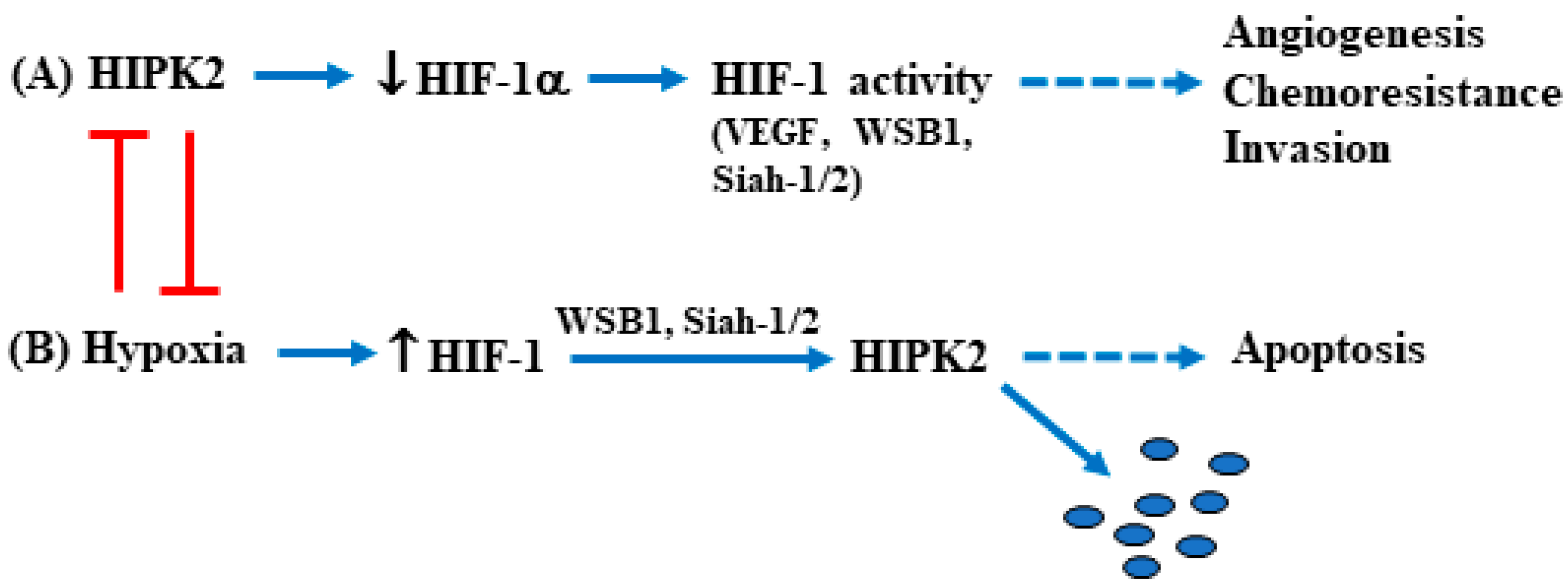
A more common way to inhibit HIPK2 function is by inducing its proteasomal degradation, and, in this regard, a few ubiquitin ligases have been discovered. In response to mild DNA damage, it has been shown that p53-induced MDM2 can trigger HIPK2 protein degradation which, in turn, impairs p53 apoptotic activity. As a proof of principle, an HIPK2 mutant vector, resistant to MDM2 inhibition, is able to induce p53 apoptotic function [14]. Interestingly, in response to severe DNA damage, HIPK2 activation leads to MDM2 phosphorylation and protein degradation [60]. This autoregulatory loop between HIPK2 and MDM2 in response to mild/severe DNA damage consequently also affects p53 apoptotic/survival activity [31]. In normal conditions, HIPK2 is degraded by binding to the RING family ligase seven in absentia homolog-1 (Siah1). The DNA damage activates HIPK2 by releasing the binding with Siah1 which is phosphorylated by the damage-induced ATM/ATR [42,43]. TNF receptor-associated factor 2 (TRAF2) has also been shown to promote HIPK2 proteasomal degradation [61]. The promyelocytic leukemia (PML) gene, which encodes a putative tumor suppressor gene, interacts with multiple factors and their coactivators involved in the control of apoptosis [62]. Among them, it has been shown that PML activates transcription by protecting HIPK2 from SCFFbx3-induced proteasomal degradation. In contrast, the leukemia-associated fusion protein PML-RARα induces the degradation of HIPK2 by inhibiting PML stabilization of HIPK2, underscoring a novel mechanism in the regulation of leukemogenesis [63]. Among other E3 ubiquitin ligases involved in HIPK2 degradation is WD40-repeat/SOCS box protein WSB-1 [64], a target of the hypoxia-inducible factor-1 (HIF-1) [65]; however, the ubiquitination and degradation of HIPK2 by WSB-1 is inhibited by genotoxic stress [64]. A hypoxic condition allows a markedly increased HIPK2 interaction with the RING family ligase Siah2, resulting in HIPK2 proteasomal degradation [66]. High-mobility group box 1 (HMGB1) is a conserved nuclear chromatin-binding protein that may have important tumor-promoting activity in several cancers, including hepatocellular carcinoma (HCC) [67]. It has been shown in vivo that HMGB1 promotes disease progression of human HCC [68]. At the molecular level, targeting HMGB1 inhibits Siah-2-induced HIPK2 ubiquitination and upregulates HIPK2 protein expression, suggesting that targeting HMGB1 could suppress HCC progression via HIPK2 activation [68]. Interestingly, HIPK2 overexpression binds to the HIF-1α promoter in a multiprotein co-repressor complex with histone deacetylase 1 (HDAC1), downmodulating both HIF-1α reporter activity and mRNA levels [51]. Hypoxia-inducible factor-1 (HIF-1) is a heterodimeric transcription factor that consists of two subunits, namely HIF-1α and HIF-1β. HIF-1β is constitutively expressed in cells, while HIF-1α stability is stimulated by hypoxia, growth factors, and several oncogenes [69]. Once activated, HIF-1 induces the transcription of several genes involved in many aspects of cancer progression, including angiogenesis, metabolic adaptation, apoptosis resistance, invasion, and metastasis [70]. HIPK2 overexpression reduces the basal levels of HIF-1α with a mechanism that does not involve hypoxia, inhibiting the HIF-1 transcriptional activity and, therefore, counteracting theHIF-1-induced angiogenesis and tumor progression, in vitro and in vivo [51,71,72,73]. In this way, the autoregulatory loop between HIF-1 and HIPK2 modifies the downstream pathways regulated by both proteins, and its balance may dictate the cancer survival/cell death outcome in response to therapies [71,72] (Figure 2).
Recently, it has been shown that microRNAs (miRNAs) may regulate HIPK2 both at the mRNA and the protein level, having an effect on cancer progression and angiogenesis as well as on other diseases [74]. For instance, circulating exomiR-1229 has been shown to target HIPK2 mRNA and downregulate it in colorectal cancer (CRC) tissues compared to the adjacent normal tissues [75]. A similar HIPK2 mRNA downregulation was previously found in CRC tissues compared to tissues of benign familial polyposis adenomas (FAP) [52]. ExomiR-1260b, derived from non-small cell lung cancer (NSCLC) has been shown to target HIPK2 in human umbilical endothelial cells (HUVECs) and promote the angiogenesis, migration, invasion, and chemoresistance of NSCLC cells [76]. The mechanisms of HIPK2 inhibition are summarized in Table 1.

2.1. HIPK2 Regulation by Hyperglycemia in Tumors

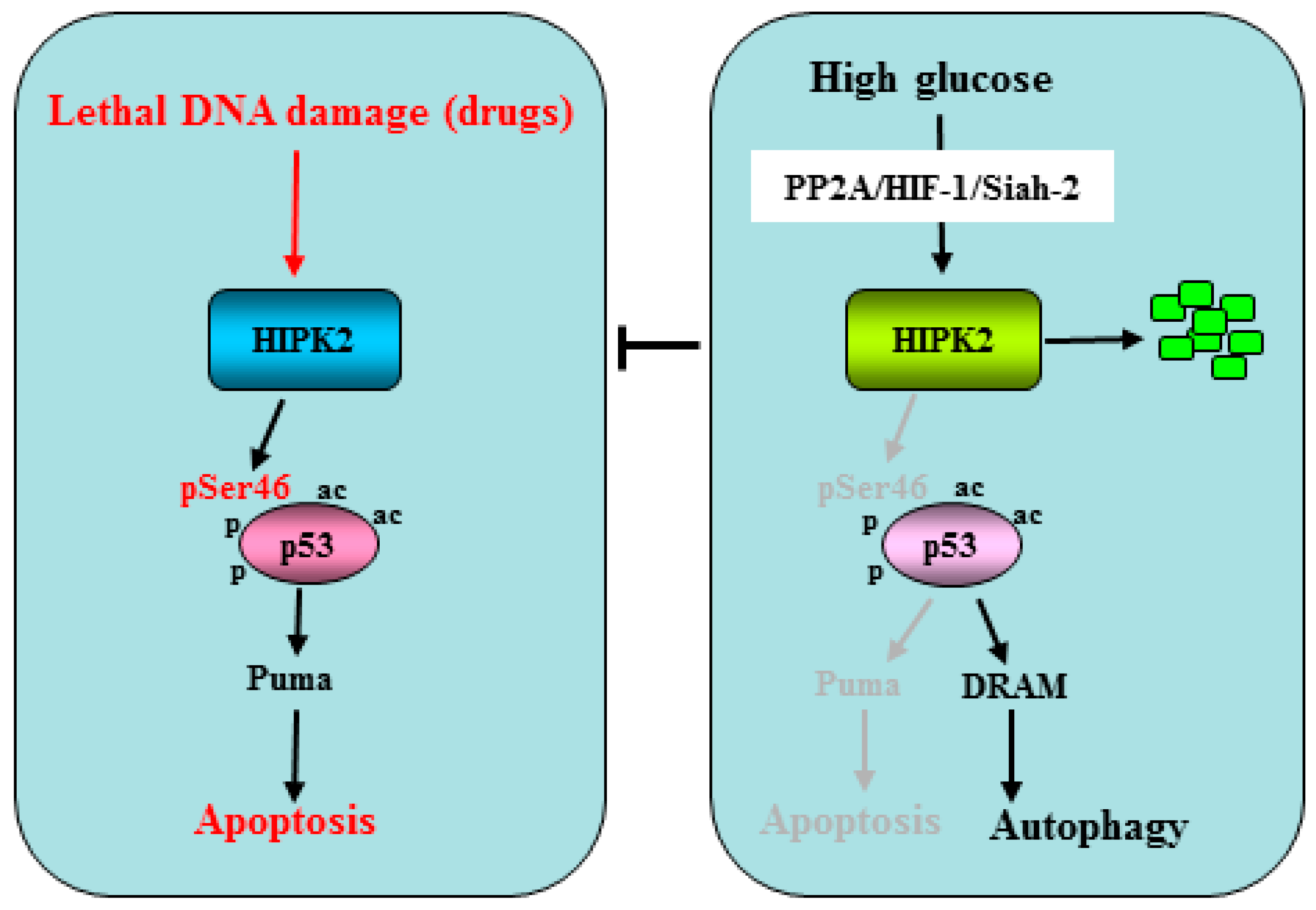
Taking advantage of the findings showing that hyperglycemia increases HIF-1α gene transcription [77] and induces the HIF-1 transcriptional activity irrespective of the oxygen levels [78], in an in vitro study, it was shown that high glucose (HG) condition promotes the degradation of HIPK2. Mechanistically, the HG-induced HIPK2 degradation was partly induced by the activity of the ubiquitin ligase Siah2, downstream of a protein cascade including protein phosphatase A2 (PP2A) and HIF-1α [79]. As a proof of principle, the HIPK2 degradation was confirmed by culturing the cancer cells in the presence of hyperglycemic sera derived from patients with T2D compared to normoglycemic sera [79]. HIPK2 protein stability regulated by the metabolic HG condition may impact on the cellular outcome: either survival or apoptosis. This can happen because the HG-induced HIPK2 inhibition consequently inhibits the p53Ser46 phosphorylation and the p53 apoptotic activity. Interestingly, the HG-mediated inhibition of the p53 apoptotic activity is counteracted by the phosphatase inhibitor of the PP2A named Calyculin A (CL-A) [80,81]. In addition, hyperglycemia increases autophagy in drug-treated cancer cells, in part depending on the p53 transcriptional switch from apoptotic target genes such as PUMA to autophagic genes such as DRAM [82] (Figure 3). As a proof of principle of the p53 transcriptional activity switch, xenograft tumors transplanted in normoglycemic mice displayed growth delay in response to chemotherapy, while those transplanted in diabetic mice did not, and such difference correlated with the PUMA to DRAM gene expression switch [82,83]. The different transcriptional activity of p53 could depend on the impairment of the HIPK2 kinase activity by the HG condition which, consequently, differentially affects the p53 transcriptional activity. Thus, the low/high genotoxic damage can differentially induce the p53 post-transcriptional modifications by modulating the upstream kinases, influencing the transcription of target genes: low damage induces mainly the cell cycle arrest-related or autophagic target genes, while high damage induces the apoptotic target genes [84,85]. The above findings highlight how metabolic conditions such as hyperglycemia might reduce the response to anticancer therapies by impairing the apoptotic activity of the HIPK2/p53 axis, in agreement with preclinical studies showing a reduction of chemotherapy cytotoxicity by hyperglycemia [86]. Interestingly, HG-induced HIPK2 degradation is counteracted in vitro by lowering the cell culture glucose amount, suggesting that this is a reversible effect [79]. Thus, an interesting strategy to pursue to keep the HIPK2/p53 axis functioning in cancer patients affected by diabetes or by other pathologies with hyperglycemia could be to reduce the glycemic load, although this hypothesis needs to be supported by further studies for clinical application.
The HG condition has been shown to trigger the antioxidant response, which correlates with the reduced Adriamycin-induced DNA damage and cell death, thus contributing to an increase in chemoresistance in cancer cells [87]. The master regulator of the antioxidant pathway is the transcription factor nuclear factor erythroid 2-related factor 2 (NRF2) [88,89] whose pharmacologic or genetic inhibition may re-establish cancer cell sensitivity to the chemotherapeutic drug cytotoxicity impaired by the HG [87,90]. The HIPK2/NRF2 relationship is quite intricate and still not completely unveiled. NRF2 may modulate HIPK2 both directly at the mRNA level and indirectly at the protein level. NRF2 has been shown to induce the HIPK2 mRNA transcription and engage with the HIPK2 protein, a pro-survival crosstalk to the detriment of the HIPK2 apoptotic activity [91]. Mechanistically, in this condition, HIPK2 has been shown to induce some antioxidant target genes which are in common with NRF2 (i.e., NQO1, HO-1) [91]. Moreover, NRF2 signaling has been shown to sustain the HIF-1 response by activating HIF-1α [92] and, consequently, although indirectly, to also modulate the HIPK2 function. Therefore, it is tempting to speculate that, in response to metabolic conditions that modify the redox state (e.g., hyperglycemia), NRF2 activation may calibrate the HIPK2 activation through, for instance, post-translational modifications [91] which impair HIPK2 apoptotic activity, favoring instead the transcription of genes promoting chemoresistance and tumor progression. This hypothesis, however, needs to be supported by further studies. HG may also promote tumor invasion [93] and epithelial–mesenchymal transition (EMT) in colon cancer cells in addition to promoting cancer cell migration and invasion [94]. Since HIPK2 has been shown to repress the vimentin gene promoter, one of the most important markers of EMT, and to restrain the cancer cell invasion [95], it is tempting to speculate that HIPK2 inhibition by the HG condition might also contribute to promoting tumor invasion.
Interestingly, it has been shown that cancer drug resistance, in part due to the HG-induced upregulation of hypoxic/glycolytic genes, such as HIF-1, GLUT-1, and HK-2, and to the inhibition of the JNK apoptotic pathway likely through NF-κB activation, can be counteracted by zinc(II) supplementation, which restores the drug cytotoxicity [77]. Zinc supplementation has been shown to play a role in cancer prevention [96], attracting studies for improving the effect of anticancer therapies. Thus, a genome-wide analysis in colon cancer cells has shown a reversal of the hypoxia-induced gene expression by zinc supplementation [97]. In addition, in vitro and in vivo murine studies have shown that zinc supplementation downregulates the HIF-1 activity and restores the cancer cell response to therapies through the re-establishment of the HIPK2/p53 apoptotic axis [71,72,98]. Zinc supplementation has been shown to ameliorate the severity of diabetic hyperglycemia and of the associated metabolic abnormalities, including hypoinsulinemia, insulin resistance, and the altered pancreatic morphology, in a murine model of diabetes [99]. Moreover, a systematic review and meta-analysis of randomized controlled trials reported that zinc supplementation significantly reduces several key glycemic indicators, particularly fasting glucose (FG), in subjects with diabetes [100]. Thus, zinc supplementation may offer a significant potential for clinical application in managing diabetic hyperglycemia and the related metabolic complications which promote cancer chemoresistance, in part by counteracting the HG-induced HIPK2/p53 inhibition.

2.2. HIPK2 and Diabetes Complications

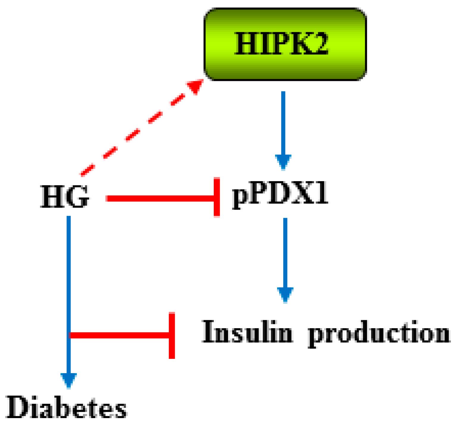
HIPK2 has been suggested to have a role in pancreatic development and mature β-cell function. Thus, it has been shown that HIPK2 is expressed in the pancreatic epithelium at an earlier stage of development, while it is expressed in the pancreatic endocrine cells at the later stage of development [101]. Mechanistically, the authors found that HIPK2 phosphorylates and induces the transcriptional activity of the insulin promoter factor (IPF)-1/pancreatic duodenal homeobox (PDX)-1, a homeodomain transcription factor which plays a crucial role in both pancreas development and maintenance of cell function, and that HIPK2 co-localizes with PDX1 in both the developing and adult pancreas [101]. A different study, using mass spectrometry and a phosphoserine-specific antibody, showed that HIPK2 regulates PDX1 function in living β-cells. Mechanistically, HIPK2 phosphorylates PDX1 at Ser-269, affecting the PDX1subnuclear distribution rather than the transcriptional activity [102]. Accordingly, a previous study has shown that HG reduces PDX phosphorylation in Ser-269 in primary rat islets [103]. The important role of IPF/PDX1 in diabetes has been shown in a mice model in which targeted disruption of the IPF1/PDX1 gene leads to overt diabetes with decreased insulin expression and secretion [104]. In humans, mutations in the IPF1 gene have been linked to diabetes [105]. The effect of high glucose in inhibiting PDX phosphorylation is also in agreement with a study showing that high glucose induces HIPK2 degradation [79]. The findings of HIPK2-mediated IPF1/PDX1 regulation highlight the key role of HIPK2 in glucose homeostasis in patients with diabetes [101] (Figure 4).
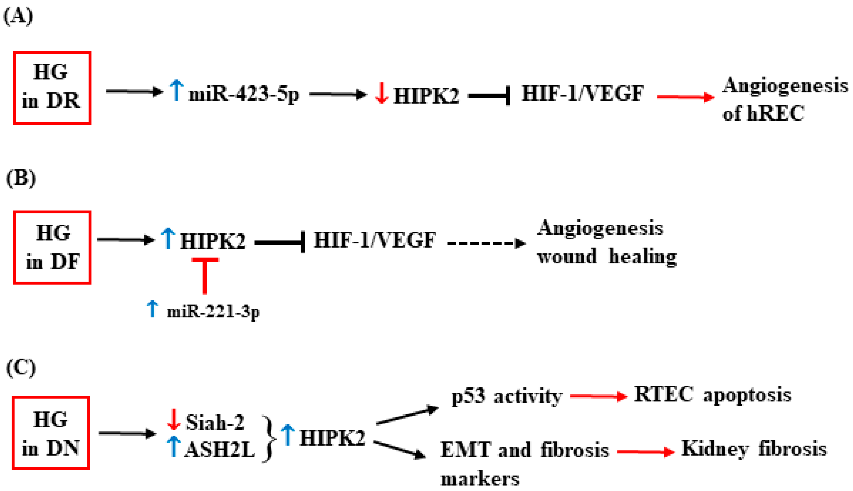
One of the complications of diabetes is diabetic retinopathy (DR), which is also the primary cause of blindness in the world [106]. An interesting link between HIPK2 and DR has been depicted due to HIPK2 regulation by microRNA (miRNA), which is usually involved in DR vascular complications [107]. Taking advantage of a previous study in which the authors found that miR-423-5p levels are elevated in the plasma of DR patients [108], the authors, following this, evaluated the role of HG in this regulation. They found that the HG condition upregulates miR-423-5p, leading to the enhanced proliferation and angiogenesis of the retinal endothelial cells (hRCEs) as well as to the increased angiogenesis in the retina of a mouse model of streptozotocin (STZ)-induced diabetes [109]. miR-423-5p targets and downregulates HIPK2, with a consequent increase in the HIF-1α and VEGF protein levels, as corroborated by the knockdown of miR-423-5p that recovers the HIPK2 levels and suppresses the HG-induced angiogenesis [109]. The results indicate that the miR-423-5p-induced downregulation of HIPK2 contributes to the progression of DR through the activation of the HIF-1/VEGF angiogenic pathway (Figure 5A).
Hyperglycemia can cause neurological disorders and impaired angiogenesis, leading to reduced wound healing [110] and resulting in diabetic foot ulcers [111,112]. miR-221-3p has been found to alleviate diabetic ulcers [113] and to counteract the inhibition of angiogenesis by HG [96]. In that study, the authors found an increased HIPK2 expression in vitro in the HG-cultured HUVECs and in vivo in skin tissues of diabetic mice compared to the skin of non-diabetic mice [114]. Thus, HG-increased HIPK2 expression correlates with reduced angiogenesis and impaired wound healing, aggravating diabetic foot ulcers, a condition that can be ameliorated by treatment with miR-221-3p. Mechanistically, it was shown that HIPK2 is targeted and inhibited by miR-221-3p overexpression, which restores angiogenesis inhibited by the HG condition, therefore improving wound healing [114] (Figure 5B).
The HG-increased expression of HIPK2 was also observed in glomerular mesangial cells (GMCs) of a mouse model of diabetic nephropathy (DN) [41]. DN is a leading cause of end-stage renal disease (ESRD) and a threat to patients with diabetes [115]. HIPK2 has been found to play a role in DN and, in particular, in inducing kidney fibrosis [116]. In a mouse model of human immunodeficiency virus (HIV)-associated nephropathy (HIVAN), the authors found that HIV increases HIPK2 stability by inhibiting Siah-1-mediated HIPK2 proteasomal degradation. In that model, the HIPK2 activity leads to the apoptosis of the renal tubular epithelial cells (RTECs) by p53 apoptotic activation. In addition, HIPK2 activation leads to kidney fibrosis by activating, in kidney epithelial cells, the TGF-β–Smad3 and Wnt–Notch pathways and by inducing the expression of EMT markers, including αSMA (smooth muscle actin), collagen I, and fibronectin [116]. The authors confirmed the role of HIPK2 in inducing kidney fibrosis and also in focal segmental glomerulosclerosis (FSGS), DN, and IgA nephropathy (IgAN) [116] (Figure 5C). Therefore, targeting HIPK2 could be a valuable strategy to inhibit renal fibrosis during DN. As a proof of principle, it has been shown that the anti-helminthic drug phosphate niclosamide (P-NICLO) [117] can inhibit TGF-β-induced HIPK2 expression, resulting in the inhibition of the downstream pathways that are responsible for fibrosis, such as Smad, Notch, NF-kB, and Wnt/β-catenin [118].
The dysfunction of glomerular mesangial cells (GMCs) is a pathological feature of DN and the main contributing factor to glomerulosclerosis. In a previous study, the authors found that HG activates the transcription factor ASH2L, which enhances HIPK2 expression, contributing to fibrosis and inflammation in GMC [119] (Figure 5C). Thus, the inhibition of ASH2L suppresses HIPK2 expression in HG-treated mesangial cells, with a consequent decrease in fibrosis (e.g., FN, collagen 3, collagen 1, MMP9, α-SMA, and CTGF) and inflammation markers (e.g., IL-6 and IL-1β); in agreement, IL-6 and α-SMA were found to be downregulated by HIPK2 inhibition with small interfering RNA (siHipk2) [119]. Although the upregulation of HIPK2 in the kidneys of STZ-induced mice and leptin receptor-deficient db/db mice has been observed [119], further studies are needed to unveil how HIPK2 affects DN.

3. Conclusions

HIPK2 is a “bona fide” oncosuppressor molecule that exerts its anticancer activity mainly by triggering p53 apoptotic activity and by inhibiting hypoxia-induced angiogenesis. Recent developments have suggested that HIPK2 deregulation by hyperglycemia may play a role not only in the chemoresistance of various cancer cell types but also in the progression of some diabetes complications, likely involving the same HIPK2-triggered molecular pathways. In this review, we highlighted that increased antiapoptotic and pro-angiogenic activities are crucial mechanisms underlying chemoresistance mediated by HG-induced HIPK2 degradation largely mediated by HIF-1-induced mechanisms. In vitro research has revealed that lowering the glucose load or inhibiting the HIF-1 pathway may be a promising strategy to overcome the HG-induced chemoresistance by restoring the HIPK2/p53 axis, improving the efficacies of chemotherapeutic regimes. Both the suppression of HIF-1 activity by zinc(II) supplementation and the targeted knockdown of HIF-1α expression by siRNA significantly enhanced chemosensitivity in cancer cells and suppressed chemoresistant xenograft tumor growth in vivo. Advances have been made in understanding the mechanisms underlying chemoresistance in response to HG-induced HIPK2 inhibition, and further development of targeted therapies provides encouraging prospects for clinical application to optimize patient prognosis in different cancers. It is noteworthy that targeting HIF-1 activity may be a safer approach rather than re-activating HIPK2, whose apoptotic activity could have side effects in normal cells. Furthermore, whether therapy resistance dependent on HG-induced inhibition of HIPK2 activity is universal or specific for certain tumor types remains largely unexplored. Intriguingly, deregulated angiogenesis in human retinal endothelial cells (hREC), by means of activation of the HIF-1/VEGF pathway upon miR-423-5p-induced HIPK2 downregulation in diabetic patients, contributes to diabetic retinopathy. On the other hand, the upregulation of HIPK2 correlates with reduced angiogenesis and impaired wound healing, aggravating diabetic foot ulcers, a condition that can be ameliorated by treatment with miR-221-3p, which targets and inhibits HIPK2. The HIPK2 upregulation in in vitro and in vivo models of diabetic nephropathy unveiled a role of HIPK2 in kidney fibrosis by apoptosis of the renal tubular epithelial cells (RTECs) induced by p53 activation and by HIPK2-induced EMT markers. These findings, underscoring the involvement of HIPK2, provide new insights into the molecular mechanism of frequent diabetes complications. As a future perspective, the detailed molecular mechanisms of how hyperglycemia (de)regulates HIPK2 activity in diabetic complications warrant further investigation to unveil the cell type and context-dependent function/dysfunction of HIPK2 in this metabolic condition to reduce the clinical complications of the hyperglycemia.

Author Contributions

Conceptualization and writing, G.D.; review and editing, A.G., V.D., G.P., M.C. and G.D. All authors have read and agreed to the published version of the manuscript.

Funding

The research in G.D.’s lab was funded by the Italian Association for Cancer Research (AIRC, number IG11377), by the Italian Ministry of Research (Miur-PRIN, n. 2005059700_003), and by G.D.’s Annunzio University Grants (Fondi Ateneo 2011).

Acknowledgments

The authors wish to thank the people in the lab for helpful discussion.

Conflicts of Interest

The authors declare no conflict of interest. The funders had no role in the design of the study; in the collection, analyses, or interpretation of data; in the writing of the manuscript; or in the decision to publish the results.

References

  1. Schmidt, A.M. Highlighting diabetes mellitus: The epidemic continues. Arterioscler. Thromb. Vasc. Biol. 2018, 38, e1–e8. [Google Scholar] [CrossRef] [PubMed]
  2. Engerman, R.L.; Kern, T.S. Hyperglycemia as a cause of diabetic retinopathy. Metabolism 1986, 35, 20–23. [Google Scholar] [CrossRef] [PubMed]
  3. Wu, J.T. Review of diabetes: Identification of markers for early detection, glycemic control, and monitoring clinical implications. J. Clin. Lab. Anal. 1993, 7, 293–300. [Google Scholar] [CrossRef]
  4. Ansari, P.; Akther, S.; Khan, J.; Islam, S.S.; Masud, S.R.; Rahman, A.; Seidel, V.; Abdel-Wahab, Y.H.A. Hyperglycaemia-linked diabetic foot complications and their management using conventional and alternative therapies. Appl. Sci. 2022, 12, 11777. [Google Scholar] [CrossRef]
  5. Martyn, J.A.J.; Kaneki, M.; Yasuhara, S. Obesity-induced insulin resistance and hyperglycemia: Etiologic factors and molecular mechanisms. Anesthesiology 2008, 109, 137–148. [Google Scholar] [CrossRef] [PubMed]
  6. Mechanick, J.I. Metabolic mechanisms of stress hyperglycemia. J. Parent. Enter. Nutr. 2006, 30, 157–163. [Google Scholar] [CrossRef]
  7. Ryu, T.Y.; Park, J.; Scherer, P.E. Hyperglycemia as a risk factor for cancer progression. Diabetes Metab. J. 2014, 38, 330–336. [Google Scholar] [CrossRef]
  8. Li, W.; Zhang, X.; Sang, H.; Zhou, Y.; Shang, C.; Wang, Y.; Zhu, H. Effects of hyperglycemia on the progression of tumor diseases. J. Exp. Clin. Cancer Res. 2019, 38, 327. [Google Scholar] [CrossRef]
  9. Vishvakarma, N.K.; Kumar, A.; Singh, V.; Singh, S.M. Hyperglicaemia of tumor microenvironment modulates stage-dependent tumor progression and multidrug resistance: Implication of cell survival regulatory molecules and altered glucose transport. Mol. Carcinog. 2013, 52, 932–945. [Google Scholar] [CrossRef]
  10. Rahman, I.; Athar, T.; Islam, M. Type 2 diabetes, obesity, and cancer share some common and critical pathways. Front. Oncol. 2021, 10, 2020. [Google Scholar] [CrossRef]
  11. Gilardini Montani, M.S.; Granato, M.; Cuomo, L.; Valia, S.; Di Renzo, L.; D’Orazi, G.; Faggioni, A.; Cirone, M. High glucose and hyperglycemic sera from type 2 diabetic patients impair DC differentiation by inducing ROS and activating Wnt/beta-catenin and p38 MAPK. Biochem. Biophys. Acta-DIS 2016, 1862, 805–813. [Google Scholar] [CrossRef]
  12. Kim, Y.H.; Choi, C.Y.; Lee, S.J.; Conti, M.A.; Kim, Y. Homeodomain-interacting protein kinases, a novel family of co-repressors for homeodomain transcription factors. J. Biol. Chem. 1998, 273, 25875–25879. [Google Scholar] [CrossRef] [PubMed]
  13. Hofmann, T.G.; Mincheva, A.; Lichter, P.; Droge, W.; Schimtz, M.L. Human homeodomain-interacting protein kinase-2 (HIPK2) is a member of the DYRK family of protein kinases abd maps to chromosome 7q32-q34. Biochimie 2000, 82, 1123–1127. [Google Scholar] [CrossRef] [PubMed]
  14. Rinaldo, C.; Prodosmo, A.; Mancini, F.; Iacovelli, S.; Sacchi, A.; Moretti, F.; Soddu, S. MDM2-regulated degradation of HIPK2 prevents p53Ser46 phosphorylation and DNA damage-induced apoptosis. Mol. Cell 2007, 25, 739–750. [Google Scholar] [CrossRef]
  15. He, Q.; Shi, J.; Sun, H.; An, J.; Huang, Y.; Sheikh, M.S. Characterization of human homeodomain-interacting protein kinase 4 (HIPK4) as a unique member of the HIPK family. Mol. Cell. Pharmacol. 2010, 2, 61–68. [Google Scholar]
  16. Feng, Y.; Zhou, L.; Sun, X.; Li, Q. Homeodomain-interacting protein kinase 2 (HIPK2): A promising target for anti-cancer therapies. Oncotarget 2017, 8, 20452–20461. [Google Scholar] [CrossRef]
  17. Schmitz, M.L.; Rodriguez-Gil, A.; Hornung, J. Integration of stress signals by homeodomain interacting protein kinases. Biol. Chem. 2014, 395, 375–386. [Google Scholar] [CrossRef]
  18. Calzado, M.A.; Renner, F.; Roscic, A.; Schitz, M.L. HIPK2: A versatile switchboard regulating the transcription machinery and cell death. Cell Cycle 2014, 6, 139–143. [Google Scholar] [CrossRef]
  19. Haas, J.; Bloesel, D.; Bacher, S.; Kracht, M.; Schmitz, M.L. Chromatin targeting of HIPK2 leads to acetylation-dependent chromatin decondensation. Front. Cell Dev. Biol. 2020, 8, 852. [Google Scholar] [CrossRef]
  20. D’Orazi, G.; Cecchinelli, B.; Bruno, T.; Manni, I.; Higashimoto, Y.; Saito, S.; Gostissa, M.; Coen, S.; Marchetti, A.; Del Sal, G.; et al. Homeodomain interacting protein kinase-2 phosphorylates p53 at Ser46 and mediates apoptosis. Nat. Cell Biol. 2002, 4, 11–19. [Google Scholar] [CrossRef]
  21. Iacovelli, S.; Ciuffini, L.; Lazzari, C.; Bracaglia, G.; Rinaldo, C.; Prodosmo, A.; Bartolazzi, A.; Sacchi, A.; Soddu, S. HIPK2 is involved in cell proliferation and its suppression promotes growth arrest independently of DNA damage. Cell Prolif. 2009, 42, 373–384. [Google Scholar] [CrossRef] [PubMed]
  22. Hofmann, T.G.; Glas, C.; Bitomsky, N. HIPK2: A tumour suppressor that controls DNA damage-induced cell fate and cytokinesis. BioEssays 2013, 35, 55–64. [Google Scholar] [CrossRef] [PubMed]
  23. Valente, D.; Bossi, G.; Moncada, A.; Tornincasa, M.; Indelicato, S.; Piscuoglio, S.; Karamitopoulou, E.D.; Bartolazzi, A.; Pierantoni, G.M.; Fusco, A.; et al. HIPK2 deficiency causes chromosomal instability by cytokinesis failure and increases tumorigenicity. Oncotarget 2015, 6, 10320–10334. [Google Scholar] [CrossRef]
  24. Blaquiere, J.A.; Verheyen, E.M. Homeodomain-interacting protein kinases: Diverse and complex roles in development and diseases. Curr. Opin. Dev. Biol. 2017, 123, 73–103. [Google Scholar] [CrossRef]
  25. Garufi, A.; Pistritto, G.; D’Orazi, G. HIPK2 as a novel regulator of fibrosis. Cancers 2023, 15, 1059. [Google Scholar] [CrossRef] [PubMed]
  26. Garufi, A.; D’Orazi, V.; Pistritto, G.; D’Orazi, G. HIPK2 in angiogenesis: A promising biomarker in cancer progression and in angiogenic diseases. Cancers 2023, 14, 1566. [Google Scholar] [CrossRef]
  27. Stanga, S.; Lanni, C.; Govono, S.; Uberti, D.; D’Orazi, G.; Racchi, M. Unfolded p53 in the pathogenesis of Alzheimer’s disease: Is HIPK2 the link? Aging 2010, 2, 545–554. [Google Scholar] [CrossRef]
  28. Sardina, F.; Conte, A.; Paladino, S.; Pierantoni, G.M.; Rinaldo, C. HIPK2 in the physiology of nervous system and its implications in neurological disorders. Biochim. Biophys. Acta Mol. Cell Res. 2023, 1870, 119465. [Google Scholar] [CrossRef]
  29. Hofmann, T.G.; Moller, A.; Sirma, H.; Zentgraf, H.; Taya, Y.; Dröge, W.; Will, H.; Schmitz, M.L. Regulation of p53 activity by its interaction with homeodomain-interacting protein kinase-2. Nat. Cell Biol. 2002, 4, 1–10. [Google Scholar] [CrossRef]
  30. Zhang, Q.; Yoshimatsu, Y.; Hildebrand, J.; Frisch, S.M.; Goodman, R.H. Homeodomain interacting protein kinase 2 promotes apoptosis by downregulating the transcriptional corepressor CtBP. Cell 2003, 115, 177–186. [Google Scholar] [CrossRef]
  31. Di Stefano, V.; Blandino, G.; Sacchi, A.; Soddu, S.; D’Orazi, G. HIPK2 neutralizes MDM2 inhibition by rescuing p53 transcriptional activity and apoptotic function. Oncogene 2004, 23, 5185–5192. [Google Scholar] [CrossRef] [PubMed]
  32. Lazzari, C.; Prodosmo, A.; Siepi, F.; Rinaldo, C.; Galli, F.; Gentileschi, M.; Bartolazzi, A.; Costanzo, A.; Sacchi, A.; Guerrini, L.; et al. HIPK2 phosphorylates DNp63α and promotes its degradation in response to DNA damage. Oncogene 2011, 30, 4802–4813. [Google Scholar] [CrossRef] [PubMed]
  33. Puca, R.; Nardinocchi, L.; Gal, H.; Rechavi, G.; Amariglio, N.; Domany, E.; Notterman, D.A.; Scarsella, M.; Leonetti, C.; Sacchi, A.; et al. Reversible dysfunction of wild-type p53 following homeodomain interacting protein kinase-2 knockdown. Cancer Res. 2008, 15, 3707–3714. [Google Scholar] [CrossRef]
  34. Puca, R.; Nardinocchi, L.; Givol, D.; D’Orazi, G. Regulation of p53 activity by HIPK2: Molecular mechanisms and therapeutical implications in human cancer cells. Oncogene 2010, 29, 4378–4387. [Google Scholar] [CrossRef]
  35. de la Vega, L.; Frobius, K.; Moreno, R.; Calzado, M.A.; Geng, H.; Schmitz, M.L. Control of nuclear HIPK2 localization and function by a SUMO interaction motif. Biochim. Biophys. Acta 2011, 1813, 283–297. [Google Scholar] [CrossRef] [PubMed]
  36. Saul, V.V.; Schmitz, M.L. Posttranslational modifications regulate HIPK2, a driver of proliferative diseases. J. Mol. Med. 2013, 91, 1051–1058. [Google Scholar] [CrossRef]
  37. Choi, D.W.; Choi, C.Y. HIPK2 modification code for cell death and survival. Mol. Cell Oncol. 2014, 1, e955999. [Google Scholar] [CrossRef]
  38. de la Vega, L.; Grishina, I.; Moreno, R.; Krüger, M.; Braun, T.; Schmitz, M.L. A redox-regulated SUMO/acetylation switch of HIPK2 controls the survival threshold to oxidative stress. Mol. Cell 2012, 46, 472–483. [Google Scholar] [CrossRef]
  39. Wong, K.K.L.; Liu, T.W.; Parker, J.M.; Sinclair, D.A.R.; Chen, Y.Y.; Khoo, K.H.; Vocadlo, D.J.; Verheyen, E.M. The nutrient sensor OGT regulates Hipk stability and tumorigenic-like activities in Drosophila. Proc. Natl. Acad. Sci. USA 2020, 117, 2004–2013. [Google Scholar] [CrossRef]
  40. Chen, J.; Verheyen, E.M. Homeodomain-interacting protein kinase regulates Yorkie activity to promote tissue growth. Curr. Biol. 2012, 22, 1582–1586. [Google Scholar] [CrossRef]
  41. Oh, H.; Kato, M.; Deshpande, S.; Zhang, E.; Das, S.; Lanting, L.; Wang, M.; Natarajan, R. Inhibition of the processing of miR-25 by HIPK2-phosphorylated-MeCP2 induces NOX4 in early diabetic nephropathy. Sci. Rep. 2016, 6, 38789. [Google Scholar] [CrossRef] [PubMed]
  42. Winter, M.; Sombroek, D.; Dauth, I.; Moehlenbrink, J.; Scheuermann, K.; Crone, J.; Hofmann, T.G. Control of HIPK2 stability by ubiquitin ligase Siah-1 and checkpoint ATM and ATR. Nat. Cell Biol. 2008, 10, 812–824. [Google Scholar] [CrossRef] [PubMed]
  43. Kim, S.Y.; Choi, D.W.; Kim, E.A.; Choi, C.Y. Stabilization of HIPK2 by escape from proteasomal degradation by the E3 ubiquitin ligase Siah1. Cancer Lett. 2009, 279, 177–184. [Google Scholar] [CrossRef] [PubMed]
  44. Hsu, P.P.; Sabatini, D.M. Cancer cell metabolism: Warburg and beyond. Cell 2008, 134, 703–707. [Google Scholar] [CrossRef] [PubMed]
  45. Ferrer, C.M.; Sodi, V.L.; Reginato, M.J. O-GlcNAcylation in cancer biology: Linking metabolism and signaling. J. Mol. Biol. 2016, 428, 3282–3294. [Google Scholar] [CrossRef] [PubMed]
  46. Deshmukh, H.; Yeh, T.H.; Yu, J.; Sharma, M.K.; Perry, A.; Leonard, J.R.; Watson, M.A.; Gutmann, D.H.; Nagarajan, R. High-resolution, dual-platform aCGH analysis reveals frequent HIPK2 amplification and increased expression in pilocytic astrocytomas. Oncogene 2008, 27, 4745–4751. [Google Scholar] [CrossRef] [PubMed]
  47. Al-Beiti, M.A.; Lu, X. Expression of HIPK2 in cervical cancer: Correlation with clinicopathology and prognosis. Aust. N. Z. J. Obstet. Gynaecol. 2008, 48, 329–336. [Google Scholar] [CrossRef]
  48. Bon, G.; Di Carlo, S.E.; Folgiero, V.; Avetrani, P.; Lazzari, C.; D’Orazi, G.; Brizzi, M.F.; Sacchi, A.; Soddu, S.; Blandino, G.; et al. Negative regulation of beta(β) integrin transcription by homeodomain-interacting protein kinase e and p53 impairs tumor progression. Cancer Res. 2009, 69, 5978–5986. [Google Scholar] [CrossRef]
  49. Polonio-Vallon, T.; Kirkpatrick, J.; Krijgsveld, J.; Hofmann, T.G. Src kinase modulates the apoptotic p53 pathway by altering HIPK2 localization. Cell Cycle 2014, 13, 115–125. [Google Scholar] [CrossRef]
  50. Bon, G.; Folgiero, V.; Di Carlo, S.; Sacchi, A.; Falcioni, R. The involvement of alpha(6)beta(4) integrin in the mechanisms that regulate breast cancer progression. Breast Cancer Res. 2007, 9, 203. [Google Scholar] [CrossRef]
  51. Nardinocchi, L.; Puca, R.; Guidolin, D.; Belloni, A.S.; Bossi, G.; Michiels, C.; Sacchi, A.; Onisto, M.; D’Orazi, G. Transcriptional regulation of hypoxia-inducible factor 1alpha by HIPK2 suggests a novel mechanism to restrain tumor growth. Biochim. Biophys. Acta (BBA)-Mol. Cell Res. 2009, 1793, 368–377. [Google Scholar] [CrossRef] [PubMed]
  52. D’Orazi, G.; Sciulli, M.G.; Di Stefano, V.; Riccioni, S.; Frattini, M.; Falcioni, R.; Bertario, L.; Sacchi, A.; Patrignani, P. Homeodomaininteracting protein kinase-2 restrains cytosolic phospholipase A2-dependent prostaglandin E2 generation in human colorectal cancer cells. Clin. Cancer Res. 2006, 12, 735–741. [Google Scholar] [CrossRef] [PubMed]
  53. Wee, H.J.; Voon, D.C.C.; Bae, S.C.; Ito, Y. PEBP2-beta/CBF-beta-dependent phosphorylation of RUNX1 and p300 by HIPK2: Implications for leukemogenesis. Blood 2008, 112, 3777–3787. [Google Scholar] [CrossRef] [PubMed]
  54. Upadhyay, M.; Bhadauriya, P.; Ganesh, S. Heat shock modulates the subcellular localization, stability and activity of HIPK2. Biochem. Biophys. Res. Commun. 2016, 472, 580–584. [Google Scholar] [CrossRef] [PubMed]
  55. Li, X.L.; Arai, Y.; Harada, H.; Shima, Y.; Yoshida, H.; Rokudai, S.; Aikawa, Y.; Kimura, A.; Kitabayashi, I. Mutations of the HIPK2 gene in acute myeloid leukemia and myelodisplatic syndrome impair AML-1 and p53-mediated transcription. Oncogene 2007, 26, 7231–7239. [Google Scholar] [CrossRef] [PubMed]
  56. Sung, K.S.; Kim, S.J.; Cho, S.W.; Park, Y.J.; Tae, K.; Choi, C.Y. Functional impairment of the HIPK2 small ubiquitin-like modifier (SUMO)-interacting motif in acute myeloid leukemia. Am. J. Cancer Res. 2019, 9, 94–107. [Google Scholar]
  57. Ricci, A.; Cherubini, E.; Ulivieri, A.; Lavra, L.; Sciacchitano, S.; Scozzi, D.; Mancini, R.; Ciliberto, G.; Bartolazzi, A.; Bruno, P.; et al. Homeodomain-interacting protein kinase2 in human idiopathic pulmonary fibrosis. J. Cell Physiol. 2013, 228, 235–241. [Google Scholar] [CrossRef]
  58. Muschik, D.; Braspenning-Wesch, I.; Stockgleth, E.; Rosl, F.; Hofmann, T.G.; Nindl, I. Cutaneous HPV23 E6 prevents p53 phosphorylation through interaction with HIPK2. PLoS ONE 2011, 6, e27655. [Google Scholar] [CrossRef]
  59. Orth, G.; Jablonska, S.; Favre, M.; Croissant, O.; Jarzabek-Chorzelska, M.; Rzesa, G. Characterization of two types of human papillomaviruses in lesions of epidermodysplasia verruciformis. Proc. Natl. Acad. Sci. USA 1978, 75, 1537–1541. [Google Scholar] [CrossRef]
  60. Di Stefano, V.; Mattiussi, M.; Sacchi, A.; D’Orazi, G. HIPK2 inhibits both MDM2 gene and protein by, respectively, p53-dependent and independent regulations. FEBS Lett. 2005, 579, 5473–5480. [Google Scholar] [CrossRef]
  61. Lee, I.; Kim, C.E.; Cho, H.; Im, H.; Shin, K.S.; Kang, S.J. TRAF2 regulates the protein stability of HIPK2. Biochem. Biophys. Res. Commun. 2022, 627, 97–102. [Google Scholar] [CrossRef] [PubMed]
  62. Rego, E.M.; Wang, Z.G.; Peruzzi, D.; He, L.Z.; Cordon-Cardo, C.; Pandolfi, P.P. Role of promyelocytic leukemia (Pml) protein in tumor suppression. J. Exp. Med. 2001, 193, 521–530. [Google Scholar] [CrossRef] [PubMed]
  63. Shima, Y.; Shima, T.; Chiba, T.; Irimura, T.; Pandolfi, P.P.; Kitabayashi, I. PML activates transcription by protecting HIPK2 and p300 from SCFFbx3-mediated degradation. Mol. Cell. Biol. 2008, 28, 7126–7138. [Google Scholar] [CrossRef] [PubMed]
  64. Choi, D.W.; Seo, Y.M.; Kim, E.A.; Sung, K.S.; Ahn, J.W.; Park, S.J.; Lee, S.R.; Choi, C.Y. Ubiquitination and degradation of homeodomain- interacting protein kinase-2 by WD40 repeat/SOCS box protein WSB-1. J. Biol. Chem. 2008, 283, 4682–4689. [Google Scholar] [CrossRef] [PubMed]
  65. Benita, Y.; Kikuchi, H.; Smith, A.D.; Zhang, M.Q.; Chung, D.C.; Xavier, R.J. An integrative genomics approach identifies Hypoxia Inducible Factor-1 (HIF-1)-target genes that form the core response to hypoxia. Nucl. Acid Res. 2009, 37, 4587–4602. [Google Scholar] [CrossRef] [PubMed]
  66. Calzado, M.A.; de la Vega, L.; Möller, A.; Bowtell, D.D.; Schmitz, M.L. An inducible autoregulatory loop between HIPK2 and Siah2 at the apex of the hypoxic response. Nat. Cell Biol. 2009, 11, 85–91. [Google Scholar] [CrossRef]
  67. Hernandez, C.; Huebener, P.; Pradere, J.P.; Antoine, D.J.; Friedman, R.A.; Schwabe, R.F. HMGB1 links chronic liver injury to progenitor responses and hepatocarcinogenesis. J. Clin. Investig. 2018, 128, 2436–2451. [Google Scholar] [CrossRef]
  68. Zhu, W.; Li, J.; Zhang, Y.; Zhu, Z.; Liu, H.; Lin, Y.; Hu, A.; Zhou, J.; Ren, H.; Shi, X. Inhibition of HMGB1 suppresses hepatocellular carcinoma progression via HIPK2-mediated autophagic degradation of ZEB1. Front. Oncol. 2021, 11, 599124. [Google Scholar] [CrossRef]
  69. Jiang, B.H.; Semenza, G.L.; Bauer, H.; Marti, H. Hypoxia-inducible factor 1 levels vary exponentially over a physiologically relevant range of O2 tension. Am. J. Physiol. 1996, 271, C1172–C1180. [Google Scholar] [CrossRef]
  70. Semenza, G.L. Targeting HIF-1 for cancer therapy. Nat. Rev. Cancer 2003, 3, 721–732. [Google Scholar] [CrossRef]
  71. Nardinocchi, L.; Puca, R.; Sacchi, A.; Rechavi, G.; Givol, D.; D’Orazi, G. Targeting hypoxia in cancer cells by restoring homeodomain interacting protein kinase 2 and p53 activity and suppressing HIF-1alpha. PLoS ONE 2009, 4, e6819. [Google Scholar] [CrossRef] [PubMed]
  72. Nardinocchi, L.; Puca, R.; Givol, D.; D’Orazi, G. HIPK2-a therapeutical target to be (re)activated for tumor suppression: Role in p53 activation and HIF-1 α inhibition. Cell Cycle 2010, 9, 1270–1275. [Google Scholar] [CrossRef] [PubMed]
  73. Nardinocchi, L.; Pantisano, V.; Puca, R.; Porru, M.; Aiello, A.; Grasselli, A.; Leonetti, C.; Safran, M.; Rechavi, G.; Givol, D.; et al. Zinc downregulates HIF-1alpha and inhibits its activity in tumor cells in vitro and in vivo. PLoS ONE 2010, 5, e15048. [Google Scholar] [CrossRef] [PubMed]
  74. Conte, A.; Pierantoni, G.M. Update on the regulation of KIPK1, HIPK2 and HIPK3 protein kinases by microRNAs. Microrna 2018, 7, 178–186. [Google Scholar] [CrossRef] [PubMed]
  75. Hu, H.Y.; Yu, C.H.; Zhang, H.H.; Zhang, S.Z.; Yu, W.Y.; Yang, Y.; Chen, Q. Exosomal miR-1229 derived from colorectal cancer cells promotes angiogenesis by targeting HIPK2. Int. J. Biol. Macromol. 2019, 132, 470–477. [Google Scholar] [CrossRef] [PubMed]
  76. Kim, D.H.; Park, H.; Choi, Y.J.; Kang, M.H. Exosomal miR-1260b derived from non-small cell lung cancer promotes tumor metastasis through the inhibition of HIPK2. Cell Death Dis. 2021, 12, 747. [Google Scholar] [CrossRef]
  77. Garufi, A.; Trisciuoglio, D.; Cirone, M.; D’Orazi, G. ZnCl2 sustains the Adriamycin-induced cell death inhibited by high glucose. Cell Death Dis. 2016, 7, e2280. [Google Scholar] [CrossRef]
  78. Isoe, T.; Makino, Y.; Mizumoto, K.; Sakagami, H.; Fujita, Y.; Honjo, J.; Takiyama, Y.; Itoh, H.; Haneda, M. High glucose activates HIF-1-mediated signal transduction in glomerular mesangial cells through a carbohydrate response element binding protein. Kidney Int. 2010, 78, 48–59. [Google Scholar] [CrossRef]
  79. Baldari, S.; Garufi, A.; Granato, M.; Cuomo, L.; Pistritto, G.; Cirone, M.; D’Orazi, G. Hyperglycemia triggers HIPK2 protein degradation. Oncotarget 2017, 8, 1190–1203. [Google Scholar] [CrossRef]
  80. Garufi, A.; D’Orazi, G. High glucose dephosphorylates serine 46 and inhibits p53 apoptotic activity. J. Exp. Clin. Cancer Res. 2014, 33, 79. [Google Scholar] [CrossRef]
  81. Mi, J.; Bolesta, E.; Brautigan, D.L.; Larner, J.M. PP2A regulates ionizing radiation-induced apoptosis through Ser46 phosphorylation of p53. Mol. Cancer Ther. 2009, 8, 135–140. [Google Scholar] [CrossRef] [PubMed]
  82. Garufi, A.; Pistritto, G.; Baldari, S.; Toietta, G.; Cirone, M.; D’Orazi, G. p53-dependent PUMA to DRAM antagonistic interplay as a key molecular switch in cell-fate decision in normal/high glucose conditions. J. Exp. Clin. Cancer Res. 2017, 36, 126. [Google Scholar] [CrossRef] [PubMed]
  83. Cirone, M.; Gilardini Montani, M.S.; Granato, M.; Garufi, A.; D’Orazi, G. Autophagy manipulation as a strategy for efficient anticancer therapies: Possible consequences. J. Exp. Clin. Cancer Res. 2019, 38, 262. [Google Scholar] [CrossRef] [PubMed]
  84. Lavin, M.; Gueven, N. The complexity of p53 stabilization and activation. Cell Death Diff. 2006, 13, 941–950. [Google Scholar] [CrossRef] [PubMed]
  85. Chen, X.; Chen, J.; Gan, S.; Guan, H.; Zhou, Y.; Ouyang, Q.; Shi, J. DNA damage strength modulates a bimodal switch of p53 dynamics for cell-fate control. BMC Biology 2013, 11, 73. [Google Scholar] [CrossRef]
  86. Gerards, M.C.; van der Velde, D.L.; Baars, J.W.; Brandjes, D.P.; Hoekstra, J.B.; Vriesendorp, T.M.; Gerdes, V.E. Impact of hyperglycemia on the efficacy of chemotherapy—A systematic review of preclinical studies. Crit. Rev. Oncol. Hematol. 2017, 113, 235–341. [Google Scholar] [CrossRef]
  87. Garufi, A.; Traversi, G.; Gilardini Montani, M.S.; D’Orazi, V.; Pistritto, G.; Cirone, M.; D’Orazi, G. Reduced chemotherapeutic sensitivity in high glucose condition: Implication of antioxidant response. Oncotarget 2019, 10, 4691–4702. [Google Scholar] [CrossRef]
  88. Ma, Q. Role of Nrf2 in oxidative stress and toxicity. Annu. Rev. Pharmacol. Toxicol. 2013, 53, 401–426. [Google Scholar] [CrossRef]
  89. Rojo de la Vega, M.; Chapman, E.; Zhang, D.D. NRF2 and the hallmarks of cancer. Cancer Cell 2018, 34, 20–43. [Google Scholar] [CrossRef]
  90. Garufi, A.; Pistritto, G.; D’Orazi, V.; Cirone, M.; D’Orazi, G. The impact of NRF2 inhibition on drug-induced colon cancer cell death and p53 activity: A pilot study. Biomolecules 2022, 12, 461. [Google Scholar] [CrossRef]
  91. Torrente, L.; Sanchez, C.; Moreno, R.; Chowdhry, S.; Cabello, P.; Isono, K.; Koseki, H.; Honda, T.; Hayes, J.D.; Dinkova-Kostova, A.T.; et al. Crosstalk between NRF2 and HIPK2 shapes cytoprotective responses. Oncogene 2017, 36, 6204–6212. [Google Scholar] [CrossRef] [PubMed]
  92. Toth, R.K.; Warfel, N.A. Strange bedfellows: Nuclear factor, Erythroid 2-like 2 (Nrf2) and hypoxia-inducible factor 1 (HIF-1) in tumor hypoxia. Antioxidants 2017, 6, 27. [Google Scholar] [CrossRef] [PubMed]
  93. Kang, X.; Kong, F.; Wu, X.; Ren, X.; Wu, S.; Wu, K.; Iang, Z.; Zhang, W. High glucose promotes tumor invasion and increases metastasis-associated protein expression in human lung epithelial cells by upregulating heme oxygenase-1 via reactive oxygen species or the TGF-β1/PI3K/Akt signaling pathway. Cell. Physiol. Biochem. 2015, 35, 1008–1022. [Google Scholar] [CrossRef] [PubMed]
  94. Wu, J.; Chen, J.; Xi, Y.; Wang, F.; Sha, H.; Luo, L.; Zhu, Y.; Hong, X.; Bu, S. High glucose induces epithelial-mesenchymal transition and results in the migration and invasion of colorectal cancer cells. Exp. Ther. Med. 2018, 16, 222–230. [Google Scholar] [CrossRef] [PubMed]
  95. Nodale, C.; Sheffer, M.; Jacob-Hirsch, J.; Folgiero, V.; Falcioni, R.; Aiello, A.; Garufi, A.; Rechavi, G.; Givol, D.; D’Orazi, D. HIPK2 downregulates vimentin and inhibits breast cancer cell invasion. Cancer Biol. Ther. 2012, 13, 198–205. [Google Scholar] [CrossRef] [PubMed]
  96. Prasad, A.S.; Beck, F.W.J.; Snell, D.C.; Kucuk, O. Zinc in cancer prevention. Nutr. Cancer 2009, 61, 879–887. [Google Scholar] [CrossRef] [PubMed]
  97. Sheffer, S.; Simon, A.J.; Jacob-Hirsch, J.; Rechavi, G.; Domany, E.; Givol, D.; D’Orazi, G. Genome-wide analysis discloses reversal of the hypoxia-induced changes of gene expression in colon cancer cells by zinc supplementation. Oncotarget 2011, 2, 1191–1202. [Google Scholar] [CrossRef]
  98. Cirone, M.; Garufi, A.; Di Renzo, L.; Granato, M.; Faggioni, A.; D’Orazi, G. Zinc supplementation is required for the cytotoxic and immunogenic effects of chemotherapy in chemoresistant p53-functionally deficient cells. Oncoimmunology 2013, 2, e26198. [Google Scholar] [CrossRef]
  99. Barman, S.; Srinivasan, K. Zinc supplementation alleviates hyperglycemia and associated metabolic abnormalities in streptozotocin-induced diabetic rats. Can. J. Physiol. Pharmacol. 2016, 94, 1356–1365. [Google Scholar] [CrossRef]
  100. Wang, X.; Wu, W.; Zheng, W.; Fang, X.; Chen, L.; Rink, L.; Min, J.; Wang, F. Zinc supplementation improves glycemic control for diabetes prevention and management: A systematic review and meta-analysis of randomized controlled trials. Am. J. Clin. Nutr. 2019, 110, 76–90. [Google Scholar] [CrossRef]
  101. Boucher, M.J.; Simoneau, M.; Edlund, H. The homeodomain-interacting protein kinase 2 regulates insulin promoter factor-1 pancreatic duodenal homeobox-1 transcriptional activity. Endocrinology 2009, 150, 87–97. [Google Scholar] [CrossRef] [PubMed]
  102. An, R.; da Silva, X.G.; Semplici, F.; Vakhshouri, S.; Hao, H.X.; Rutter, J.; Pagano, M.A.; Meggio, F.; Pinna, L.A.; Rutter, G.A. Pancreatic and duodenal homeobox 1 (PDX1) phosphorylation at serine-269 is HIPK2-dependent and affects PDX1 subnuclear localization. Biochem. Biophys. Res. Commun. 2010, 399, 155–161. [Google Scholar] [CrossRef] [PubMed]
  103. Humphrey, R.K.; Yu, S.M.; Flores, L.E.; Jhala, U.S. Glucose regulates steady-state levels of PDX1 via the reciprocal actions of GSK3 and Akt kinases. J. Biol. Chem. 2009, 285, 3406–3416. [Google Scholar] [CrossRef] [PubMed]
  104. Ahlgren, U.; Jonsson, J.; Jonsson, L.; Simu, K.; Edlund, H. Beta-cell-specific inactivation of the mouse Ipf1/PDX1 gene results in loss of the beta-cell phenotype and maturity onset diabetes. Genes Dev. 1998, 12, 1763–1768. [Google Scholar] [CrossRef] [PubMed]
  105. Hani, E.H.; Stoffers, D.A.; Chevre, J.C.; Durand, E.; Stanojevic, V.; Dina, C.; Habener, J.F.; Froguel, P. Defective mutations in the insulin promoter factor-1 (IPF-1) gene in late-onset type 2 diabetes mellitus. J. Clin. Investig. 1999, 104, R41–R48. [Google Scholar] [CrossRef] [PubMed]
  106. Wang, W.; Lo, A.C.-Y. Diabetic retinopathy: Pathophysiology and treatments. Int. J. Mol. Sci. 2018, 19, 1816. [Google Scholar] [CrossRef] [PubMed]
  107. Cataldi, S.; Tramontano, M.; Costa, V.; Aprile, M.; Ciccodicola, A. Diabetic retinopathy: Are lncRNAs new molecular players and targets? Antioxidant 2022, 11, 2021. [Google Scholar] [CrossRef] [PubMed]
  108. Xiao, Q.; Zhao, Y.; Xu, J.; Li, W.J.; Chen, Y.; Sun, H.J. NFE2/miR-423-5p/TFF1 axis regulates high glucose-induced apoptosis in retinal pigment epithelial cells. BMC Mol. Cell Biol. 2019, 20, 39. [Google Scholar] [CrossRef]
  109. Xiao, Q.; Zhao, Y.; Sun, H.; Hu, J.; Li, W.; Gao, L. MiR-423-5p activated by E2F1 promotes neovascularization in diabetic retinopathy by targeting HIPK2. Diabetol. Metab. Syndr. 2021, 13, 152. [Google Scholar] [CrossRef]
  110. Fui, L.W.; Lok, M.P.W.; Govindasamy, V.; Yong, T.K.; Lek, T.K.; Das, A.K. Understanding the multifaceted mechanisms of diabetic wound healing and therapeutic applications of stem cells conditioned medium in the healing process. J. Tissue Eng. Regen. Med. 2019, 13, 2218–2233. [Google Scholar] [CrossRef]
  111. Zhang, P.; Lu, J.; Jing, Y.; Tang, S.; Zhu, D.; Bi, Y. Global epidemiology of diabetic foot ulceration: A systematic review and meta-analysis. Ann. Med. 2017, 49, 106–116. [Google Scholar] [CrossRef] [PubMed]
  112. Panunzi, A.; Madotto, F.; Sangalli, E.; Riccio, F.; Sganzaroli, A.B.; Galenda, P.; Bertulessi, A.; Barmina, M.F.; Ludovico, O.; Fortunato, O.; et al. Results of a prospective observational study of autologous peripheral blood mononuclear cell therapy for no-option critical limb-threatening ischemia and severe diabetic foot ulcers. Cardiovasc. Diabetol. 2022, 21, 196. [Google Scholar] [CrossRef] [PubMed]
  113. Xu, J.; Bai, S.; Cao, Y.; Liu, L.; Fang, Y.; Du, J.; Luo, L.; Chen, M.; Shen, B.; Zhang, Q. miRNA-221-3p in endothelial progenitor cell-derived exosomes accelerates skin wound healing in diabetic mice. Diabetes Metab. Syndr. Obes. Targets Ther. 2020, 13, 1259–1270. [Google Scholar] [CrossRef] [PubMed]
  114. Yu, Q.; Liu, L.; Zhang, X.; Chang, H.; Ma, S.; Xie, Z.; Tang, S.; Ju, X.; Zhu, H.; Shen, B.; et al. MiR-221-3p targets HIPK2 to promote diabetic wound healing. Microvasc. Res. 2022, 140, 104306. [Google Scholar] [CrossRef] [PubMed]
  115. Thomas, M.C.; Brownlee, M.; Susztak, K.; Sharma, K.; Jandeleit-Dahm, K.A.M.; Zoungas, S.; Rossing, P.; Groop, P.-H.; Cooper, M.E. Diabetic kidney disease. Nat. Rev. Dis. Primers 2015, 1, 15018. [Google Scholar] [CrossRef]
  116. Jin, Y.; Ratnam, K.; Chuang, P.Y.; Fan, Y.; Zhong, Y.; Dai, Y.; Mazloom, A.R.; Chen, E.Y.; D’Agati, V.; Xiong, H.; et al. A systems approach identifies HIPK2 as a key regulator of kidney fibrosis. Nat. Med. 2012, 18, 580–588. [Google Scholar] [CrossRef]
  117. Chen, W.; Mook, R.A., Jr.; Premont, R.T.; Wang, J. Niclosamide: Beyond an antihelminthic drug. Cell. Signal. 2018, 41, 89–96. [Google Scholar] [CrossRef]
  118. Chang, X.; Zhen, X.; Liu, J.; Ren, X.; Hu, Z.; Zhou, Z.; Zhu, F.; Ding, K.; Nie, J. The antihelmenthic phosphate niclosamide impedes renal fibrosis by inhibiting homeodomain-interacting protein kinase 2 expression. Kidney Int. 2017, 92, 612–624. [Google Scholar] [CrossRef]
  119. Zhong, W.; Hong, C.; Dong, Y.; Li, Y.; Xiao, C.; Liu, X. ASH2L aggravates fibrosis and inflammation through HIPK2 in high glucose-induced glomerular mesangial cells. Genes 2022, 13, 2244. [Google Scholar] [CrossRef]
Figure 1. Schematic representation of HIPK2 apoptotic activity. Following activation by drugs and radiations (UV and IR) (red arrow), HIPK2 activates (↑) or inhibits (↓) molecules involved in apoptosis regulation, inducing apoptosis.
Cancers 15 02678 g001
Figure 2. Schematic representation of the regulatory balance between HIF-1 and HIPK2 (red symbols) (see text). (A) HIPK2 activation inhibits HIF-1α expression (↓) and, therefore, HIF-1 activity, impairing the HIF-1-induced angiogenesis, chemoresistance, and tumor invasion (dotted blue arrow); (B) Hypoxia induces HIF-1 transcription (↑) which, through its targets WSB1 and/or Siah-1/2, induces HIPK2 proteasomal degradation (blue dots), impairing the HIPK2-induced apoptosis (dotted blue arrow).
Cancers 15 02678 g002
Figure 3. Schematic representation of HIPK2/p53 apoptotic regulation by HG. Left panel: lethal damage, such as that triggered by drugs, activates HIPK2 to induce p53 phosphorylation in serine 46 (pSer46) that consequently induces apoptosis through Puma transcription. Right panel: high glucose induces HIPK2 degradation (green squares) through the PP2A/HIF-1/Siah-2 axis, impairing the apoptotic activity of p53 that is instead switched toward transcription of the autophagic gene DRAM.
Cancers 15 02678 g003
Figure 4. Schematical representation of the outcome of HIPK2-induced regulation of insulin promoter factor (IPF)-1/pancreatic duodenal homeobox (PDX)-1 transcription factor. HIPK2 phosphorylates and induces the transcriptional activity of IPF/PDX1 (blue arrow), which plays a crucial role in both pancreas development and maintenance of mature β-cell function for insulin production (blue arrow). High glucose (HG) inhibits PDX1 phosphorylation, likely through HG-induced HIPK2 inhibition (dot red arrow), which correlates with diabetes (blue arrow).
Cancers 15 02678 g004
Figure 5. Schematic representation of HIPK2 regulation in diabetes complications. (A) High glucose (HG) induces overexpression (blue ↑) of miR-423-5p that targets and downregulates (red ↓) HIPK2: the HIPK2 downregulation de-represses the HIF-1/VEGF axis which induces angiogenesis in hREC (human retinal endothelial cells) (red arrow), contributing to diabetic retinopathy (DR). (B) HG induces overexpression (blue ↑) of HIPK2 in HUVECs (human umbilical vascular endothelial cells) which downregulates the HIF-1/VEGF-pathway, impairing angiogenesis and wound healing (dotted arrow) and contributing to diabetic foot ulcer (DFU). Upregulation (blue arrow) of miR-221-3p block HIPK2 (red symbol) counteracting the inhibition of angiogenesis induced by HG. (C) In diabetic nephropathy (DN), the overexpression (blue ↑) of HIPK2, as a consequence of Siah-2 inhibition (red ↓) or ASH2L upregulation (blue ↑), contributes to kidney fibrosis by p53-induced apoptosis of RTECs (renal tubular epithelial cells) (red arrow) and upregulation of epithelial–mesenchymal transition (EMT) and fibrosis markers (red arrow). See text for details.
Cancers 15 02678 g005
Table 1. Mechanisms of HIPK2 deregulation.
Molecules and Stimuli InvolvedMechanisms of HIPK2 InactivationTissueCellsRef.
Integrin alpha(6)beta(4)Cytoplasmic localization++[48]
SrcCytoplasmic localization +[49]
CBF-β-SMMHCCytoplasmic localization +[53]
HPV23 E6Impairment of PML-NB localization++[58]
Sub-lethal heat shockCytoplasmic localization +[54]
Gene mutation (in AML)Downregulation++[55,56]
LOH (in IPF)Downregulation+ [57]
Overexpressed ExomiR-1229Downregulation++[75]
Overexpressed ExomiR-1260bDownregulation++[76]
MDM2Protein degradation +[14]
Siah-1Protein degradation +[42,43]
Siah-2Protein degradation +[66,68]
PML-RARαProtein degradation [63]
TRAF-2Protein degradation +[61]
WSB-1Protein degradation +[64]
+: experiments performed in cell lines (Cells) and/or in tissue samples (Tissues).
Disclaimer/Publisher’s Note: The statements, opinions and data contained in all publications are solely those of the individual author(s) and contributor(s) and not of MDPI and/or the editor(s). MDPI and/or the editor(s) disclaim responsibility for any injury to people or property resulting from any ideas, methods, instructions or products referred to in the content.